Bispecific antibodies specific for a costimulatory TNF receptor
FIELD OF THE INVENTION
The invention relates to novel bispecific antigen binding molecules, comprising (a) at least one moiety capable of specific binding to a costimulatory TNF receptor family member, (b) at least one moiety capable of specific binding to a target cell antigen, and (c) a Fc domain composed of a first and a second subunit capable of stable association. The invention further relates to methods of producing these molecules and to methods of using the same.
BACKGROUND
Several members of the tumor necrosis factor receptor (TNFR) family function after initial T cell activation to sustain T cell responses and thus have pivotal roles in the organization and function of the immune system. CD27, 4-1BB (CD137), OX40 (CD134), HVEM, CD30, and GITR can have costimulatory effects on T cells, meaning that they sustain T-cell responses after initial T cell activation (Watts T.H. (2005) Annu. Rev. Immunol. 23, 23-68). The effects of these costimulatory TNFR family members can often be functionally, temporally, or spatially segregated from those of CD28 and from each other. The sequential and transient regulation of T cell activation/survival signals by different costimulators may function to allow longevity of the response while maintaining tight control of T cell survival. Depending on the disease condition, stimulation via costimulatory TNF family members can exacerbate or ameliorate disease.
Despite these complexities, stimulation or blockade of TNFR family costimulators shows promise for several therapeutic applications, including cancer, infectious disease, transplantation, and autoimmunity.
Among several costimulatory molecules, the tumor necrosis factor (TNF) receptor family member OX40 (CD 134) plays a key role in the survival and homeostasis of effector and memory T cells (Croft M. et al. (2009), Immunological Reviews 229, 173-191). OX40 (CD134) is expressed in several types of cells and regulates immune responses against infections, tumors and self-antigens and its expression has been demonstrated on the surface of T-cells, NKT-cells and NK-cells as well as neutrophils (Baumann R. et al. (2004), Eur. J. Immunol. 34, 2268-2275) and shown to be strictly inducible or strongly upregulated in response to various stimulatory signals. Functional activity of the molecule has been demonstrated in every OX40-expressing cell type suggesting complex regulation of OX40-mediated activity in vivo. Combined with T-
cell receptor triggering, OX40 engagement on T-cells by its natural ligand or agonistic antibodies leads to synergistic activation of the PI3K and NFKB signalling pathways (Song J. et al. (2008) J. Immunology 180(11), 7240-7248). In turn, this results in enhanced proliferation, increased cytokine receptor and cytokine production and better survival of activated T-cells. In addition to its co- stimulatory activity in effector CD4+ or CD8+ T-cells, OX40 triggering has been recently shown to inhibit the development and immunosuppressive function of T regulatory cells. This effect is likely to be responsible, at least in part, for the enhancing activity of OX40 on anti-tumor or anti-microbial immune responses. Given that OX40 engagement can expand T- cell populations, promote cytokine secretion, and support T-cell memory, agonists including antibodies and soluble forms of the ligand OX40L have been used successfully in a variety of preclinical tumor models (Weinberg et al. (2000), J. Immunol. 164, 2160-2169).
4-1BB (CD137), a member of the TNF receptor superfamily, has been first identified as a molecule whose expression is induced by T-cell activation (Kwon Y.H. and Weissman S.M. (1989), Proc. Natl. Acad. Sci. USA 86, 1963-1967). Subsequent studies demonstrated expression of 4-1BB in T- and B-lymphocytes (Snell L.M. et al. (2011) Immunol. Rev. 244, 197-217 or Zhang X.et al. (2010), J. Immunol. 184, 787-795), NK-cells (Lin W. et al. (2008), Blood 112, 699-707, NKT-cells (Kim D.H. et al. (2008), J. Immunol. 180, 2062-2068), monocytes (Kienzle G. and von Kempis J. (2000), Int. Immunol. 12, 73-82, or Schwarz H. et al. (1995), Blood 85, 1043-1052), neutrophils (Heinisch I.V. et al. (2000), Eur. J. Immunol. 30, 3441-3446), mast (Nishimoto H. et al. (2005), Blood 106, 4241-4248), and dendritic cells as well as cells of non- hematopoietic origin such as endothelial and smooth muscle cells (Broil K. et al. (2001), Am. J. Clin. Pathol. 115, 543-549 or Olofsson P.S. et al. (2008), Circulation 117, 1292-1301).
Expression of 4- IBB in different cell types is mostly inducible and driven by various stimulatory signals, such as T-cell receptor (TCR) or B-cell receptor triggering, as well as signaling induced through co- stimulatory molecules or receptors of pro-inflammatory cytokines (Diehl L. et al.
(2002), J. Immunol. 168, 3755-3762; von Kempis J. et al. (1997), Osteoarthritis Cartilage 5, 394- 406; Zhang X.et al. (2010), J. Immunol. 184, 787-795).
CD 137 signaling is known to stimulate IFNy secretion and proliferation of NK cells (Buechele C. et al. (2012), Eur. J. Immunol. 42, 737-748; Lin W. et al. (2008), Blood 112, 699- 707; Melero I. et al. (1998), Cell Immunol. 190, 167-172) as well as to promote DC activation as indicated by their increased survival and capacity to secret cytokines and upregulate co- stimulatory molecules (Choi B. K. et al. (2009), J. Immunol. 182, 4107-4115; Futagawa T. et al. (2002), Int. Immunol. 14, 275-286; Wilcox R. A. et al. (2002), J. Immunol. 168, 4262-4267). However, CD 137 is best characterized as a co-stimulatory molecule which modulates TCR- induced activation in both the CD4+ and CD8+ subsets of T-cells. In combination with TCR triggering, agonistic 4-lBB-specific antibodies enhance proliferation of T-cells, stimulate lymphokine secretion and decrease sensitivity of T-lymphocytes to activation-induced cells
death (Snell L.M. et al. (2011) Immunol. Rev. 244, 197-217). In line with these co- stimulatory effects of 4- IBB antibodies on T-cells in vitro, their administration to tumor bearing mice leads to potent anti-tumor effects in many experimental tumor models (Melero I. et al. (1997), Nat. Med. 3, 682-685; Narazaki H. et al. (2010), Blood 115, 1941-1948). In vivo depletion experiments demonstrated that CD8+ T-cells play the most critical role in anti-tumoral effect of 4-lBB-specific antibodies. However, depending on the tumor model or combination therapy, which includes anti-4-lBB, contributions of other types of cells such as DCs, NK-cells or CD4+ T-cells have been reported (MuriUo O. et al. (2009), Eur. J. Immunol. 39, 2424-2436; Stagg J. et al. (2011), Proc. Natl. Acad. Sci. USA 108, 7142-7147). In addition to their direct effects on different lymphocyte subsets, 4- IBB agonists can also induce infiltration and retention of activated T-cells in the tumor through 4- IBB -mediated upregulation of intercellular adhesion molecule 1 (ICAM1) and vascular cell adhesion molecule 1 (VCAM1) on tumor vascular endothelium (Palazon A. et al. (2011), Cancer Res. 71, 801-811). 4- IBB triggering may also reverse the state of T-cell anergy induced by exposure to soluble antigen that may contribute to disruption of immunological tolerance in the tumor micro- environment or during chronic infections (Wilcox R.A. et al. (2004), Blood 103, 177-184).
It appears that the immunomodulatory properties of 4- IBB agonistic antibodies in vivo require the presence of the wild type Fc -portion on the antibody molecule thereby implicating Fc-receptor binding as an important event required for the pharmacological activity of such reagents as has been described for agonistic antibodies specific to other apoptosis-inducing or immunomodulatory members of the TNFR-superfamily (Li F. and Ravetch J.V. (2011), Science 333, 1030-1034; Teng M.W. et al. (2009), J. Immunol. 183, 1911-1920). However, systemic administration of 4- IBB- specific agonistic antibodies with the functionally active Fc domain also induces expansion of CD8+ T-cells associated with liver toxicity (Dubrot J. et al. (2010), Cancer Immunol. Immunother. 59, 1223-1233) that is diminished or significantly ameliorated in the absence of functional Fc-receptors in mice. In human clinical trials (ClinicalTrials.gov, NCT00309023), Fc-competent 4- IBB agonistic antibodies (BMS-663513) administered once every three weeks for 12 weeks induced stabilization of the disease in patients with melanoma, ovarian or renal cell carcinoma. However, the same antibody given in another trial
(NCT00612664) caused grade 4 hepatitis leading to termination of the trial (Simeone E. and
Ascierto P.A. (2012), J. Immunotoxicology 9, 241-247). Thus, there is a need for new generation agonists that should not only effectively engage 4- IBB on the surface of hematopoietic and endothelial cells but also be capable of achieving that through mechanisms other than binding to Fc-receptors in order to avoid uncontrollable side effects. The available pre-clinical and clinical data clearly demonstrate that there is a high clinical need for effective agonists of costimulatory TNFR family members such as Ox40 and 4- IBB
that are able to induce and enhance effective endogenous immune responses to cancer. However, almost never are the effects limited to a single cell type or acting via a single mechanism and studies designed to elucidate inter- and intracellular signaling mechanisms have revealed increasing levels of complexity. Thus, there is a need of "targeted" agonists that preferably act on a single cell type. The antigen binding molecules of the invention combine a moiety capable of preferred binding to tumor- specific or tumor- associated targets with a moiety capable of agonistic binding to costimulatory TNF receptors. The antigen binding molecules of this invention may be able to trigger TNF receptors not only effectively, but also very selectively at the desired site thereby reducing undesirable side effects. SUMMARY OF THE INVENTION
The present invention relates to bispecific antigen binding molecules combining at least one moiety capable of specific binding to a costimulatory TNF receptor family member, i.e at least one antigen binding site that targets costimulatory TNF receptors with at least one moiety capable of specific binding to a target cell antigen, i.e. with at least one antigen binding side targeting a target cell antigen. These bispecific antigen binding molecules are advantageous as they will preferably activate costimulatory TNF receptors at the site where the target cell antigen is expressed, due to their binding capability towards a target cell antigen. The invention also provides novel antibodies capable of specific binding to a costimulatory TNF receptor family member. In comparison to known antibodies to costimulatory TNF receptors these antibodies have properties that are advantageous for incorporating them into bispecific antigen binding molecules in combination with moieties capable of specific binding to a target cell antigen.
In one aspect, the invention provides a bispecific antigen binding molecule, comprising (a) at least one moiety capable of specific binding to a costimulatory TNF receptor family member,
(b) at least one moiety capable of specific binding to a target cell antigen,
and
(c) a Fc domain composed of a first and a second subunit capable of stable association.
In a particular aspect, the bispecific antigen binding molecule comprises (a) at least one moiety capable of specific binding to a costimulatory TNF receptor family member, wherein the costimulatory TNF receptor family member is selected from the group consisting of OX40 and 4- IBB, (b) at least one moiety capable of specific binding to a target cell antigen, and (c) a Fc domain composed of a first and a second subunit capable of stable association.
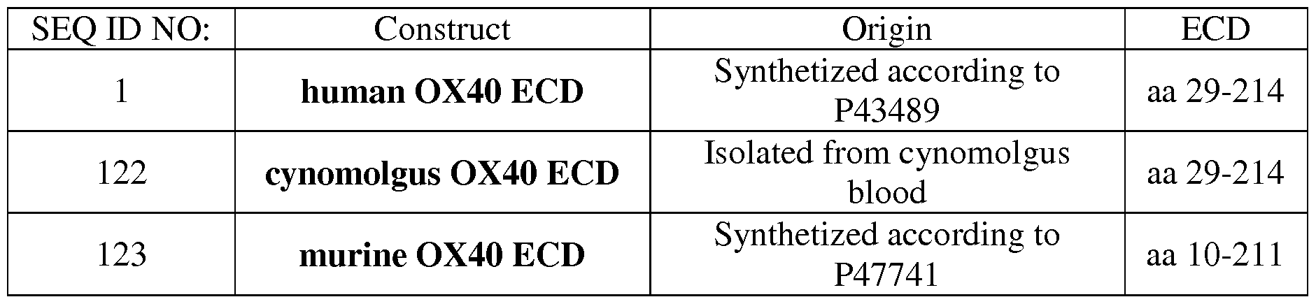
In one aspect, the costimulatory TNF receptor family member is OX40. Thus, in a particular aspect, the moiety capable of specific binding to a costimulatory TNF receptor family member binds to a polypeptide comprising the amino acid sequence of SEQ ID NO: l.
In a further aspect, provided is a bispecific antigen binding molecule, comprising at least one moiety capable of specific binding to OX40, wherein said moiety comprises a VH domain comprising
(i) a CDR-H1 comprising the amino acid sequence selected from the goup consisting of SEQ ID NO:2 and SEQ ID NO:3,
(ii) a CDR-H2 comprising the amino acid sequence selected from the group consisting of SEQ ID NO:4 and SEQ ID NO:5, and
(iii) a CDR-H3 comprising the amino acid sequence selected from the group consisting of SEQ ID NO:6, SEQ ID NO:7, SEQ ID NO:8, SEQ ID NO: 9, SEQ ID NO: 10, SEQ ID NO: 11 and SEQ ID NO: 12,
and a VL domain comprising
(iv) a CDR-L1 comprising the amino acid sequence selected from the group consisting of SEQ ID NO: 13, SEQ ID NO: 14 and SEQ ID NO: 15,
(v) a CDR-L2 comprising the amino acid sequence selected from the group consisting of SEQ ID NO: 16, SEQ ID NO: 17 and SEQ ID NO: 18, and
(vi) a CDR-L3 comprising the amino acid sequence selected from the group consisting of SEQ ID NO: 19, SEQ ID NO:20, SEQ ID NO:21, SEQ ID NO:22, SEQ ID NO:23 and SEQ
ID NO:24.
In another aspect, the invention provides a bispecific antigen binding molecule, wherein the moiety capable of specific binding to OX40 comprises a heavy chain variable region VH comprising an amino acid sequence that is at least about 95%, 96%, 97%, 98%, 99% or 100% identical to an amino acid sequence selected from the group consisting of SEQ ID NO:25, SEQ ID NO: 27, SEQ ID NO:29, SEQ ID NO:31, SEQ ID NO:33, SEQ ID NO:35 and SEQ ID NO:37 and a light chain variable region VL comprising an amino acid sequence that is at least about 95%, 96%, 97%, 98%, 99% or 100% identical to an amino acid sequence of SEQ ID NO:26, SEQ ID NO: 28, SEQ ID NO:30, SEQ ID NO:30, SEQ ID NO:32, SEQ ID NO:34, SEQ ID NO:36 and SEQ ID NO:38.
Particularly, a bispecific antigen binding molecule is provided, wherein the moiety capable of specific binding to OX40 comprises
(i) a heavy chain variable region VH comprising an amino acid sequence of SEQ ID NO:25 and and a light chain variable region VL comprising an amino acid sequence of SEQ ID NO:26,
(ii) a heavy chain variable region VH comprising an amino acid sequence of SEQ ID NO:27 and and a light chain variable region VL comprising an amino acid sequence of SEQ ID NO:28,
(iii) a heavy chain variable region VH comprising an amino acid sequence of SEQ ID NO:29 and and a light chain variable region VL comprising an amino acid sequence of SEQ ID
NO:30,
(iv) a heavy chain variable region VH comprising an amino acid sequence of SEQ ID NO:31 and and a light chain variable region VL comprising an amino acid sequence of SEQ ID NO:32,
(v) a heavy chain variable region VH comprising an amino acid sequence of SEQ ID NO:33 and and a light chain variable region VL comprising an amino acid sequence of SEQ ID NO:34,
(vi) a heavy chain variable region VH comprising an amino acid sequence of SEQ ID NO:35 and and a light chain variable region VL comprising an amino acid sequence of SEQ ID NO:36, or
(vii) a heavy chain variable region VH comprising an amino acid sequence of SEQ ID NO:37 and and a light chain variable region VL comprising an amino acid sequence of SEQ ID NO:38.
In another aspect, the costimulatory TNF receptor family member is 4- IBB. Thus, in a particular aspect, the moiety capable of specific binding to a costimulatory TNF receptor family member binds to a polypeptide comprising the amino acid sequence of SEQ ID NO:39.
Furthermore, provided is a bispecific antigen binding molecule, comprising at least one moiety capable of specific binding to 4- IBB, wherein said moiety comprises a VH domain comprising
(i) a CDR-H1 comprising the amino acid sequence selected from the goup consisting of SEQ ID NO:40 and SEQ ID NO:41,
(ii) a CDR-H2 comprising the amino acid sequence selected from the group consisting of SEQ ID NO:42 and SEQ ID NO:43, and
(iii) a CDR-H3 comprising the amino acid sequence selected from the group consisting of SEQ ID NO:44, SEQ ID NO:45, SEQ ID NO:46, SEQ ID NO:47 and SEQ ID NO:48 and a VL domain comprising
(iv) a CDR-L1 comprising the amino acid sequence selected from the group consisting of SEQ ID NO:49 and SEQ ID NO:50,
(v) a CDR-L2 comprising the amino acid sequence selected from the group consisting of SEQ ID NO:51 and SEQ ID NO:52, and
(vi) a CDR-L3 comprising the amino acid sequence selected from the group consisting of SEQ ID NO:53, SEQ ID NO:54, SEQ ID NO:55, SEQ ID NO:56 and SEQ ID NO:57.
In another aspect, the invention provides a bispecific antigen binding molecule, wherein the moiety capable of specific binding to 4- IBB comprises a heavy chain variable region VH comprising an amino acid sequence that is at least about 95%, 96%, 97%, 98%, 99% or 100% identical to an amino acid sequence selected from the group consisting of SEQ ID NO:58, SEQ ID NO:60, SEQ ID NO:62, SEQ ID NO:64 and SEQ ID NO:66 and a light chain variable region comprising an amino acid sequence that is at least about 95%, 96%, 97%, 98%, 99% or 100% identical to an amino acid sequence selected from the group consisting of SEQ ID NO:59, SEQ ID NO:61, SEQ ID NO:63, SEQ ID NO:65 and SEQ ID NO:67.
Particularly, a bispecific antigen binding molecule is provided, wherein the moiety capable of specific binding to 4- IBB comprises
(i) a heavy chain variable region VH comprising an amino acid sequence of SEQ ID NO:58 and and a light chain variable region VL comprising an amino acid sequence of SEQ ID NO:59,
(ii) a heavy chain variable region VH comprising an amino acid sequence of SEQ ID NO:60 and and a light chain variable region VL comprising an amino acid sequence of SEQ ID NO:61,
(iii) a heavy chain variable region VH comprising an amino acid sequence of SEQ ID NO:62 and and a light chain variable region VL comprising an amino acid sequence of SEQ ID
NO:63,
(iv) a heavy chain variable region VH comprising an amino acid sequence of SEQ ID NO:64 and and a light chain variable region VL comprising an amino acid sequence of SEQ ID NO:65, or
(v) a heavy chain variable region VH comprising an amino acid sequence of SEQ ID NO:66 and and a light chain variable region VL comprising an amino acid sequence of SEQ ID NO:67.
In a particular aspect, the invention provides a bispecific antigen binding molecule , comprising
(a) at least one moiety capable of specific binding to a costimulatory TNF receptor family member,
(b) at least one moiety capable of specific binding to a target cell antigen,
and
(c) a Fc domain composed of a first and a second subunit capable of stable association., wherein the moiety capable of specific binding to a costimulatory TNF receptor family member is a Fab fragment. In one aspect, if the bispecific antigen binding molecule , comprises more than one moiety capable of specific binding to a costimulatory TNF receptor family member, all moieties capable of specific binding to a costimulatory TNF receptor family member are Fab fragments.
In another aspect, the bispecific antigen binding molecule comprises (a) at least one moiety capable of specific binding to a costimulatory TNF receptor family member, (b) at least one moiety capable of specific binding to a target cell antigen, wherein the target cell antigen is selected from the group consisting of Fibroblast Activation Protein (FAP), Melanoma-associated Chondroitin Sulfate Proteoglycan (MCSP), Epidermal Growth Factor Receptor (EGFR),
Carcinoembryonic Antigen (CEA), CD19, CD20 and CD33, and (c) a Fc domain composed of a first and a second subunit capable of stable association. More particularly, the target cell antigen is Fibroblast Activation Protein (FAP).
In a particular aspect, provided is a bispecific antigen binding molecule, wherein the moiety capable of specific binding to FAP comprises a VH domain comprising
(i) a CDR-H1 comprising the amino acid sequence selected from the goup consisting of SEQ ID NO:68 and SEQ ID NO:69,
(ii) a CDR-H2 comprising the amino acid sequence selected from the group consisting of SEQ ID NO:70 and SEQ ID NO:71, and
(iii) a CDR-H3 comprising the amino acid sequence selected from the group consisting of SEQ ID NO:72 and SEQ ID NO:73,
and a VL domain comprising
(iv) a CDR-L1 comprising the amino acid sequence selected from the group consisting of SEQ ID NO:74 and SEQ ID NO:75,
(v) a CDR-L2 comprising the amino acid sequence selected from the group consisting of SEQ ID NO:76 and SEQ ID NO:77, and
(vi) a CDR-L3 comprising the amino acid sequence selected from the group consisting of SEQ ID NO:78 and SEQ ID NO:79.
In a further aspect, the invention thus provides a bispecific antigen binding molecule, wherein
(i) the moiety capable of specific binding to OX40 comprises a heavy chain variable region VH comprising an amino acid sequence that is at least about 95%, 96%, 97%, 98%, 99% or 100% identical to the amino acid sequence of SEQ ID NO:25, SEQ ID NO: 27, SEQ ID NO:29, SEQ ID NO:31, SEQ ID NO:33, SEQ ID NO:35 or SEQ ID NO:37 and a light chain variable region comprising an amino acid sequence that is at least about 95%, 96%, 97%, 98%, 99% or 100%
identical to the amino acid sequence of SEQ ID NO:26, SEQ ID NO: 28, SEQ ID NO:30, SEQ ID NO:32, SEQ ID NO:34, SEQ ID NO:36 or SEQ ID NO:38 and
(ii) the moiety capable of specific binding to FAP comprises a heavy chain variable region VH comprising an amino acid sequence that is at least about 95%, 96%, 97%, 98%, 99% or 100% identical to the amino acid sequence of SEQ ID NO: 80 or SEQ ID NO: 82 and a light chain variable region comprising an amino acid sequence that is at least about 95%, 96%, 97%, 98%, 99% or 100% identical to the amino acid sequence of SEQ ID NO:81 or SEQ ID NO:83.
In one aspect, the invention provides a bispecific antigen binding molecule, wherein
(i) the moiety capable of specific binding to OX40 comprises a heavy chain variable region VH comprising the amino acid sequence of SEQ ID NO: 27 and a light chain variable region comprising an amino acid sequence of SEQ ID NO: 28 and
(ii) the moiety capable of specific binding to FAP comprises a heavy chain variable region VH comprising an amino acid sequence of SEQ ID NO: 82 and a light chain variable region comprising an amino acid sequence of SEQ ID NO: 83. In another aspect, the invention provides a bispecific antigen binding molecule, wherein
(i) the moiety capable of specific binding to 4- IBB comprises a heavy chain variable region VH comprising an amino acid sequence that is at least about 95%, 96%, 97%, 98%, 99% or 100% identical to the amino acid sequence of SEQ ID NO:58, SEQ ID NO:60, SEQ ID NO:62, SEQ ID NO:64 or SEQ ID NO:66 and a light chain variable region comprising an amino acid sequence that is at least about 95%, 96%, 97%, 98%, 99% or 100% identical to the amino acid sequence of SEQ ID NO:59, SEQ ID NO:61, SEQ ID NO:63, SEQ ID NO:65 or SEQ ID NO:67 and
(ii) the moiety capable of specific binding to FAP comprises a heavy chain variable region VH comprising an amino acid sequence that is at least about 95%, 96%, 97%, 98%, 99% or 100% identical to the amino acid sequence of SEQ ID NO: 80 or SEQ ID NO: 82 and a light chain variable region comprising an amino acid sequence that is at least about 95%, 96%, 97%, 98%, 99% or 100% identical to the amino acid sequence of SEQ ID NO:81 or SEQ ID NO:83.
In a further aspect, provided is a bispecific antigen binding molecule, wherein said molecule comprises
(a) a first Fab fragment capable of specific binding to a costimulatory TNF receptor family member,
(b) a second Fab fragment capable of specific binding to a target cell antigen, and
(c) a Fc domain composed of a first and a second subunit capable of stable association.
In one aspect, the bispecific antigen binding molecule is a human, a humanized or a chimeric antibody. In particular, the Fc domain is a human IgG domain, particularly an IgGl Fc domain or an IgG4 Fc domain.
In a further aspect, provided is a bispecific antigen binding molecule, wherein the Fc domain comprises one or more amino acid substitution that reduces the binding affinity of the antibody to an Fc receptor and/or effector function. In particular, the Fc domain is of human IgGl subclass with the amino acid mutations L234A, L235A and P329G (numbering according to Kabat EU index).
In a further aspect, provided is a bispecific antigen binding molecule, wherein the Fc domain comprises a modification promoting the association of the first and second subunit of the Fc domain. In a particular aspect, the invention provides a bispecific antigen binding molecule, wherein the first subunit of the Fc domain comprises knobs and the second subunit of the Fc domain comprises holes according to the knobs into holes method. More particularly, the first subunit of the Fc domain comprises the amino acid substitutions S354C and T366W (numbering according to Kabat EU index) and the second subunit of the Fc domain comprises the amino acid substitutions Y349C, T366S and Y407V (numbering according to Kabat EU index).
In particular the invention provides a bispecific antigen binding molecule, comprising
(a) two moieties capable of specific binding to a costimulatory TNF receptor family member,
(b) two moieties capable of specific binding to a target cell antigen,
and
(c) a Fc domain composed of a first and a second subunit capable of stable association.
Thus, provided is a bispecific antigen binding molecule, wherein the bispecific antigen binding molecule is bivalent both for the costimulatory TNF receptor family member and for the target cell antigen. In a particular aspect, the bispecific antigen binding molecule comprises
(a) two light chains and two heavy chains of an antibody comprising two Fab fragments capable of specific binding to a costimulatory TNF receptor family member and the Fc domain, and
(b) two additional Fab fragments capable of specific binding to a target cell antigen, wherein said additional Fab fragments are each connected via a peptide linker to the C-terminus of the heavy chains of (a). More particularly, the two additional Fab fragments capable of specific binding to a target cell antigen are cross-Fab fragments wherein the variable domains VL and VH are replaced by each other and the VL-CH chains are each connected via a peptide linker to the C- terminus of the heavy chains of (a).
In one aspect, the two Fab fragments capable of specific binding to a costimulatory TNF receptor family member are two Fab fragments capable of specific binding to OX40 or 4- IBB and the two additional Fab fragments capable of specific binding to a target cell antigen are cross-Fab fragments capable of specific binding to FAP. In another aspect, the invention provides a bispecific antigen binding molecule comprising
(a) two moieties capable of specific binding to a costimulatory TNF receptor family member,
(b) one moiety capable of specific binding to a target cell antigen,
and
(c) a Fc domain composed of a first and a second subunit capable of stable association. Thus, provided is a bispecific antigen binding molecule, wherein the bispecific antigen binding molecule is bivalent for the costimulatory TNF receptor family member and monovalent for the target cell antigen.
In a particular aspect, the bispecific antigen binding molecule comprises
(a) two light chains and two heavy chains of an antibody comprising two Fab fragments capable of specific binding to a costimulatory TNF receptor family member and the Fc domain, and
(b) a VH and VL domain capable of specific binding to a target cell antigen, wherein the VH domain is connected via a peptide linker to the C-terminus of one of the heavy chains and wherein the VL domain is connected via a peptide linker to the C-terminus of the second heavy chain. In another aspect, the invention provides an antibody that specifically binds to OX40, wherein said antibody comprises
(i) a heavy chain variable region VH comprising an amino acid sequence of SEQ ID NO:25 and and a light chain variable region VL comprising an amino acid sequence of SEQ ID NO:26,
(ii) a heavy chain variable region VH comprising an amino acid sequence of SEQ ID NO:27 and and a light chain variable region VL comprising an amino acid sequence of SEQ ID NO:28,
(iii) a heavy chain variable region VH comprising an amino acid sequence of SEQ ID NO:29 and and a light chain variable region VL comprising an amino acid sequence of SEQ ID NO:30,
(iv) a heavy chain variable region VH comprising an amino acid sequence of SEQ ID NO:31 and and a light chain variable region VL comprising an amino acid sequence of SEQ ID NO:32,
(v) a heavy chain variable region VH comprising an amino acid sequence of SEQ ID NO:33 and and a light chain variable region VL comprising an amino acid sequence of SEQ ID NO:34,
(vi) a heavy chain variable region VH comprising an amino acid sequence of SEQ ID NO:35 and and a light chain variable region VL comprising an amino acid sequence of SEQ ID
NO:36, or
(vii) a heavy chain variable region VH comprising an amino acid sequence of SEQ ID NO:37 and and a light chain variable region VL comprising an amino acid sequence of SEQ ID NO:38. In yet another aspect, provided is an antibody that specifically binds to 4- IBB, wherein said antibody comprises
(i) a heavy chain variable region VH comprising an amino acid sequence of SEQ ID NO:58 and and a light chain variable region VL comprising an amino acid sequence of SEQ ID NO:59,
(ii) a heavy chain variable region VH comprising an amino acid sequence of SEQ ID NO:60 and and a light chain variable region VL comprising an amino acid sequence of SEQ ID NO:61,
(iii) a heavy chain variable region VH comprising an amino acid sequence of SEQ ID NO:62 and and a light chain variable region VL comprising an amino acid sequence of SEQ ID NO:63,
(iv) a heavy chain variable region VH comprising an amino acid sequence of SEQ ID NO:64 and and a light chain variable region VL comprising an amino acid sequence of SEQ ID NO:65, or
(v) a heavy chain variable region VH comprising an amino acid sequence of SEQ ID NO:66 and and a light chain variable region VL comprising an amino acid sequence of SEQ ID
NO:67.
According to another aspect of the invention, there is provided an isolated polynucleotide encoding a bispecific antigen binding molecule as described herein before or an antibody that specifically binds to OX40 as described herein before or an antibody that specifically binds to 4- IBB as described herein before. The invention further provides a vector, particularly an expression vector, comprising the isolated polynucleotide of the invention and a host cell comprising the isolated polynucleotide or the vector of the invention. In some aspects the host cell is a eukaryotic cell, particularly a mammalian cell.
In another aspect, provided is a method for producing a bispecific antigen binding molecule as described herein before or an antibody that specifically binds to OX40 as described herein before or an antibody that specifically binds to 4- IBB as described herein before, comprising the steps of (i) culturing the host cell of the invention under conditions suitable for expression of the antigen binding molecule, and (ii) recovering the antigen binding molecule. The invention also encompasses the bispecific antigen binding molecule or the antibody that specifically binds to OX40 or the antibody that specifically binds to 4- IBB produced by the method of the invention.
The invention further provides a pharmaceutical composition comprising a bispecific antigen binding molecule as described herein before or an antibody that specifically binds to OX40 as described herein before or an antibody that specifically binds to 4- IBB as described herein before and at least one pharmaceutically acceptable excipient.
Also encompassed by the invention is the bispecific antigen binding molecule as described herein before or the antibody that specifically binds to OX40 as described herein before or the antibody that specifically binds to 4- IBB as described herein before, or the pharmaceutical composition comprising the bispecific antigen binding molecule or the antibody that specifically binds to OX40 or the antibody that specifically binds to 4- IBB, for use as a medicament.
In one aspect, provided is a bispecific antigen binding molecule as described herein before or an antibody that specifically binds to OX40 as described herein before or an antibody that specifically binds to 4- IBB as described herein before or the pharmaceutical composition of the invention, for use
(i) in stimulating T cell response,
(ii) in supporting survival of activated T cells,
(iii) in the treatment of infections,
(iv) in the treatment of cancer,
(v) in delaying progression of cancer, or
(vi) in prolonging the survival of a patient suffering from cancer.
In a specific embodiment, provided is the bispecific antigen binding molecule as described herein before or the antibody that specifically binds to OX40 as described herein before or the antibody that specifically binds to 4- IBB as described herein before, or the pharmaceutical composition of the invention, for use in the treatment of cancer.
In another specific aspect, the invention provides the bispecific antigen binding molecule as described herein before or the antibody that specifically binds to OX40 as described herein before or the antibody that specifically binds to 4- IBB as described herein for use in the treatment of cancer, wherein the bispecific antigen binding molecule is administered in
combination with a chemotherapeutic agent, radiation and/ or other agents for use in cancer immunotherapy.
In a further aspect, the invention provides a method of inhibiting the growth of tumor cells in an individual comprising administering to the individual an effective amount of the bispecific antigen binding molecule as described herein before or the antibody that specifically binds to OX40 as described herein before or the antibody that specifically binds to 4- IBB as described herein before, or the pharmaceutical composition of the invention, to inhibit the growth of the tumor cells.
Also provided is the use of the the bispecific antigen binding molecule as described herein before or the antibody that specifically binds to OX40 as described herein before or the antibody that specifically binds to 4- IBB as described herein before for the manufacture of a medicament for the treatment of a disease in an individual in need thereof, in particular for the manufacture of a medicament for the treatment of cancer, as well as a method of treating a disease in an individual, comprising administering to said individual a therapeutically effective amount of a composition comprising the TNF family ligand trimer-containing antigen binding molecule of the invention in a pharmaceutically acceptable form. In a specific aspect, the disease is cancer. In any of the above aspects the individual is a mammal, particularly a human.
BRIEF DESCRIPTION OF THE DRAWINGS
Figure 1A shows the monomeric form of Fc-linked TNF receptor antigen that was used for the preparation of TNF receptor antibodies. Figure IB shows a dimeric human TNF receptor antigen Fc fusion molecule with a C-terminal Ha tag that was used for the testing of the binding of TNF receptor antibodies in the presence of TNF ligand (ligand blocking property). The schematic setup of the experiment described in Example 2.4 is shown in Figure 1C.
Figure 2 shows the binding of anti-OX40 antibodies to activated human CD4+ and CD8+ T cells. OX40 is not expressed on resting human PBMCs (Figure 2A and 2C). After activation of human PBMCs OX40 is up-regulated on CD4+ and CD8+ T cells (Figure 2B and 2D). OX40 expression on human CD8+ T cells is lower than on CD4+ T cells. The depicted clones varied in their binding strength (EC50 values as well as signal strength) to OX40 positive cells. Shown is the binding as median of fluorescence intensity (MFI) of FITC labeled anti-human IgG Fcy- specific goat IgG F(ab )2 fragment which is used as secondary detection antibody. MFI was measured by flow cytometry and baseline corrected by subtracting the MFI of the blank control. The x-axis shows the concentration of antibody constructs. All OX40 clones do bind to activated, OX40 expressing human CD4+ T cells, and to a lower extent to activated human CD8+ T cells.
Figure 3 shows the binding of the anti-OX40 antibodies to activated mouse CD4+ and CD8+ T cells. OX40 was not detected on resting mouse splencoytes (Figure 3 A and 3C). After activation OX40 is up-regulated on CD4+ and CD8+ T cells (Figure 3B and 3D). Mouse splenocytes were isolated by erythrolysis with ACK lysis buffer of mechanically-homogenized spleens obtained from 6-8 weeks old female C57BL/6 mice. Binding of anti-OX40 antibodies to cell surface proteins was detected with a goat anti-human IgG Fc-specific secondary antibody conjugated to FITC using FACS analysis. MFI was measured by flow cytometry and baseline corrected by subtracting the MFI of the blank control. The x-axis shows the concentration of antibody constructs. Only clone 20B7 does bind to activated, OX40 expressing mouse CD4 and CD8 T Cells, but not to resting T cells.
Figure 4 shows the binding of anti-OX40 antibodies on cynomolgus activated CD4+ and CD8+ T cells. The depicted clones varied in their binding strength (EC50 values as well as signal strength) to OX40 positive activated cynomolgus CD4+ T cells (Fig. 4A). OX40 expression on activated CD8+ T cells is low under this condition and hardly any binding of the selected clones was found (Fig 4B). Binding of anti-OX40 antibodies to cell surface proteins was detected with a goat anti-human IgG Fc-specific secondary antibody conjugated to FITC using FACS analysis. MFI was measured by flow cytometry and baseline corrected by subtracting the MFI of the blank control. The x-axis shows the concentration of antibody constructs. All OX40 clones do bind to activated, OX40 expressing cynomolgus CD4+ T cells, and to a lower extent to activated cynomolgus CD8+ T cells.
Figure 5 shows the lack of binding to OX40 negative tumor cells. The depicted clones showed no binding to OX40 negative U-78 MG (Fig. 5A) and WM266-4 tumor cells (Fig. 5B). Shown is the binding as median of fluorescence intensity (MFI) of FITC labeled anti-human IgG Fcy-specific goat IgG F(ab )2 fragment which is used as secondary detection antibody. MFI was measured by flow cytometry and baseline corrected by subtracting the MFI of the blank control. The x-axis shows the concentration of antibody constructs. All clones in an IgG format do not bind to OX40 negative tumor cells. Binding is specific for OX40 on activated leukocytes.
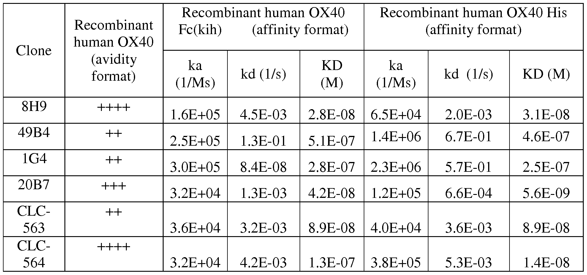
Figures 6A to 6F show the interaction between anti-Ox40 antibodies 8H9 (Fig. 6A), 20B7 (Fig. 6B), 1G4 (Fig. 6C), 49B4 (Fig. 6D), CLC-563 (Fig. 6E) and CLC-564 (Fig. 6F) and the preformed complex huOx40 Ligand / huOx40-Fc as measured by surface plasmon resonance.
Figure 7 shows the effect of the anti-human OX40 antibodies of the invention on HeLa cells expressing human OX40 and reporter gene NF-KB-luciferase. Shown is the activation of NF-κΒ signaling pathway in the reporter cell line with various anti-OX40 binders in a
P329GLALA hulgGI format with (Fig. 7B) or without (Fig. 7B) crosslinking by secondary- antibody. The reporter cells were cultured for 6 hours in the presence of anti-OX40 constructs at
the indicated concentrations w/ or w/o crosslinking secondary poly-clonal anti-huIgGl Fcy- specific goat IgG F(ab)2 fragment in a 1:2 ratio. Activity is characterized by blotting the units of released light (URL) measured during 0.5 s versus the concentration in nM of tested anti-Ox40 construct. URLs are emitted due to luciferase-mediated oxidation of luciferin to oxyluciferin. All clones are able to induce NFKB activation when the OX40 axis is triggered in a human OX40+ reporter cell line. All clones are thus agonistic and activate in a dose dependent way.
Crosslinking by secondary Fc part specific Abs strongly increases this agonism.
Figures 8A-8F shows the bioactivity of the anti-human OX40 antibodies in preactivated human CD4 T cells. Costimulation with plate-immobilized anti-Ox40 binders (hulgGI
P329GLALA format) promoted cell proliferation and maturation of sub-optimally restimulated human CD4 T cells and induced an enhanced activated phenotype. PHA-L pre-activated CFSE- labeled human CD4 T cells were cultured for four days on plates pre-coated with mouse IgG Fey specific antibodies, human IgG Fey specific antibodies (both 2 μg/mL), mouse anti-human CD3 antibodies (clone OKT3, [3 ng/mL]) and titrated anti-Ox40 binders (hulgGI P329GLALA format ). Shown is the event count (Fig. 8A), the percentage of proliferating (CFSE-low) cells (Fig. 8B), the percentage of effector T cells (CD 127 low/CD45RA low) (FIG. 8C) and the percentage of CD62L low (FIG. 8D), OX40 positive (Fig. 8F) or Tim-3 positive cells (FIG. 8E) at day 4. Baseline values of samples containing only the plate-immobilized anti-human CD3 were substracted. Therefore, the enhancing effect of OX40 stimulation but not the effect of suboptimal anti-CD3 stimulation per se is visible here. All clones are able support suboptimal TCR stimulation in OX40 positive preactived CD4 T cells when they are coated to plate. Cells do survive better and proliferate more. In the tumor microenvironment this could lead to increased anti-tumor activity of T cells.
Figure 9 summarizes the EC50 values (for all biomarkers) as marker for the agonistic capacity of the respective clone (values calculated from the curves shown in Figure 8). The potency increases from left to right. The event count, the percentage of proliferating (CFSE-low) cells and the percentage of CD62L low, CD45RA low or Tim-3 positive cells at day 4 were plotted vs the anti-Ox40 antibody concentration and EC50 values were calculated using the inbuilt sigmoidal dose response quotation in Prism4 (GraphPad Software, USA). Figures 10A-10F show the bioactivity of the anti- human OX40 antibodies in preactivated human CD4 T cells in solution. No effect on cell proliferation, maturation or activation status of sub-optimally restimulated human CD4 T cells was detected in the absence of plate
immobilization of anti-Ox40 binders (hu IgGl P329GLALA format). PHA-L pre-activated CFSE-labeled human CD4 T cells were cultured for four days on plates pre-coated with mouse IgG Fey specific antibodies and mouse anti-human CD3 antibodies (clone OKT3, [3 ng/mL]). Titrated anti-Ox40 binders (hu IgGl P329GLALA format) were added to the media and were
present in solution throughout the experiment. Shown is the event count (Fig. 10A), the percentage of proliferating (CFSE-low) cells (Fig. 10B), the percentage of effector T cells (CD1271ow CD45RAlow) (Fig. IOC) and the percentage of CD62L low (Fig. 10E), OX40 positive (Fig. 10F) or Tim-3 positive cells (Fig. 10E) at day 4. Baseline values of samples containing only the plate-immobilized anti-human CD3 were substracted. Therefore, the enhancing effect of OX40 stimulation but not the effect of suboptimal anti-CD3 stimulation per se is visible here. There is no improved TCR stimulation in the absence of strong crosslinking (P329GLALA format in solution). Crosslinking is therefore essential for a bivalent aOx40 format to be agonistic on T cells. This crosslinking will be provided by FAP expressed on the cell surface of tumor or tumor- stromal cells in targeted formats.
Figure 11 shows a correlation between the binding strength and agonistic capacity of the different anti-OX40 clones. Binding of anti-Ox40 clones (hulgGI P329GLALA format) on activated CD4 T cells was performed as described in Example 2.1.2. Plateau values were normalized to the value obtained with clone 8H9 (hulgGI P329GLALA format). Bioactivity testing of anti-Ox40 clones (hulgGI P329GLALA format) was performed as described in
Example 3.2 and plateau values of PD-1 expression were normalized to the values obtained for clone 8H9 (hulgGI P329GLALA format). Normalized binding was plotted against normalized bioactivity, to test for a correlation between binding strength and agonistic capacity. For most clones there was a direct correlation (linear regression is shown, p value 0.96; slope 0.91).
However, two clones (49B4, 1G4) showed a much stronger bioactivity then could be predicted from their binding strength. This subgroup of clones which show unexpectedly high agonistic potency in the face of low binding abilility is of particular interest for the bispecific antigen binding molecules of the invention.
In Figure 12A is shown a schematic scheme of an exemplary bispecific, bivalent antigen binding molecule of the invention comprising two Fab fragments binding to OX40 and two cross-Fab fragments binding to FAP (2+2 format). Figure 12B shows a schematic scheme of an exemplary bispecific, monovalent antigen binding molecule (1+1 format) of the invention comprising one Fab fragment binding to OX40 and one cross-Fab fragment binding to FAP. In Figure 12C the setup for the SPR experiments showing simultaneous binding to immobilized human OX40 or human 4- IBB and human FAP is shown. In Figure 12D is shown a schematic drawing of an exemplary bispecific antigen binding molecule, that is bivalent for binding to OX40 and monovalent for binding to FAP. It comprises two Fab fragments binding to OX40 and a VH and VL domain binding to FAP. The black point symbolizes the knob into hole modifications in the heavy chains. In Figure 12E is shown the setup for the SPR experiments demonstrating binding to FAP as described in Example 5.4.1. Figure 12F shows how simultaneous binding to immobilized human OX40 and human FAP was measured (Example 5.4.1).
Figures 13A-13D show the SPR diagrams of simultaneous binding of bispecific bivalent 2+2 constructs (analyte 1) to immobilized human OX40 and human FAP (analyte 2). In Figures 13E-13H, the simultaneous binding of bispecific monovalent 1+1 constructs (analyte 1) to immobilized human OX40 and human FAP (analyte 2) is shown. In Figures 13J-13L, the simultaneous binding of bispecific 2+1 constructs (analyte 1) to immobilized human OX40 and human FAP (analyte 2) is shown. Figure 13M shows the binding to hu OX40 of the bispecific 2+1 constructs in a cell-based FRET assay (TagLite) (Example 5.4.2). The data show that the two anti-OX40 Fab domains in the 2+1 constucts bind to hu OX40 in comparable manner as a common IgG antibody. Figures 14A-14D and 14E-14H show the binding of selected anti-OX40 binders (clone
8H9, 1G4) in a FAP targeted monovalent or bivalent format to resting and activated human PBMC, respectively. Binding characteristics to OX40 positive T cells (Figures 14B and 14D) were comparable for clones in a conventional bivalent hu IgG format (open square) and a FAP- targeted bivalent format (filled square). Binding of the same clone in a FAP- targeted monovalent format (filled triangle) was clearly weaker due to loss of avidity binding. In the absence of human Ox40 expressing cells no binding can be observed (resting cells, left graphs). Shown is the binding as median of fluorescence intensity (MFI) of FITC labeled anti-human IgG Fcy-specific goat IgG F(ab )2 fragment which is used as secondary detection antibody. MFI was measured by flow cytometry and baseline corrected by subtracting the MFI of the blank control (see Example 4.3.2.1). DP88 hulgGI P329G LALA is isotype antibody used as control. The x- axis shows the concentration of antibody constructs. In Figures 14A-14D it can be seen that clone 1G4 binds to activated, OX40 expressing human CD4 T cells, and to a lower extent to activated human CD8 T cells. The bivalent construct binds stronger than the monovalent construct. The constructs do not bind to OX40 negative resting T cells. Figures 14E-14H show that clone 8H9 binds to activated, OX40-expressing human CD4 T cells, and to a lower extent to activated human CD8 T cells. The bivalent construct binds stronger than the monovalent construct. The constructs do not bind to OX40 negative resting T cells. In Figures 14J-14M it is also shown that the bivalent FAP-targeted OX40 constructs showed stronger binding
characteristics to OX40 positive cells as respective clone in a monovalent antibody format. Figures 14N-14Q show that different 2+1 constructs bound with similar strength to OX40 positive T cells, independently of the second binding moiety.
Figures 15A and 15B show the binding of selected anti-OX40 binders (clone 8H9, 1G4) in a FAP targeted monovalent or bivalent format to FAP positive tumor cells. Transgenic modified mouse embryonic fibroblast NIH/3T3-huFAP clone 39 or WM266-4 cells express high levels of human fibroblast activation protein (huFAP). Only FAP- targeted mono- and bivalent anti-Ox40 constructs (filled square and triangle) but not the same clone in a human IgGl P329GLALA format (open square) binds to NIH/3T3-huFAP clone 39 cells (Fig. 15 A) and WM266-4 cells
(Fig. 15B), respectively. Shown is the binding as median of fluorescence intensity (MFI) of Fluorescein isothiocyanate (FITC)-labeled anti-human IgG Fcy-specific goat IgG F(ab )2 fragment which is used as secondary detection antibody. MFI was measured by flow cytometry. The x-axis shows the concentration of antibody constructs. The bivalent FAP construct binds stronger than the monovalent construct. Binding to human FAP-expressing tumor cells is also shown in Figures 15C, 15D, 15E and 15F (see Example 4.5.4.2 for more details).
Figures 16A to 16G show the NFKB activation by selected binders (8H9, 1G4) in a monovalent or bivalent FAP targeted format in the presence or absence of hyper-crosslinking. Shown is the activation of NF-κΒ signaling pathway in the reporter cells by selected binders 1G4 (Fig. 16A-16D) and 8H9 (Fig. 16E-16G) in a monovalent (filled triangle) or bivalent (filled square) FAP targeted format or as non-targeted hu IgG P329GLALA antibodies (open square). Hyper-crosslinking was provided by either anti-hu IgG Fcy-specific secondary antibodies (ratio 1:2 of primary to secondary antibodies) or via FAP-expressing NIH/3T3-huFAP clone 39 and WM266-4 tumor cells (ratio 2: 1 of FAP+ tumor cells to reporter cells). The NF-KB-mediated lucif erase activity was characterized by blotting the units of released light (URL), measured during 0.5 s, versus the concentration in nM of tested compounds. URLs are emitted due to luciferase-mediated oxidation of luciferin to oxyluciferin. Values are baseline corrected by subtracting the URLs of the blank control. In Figure 16A it can be seen that all constructs containing clone 1G4 were able to induce NFkB activation in Ox40+ HeLa reporter cells.
Crosslinking by secondary anti IgG Fey specific antibody strongly increased NFKB activation. Addition of FAP positive cells (NIH or WM266-4) however increased only the agonistic potential of FAP targeted molecules, but not that of the P329GLALA IgG format. Bivalent constructs performed clearly better than monovalent constructs. In Figure 16E-16G it is shown that all constructs containing clone 8H9 were able to induce NFKB activation in OX40+ HeLa reporter cells. Crosslinking by secondary anti IgG Fey specific antibody strongly increased
NFKB activation. Addition of FAP positive cells (NIH) however increased only the agonistic potential of FAP targeted molecules, but not that of the P329GLALA IgG format. Bivalent constructs performed slightly better than monovalent constructs. In Figures 16H, 16 J and 16K it can also be seen that constructs with monovalent binding to OX40 (1+1) are less efficient than constructs with bivalent binding to OX40 (2+1 and 2+2 constructs. Further data are given in Figures 16L, 16M and 16N and in Example 5.1.
The rescue of suboptimal TCR restimulation of preactivated CD4 T cells with plate- immobilized FAP targeted mono and bivalent anti-OX40 (1G4 and 8H9) constructs is shown in Figures 17A and 17B and in Figures 18A-18D. Costimulation with plate-immobilized anti- Ox40 binders (hulgGI P329GLALA format) promoted cell proliferation and maturation of sub- optimally restimulated human CD4 T cells and induced an enhanced activated phenotype. PHA- L pre-activated CFSE-labeled human CD4 T cells were cultured for four days on plates pre-
coated with mouse IgG Fey spec, antibodies, human IgG Fey spec, antibodies (both 2 μg/mL), mouse anti-human CD3 antibodies (clone OKT3, [3 ng/mL]) and titrated anti-Ox40 binders (hulgGI P329GLALA format ). Shown is the event count, the percentage of proliferating
(CFSE-low) cells, the percentage of effector T cells (CD1271ow CD45RAlow) and the
percentage of CD62L low, OX40 positive or Tim-3 positive cells at day 4. Baseline values of samples containing only the plate-immobilized anti-human CD3 were substracted. Therefore, the enhancing effect of OX40 stimulation but not the effect of suboptimal anti-CD3 stimulation per se is visible here. In Figure 17 it can be seen that all constructs containing clone 8H9 were able to rescue suboptimal TCR stimulation of preactivated, Ox40+ CD4 T cells when coated to plate. Cells showed a more activated (Tim3+ FSC+) phenotype. Bivalent constructs performed slightly better than monovalent constructs. In Figure 18 it can be observed that all constructs containing clone 1G4 were able to rescue suboptimal TCR stimulation of preactivated, Ox40+ CD4 T cells when coated to plate. Cells proliferate more and present an activated (Tim3+ Ox40+) phenotype. Bivalent constructs perform better than monovalent constructs. Both bivalent constructs performed comparable when coated to plate.
In Figure 19 the EC50 values as calculated from rescuing suboptimal TCR stimulation with plate-immobilized FAP targeted mono and bivalent anti-OX40 (clone 1G4) constructs are shown. The percentage of proliferating (CFSE-low) cells and the percentage of CD127L low, Tim-3 positive and OX40 positive cells at day 4 were plotted vs the anti-OX40 antibody concentration and EC50 values as measure for agonistic strength were calculated using the inbuilt sigmoidal dose response quotation inPrism4 (GraphPad Software, USA). All constructs containing clone 1G4 were able to rescue suboptimal TCR stimulation of preactivated, Ox40+ CD4 T cells when coated to plate. However, the bivalent (2+2) constructs performed better than monovalent (1+1) constructs. Figures 20A-20H relate to the OX40 mediated costimulation of suboptimally TCR triggered resting human PBMC and hypercrosslinking by cell surface FAP. Only constructs containing FAP binding moiety were able to rescue suboptimal TCR stimulation of preactivated, Ox40+ CD4 T cells when crosslinking was provided by FAP positive cells (NIH). Shown is either the event count (Figures 20B and 20D), the percentage of low-proliferating (CFSE-high) cells (Figures 20A and 20C) or the MFI of CD62L(Figures 20F and 20H), CD 127 (Fig. 20E) or GranzymeB vital CD4+ (Fig. 20G) and CD8+ T cells. Baseline values of samples containing only the anti-human CD3 (clone V9, hulgGI), resting human PBMC and NIH/3T3-huFAP clone 39 were substracted. Thus the enhancing effect of OX40 costimulation but not the effect of suboptimal anti-CD3 stimulation per se is shown here. Cells survived better, proliferated more and showed a stronger activated (CD62L and CD 127 low) phenotype. Targeted bivalent constructs performed only slightly better than monovalent constructs. Clone 8H9 in a non- targeted hulgGI P329GLALA was not able to rescue suboptimal TCR stimulation in the absence
of further crosslinking. In a FAP positive tumor micro environment this could lead to increased anti-tumor activity of T cells whereby systemic OX40 activation is avoided.
Figures 21A-21C relate to the activation of resting human CD4 cell using surface immobilized FAP targeted mono and bivalent anti-OX40 (1G4) constructs. Costimulation with non-targeted anti-Ox40 (1G4) hulgGI did not rescue suboptimally TCR stimulated CD4 and CD8 (data not shown) T cells. Hyper-crosslinking of the FAP targeted mono and bivalent anti- Ox40 constructs by the present NIH/3T3-huFAP clone 39 cells strongly promoted proliferation, survival (data not shown) and induced an enhanced activated phenotype in human CD4 cells. Shown is the MFI of CD25 expression on CD4 T cells and the percentage of CD25+ CD4 T cells. Baseline values of samples containing only the anti-human CD3 (clone V9, hulgGI), resting human PBMC and NIH/3T3-huFAP clone 39 were substracted. The agonistic effect of the compounds were quantified as area under the curve using the inbuilt function in GraphPad Prism and is shown for the three different anti-Ox40 (1G4) constructs. Targeted bivalent (2+2) constructs performed better than monovalent (1+1) constructs. Data as obtained in a second experiment are shown in Figures 21D-21H and 21J-21N, respectively. Monovalent anti-OX40 construct (1+1; filled triangle) was less able to rescue TCR stimulation than bivalent anti-OX40 targeting constructs (semi-filled circle, filled square). The bivalently to FAP binding 2+2 construct was already able at lower concentrations to rescue suboptimal TCR stimulation compared to the monovalently to FAP binding 2+1 constructs. In the 2+1 format the high affinity FAP binding clone 4B9 was clearly superior to the low affinity clone 28H1 (Figures 21J-21N). This suggests that the EC50 values of the observed bioactivity were driven by the binding to FAP (2+2 > 2+1 (4B9) > 2+1 (28H1). In Figure 21P the agonistic capacity of each construct was quantified for the analyzed markers as area under the curve and plotted against each other. The observed bioactivity was best for the FAP (28H1) (2+2) construct, followed by the FAP (4B9) (2+1) construct and then the FAP (28H1) (2+1) construct.
In Figures 22A and 22B it is shown that anti-Ox40 antibodies containing human IgGl Fc regions can indue lysis of OX40 positive cells. PkH26 labeled HeLa_hOx40_NFkB_Lucl and freshly isolated NK cells were cocultured at an E to T ratio 3: 1 for 24 hours in the presence of OX40 antibodies (human IgGl and human IgGl P329GLALA). The LDH content was analyzed after 4 hours using the cytotoxicity detection kit - LDH (Roche, Cat.No. 11644793001). After 24 hrs cells were stained with Dapi and were analyzed by flow cytometry. The percentage of Dapi positive dead cells was used to calculate specific lysis. Anti OX40 binders in a human IgG format bind to Fc receptors on NK cells and induce ADCC of OX40 positive target cells. Using the hu IgGl P329GLALA format instead prevents ADCC of OX40 positive cells (e.g. recently activated T cells).
Figures 23A-23D show the binding to resting and activated human T cells of four anti- human 4-lBB-specific clones transferred to a hulgGI P329G LALA format (filled diamond: clone 25G7, filled square: clone 12B3, filled star: clone 11D5, pointing-up triangle: clone 9B11) and one anti-mouse 4- IBB specific clone 20G2 transferred to a hulgGI P329G LALA format (pointing down triangle). As negative control a non-4- lBB-specific clone DP47 hulgGI P329G LALA antibody was used (open grey circle). The upper panels show binding to resting CD4+ T cells (Fig. 23A) and activated CD4+ T cells (Fig. 23B), whereas the lower panels show binding to resting CD8+ T cells (Fig. 23C) and activated CD8+ T cells (Fig. 23D). The binding is characterized by plotting the median of fluorescence (MFI) of FITC-labeled or PE-labeled anti- human IgG Fcy-specific goat IgG F(ab )2 fragment that is used as secondary detection antibody versus the concentration in nM of the tested primary anti-4- IBB -binding hulgGI P329G LALA antibodies. MFI was measured by flow cytometry and baseline corrected by subtracting the MFI of the blank control (no primary antibody).
Figures 24A-24D show the binding to 4- IBB expressing mouse T cells. Shown is the binding to resting and activated mouse T cells of four anti-human 4- IBB binding hulgGI P329G LALA antibody clones (filled diamond: clone 25G7, filled square: clone 12B3, filled star: clone 11D5, pointing-up triangle: clone 9B11) and one anti-mouse 4- IBB binding hulgGI P329G LALA antibody clone 20G2 (pointing-down tringle). As negative control a non-4- IBB binding DP47 hulgGI P329G LALA antibody was used (open grey circle). The upper panels show binding to resting mouse CD4+ T cells (Fig. 24A) and activated CD4+ T cells (Fig. 24B), whereas the lower panels show binding to resting mouse CD8+ T cells (Fig. 24C) and activated CD8+ T cells (Fig. 24D). The binding is characterized by plotting the MFI of FITC-labeled anti- human IgG Fcy-specific goat IgG F(ab )2 fragment that is used as secondary detection antibody versus the concentration in nM of the tested primary anti-4- IBB -binding hulgGI P329G LALA antibodies. MFI was measured by flow cytometry and baseline corrected by subtracting the MFI of the blank control (no primary antibody).
Figures 25A-25D show the binding of mouse IgGs to 4- IBB expressing mouse T cells. Shown is the binding to resting and activated mouse T cells of the anti-mouse 4- IBB binding clone 20G2 transferred to the formats mouse IgGl DAPG and mouse IgGl wildtype (wt). As negative control a commercial non-4- IBB binding mouse IgGl wt isotype control was used (open grey circle, BioLegend, Cat.-No. 400153). In the upper panels binding to resting CD4+ T cells (Fig. 25A) and activated CD4+ T cells (Fig. 25B) is shown, whereas in the lower panels binding to resting CD8+ T cells (Fig. 25C) and activated CD8+ T cells (Fig. 25D) is shown. The binding is characterized by plotting the median of fluorescence of intensity (MFI) of FITC- labeled anti-mouse IgG Fcy-specific goat IgG F(ab )2 fragment that is used as secondary detection antibody versus the concentration in nM of the tested primary anti -4-lBB-binding
moIgG antibodies. MFI was measured by flow cytometry and baseline corrected by subtracting the MFI of the blank control (no primary antibody).
Figures 26A and 26B show the binding to 4- IBB expressing cynomolgus T cells. Shown is the binding to activated cynomolgus T cells of four anti-human 4- IBB binding hulgGI P329G LALA antibody clones (filled diamond: clone 25G7, filled square: clone 12B3, filled star: clone 11D5, pointing-up triangle: clone 9B11). As negative control a non-4-lBB binding DP47 hulgGI P329G LALA antibody was used (open grey circle). Shown is the binding to activated CD4+ T cells (Fig. 26A) and to activated CD8+ T cells (Fig. 26B) respectively. The binding is characterized by plotting the median of fluorescence of intensity (MFI) of FITC-labeled anti- human IgG Fcy-specific goat IgG F(ab )2 fragment that is used as secondary detection antibody versus the concentration in nM of the tested primary anti-4- IBB -binding hulgGI P329G LALA antibodies. MFI was measured by flow cytometry and baseline corrected by subtracting the MFI of the blank control (no primary antibody).
Figures 27A-27E refer to ligand binding properties of the anti-4- IBB antibodies of the invention as determined by surface plasmon resonance. The interaction between human anti-4- 1BB IgG 25G7, 11D5, 9B11 and 12B3 and the preformed complex hu4-lBB Ligand / hu4-lBB is shown as well as the interaction of mouse anti-4- IBB clone 20G2 and the preformed complex mu4-lBB Ligand / mu4-lBB.
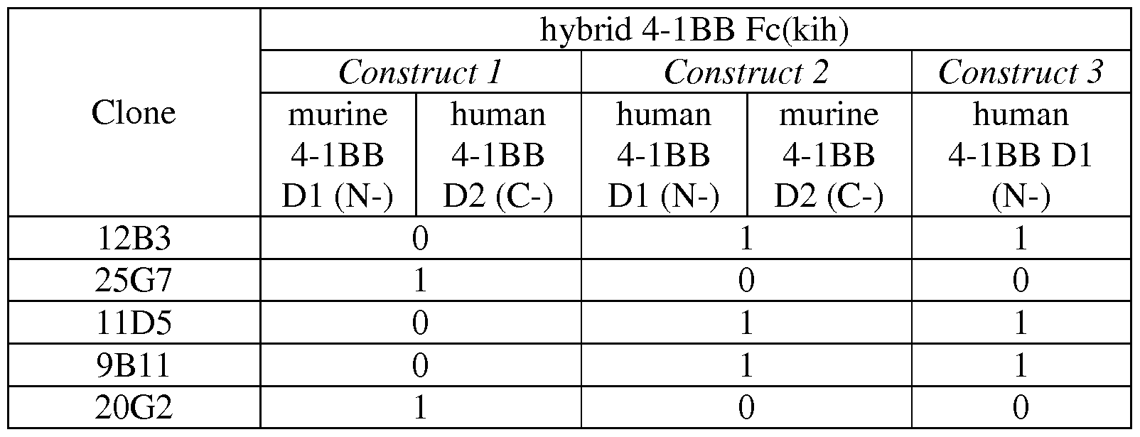
Figures 28A-28C and 28D-28F relate to competition binding experiments. Figures 28A- 28C show the interaction between anti-4- IBB IgG clones 12B3, 11D5 and 25G7 and a preformed complex of clone 9B11 and hu4-lBB. Figures 28D-28F show the interaction between anti-4- IBB IgG clones 12B3, 9B11 and 25G7 and a preformed complex of clone 11D5 and hu4- 1BB. It can be concluded that anti-4- IBB clones 12B3, 11D5 and 9B11 share a different spatial epitope as 25G7, since the two antibodies can bind simultaneously to human 4- IBB. Figures 29A-29D show the binding of hybrid 4- IBB Fc(kih) variants to anti-4- IBB antibodies, i.e. binding of hu4-lBBDl/mu4-lBBD2-Fc(kih) and mu4-lBBDl/hu4-lBBD2- Fc(kih) variants to anti-4-lBB antibodies. Underlined is the 4-1BB domain recognized by the antibody. In Figures 30A-30D is shown the binding of anti-human 4- IBB antibodies 11D5, 12B3, 25G7 and 9B11 to human 4-1BB Domain 1. Anti-human 4-1BB antibodies 11D5, 12B3 and 9B11 bind human domain 1 containing 4- IBB constructs.
Figures 31A-31D show functional properties of different anti-human 4- IBB clones in vitro. Pre-activated human CD8+ T cells were activated with different concentrations of surface immobilized anti-human-4-lBB-specific hulgGI P329G LALA antibodies in the absence of anti-human CD3 antibody (Figures 31 A and 31C) or in the presence of sub-optimal
concentration of surface immobilized anti-human CD3 antibody (Figures 3 IB and 3 ID). Shown
is the frequency of IFNy+ (A and B) and TNF + (C and D) CD8+ T cells in the total CD8+ T cell population versus the concentration of surface immobilized 4-lBB-binding hulgGI P329G LALA in pM. In the presence of CD3-stimulation 4-lBB-co-stimulation could increase IFNy (Fig. 3 IB) and TNFa (Fig. 3 ID) secretion in a concentration dependent manner. In the absence of CD3-stimulation, activation of 4- IBB had no effect on IFNy (Fig. 31 A) and TNFa (Fig. 31C) secretion.
Figures 32A and 32B show functional properties of anti-mouse 4- IBB clone 20G2 in vitro. Mouse splenocytes were incubated in the presence of 0.5 ug/mL anti-mouse IgGl CD3 hamster IgG (clone 145-2C11) and different concentration of anti-mouse 4-1BB antibodies (filled black diamond: mouse IgG, open black diamond: mouse IgG DAPG) or fitting isotype controls (filled grey circle: mouse IgGl, open grey circle: mouse IgGl DAPG) in solution. The concentration is indicated on the x-axis in nM. Only if the anti-mouse 4- IBB clone 20G2 mouse IgGl (black diamonds) could be cross-linked via FcR-expressing cells, activation of Granzyme B (Fig. 32A) and Eomesodermin (Fig. 32B) could be increased in a concentration dependent manner.
Figure 33 shows functional properties of anti-mouse 4- IBB clone 20G2 in vivo. Shown are the results of three mice per group. After treatment with anti-mouse 4- IBB clone 20G2 mouse IgGl (grey bars) CD8+ T cells are accumulating in the liver in total number (a). Further proliferation marker Ki67 was upregulated in frequency (b) and total number (c) on CD8+ T cells. It also induced a positive feedback loop by upregulation of 4-1BB (CD137) in frequency (d) and total number (e) on CD8+ T cells. The strongest effect was seen 1 day after third injection. If mice were treated with anti-mouse 4- IBB clone 20G2 mouse IgGl DAPG (black bars) crosslinking of antibody was prevented and no 4- IBB activation occurred. Therefore CD8+ T cells in the liver were similar in number and phenotype as in the PBS treated mice (white bars). In Figure 34A is shown a schematic scheme of an exemplary bispecific, bivalent antigen binding molecule of the invention comprising two Fab fragments binding to 4- IBB and two cross-Fab fragments binding to FAP (2+2 format). In Figure 34B the setup for the SPR experiments showing simultaneous binding to immobilized human 4- IBB and human FAP is shown. Figure 34C shows a schematic scheme of an exemplary bispecific, monovalent antigen binding molecule of the invention comprising one Fab fragment binding to 4- IBB and one cross- Fab fragment binding to FAP (1+1 format).
Simultaneous binding of bispecific bivalent anti-4-lBB/anti-FAP constructs is shown in Figures 35A-35D. The bispecific constructs were used as analyte 1 to immobilized human 4- 1BB and human FAP was used as analyte 2. All bispecific constructs could bind simultaneously human 4- IBB and human FAP.
Figures 36A and 36B shows exemplary bispecific antigen binding molecules that are bivalent anti-4-lBB and monovalent anti-FAP hulgGI P329GLALA, termed also 2+1 format. The bispecific antigen binding molecules comprise two Fab fragments binding to 4- IBB and a VH and VL domain binding to FAP. Figures 37A-37C relate to the simultaneous binding of bispecific 2+1 anti-4-lBB and anti-
FAP constructs. Figure 37A is a pictogram of the assay setup; Figures 37B and 37C show the detected simultaneous binding of the bispecific antigen binding molecules in 2+1 format (analyte 1) to immobilized human 4- IBB and human FAP.
Figures 38A-38F show the binding to resting CD4+ (upper panels) and CD8+ T cells (lower panels) of the human-4-lBB-specific clone 11D5 (Figures 38A and C), 12B3 (Figures 38B and D) and 25G7 (Figures 38E and F). Binding is presented as geo mean of fluorescence of intensity of secondary detection antibody PE-conjugated anti-human IgG Fcy-fragment- specific goat IgG F(ab2 ) fragment versus the concentration of primary 4-lBB-binding antibody. In all blots the negative control DP47-untargeted hulgGI P329G LALA was used (open black circle, dotted line). None of the constructs showed specific binding to resting human CD4+ T cells (Figures 38 A,B and E) or resting CD8+ T cells (Figures 38C,D and F).
Figures 39A-39F show the binding to activated CD4+ (upper panels) and CD8+ T cells (lower panels) of the human-4-lBB-specific clone 11D5 (Figures 39A and C), 12B3 (Figures 39B and D) and 25G7 (Figures 39E and F). Binding is shown as geo mean of fluorescence of intensity of secondary detection antibody PE-conjugated anti-human IgG Fey- fragment- specific goat IgG F(ab2 ) fragment versus the concentration of primary 4-lBB-binding antibody. In all blots the negative control DP47-untargeted hulgGI P329G LALA was used (open black circle, dotted line). All constructs bound mainly to activated human CD8+ T cells (Figure 39 C, D and F), which display a higher 4-lBB-expression than activated human CD4+ T cells (Figure 40 A,B and E).
Figure 40 summarizes the binding to activated human CD8+ T cells of different clones and formats as area under the curve (AUC) of binding curves. The different formats and used anti- FAP binding clones are indicated as pictograms below the graph, the 4-lBB-binding clones are indicated by column pattern: DP47 control molecule in white, 25G7 containing molecules in black, if DP47-untargeted in black with white stripes, clone 11D5 in greyand clone 12B3 in white/black-check.
Figures 41A-41F show the binding to human FAP-expressing melanoma cell line WM- 266-4 (Figure 42 A, B and E) and NIH/3T3-huFAP cone 19 cells (Figure 42 C, D and F).
Binding is shown as geo mean of fluorescence of intensity of secondary detection antibody PE- conjugated anti-human IgG Fcy-fragment- specific goat IgG F(ab2 ) fragment versus the
concentration of primary 4-lBB-binding antibody. Binding curves using constructs containing 4- lBB-binding clone 11D5 are shown in Figures 41 A and 41C, with clone 12B3 in Figures 41B and 41D and with clone 25G7 in Figures 41E and 41F. In all blots the negative control DP47- untargeted hulgGI P329G LALA was used (open black circle, dotted line). Only FAP-targeted formats bind to the FAP-expressing cells and not their parental anti-4-lBB huIgGlP329G LALA antibodies. Therefore independent of the format all shown FAP-targeted molecules feature a FAP-specific targeting property.Depending on the format, FAP-binding clone and targeting moiety, some molecules possess a better FAP-targeting property than others.
Figure 42 summarizes the binding to NIH/3T3-huFAP cells. Shown is the area under the curve (AUC) of the binding curves. Used antibody formats are indicated as picto grams under the graph, the 4-lBB-binding clones are indicated by the column color: DP47 control molecule in white, 25G7 containing molecules in black, if DP47-untargeted in black with white stripes, clone 11D5 in greyand clone 12B3 in white/black-check. The graph shows that only FAP-targeted molecules but not their 4-lBB-binding parental hulgGI P329G LALA nor the DP47-targeted 4- IBB (25G7)-binding molecules can bind to FAP-expressing cells.
Figures 43A-43I show NF-κΒ -mediated luciferase activity in the 4-lBB-expressing reporter cell line HeLa-hu4-lBB-NFkB-luc. Luciferase activity is shown on the y-axis as units of released light (URLs) versus the added concentration of agonistic human4- IBB -binding molecules after 6 hours of incubation. In Figure 43A, D and G no FAP-expressing tumor cells were added. In Figure 43B, E and H FAP-expressing human melanoma cell line WM-266-4 and in Figure 43 C, F and I human FAP-transfected NIH/3T3 cells were added in a ratio 5: 1 to the reporter cell line and incubated for 6 h. Activation curves using constructs containing 4-1BB- binding clone 11D5 are shown in Figures 43A, 43B and 43C, with clone 12B3 in Figures 43D, 43E and 43F and with clone 25G7 in Figures 43G, 43H and 431. Only FAP-targeted formats induce a luciferase activity in the presence of FAP-expressing tumor cells. Activation levels depend on the clone, the format and the FAP-expressing tumor cell line.
Figures 44A and 44B summarize the NF-κΒ -mediated luciferase activity in the 4-1BB- expressing reporter cell line HeLa-hu4-lBB-NFkB-luc in the presence of NIH/3T3-huFAP cells. Shown is the area under the curve (AUC) of the activation curves in the presence of NIH/3T3- huFAP cells. Used antibody formats and anti-FAP clones are indicated as picto grams under the graph, the different agonistic 4- IBB clones are indicated with different column patterns: DP47 control molecule in white, 25G7 containing molecules in black, clone 11D5 in grey and clone 12B3 in white/black-check. The graph shows that only FAP-targeted molecules can induce a strong activation above background. Activation levels depend on the clone, FAP-targeting and the format.
DETAILED DESCRIPTION OF THE INVENTION
Definitions
Unless defined otherwise, technical and scientific terms used herein have the same meaning as generally used in the art to which this invention belongs. For purposes of interpreting this specification, the following definitions will apply and whenever appropriate, terms used in the singular will also include the plural and vice versa.
As used herein, the term "antigen binding molecule" refers in its broadest sense to a molecule that specifically binds an antigenic determinant. Examples of antigen binding molecules are antibodies, antibody fragments and scaffold antigen binding proteins. As used herein, the term "moiety capable of specific binding to a target cell antigen" refers to a polypeptide molecule that specifically binds to an antigenic determinant. In one aspect, the antigen binding moiety is able to activate signaling through its target cell antigen. In a particular aspect, the antigen binding moiety is able to direct the entity to which it is attached (e.g. the TNF family ligand trimer) to a target site, for example to a specific type of tumor cell or tumor stroma bearing the antigenic determinant. Moieties capable of specific binding to a target cell antigen include antibodies and fragments thereof as further defined herein. In addition, moieties capable of specific binding to a target cell antigen include scaffold antigen binding proteins as further defined herein, e.g. binding domains which are based on designed repeat proteins or designed repeat domains (see e.g. WO 2002/020565). In relation to an antibody or fragment thereof, the term "moiety capable of specific binding to a target cell antigen" refers to the part of the molecule that comprises the area which specifically binds to and is complementary to part or all of an antigen. A moiety capable of specific antigen binding may be provided, for example, by one or more antibody variable domains (also called antibody variable regions). Particularly, a moiety capable of specific antigen binding comprises an antibody light chain variable region (VL) and an antibody heavy chain variable region (VH). In a particular aspect, the "moiety capable of specific binding to a target cell antigen " is a Fab fragment or a cross-Fab fragment.
The term "moiety capable of specific binding to a costimulatory TNF receptor family member " refers to a polypeptide molecule that specifically binds to a costimulatory TNF receptor family member. In one aspect, the antigen binding moiety is able to activate signaling through a costimulatory TNF receptor family member. Moieties capable of specific binding to a target cell antigen include antibodies and fragments thereof as further defined herein. In addition, moieties capable of specific binding to a costimulatory TNF receptor family member include scaffold antigen binding proteins as further defined herein, e.g. binding domains which are based
on designed repeat proteins or designed repeat domains (see e.g. WO 2002/020565). Particularly, a moiety capable of specific binding to a costimulatory TNF receptor family member comprises an antibody light chain variable region (VL) and an antibody heavy chain variable region (VH). In a particular aspect, the "moiety capable of specific binding to a costimulatory TNF receptor family member " is a Fab fragment or a cross-Fab fragment.
The term "antibody" herein is used in the broadest sense and encompasses various antibody structures, including but not limited to monoclonal antibodies, polyclonal antibodies, monospecific and multispecific antibodies (e.g., bispecific antibodies), and antibody fragments so long as they exhibit the desired antigen-binding activity. The term "monoclonal antibody" as used herein refers to an antibody obtained from a population of substantially homogeneous antibodies, i.e., the individual antibodies comprising the population are identical and/or bind the same epitope, except for possible variant antibodies, e.g. containing naturally occurring mutations or arising during production of a monoclonal antibody preparation, such variants generally being present in minor amounts. In contrast to polyclonal antibody preparations, which typically include different antibodies directed against different determinants (epitopes), each monoclonal antibody of a monoclonal antibody preparation is directed against a single determinant on an antigen.
The term "monospecific" antibody as used herein denotes an antibody that has one or more binding sites each of which bind to the same epitope of the same antigen. The term
"bispecific" means that the antigen binding molecule is able to specifically bind to at least two distinct antigenic determinants. Typically, a bispecific antigen binding molecule comprises two antigen binding sites, each of which is specific for a different antigenic determinant. In certain embodiments the bispecific antigen binding molecule is capable of simultaneously binding two antigenic determinants, particularly two antigenic determinants expressed on two distinct cells. The term "valent" as used within the current application denotes the presence of a specified number of binding sites specific for one distinct antigenic determinant in an antigen binding molecule that are specific for one distinct antigenic determinant. As such, the terms "bivalent", "tetravalent", and "hexavalent" denote the presence of two binding sites, four binding sites, and six binding sites specific for a certain antigenic determinant, respectively, in an antigen binding molecule. In particular aspects of the invention, the bispecific antigen binding molecules according to the invention can be monovalent for a certain antigenic determinant, menaing that they have only one binding site for said antigenic determinant or they can be bivalent for a certain antigenic determinant, meaning that they have two binding sites for said antigenic determinant.
The terms "full length antibody", "intact antibody", and "whole antibody" are used herein interchangeably to refer to an antibody having a structure substantially similar to a native antibody structure. "Native antibodies" refer to naturally occurring immunoglobulin molecules with varying structures. For example, native IgG-class antibodies are heterotetrameric glycoproteins of about 150,000 daltons, composed of two light chains and two heavy chains that are disulfide-bonded. From N- to C-terminus, each heavy chain has a variable region (VH), also called a variable heavy domain or a heavy chain variable domain, followed by three constant domains (CHI, CH2, and CH3), also called a heavy chain constant region. Similarly, from N- to C-terminus, each light chain has a variable region (VL), also called a variable light domain or a light chain variable domain, followed by a light chain constant domain (CL), also called a light chain constant region. The heavy chain of an antibody may be assigned to one of five types, called a (IgA), δ (IgD), ε (IgE), γ (IgG), or μ (IgM), some of which may be further divided into subtypes, e.g. l (IgGl), γ2 (IgG2), γ3 (IgG3), γ4 (IgG4), al (IgAl) and a2 (IgA2). The light chain of an antibody may be assigned to one of two types, called kappa (κ) and lambda (λ), based on the amino acid sequence of its constant domain.
An "antibody fragment" refers to a molecule other than an intact antibody that comprises a portion of an intact antibody that binds the antigen to which the intact antibody binds.
Examples of antibody fragments include but are not limited to Fv, Fab, Fab', Fab'-SH, F(ab')2; diabodies, triabodies, tetrabodies, cross-Fab fragments; linear antibodies; single-chain antibody molecules (e.g. scFv); and single domain antibodies. For a review of certain antibody fragments, see Hudson et al., Nat Med 9, 129-134 (2003). For a review of scFv fragments, see e.g.
Pliickthun, in The Pharmacology of Monoclonal Antibodies, vol. 113, Rosenburg and Moore eds., Springer- Verlag, New York, pp. 269-315 (1994); see also WO 93/16185; and U.S. Patent Nos. 5,571,894 and 5,587,458. For discussion of Fab and F(ab')2 fragments comprising salvage receptor binding epitope residues and having increased in vivo half-life, see U.S. Patent No.
5,869,046. Diabodies are antibody fragments with two antigen-binding sites that may be bivalent or bispecific, see, for example, EP 404,097; WO 1993/01161; Hudson et al., Nat Med 9, 129-134 (2003); and Hollinger et al., Proc Natl Acad Sci USA 90, 6444-6448 (1993). Triabodies and tetrabodies are also described in Hudson et al., Nat Med 9, 129-134 (2003). Single-domain antibodies are antibody fragments comprising all or a portion of the heavy chain variable domain or all or a portion of the light chain variable domain of an antibody. In certain embodiments, a single-domain antibody is a human single-domain antibody (Domantis, Inc., Waltham, MA; see e.g. U.S. Patent No. 6,248,516 Bl). Antibody fragments can be made by various techniques, including but not limited to proteolytic digestion of an intact antibody as well as production by recombinant host cells (e.g. E. coli or phage), as described herein.
Papain digestion of intact antibodies produces two identical antigen-binding fragments, called "Fab" fragments containing each the heavy- and light-chain variable domains and also the
constant domain of the light chain and the first constant domain (CHI) of the heavy chain. As used herein, Thus, the term "Fab fragment" refers to an antibody fragment comprising a light chain fragment comprising a VL domain and a constant domain of a light chain (CL), and a VH domain and a first constant domain (CHI) of a heavy chain. Fab' fragments differ from Fab fragments by the addition of a few residues at the carboxy terminus of the heavy chain CHI domain including one or more cysteins from the antibody hinge region. Fab'-SH are Fab' fragments wherein the cysteine residue(s) of the constant domains bear a free thiol group. Pepsin treatment yields an F(ab')2 fragment that has two antigen-combining sites (two Fab fragments) and a part of the Fc region. According to the present invention, the term "Fab fragment" also includes "cross-Fab fragments" or "crossover Fab fragments" as defined below.
The term "cross-Fab fragment" or "xFab fragment" or "crossover Fab fragment" refers to a Fab fragment, wherein either the variable regions or the constant regions of the heavy and light chain are exchanged. Two different chain compositions of a cross-Fab molecule are possible and comprised in the bispecific antibodies of the invention: On the one hand, the variable regions of the Fab heavy and light chain are exchanged, i.e. the crossover Fab molecule comprises a peptide chain composed of the light chain variable region (VL) and the heavy chain constant region (CHI), and a peptide chain composed of the heavy chain variable region (VH) and the light chain constant region (CL). This crossover Fab molecule is also referred to as CrossFab (VLVH> On the other hand, when the constant regions of the Fab heavy and light chain are exchanged, the crossover Fab molecule comprises a peptide chain composed of the heavy chain variable region (VH) and the light chain constant region (CL), and a peptide chain composed of the light chain variable region (VL) and the heavy chain constant region (CHI). This crossover Fab molecule is also referred to as CrossFab (CLCHI>
A "single chain Fab fragment" or "scFab" is a polypeptide consisting of an antibody heavy chain variable domain (VH), an antibody constant domain 1 (CHI), an antibody light chain variable domain (VL), an antibody light chain constant domain (CL) and a linker, wherein said antibody domains and said linker have one of the following orders in N-terminal to C-terminal direction: a) VH-CH1 -linker- VL-CL, b) VL-CL-linker-VH-CHl, c) VH-CL-linker-VL-CHl or d) VL-CH1 -linker- VH-CL; and wherein said linker is a polypeptide of at least 30 amino acids, preferably between 32 and 50 amino acids. Said single chain Fab fragments are stabilized via the natural disulfide bond between the CL domain and the CHI domain. In addition, these single chain Fab molecules might be further stabilized by generation of interchain disulfide bonds via insertion of cysteine residues (e.g. position 44 in the variable heavy chain and position 100 in the variable light chain according to Kabat numbering). A "crossover single chain Fab fragment" or "x-scFab" is a is a polypeptide consisting of an antibody heavy chain variable domain (VH), an antibody constant domain 1 (CHI), an
antibody light chain variable domain (VL), an antibody light chain constant domain (CL) and a linker, wherein said antibody domains and said linker have one of the following orders in N- terminal to C-terminal direction: a) VH-CL-linker-VL-CHl and b) VL-CH1 -linker- VH-CL; wherein VH and VL form together an antigen-binding site which binds specifically to an antigen and wherein said linker is a polypeptide of at least 30 amino acids. In addition, these x-scFab molecules might be further stabilized by generation of interchain disulfide bonds via insertion of cysteine residues (e.g. position 44 in the variable heavy chain and position 100 in the variable light chain according to Kabat numbering).
A "single-chain variable fragment (scFv)" is a fusion protein of the variable regions of the heavy (VH) and light chains (VL) of an antibody, connected with a short linker peptide of ten to about 25 amino acids. The linker is usually rich in glycine for flexibility, as well as serine or threonine for solubility, and can either connect the N-terminus of the VH with the C-terminus of the VL, or vice versa. This protein retains the specificity of the original antibody, despite removal of the constant regions and the introduction of the linker. scFv antibodies are, e.g. described in Houston, J.S., Methods in Enzymol. 203 (1991) 46-96). In addition, antibody fragments comprise single chain polypeptides having the characteristics of a VH domain, namely being able to assemble together with a VL domain, or of a VL domain, namely being able to assemble together with a VH domain to a functional antigen binding site and thereby providing the antigen binding property of full length antibodies. "Scaffold antigen binding proteins" are known in the art, for example, fibronectin and designed ankyrin repeat proteins (DARPins) have been used as alternative scaffolds for antigen- binding domains, see, e.g., Gebauer and Skerra, Engineered protein scaffolds as next- generation antibody therapeutics. Curr Opin Chem Biol 13:245-255 (2009) and Stumpp et al., Darpins: A new generation of protein therapeutics. Drug Discovery Today 13: 695-701 (2008). In one aspect of the invention, a scaffold antigen binding protein is selected from the group consisting of CTLA-4 (Evibody), Lipocalins (Anticalin), a Protein A-derived molecule such as Z-domain of Protein A (Affibody), an A-domain (Avimer/Maxibody), a serum transferrin (trans-body); a designed ankyrin repeat protein (DARPin), a variable domain of antibody light chain or heavy chain (single-domain antibody, sdAb), a variable domain of antibody heavy chain (nanobody, aVH), VNAR fragments, a fibronectin (AdNectin), a C-type lectin domain (Tetranectin); a variable domain of a new antigen receptor beta-lactamase (VNAR fragments), a human gamma- crystallin or ubiquitin (Affilin molecules); a kunitz type domain of human protease inhibitors, microbodies such as the proteins from the knottin family, peptide aptamers and fibronectin (adnectin). CTLA-4 (Cytotoxic T Lymphocyte-associated Antigen 4) is a CD28-family receptor expressed on mainly CD4+ T-cells. Its extracellular domain has a variable domain- like Ig fold. Loops corresponding to CDRs of antibodies can be substituted with heterologous sequence to confer different binding properties. CTLA-4 molecules engineered to have different binding
specificities are also known as Evibodies (e.g. US7166697B1). Evibodies are around the same size as the isolated variable region of an antibody (e.g. a domain antibody). For further details see Journal of Immunological Methods 248 (1-2), 31-45 (2001). Lipocalins are a family of extracellular proteins which transport small hydrophobic molecules such as steroids, bilins, retinoids and lipids. They have a rigid beta-sheet secondary structure with a number of loops at the open end of the conical structure which can be engineered to bind to different target antigens. Anticalins are between 160-180 amino acids in size, and are derived from lipocalins. For further details see Biochim Biophys Acta 1482: 337-350 (2000), US7250297B1 and US20070224633. An affibody is a scaffold derived from Protein A of Staphylococcus aureus which can be engineered to bind to antigen. The domain consists of a three-helical bundle of approximately 58 amino acids. Libraries have been generated by randomization of surface residues. For further details see Protein Eng. Des. Sel. 2004, 17, 455-462 and EP 1641818A1. Avimers are
multidomain proteins derived from the A-domain scaffold family. The native domains of approximately 35 amino acids adopt a defined disulfide bonded structure. Diversity is generated by shuffling of the natural variation exhibited by the family of A-domains. For further details see Nature Biotechnology 23(12), 1556 - 1561 (2005) and Expert Opinion on Investigational Drugs 16(6), 909-917 (June 2007). A transferrin is a monomeric serum transport glycoprotein.
Transferrins can be engineered to bind different target antigens by insertion of peptide sequences in a permissive surface loop. Examples of engineered transferrin scaffolds include the Trans- body. For further details see J. Biol. Chem 274, 24066-24073 (1999). Designed Ankyrin Repeat Proteins (DARPins) are derived from Ankyrin which is a family of proteins that mediate attachment of integral membrane proteins to the cytoskeleton. A single ankyrin repeat is a 33 residue motif consisting of two alpha-helices and a beta-turn. They can be engineered to bind different target antigens by randomizing residues in the first alpha-helix and a beta-turn of each repeat. Their binding interface can be increased by increasing the number of modules (a method of affinity maturation). For further details see J. Mol. Biol. 332, 489-503 (2003), PNAS 100(4), 1700-1705 (2003) and J. Mol. Biol. 369, 1015-1028 (2007) and US20040132028A1. A single- domain antibody is an antibody fragment consisting of a single monomeric variable antibody domain. The first single domains were derived from the variable domain of the antibody heavy chain from camelids (nanobodies or VHH fragments). Furthermore, the term single-domain antibody includes an autonomous human heavy chain variable domain (aVH) or VNAR fragments derived from sharks. Fibronectin is a scaffold which can be engineered to bind to antigen.
Adnectins consists of a backbone of the natural amino acid sequence of the 10th domain of the 15 repeating units of human fibronectin type III (FN3). Three loops at one end of the .beta.- sandwich can be engineered to enable an Adnectin to specifically recognize a therapeutic target of interest. For further details see Protein Eng. Des. Sel. 18, 435- 444 (2005), US20080139791, WO2005056764 and US6818418B1. Peptide aptamers are combinatorial recognition molecules that consist of a constant scaffold protein, typically thioredoxin (TrxA) which contains a
constrained variable peptide loop inserted at the active site. For further details see Expert Opin. Biol. Ther. 5, 783-797 (2005). Microbodies are derived from naturally occurring microproteins of 25-50 amino acids in length which contain 3-4 cysteine bridges - examples of microproteins include KalataBI and conotoxin and knottins. The microproteins have a loop which can beengineered to include upto 25 amino acids without affecting the overall fold of the
microprotein. For further details of engineered knottin domains, see WO2008098796.
An "antigen binding molecule that binds to the same epitope" as a reference molecule refers to an antigen binding molecule that blocks binding of the reference molecule to its antigen in a competition assay by 50% or more, and conversely, the reference molecule blocks binding of the antigen binding molecule to its antigen in a competition assay by 50% or more.
The term "antigen binding domain" or "antigen-binding site" refers to the part of an antigen binding molecule that comprises the area which specifically binds to and is
complementary to part or all of an antigen. Where an antigen is large, an antigen binding molecule may only bind to a particular part of the antigen, which part is termed an epitope. An antigen binding domain may be provided by, for example, one or more variable domains (also called variable regions). Preferably, an antigen binding domain comprises an antibody light chain variable region (VL) and an antibody heavy chain variable region (VH).
As used herein, the term "antigenic determinant" is synonymous with "antigen" and "epitope," and refers to a site (e.g. a contiguous stretch of amino acids or a conformational configuration made up of different regions of non-contiguous amino acids) on a polypeptide macromolecule to which an antigen binding moiety binds, forming an antigen binding moiety- antigen complex. Useful antigenic determinants can be found, for example, on the surfaces of tumor cells, on the surfaces of virus-infected cells, on the surfaces of other diseased cells, on the surface of immune cells, free in blood serum, and/or in the extracellular matrix (ECM). The proteins useful as antigens herein can be any native form the proteins from any vertebrate source, including mammals such as primates (e.g. humans) and rodents (e.g. mice and rats), unless otherwise indicated. In a particular embodiment the antigen is a human protein. Where reference is made to a specific protein herein, the term encompasses the "full-length", unprocessed protein as well as any form of the protein that results from processing in the cell. The term also encompasses naturally occurring variants of the protein, e.g. splice variants or allelic variants.
By "specific binding" is meant that the binding is selective for the antigen and can be discriminated from unwanted or non-specific interactions. The ability of an antigen binding molecule to bind to a specific antigen can be measured either through an enzyme-linked immunosorbent assay (ELISA) or other techniques familiar to one of skill in the art, e.g. Surface Plasmon Resonance (SPR) technique (analyzed on a BIAcore instrument) (Liljeblad et al., Glyco
J 17, 323-329 (2000)), and traditional binding assays (Heeley, Endocr Res 28, 217-229 (2002)). In one embodiment, the extent of binding of an antigen binding molecule to an unrelated protein is less than about 10% of the binding of the antigen binding molecule to the antigen as measured, e.g. by SPR. In certain embodiments, an molecule that binds to the antigen has a dissociation constant (Kd) of < 1 μΜ, < 100 nM, < 10 nM, < 1 nM, < 0.1 nM, < 0.01 nM, or < 0.001 nM (e.g. 10~8 M or less, e.g. from 10~8 M to 10"13 M, e.g. from 10"9 M to 10"13 M).
"Affinity" or "binding affinity" refers to the strength of the sum total of non-covalent interactions between a single binding site of a molecule (e.g. an antibody) and its binding partner (e.g. an antigen). Unless indicated otherwise, as used herein, "binding affinity" refers to intrinsic binding affinity which reflects a 1: 1 interaction between members of a binding pair (e.g.
antibody and antigen). The affinity of a molecule X for its partner Y can generally be represented by the dissociation constant (Kd), which is the ratio of dissociation and association rate constants (koff and kon, respectively). Thus, equivalent affinities may comprise different rate constants, as long as the ratio of the rate constants remains the same. Affinity can be measured by common methods known in the art, including those described herein. A particular method for measuring affinity is Surface Plasmon Resonance (SPR).
An "affinity matured" antibody refers to an antibody with one or more alterations in one or more hypervariable regions (HVRs), compared to a parent antibody which does not possess such alterations, such alterations resulting in an improvement in the affinity of the antibody for antigen.
A "target cell antigen" as used herein refers to an antigenic determinant presented on the surface of a target cell, for example a cell in a tumor such as a cancer cell or a cell of the tumor stroma. In certain embodiments, the target cell antigen is an antigen on the surface of a tumor cell. In one embodiment, target cell antigen is selected from the group consisting of Fibroblast Activation Protein (FAP), Carcinoembryonic Antigen (CEA), Melanoma-associated Chondroitin Sulfate Proteoglycan (MCSP), Epidermal Growth Factor Receptor (EGFR), CD19, CD20 and CD33. In particular, the target cell antigen is Fibroblast Activation Protein (FAP).
The term "Fibroblast activation protein (FAP)", also known as Prolyl endopeptidase FAP or Seprase (EC 3.4.21), refers to any native FAP from any vertebrate source, including mammals such as primates (e.g. humans) non-human primates (e.g. cynomolgus monkeys) and rodents (e.g. mice and rats), unless otherwise indicated. The term encompasses "full-length," unprocessed FAP as well as any form of FAP that results from processing in the cell. The term also encompasses naturally occurring variants of FAP, e.g., splice variants or allelic variants. In one embodiment, the antigen binding molecule of the invention is capable of specific binding to human, mouse and/or cynomolgus FAP. The amino acid sequence of human FAP is shown in
UniProt (www.uniprot.org) accession no. Q12884 (version 149, SEQ ID NO:84), or NCBI (www.ncbi.nlm.nih.gov/) RefSeq NP_004451.2. The extracellular domain (ECD) of human FAP extends from amino acid position 26 to 760. The amino acid and nucleotide sequences of a His- tagged human FAP ECD is shown in SEQ ID NOs 85 and 86, respectively. The amino acid sequence of mouse FAP is shown in UniProt accession no. P97321 (version 126, SEQ ID
NO:87), or NCBI RefSeq NP_032012.1. The extracellular domain (ECD) of mouse FAP extends from amino acid position 26 to 761. SEQ ID NOs 88 and 89 show the amino acid and nucleotide sequences, respectively, of a His-tagged mouse FAP ECD. SEQ ID NOs 90 and 91 show the amino acid and nucleotide sequences, respectively, of a His-tagged cynomolgus FAP ECD. Preferably, an anti-FAP binding molecule of the invention binds to the extracellular domain of FAP.
The term "Carcinoembroynic antigen (CEA)", also known as Carcinoembryonic antigen- related cell adhesion molecule 5 (CEACAM5), refers to any native CEA from any vertebrate source, including mammals such as primates (e.g. humans) non-human primates (e.g.
cynomolgus monkeys) and rodents (e.g. mice and rats), unless otherwise indicated. The amino acid sequence of human CEA is shown in UniProt accession no. P06731 (version 151, SEQ ID NO:92). The term "Melanoma-associated Chondroitin Sulfate Proteoglycan (MCSP)", also known as Chondroitin Sulfate Proteoglycan 4 (CSPG4) refers to any native MCSP from any vertebrate source, including mammals such as primates (e.g. humans) non-human primates (e.g. cynomolgus monkeys) and rodents (e.g. mice and rats), unless otherwise indicated. The amino acid sequence of human MCSP is shown in UniProt accession no. Q6UVK1 (version 103, SEQ ID NO:93). The term "Epidermal Growth Factor Receptor (EGFR)", also named Proto- oncogene c-ErbB-1 or Receptor tyrosine -protein kinase erbB-1, refers to any native EGFR from any vertebrate source, including mammals such as primates (e.g. humans) non-human primates (e.g. cynomolgus monkeys) and rodents (e.g. mice and rats), unless otherwise indicated. The amino acid sequence of human EGFR is shown in UniProt accession no. P00533 (version 211, SEQ ID NO:94). The term "CD19" refers to B-lymphocyte antigen CD19, also known as B- lymphocyte surface antigen B4 or T-cell surface antigen Leu- 12 and includes any native CD 19 from any vertebrate source, including mammals such as primates (e.g. humans) non-human primates (e.g. cynomolgus monkeys) and rodents (e.g. mice and rats), unless otherwise indicated. The amino acid sequence of human CD19 is shown in Uniprot accession no. P15391 (version 160, SEQ ID NO:95). "CD20" refers to B-lymphocyte antigen CD20, also known as membrane- spanning 4-domains subfamily A member 1 (MS4A1), B-lymphocyte surface antigen Bl or Leukocyte surface antigen Leu- 16, and includes any native CD20 from any vertebrate source, including mammals such as primates (e.g. humans) non-human primates (e.g. cynomolgus monkeys) and rodents (e.g. mice and rats), unless otherwise indicated. The amino acid sequence of human CD20 is shown in Uniprot accession no. PI 1836 (version 149, SEQ ID NO:96).
"CD33" refers to Myeloid cell surface antigen CD33, also known as SIGLEC3 or gp67, and includes any native CD33 from any vertebrate source, including mammals such as primates (e.g. humans) non-human primates (e.g. cynomolgus monkeys) and rodents (e.g. mice and rats), unless otherwise indicated. The amino acid sequence of human CD33 is shown in Uniprot accession no. P20138 (version 157, SEQ ID NO:97).
The term "variable region" or "variable domain" refers to the domain of an antibody heavy or light chain that is involved in binding the antigen binding molecule to antigen. The variable domains of the heavy chain and light chain (VH and VL, respectively) of a native antibody generally have similar structures, with each domain comprising four conserved framework regions (FRs) and three hypervariable regions (HVRs). See, e.g., Kindt et al., Kuby Immunology, 6th ed., W.H. Freeman and Co., page 91 (2007). A single VH or VL domain may be sufficient to confer antigen-binding specificity.
The term "hypervariable region" or "HVR," as used herein refers to each of the regions of an antibody variable domain which are hypervariable in sequence and/or form structurally defined loops ("hypervariable loops"). Generally, native four-chain antibodies comprise six HVRs; three in the VH (HI, H2, H3), and three in the VL (LI, L2, L3). HVRs generally comprise amino acid residues from the hypervariable loops and/or from the "complementarity determining regions" (CDRs), the latter being of highest sequence variability and/or involved in antigen recognition. Exemplary hypervariable loops occur at amino acid residues 26-32 (LI), 50- 52 (L2), 91-96 (L3), 26-32 (HI), 53-55 (H2), and 96-101 (H3). (Chothia and Lesk, J. Mol. Biol. 196:901-917 (1987).) Exemplary CDRs (CDR-L1, CDR-L2, CDR-L3, CDR-H1, CDR-H2, and CDR-H3) occur at amino acid residues 24-34 of LI, 50-56 of L2, 89-97 of L3, 31-35B of HI, 50-65 of H2, and 95-102 of H3. (Kabat et al., Sequences of Proteins of Immunological Interest, 5th Ed. Public Health Service, National Institutes of Health, Bethesda, MD (1991).)
Hypervariable regions (HVRs) are also referred to as complementarity determining regions
(CDRs), and these terms are used herein interchangeably in reference to portions of the variable region that form the antigen binding regions. This particular region has been described by Kabat et al., U.S. Dept. of Health and Human Services, "Sequences of Proteins of Immunological Interest" (1983) and by Chothia et al, J. Mol. Biol. 196:901-917 (1987), where the definitions include overlapping or subsets of amino acid residues when compared against each other.
Nevertheless, application of either definition to refer to a CDR of an antibody or variants thereof is intended to be within the scope of the term as defined and used herein. The appropriate amino acid residues which encompass the CDRs as defined by each of the above cited references are set forth below in Table A as a comparison. The exact residue numbers which encompass a particular CDR will vary depending on the sequence and size of the CDR. Those skilled in the art can routinely determine which residues comprise a particular CDR given the variable region amino acid sequence of the antibody.
TABLE A. CDR Definitions1
1 Numbering of all CDR definitions in Table A is according to the numbering
conventions set forth by Kabat et al. (see below).
"AbM" with a lowercase "b" as used in Table A refers to the CDRs as
defined by Oxford Molecular's "AbM" antibody modeling software.
Kabat et al. also defined a numbering system for variable region sequences that is applicable to any antibody. One of ordinary skill in the art can unambiguously assign this system of "Kabat numbering" to any variable region sequence, without reliance on any experimental data beyond the sequence itself. As used herein, "Kabat numbering" refers to the numbering system set forth by Kabat et al., U.S. Dept. of Health and Human Services, "Sequence of Proteins of Immunological Interest" (1983). Unless otherwise specified, references to the numbering of specific amino acid residue positions in an antibody variable region are according to the Kabat numbering system.
With the exception of CDRl in VH, CDRs generally comprise the amino acid residues that form the hypervariable loops. CDRs also comprise "specificity determining residues," or "SDRs," which are residues that contact antigen. SDRs are contained within regions of the CDRs called abbreviated-CDRs, or a-CDRs. Exemplary a-CDRs (a-CDR-Ll, a-CDR-L2, a-CDR-L3, a- CDR-H1, a-CDR-H2, and a-CDR-H3) occur at amino acid residues 31-34 of LI, 50-55 of L2, 89-96 of L3, 31-35B of HI, 50-58 of H2, and 95-102 of H3. (See Almagro and Fransson, Front. Biosci. 13: 1619-1633 (2008).) Unless otherwise indicated, HVR residues and other residues in the variable domain (e.g., FR residues) are numbered herein according to Kabat et al., supra.
"Framework" or "FR" refers to variable domain residues other than hypervariable region (HVR) residues. The FR of a variable domain generally consists of four FR domains: FR1, FR2, FR3, and FR4. Accordingly, the HVR and FR sequences generally appear in the following sequence in VH (or VL): FR1-H1(L1)-FR2-H2(L2)-FR3-H3(L3)-FR4.
The term "chimeric" antibody refers to an antibody in which a portion of the heavy and/or light chain is derived from a particular source or species, while the remainder of the heavy and/or light chain is derived from a different source or species.
The "class" of an antibody refers to the type of constant domain or constant region possessed by its heavy chain. There are five major classes of antibodies: IgA, IgD, IgE, IgG, and IgM, and several of these may be further divided into subclasses (isotypes), e.g. IgGi, IgG2, IgG3, IgG4, IgAi, and IgA2. The heavy chain constant domains that correspond to the different classes of immunoglobulins are called , δ, ε, γ, and μ respectively..
A "humanized" antibody refers to a chimeric antibody comprising amino acid residues from non-human HVRs and amino acid residues from human FRs. In certain embodiments, a humanized antibody will comprise substantially all of at least one, and typically two, variable domains, in which all or substantially all of the HVRs (e.g., CDRs) correspond to those of a non- human antibody, and all or substantially all of the FRs correspond to those of a human antibody. A humanized antibody optionally may comprise at least a portion of an antibody constant region derived from a human antibody. A "humanized form" of an antibody, e.g., a non-human antibody, refers to an antibody that has undergone humanization. Other forms of "humanized antibodies" encompassed by the present invention are those in which the constant region has been additionally modified or changed from that of the original antibody to generate the properties according to the invention, especially in regard to Clq binding and/or Fc receptor (FcR) binding.
A "human" antibody is one which possesses an amino acid sequence which corresponds to that of an antibody produced by a human or a human cell or derived from a non-human source that utilizes human antibody repertoires or other human antibody-encoding sequences. This definition of a human antibody specifically excludes a humanized antibody comprising non- human antigen-binding residues.
The term "Fc domain" or "Fc region" herein is used to define a C-terminal region of an antibody heavy chain that contains at least a portion of the constant region. The term includes native sequence Fc regions and variant Fc regions. An IgG Fc region comprises an IgG CH2 and an IgG CH3 domain. The "CH2 domain" of a human IgG Fc region usually extends from an amino acid residue at about position 231 to an amino acid residue at about position 340. In one embodiment, a carbohydrate chain is attached to the CH2 domain. The CH2 domain herein may be a native sequence CH2 domain or variant CH2 domain. The "CH3 domain" comprises the stretch of residues C-terminal to a CH2 domain in an Fc region (i.e. from an amino acid residue at about position 341 to an amino acid residue at about position 447 of an IgG). The CH3 region herein may be a native sequence CH3 domain or a variant CH3 domain (e.g. a CH3 domain with an introduced "protuberance" ("knob") in one chain thereof and a corresponding introduced "cavity" ("hole") in the other chain thereof; see US Patent No. 5,821,333, expressly incorporated herein by reference). Such variant CH3 domains may be used to promote heterodimerization of two non-identical antibody heavy chains as herein described. In one embodiment, a human IgG
heavy chain Fc region extends from Cys226, or from Pro230, to the carboxyl-terminus of the heavy chain. However, the C-terminal lysine (Lys447) of the Fc region may or may not be present. Unless otherwise specified herein, numbering of amino acid residues in the Fc region or constant region is according to the EU numbering system, also called the EU index, as described in Kabat et al., Sequences of Proteins of Immunological Interest, 5th Ed. Public Health Service, National Institutes of Health, Bethesda, MD, 1991.
The "knob-into-hole" technology is described e.g. in US 5,731,168; US 7,695,936;
Ridgway et al., Prot Eng 9, 617-621 (1996) and Carter, J Immunol Meth 248, 7-15 (2001).
Generally, the method involves introducing a protuberance ("knob") at the interface of a first polypeptide and a corresponding cavity ("hole") in the interface of a second polypeptide, such that the protuberance can be positioned in the cavity so as to promote heterodimer formation and hinder homodimer formation. Protuberances are constructed by replacing small amino acid side chains from the interface of the first polypeptide with larger side chains (e.g. tyrosine or tryptophan). Compensatory cavities of identical or similar size to the protuberances are created in the interface of the second polypeptide by replacing large amino acid side chains with smaller ones (e.g. alanine or threonine). The protuberance and cavity can be made by altering the nucleic acid encoding the polypeptides, e.g. by site-specific mutagenesis, or by peptide synthesis. In a specific embodiment a knob modification comprises the amino acid substitution T366W in one of the two subunits of the Fc domain, and the hole modification comprises the amino acid substitutions T366S, L368A and Y407V in the other one of the two subunits of the Fc domain. In a further specific embodiment, the subunit of the Fc domain comprising the knob modification additionally comprises the amino acid substitution S354C, and the subunit of the Fc domain comprising the hole modification additionally comprises the amino acid substitution Y349C. Introduction of these two cysteine residues results in the formation of a disulfide bridge between the two subunits of the Fc region, thus further stabilizing the dimer (Carter, J Immunol Methods 248, 7-15 (2001)).
A "region equivalent to the Fc region of an immunoglobulin" is intended to include naturally occurring allelic variants of the Fc region of an immunoglobulin as well as variants having alterations which produce substitutions, additions, or deletions but which do not decrease substantially the ability of the immunoglobulin to mediate effector functions (such as antibody- dependent cellular cytotoxicity). For example, one or more amino acids can be deleted from the N-terminus or C-terminus of the Fc region of an immunoglobulin without substantial loss of biological function. Such variants can be selected according to general rules known in the art so as to have minimal effect on activity (see, e.g., Bowie, J. U. et al., Science 247: 1306-10 (1990)). The term "effector functions" refers to those biological activities attributable to the Fc region of an antibody, which vary with the antibody isotype. Examples of antibody effector
functions include: Clq binding and complement dependent cytotoxicity (CDC), Fc receptor binding, antibody-dependent cell-mediated cytotoxicity (ADCC), antibody-dependent cellular phagocytosis (ADCP), cytokine secretion, immune complex-mediated antigen uptake by antigen presenting cells, down regulation of cell surface receptors (e.g. B cell receptor), and B cell activation.
Fc receptor binding dependent effector functions can be mediated by the interaction of the Fc-region of an antibody with Fc receptors (FcRs), which are specialized cell surface receptors on hematopoietic cells. Fc receptors belong to the immunoglobulin superfamily, and have been shown to mediate both the removal of antibody-coated pathogens by phagocytosis of immune complexes, and the lysis of erythrocytes and various other cellular targets (e.g. tumor cells) coated with the corresponding antibody, via antibody dependent cell mediated cytotoxicity (ADCC) (see e.g. Van de Winkel, J.G. and Anderson, C.L., J. Leukoc. Biol. 49 (1991) 511-524). FcRs are defined by their specificity for immunoglobulin isotypes: Fc receptors for IgG antibodies are referred to as FcyR. Fc receptor binding is described e.g. in Ravetch, J.V. and Kinet, J.P., Annu. Rev. Immunol. 9 (1991) 457-492; Capel, P.J., et al., Immunomethods 4 (1994) 25-34; de Haas, M., et al., J. Lab. Clin. Med. 126 (1995) 330-341; and Gessner, J.E., et al., Ann. Hematol. 76 (1998) 231-248.
Cross-linking of receptors for the Fc-region of IgG antibodies (FcyR) triggers a wide variety of effector functions including phagocytosis, antibody-dependent cellular cytotoxicity, and release of inflammatory mediators, as well as immune complex clearance and regulation of antibody production. In humans, three classes of FcyR have been characterized, which are:
- FcyRI (CD64) binds monomeric IgG with high affinity and is expressed on macrophages, monocytes, neutrophils and eosinophils. Modification in the Fc-region IgG at least at one of the amino acid residues E233-G236, P238, D265, N297, A327 and P329 (numbering according to EU index of Kabat) reduce binding to FcyRI. IgG2 residues at positions 233-236, substituted into IgGl and IgG4, reduced binding to FcyRI by 103-fold and eliminated the human monocyte response to antibody-sensitized red blood cells (Armour, K.L., et al., Eur. J. Immunol. 29 (1999) 2613-2624).
-FcyRII (CD32) binds complexed IgG with medium to low affinity and is widely expressed. This receptor can be divided into two sub-types, FcyRIIA and FcyRIIB. FcyRIIA is found on many cells involved in killing (e.g. macrophages, monocytes, neutrophils) and seems able to activate the killing process. FcyRIIB seems to play a role in inhibitory processes and is found on B cells, macrophages and on mast cells and eosinophils. On B-cells it seems to function to suppress further immunoglobulin production and isotype switching to, for example, the IgE class. On macrophages, FcyRIIB acts to inhibit phagocytosis as mediated through FcyRIIA. On eosinophils and mast cells the B-form may help to suppress activation of these cells through IgE binding to its separate receptor. Reduced binding for FcyRIIA is found e.g. for antibodies
comprising an IgG Fc-region with mutations at least at one of the amino acid residues E233- G236, P238, D265, N297, A327, P329, D270, Q295, A327, R292, and K414 (numbering according to EU index of Kabat).
- FcyRIII (CD 16) binds IgG with medium to low affinity and exists as two types. FcyRIIIA is found on NK cells, macrophages, eosinophils and some monocytes and T cells and mediates ADCC. Fc γ RIIIB is highly expressed on neutrophils. Reduced binding to FcyRIIIA is found e.g. for antibodies comprising an IgG Fc-region with mutation at least at one of the amino acid residues E233-G236, P238, D265, N297, A327, P329, D270, Q295, A327, S239, E269, E293, Y296, V303, A327, K338 and D376 (numbering according to EU index of Kabat).
Mapping of the binding sites on human IgGl for Fc receptors, the above mentioned mutation sites and methods for measuring binding to FcyRI and FcyRIIA are described in Shields, R.L., et al. J. Biol. Chem. 276 (2001) 6591-6604.
The term "ADCC" or "antibody-dependent cellular cytotoxicity" is a function mediated by Fc receptor binding and refers to lysis of target cells by an antibody as reported herein in the presence of effector cells. The capacity of the antibody to induce the initial steps mediating
ADCC is investigated by measuring their binding to Fey receptors expressing cells, such as cells, recombinantly expressing FcyRI and/or FcyRIIA or NK cells (expressing essentially FcyRIIIA). In particular, binding to FcyR on NK cells is measured.
An "activating Fc receptor" is an Fc receptor that following engagement by an Fc region of an antibody elicits signaling events that stimulate the receptor-bearing cell to perform effector functions. Activating Fc receptors include FcyRIIIa (CD16a), FcyRI (CD64), FcyRIIa (CD32), and FcaRI (CD89). A particular activating Fc receptor is human FcyRIIIa (see UniProt accession no. P08637, version 141).
The "Tumor Necrosis factor receptor superfamily" or "TNF receptor superfamily" currently consists of 27 receptors. It is a group of cytokine receptors characterized by the ability to bind tumor necrosis factors (TNFs) via an extracellular cysteine -rich domain (CRD). These pseudorepeats are defined by intrachain disulphides generated by highly conserved cysteine residues within the receptor chains. With the exception of nerve growth factor (NGF), all TNFs are homologous to the archetypal TNF- alpha. In their active form, the majority of TNF receptors form trimeric complexes in the plasma membrame. Accordingly, most TNF receptors contain transmembrane domains (TMDs). Several of these receptors also contain intracellular death domains (DDs) that recruit caspase-interacting proteins following ligand binding to initiate the extrinsic pathway of caspase activation. Other TNF superfamily receptors that lack death domains bind TNF receptor-associated factors and activate intracellular signaling pathways that can lead to proliferation or differentiation. These receptors can also initiate apoptosis, but they do so via indirect mechanisms. In addition to regulating apoptosis, several TNF superfamily
receptors are involved in regulating immune cell functions such as B cell homeostasis and activation, natural killer cell activation, and T cell co-stimulation. Several others regulate cell type-specific responses such as hair follicle development and osteoclast development. Members of the TNF receptor superfamily include the following: Tumor necrosis factor receptor 1 (1 A) (TNFRSF1A, CD120a), Tumor necrosis factor receptor 2 (IB) (TNFRSF1B, CD120b),
Lymphotoxin beta receptor (LTBR, CD18), OX40 (TNFRSF4, CD134), CD40 (Bp50), Fas receptor (Apo-1, CD95, FAS), Decoy receptor 3 (TR6, M68, TNFRSF6B), CD27 (S152, Tp55), CD30 (Ki-1, TNFRSF8), 4-1BB (CD137, TNFRSF9), DR4 (TRAILR1, Apo-2, CD261, TNFRSF10A), DR5 (TRAILR2, CD262, TNFRSF10B), Decoy Receptor 1 (TRAILR3, CD263, TNFRSF10C), Decoy Receptor 2 (TRAILR4, CD264, TNFRSF10D), RANK (CD265,
TNFRSF11A), Osteoprotegerin (OCIF, TR1, TNFRSF11B), TWEAK receptor (Fnl4, CD266, TNFRSF12A), TACI (CD267, TNFRSF13B), BAFF receptor (CD268, TNFRSF13C),
Herpesvirus entry mediator (HVEM, TR2, CD270, TNFRSF14), Nerve growth factor receptor (p75NTR, CD271, NGFR), B-cell maturation antigen (CD269, TNFRSF17), Glucocorticoid- induced TNFR-related (GITR, AITR, CD357, TNFRSF18), TROY (TNFRSF19), DR6 (CD358, TNFRSF21), DR3 (Apo-3, TRAMP, WS-1, TNFRSF25) and Ectodysplasin A2 receptor (XEDAR, EDA2R).
Several members of the tumor necrosis factor receptor (TNFR) family function after initial T cell activation to sustain T cell responses. The term "costimulatory TNF receptor family member" or "costimulatory TNF family receptor" refers to a subgroup of TNF receptor family members, which are able to costimulate proliferation and cytokine production of T-cells. The term refers to any native TNF family receptor from any vertebrate source, including mammals such as primates (e.g. humans), non-human primates (e.g. cynomolgus monkeys) and rodents (e.g. mice and rats), unless otherwise indicated. In specific embodiments of the invention, costimulatory TNF receptor family members are selected from the group consisting of OX40 (CD134), 4-1BB (CD137), CD27, HVEM (CD270), CD30, and GITR, all of which can have costimulatory effects on T cells. More particularly, the costimulatory TNF receptor family member is selected from the group consisting of OX40 and 4- IBB.
Further information, in particular sequences, of the TNF receptor family members may be obtained from publically accessible databases such as Uniprot (www.uniprot.org). For instance, the human costimulatory TNF receptors have the following amino acid sequences: human OX40 (UniProt accession no. P43489, SEQ ID NO:98), human 4-1BB (UniProt accession no. Q07011, SEQ ID NO:99), human CD27 (UniProt accession no. P26842, SEQ ID NO: 100), human HVEM (UniProt accession no. Q92956, SEQ ID NO: 101), human CD30 (UniProt accession no. P28908, SEQ ID NO: 102), and human GITR (UniProt accession no. Q9Y5U5, SEQ ID NO: 103).
The term "OX40", as used herein, refers to any native OX40 from any vertebrate source, including mammals such as primates (e.g. humans) and rodents (e.g., mice and rats), unless otherwise indicated. The term encompasses "full-length," unprocessed OX40 as well as any form of OX40 that results from processing in the cell. The term also encompasses naturally occurring variants of OX40, e.g., splice variants or allelic variants. The amino acid sequence of an exemplary human OX40 is shown in SEQ ID NO: 98 (Uniprot P43489, version 112) and the amino acid sequence of an exemplary murine OX40 is shown in SEQ ID NO: 104 (Uniprot P47741, version 101).
The terms "anti-OX40 antibody", "anti-OX40", "OX40 antibody and "an antibody that specifically binds to OX40" refer to an antibody that is capable of binding OX40 with sufficient affinity such that the antibody is useful as a diagnostic and/or therapeutic agent in targeting OX40. In one embodiment, the extent of binding of an anti-OX40 antibody to an unrelated, non- OX40 protein is less than about 10% of the binding of the antibody to OX40 as measured, e.g., by a radioimmunoassay (RIA) or flow cytometry (FACS). In certain embodiments, an antibody that binds to OX40 has a dissociation constant ( KD) of < ΙμΜ, < 100 nM, < 10 nM, < 1 nM,
< 0.1 nM, < 0.01 nM, or < 0.001 nM (e.g. 10"6 M or less, e.g. from 10~68 M to 10"13 M, e.g., from 10~8 M to 10"10 M).
The term "4-lBB", as used herein, refers to any native 4- IBB from any vertebrate source, including mammals such as primates (e.g. humans) and rodents (e.g., mice and rats), unless otherwise indicated. The term encompasses "full-length," unprocessed 4- IBB as well as any form of 4- IBB that results from processing in the cell. The term also encompasses naturally occurring variants of 4-lBB, e.g., splice variants or allelic variants. The amino acid sequence of an exemplary human 4-lBB is shown in SEQ ID NO: 99 (Uniprot accession no. Q07011), the amino acid sequence of an exemplary murine 4- IBB is shown in SEQ ID NO: 105 (Uniprot accession no. P20334) and the amino acid sequence of an exemplary cynomolgous 4-lBB (from Macaca mulatta) is shown in SEQ ID NO: 106 (Uniprot accession no. F6W5G6).
The terms "anti-4-lBB antibody", "anti-4-lBB", "4- IBB antibody and "an antibody that specifically binds to 4- IBB" refer to an antibody that is capable of binding 4- IBB with sufficient affinity such that the antibody is useful as a diagnostic and/or therapeutic agent in targeting 4- IBB. In one embodiment, the extent of binding of an anti-4-lBB antibody to an unrelated, non- 4-1BB protein is less than about 10% of the binding of the antibody to 4-lBB as measured, e.g., by a radioimmunoassay (RIA) or flow cytometry (FACS). In certain embodiments, an antibody that binds to 4- IBB has a dissociation constant ( KD) of < ΙμΜ, < 100 nM, < 10 nM, < 1 nM,
< 0.1 nM, < 0.01 nM, or < 0.001 nM (e.g. 10"6 M or less, e.g. from 10~68 M to 10"13 M, e.g., from 10"8 M to 10"10 M).
The term "peptide linker" refers to a peptide comprising one or more amino acids, typically about 2 to 20 amino acids. Peptide linkers are known in the art or are described herein. Suitable, non-immunogenic linker peptides are, for example, (G4S)n, (SG4)n or G4(SG4)n peptide linkers, wherein "n" is generally a number between 1 and 10, typically between 2 and 4, in particular 2, i.e. the peptides selected from the group consisting of GGGGS (SEQ ID NO: 107) GGGGSGGGGS (SEQ ID NO: 108), SGGGGSGGGG (SEQ ID NO: 109) and
GGGGSGGGGSGGGG (SEQ ID NO: 110), but also include the sequences GSPGSSSSGS (SEQ ID NO: 111), (G4S)3 (SEQ ID NO: 112), (G4S)4 (SEQ ID NO: 113), GSGSGSGS (SEQ ID NO: 114), GSGSGNGS (SEQ ID NO: 115), GGSGSGSG (SEQ ID NO: 116), GGSGSG (SEQ ID NO: 117), GGSG (SEQ ID NO: 118), GGSGNGSG (SEQ ID NO: 119), GGNGSGSG (SEQ ID NO: 120) and GGNGSG (SEQ ID NO: 121). Peptide linkers of particular interest are (G4S) (SEQ ID NO: 107), (G4S)2 or GGGGSGGGGS (SEQ ID NO: 108) and GSPGSSSSGS (SEQ ID NO: l l l).
The term "amino acid" as used within this application denotes the group of naturally occurring carboxy a-amino acids comprising alanine (three letter code: ala, one letter code: A), arginine (arg, R), asparagine (asn, N), aspartic acid (asp, D), cysteine (cys, C), glutamine (gin, Q), glutamic acid (glu, E), glycine (gly, G), histidine (his, H), isoleucine (ile, I), leucine (leu, L), lysine (lys, K), methionine (met, M), phenylalanine (phe, F), proline (pro, P), serine (ser, S), threonine (thr, T), tryptophan (trp, W), tyrosine (tyr, Y), and valine (val, V). By "fused" or "connected" is meant that the components (e.g. a heavy chain of an antibody and a Fab fragment) are linked by peptide bonds, either directly or via one or more peptide linkers.
"Percent (%) amino acid sequence identity" with respect to a reference polypeptide (protein) sequence is defined as the percentage of amino acid residues in a candidate sequence that are identical with the amino acid residues in the reference polypeptide sequence, after aligning the sequences and introducing gaps, if necessary, to achieve the maximum percent sequence identity, and not considering any conservative substitutions as part of the sequence identity. Alignment for purposes of determining percent amino acid sequence identity can be achieved in various ways that are within the skill in the art, for instance, using publicly available computer software such as BLAST, BLAST-2, ALIGN. SAWI or Megalign (DNASTAR) software. Those skilled in the art can determine appropriate parameters for aligning sequences, including any algorithms needed to achieve maximal alignment over the full length of the sequences being compared. For purposes herein, however, % amino acid sequence identity values are generated using the sequence comparison computer program ALIGN-2. The ALIGN- 2 sequence comparison computer program was authored by Genentech, Inc., and the source code has been filed with user documentation in the U.S. Copyright Office, Washington D.C., 20559,
where it is registered under U.S. Copyright Registration No. TXU510087. The ALIGN-2 program is publicly available from Genentech, Inc., South San Francisco, California, or may be compiled from the source code. The ALIGN-2 program should be compiled for use on a UNIX operating system, including digital UNIX V4.0D. All sequence comparison parameters are set by the ALIGN-2 program and do not vary. In situations where ALIGN-2 is employed for amino acid sequence comparisons, the % amino acid sequence identity of a given amino acid sequence A to, with, or against a given amino acid sequence B (which can alternatively be phrased as a given amino acid sequence A that has or comprises a certain % amino acid sequence identity to, with, or against a given amino acid sequence B) is calculated as follows: 100 times the fraction X/Y where X is the number of amino acid residues scored as identical matches by the sequence alignment program ALIGN-2 in that program's alignment of A and B, and where Y is the total number of amino acid residues in B. It will be appreciated that where the length of amino acid sequence A is not equal to the length of amino acid sequence B, the % amino acid sequence identity of A to B will not equal the % amino acid sequence identity of B to A. Unless specifically stated otherwise, all % amino acid sequence identity values used herein are obtained as described in the immediately preceding paragraph using the ALIGN-2 computer program.
In certain embodiments, amino acid sequence variants of the TNF ligand trimer- containing antigen binding molecules provided herein are contemplated. For example, it may be desirable to improve the binding affinity and/or other biological properties of the TNF ligand trimer-containing antigen binding molecules. Amino acid sequence variants of the TNF ligand trimer-containing antigen binding molecules may be prepared by introducing appropriate modifications into the nucleotide sequence encoding the molecules, or by peptide synthesis. Such modifications include, for example, deletions from, and/or insertions into and/or substitutions of residues within the amino acid sequences of the antibody. Any combination of deletion, insertion, and substitution can be made to arrive at the final construct, provided that the final construct possesses the desired characteristics, e.g., antigen-binding. Sites of interest for substitutional mutagenesis include the HVRs and Framework (FRs). Conservative substitutions are provided in Table B under the heading "Preferred Substitutions" and further described below in reference to amino acid side chain classes (1) to (6). Amino acid substitutions may be introduced into the molecule of interest and the products screened for a desired activity, e.g., retained/improved antigen binding, decreased immunogenicity, or improved ADCC or CDC.
TABLE B
Amino acids may be grouped according to common side-chain properties:
(1) hydrophobic: Norleucine, Met, Ala, Val, Leu, He;
(2) neutral hydrophilic: Cys, Ser, Thr, Asn, Gin;
(3) acidic: Asp, Glu;
(4) basic: His, Lys, Arg;
(5) residues that influence chain orientation: Gly, Pro;
(6) aromatic: Trp, Tyr, Phe.
Non-conservative substitutions will entail exchanging a member of one of these clasother class.
The term "amino acid sequence variants" includes substantial variants wherein there are amino acid substitutions in one or more hypervariable region residues of a parent antigen binding molecule (e.g. a humanized or human antibody). Generally, the resulting variant(s) selected for further study will have modifications (e.g., improvements) in certain biological properties (e.g., increased affinity, reduced immunogenicity) relative to the parent antigen binding molecule and/or will have substantially retained certain biological properties of the parent antigen binding molecule. An exemplary substitutional variant is an affinity matured antibody, which may be conveniently generated, e.g., using phage display-based affinity maturation techniques such as those described herein. Briefly, one or more HVR residues are mutated and the variant antigen binding molecules displayed on phage and screened for a particular biological activity (e.g. binding affinity). In certain embodiments, substitutions, insertions, or deletions may occur within one or more HVRs so long as such alterations do not substantially reduce the ability of the antigen binding molecule to bind antigen. For example, conservative alterations (e.g., conservative substitutions as provided herein) that do not substantially reduce binding affinity may be made in HVRs. A useful method for identification of residues or regions of an antibody that may be targeted for mutagenesis is called "alanine scanning mutagenesis" as described by Cunningham and Wells (1989) Science, 244: 1081- 1085. In this method, a residue or group of target residues (e.g., charged residues such as Arg, Asp, His, Lys, and Glu) are identified and replaced by a neutral or negatively charged amino acid (e.g., alanine or polyalanine) to determine whether the interaction of the antibody with antigen is affected. Further substitutions may be introduced at the amino acid locations demonstrating functional sensitivity to the initial substitutions. Alternatively, or additionally, a crystal structure of an antigen- antigen binding molecule complex to identify contact points between the antibody and antigen. Such contact residues and neighboring residues may be targeted or eliminated as candidates for substitution. Variants may be screened to determine whether they contain the desired properties.
Amino acid sequence insertions include amino- and/or carboxyl-terminal fusions ranging in length from one residue to polypeptides containing a hundred or more residues, as well as intrasequence insertions of single or multiple amino acid residues. Examples of terminal insertions include bispecific antigen binding molecules of the invention with an N-terminal methionyl residue. Other insertional variants of the molecule include the fusion to the N- or C- terminus to a polypeptide which increases the serum half-life of the bispecific antigen binding molecules.
In certain embodiments, the bispecific antigen binding molecules provided herein are altered to increase or decrease the extent to which the antibody is glycosylated. Glycosylation variants of the molecules may be conveniently obtained by altering the amino acid sequence such that one or more glycosylation sites is created or removed. Where the TNF ligand trimer-
containing antigen binding molecule comprises an Fc region, the carbohydrate attached thereto may be altered. Native antibodies produced by mammalian cells typically comprise a branched, biantennary oligosaccharide that is generally attached by an N-linkage to Asn297 of the CH2 domain of the Fc region. See, e.g., Wright et al. TIBTECH 15:26-32 (1997). The oligosaccharide may include various carbohydrates, e.g., mannose, N-acetyl glucosamine (GlcNAc), galactose, and sialic acid, as well as a fucose attached to a GlcNAc in the "stem" of the biantennary oligosaccharide structure. In some embodiments, modifications of the oligosaccharide in TNF family ligand trimer-containing antigen binding molecule may be made in order to create variants with certain improved properties. In one aspect, variants of bispecific antigen binding molecules or antibodies of the invention are provided having a carbohydrate structure that lacks fucose attached (directly or indirectly) to an Fc region. Such fucosylation variants may have improved ADCC function, see e.g. US Patent Publication Nos. US 2003/0157108 (Presta, L.) or US 2004/0093621 (Kyowa Hakko Kogyo Co., Ltd). In another aspect, variants of the bispecific antigen binding molecules or antibodies of the invention are provided with bisected
oligosaccharides, e.g., in which a biantennary oligosaccharide attached to the Fc region is bisected by GlcNAc. Such variants may have reduced fucosylation and/or improved ADCC function., see for example WO 2003/011878 (Jean-Mairet et al.); US Patent No. 6,602,684 (Umana et al.); and US 2005/0123546 (Umana et al.). Variants with at least one galactose residue in the oligosaccharide attached to the Fc region are also provided. Such antibody variants may have improved CDC function and are described, e.g., in WO 1997/30087 (Patel et al.); WO 1998/58964 (Raju, S.); and WO 1999/22764 (Raju, S.).
In certain aspects, it may be desirable to create cysteine engineered variants of the bispecific antigen binding molecules of the invention, e.g., "thioMAbs," in which one or more residues of the molecule are substituted with cysteine residues. In particular aspects, the substituted residues occur at accessible sites of the molecule. By substituting those residues with cysteine, reactive thiol groups are thereby positioned at accessible sites of the antibody and may be used to conjugate the antibody to other moieties, such as drug moieties or linker-drug moieties, to create an immunoconjugate. In certain aspects, any one or more of the following residues may be substituted with cysteine: V205 (Kabat numbering) of the light chain; Al 18 (EU numbering) of the heavy chain; and S400 (EU numbering) of the heavy chain Fc region.
Cysteine engineered antigen binding molecules may be generated as described, e.g., in U.S.
Patent No. 7,521,541.
The term "polynucleotide" refers to an isolated nucleic acid molecule or construct, e.g. messenger RNA (mRNA), virally-derived RNA, or plasmid DNA (pDNA). A polynucleotide may comprise a conventional phosphodiester bond or a non-conventional bond (e.g. an amide bond, such as found in peptide nucleic acids (PNA). The term "nucleic acid molecule" refers to any one or more nucleic acid segments, e.g. DNA or RNA fragments, present in a polynucleotide.
By "isolated" nucleic acid molecule or polynucleotide is intended a nucleic acid molecule, DNA or RNA, which has been removed from its native environment. For example, a
recombinant polynucleotide encoding a polypeptide contained in a vector is considered isolated for the purposes of the present invention. Further examples of an isolated polynucleotide include recombinant polynucleotides maintained in heterologous host cells or purified (partially or substantially) polynucleotides in solution. An isolated polynucleotide includes a polynucleotide molecule contained in cells that ordinarily contain the polynucleotide molecule, but the polynucleotide molecule is present extrachromosomally or at a chromosomal location that is different from its natural chromosomal location. Isolated RNA molecules include in vivo or in vitro RNA transcripts of the present invention, as well as positive and negative strand forms, and double- stranded forms. Isolated polynucleotides or nucleic acids according to the present invention further include such molecules produced synthetically. In addition, a polynucleotide or a nucleic acid may be or may include a regulatory element such as a promoter, ribosome binding site, or a transcription terminator. By a nucleic acid or polynucleotide having a nucleotide sequence at least, for example,
95% "identical" to a reference nucleotide sequence of the present invention, it is intended that the nucleotide sequence of the polynucleotide is identical to the reference sequence except that the polynucleotide sequence may include up to five point mutations per each 100 nucleotides of the reference nucleotide sequence. In other words, to obtain a polynucleotide having a nucleotide sequence at least 95% identical to a reference nucleotide sequence, up to 5% of the nucleotides in the reference sequence may be deleted or substituted with another nucleotide, or a number of nucleotides up to 5% of the total nucleotides in the reference sequence may be inserted into the reference sequence. These alterations of the reference sequence may occur at the 5' or 3' terminal positions of the reference nucleotide sequence or anywhere between those terminal positions, interspersed either individually among residues in the reference sequence or in one or more contiguous groups within the reference sequence. As a practical matter, whether any particular polynucleotide sequence is at least 80%, 85%, 90%, 95%, 96%, 97%, 98% or 99% identical to a nucleotide sequence of the present invention can be determined conventionally using known computer programs, such as the ones discussed above for polypeptides (e.g.
ALIGN-2).
The term "expression cassette" refers to a polynucleotide generated recombinantly or synthetically, with a series of specified nucleic acid elements that permit transcription of a particular nucleic acid in a target cell. The recombinant expression cassette can be incorporated into a plasmid, chromosome, mitochondrial DNA, plastid DNA, virus, or nucleic acid fragment. Typically, the recombinant expression cassette portion of an expression vector includes, among other sequences, a nucleic acid sequence to be transcribed and a promoter. In certain
embodiments, the expression cassette of the invention comprises polynucleotide sequences that encode bispecific antigen binding molecules of the invention or fragments thereof.
The term "vector" or "expression vector" is synonymous with "expression construct" and refers to a DNA molecule that is used to introduce and direct the expression of a specific gene to which it is operably associated in a target cell. The term includes the vector as a self -replicating nucleic acid structure as well as the vector incorporated into the genome of a host cell into which it has been introduced. The expression vector of the present invention comprises an expression cassette. Expression vectors allow transcription of large amounts of stable mRNA. Once the expression vector is inside the target cell, the ribonucleic acid molecule or protein that is encoded by the gene is produced by the cellular transcription and/or translation machinery. In one embodiment, the expression vector of the invention comprises an expression cassette that comprises polynucleotide sequences that encode bispecific antigen binding molecules of the invention or fragments thereof.
The terms "host cell", "host cell line," and "host cell culture" are used interchangeably and refer to cells into which exogenous nucleic acid has been introduced, including the progeny of such cells. Host cells include "transformants" and "transformed cells," which include the primary transformed cell and progeny derived therefrom without regard to the number of passages. Progeny may not be completely identical in nucleic acid content to a parent cell, but may contain mutations. Mutant progeny that have the same function or biological activity as screened or selected for in the originally transformed cell are included herein. A host cell is any type of cellular system that can be used to generate the bispecific antigen binding molecules of the present invention. Host cells include cultured cells, e.g. mammalian cultured cells, such as CHO cells, BHK cells, NS0 cells, SP2/0 cells, YO myeloma cells, P3X63 mouse myeloma cells, PER cells, PER.C6 cells or hybridoma cells, yeast cells, insect cells, and plant cells, to name only a few, but also cells comprised within a transgenic animal, transgenic plant or cultured plant or animal tissue.
An "effective amount" of an agent refers to the amount that is necessary to result in a physiological change in the cell or tissue to which it is administered.
A "therapeutically effective amount" of an agent, e.g. a pharmaceutical composition, refers to an amount effective, at dosages and for periods of time necessary, to achieve the desired therapeutic or prophylactic result. A therapeutically effective amount of an agent for example eliminates, decreases, delays, minimizes or prevents adverse effects of a disease.
An "individual" or "subject" is a mammal. Mammals include, but are not limited to, domesticated animals (e.g. cows, sheep, cats, dogs, and horses), primates (e.g. humans and non-
human primates such as monkeys), rabbits, and rodents (e.g. mice and rats). Particularly, the individual or subject is a human.
The term "pharmaceutical composition" refers to a preparation which is in such form as to permit the biological activity of an active ingredient contained therein to be effective, and which contains no additional components which are unacceptably toxic to a subject to which the formulation would be administered.
A "pharmaceutically acceptable excipient" refers to an ingredient in a pharmaceutical composition, other than an active ingredient, which is nontoxic to a subject. A pharmaceutically acceptable excipient includes, but is not limited to, a buffer, a stabilizer, or a preservative. The term "package insert" is used to refer to instructions customarily included in commercial packages of therapeutic products, that contain information about the indications, usage, dosage, administration, combination therapy, contraindications and/or warnings concerning the use of such therapeutic products.
As used herein, "treatment" (and grammatical variations thereof such as "treat" or
"treating") refers to clinical intervention in an attempt to alter the natural course of the individual being treated, and can be performed either for prophylaxis or during the course of clinical pathology. Desirable effects of treatment include, but are not limited to, preventing occurrence or recurrence of disease, alleviation of symptoms, diminishment of any direct or indirect
pathological consequences of the disease, preventing metastasis, decreasing the rate of disease progression, amelioration or palliation of the disease state, and remission or improved prognosis. In some embodiments, the molecules of the invention are used to delay development of a disease or to slow the progression of a disease.
The term "cancer" as used herein refers to proliferative diseases, such as lymphomas, lymphocytic leukemias, lung cancer, non-small cell lung (NSCL) cancer, bronchioloalviolar cell lung cancer, bone cancer, pancreatic cancer, skin cancer, cancer of the head or neck, cutaneous or intraocular melanoma, uterine cancer, ovarian cancer, rectal cancer, cancer of the anal region, stomach cancer, gastric cancer, colon cancer, breast cancer, uterine cancer, carcinoma of the fallopian tubes, carcinoma of the endometrium, carcinoma of the cervix, carcinoma of the vagina, carcinoma of the vulva, Hodgkin's Disease, cancer of the esophagus, cancer of the small intestine, cancer of the endocrine system, cancer of the thyroid gland, cancer of the parathyroid gland, cancer of the adrenal gland, sarcoma of soft tissue, cancer of the urethra, cancer of the penis, prostate cancer, cancer of the bladder, cancer of the kidney or ureter, renal cell carcinoma, carcinoma of the renal pelvis, mesothelioma, hepatocellular cancer, biliary cancer, neoplasms of the central nervous system (CNS), spinal axis tumors, brain stem glioma, glioblastoma multiforme, astrocytomas, schwanomas, ependymonas, medulloblastomas, meningiomas,
squamous cell carcinomas, pituitary adenoma and Ewings sarcoma, including refractory versions of any of the above cancers, or a combination of one or more of the above cancers.
Bispecific antibodies of the invention
The invention provides novel biospecific antigen binding molecules with particularly advantageous properties such as producibility, stability, binding affinity, biological activity, targeting efficiency and reduced toxicity.
Exemplary bispecific antigen binding molecules
In one aspect, the invention provides bispecific antigen binding molecules, comprising
(a) at least one moiety capable of specific binding to a costimulatory TNF receptor family member,
(b) at least one moiety capable of specific binding to a target cell antigen, and
(c) a Fc domain composed of a first and a second subunit capable of stable association.
In a particular aspect, these bispecific antigen binding molecules are characterized by agonistic binding to a costimulatory TNF receptor family member. Particularly, the
costimulatory TNF receptor family member is selected from the group consisting of OX40 and 4-1BB.
Bispecific antigen binding molecules binding to OX40
In one aspect, the costimulatory TNF receptor family member is OX40. Particularly, the invention provides bispecific antigen binding molecules, wherein the moiety capable of specific binding to a costimulatory TNF receptor family member binds to a polypeptide comprising the amino acid sequence of SEQ ID NO: l.
In one aspect, provided is a bispecific antigen binding molecule, comprising at least one moiety capable of specific binding to OX40, wherein said moiety comprises a VH domain comprising
(i) a CDR-H1 comprising the amino acid sequence selected from the goup consisting of SEQ ID NO:2 and SEQ ID NO:3,
(ii) a CDR-H2 comprising the amino acid sequence selected from the group consisting of SEQ ID NO:4 and SEQ ID NO:5, and
(iii) a CDR-H3 comprising the amino acid sequence selected from the group consisting of SEQ ID NO:6, SEQ ID NO:7, SEQ ID NO:8, SEQ ID NO: 9, SEQ ID NO: 10, SEQ ID
NO: 11 and SEQ ID NO: 12,
and a VL domain comprising
(iv) a CDR-L1 comprising the amino acid sequence selected from the group consisting of SEQ ID NO: 13, SEQ ID NO: 14 and SEQ ID NO: 15,
(v) a CDR-L2 comprising the amino acid sequence selected from the group consisting of SEQ ID NO: 16, SEQ ID NO: 17 and SEQ ID NO: 18, and
(vi) a CDR-L3 comprising the amino acid sequence selected from the group consisting of
SEQ ID NO: 19, SEQ ID NO:20, SEQ ID NO:21, SEQ ID NO:22, SEQ ID NO:23 and SEQ ID NO:24.
In particular, provided is a bispecific antigen binding molecule, comprising at least one moiety capable of specific binding to OX40, wherein said moiety comprises
(a) a VH domain comprising CDR-H1 comprising the amino acid sequence of SEQ ID
NO:2, CDR-H2 comprising the amino acid sequence of SEQ ID NO:4, CDR-H3 comprising the amino acid sequence of SEQ ID NO:6 and a VL domain comprising CDR-L1 comprising the amino acid sequence of SEQ ID NO: 13, CDR-H2 comprising the amino acid sequence of SEQ ID NO: 16 and CDR-H3 comprising the amino acid sequence of SEQ ID NO: 19,
(b) a VH domain comprising CDR-H1 comprising the amino acid sequence of SEQ ID
NO:2, CDR-H2 comprising the amino acid sequence of SEQ ID NO:4, CDR-H3 comprising the amino acid sequence of SEQ ID NO:7 and a VL domain comprising CDR-L1 comprising the amino acid sequence of SEQ ID NO: 13, CDR-H2 comprising the amino acid sequence of SEQ ID NO: 16 and CDR-H3 comprising the amino acid sequence of SEQ ID NO:20,
(c) a VH domain comprising CDR-H1 comprising the amino acid sequence of SEQ ID
NO:2, CDR-H2 comprising the amino acid sequence of SEQ ID NO:4, CDR-H3 comprising the amino acid sequence of SEQ ID NO:8 and a VL domain comprising CDR-L1 comprising the amino acid sequence of SEQ ID NO: 13, CDR-H2 comprising the amino acid sequence of SEQ ID NO: 16 and CDR-H3 comprising the amino acid sequence of SEQ ID NO:21,
(d) a VH domain comprising CDR-H1 comprising the amino acid sequence of SEQ ID
NO:2, CDR-H2 comprising the amino acid sequence of SEQ ID NO:4, CDR-H3 comprising the amino acid sequence of SEQ ID NO:9 and a VL domain comprising CDR-L1 comprising the amino acid sequence of SEQ ID NO: 13, CDR-H2 comprising the amino acid sequence of SEQ ID NO: 16 and CDR-H3 comprising the amino acid sequence of SEQ ID NO:22,
(e) a VH domain comprising CDR-H1 comprising the amino acid sequence of SEQ ID
NO:3, CDR-H2 comprising the amino acid sequence of SEQ ID NO:5, CDR-H3 comprising the amino acid sequence of SEQ ID NO: 10 and a VL domain comprising CDR-L1 comprising the amino acid sequence of SEQ ID NO: 14, CDR-H2 comprising the amino acid sequence of SEQ ID NO: 17 and CDR-H3 comprising the amino acid sequence of SEQ ID NO:23,
(f) a VH domain comprising CDR-H1 comprising the amino acid sequence of SEQ ID NO:3, CDR-H2 comprising the amino acid sequence of SEQ ID NO:5, CDR-H3 comprising the amino acid sequence of SEQ ID NO: 11 and a VL domain comprising CDR-L1 comprising the amino acid sequence of SEQ ID NO: 14, CDR-H2 comprising the amino acid sequence of SEQ ID NO: 17 and CDR-H3 comprising the amino acid sequence of SEQ ID NO:23, or
(g) a VH domain comprising CDR-H1 comprising the amino acid sequence of SEQ ID NO:3, CDR-H2 comprising the amino acid sequence of SEQ ID NO:5, CDR-H3 comprising the amino acid sequence of SEQ ID NO: 12 and a VL domain comprising CDR-L1 comprising the amino acid sequence of SEQ ID NO: 15, CDR-H2 comprising the amino acid sequence of SEQ ID NO: 18 and CDR-H3 comprising the amino acid sequence of SEQ ID NO:24.
In one aspect, the invention provides a bispecific antigen binding molecule, comprising at least one moiety capable of specific binding to OX40, wherein said moiety comprises a VH domain comprising CDR-H1 comprising the amino acid sequence of SEQ ID NO:2, CDR-H2 comprising the amino acid sequence of SEQ ID NO:4, CDR-H3 comprising the amino acid sequence of SEQ ID NO:7 and a VL domain comprising CDR-L1 comprising the amino acid sequence of SEQ ID NO: 13, CDR-H2 comprising the amino acid sequence of SEQ ID NO: 16 and CDR-H3 comprising the amino acid sequence of SEQ ID NO:20.
In another aspect, the invention provides a bispecific antigen binding molecule, wherein the moiety capable of specific binding to OX40 comprises a heavy chain variable region VH comprising an amino acid sequence that is at least about 95%, 96%, 97%, 98%, 99% or 100% identical to an amino acid sequence selected from the group consisting of SEQ ID NO:25, SEQ ID NO: 27, SEQ ID NO:29, SEQ ID NO:31, SEQ ID NO:33, SEQ ID NO:35 and SEQ ID NO:37 and a light chain variable region VL comprising an amino acid sequence that is at least about 95%, 96%, 97%, 98%, 99% or 100% identical to an amino acid sequence of SEQ ID NO:26, SEQ ID NO: 28, SEQ ID NO:30, SEQ ID NO:30, SEQ ID NO:32, SEQ ID NO:34, SEQ ID NO:36 and SEQ ID NO:38.
Particularly, provided is a bispecific antigen binding molecule, wherein the moiety capable of specific binding to OX40 comprises
(i) a heavy chain variable region VH comprising an amino acid sequence of SEQ ID NO:25 and a light chain variable region VL comprising an amino acid sequence of SEQ ID NO:26,
(ii) a heavy chain variable region VH comprising an amino acid sequence of SEQ ID NO:27 and a light chain variable region VL comprising an amino acid sequence of SEQ ID NO:28,
(iii) a heavy chain variable region VH comprising an amino acid sequence of SEQ ID NO:29 and a light chain variable region VL comprising an amino acid sequence of SEQ ID NO:30,
(iv) a heavy chain variable region VH comprising an amino acid sequence of SEQ ID NO:31 and a light chain variable region VL comprising an amino acid sequence of SEQ ID NO:32,
(v) a heavy chain variable region VH comprising an amino acid sequence of SEQ ID NO:33 and a light chain variable region VL comprising an amino acid sequence of SEQ ID NO:34, (vi) a heavy chain variable region VH comprising an amino acid sequence of SEQ ID NO:35 and a light chain variable region VL comprising an amino acid sequence of SEQ ID NO:36, or
(vii) a heavy chain variable region VH comprising an amino acid sequence of SEQ ID NO:37 and a light chain variable region VL comprising an amino acid sequence of SEQ ID NO:38. In a particular aspect, provided is a bispecific antigen binding molecule, wherein the moiety capable of specific binding to OX40 comprises a heavy chain variable region VH comprising an amino acid sequence of SEQ ID NO:27 and a light chain variable region VL comprising an amino acid sequence of SEQ ID NO:28.
Bispecific antigen binding molecules binding to 4- IBB In another aspect, the costimulatory TNF receptor family member is 4- IBB. Particularly, the invention provides bispecific antigen binding molecules, wherein the moiety capable of specific binding to a costimulatory TNF receptor family member binds to a polypeptide comprising the amino acid sequence of SEQ ID NO:39.
In one aspect, provided is a bispecific antigen binding molecule, comprising at least one moiety capable of specific binding to 4- IBB, wherein said moiety comprises a VH domain comprising
(i) a CDR-Hl comprising the amino acid sequence selected from the goup consisting of SEQ ID NO:40 and SEQ ID NO:41,
(ii) a CDR-H2 comprising the amino acid sequence selected from the group consisting of SEQ ID NO:42 and SEQ ID NO:43, and
(iii) a CDR-H3 comprising the amino acid sequence selected from the group consisting of SEQ ID NO:44, SEQ ID NO:45, SEQ ID NO:46, SEQ ID NO:47 and SEQ ID NO:48 and a VL domain comprising
(iv) a CDR-L1 comprising the amino acid sequence selected from the group consisting of SEQ ID NO:49 and SEQ ID NO:50,
(v) a CDR-L2 comprising the amino acid sequence selected from the group consisting of SEQ ID NO:51 and SEQ ID NO:52, and
(vi) a CDR-L3 comprising the amino acid sequence selected from the group consisting of SEQ ID NO:53, SEQ ID NO:54, SEQ ID NO:55, SEQ ID NO:56 and SEQ ID NO:57.
In particular, provided is a bispecific antigen binding molecule, comprising at least one moiety capable of specific binding to 4- IBB, wherein said moiety comprises
(a) a VH domain comprising CDR-H1 comprising the amino acid sequence of SEQ ID NO:40, CDR-H2 comprising the amino acid sequence of SEQ ID NO:42, CDR-H3 comprising the amino acid sequence of SEQ ID NO:44 and a VL domain comprising CDR-L1 comprising the amino acid sequence of SEQ ID NO:49, CDR-H2 comprising the amino acid sequence of SEQ ID NO:51 and CDR-H3 comprising the amino acid sequence of SEQ ID NO:53,
(b) a VH domain comprising CDR-H1 comprising the amino acid sequence of SEQ ID NO:41, CDR-H2 comprising the amino acid sequence of SEQ ID NO:43, CDR-H3 comprising the amino acid sequence of SEQ ID NO:45 and a VL domain comprising CDR-L1 comprising the amino acid sequence of SEQ ID NO:50, CDR-H2 comprising the amino acid sequence of SEQ ID NO:52 and CDR-H3 comprising the amino acid sequence of SEQ ID NO:54,
(c) a VH domain comprising CDR-H1 comprising the amino acid sequence of SEQ ID NO:40, CDR-H2 comprising the amino acid sequence of SEQ ID NO:42, CDR-H3 comprising the amino acid sequence of SEQ ID NO:46 and a VL domain comprising CDR-L1 comprising the amino acid sequence of SEQ ID NO:49, CDR-H2 comprising the amino acid sequence of SEQ ID NO:51 and CDR-H3 comprising the amino acid sequence of SEQ ID NO:55,
(d) a VH domain comprising CDR-H1 comprising the amino acid sequence of SEQ ID NO:40, CDR-H2 comprising the amino acid sequence of SEQ ID NO:42, CDR-H3 comprising the amino acid sequence of SEQ ID NO:47 and a VL domain comprising CDR-L1 comprising the amino acid sequence of SEQ ID NO:49, CDR-H2 comprising the amino acid sequence of SEQ ID NO:51 and CDR-H3 comprising the amino acid sequence of SEQ ID NO:56, or
(e) a VH domain comprising CDR-H1 comprising the amino acid sequence of SEQ ID NO:40, CDR-H2 comprising the amino acid sequence of SEQ ID NO:42, CDR-H3 comprising the amino acid sequence of SEQ ID NO:48 and a VL domain comprising CDR-L1 comprising the amino acid sequence of SEQ ID NO:49, CDR-H2 comprising the amino acid sequence of SEQ ID NO:51 and CDR-H3 comprising the amino acid sequence of SEQ ID NO:57.
In another aspect, the invention provides a bispecific antigen binding molecule, wherein the moiety capable of specific binding to 4- IBB comprises a heavy chain variable region VH comprising an amino acid sequence that is at least about 95%, 96%, 97%, 98%, 99% or 100% identical to an amino acid sequence selected from the group consisting of SEQ ID NO:58, SEQ ID NO:60, SEQ ID NO:62, SEQ ID NO:64 and SEQ ID NO:66 and a light chain variable region comprising an amino acid sequence that is at least about 95%, 96%, 97%, 98%, 99% or 100% identical to an amino acid sequence selected from the group consisting of SEQ ID NO:59, SEQ ID NO:61, SEQ ID NO:63, SEQ ID NO:65 and SEQ ID NO:67.
Particularly, provided is a bispecific antigen binding molecule, wherein the moiety capable of specific binding to 4- IBB comprises
(i) a heavy chain variable region VH comprising an amino acid sequence of SEQ ID NO:58 and and a light chain variable region VL comprising an amino acid sequence of SEQ ID NO:59,
(ii) a heavy chain variable region VH comprising an amino acid sequence of SEQ ID NO:60 and and a light chain variable region VL comprising an amino acid sequence of SEQ ID NO:61,
(iii) a heavy chain variable region VH comprising an amino acid sequence of SEQ ID NO:62 and and a light chain variable region VL comprising an amino acid sequence of SEQ ID
NO:63,
(iv) a heavy chain variable region VH comprising an amino acid sequence of SEQ ID NO:64 and and a light chain variable region VL comprising an amino acid sequence of SEQ ID NO:65, or
(v) a heavy chain variable region VH comprising an amino acid sequence of SEQ ID NO:66 and and a light chain variable region VL comprising an amino acid sequence of SEQ ID NO:67.
The bispecific antigen binding molecules of the invention are further characterized by comprising at least one moiety capable of specific binding to a target cell antigen. The bispecific antigen binding molecules thus possess the advantage over conventional antibodies capable of specific binding to a costimulatory TNF receptor family member, that they selectively induce a costimulatory T cell response at the target cells, which are typically cancer cells. In one aspect, the target cell antigen is selected from the group consisting of Fibroblast Activation Protein (FAP), Melanoma-associated Chondroitin Sulfate Proteoglycan (MCSP), Epidermal Growth Factor Receptor (EGFR), Carcinoembryonic Antigen (CEA), CD 19, CD20 and CD33.
Bispecific antigen binding molecules wherein the target cell antigen is FAP
In a particular aspect, the target cell antigen is Fibroblast Activation Protein (FAP). FAP binding moieties have been described in WO 2012/02006 which is included by reference in its entirety. FAP binding moieties of particular interest are described below. In one aspect, the invention provides a bispecific antigen binding molecule, wherein the moiety capable of specific binding to FAP comprises a VH domain comprising
(i) a CDR-H1 comprising the amino acid sequence selected from the goup consisting of SEQ ID NO:68 and SEQ ID NO:69,
(ii) a CDR-H2 comprising the amino acid sequence selected from the group consisting of SEQ ID NO:70 and SEQ ID NO:71, and
(iii) a CDR-H3 comprising the amino acid sequence selected from the group consisting of SEQ ID NO:72 and SEQ ID NO:73,
and a VL domain comprising
(iv) a CDR-L1 comprising the amino acid sequence selected from the group consisting of SEQ ID NO:74 and SEQ ID NO:75,
(v) a CDR-L2 comprising the amino acid sequence selected from the group consisting of SEQ ID NO:76 and SEQ ID NO:77, and
(vi) a CDR-L3 comprising the amino acid sequence selected from the group consisting of SEQ ID NO:78 and SEQ ID NO:79.
In particular, provided is a a bispecific antigen binding molecule, wherein the moiety capable of specific binding to FAP comprises
(a) a VH domain comprising CDR-Hl comprising the amino acid sequence of SEQ ID NO:68, CDR-H2 comprising the amino acid sequence of SEQ ID NO:70, CDR-H3 comprising the amino acid sequence of SEQ ID NO:72 and a VL domain comprising CDR-L1 comprising the amino acid sequence of SEQ ID NO:74, CDR-H2 comprising the amino acid sequence of SEQ ID NO:76 and CDR-H3 comprising the amino acid sequence of SEQ ID NO:78, or
(b) a VH domain comprising CDR-Hl comprising the amino acid sequence of SEQ ID NO:69, CDR-H2 comprising the amino acid sequence of SEQ ID NO:71, CDR-H3 comprising the amino acid sequence of SEQ ID NO:73 and a VL domain comprising CDR-L1 comprising the amino acid sequence of SEQ ID NO:75, CDR-H2 comprising the amino acid sequence of SEQ ID NO:77 and CDR-H3 comprising the amino acid sequence of SEQ ID NO:79.
In a particular aspect, the moiety capable of specific binding to FAP comprises a VH domain comprising CDR-Hl comprising the amino acid sequence of SEQ ID NO:69, CDR-H2 comprising the amino acid sequence of SEQ ID NO:71, CDR-H3 comprising the amino acid sequence of SEQ ID NO:73 and a VL domain comprising CDR-L1 comprising the amino acid sequence of SEQ ID NO:75, CDR-H2 comprising the amino acid sequence of SEQ ID NO:77 and CDR-H3 comprising the amino acid sequence of SEQ ID NO:79. Particularly, provided is a bispecific antigen binding molecule, wherein the moiety capable of specific binding to FAP comprises
(i) a heavy chain variable region VH comprising an amino acid sequence of SEQ ID NO: 80 and and a light chain variable region VL comprising an amino acid sequence of SEQ ID NO:81, or
(ii) a heavy chain variable region VH comprising an amino acid sequence of SEQ ID NO: 82 and and a light chain variable region VL comprising an amino acid sequence of SEQ ID NO:83.
Bispecific antigen binding molecules binding to OX40 and FAP In a further aspect, provided is a bispecific antigen binding molecule, wherein
(i) the moiety capable of specific binding to OX40 comprises a heavy chain variable region VH comprising an amino acid sequence that is at least about 95%, 96%, 97%, 98%, 99% or 100% identical to the amino acid sequence of SEQ ID NO:25, SEQ ID NO: 27, SEQ ID NO:29, SEQ ID NO:31, SEQ ID NO:33, SEQ ID NO:35 or SEQ ID NO:37 and a light chain variable region comprising an amino acid sequence that is at least about 95%, 96%, 97%, 98%, 99% or 100% identical to the amino acid sequence of SEQ ID NO:26, SEQ ID NO: 28, SEQ ID NO:30, SEQ ID NO:32, SEQ ID NO:34, SEQ ID NO:36 or SEQ ID NO:38 and
(ii) the moiety capable of specific binding to FAP comprises a heavy chain variable region VH comprising an amino acid sequence that is at least about 95%, 96%, 97%, 98%, 99% or 100% identical to the amino acid sequence of SEQ ID NO: 80 or SEQ ID NO: 82 and a light chain variable region comprising an amino acid sequence that is at least about 95%, 96%, 97%, 98%, 99% or 100% identical to the amino acid sequence of SEQ ID NO:81 or SEQ ID NO:83.
In a particular aspect, provided is a bispecific antigen binding molecule, wherein
(a) the moiety capable of specific binding to OX40 comprises a heavy chain variable region VH comprising an amino acid sequence of SEQ ID NO:25 and a light chain variable region comprising an amino acid sequence of SEQ ID NO:26 and the moiety capable of specific binding to FAP comprises a heavy chain variable region VH comprising an amino acid sequence of SEQ ID NO: 80 and and a light chain variable region comprising an amino acid sequence of SEQ ID NO:81,
(b) the moiety capable of specific binding to OX40 comprises a heavy chain variable region VH comprising an amino acid sequence of SEQ ID NO:25 and a light chain variable region comprising an amino acid sequence of SEQ ID NO:26 and the moiety capable of specific binding to FAP comprises a heavy chain variable region VH comprising an amino acid sequence of SEQ ID NO: 82 and and a light chain variable region comprising an amino acid sequence of SEQ ID NO:83,
(c) the moiety capable of specific binding to OX40 comprises a heavy chain variable region VH comprising an amino acid sequence of SEQ ID NO:27 and a light chain variable region comprising an amino acid sequence of SEQ ID NO:28 and the moiety capable of specific binding to FAP comprises a heavy chain variable region VH comprising an amino acid
sequence of SEQ ID NO: 80 and and a light chain variable region comprising an amino acid sequence of SEQ ID NO:81,
(d) the moiety capable of specific binding to OX40 comprises a heavy chain variable region VH comprising an amino acid sequence of SEQ ID NO:27 and a light chain variable region comprising an amino acid sequence of SEQ ID NO:28 and the moiety capable of specific binding to FAP comprises a heavy chain variable region VH comprising an amino acid sequence of SEQ ID NO: 82 and and a light chain variable region comprising an amino acid sequence of SEQ ID NO:83,
(e) the moiety capable of specific binding to OX40 comprises a heavy chain variable region VH comprising an amino acid sequence of SEQ ID NO:29 and a light chain variable region comprising an amino acid sequence of SEQ ID NO:30 and the moiety capable of specific binding to FAP comprises a heavy chain variable region VH comprising an amino acid sequence of SEQ ID NO: 80 and and a light chain variable region comprising an amino acid sequence of SEQ ID NO:81,
(f) the moiety capable of specific binding to OX40 comprises a heavy chain variable region VH comprising an amino acid sequence of SEQ ID NO:29 and a light chain variable region comprising an amino acid sequence of SEQ ID NO:30 and the moiety capable of specific binding to FAP comprises a heavy chain variable region VH comprising an amino acid sequence of SEQ ID NO: 82 and and a light chain variable region comprising an amino acid sequence of SEQ ID NO:83,
(g) the moiety capable of specific binding to OX40 comprises a heavy chain variable region VH comprising an amino acid sequence of SEQ ID NO:31 and a light chain variable region comprising an amino acid sequence of SEQ ID NO:32 and the moiety capable of specific binding to FAP comprises a heavy chain variable region VH comprising an amino acid sequence of SEQ ID NO: 80 and and a light chain variable region comprising an amino acid sequence of SEQ ID NO:81,
(h) the moiety capable of specific binding to OX40 comprises a heavy chain variable region VH comprising an amino acid sequence of SEQ ID NO:31 and a light chain variable region comprising an amino acid sequence of SEQ ID NO:32 and the moiety capable of specific binding to FAP comprises a heavy chain variable region VH comprising an amino acid sequence of SEQ ID NO: 82 and and a light chain variable region comprising an amino acid sequence of SEQ ID NO:83,
(i) the moiety capable of specific binding to OX40 comprises a heavy chain variable region VH comprising an amino acid sequence of SEQ ID NO:33 and a light chain variable region comprising an amino acid sequence of SEQ ID NO:34 and the moiety capable of specific binding to FAP comprises a heavy chain variable region VH comprising an amino acid sequence of SEQ ID NO: 80 and and a light chain variable region comprising an amino acid sequence of SEQ ID NO:81,
(j) the moiety capable of specific binding to OX40 comprises a heavy chain variable region VH comprising an amino acid sequence of SEQ ID NO:33 and a light chain variable region comprising an amino acid sequence of SEQ ID NO:34 and the moiety capable of specific binding to FAP comprises a heavy chain variable region VH comprising an amino acid sequence of SEQ ID NO: 82 and and a light chain variable region comprising an amino acid sequence of SEQ ID NO:83,
(k) the moiety capable of specific binding to OX40 comprises a heavy chain variable region VH comprising an amino acid sequence of SEQ ID NO:35 and a light chain variable region comprising an amino acid sequence of SEQ ID NO:36 and the moiety capable of specific binding to FAP comprises a heavy chain variable region VH comprising an amino acid sequence of SEQ ID NO: 80 and and a light chain variable region comprising an amino acid sequence of SEQ ID NO:81,
(1) the moiety capable of specific binding to OX40 comprises a heavy chain variable region VH comprising an amino acid sequence of SEQ ID NO:35 and a light chain variable region comprising an amino acid sequence of SEQ ID NO:36 and the moiety capable of specific binding to FAP comprises a heavy chain variable region VH comprising an amino acid sequence of SEQ ID NO: 82 and and a light chain variable region comprising an amino acid sequence of SEQ ID NO:83,
(m) the moiety capable of specific binding to OX40 comprises a heavy chain variable region VH comprising an amino acid sequence of SEQ ID NO:37 and a light chain variable region comprising an amino acid sequence of SEQ ID NO:38 and the moiety capable of specific binding to FAP comprises a heavy chain variable region VH comprising an amino acid sequence of SEQ ID NO: 80 and and a light chain variable region comprising an amino acid sequence of SEQ ID NO:81, or
(n) the moiety capable of specific binding to OX40 comprises a heavy chain variable region VH comprising an amino acid sequence of SEQ ID NO:37 and a light chain variable region comprising an amino acid sequence of SEQ ID NO:38 and the moiety capable of specific binding to FAP comprises a heavy chain variable region VH comprising an amino acid sequence of SEQ ID NO: 82 and and a light chain variable region comprising an amino acid sequence of SEQ ID NO: 83.
In a particular aspect, the invention provides a bispecific antigen binding molecule, wherein the moiety capable of specific binding to OX40 comprises a heavy chain variable region VH comprising an amino acid sequence of SEQ ID NO:27 and a light chain variable region comprising an amino acid sequence of SEQ ID NO:28 and the moiety capable of specific binding to FAP comprises a heavy chain variable region VH comprising an amino acid sequence of SEQ ID NO: 80 and and a light chain variable region comprising an amino acid sequence of SEQ ID NO:81, or wherein the moiety capable of specific binding to OX40
comprises a heavy chain variable region VH comprising an amino acid sequence of SEQ ID NO:27 and a light chain variable region comprising an amino acid sequence of SEQ ID NO:28 and the moiety capable of specific binding to FAP comprises a heavy chain variable region VH comprising an amino acid sequence of SEQ ID NO: 82 and and a light chain variable region comprising an amino acid sequence of SEQ ID NO: 83.
Bispecific antigen binding molecules binding to 4- IBB and FAP
In another aspect, provided is a bispecific antigen binding molecule, wherein
(i) the moiety capable of specific binding to 4- IBB comprises a heavy chain variable region VH comprising an amino acid sequence that is at least about 95%, 96%, 97%, 98%, 99% or 100% identical to the amino acid sequence of SEQ ID NO:58, SEQ ID NO:60, SEQ ID NO:62, SEQ ID NO:64 or SEQ ID NO:66 and a light chain variable region comprising an amino acid sequence that is at least about 95%, 96%, 97%, 98%, 99% or 100% identical to the amino acid sequence of SEQ ID NO:59, SEQ ID NO:61, SEQ ID NO:63, SEQ ID NO:65 or SEQ ID NO:67 and (ii) the moiety capable of specific binding to FAP comprises a heavy chain variable region VH comprising an amino acid sequence that is at least about 95%, 96%, 97%, 98%, 99% or 100% identical to the amino acid sequence of SEQ ID NO: 80 or SEQ ID NO: 82 and a light chain variable region comprising an amino acid sequence that is at least about 95%, 96%, 97%, 98%, 99% or 100% identical to the amino acid sequence of SEQ ID NO:81 or SEQ ID NO:83. In a particular aspect, provided is a bispecific antigen binding molecule, wherein
(a) the moiety capable of specific binding to 4- IBB comprises a heavy chain variable region VH comprising an amino acid sequence of SEQ ID NO:58 and a light chain variable region comprising an amino acid sequence of SEQ ID NO:59 and the moiety capable of specific binding to FAP comprises a heavy chain variable region VH comprising an amino acid sequence of SEQ ID NO: 80 and and a light chain variable region comprising an amino acid sequence of SEQ ID NO:81,
(b) the moiety capable of specific binding to 4- IBB comprises a heavy chain variable region VH comprising an amino acid sequence of SEQ ID NO:58 and a light chain variable region comprising an amino acid sequence of SEQ ID NO:95 and the moiety capable of specific binding to FAP comprises a heavy chain variable region VH comprising an amino acid sequence of SEQ ID NO: 82 and and a light chain variable region comprising an amino acid sequence of SEQ ID NO:83,
(c) the moiety capable of specific binding to 4- IBB comprises a heavy chain variable region VH comprising an amino acid sequence of SEQ ID NO:60 and a light chain variable region comprising an amino acid sequence of SEQ ID NO:61 and the moiety capable of specific
binding to FAP comprises a heavy chain variable region VH comprising an amino acid sequence of SEQ ID NO: 80 and and a light chain variable region comprising an amino acid sequence of SEQ ID NO:81,
(d) the moiety capable of specific binding to 4- IBB comprises a heavy chain variable region VH comprising an amino acid sequence of SEQ ID NO:60 and a light chain variable region comprising an amino acid sequence of SEQ ID NO:61 and the moiety capable of specific binding to FAP comprises a heavy chain variable region VH comprising an amino acid sequence of SEQ ID NO: 82 and and a light chain variable region comprising an amino acid sequence of SEQ ID NO:83,
(e) the moiety capable of specific binding to 4- IBB comprises a heavy chain variable region VH comprising an amino acid sequence of SEQ ID NO:62 and a light chain variable region comprising an amino acid sequence of SEQ ID NO:63 and the moiety capable of specific binding to FAP comprises a heavy chain variable region VH comprising an amino acid sequence of SEQ ID NO: 80 and and a light chain variable region comprising an amino acid sequence of SEQ ID NO:81,
(f) the moiety capable of specific binding to 4- IBB comprises a heavy chain variable region VH comprising an amino acid sequence of SEQ ID NO:62 and a light chain variable region comprising an amino acid sequence of SEQ ID NO:63and the moiety capable of specific binding to FAP comprises a heavy chain variable region VH comprising an amino acid sequence of SEQ ID NO: 82 and and a light chain variable region comprising an amino acid sequence of SEQ ID NO:83,
(g) the moiety capable of specific binding to 4- IBB comprises a heavy chain variable region VH comprising an amino acid sequence of SEQ ID NO:64 and a light chain variable region comprising an amino acid sequence of SEQ ID NO:65 and the moiety capable of specific binding to FAP comprises a heavy chain variable region VH comprising an amino acid sequence of SEQ ID NO: 80 and and a light chain variable region comprising an amino acid sequence of SEQ ID NO:81,
(h) the moiety capable of specific binding to 4- IBB comprises a heavy chain variable region VH comprising an amino acid sequence of SEQ ID NO:64 and a light chain variable region comprising an amino acid sequence of SEQ ID NO:65 and the moiety capable of specific binding to FAP comprises a heavy chain variable region VH comprising an amino acid sequence of SEQ ID NO: 82 and and a light chain variable region comprising an amino acid sequence of SEQ ID NO:83,
(i) the moiety capable of specific binding to 4- IBB comprises a heavy chain variable region VH comprising an amino acid sequence of SEQ ID NO:66 and a light chain variable region comprising an amino acid sequence of SEQ ID NO:67 and the moiety capable of specific binding to FAP comprises a heavy chain variable region VH comprising an amino acid
sequence of SEQ ID NO: 80 and and a light chain variable region comprising an amino acid sequence of SEQ ID NO:81, or
(j) the moiety capable of specific binding to 4- IBB comprises a heavy chain variable region VH comprising an amino acid sequence of SEQ ID NO:66 and a light chain variable region comprising an amino acid sequence of SEQ ID NO:67 and the moiety capable of specific binding to FAP comprises a heavy chain variable region VH comprising an amino acid sequence of SEQ ID NO: 82 and and a light chain variable region comprising an amino acid sequence of SEQ ID NO: 83.
Bispecific, monovalent antigen binding molecules (1+1 format) In one aspect, the invention relates to bispecifc antigen binding molecules comprising (a) one moiety capable of specific binding to a costimulatory TNF receptor family member, (b) one moiety capable of specific binding to a target cell antigen, and (c) a Fc domain composed of a first and a second subunit capable of stable association.
In a particular aspect, provided is a bispecific antigen binding molecule, wherein said molecule comprises
(a) a first Fab fragment capable of specific binding to a costimulatory TNF receptor family member,
(b) a second Fab fragment capable of specific binding to a target cell antigen, and
(c) a Fc domain composed of a first and a second subunit capable of stable association. In one aspect, provided is a bispecific antigen binding molecule, wherein said molecule comprises
(a) a first Fab fragment capable of specific binding to OX40,
(b) a second Fab fragment capable of specific binding to a target cell antigen, and
(c) a Fc domain composed of a first and a second subunit capable of stable association. In a further aspect, provided is a bispecific antigen binding molecule, wherein said molecule comprises
(a) a first Fab fragment capable of specific binding to OX40,
(b) a second Fab fragment capable of specific binding to FAP, and
(c) a Fc domain composed of a first and a second subunit capable of stable association. In a particular aspect, the invention provides a bispecific antigen binding molecule comprising
(a) a first heavy chain comprising the amino acid sequence of SEQ ID NO:303, a first light chain comprising the amino acid sequence of SEQ ID NO: 182,
a second heavy chain comprising the amino acid sequence of SEQ ID NO:229, and a second light chain comprising the amino acid sequence of SEQ ID NO:217, or
(b) a first heavy chain comprising the amino acid sequence of SEQ ID NO:231, a first light chain comprising the amino acid sequence of SEQ ID NO: 186,
a second heavy chain comprising the amino acid sequence of SEQ ID NO:229, and a second light chain comprising the amino acid sequence of SEQ ID NO:217, or
(c) a first heavy chain comprising the amino acid sequence of SEQ ID NO:233, a first light chain comprising the amino acid sequence of SEQ ID NO: 190,
a second heavy chain comprising the amino acid sequence of SEQ ID NO:229, and a second light chain comprising the amino acid sequence of SEQ ID NO:217, or
(d) a first heavy chain comprising the amino acid sequence of SEQ ID NO:235, a first light chain comprising the amino acid sequence of SEQ ID NO: 194,
a second heavy chain comprising the amino acid sequence of SEQ ID NO:229, and a second light chain comprising the amino acid sequence of SEQ ID NO:217, or
(e) a first heavy chain comprising the amino acid sequence of SEQ ID NO: 237, a first light chain comprising the amino acid sequence of SEQ ID NO: 198,
a second heavy chain comprising the amino acid sequence of SEQ ID NO:229, and a second light chain comprising the amino acid sequence of SEQ ID NO:217, or
(f) a first heavy chain comprising the amino acid sequence of SEQ ID NO:239, a first light chain comprising the amino acid sequence of SEQ ID NO:202,
a second heavy chain comprising the amino acid sequence of SEQ ID NO:229, and a second light chain comprising the amino acid sequence of SEQ ID NO:217.
In one aspect, provided is a bispecific antigen binding molecule, wherein said molecule comprises
(a) a first Fab fragment capable of specific binding to 4- IBB,
(b) a second Fab fragment capable of specific binding to a target cell antigen, and
(c) a Fc domain composed of a first and a second subunit capable of stable association.
In a further aspect, provided is a bispecific antigen binding molecule, wherein said molecule comprises
(a) a first Fab fragment capable of specific binding to 4- IBB,
(b) a second Fab fragment capable of specific binding to FAP, and
(c) a Fc domain composed of a first and a second subunit capable of stable association.
In a particular aspect, the invention provides a bispecific antigen binding molecule comprising
(a) a first heavy chain comprising the amino acid sequence of SEQ ID NO:295, a first light chain comprising the amino acid sequence of SEQ ID NO:261,
a second heavy chain comprising the amino acid sequence of SEQ ID NO:229, and a second light chain comprising the amino acid sequence of SEQ ID NO:217, or
(b) a first heavy chain comprising the amino acid sequence of SEQ ID NO:297, a first light chain comprising the amino acid sequence of SEQ ID NO:265,
a second heavy chain comprising the amino acid sequence of SEQ ID NO:229, and a second light chain comprising the amino acid sequence of SEQ ID NO:217, or
(c) a first heavy chain comprising the amino acid sequence of SEQ ID NO:299, a first light chain comprising the amino acid sequence of SEQ ID NO:269,
a second heavy chain comprising the amino acid sequence of SEQ ID NO:229, and a second light chain comprising the amino acid sequence of SEQ ID NO:217, or
(d) a first heavy chain comprising the amino acid sequence of SEQ ID NO:301, a first light chain comprising the amino acid sequence of SEQ ID NO:273,
a second heavy chain comprising the amino acid sequence of SEQ ID NO:229, and a second light chain comprising the amino acid sequence of SEQ ID NO:217.
Bispecific, bivalent antigen binding molecules (2+2 format)
In another aspect, the invention relates to a bispecific antigen binding molecule, comprising
(a) two moieties capable of specific binding to a costimulatory TNF receptor family member,
(b) two moieties capable of specific binding to a target cell antigen,
and
(c) a Fc domain composed of a first and a second subunit capable of stable association.
In one aspect, the bispecific antigen binding molecule is bivalent both for the costimulatory TNF receptor family member and for the target cell antigen.
In one aspect, the bispecific antigen binding molecule of the invention comprises
(a) two light chains and two heavy chains of an antibody comprising two Fab fragments capable of specific binding to a costimulatory TNF receptor family member and the Fc domain, and
(b) two additional Fab fragments capable of specific binding to a target cell antigen, wherein said additional Fab fragments are each connected via a peptide linker to the C-terminus of the heavy chains of (a).
In a particular aspect, the peptide linker is (G4S)4.
In another aspect, the two additional Fab fragments capable of specific binding to a target cell antigen are crossover Fab fragments wherein the variable domains VL and VH are replaced by each other and the VL-CH chains are each connected via a peptide linker to the C-terminus of the heavy chains of (a). In particular, the invention relates to bispecific antigen binding molecules, wherein the two
Fab fragments capable of specific binding to a costimulatory TNF receptor family member are two Fab fragments capable of specific binding to OX40 or 4- IBB and the two additional Fab fragments capable of specific binding to a target cell antigen are crossover Fab fragments capable of specific binding to FAP. In one aspect, the invention relates to a bispecific antigen binding molecule, comprising
(a) two moieties capable of specific binding to OX40, (b) two moieties capable of specific binding to FAP, and (c) a Fc domain composed of a first and a second subunit capable of stable association.
In a particular aspect, the invention provides a bispecific antigen binding molecule comprising
(a) two heavy chains, each comprising the amino acid sequence of SEQ ID NO:216, a first light chain comprising the amino acid sequence of SEQ ID NO: 182, and a second light chain comprising the amino acid sequence of SEQ ID NO:217, or
(b) two heavy chains, each comprising the amino acid sequence of SEQ ID NO:219, a first light chain comprising the amino acid sequence of SEQ ID NO: 186, and a second light chain comprising the amino acid sequence of SEQ ID NO:217, or
(c) two heavy chains, each comprising the amino acid sequence of SEQ ID NO:221, a first light chain comprising the amino acid sequence of SEQ ID NO: 190, and a second light chain comprising the amino acid sequence of SEQ ID NO:217, or
(d) two heavy chains, each comprising the amino acid sequence of SEQ ID NO:223, a first light chain comprising the amino acid sequence of SEQ ID NO: 194, and a second light chain comprising the amino acid sequence of SEQ ID NO:217, or
(e) two heavy chains, each comprising the amino acid sequence of SEQ ID NO:225, a first light chain comprising the amino acid sequence of SEQ ID NO: 198, and a second light chain comprising the amino acid sequence of SEQ ID NO:217, or
(f) two heavy chains, each comprising the amino acid sequence of SEQ ID NO:227, a first light chain comprising the amino acid sequence of SEQ ID NO:202, and a second light chain comprising the amino acid sequence of SEQ ID NO:217.
In one aspect, the invention relates to a bispecific antigen binding molecule, comprising (a) two moieties capable of specific binding to 4- IBB, (b) two moieties capable of specific binding to FAP, and (c) a Fc domain composed of a first and a second subunit capable of stable association. In a particular aspect, the invention provides a bispecific antigen binding molecule comprising
(a) two heavy chains, each comprising the amino acid sequence of SEQ ID NO:287, a first light chain comprising the amino acid sequence of SEQ ID NO:261, and a second light chain comprising the amino acid sequence of SEQ ID NO:217, or
(b) two heavy chains, each comprising the amino acid sequence of SEQ ID NO:289, a first light chain comprising the amino acid sequence of SEQ ID NO:265, and a second light chain comprising the amino acid sequence of SEQ ID NO:217, or
(c) two heavy chains, each comprising the amino acid sequence of SEQ ID NO:291, a first light chain comprising the amino acid sequence of SEQ ID NO:269, and a second light chain comprising the amino acid sequence of SEQ ID NO:217, or
(d) two heavy chains, each comprising the amino acid sequence of SEQ ID NO:293, a first light chain comprising the amino acid sequence of SEQ ID NO:273, and a second light chain comprising the amino acid sequence of SEQ ID NO:217.
Bispecific antigen binding molecules bivalent for binding to a costimulatory TNF receptor family member and monovalent for binding to a target cell antigen (2+1 format)
In another aspect, the invention provides a bispecific antigen binding molecule comprising
(a) two moieties capable of specific binding to a costimulatory TNF receptor family member,
(b) one moiety capable of specific binding to a target cell antigen,
and
(c) a Fc domain composed of a first and a second subunit capable of stable association.
Thus, provided is a bispecific antigen binding molecule, wherein the bispecific antigen binding molecule is bivalent for the costimulatory TNF receptor family member and monovalent for the target cell antigen.
In a particular aspect, the bispecific antigen binding molecule comprises
(a) two light chains and two heavy chains of an antibody comprising two Fab fragments capable of specific binding to a costimulatory TNF receptor family member and the Fc domain, and (b) a VH and VL domain capable of specific binding to a target cell antigen, wherein the VH domain is connected via a peptide linker to the C-terminus of one of the heavy chains and
wherein the VL domain is connected via a peptide linker to the C-terminus of the second heavy chain.
In another particular aspect, the bispecific antigen binding molecule comprises
(a) two light chains and two heavy chains of an antibody comprising two Fab fragments capable of specific binding to a costimulatory TNF receptor family member and the Fc domain, and
(b) a VH and VL domain capable of specific binding to a target cell antigen, wherein the VH domain is connected via a peptide linker to the C-terminus of the Fc knob heavy chain and wherein the VL domain is connected via a peptide linker to the C-terminus of the Fc hole heavy chain. In another particular aspect, the bispecific antigen binding molecule comprises
(a) two light chains and two heavy chains of an antibody comprising two Fab fragments capable of specific binding to a costimulatory TNF receptor family member and the Fc domain, and
(b) a VH and VL domain capable of specific binding to a target cell antigen, wherein the VH domain is connected via a peptide linker to the C-terminus of the Fc hole heavy chain and wherein the VL domain is connected via a peptide linker to the C-terminus of the Fc knob heavy chain. In particular, the invention relates to bispecific antigen binding molecules, wherein the two Fab fragments capable of specific binding to a costimulatory TNF receptor family member are two Fab fragments capable of specific binding to OX40 or 4- IBB and the VH and VL domain capable of specific binding to a target cell antigen are capable of specific binding to FAP. In one aspect, the invention relates to a bispecific antigen binding molecule, comprising
(a) two Fab fragments capable of specific binding to OX40, (b) a VH and a VL domain capable of specific binding to FAP, and (c) a Fc domain composed of a first and a second subunit capable of stable association.
In a particular aspect, the invention provides a bispecific antigen binding molecule comprising
(a) two light chains, each comprising the amino acid sequence of SEQ ID NO: 186, a first heavy chain comprising the amino acid sequence of SEQ ID NO:306, and a second heavy chain comprising the amino acid sequence of SEQ ID NO:307, or
(b) two light chains, each comprising the amino acid sequence of SEQ ID NO: 186, a first heavy chain comprising the amino acid sequence of SEQ ID NO:310, and a second heavy chain comprising the amino acid sequence of SEQ ID NO:311.
In one aspect, the invention relates to a bispecific antigen binding molecule, comprising (a) two Fab fragments capable of specific binding to 4- IBB, (b) a VH and a VL domain capable
of specific binding to FAP, and (c) a Fc domain composed of a first and a second subunit capable of stable association.
In a particular aspect, the invention provides a bispecific antigen binding molecule comprising
(a) two light chains, each comprising the amino acid sequence of SEQ ID NO:261, a first heavy chain comprising the amino acid sequence of SEQ ID NO:318, and a second heavy chain comprising the amino acid sequence of SEQ ID NO:319, or
(b) two light chains, each comprising the amino acid sequence of SEQ ID NO:265, a first heavy chain comprising the amino acid sequence of SEQ ID NO:322, and a second heavy chain comprising the amino acid sequence of SEQ ID NO:323, or
(c) two light chains, each comprising the amino acid sequence of SEQ ID NO:269, a first heavy chain comprising the amino acid sequence of SEQ ID NO:326, and a second heavy chain comprising the amino acid sequence of SEQ ID NO:327, or
(d) two light chains, each comprising the amino acid sequence of SEQ ID NO:273, a first heavy chain comprising the amino acid sequence of SEQ ID NO:330, and a second heavy chain comprising the amino acid sequence of SEQ ID NO:331, or
(e) two light chains, each comprising the amino acid sequence of SEQ ID NO:261, a first heavy chain comprising the amino acid sequence of SEQ ID NO:334, and a second heavy chain comprising the amino acid sequence of SEQ ID NO:335, or
(f) two light chains, each comprising the amino acid sequence of SEQ ID NO:265, a first heavy chain comprising the amino acid sequence of SEQ ID NO:338, and a second heavy chain comprising the amino acid sequence of SEQ ID NO:339, or
(g) two light chains, each comprising the amino acid sequence of SEQ ID NO:269, a first heavy chain comprising the amino acid sequence of SEQ ID NO:342, and a second heavy chain comprising the amino acid sequence of SEQ ID NO:343, or
(h) two light chains, each comprising the amino acid sequence of SEQ ID NO:273, a first heavy chain comprising the amino acid sequence of SEQ ID NO:346, and a second heavy chain comprising the amino acid sequence of SEQ ID NO:347.
Fc domain modifications reducing Fc receptor binding and/or effector function The bispecific antigen binding molecules of the invention further comprise a Fc domain composed of a first and a second subunit capable of stable association.
In certain aspects, one or more amino acid modifications may be introduced into the Fc region of an antibody provided herein, thereby generating an Fc region variant. The Fc region variant may comprise a human Fc region sequence (e.g., a human IgGl, IgG2, IgG3 or IgG4 Fc region) comprising an amino acid modification (e.g. a substitution) at one or more amino acid positions.
The Fc domain confers favorable pharmacokinetic properties to the bispecific antibodies of the invention, including a long serum half-life which contributes to good accumulation in the target tissue and a favorable tissue-blood distribution ratio. At the same time it may, however, lead to undesirable targeting of the bispecific antibodies of the invention to cells expressing Fc receptors rather than to the preferred antigen-bearing cells. Accordingly, in particular
embodiments the Fc domain of the the bispecific antibodies of the invention exhibits reduced binding affinity to an Fc receptor and/or reduced effector function, as compared to a native IgG Fc domain, in particular an IgGl Fc domain or an IgG4 Fc domain. More particularly, the Fc domain is an IgGl Fc domain. In one such aspect the Fc domain (or the bispecific antigen binding molecule of the invention comprising said Fc domain) exhibits less than 50%, preferably less than 20%, more preferably less than 10% and most preferably less than 5% of the binding affinity to an Fc receptor, as compared to a native IgGl Fc domain (or the bispecific antigen binding molecule of the invention comprising a native IgGl Fc domain), and/or less than 50%, preferably less than 20%, more preferably less than 10% and most preferably less than 5% of the effector function, as compared to a native IgGl Fc domain (or the bispecific antigen binding molecule of the invention comprising a native IgGl Fc domain). In one aspect, the Fc domain (or the bispecific antigen binding molecule of the invention comprising said Fc domain) does not substantially bind to an Fc receptor and/or induce effector function. In a particular aspect the Fc receptor is an Fey receptor. In one aspect, the Fc receptor is a human Fc receptor. In one aspect, the Fc receptor is an activating Fc receptor. In a specific aspect, the Fc receptor is an activating human Fey receptor, more specifically human FcyRIIIa, FcyRI or FcyRIIa, most specifically human
FcyRIIIa. In one aspect, the Fc receptor is an inhibitory Fc receptor. In a specific aspect, the Fc receptor is an inhibitory human Fey receptor, more specifically human FcyRIIB. In one aspect the effector function is one or more of CDC, ADCC, ADCP, and cytokine secretion. In a particular aspect, the effector function is ADCC. In one aspect, the Fc domain domain exhibits substantially similar binding affinity to neonatal Fc receptor (FcRn), as compared to a native IgGl Fc domain. Substantially similar binding to FcRn is achieved when the Fc domain (or the the bispecific antigen binding molecule of the invention comprising said Fc domain) exhibits greater than about 70%, particularly greater than about 80%, more particularly greater than about 90% of the binding affinity of a native IgGl Fc domain (or the the bispecific antigen binding molecule of the invention comprising a native IgGl Fc domain) to FcRn.
In a particular aspect, the Fc domain is engineered to have reduced binding affinity to an Fc receptor and/or reduced effector function, as compared to a non-engineered Fc domain. In a particular aspect, the Fc domain of the bispecific antigen binding molecule of the invention comprises one or more amino acid mutation that reduces the binding affinity of the Fc domain to an Fc receptor and/or effector function. Typically, the same one or more amino acid mutation is present in each of the two subunits of the Fc domain. In one aspect, the amino acid mutation reduces the binding affinity of the Fc domain to an Fc receptor. In another aspect, the amino acid mutation reduces the binding affinity of the Fc domain to an Fc receptor by at least 2-fold, at least 5-fold, or at least 10-fold. In one aspect, the bispecific antigen binding molecule of the invention comprising an engineered Fc domain exhibits less than 20%, particularly less than 10%, more particularly less than 5% of the binding affinity to an Fc receptor as compared to bispecific antibodies of the invention comprising a non-engineered Fc domain. In a particular aspect, the Fc receptor is an Fey receptor. In other aspects, the Fc receptor is a human Fc receptor. In one aspect, the Fc receptor is an inhibitory Fc receptor. In a specific aspect, the Fc receptor is an inhibitory human Fey receptor, more specifically human FcyRIIB. In some aspects the Fc receptor is an activating Fc receptor. In a specific aspect, the Fc receptor is an activating human Fey receptor, more specifically human FcyRIIIa, FcyRI or FcyRIIa, most specifically human FcyRIIIa. Preferably, binding to each of these receptors is reduced. In some aspects, binding affinity to a complement component, specifically binding affinity to Clq, is also reduced. In one aspect, binding affinity to neonatal Fc receptor (FcRn) is not reduced. Substantially similar binding to FcRn, i.e. preservation of the binding affinity of the Fc domain to said receptor, is achieved when the Fc domain (or the bispecific antigen binding molecule of the invention comprising said Fc domain) exhibits greater than about 70% of the binding affinity of a non- engineered form of the Fc domain (or the bispecific antigen binding molecule of the invention comprising said non-engineered form of the Fc domain) to FcRn. The Fc domain, or the the bispecific antigen binding molecule of the invention comprising said Fc domain, may exhibit greater than about 80% and even greater than about 90% of such affinity. In certain
embodiments the Fc domain of the bispecific antigen binding molecule of the invention is engineered to have reduced effector function, as compared to a non-engineered Fc domain. The reduced effector function can include, but is not limited to, one or more of the following:
reduced complement dependent cytotoxicity (CDC), reduced antibody-dependent cell-mediated cytotoxicity (ADCC), reduced antibody-dependent cellular phagocytosis (ADCP), reduced cytokine secretion, reduced immune complex-mediated antigen uptake by antigen-presenting cells, reduced binding to NK cells, reduced binding to macrophages, reduced binding to monocytes, reduced binding to polymorphonuclear cells, reduced direct signaling inducing apoptosis, reduced dendritic cell maturation, or reduced T cell priming.
Antibodies with reduced effector function include those with substitution of one or more of Fc region residues 238, 265, 269, 270, 297, 327 and 329 (U.S. Patent No. 6,737,056). Such Fc mutants include Fc mutants with substitutions at two or more of amino acid positions 265, 269, 270, 297 and 327, including the so-called "DANA" Fc mutant with substitution of residues 265 and 297 to alanine (US Patent No. 7,332,581). Certain antibody variants with improved or diminished binding to FcRs are described, (e.g. U.S. Patent No. 6,737,056; WO 2004/056312, and Shields, R.L. et al., J. Biol. Chem. 276 (2001) 6591-6604).
In one aspect of the invention, the Fc domain comprises an amino acid substitution at a position of E233, L234, L235, N297, P331 and P329. In some aspects, the Fc domain comprises the amino acid substitutions L234A and L235A ("LALA"). In one such embodiment, the Fc domain is an IgGl Fc domain, particularly a human IgGl Fc domain. In one aspect, the Fc domain comprises an amino acid substitution at position P329. In a more specific aspect, the amino acid substitution is P329A or P329G, particularly P329G. In one embodiment the Fc domain comprises an amino acid substitution at position P329 and a further amino acid substitution selected from the group consisting of E233P, L234A, L235A, L235E, N297A, N297D or P331S. In more particular embodiments the Fc domain comprises the amino acid mutations L234A, L235A and P329G ("P329G LALA"). The "P329G LALA" combination of amino acid substitutions almost completely abolishes Fey receptor binding of a human IgGl Fc domain, as described in PCT Patent Application No. WO 2012/130831 Al. Said document also describes methods of preparing such mutant Fc domains and methods for determining its properties such as Fc receptor binding or effector functions. such antibody is an IgGl with mutations L234A and L235A or with mutations L234A, L235A and P329G (numbering according to EU index of Kabat et al , Kabat et al., Sequences of Proteins of Immunological Interest, 5th Ed. Public Health Service, National Institutes of Health, Bethesda, MD, 1991). In one aspect, the Fc domain is an IgG4 Fc domain. In a more specific embodiment, the Fc domain is an IgG4 Fc domain comprising an amino acid substitution at position S228 (Kabat numbering), particularly the amino acid substitution S228P. In a more specific embodiment, the Fc domain is an IgG4 Fc domain comprising amino acid substitutions L235E and S228P and P329G. This amino acid substitution reduces in vivo Fab arm exchange of IgG4 antibodies (see Stubenrauch et al., Drug Metabolism and Disposition 38, 84-91 (2010)).
Antibodies with increased half lives and improved binding to the neonatal Fc receptor (FcRn), which is responsible for the transfer of maternal IgGs to the fetus (Guyer, R.L. et al., J. Immunol. 117 (1976) 587-593, and Kim, J.K. et al., J. Immunol. 24 (1994) 2429-2434), are described in US 2005/0014934. Those antibodies comprise an Fc region with one or more substitutions therein which improve binding of the Fc region to FcRn. Such Fc variants include those with substitutions at one or more of Fc region residues: 238, 256, 265, 272, 286, 303, 305,
307, 311, 312, 317, 340, 356, 360, 362, 376, 378, 380, 382, 413, 424 or 434, e.g., substitution of Fc region residue 434 (US Patent No. 7,371,826). See also Duncan, A.R. and Winter, G., Nature 322 (1988) 738-740; US 5,648,260; US 5,624,821; and WO 94/29351 concerning other examples of Fc region variants. Binding to Fc receptors can be easily determined e.g. by ELISA, or by Surface Plasmon
Resonance (SPR) using standard instrumentation such as a BIAcore instrument (GE Healthcare), and Fc receptors such as may be obtained by recombinant expression. A suitable such binding assay is described herein. Alternatively, binding affinity of Fc domains or cell activating bispecific antigen binding molecules comprising an Fc domain for Fc receptors may be evaluated using cell lines known to express particular Fc receptors, such as human NK cells expressing Fcyllla receptor. Effector function of an Fc domain, or bispecific antibodies of the invention comprising an Fc domain, can be measured by methods known in the art. A suitable assay for measuring ADCC is described herein. Other examples of in vitro assays to assess ADCC activity of a molecule of interest are described in U.S. Patent No. 5,500,362; Hellstrom et al. Proc Natl Acad Sci USA 83, 7059-7063 (1986) and Hellstrom et al., Proc Natl Acad Sci USA 82, 1499- 1502 (1985); U.S. Patent No. 5,821,337; Bruggemann et al., J Exp Med 166, 1351-1361 (1987). Alternatively, non-radioactive assays methods may be employed (see, for example, ACTI™ nonradioactive cytotoxicity assay for flow cytometry (CellTechnology, Inc. Mountain View, CA); and CytoTox 96® non-radioactive cytotoxicity assay (Promega, Madison, WI)). Useful effector cells for such assays include peripheral blood mononuclear cells (PBMC) and Natural Killer (NK) cells. Alternatively, or additionally, ADCC activity of the molecule of interest may be assessed in vivo, e.g. in a animal model such as that disclosed in Clynes et al., Proc Natl Acad Sci USA 95, 652-656 (1998).
The following section describes preferred aspects of the bispecific antigen binding molecules of the invention comprising Fc domain modifications reducing Fc receptor binding and/or effector function. In one aspect, the invention relates to the bispecific antigen binding molecule (a) at least one moiety capable of specific binding to a costimulatory TNF receptor family member,(b) at least one moiety capable of specific binding to a target cell antigen, and (c) a Fc domain composed of a first and a second subunit capable of stable association, wherein the Fc domain comprises one or more amino acid substitution that reduces the binding affinity of the antibody to an Fc receptor, in particular towards Fey receptor. In another aspect, the invention relates to the bispecific antigen binding molecule comprising (a) at least one moiety capable of specific binding to a costimulatory TNF receptor family member,(b) at least one moiety capable of specific binding to a target cell antigen, and (c) a Fc domain composed of a first and a second subunit capable of stable association, wherein the Fc domain comprises one or more amino acid substitution that reduces effector function. In particular aspect, the Fc domain is of human IgGl
subclass with the amino acid mutations L234A, L235A and P329G (numbering according to Kabat EU index).
Fc domain modifications promoting heterodimerization
The bispecific antigen binding molecules of the invention comprise different antigen- binding sites, fused to one or the other of the two subunits of the Fc domain, thus the two subunits of the Fc domain may be comprised in two non-identical polypeptide chains.
Recombinant co-expression of these polypeptides and subsequent dimerization leads to several possible combinations of the two polypeptides. To improve the yield and purity of the bispecific antibodies of the invention in recombinant production, it will thus be advantageous to introduce in the Fc domain of the bispecific antigen binding molecules of the invention a modification promoting the association of the desired polypeptides.
Accordingly, in particular aspects the invention relates to the bispecific antigen binding molecule comprising (a) at least one moiety capable of specific binding to a costimulatory TNF receptor family member, (b) at least one moiety capable of specific binding to a target cell antigen, and (c) a Fc domain composed of a first and a second subunit capable of stable association, wherein the Fc domain comprises a modification promoting the association of the first and second subunit of the Fc domain. The site of most extensive protein-protein interaction between the two subunits of a human IgG Fc domain is in the CH3 domain of the Fc domain. Thus, in one aspect said modification is in the CH3 domain of the Fc domain. In a specific aspect said modification is a so-called "knob-into-hole" modification, comprising a "knob" modification in one of the two subunits of the Fc domain and a "hole" modification in the other one of the two subunits of the Fc domain. Thus, the invention relates to the bispecific antigen binding molecule comprising (a) at least one moiety capable of specific binding to a costimulatory TNF receptor family member,(b) at least one moiety capable of specific binding to a target cell antigen, and (c) a Fc domain composed of a first and a second subunit capable of stable association, wherein the first subunit of the Fc domain comprises knobs and the second subunit of the Fc domain comprises holes according to the knobs into holes method. In a particular aspect, the first subunit of the Fc domain comprises the amino acid substitutions S354C and T366W (EU numbering) and the second subunit of the Fc domain comprises the amino acid substitutions Y349C, T366S and Y407V (numbering according to Kabat EU index).
The knob-into-hole technology is described e.g. in US 5,731,168; US 7,695,936; Ridgway et al., Prot Eng 9, 617-621 (1996) and Carter, J Immunol Meth 248, 7-15 (2001). Generally, the method involves introducing a protuberance ("knob") at the interface of a first polypeptide and a corresponding cavity ("hole") in the interface of a second polypeptide, such that the
protuberance can be positioned in the cavity so as to promote heterodimer formation and hinder homodimer formation. Protuberances are constructed by replacing small amino acid side chains from the interface of the first polypeptide with larger side chains (e.g. tyrosine or tryptophan). Compensatory cavities of identical or similar size to the protuberances are created in the interface of the second polypeptide by replacing large amino acid side chains with smaller ones (e.g. alanine or threonine).
Accordingly, in one aspect, in the CH3 domain of the first subunit of the Fc domain of the bispecific antigen binding molecules of the invention an amino acid residue is replaced with an amino acid residue having a larger side chain volume, thereby generating a protuberance within the CH3 domain of the first subunit which is positionable in a cavity within the CH3 domain of the second subunit, and in the CH3 domain of the second subunit of the Fc domain an amino acid residue is replaced with an amino acid residue having a smaller side chain volume, thereby generating a cavity within the CH3 domain of the second subunit within which the protuberance within the CH3 domain of the first subunit is positionable. The protuberance and cavity can be made by altering the nucleic acid encoding the polypeptides, e.g. by site-specific mutagenesis, or by peptide synthesis. In a specific aspect, in the CH3 domain of the first subunit of the Fc domain the threonine residue at position 366 is replaced with a tryptophan residue (T366W), and in the CH3 domain of the second subunit of the Fc domain the tyrosine residue at position 407 is replaced with a valine residue (Y407V). In one aspect, in the second subunit of the Fc domain additionally the threonine residue at position 366 is replaced with a serine residue (T366S) and the leucine residue at position 368 is replaced with an alanine residue (L368A).
In yet a further aspect, in the first subunit of the Fc domain additionally the serine residue at position 354 is replaced with a cysteine residue (S354C), and in the second subunit of the Fc domain additionally the tyrosine residue at position 349 is replaced by a cysteine residue (Y349C). Introduction of these two cysteine residues results in formation of a disulfide bridge between the two subunits of the Fc domain, further stabilizing the dimer (Carter (2001), J Immunol Methods 248, 7-15). In a particular aspect, the first subunit of the Fc domain comprises the amino acid substitutions S354C and T366W (EU numbering) and the second subunit of the Fc domain comprises the amino acid substitutions Y349C, T366S and Y407V (numbering according to Kabat EU index).
In an alternative aspect, a modification promoting association of the first and the second subunit of the Fc domain comprises a modification mediating electrostatic steering effects, e.g. as described in PCT publication WO 2009/089004. Generally, this method involves replacement of one or more amino acid residues at the interface of the two Fc domain subunits by charged amino acid residues so that homodimer formation becomes electrostatically unfavorable but heterodimerization electrostatically favorable.
The C-terminus of the heavy chain of the bispecific antibody as reported herein can be a complete C-terminus ending with the amino acid residues PGK. The C-terminus of the heavy chain can be a shortened C-terminus in which one or two of the C terminal amino acid residues have been removed. In one preferred aspect, the C-terminus of the heavy chain is a shortened C- terminus ending PG. In one aspect of all aspects as reported herein, a bispecific antibody comprising a heavy chain including a C-terminal CH3 domain as specified herein, comprises the C-terminal glycine-lysine dipeptide (G446 and K447, numbering according to Kabat EU index). In one embodiment of all aspects as reported herein, a bispecific antibody comprising a heavy chain including a C-terminal CH3 domain, as specified herein, comprises a C-terminal glycine residue (G446, numbering according to Kabat EU index).
Modifications in the Fab domains
In one aspect, the invention relates to a bispecific antigen binding molecule comprising (a) a first Fab fragment capable of specific binding to a costimulatory TNF receptor family member, (b) a second Fab fragment capable of specific binding to a target cell antigen, and (c) a Fc domain composed of a first and a second subunit capable of stable association, wherein in one of the Fab fragments either the variable domains VH and VL or the constant domains CHI and CL are exchanged. The bispecific antibodies are prepared according to the Crossmab technology.
Multispecific antibodies with a domain replacement/exchange in one binding arm
(CrossMabVH-VL or CrossMabCH-CL) are described in detail in WO2009/080252 and
Schaefer, W. et al, PNAS, 108 (2011) 11187-1191. They clearly reduce the byproducts caused by the mismatch of a light chain against a first antigen with the wrong heavy chain against the second antigen (compared to approaches without such domain exchange).
In one aspect, the invention relates to a bispecific antigen binding molecule comprising (a) a first Fab fragment capable of specific binding to a costimulatory TNF receptor family member, (b) a second Fab fragment capable of specific binding to a target cell antigen, and (c) a Fc domain composed of a first and a second subunit capable of stable association, wherein in one of the Fab fragments the constant domains CL and CHI are replaced by each other so that the CHI domain is part of the light chain and the CL domain is part of the heavy chain. More particularly, in the second Fab fragment capable of specific binding to a target cell antigen the constant domains CL and CHI are replaced by each other so that the CHI domain is part of the light chain and the CL domain is part of the heavy chain.
In a particular aspect, the invention relates a bispecific antigen binding molecule comprising (a) a first Fab fragment capable of specific binding to a costimulatory TNF receptor family member, (b) a second Fab fragment capable of specific binding to a target cell antigen, wherein the constant domains CL and CHI are replaced by each other so that the CHI domain is
part of the light chain and the CL domain is part of the heavy chain. Such a molecule is called a monvalent bispecific antigen binding molecule.
In another aspect, the invention relates to a bispecific antigen binding molecule,
comprising (a) two light chains and two heavy chains of an antibody comprising two Fab fragments capable of specific binding to a costimulatory TNF receptor family member and the Fc domain, and (b) two additional Fab fragments capable of specific binding to a target cell antigen, wherein said additional Fab fragments are each connected via a peptide linker to the C- terminus of the heavy chains of (a). In a particular aspect, the additional Fab fragments are Fab fragments, wherein the variable domains VL and VH are replaced by each other so that the VH domain is part of the light chain and the VL domain is part of the heavy chain.
Thus, in a particular aspect, the invention comprises a bispecific, antigen binding molecule, comprising (a) two light chains and two heavy chains of an antibody comprising two Fab fragments capable of specific binding to a costimulatory TNF receptor family member and the Fc domain, and (b) two additional Fab fragments capable of specific binding to a target cell antigen, wherein said two additional Fab fragments capable of specific binding to a target cell antigen are crossover Fab fragments wherein the variable domains VL and VH are replaced by each other and the VL-CH chains are each connected via a peptide linker to the C-terminus of the heavy chains of (a).
In another aspect, and to further improve correct pairing, the bispecific antigen binding molecule comprising (a) a first Fab fragment capable of specific binding to a costimulatory TNF receptor family member, (b) a second Fab fragment capable of specific binding to a target cell antigen, and (c) a Fc domain composed of a first and a second subunit capable of stable association, can contain different charged amino acid substitutions (so-called "charged residues"). These modifications are introduced in the crossed or non-crossed CHI and CL domains. In a particular aspect, the invention relates to a bispecific antigen binding molecule, wherein in one of CL domains the amino acid at position 123 (EU numbering) has been replaced by arginine (R) and the amino acid at position 124 (EU numbering) has been substituted by lysine (K) and wherein in one of the CHI domains the amino acids at position 147 (EU numbering) and at position 213 (EU numbering) have been substituted by glutamic acid (E). More particularly, the invention relates to a bispecific binding molecule comprising a Fab, wherein in the CL domain adjacent to the TNF ligand family member the amino acid at position 123 (EU numbering) has been replaced by arginine (R) and the amino acid at position 124 (EU numbering) has been substituted by lysine (K), and wherein in the CHI domain adjacent to the TNF ligand family member the amino acids at position 147 (EU numbering) and at position 213 (EU numbering) have been substituted by glutamic acid (E).
Exemplary antibodes of the invention
In one aspect, the invention provides new antibodies and antibody fragments that specifically bind to OX40. These antibodies have superior properties compared to known OX40 antibodies that make them especially suitable for the incorporation into bispecific antigen binding molecules comprising another antigen binding moiety capable of specific binding to a target cell antigen.
In particular, provided is an antibody that specifically binds to OX40, wherein said antibody comprises
(a) a VH domain comprising CDR-H1 comprising the amino acid sequence of SEQ ID NO:2, CDR-H2 comprising the amino acid sequence of SEQ ID NO:4, CDR-H3 comprising the amino acid sequence of SEQ ID NO:6 and a VL domain comprising CDR-L1 comprising the amino acid sequence of SEQ ID NO: 13, CDR-H2 comprising the amino acid sequence of SEQ ID NO: 16 and CDR-H3 comprising the amino acid sequence of SEQ ID NO: 19,
(b) a VH domain comprising CDR-H1 comprising the amino acid sequence of SEQ ID NO:2, CDR-H2 comprising the amino acid sequence of SEQ ID NO:4, CDR-H3 comprising the amino acid sequence of SEQ ID NO:7 and a VL domain comprising CDR-L1 comprising the amino acid sequence of SEQ ID NO: 13, CDR-H2 comprising the amino acid sequence of SEQ ID NO: 16 and CDR-H3 comprising the amino acid sequence of SEQ ID NO:20,
(c) a VH domain comprising CDR-H1 comprising the amino acid sequence of SEQ ID NO:2, CDR-H2 comprising the amino acid sequence of SEQ ID NO:4, CDR-H3 comprising the amino acid sequence of SEQ ID NO:8 and a VL domain comprising CDR-L1 comprising the amino acid sequence of SEQ ID NO: 13, CDR-H2 comprising the amino acid sequence of SEQ ID NO: 16 and CDR-H3 comprising the amino acid sequence of SEQ ID NO:21,
(d) a VH domain comprising CDR-H1 comprising the amino acid sequence of SEQ ID NO:2, CDR-H2 comprising the amino acid sequence of SEQ ID NO:4, CDR-H3 comprising the amino acid sequence of SEQ ID NO:9 and a VL domain comprising CDR-L1 comprising the amino acid sequence of SEQ ID NO: 13, CDR-H2 comprising the amino acid sequence of SEQ ID NO: 16 and CDR-H3 comprising the amino acid sequence of SEQ ID NO:22,
(e) a VH domain comprising CDR-H1 comprising the amino acid sequence of SEQ ID NO:3, CDR-H2 comprising the amino acid sequence of SEQ ID NO:5, CDR-H3 comprising the amino acid sequence of SEQ ID NO: 10 and a VL domain comprising CDR-L1 comprising the amino acid sequence of SEQ ID NO: 14, CDR-H2 comprising the amino acid sequence of SEQ ID NO: 17 and CDR-H3 comprising the amino acid sequence of SEQ ID NO:23,
(f) a VH domain comprising CDR-H1 comprising the amino acid sequence of SEQ ID NO:3, CDR-H2 comprising the amino acid sequence of SEQ ID NO:5, CDR-H3 comprising the
amino acid sequence of SEQ ID NO: 11 and a VL domain comprising CDR-L1 comprising the amino acid sequence of SEQ ID NO: 14, CDR-H2 comprising the amino acid sequence of SEQ ID NO: 17 and CDR-H3 comprising the amino acid sequence of SEQ ID NO:23, or
(g) a VH domain comprising CDR-H1 comprising the amino acid sequence of SEQ ID NO:3, CDR-H2 comprising the amino acid sequence of SEQ ID NO:5, CDR-H3 comprising the amino acid sequence of SEQ ID NO: 12 and a VL domain comprising CDR-L1 comprising the amino acid sequence of SEQ ID NO: 15, CDR-H2 comprising the amino acid sequence of SEQ ID NO: 18 and CDR-H3 comprising the amino acid sequence of SEQ ID NO:24.
In one aspect, provided is an antibody that specifically binds to OX40, wherein said antibody comprises
(i) a heavy chain variable region VH comprising an amino acid sequence of SEQ ID NO:25 and and a light chain variable region VL comprising an amino acid sequence of SEQ ID NO:26,
(ii) a heavy chain variable region VH comprising an amino acid sequence of SEQ ID NO:27 and and a light chain variable region VL comprising an amino acid sequence of SEQ ID
NO:28,
(iii) a heavy chain variable region VH comprising an amino acid sequence of SEQ ID NO:29 and and a light chain variable region VL comprising an amino acid sequence of SEQ ID NO:30,
(iv) a heavy chain variable region VH comprising an amino acid sequence of SEQ ID NO:31 and and a light chain variable region VL comprising an amino acid sequence of SEQ ID NO:32,
(v) a heavy chain variable region VH comprising an amino acid sequence of SEQ ID NO:33 and and a light chain variable region VL comprising an amino acid sequence of SEQ ID NO:34,
(vi) a heavy chain variable region VH comprising an amino acid sequence of SEQ ID NO:35 and and a light chain variable region VL comprising an amino acid sequence of SEQ ID NO:36, or
(vii) a heavy chain variable region VH comprising an amino acid sequence of SEQ ID NO:37 and and a light chain variable region VL comprising an amino acid sequence of SEQ ID
NO:38.
In a further aspect, provided is an antibody that competes for binding with an antibody that specifically binds to OX40, wherein said antibody comprises
(i) a heavy chain variable region VH comprising an amino acid sequence of SEQ ID NO:25 and and a light chain variable region VL comprising an amino acid sequence of SEQ ID NO:26,
(ii) a heavy chain variable region VH comprising an amino acid sequence of SEQ ID NO:27 and and a light chain variable region VL comprising an amino acid sequence of SEQ ID
NO:28,
(iii) a heavy chain variable region VH comprising an amino acid sequence of SEQ ID NO:29 and and a light chain variable region VL comprising an amino acid sequence of SEQ ID NO:30,
(iv) a heavy chain variable region VH comprising an amino acid sequence of SEQ ID NO:31 and and a light chain variable region VL comprising an amino acid sequence of SEQ ID NO:32,
(v) a heavy chain variable region VH comprising an amino acid sequence of SEQ ID NO:33 and and a light chain variable region VL comprising an amino acid sequence of SEQ ID NO:34,
(vi) a heavy chain variable region VH comprising an amino acid sequence of SEQ ID NO:35 and and a light chain variable region VL comprising an amino acid sequence of SEQ ID NO:36, or
(vii) a heavy chain variable region VH comprising an amino acid sequence of SEQ ID NO:37 and and a light chain variable region VL comprising an amino acid sequence of SEQ ID
NO:38.
In one aspect, provided is an antibody that competes for binding with an antibody that specifically binds to OX40, wherein said antibody comprises a heavy chain variable region VH comprising an amino acid sequence of SEQ ID NO:27 and and a light chain variable region VL comprising an amino acid sequence of SEQ ID NO:28. In particular, provided is an antibody that specifically binds to OX40, wherein said antibody comprises a heavy chain variable region VH comprising an amino acid sequence of SEQ ID NO:27 and and a light chain variable region VL comprising an amino acid sequence of SEQ ID NO:28.
In a further aspect, provided is an antibody that specifically binds to OX40 and is cross- reactive for human and murine OX40, wherein said antibody comprises a heavy chain variable region VH comprising an amino acid sequence of SEQ ID NO:31 and and a light chain variable region VL comprising an amino acid sequence of SEQ ID NO:32.
In another aspect, the invention provides new antibodies and antibody fragments that specifically bind to 4-lBB. These antibodies have superior properties compared to known 4-lBB
antibodies so that they are especially suitable for the incorporation into bispecific antigen binding molecules comprising another antigen binding moiety capable of specific binding to a target cell antigen.
In particular, provided is an antibody that specifically binds to 4- IBB, wherein said antibody comprises
(a) a VH domain comprising CDR-H1 comprising the amino acid sequence of SEQ ID NO:40, CDR-H2 comprising the amino acid sequence of SEQ ID NO:42, CDR-H3 comprising the amino acid sequence of SEQ ID NO:44 and a VL domain comprising CDR-L1 comprising the amino acid sequence of SEQ ID NO:49, CDR-H2 comprising the amino acid sequence of SEQ ID NO:51 and CDR-H3 comprising the amino acid sequence of SEQ ID NO:53,
(b) a VH domain comprising CDR-H1 comprising the amino acid sequence of SEQ ID NO:41, CDR-H2 comprising the amino acid sequence of SEQ ID NO:43, CDR-H3 comprising the amino acid sequence of SEQ ID NO:45 and a VL domain comprising CDR-L1 comprising the amino acid sequence of SEQ ID NO:50, CDR-H2 comprising the amino acid sequence of SEQ ID NO:52 and CDR-H3 comprising the amino acid sequence of SEQ ID NO:54,
(c) a VH domain comprising CDR-H1 comprising the amino acid sequence of SEQ ID NO:40, CDR-H2 comprising the amino acid sequence of SEQ ID NO:42, CDR-H3 comprising the amino acid sequence of SEQ ID NO:46 and a VL domain comprising CDR-L1 comprising the amino acid sequence of SEQ ID NO:49, CDR-H2 comprising the amino acid sequence of SEQ ID NO:51 and CDR-H3 comprising the amino acid sequence of SEQ ID NO:55,
(d) a VH domain comprising CDR-H1 comprising the amino acid sequence of SEQ ID NO:40, CDR-H2 comprising the amino acid sequence of SEQ ID NO:42, CDR-H3 comprising the amino acid sequence of SEQ ID NO:47 and a VL domain comprising CDR-L1 comprising the amino acid sequence of SEQ ID NO:49, CDR-H2 comprising the amino acid sequence of SEQ ID NO:51 and CDR-H3 comprising the amino acid sequence of SEQ ID NO:56, or
(e) a VH domain comprising CDR-H1 comprising the amino acid sequence of SEQ ID NO:40, CDR-H2 comprising the amino acid sequence of SEQ ID NO:42, CDR-H3 comprising the amino acid sequence of SEQ ID NO:48 and a VL domain comprising CDR-L1 comprising the amino acid sequence of SEQ ID NO:49, CDR-H2 comprising the amino acid sequence of SEQ ID NO:51 and CDR-H3 comprising the amino acid sequence of SEQ ID NO:57.
In one aspect, the invention provides an antibody that specifically binds to 4- IBB, wherein said antibody comprises
(i) a heavy chain variable region VH comprising an amino acid sequence of SEQ ID NO:58 and and a light chain variable region VL comprising an amino acid sequence of SEQ ID NO:59,
(ii) a heavy chain variable region VH comprising an amino acid sequence of SEQ ID NO:60 and and a light chain variable region VL comprising an amino acid sequence of SEQ ID NO:61,
(iii) a heavy chain variable region VH comprising an amino acid sequence of SEQ ID NO:62 and and a light chain variable region VL comprising an amino acid sequence of SEQ ID
NO:63,
(iv) a heavy chain variable region VH comprising an amino acid sequence of SEQ ID NO:64 and and a light chain variable region VL comprising an amino acid sequence of SEQ ID NO:65, or
(v) a heavy chain variable region VH comprising an amino acid sequence of SEQ ID NO:66 and and a light chain variable region VL comprising an amino acid sequence of SEQ ID NO:67.
In a further aspect, the invention provides an antibody that competes for binding with an antibody that specifically binds to 4- IBB, wherein said antibody comprises
(i) a heavy chain variable region VH comprising an amino acid sequence of SEQ ID NO:58 and and a light chain variable region VL comprising an amino acid sequence of SEQ ID NO:59,
(ii) a heavy chain variable region VH comprising an amino acid sequence of SEQ ID NO:60 and and a light chain variable region VL comprising an amino acid sequence of SEQ ID NO:61,
(iii) a heavy chain variable region VH comprising an amino acid sequence of SEQ ID NO:62 and and a light chain variable region VL comprising an amino acid sequence of SEQ ID NO:63,
(iv) a heavy chain variable region VH comprising an amino acid sequence of SEQ ID NO:64 and and a light chain variable region VL comprising an amino acid sequence of SEQ ID
NO:65, or
(v) a heavy chain variable region VH comprising an amino acid sequence of SEQ ID NO:66 and and a light chain variable region VL comprising an amino acid sequence of SEQ ID NO:67. Polynucleotides
The invention further provides isolated polynucleotides encoding a bispecific antigen binding molecule as described herein or a fragment thereof.
The isolated polynucleotides encoding bispecific antibodies of the invention may be expressed as a single polynucleotide that encodes the entire antigen binding molecule or as multiple (e.g., two or more) polynucleotides that are co-expressed. Polypeptides encoded by polynucleotides that are co-expressed may associate through, e.g., disulfide bonds or other means to form a functional antigen binding molecule. For example, the light chain portion of an immunoglobulin may be encoded by a separate polynucleotide from the heavy chain portion of the immunoglobulin. When co-expressed, the heavy chain polypeptides will associate with the light chain polypeptides to form the immunoglobulin.
In some aspects, the isolated polynucleotide encodes a polypeptide comprised in the bispecific molecule according to the invention as described herein.
In one aspect, the present invention is directed to an isolated polynucleotide encoding a bispecific antigen binding molecule, comprising (a) at least one moiety capable of specific binding to OX40, (b) at least one moiety capable of specific binding to a target cell antigen, and (c) a Fc domain composed of a first and a second subunit capable of stable association. In another aspect, provided is an isolated polynucleotide encoding a bispecific antigen binding molecule, comprising (a) at least one moiety capable of specific binding to 4- IBB, (b) at least one moiety capable of specific binding to a target cell antigen, and (c) a Fc domain composed of a first and a second subunit capable of stable association.
In a further aspect, the invention is directed to an isolated polynucleotide comprising a sequence that encodes an antibody or antibody fragment that specifically binds at OX40.
In another aspect, provided is an isolated polynucleotide encoding an isolated
polynucleotide comprising a sequence that encodes an antibody or antibody fragment that specifically binds at 4- IBB.
In certain embodiments the polynucleotide or nucleic acid is DNA. In other embodiments, a polynucleotide of the present invention is RNA, for example, in the form of messenger RNA (mRNA). RNA of the present invention may be single stranded or double stranded.
Recombinant Methods
Bispecific antibodies of the invention may be obtained, for example, by solid-state peptide synthesis (e.g. Merrifield solid phase synthesis) or recombinant production. For recombinant production one or more polynucleotide encoding the bispecific antigen binding molecule or polypeptide fragments thereof, e.g., as described above, is isolated and inserted into one or more vectors for further cloning and/or expression in a host cell. Such polynucleotide may be readily isolated and sequenced using conventional procedures. In one aspect of the invention, a vector,
preferably an expression vector, comprising one or more of the polynucleotides of the invention is provided. Methods which are well known to those skilled in the art can be used to construct expression vectors containing the coding sequence of the bispecific antigen binding molecule (fragment) along with appropriate transcriptional/translational control signals. These methods include in vitro recombinant DNA techniques, synthetic techniques and in vivo
recombination/genetic recombination. See, for example, the techniques described in Maniatis et al., MOLECULAR CLONING: A LABORATORY MANUAL, Cold Spring Harbor Laboratory, N.Y. (1989); and Ausubel et al., CURRENT PROTOCOLS IN MOLECULAR BIOLOGY, Greene Publishing Associates and Wiley Interscience, N.Y. (1989). The expression vector can be part of a plasmid, virus, or may be a nucleic acid fragment. The expression vector includes an expression cassette into which the polynucleotide encoding the bispecific antigen binding molecule or polypeptide fragments thereof (i.e. the coding region) is cloned in operable association with a promoter and/or other transcription or translation control elements. As used herein, a "coding region" is a portion of nucleic acid which consists of codons translated into amino acids. Although a "stop codon" (TAG, TGA, or TAA) is not translated into an amino acid, it may be considered to be part of a coding region, if present, but any flanking sequences, for example promoters, ribosome binding sites, transcriptional terminators, introns, 5' and 3' untranslated regions, and the like, are not part of a coding region. Two or more coding regions can be present in a single polynucleotide construct, e.g. on a single vector, or in separate polynucleotide constructs, e.g. on separate (different) vectors. Furthermore, any vector may contain a single coding region, or may comprise two or more coding regions, e.g. a vector of the present invention may encode one or more polypeptides, which are post- or co-translationally separated into the final proteins via proteolytic cleavage. In addition, a vector, polynucleotide, or nucleic acid of the invention may encode heterologous coding regions, either fused or unfused to a polynucleotide encoding the bispecific antigen binding molecule of the invention or polypeptide fragments thereof, or variants or derivatives thereof. Heterologous coding regions include without limitation specialized elements or motifs, such as a secretory signal peptide or a heterologous functional domain. An operable association is when a coding region for a gene product, e.g. a polypeptide, is associated with one or more regulatory sequences in such a way as to place expression of the gene product under the influence or control of the regulatory sequence(s). Two DNA fragments (such as a polypeptide coding region and a promoter associated therewith) are "operably associated" if induction of promoter function results in the transcription of mRNA encoding the desired gene product and if the nature of the linkage between the two DNA fragments does not interfere with the ability of the expression regulatory sequences to direct the expression of the gene product or interfere with the ability of the DNA template to be transcribed. Thus, a promoter region would be operably associated with a nucleic acid encoding a polypeptide if the promoter was capable of effecting transcription of that nucleic acid. The promoter may be a cell-specific promoter that directs substantial transcription of the
DNA only in predetermined cells. Other transcription control elements, besides a promoter, for example enhancers, operators, repressors, and transcription termination signals, can be operably associated with the polynucleotide to direct cell-specific transcription.
Suitable promoters and other transcription control regions are disclosed herein. A variety of transcription control regions are known to those skilled in the art. These include, without limitation, transcription control regions, which function in vertebrate cells, such as, but not limited to, promoter and enhancer segments from cytomegaloviruses (e.g. the immediate early promoter, in conjunction with intron-A), simian virus 40 (e.g. the early promoter), and retroviruses (such as, e.g. Rous sarcoma virus). Other transcription control regions include those derived from vertebrate genes such as actin, heat shock protein, bovine growth hormone and rabbit a-globin, as well as other sequences capable of controlling gene expression in eukaryotic cells. Additional suitable transcription control regions include tissue-specific promoters and enhancers as well as inducible promoters (e.g. promoters inducible tetracyclins). Similarly, a variety of translation control elements are known to those of ordinary skill in the art. These include, but are not limited to ribosome binding sites, translation initiation and termination codons, and elements derived from viral systems (particularly an internal ribosome entry site, or IRES, also referred to as a CITE sequence). The expression cassette may also include other features such as an origin of replication, and/or chromosome integration elements such as retroviral long terminal repeats (LTRs), or adeno-associated viral (AAV) inverted terminal repeats (ITRs).
Polynucleotide and nucleic acid coding regions of the present invention may be associated with additional coding regions which encode secretory or signal peptides, which direct the secretion of a polypeptide encoded by a polynucleotide of the present invention. For example, if secretion of the bispecific antigen binding molecule or polypeptide fragments thereof is desired, DNA encoding a signal sequence may be placed upstream of the nucleic acid encoding the bispecific antigen binding molecule of the invention or polypeptide fragments thereof.
According to the signal hypothesis, proteins secreted by mammalian cells have a signal peptide or secretory leader sequence which is cleaved from the mature protein once export of the growing protein chain across the rough endoplasmic reticulum has been initiated. Those of ordinary skill in the art are aware that polypeptides secreted by vertebrate cells generally have a signal peptide fused to the N-terminus of the polypeptide, which is cleaved from the translated polypeptide to produce a secreted or "mature" form of the polypeptide. In certain embodiments, the native signal peptide, e.g. an immunoglobulin heavy chain or light chain signal peptide is used, or a functional derivative of that sequence that retains the ability to direct the secretion of the polypeptide that is operably associated with it. Alternatively, a heterologous mammalian signal peptide, or a functional derivative thereof, may be used. For example, the wild-type leader
sequence may be substituted with the leader sequence of human tissue plasminogen activator (TPA) or mouse β- glucuronidase.
DNA encoding a short protein sequence that could be used to facilitate later purification (e.g. a histidine tag) or assist in labeling the fusion protein may be included within or at the ends of the polynucleotide encoding a bispecific antigen binding molecule of the invention or polypeptide fragments thereof.
In a further aspect of the invention, a host cell comprising one or more polynucleotides of the invention is provided. In certain aspects, a host cell comprising one or more vectors of the invention is provided. The polynucleotides and vectors may incorporate any of the features, singly or in combination, described herein in relation to polynucleotides and vectors, respectively. In one aspect, a host cell comprises (e.g. has been transformed or transfected with) a vector comprising a polynucleotide that encodes (part of) a bispecific antigen binding molecule of the invention of the invention. As used herein, the term "host cell" refers to any kind of cellular system which can be engineered to generate the fusion proteins of the invention or fragments thereof. Host cells suitable for replicating and for supporting expression of antigen binding molecules are well known in the art. Such cells may be transfected or transduced as appropriate with the particular expression vector and large quantities of vector containing cells can be grown for seeding large scale fermenters to obtain sufficient quantities of the antigen binding molecule for clinical applications. Suitable host cells include prokaryotic
microorganisms, such as E. coli, or various eukaryotic cells, such as Chinese hamster ovary cells (CHO), insect cells, or the like. For example, polypeptides may be produced in bacteria in particular when glycosylation is not needed. After expression, the polypeptide may be isolated from the bacterial cell paste in a soluble fraction and can be further purified. In addition to prokaryotes, eukaryotic microbes such as filamentous fungi or yeast are suitable cloning or expression hosts for polypeptide-encoding vectors, including fungi and yeast strains whose glycosylation pathways have been "humanized", resulting in the production of a polypeptide with a partially or fully human glycosylation pattern. See Gerngross, Nat Biotech 22, 1409-1414 (2004), and Li et al., Nat Biotech 24, 210-215 (2006).
Suitable host cells for the expression of (glycosylated) polypeptides are also derived from multicellular organisms (invertebrates and vertebrates). Examples of invertebrate cells include plant and insect cells. Numerous baculoviral strains have been identified which may be used in conjunction with insect cells, particularly for transfection of Spodoptera frugiperda cells. Plant cell cultures can also be utilized as hosts. See e.g. US Patent Nos. 5,959,177, 6,040,498, 6,420,548, 7,125,978, and 6,417,429 (describing PLANTIBODIES™ technology for producing antibodies in transgenic plants). Vertebrate cells may also be used as hosts. For example, mammalian cell lines that are adapted to grow in suspension may be useful. Other examples of
useful mammalian host cell lines are monkey kidney CV1 line transformed by SV40 (COS-7); human embryonic kidney line (293 or 293T cells as described, e.g., in Graham et al., J Gen Virol 36, 59 (1977)), baby hamster kidney cells (BHK), mouse Sertoli cells (TM4 cells as described, e.g., in Mather, Biol Reprod 23, 243-251 (1980)), monkey kidney cells (CV1), African green monkey kidney cells (VERO-76), human cervical carcinoma cells (HELA), canine kidney cells (MDCK), buffalo rat liver cells (BRL 3A), human lung cells (W138), human liver cells (Hep G2), mouse mammary tumor cells (MMT 060562), TRI cells (as described, e.g., in Mather et al., Annals N.Y. Acad Sci 383, 44-68 (1982)), MRC 5 cells, and FS4 cells. Other useful mammalian host cell lines include Chinese hamster ovary (CHO) cells, including dhfr- CHO cells (Urlaub et al., Proc Natl Acad Sci USA 77, 4216 (1980)); and myeloma cell lines such as YO, NSO, P3X63 and Sp2/0. For a review of certain mammalian host cell lines suitable for protein production, see, e.g., Yazaki and Wu, Methods in Molecular Biology, Vol. 248 (B.K.C. Lo, ed., Humana Press, Totowa, NJ), pp. 255-268 (2003). Host cells include cultured cells, e.g., mammalian cultured cells, yeast cells, insect cells, bacterial cells and plant cells, to name only a few, but also cells comprised within a transgenic animal, transgenic plant or cultured plant or animal tissue. In one embodiment, the host cell is a eukaryotic cell, preferably a mammalian cell, such as a Chinese Hamster Ovary (CHO) cell, a human embryonic kidney (HEK) cell or a lymphoid cell (e.g., Y0, NSO, Sp20 cell). Standard technologies are known in the art to express foreign genes in these systems. Cells expressing a polypeptide comprising either the heavy or the light chain of an immunoglobulin, may be engineered so as to also express the other of the immunoglobulin chains such that the expressed product is an immunoglobulin that has both a heavy and a light chain.
In one aspect, a method of producing a bispecific antigen binding molecule of the invention or polypeptide fragments thereof is provided, wherein the method comprises culturing a host cell comprising polynucleotides encoding the bispecific antigen binding molecule of the invention or polypeptide fragments thereof, as provided herein, under conditions suitable for expression of the bispecific antigen binding molecule of the invention or polypeptide fragments thereof, and recovering the bispecific antigen binding molecule of the invention or polypeptide fragments thereof from the host cell (or host cell culture medium). Bispecific molecules of the invention prepared as described herein may be purified by art- known techniques such as high performance liquid chromatography, ion exchange
chromatography, gel electrophoresis, affinity chromatography, size exclusion chromatography, and the like. The actual conditions used to purify a particular protein will depend, in part, on factors such as net charge, hydrophobicity, hydrophilicity etc., and will be apparent to those having skill in the art. For affinity chromatography purification an antibody, ligand, receptor or antigen can be used to which the bispecific antigen binding molecule binds. For example, for affinity chromatography purification of fusion proteins of the invention, a matrix with protein A
or protein G may be used. Sequential Protein A or G affinity chromatography and size exclusion chromatography can be used to isolate an antigen binding molecule essentially as described in the examples. The purity of the bispecific antigen binding molecule or fragments thereof can be determined by any of a variety of well-known analytical methods including gel electrophoresis, high pressure liquid chromatography, and the like. For example, the bispecific antigen binding molecules expressed as described in the Examples were shown to be intact and properly assembled as demonstrated by reducing and non-reducing SDS-PAGE.
Assays
The antigen binding molecules provided herein may be identified, screened for, or characterized for their physical/chemical properties and/or biological activities by various assays known in the art.
1. Affinity assays
The affinity of the bispecific antigen binding molecules, antibodies and antibody fragments provided herein for the corresponding TNF receptor can be determined in accordance with the methods set forth in the examples by surface plasmon resonance (SPR), using standard instrumentation such as a BIAcore instrument (GE Healthcare), and receptors or target proteins such as may be obtained by recombinant expression. The affinity of the bispecific antigen binding molecule for the target cell antigen can also be determined by surface plasmon resonance (SPR), using standard instrumentation such as a BIAcore instrument (GE Healthcare), and receptors or target proteins such as may be obtained by recombinant expression. A specific illustrative and exemplary embodiment for measuring binding affinity is described in Example 2. According to one aspect, KD is measured by surface plasmon resonance using a BIACORE® T100 machine (GE Healthcare) at 25 °C.
2. Binding assays and other assays Binding of the bispecific antigen binding molecule provided herein to the corresponding receptor expressing cells may be evaluated using cell lines expressing the particular receptor or target antigen, for example by flow cytometry (FACS). In one aspect, peripheral blood mononuclear cells (PBMCs) expressing the TNF receptor are used in the binding assay. These cells are used directly after isolation (naive PMBCs) or after stimulation (activated PMBCs). In another aspect, activated mouse splenocytes (expressing the TNF receptor molecule) were used to demonstrate the binding of the bispecific antigen binding molecule or antibody of the invention to the corresponding TNF receptor expressing cells. In a further aspect, PBMC isolated from heparinized blood of healthy Macaca fascicularis were used to show of the bispecific
antigen binding molecule or antibody to the corresponding cynomolgus TNF receptor expressing cells.
In a further aspect, cancer cell lines expressing the target cell antigen, for example FAP, were used to demonstrate the binding of the antigen binding molecules to the target cell antigen. In another aspect, competition assays may be used to identify an antigen binding molecule that competes with a specific antibody or antigen binding molecule for binding to the target or TNF receptor, respectively. In certain embodiments, such a competing antigen binding molecule binds to the same epitope (e.g., a linear or a conformational epitope) that is bound by a specific anti-target antibody or a specific anti-TNF receptor antibody. Detailed exemplary methods for mapping an epitope to which an antibody binds are provided in Morris (1996) "Epitope Mapping Protocols," in Methods in Molecular Biology vol. 66 (Humana Press, Totowa, NJ).
3. Activity assays
In one aspect, assays are provided for identifying bispecific antigen binding molecules that bind to a specific target cell antigen and to a specific TNF receptor having biological activity. Biological activity may include, e.g., agonistic signalling through the TNF receptor on cells expressing the target cell antigen. TNF family ligand trimer-containing antigen binding molecules identified by the assays as having such biological activity in vitro are also provided.
In certain aspects, a bispecific antigen binding molecule of the invention is tested for such biological activity. Furthermore, assays for detecting cell lysis (e.g. by measurement of LDH release), induced apoptosis kinetics (e.g. by measurement of Caspase 3/7 activity) or apoptosis (e.g. using the TUNEL assay) are well known in the art. In addition the biological activity of such complexes can be assessed by evaluating their effects on survival, proliferation and lymphokine secretion of various lymphocyte subsets such as NK cells, NKT-cells or γδ T-cells or assessing their capacity to modulate phenotype and function of antigen presenting cells such as dendritic cells, monocytes/macrophages or B-cells.
Pharmaceutical Compositions, Formulations and Routes of Administation
In a further aspect, the invention provides pharmaceutical compositions comprising any of the bispecific antigen binding molecules or antibodies provided herein, e.g., for use in any of the below therapeutic methods. In one embodiment, a pharmaceutical composition comprises any of the bispecific antigen binding molecules provided herein and at least one pharmaceutically acceptable excipient. In another embodiment, a pharmaceutical composition comprises any of the bispecific antigen binding molecules provided herein and at least one additional therapeutic agent, e.g., as described below.
Pharmaceutical compositions of the present invention comprise a therapeutically effective amount of one or more bispecific antigen binding molecules dissolved or dispersed in a pharmaceutically acceptable excipient. The phrases "pharmaceutical or pharmacologically acceptable" refers to molecular entities and compositions that are generally non-toxic to recipients at the dosages and concentrations employed, i.e. do not produce an adverse, allergic or other untoward reaction when administered to an animal, such as, for example, a human, as appropriate. The preparation of a pharmaceutical composition that contains at least one bispecific antigen binding molecule or antibody according to the invention and optionally an additional active ingredient will be known to those of skill in the art in light of the present disclosure, as exemplified by Remington's Pharmaceutical Sciences, 18th Ed. Mack Printing Company, 1990, incorporated herein by reference. In particular, the compositions are lyophilized formulations or aqueous solutions. As used herein, "pharmaceutically acceptable excipient" includes any and all solvents, buffers, dispersion media, coatings, surfactants, antioxidants, preservatives (e.g.
antibacterial agents, antifungal agents), isotonic agents, salts, stabilizers and combinations thereof, as would be known to one of ordinary skill in the art.
Parenteral compositions include those designed for administration by injection, e.g.
subcutaneous, intradermal, intralesional, intravenous, intraarterial intramuscular, intrathecal or intraperitoneal injection. For injection, the bispecific antigen binding molecules or antibodies of the invention may be formulated in aqueous solutions, preferably in physiologically compatible buffers such as Hanks' solution, Ringer's solution, or physiological saline buffer. The solution may contain formulatory agents such as suspending, stabilizing and/or dispersing agents.
Alternatively, the bispecific antigen binding molecules or antibodies may be in powder form for constitution with a suitable vehicle, e.g., sterile pyrogen-free water, before use. Sterile injectable solutions are prepared by incorporating the antigen binding molecules of the invention in the required amount in the appropriate solvent with various of the other ingredients enumerated below, as required. Sterility may be readily accomplished, e.g., by filtration through sterile filtration membranes. Generally, dispersions are prepared by incorporating the various sterilized active ingredients into a sterile vehicle which contains the basic dispersion medium and/or the other ingredients. In the case of sterile powders for the preparation of sterile injectable solutions, suspensions or emulsion, the preferred methods of preparation are vacuum-drying or freeze- drying techniques which yield a powder of the active ingredient plus any additional desired ingredient from a previously sterile-filtered liquid medium thereof. The liquid medium should be suitably buffered if necessary and the liquid diluent first rendered isotonic prior to injection with sufficient saline or glucose. The composition must be stable under the conditions of manufacture and storage, and preserved against the contaminating action of microorganisms, such as bacteria and fungi. It will be appreciated that endotoxin contamination should be kept minimally at a safe level, for example, less that 0.5 ng/mg protein. Suitable pharmaceutically acceptable excipients
include, but are not limited to: buffers such as phosphate, citrate, and other organic acids;
antioxidants including ascorbic acid and methionine; preservatives (such as
octadecyldimethylbenzyl ammonium chloride; hexamethonium chloride; benzalkonium chloride; benzethonium chloride; phenol, butyl or benzyl alcohol; alkyl parabens such as methyl or propyl paraben; catechol; resorcinol; cyclohexanol; 3-pentanol; and m-cresol); low molecular weight (less than about 10 residues) polypeptides; proteins, such as serum albumin, gelatin, or immunoglobulins; hydrophilic polymers such as polyvinylpyrrolidone; amino acids such as glycine, glutamine, asparagine, histidine, arginine, or lysine; monosaccharides, disaccharides, and other carbohydrates including glucose, mannose, or dextrins; chelating agents such as EDTA; sugars such as sucrose, mannitol, trehalose or sorbitol; salt-forming counter-ions such as sodium; metal complexes (e.g. Zn-protein complexes); and/or non-ionic surfactants such as polyethylene glycol (PEG). Aqueous injection suspensions may contain compounds which increase the viscosity of the suspension, such as sodium carboxymethyl cellulose, sorbitol, dextran, or the like. Optionally, the suspension may also contain suitable stabilizers or agents which increase the solubility of the compounds to allow for the preparation of highly concentrated solutions.
Additionally, suspensions of the active compounds may be prepared as appropriate oily injection suspensions. Suitable lipophilic solvents or vehicles include fatty oils such as sesame oil, or synthetic fatty acid esters, such as ethyl cleats or triglycerides, or liposomes.
Active ingredients may be entrapped in microcapsules prepared, for example, by
coacervation techniques or by interfacial polymerization, for example, hydroxymethylcellulose or gelatin-microcapsules and poly-(methylmethacylate) microcapsules, respectively, in colloidal drug delivery systems (for example, liposomes, albumin microspheres, microemulsions, nano- particles and nanocapsules) or in macroemulsions. Such techniques are disclosed in Remington's Pharmaceutical Sciences (18th Ed. Mack Printing Company, 1990). Sustained-release
preparations may be prepared. Suitable examples of sustained-release preparations include semipermeable matrices of solid hydrophobic polymers containing the polypeptide, which matrices are in the form of shaped articles, e.g. films, or microcapsules. In particular
embodiments, prolonged absorption of an injectable composition can be brought about by the use in the compositions of agents delaying absorption, such as, for example, aluminum
monostearate, gelatin or combinations thereof.
Exemplary pharmaceutically acceptable excipients herein further include insterstitial drug dispersion agents such as soluble neutral- active hyaluronidase glycoproteins (sHASEGP), for example, human soluble PH-20 hyaluronidase glycoproteins, such as rHuPH20 (HYLENEX®, Baxter International, Inc.). Certain exemplary sHASEGPs and methods of use, including rHuPH20, are described in US Patent Publication Nos. 2005/0260186 and 2006/0104968. In one aspect, a sHASEGP is combined with one or more additional glycosaminoglycanases such as chondroitinases.
Exemplary lyophilized antibody formulations are described in US Patent No. 6,267,958. Aqueous antibody formulations include those described in US Patent No. 6,171,586 and
WO2006/044908, the latter formulations including a histidine-acetate buffer.
In addition to the compositions described previously, the antigen binding molecules may also be formulated as a depot preparation. Such long acting formulations may be administered by implantation (for example subcutaneously or intramuscularly) or by intramuscular injection. Thus, for example, the fusion proteins may be formulated with suitable polymeric or
hydrophobic materials (for example as emulsion in an acceptable oil) or ion exchange resins, or as sparingly soluble derivatives, for example, as a sparingly soluble salt. Pharmaceutical compositions comprising the bispecific antigen binding molecules or antibodies of the invention may be manufactured by means of conventional mixing, dissolving, emulsifying, encapsulating, entrapping or lyophilizing processes. Pharmaceutical compositions may be formulated in conventional manner using one or more physiologically acceptable carriers, diluents, excipients or auxiliaries which facilitate processing of the proteins into preparations that can be used pharmaceutically. Proper formulation is dependent upon the route of administration chosen.
The bispecific antigen binding molecules may be formulated into a composition in a free acid or base, neutral or salt form. Pharmaceutically acceptable salts are salts that substantially retain the biological activity of the free acid or base. These include the acid addition salts, e.g. those formed with the free amino groups of a proteinaceous composition, or which are formed with inorganic acids such as for example, hydrochloric or phosphoric acids, or such organic acids as acetic, oxalic, tartaric or mandelic acid. Salts formed with the free carboxyl groups can also be derived from inorganic bases such as for example, sodium, potassium, ammonium, calcium or ferric hydroxides; or such organic bases as isopropylamine, trimethylamine, histidine or procaine. Pharmaceutical salts tend to be more soluble in aqueous and other protic solvents than are the corresponding free base forms.
The composition herein may also contain more than one active ingredients as necessary for the particular indication being treated, preferably those with complementary activities that do not adversely affect each other. Such active ingredients are suitably present in combination in amounts that are effective for the purpose intended.
The formulations to be used for in vivo administration are generally sterile. Sterility may be readily accomplished, e.g., by filtration through sterile filtration membranes.
Therapeutic methods and compositions
Any of the bispecific antigen binding molecules or antibodies provided herein may be used in therapeutic methods.
For use in therapeutic methods, bispecific antigen binding molecules or antibodies of the invention can be formulated, dosed, and administered in a fashion consistent with good medical practice. Factors for consideration in this context include the particular disorder being treated, the particular mammal being treated, the clinical condition of the individual patient, the cause of the disorder, the site of delivery of the agent, the method of administration, the scheduling of administration, and other factors known to medical practitioners. In one aspect, bispecific antigen binding molecules or antibodies of the invention for use as a medicament are provided.
In further aspects, bispecific antigen binding molecules or antibodies of the invention for use (i) in stimulating or enhancing T cell response, (ii) for use in supporting survival of activated T cells, (iii) for use in the treatment of infections, (iv) for use in the treatment of cancer, (v) for use in delaying progression of cancer, or (vi) for use in prolonging the survival of a patient suffering from cancer, are provided. In a particular aspect, TNF family ligand trimer-containing antigen binding molecules or antibodies of the invention for use in treating a disease, in particular for use in the treatment of cancer, are provided.
In certain aspects, bispecific antigen binding molecules or anitbodies of the invention for use in a method of treatment are provided. In one aspect, the invention provides a bispecific antigen binding molecule or antibody as described herein for use in the treatment of a disease in an individual in need thereof. In certain aspects, the invention provides a bispecific antigen binding molecule or antibody for use in a method of treating an individual having a disease comprising administering to the individual a therapeutically effective amount of the bispecific antigen binding molecule or antibody. In certain aspects the disease to be treated is cancer. The subject, patient, or "individual" in need of treatment is typically a mammal, more specifically a human.
In one aspect, provided is a method for (i) stimulating or enhancing T-cell response, (ii) supporting survival of activated T cells, (iii) treating infections, (iv) treating cancer, (v) delaying progression of cancer or (vi) prolonging the survival of a patient suffering from cancer, wherein the method comprises administering a therapeutically effective amount of the bispecific antigen binding molecule or antibody of the invention to an individual in need thereof.
In a further aspect, the invention provides for the use of the bispecific antigen binding molecule or antibody of the invention in the manufacture or preparation of a medicament for the treatment of a disease in an individual in need thereof. In one aspect the medicament is for use in a method of treating a disease comprising administering to an individual having the disease a therapeutically effective amount of the medicament. In certain aspects, the disease to be treated is a proliferative disorder, particularly cancer. Examples of cancers include, but are not limited to, bladder cancer, brain cancer, head and neck cancer, pancreatic cancer, lung cancer, breast cancer, ovarian cancer, uterine cancer, cervical cancer, endometrial cancer, esophageal cancer, colon cancer, colorectal cancer, rectal cancer, gastric cancer, prostate cancer, blood cancer, skin cancer, squamous cell carcinoma, bone cancer, and kidney cancer. Other examples of cancer include carcinoma, lymphoma (e.g., Hodgkin's and non-Hodgkin's lymphoma), blastoma, sarcoma, and leukemia. Other cell proliferation disorders that can be treated using the bispecific antigen binding molecule or antibody of the invention include, but are not limited to neoplasms located in the: abdomen, bone, breast, digestive system, liver, pancreas, peritoneum, endocrine glands (adrenal, parathyroid, pituitary, testicles, ovary, thymus, thyroid), eye, head and neck, nervous system (central and peripheral), lymphatic system, pelvic, skin, soft tissue, spleen, thoracic region, and urogenital system. Also included are pre-cancerous conditions or lesions and cancer metastases. In certain embodiments the cancer is chosen from the group consisting of renal cell cancer, skin cancer, lung cancer, colorectal cancer, breast cancer, brain cancer, head and neck cancer. A skilled artisan readily recognizes that in many cases the the bispecific antigen binding molecule or antibody of the invention may not provide a cure but may provide a benefit. In some aspects, a physiological change having some benefit is also considered therapeutically beneficial. Thus, in some aspects, an amount of the bispecific antigen binding molecule or antibody of the invention that provides a physiological change is considered an "effective amount" or a
"therapeutically effective amount".
For the prevention or treatment of disease, the appropriate dosage of a bispecific antigen binding molecule or antibody of the invention (when used alone or in combination with one or more other additional therapeutic agents) will depend on the type of disease to be treated, the route of administration, the body weight of the patient, the specific molecule, the severity and course of the disease, whether the the bispecific antigen binding molecule or antibody of the invention is administered for preventive or therapeutic purposes, previous or concurrent therapeutic interventions, the patient's clinical history and response to the fusion protein, and the discretion of the attending physician. The practitioner responsible for administration will, in any event, determine the concentration of active ingredient(s) in a composition and appropriate dose(s) for the individual subject. Various dosing schedules including but not limited to single or multiple administrations over various time-points, bolus administration, and pulse infusion are contemplated herein.
The bispecific antigen binding molecule or antibody of the invention is suitably
administered to the patient at one time or over a series of treatments. Depending on the type and severity of the disease, about 1 μg/kg to 15 mg/kg (e.g. 0.1 mg/kg - 10 mg/kg) of TNF family ligand trimer-containing antigen binding molecule can be an initial candidate dosage for administration to the patient, whether, for example, by one or more separate administrations, or by continuous infusion. One typical daily dosage might range from about 1 μg/kg to 100 mg/kg or more, depending on the factors mentioned above. For repeated administrations over several days or longer, depending on the condition, the treatment would generally be sustained until a desired suppression of disease symptoms occurs. One exemplary dosage of the the bispecific antigen binding molecule or antibody of the invention would be in the range from about 0.005 mg/kg to about 10 mg/kg. In other examples, a dose may also comprise from about 1 μg/kg body weight, about 5 μg/kg body weight, about 10 μg/kg body weight, about 50 μg/kg body weight, about 100 μg/kg body weight, about 200 μg/kg body weight, about 350 μg/kg body weight, about 500 μg/kg body weight, about 1 mg/kg body weight, about 5 mg/kg body weight, about 10 mg/kg body weight, about 50 mg/kg body weight, about 100 mg/kg body weight, about 200 mg/kg body weight, about 350 mg/kg body weight, about 500 mg/kg body weight, to about 1000 mg/kg body weight or more per administration, and any range derivable therein. In examples of a derivable range from the numbers listed herein, a range of about 5 mg/kg body weight to about 100 mg/kg body weight, about 5 μg/kg body weight to about 500 mg/kg body weight etc., can be administered, based on the numbers described above. Thus, one or more doses of about 0.5 mg/kg, 2.0 mg/kg, 5.0 mg/kg or 10 mg/kg (or any combination thereof) may be administered to the patient. Such doses may be administered intermittently, e.g. every week or every three weeks (e.g. such that the patient receives from about two to about twenty, or e.g. about six doses of the fusion protein). An initial higher loading dose, followed by one or more lower doses may be administered. However, other dosage regimens may be useful. The progress of this therapy is easily monitored by conventional techniques and assays.
The bispecific antigen binding molecule or antibody of the invention will generally be used in an amount effective to achieve the intended purpose. For use to treat or prevent a disease condition, the bispecific antigen binding molecule or antibody of the invention, or
pharmaceutical compositions thereof, are administered or applied in a therapeutically effective amount. Determination of a therapeutically effective amount is well within the capabilities of those skilled in the art, especially in light of the detailed disclosure provided herein.
For systemic administration, a therapeutically effective dose can be estimated initially from in vitro assays, such as cell culture assays. A dose can then be formulated in animal models to achieve a circulating concentration range that includes the IC50 as determined in cell culture. Such information can be used to more accurately determine useful doses in humans.
Initial dosages can also be estimated from in vivo data, e.g., animal models, using techniques that are well known in the art. One having ordinary skill in the art could readily optimize administration to humans based on animal data.
Dosage amount and interval may be adjusted individually to provide plasma levels of the the bispecific antigen binding molecule or antibody of the invention which are sufficient to maintain therapeutic effect. Usual patient dosages for administration by injection range from about 0.1 to 50 mg/kg/day, typically from about 0.5 to 1 mg/kg/day. Therapeutically effective plasma levels may be achieved by administering multiple doses each day. Levels in plasma may be measured, for example, by HPLC. In cases of local administration or selective uptake, the effective local concentration of the bispecific antigen binding molecule or antibody of the invention may not be related to plasma concentration. One skilled in the art will be able to optimize therapeutically effective local dosages without undue experimentation.
A therapeutically effective dose of the bispecific antigen binding molecule or antibody of the invention described herein will generally provide therapeutic benefit without causing substantial toxicity. Toxicity and therapeutic efficacy of a fusion protein can be determined by standard pharmaceutical procedures in cell culture or experimental animals. Cell culture assays and animal studies can be used to determine the LD50 (the dose lethal to 50% of a population) and the ED50 (the dose therapeutically effective in 50% of a population). The dose ratio between toxic and therapeutic effects is the therapeutic index, which can be expressed as the ratio LD50/ED50. Bispecific antigen binding molecules that exhibit large therapeutic indices are preferred. In one aspect, the the bispecific antigen binding molecule or antibody of the invention exhibits a high therapeutic index. The data obtained from cell culture assays and animal studies can be used in formulating a range of dosages suitable for use in humans. The dosage lies preferably within a range of circulating concentrations that include the ED50 with little or no toxicity. The dosage may vary within this range depending upon a variety of factors, e.g., the dosage form employed, the route of administration utilized, the condition of the subject, and the like. The exact formulation, route of administration and dosage can be chosen by the individual physician in view of the patient's condition (see, e.g., Fingl et al., 1975, in: The Pharmacological Basis of Therapeutics, Ch. 1, p. 1, incorporated herein by reference in its entirety).
The attending physician for patients treated with fusion proteins of the invention would know how and when to terminate, interrupt, or adjust administration due to toxicity, organ dysfunction, and the like. Conversely, the attending physician would also know to adjust treatment to higher levels if the clinical response were not adequate (precluding toxicity). The magnitude of an administered dose in the management of the disorder of interest will vary with
the severity of the condition to be treated, with the route of administration, and the like. The severity of the condition may, for example, be evaluated, in part, by standard prognostic evaluation methods. Further, the dose and perhaps dose frequency will also vary according to the age, body weight, and response of the individual patient. Other agents and treatments
The bispecific antigen binding molecule or antibody of the invention may be administered in combination with one or more other agents in therapy. For instance, the bispecific antigen binding molecule or antibody of the invention of the invention may be co-administered with at least one additional therapeutic agent. The term "therapeutic agent" encompasses any agent that can be administered for treating a symptom or disease in an individual in need of such treatment. Such additional therapeutic agent may comprise any active ingredients suitable for the particular indication being treated, preferably those with complementary activities that do not adversely affect each other. In certain embodiments, an additional therapeutic agent is another anti-cancer agent, for example a microtubule disruptor, an antimetabolite, a topoisomerase inhibitor, a DNA intercalator, an alkylating agent, a hormonal therapy, a kinase inhibitor, a receptor antagonist, an activator of tumor cell apoptosis, or an antiangiogenic agent. In certain aspects, an additional therapeutic agent is an immunomodulatory agent, a cytostatic agent, an inhibitor of cell adhesion, a cytotoxic or cytostatic agent, an activator of cell apoptosis, or an agent that increases the sensitivity of cells to apoptotic inducers. Such other agents are suitably present in combination in amounts that are effective for the purpose intended. The effective amount of such other agents depends on the amount of fusion protein used, the type of disorder or treatment, and other factors discussed above. The the bispecific antigen binding molecule or antibody of the invention are generally used in the same dosages and with administration routes as described herein, or about from 1 to 99% of the dosages described herein, or in any dosage and by any route that is empirically/clinically determined to be appropriate.
Such combination therapies noted above encompass combined administration (where two or more therapeutic agents are included in the same or separate compositions), and separate administration, in which case, administration of the bispecific antigen binding molecule or antibody of the invention can occur prior to, simultaneously, and/or following, administration of the additional therapeutic agent and/or adjuvant.
Articles of Manufacture
In another aspect of the invention, an article of manufacture containing materials useful for the treatment, prevention and/or diagnosis of the disorders described above is provided. The
article of manufacture comprises a container and a label or package insert on or associated with the container. Suitable containers include, for example, bottles, vials, syringes, IV solution bags, etc. The containers may be formed from a variety of materials such as glass or plastic. The container holds a composition which is by itself or combined with another composition effective for treating, preventing and/or diagnosing the condition and may have a sterile access port (for example the container may be an intravenous solution bag or a vial having a stopper that is pierceable by a hypodermic injection needle). At least one active agent in the composition is a bispecific antigen binding molecule or antibody of the invention.
The label or package insert indicates that the composition is used for treating the condition of choice. Moreover, the article of manufacture may comprise (a) a first container with a composition contained therein, wherein the composition comprises a bispecific antigen binding molecule of the invention; and (b) a second container with a composition contained therein, wherein the composition comprises a further cytotoxic or otherwise therapeutic agent. The article of manufacture in this embodiment of the invention may further comprise a package insert indicating that the compositions can be used to treat a particular condition.
Alternatively, or additionally, the article of manufacture may further comprise a second (or third) container comprising a pharmaceutically-acceptable buffer, such as bacteriostatic water for injection (BWFI), phosphate-buffered saline, Ringer's solution and dextrose solution. It may further include other materials desirable from a commercial and user standpoint, including other buffers, diluents, filters, needles, and syringes.
Table C (Sequences):
SEQ Name Sequence
ID
NO:
1 Human OX40 ECD Uniprot No. P43489, aa 29-214
2 OX40(8H9,49B4,1G4, SYAIS
20B7) CDR-H1
3 OX40(CLC-563, CLC- SYAMS
564, 17A9) CDR-H1
4 OX40(8H9,49B4,1G4, GIIPIFGTANYAQKFQG
20B7) CDR-H2
5 OX40(CLC-563, CLC- AISGSGGSTYYADSVKG
564, 17A9) CDR-H2
6 OX40(8H9) CDR-H3 EYGWMDY
7 OX40(49B4) CDR-H3 EYYRGPYDY
8 OX40(1G4) CDR-H3 EYGSMDY
9 OX40(20B7) CDR-H3 VNYPYSYWGDFDY
10 OX40(CLC-563) CDR-H3 DVGAFDY
11 OX40(CLC-564) CDR-H3 DVGPFDY
SEQ Name Sequence
ID NO:
12 OX40(17A9)-CDR-H3 VFYRGGVSMDY
13 OX40(8H9,49B4,1G4, RASQSISSWLA
20B7) CDR-L1
14 OX40(CLC-563, CLC564) RASQSVSSSYLA
CDR-L1
15 OX40(17A9) CDR-L1 QGDSLRSYYAS
16 OX40(8H9,49B4,1G4, DASSLES
20B7) CDR-L2
17 OX40(CLC-563, CLC564) GASSRAT
CDR-L2
18 OX40(17A9) CDR-L2 GKNNRPS
19 OX40(8H9) CDR-L3 QQYLTYSRFT
20 OX40(49B4) CDR-L3 QQYSSQPYT
21 OX40(1G4) CDR-L3 QQYISYSMLT
22 OX40(20B7) CDR-L3 QQYQAFSLT
23 OX40(CLC-563, CLC- QQYGSSPLT
164) CDR-L3
24 OX40(17A9) CDR-L3 NSRVMPHNRV
25 OX40(8H9) VH OVOLVOSGAEVKKPGSSVKVSCKASGGTFSSYAISW
VROAPGOGLEWMGGIIPIFGTANYAOKFOGRVTITA DKSTSTAYMELSSLRSEDTAVYYCAREYGWMDYW GQGTTVTVSS
26 OX40(8H9) VL DIOMTOSPSTLSASVGDRVTITCRASOSISSWLAWYO
OKPGKAPKLLIYDASSLESGVPSRFSGSGSGTEFTLTI SSLOPDDFATYYCOOYLTYSRFTFGOGTKVEIK
27 OX40(49B4) VH OVOLVOSGAEVKKPGSSVKVSCKASGGTFSSYAISW
VROAPGOGLEWMGGIIPIFGTANYAOKFOGRVTITA DKSTSTAYMELSSLRSEDTAVYYCAREYYRGPYDY WGQGTTVTVSS
28 OX40(49B4) VL DIOMTOSPSTLSASVGDRVTITCRASOSISSWLAWYO
OKPGKAPKLLIYDASSLESGVPSRFSGSGSGTEFTLTI SSLOPDDFATYYCOOYSSOPYTFGOGTKVEIK
29 OX40(1G4) VH OVOLVOSGAEVKKPGSSVKVSCKASGGTFSSYAISW
VROAPGOGLEWMGGIIPIFGTANYAOKFOGRVTITA DKSTSTAYMELSSLRSEDTAVYYCAREYGSMDYWG QGTTVTVSS
30 OX40(1G4) VL DIOMTOSPSTLSASVGDRVTITCRASOSISSWLAWYO
OKPGKAPKLLIYDASSLESGVPSRFSGSGSGTEFTLTI SSLOPDDFATYYCOOYISYSMLTFGOGTKVEIK
31 OX40(20B7) VH OVOLVOSGAEVKKPGSSVKVSCKASGGTFSSYAISW
VROAPGOGLEWMGGIIPIFGTANYAOKFOGRVTITA DKSTSTAYMELSSLRSEDTAVYYCARVNYPYSYWG DFD YWGOGTT VT VS S
32 OX40(20B7) VL DIOMTOSPSTLSASVGDRVTITCRASOSISSWLAWYO
OKPGKAPKLLIYDASSLESGVPSRFSGSGSGTEFTLTI SSLOPDDFATYYCOOYOAFSLTFGOGTKVEIK
33 OX40(CLC-563) VH EVOLLESGGGLVOPGGSLRLSCAASGFTFSSYAMSW
VROAPGKGLEWVSAISGSGGSTYYADSVKGRFTISR DNSKNTLYLOMNSLRAEDTAVYYCALDVGAFDYW
SEQ Name Sequence
ID NO:
GQGALVTVSS
34 OX40(CLC-563) VL EIVLTOSPGTLSLSPGERATLSCRASOSVSSSYLAWY
OOKPGOAPRLLIYGASSRATGIPDRFSGSGSGTDFTLT
ISRLEPEDFAVYYCOOYGSSPLTFGOGTKVEIK
35 OX40(CLC-564) VH EVOLLESGGGLVOPGGSLRLSCAASGFTFSSYAMSW
VROAPGKGLEWVSAISGSGGSTYYADSVKGRFTISR DNSKNTLYLOMNSLRAEDTAVYYCAFDVGPFDYWG QGTLVTVSS
36 OX40(CLC-564) VL EIVLTOSPGTLSLSPGERATLSCRASOSVSSSYLAWY
OOKPGOAPRLLIYGASSRATGIPDRFSGSGSGTDFTLT
ISRLEPEDFAVYYCOOYGSSPLTFGOGTKVEIK
37 OX40(17A9) VH EVOLLESGGGLVOPGGSLRLSCAASGFTFSSYAMSW
VROAPGKGLEWVSAISGSGGSTYYADSVKGRFTISR DNSKNTLYLOMNSLRAEDTAVYYCARVFYRGGVSM DYWGOGTLVTVSS
38 OX40(17A9) VL SSELTODPAVSVALGOTVRITCOGDSLRSYYASWYO
OKPGOAPVLVIYGKNNRPSGIPDRFSGSSSGNTASLTI TGAOAEDEADYYCNSRVMPHNRVFGGGTKLTV
39 Human 4- IBB ECD Uniprot No. Q07011, aa 24-186
40 4-lBB(12B3,llD5, 9B11, SYAIS
20G2) CDR-H1
41 4-lBB(25G7) CDR-H1 SYAMS
42 4-lBB(12B3,llD5, 9B11, GIIPIFGTANYAQKFQG
20G2) CDR-H2
43 4-lBB(25G7) CDR-H2 AISGSGGSTYYADSVKG
44 4-lBB(12B3) CDR-H3 SEFRFYADFDY
45 4-lBB(25G7) CDR-H3 DDPWPPFDY
46 4-lBB(llD5) CDR-H3 STLIYGYFDY
47 4-lBB(9Bll) CDR-H3 SSGAYPGYFDY
48 4-lBB(20G2) CDR-H3 SYYWESYPFDY
49 4-lBB(12B3,llD5, 9B11, RASQSISSWLA
20G2) CDR-L1
50 4-lBB(25G7) CDR-L1 QGDSLRSYYAS
51 4-lBB(12B3,llD5, 9B11, DASSLES
20G2) CDR-L2
52 4-lBB(25G7) CDR-L2 GKNNRPS
53 4-lBB(12B3) CDR-L3 QQYHSYPQT
54 4-lBB(25G7) CDR-L3 NSLDRRGMWV
55 4-lBB(llD5) CDR-L3 QQLNSYPQT
56 4-lBB(9Bll) CDR-L3 QQVNSYPQT
57 4-lBB(20G2) CDR-L3 QQQHSYYT
58 4-lBB(12B3) VH OVOLVOSGAEVKKPGSSVKVSCKASGGTFSSYAISW
VROAPGOGLEWMGGIIPIFGTANYAOKFOGRVTITA DKSTSTAYMELSSLRSEDTAVYYCARSEFRFYADFD YWGQGTTVTVSS
59 4-lBB(12B3) VL DIOMTOSPSTLSASVGDRVTITCRASOSISSWLAWYO
OKPGKAPKLLIYDASSLESGVPSRFSGSGSGTEFTLTI SSLOPDDFATYYCOOYHSYPOTFGOGTKVEIK
SEQ Name Sequence
ID NO:
60 4- lBB(25G7) VH EVOLLESGGGLVOPGGSLRLSCAASGFTFSSYAMSW
VROAPGKGLEWVSAISGSGGSTYYADSVKGRFTISR DNSKNTLYLOMNSLRAEDTAVYYCARDDPWPPFDY WGQGTLVTVSS
61 4- lBB(25G7) VL SSELTODPAVSVALGOTVRITCOGDSLRSYYASWYO
OKPGOAPVLVIYGKNNRPSGIPDRFSGSSSGNTASLTI TGAOAEDEADYYCNSLDRRGMWVFGGGTKLTV
62 4- lBB(l lD5) VH OVOLVOSGAEVKKPGSSVKVSCKASGGTFSSYAISW
VROAPGOGLEWMGGIIPIFGTANYAOKFOGRVTITA DKSTSTAYMELSSLRSEDTAVYYCARSTLIYGYFDY WGQGTTVTVSS
63 4- lBB(l lD5) VL DIOMTOSPSTLSASVGDRVTITCRASOSISSWLAWYO
OKPGKAPKLLIYDASSLESGVPSRFSGSGSGTEFTLTI SSLOPDDFATYYCOOLNSYPOTFGOGTKVEIK
64 4- lBB(9B l l) VH OVOLVOSGAEVKKPGSSVKVSCKASGGTFSSYAISW
VROAPGOGLEWMGGIIPIFGTANYAOKFOGRVTITA DKSTSTAYMELSSLRSEDTAVYYCARSSGAYPGYFD YWGQGTTVTVSS
65 4- lBB(9B l l) VL DIOMTOSPSTLSASVGDRVTITCRASOSISSWLAWYO
OKPGKAPKLLIYDASSLESGVPSRFSGSGSGTEFTLTI SSLOPDDFATYYCOOVNSYPOTFGOGTKVEIK
66 4- lBB(20G2) VH OVOLVOSGAEVKKPGSSVKVSCKASGGTFSSYAISW
VROAPGOGLEWMGGIIPIFGTANYAOKFOGRVTITA DKSTSTAYMELSSLRSEDTAVYYCARSYYWESYPFD YWGQGTTVTVSS
67 4- lBB(20G2) VL DIOMTOSPSTLSASVGDRVTITCRASOSISSWLAWYO
OKPGKAPKLLIYDASSLESGVPSRFSGSGSGTEFTLTI SSLOPDDFATYYCOOOHSYYTFGOGTKVEIK
68 FAP(28H1) CDR-H1 SHAMS
69 FAP(4B9) CDR-H1 SYAMS
70 FAP(28H1) CDR-H2 AIWASGEQYYADSVKG
71 FAP(4B9) CDR-H2 AIIGSGASTYYADSVKG
72 FAP(28H1) CDR-H3 GWLGNFDY
73 FAP(4B9) CDR-H3 GWFGGFNY
74 FAP(28H1) CDR-L1 RASQSVSRSYLA
75 FAP(4B9) CDR-L1 RASQSVTSSYLA
76 FAP(28H1) CDR-L2 GASTRAT
77 FAP(4B9) CDR-L2 VGSRRAT
78 FAP(28H1) CDR-L3 QQGQVIPPT
79 FAP(4B9) CDR-L3 QQGIMLPPT
80 FAP(28H1) VH EVOLLESGGGLVOPGGSLRLSCAASGFTFSSHAMSW
VROAPGKGLEWVSAIWASGEOYYADSVKGRFTISRD NSKNTLYLOMNSLRAEDTAVYYCAKGWLGNFDYW GQGTLVTVSS
81 FAP(28H1) VL EIVLTOSPGTLSLSPGERATLSCRASOSVSRSYLAWY
OOKPGOAPRLLIIGASTRATGIPDRFSGSGSGTDFTLTI
SRLEPEDFAVYYCOOGOVIPPTFGOGTKVEIK
82 FAP(4B9) VH EVOLLESGGGLVOPGGSLRLSCAASGFTFSSYAMSW
VROAPGKGLEWVSAIIGSGASTYYADSVKGRFTISRD
SEQ Name Sequence
ID NO:
NSKNTLYLOMNSLRAEDTAVYYCAKGWFGGFNYW GQGTLVTVSS
83 FAP(4B9) VL EIVLTOSPGTLSLSPGERATLSCRASOSVTSSYLAWY
OOKPGOAPRLLINVGSRRATGIPDRFSGSGSGTDFTL TISRLEPEDFAVYYCOOGIMLPPTFGOGTKVEIK
84 Human (hu) FAP UniProt no. Q 12884
85 hu FAP ectodomain+poly- RPSRVHNSEENTMRALTLKDILNGTFSYKTFFPNWIS lys-tag+his6-tag GQEYLHQSADNNIVLYNIETGQSYTILSNRTMKSVNA
SNYGLSPDRQFVYLESDYSKLWRYSYTATYYIYDLS
NGEFVRGNELPRPIQYLCWSPVGSKLAYVYQNNIYL
KQRPGDPPFQITFNGRENKIFNGIPDWVYEEEMLATK
YALWWSPNGKFLAYAEFNDTDIPVIAYSYYGDEQYP
RTINIPYPKAGAKNPVVRIFIIDTTYPAYVGPQEVPVP
AMIASSDYYFSWLTWVTDERVCLQWLKRVQNVSVL
SICDFREDWQTWDCPKTQEHIEESRTGWAGGFFVST
PVFSYDAISYYKIFSDKDGYKHIHYIKDTVENAIQITS
GKWEAINIFRVTQDSLFYSSNEFEEYPGRRNIYRISIGS
YPPSKKCVTCHLRKERCQYYTASFSDYAKYYALVCY
GPGIPISTLHDGRTDQEIKILEENKELENALKNIQLPKE
EIKKLEVDEITLWYKMILPPQFDRSKKYPLLIQVYGG
PCSQSVRSVFAVNWISYLASKEGMVIALVDGRGTAF
QGDKLLYAVYRKLGVYEVEDQITAVRKFIEMGFIDE
KRIAIWGWSYGGYVSSLALASGTGLFKCGIAVAPVSS
WEYYASVYTERFMGLPTKDDNLEHYKNSTVMARAE
YFRNVDYLLIHGTADDNVHFQNSAQIAKALVNAQV
DFQAMWYSDQNHGLSGLSTNHLYTHMTHFLKQCFS
LSDGKKKKKKGHHHHHH
86 nucleotide sequence CGCCCTTCAAGAGTTCATAACTCTGAAGAAAATAC hu FAP ectodomain+poly- AATGAGAGCACTCACACTGAAGGATATTTTAAATG lys-tag+his6-tag GAACATTTTCTTATAAAACATTTTTTCCAAACTGGA
TTTCAGGACAAGAATATCTTCATCAATCTGCAGAT
AACAATATAGTACTTTATAATATTGAAACAGGACA
ATCATATACCATTTTGAGTAATAGAACCATGAAAA
GTGTGAATGCTTCAAATTACGGCTTATCACCTGAT
CGGCAATTTGTATATCTAGAAAGTGATTATTCAAA
GCTTTGGAGATACTCTTACACAGCAACATATTACA
TCTATGACCTTAGCAATGGAGAATTTGTAAGAGGA
AATGAGCTTCCTCGTCCAATTCAGTATTTATGCTGG
TCGCCTGTTGGGAGTAAATTAGCATATGTCTATCA
AAACAATATCTATTTGAAACAAAGACCAGGAGAT
CCACCTTTTCAAATAACATTTAATGGAAGAGAAAA
TAAAATATTTAATGGAATCCCAGACTGGGTTTATG
AAGAGGAAATGCTTGCTACAAAATATGCTCTCTGG
ATTTAATGATACGGATATACCAGTTATTGCCTATTC
CTATTATGGCGATGAACAATATCCTAGAACAATAA
ATATTCCATACCCAAAGGCTGGAGCTAAGAATCCC
GTTGTTCGGATATTTATTATCGATACCACTTACCCT
GCGTATGTAGGTCCCCAGGAAGTGCCTGTTCCAGC
AATGATAGCCTCAAGTGATTATTATTTCAGTTGGC
TCACGTGGGTTACTGATGAACGAGTATGTTTGCAG
SEQ Name Sequence
ID NO:
TGGCTAAAAAGAGTCCAGAATGTTTCGGTCCTGTC
TATATGTGACTTCAGGGAAGACTGGCAGACATGGG
ATTGTCCAAAGACCCAGGAGCATATAGAAGAAAG
CAGAACTGGATGGGCTGGTGGATTCTTTGTTTCAA
CACCAGTTTTCAGCTATGATGCCATTTCGTACTACA
AAATATTTAGTGACAAGGATGGCTACAAACATATT
CACTATATCAAAGACACTGTGGAAAATGCTATTCA
AATTACAAGTGGCAAGTGGGAGGCCATAAATATA
TTCAGAGTAACACAGGATTCACTGTTTTATTCTAG
CAATGAATTTGAAGAATACCCTGGAAGAAGAAAC
ATCTACAGAATTAGCATTGGAAGCTATCCTCCAAG
CAAGAAGTGTGTTACTTGCCATCTAAGGAAAGAAA
GGTGCCAATATTACACAGCAAGTTTCAGCGACTAC
GCCAAGTACTATGCACTTGTCTGCTACGGCCCAGG
CATCCCCATTTCCACCCTTCATGATGGACGCACTG
ATCAAGAAATTAAAATCCTGGAAGAAAACAAGGA
ATTGGAAAATGCTTTGAAAAATATCCAGCTGCCTA
AAGAGGAAATTAAGAAACTTGAAGTAGATGAAAT
TACTTTATGGTACAAGATGATTCTTCCTCCTCAATT
TGACAGATCAAAGAAGTATCCCTTGCTAATTCAAG
TGTATGGTGGTCCCTGCAGTCAGAGTGTAAGGTCT
GTATTTGCTGTTAATTGGATATCTTATCTTGCAAGT
AAGGAAGGGATGGTCATTGCCTTGGTGGATGGTCG
AGGAACAGCTTTCCAAGGTGACAAACTCCTCTATG
CAGTGTATCGAAAGCTGGGTGTTTATGAAGTTGAA
GACCAGATTACAGCTGTCAGAAAATTCATAGAAAT
GGGTTTCATTGATGAAAAAAGAATAGCCATATGGG
GCTGGTCCTATGGAGGATACGTTTCATCACTGGCC
CTTGCATCTGGAACTGGTCTTTTCAAATGTGGTATA
GCAGTGGCTCCAGTCTCCAGCTGGGAATATTACGC
GTCTGTCTACACAGAGAGATTCATGGGTCTCCCAA
CAAAGGATGATAATCTTGAGCACTATAAGAATTCA
ACTGTGATGGCAAGAGCAGAATATTTCAGAAATGT
AGACTATCTTCTCATCCACGGAACAGCAGATGATA
ATGTGCACTTTCAAAACTCAGCACAGATTGCTAAA
GCTCTGGTTAATGCACAAGTGGATTTCCAGGCAAT
GTGGTACTCTGACCAGAACCACGGCTTATCCGGCC
TGTCCACGAACCACTTATACACCCACATGACCCAC
TTCCTAAAGCAGTGTTTCTCTTTGTCAGACGGCAA
AAAGAAAAAGAAAAAGGGCCACCACCATCACCAT
CAC
87 mouse FAP UniProt no. P97321
88 Murine FAP RPSRVYKPEGNTKRALTLKDILNGTFSYKTYFPNWIS ectodomain+poly-lys- EQEYLHQSEDDNIVFYNIETRESYIILSNSTMKSVNAT tag+his6-tag DYGLSPDRQFVYLESDYSKLWRYSYTATYYIYDLQN
GEFVRGYELPRPIQYLCWSPVGSKLAYVYQNNIYLK
QRPGDPPFQITYTGRENRIFNGIPDWVYEEEMLATKY
ALWWSPDGKFLAYVEFNDSDIPIIAYSYYGDGQYPR
TINIPYPKAGAKNPVVRVFIVDTTYPHHVGPMEVPVP
EMIASSDYYFSWLTWVSSERVCLQWLKRVQNVSVL
SICDFREDWHAWECPKNQEHVEESRTGWAGGFFVST
SEQ Name Sequence
ID NO:
PAFSQDATSYYKIFSDKDGYKHIHYIKDTVENAIQITS
GKWEAIYIFRVTQDSLFYSSNEFEGYPGRRNIYRISIG
NSPPSKKCVTCHLRKERCQYYTASFSYKAKYYALVC
YGPGLPISTLHDGRTDQEIQVLEENKELENSLRNIQLP
KVEIKKLKDGGLTFWYKMILPPQFDRSKKYPLLIQVY
GGPCSQSVKSVFAVNWITYLASKEGIVIALVDGRGTA
FQGDKFLHAVYRKLGVYEVEDQLTAVRKFIEMGFID
EERIAIWGWSYGGYVSSLALASGTGLFKCGIAVAPVS
SWEYYASIYSERFMGLPTKDDNLEHYKNSTVMARA
EYFRNVDYLLIHGTADDNVHFQNSAQIAKALVNAQV
DFQAMWYSDQNHGILSGRSQNHLYTHMTHFLKQCF
SLSDGKKKKKKGHHHHHH
89 nucleotide sequence CGTCCCTCAAGAGTTTACAAACCTGAAGGAAACAC Murine FAP AAAGAGAGCTCTTACCTTGAAGGATATTTTAAATG ectodomain+poly-lys- GAACATTCTCATATAAAACATATTTTCCCAACTGG tag+his6-tag ATTTCAGAACAAGAATATCTTCATCAATCTGAGGA
TGATAACATAGTATTTTATAATATTGAAACAAGAG
AATCATATATCATTTTGAGTAATAGCACCATGAAA
AGTGTGAATGCTACAGATTATGGTTTGTCACCTGA
TCGGCAATTTGTGTATCTAGAAAGTGATTATTCAA
AGCTCTGGCGATATTCATACACAGCGACATACTAC
ATCTACGACCTTCAGAATGGGGAATTTGTAAGAGG
ATACGAGCTCCCTCGTCCAATTCAGTATCTATGCT
GGTCGCCTGTTGGGAGTAAATTAGCATATGTATAT
CAAAACAATATTTATTTGAAACAAAGACCAGGAG
ATCCACCTTTTCAAATAACTTATACTGGAAGAGAA
AATAGAATATTTAATGGAATACCAGACTGGGTTTA
TGAAGAGGAAATGCTTGCCACAAAATATGCTCTTT
GAATTTAATGATTCAGATATACCAATTATTGCCTA TTCTTATTATGGTGATGGACAGTATCCTAGAACTA TAAATATTCCATATCCAAAGGCTGGGGCTAAGAAT
CCTCACCACGTGGGCCCAATGGAAGTGCCAGTTCC
AGAAATGATAGCCTCAAGTGACTATTATTTCAGCT
GGCTCACATGGGTGTCCAGTGAACGAGTATGCTTG
CAGTGGCTAAAAAGAGTGCAGAATGTCTCAGTCCT
GTCTATATGTGATTTCAGGGAAGACTGGCATGCAT
GGGAATGTCCAAAGAACCAGGAGCATGTAGAAGA
AAGCAGAACAGGATGGGCTGGTGGATTCTTTGTTT
CGACACCAGCTTTTAGCCAGGATGCCACTTCTTAC
TACAAAATATTTAGCGACAAGGATGGTTACAAACA
TATTCACTACATCAAAGACACTGTGGAAAATGCTA
TTCAAATTACAAGTGGCAAGTGGGAGGCCATATAT
ATATTCCGCGTAACACAGGATTCACTGTTTTATTCT
AGCAATGAATTTGAAGGTTACCCTGGAAGAAGAA
ACATCTACAGAATTAGCATTGGAAACTCTCCTCCG
AGCAAGAAGTGTGTTACTTGCCATCTAAGGAAAGA
AAGGTGCCAATATTACACAGCAAGTTTCAGCTACA
AAGCCAAGTACTATGCACTCGTCTGCTATGGCCCT
GGCCTCCCCATTTCCACCCTCCATGATGGCCGCAC
SEQ Name Sequence
ID NO:
AGACCAAGAAATACAAGTATTAGAAGAAAACAAA
GAACTGGAAAATTCTCTGAGAAATATCCAGCTGCC
TAAAGTGGAGATTAAGAAGCTCAAAGACGGGGGA
CTGACTTTCTGGTACAAGATGATTCTGCCTCCTCAG
TTTGACAGATCAAAGAAGTACCCTTTGCTAATTCA
AGTGTATGGTGGTCCTTGTAGCCAGAGTGTTAAGT
CTGTGTTTGCTGTTAATTGGATAACTTATCTCGCAA
GTAAGGAGGGGATAGTCATTGCCCTGGTAGATGGT
CGGGGCACTGCTTTCCAAGGTGACAAATTCCTGCA
TGCCGTGTATCGAAAACTGGGTGTATATGAAGTTG
AGGACCAGCTCACAGCTGTCAGAAAATTCATAGA
AATGGGTTTCATTGATGAAGAAAGAATAGCCATAT
GGGGCTGGTCCTACGGAGGTTATGTTTCATCCCTG
GCCCTTGCATCTGGAACTGGTCTTTTCAAATGTGG
CATAGCAGTGGCTCCAGTCTCCAGCTGGGAATATT
ACGCATCTATCTACTCAGAGAGATTCATGGGCCTC
CCAACAAAGGACGACAATCTCGAACACTATAAAA
ATTCAACTGTGATGGCAAGAGCAGAATATTTCAGA
AATGTAGACTATCTTCTCATCCACGGAACAGCAGA
TGATAATGTGCACTTTCAGAACTCAGCACAGATTG
CTAAAGCTTTGGTTAATGCACAAGTGGATTTCCAG
GCGATGTGGTACTCTGACCAGAACCATGGTATATT
ATCTGGGCGCTCCCAGAATCATTTATATACCCACA
TGACGCACTTCCTCAAGCAATGCTTTTCTTTATCAG
ACGGCAAAAAGAAAAAGAAAAAGGGCCACCACCA
TCACCATCAC
90 Cynomolgus FAP RPPRVHNSEENTMRALTLKDILNGTFSYKTFFPNWIS ectodomain+poly-lys- GQEYLHQSADNNIVLYNIETGQSYTILSNRTMKSVNA tag+his6-tag SNYGLSPDRQFVYLESDYSKLWRYSYTATYYIYDLS
NGEFVRGNELPRPIQYLCWSPVGSKLAYVYQNNIYL
KQRPGDPPFQITFNGRENKIFNGIPDWVYEEEMLATK
YALWWSPNGKFLAYAEFNDTDIPVIAYSYYGDEQYP
RTINIPYPKAGAKNPFVRIFIIDTTYPAYVGPQEVPVP
AMIASSDYYFSWLTWVTDERVCLQWLKRVQNVSVL
SICDFREDWQTWDCPKTQEHIEESRTGWAGGFFVST
PVFSYDAISYYKIFSDKDGYKHIHYIKDTVENAIQITS
GKWEAINIFRVTQDSLFYSSNEFEDYPGRRNIYRISIG
SYPPSKKCVTCHLRKERCQYYTASFSDYAKYYALVC
YGPGIPISTLHDGRTDQEIKILEENKELENALKNIQLP
KEEIKKLEVDEITLWYKMILPPQFDRSKKYPLLIQVY
GGPCSQSVRSVFAVNWISYLASKEGMVIALVDGRGT
AFQGDKLLYAVYRKLGVYEVEDQITAVRKFIEMGFI
DEKRIAIWGWSYGGYVSSLALASGTGLFKCGIAVAP
VSSWEYYASVYTERFMGLPTKDDNLEHYKNSTVMA
RAEYFRNVDYLLIHGTADDNVHFQNSAQIAKALVNA
QVDFQAMWYSDQNHGLSGLSTNHLYTHMTHFLKQ
CFSLSDGKKKKKKGHHHHHH
91 nucleotide sequence CGCCCTCCAAGAGTTCATAACTCTGAAGAAAATAC Cynomolgus FAP AATGAGAGCACTCACACTGAAGGATATTTTAAATG ectodomain+poly-lys- GGACATTTTCTTATAAAACATTTTTTCCAAACTGGA tag+his6-tag TTTCAGGACAAGAATATCTTCATCAATCTGCAGAT
SEQ Name Sequence
ID NO:
AACAATATAGTACTTTATAATATTGAAACAGGACA
ATCATATACCATTTTGAGTAACAGAACCATGAAAA
GTGTGAATGCTTCAAATTATGGCTTATCACCTGAT
CGGCAATTTGTATATCTAGAAAGTGATTATTCAAA
GCTTTGGAGATACTCTTACACAGCAACATATTACA
TCTATGACCTTAGCAATGGAGAATTTGTAAGAGGA
AATGAGCTTCCTCGTCCAATTCAGTATTTATGCTGG
TCGCCTGTTGGGAGTAAATTAGCATATGTCTATCA
AAACAATATCTATTTGAAACAAAGACCAGGAGAT
CCACCTTTTCAAATAACATTTAATGGAAGAGAAAA
TAAAATATTTAATGGAATCCCAGACTGGGTTTATG
AAGAGGAAATGCTTGCTACAAAATATGCTCTCTGG
ATTTAATGATACAGATATACCAGTTATTGCCTATTC
CTATTATGGCGATGAACAATATCCCAGAACAATAA
ATATTCCATACCCAAAGGCCGGAGCTAAGAATCCT
TTTGTTCGGATATTTATTATCGATACCACTTACCCT
GCGTATGTAGGTCCCCAGGAAGTGCCTGTTCCAGC
AATGATAGCCTCAAGTGATTATTATTTCAGTTGGC
TCACGTGGGTTACTGATGAACGAGTATGTTTGCAG
TGGCTAAAAAGAGTCCAGAATGTTTCGGTCTTGTC
TATATGTGATTTCAGGGAAGACTGGCAGACATGGG
ATTGTCCAAAGACCCAGGAGCATATAGAAGAAAG
CAGAACTGGATGGGCTGGTGGATTCTTTGTTTCAA
CACCAGTTTTCAGCTATGATGCCATTTCATACTACA
AAATATTTAGTGACAAGGATGGCTACAAACATATT
CACTATATCAAAGACACTGTGGAAAATGCTATTCA
AATTACAAGTGGCAAGTGGGAGGCCATAAATATA
TTCAGAGTAACACAGGATTCACTGTTTTATTCTAG
CAATGAATTTGAAGATTACCCTGGAAGAAGAAAC
ATCTACAGAATTAGCATTGGAAGCTATCCTCCAAG
CAAGAAGTGTGTTACTTGCCATCTAAGGAAAGAAA
GGTGCCAATATTACACAGCAAGTTTCAGCGACTAC
GCCAAGTACTATGCACTTGTCTGCTATGGCCCAGG
CATCCCCATTTCCACCCTTCATGACGGACGCACTG
ATCAAGAAATTAAAATCCTGGAAGAAAACAAGGA
ATTGGAAAATGCTTTGAAAAATATCCAGCTGCCTA
AAGAGGAAATTAAGAAACTTGAAGTAGATGAAAT
TACTTTATGGTACAAGATGATTCTTCCTCCTCAATT
TGACAGATCAAAGAAGTATCCCTTGCTAATTCAAG
TGTATGGTGGTCCCTGCAGTCAGAGTGTAAGGTCT
GTATTTGCTGTTAATTGGATATCTTATCTTGCAAGT
AAGGAAGGGATGGTCATTGCCTTGGTGGATGGTCG
GGGAACAGCTTTCCAAGGTGACAAACTCCTGTATG
CAGTGTATCGAAAGCTGGGTGTTTATGAAGTTGAA
GACCAGATTACAGCTGTCAGAAAATTCATAGAAAT
GGGTTTCATTGATGAAAAAAGAATAGCCATATGGG
GCTGGTCCTATGGAGGATATGTTTCATCACTGGCC
CTTGCATCTGGAACTGGTCTTTTCAAATGTGGGAT
AGCAGTGGCTCCAGTCTCCAGCTGGGAATATTACG
CGTCTGTCTACACAGAGAGATTCATGGGTCTCCCA
SEQ Name Sequence
ID
NO:
ACAAAGGATGATAATCTTGAGCACTATAAGAATTC
AACTGTGATGGCAAGAGCAGAATATTTCAGAAAT
GTAGACTATCTTCTCATCCACGGAACAGCAGATGA
TAATGTGCACTTTCAAAACTCAGCACAGATTGCTA
AAGCTCTGGTTAATGCACAAGTGGATTTCCAGGCA
ATGTGGTACTCTGACCAGAACCACGGCTTATCCGG
CCTGTCCACGAACCACTTATACACCCACATGACCC
ACTTCCTAAAGCAGTGTTTCTCTTTGTCAGACGGC
AAAAAGAAAAAGAAAAAGGGCCACCACCATCACC
ATCAC
92 human CEA UniProt no. P06731
93 human MCSP UniProt no. Q6UVK1
94 human EGFR UniProt no. P00533
95 human CD 19 UniProt no. P15391
96 human CD20 Uniprot no. PI 1836
97 human CD33 UniProt no. P20138
98 human OX40 UniProt no. P43489
99 human 4- IBB UniProt no. Q07011
100 human CD27 UniProt no. P26842
101 human HVEM UniProt no. Q92956
102 human CD30 UniProt no. P28908
103 human GITR UniProt no. Q9Y5U5
104 murine OX40 UniProt no. P47741
105 murine 4- IBB UniProt no. P20334
106 cynomolgus 4- IBB Uniprot no. F6W5G6
107 Peptide linker (G4S) GGGGS
108 Peptide linker (G4S)2 GGGGSGGGGS
109 Peptide linker (SG4)2 SGGGGSGGGG
110 Peptide linker G4(SG4)2 GGGGS GGGGS GGGG
111 Peptide linker GSPGSSSSGS
112 Peptide linker (G4S)3 GGGGS GGGGSGGGGS
113 Peptide linker (G4S)4 GGGGS GGGGS GGGGS GGGGS
114 Peptide linker GSGSGSGS
115 Peptide linker GSGSGNGS
116 Peptide linker GGSGSGSG
117 Peptide linker GGSGSG
118 Peptide linker GGSG
119 Peptide linker GGSGNGSG
120 Peptide linker GGNGSGSG
121 Peptide linker GGNGSG
122 cynomolgus Ox40 ECD aa 29-214
123 murine OX40 ECD aa 10-211
124 Nucleotide sequence see Table 2
Fc hole chain
125 Nucleotide sequence see Table 2
human OX40 antigen Fc
SEQ Name Sequence ID NO:
knob chain
126 Nucleotide sequence see Table 2
cynomolgus OX40 antigen
Fc knob chain
127 Nucleotide sequence see Table 2
murine OX40 antigen Fc
knob chain
128 Fc hole chain see Table 2
129 human OX40 antigen Fc see Table 2
knob chain
130 cynomolgus OX40 antigen see Table 2
Fc knob chain
131 murine OX40 antigen Fc see Table 2
knob chain
132 nucleotide sequence of see Table 3
library DP88-4
133 nucleotide sequence of see Table 4
Fab light chain Vkl_5
134 Fab light chain Vkl_5 see Table 4
135 nucleotide sequence of see Table 4
Fab heavy chain VH1_69
136 Fab heavy chain VH1_69 see Table 4
137 LMB3 see Table 5
138 Vkl_5_L3r_S see Table 5
139 Vkl_5_L3r_SY see Table 5
140 Vkl_5_L3r_SPY see Table 5
141 RJH31 see Table 5
142 RJH32 see Table 5
143 DP88-v4-4 see Table 5
144 DP88-v4-6 see Table 5
145 DP88-v4-8 see Table 5
146 fdseqlong see Table 5
147 (Vk3_20/VH3_23) see Table 6
template
148 nucleotide sequence of see Table 7
Fab light chain Vk3_20
149 Fab light chain Vk3_20 see Table 7
150 nucleotide sequence of see Table 7
Fab heavy chain VH3_23
151 Fab heavy chain VH3_23 see Table 7
(DP47)
152 MS64 see Table 8
153 DP47CDR3_ba (mod.) see Table 8
154 DP47-v4-4 see Table 8
SEQ Name Sequence ID NO:
155 DP47-v4-6 see Table 8
156 DP47-v4-8 see Table 8
157 fdseqlong see Table 8
158 V13_19/VH3_23 library see Table 9
template
159 nucleotide sequence of see Table 10
Fab light chain V13_19
160 Fab light chain V13_19 see Table 10
161 LMB3 see Table 11
162 Vl_3_19_L3r_V see Table 11
163 Vl_3_19_L3r_HV see Table 11
164 Vl_3_19_L3r_HLV see Table 11
165 RJH80 see Table 11
166 Nucleotide sequence see Table 12
OX40(8H9) VL
167 Nucleotide sequence see Table 12
OX40(8H9) VH
168 Nucleotide sequence see Table 12
OX40(49B4) VL
169 Nucleotide sequence see Table 12
OX40(49B4) VH
170 Nucleotide sequence see Table 12
OX40(1G4) VL
171 Nucleotide sequence see Table 12
OX40(1G4) VH
172 Nucleotide sequence see Table 12
OX40(20B7) VL
173 Nucleotide sequence see Table 12
OX40(20B7) VH
174 Nucleotide sequence see Table 12
OX40(CLC-563) VL
175 Nucleotide sequence see Table 12
OX40(CLC-563) VH
176 Nucleotide sequence see Table 12
OX40(CLC-564) VL
177 Nucleotide sequence see Table 12
OX40(CLC-564) VH
178 Nucleotide sequence see Table 12
OX40(17A9) VL
179 Nucleotide sequence see Table 12
OX40(17A9) VH
SEQ Name Sequence
ID
NO:
180 8H9 P329GLALA IgGl nucleotide sequence, see Table 13 (light chain)
181 8H9 P329GLALA IgGl nucleotide sequence, see Table 13 (heavy chain)
182 8H9 P329GLALA IgGl see Table 13
(light chain)
183 8H9 P329GLALA IgGl see Table 13
(heavy chain)
184 49B4 P329GLALA IgGl nucleotide sequence, see Table 13 (light chain)
185 49B4 P329GLALA IgGl nucleotide sequence, see Table 13 (heavy chain)
186 49B4 P329GLALA IgGl see Table 13
(light chain)
187 49B4 P329GLALA IgGl see Table 13
(heavy chain)
188 1G4 P329GLALA IgGl nucleotide sequence, see Table 13 (light chain)
189 1G4 P329GLALA IgGl nucleotide sequence, see Table 13 (heavy chain)
190 1G4 P329GLALA IgGl see Table 13
(light chain)
191 1G4 P329GLALA IgGl see Table 13
(heavy chain)
192 20B7 P329GLALA IgGl nucleotide sequence, see Table 13 (light chain)
193 20B7 P329GLALA IgGl nucleotide sequence, see Table 13 (heavy chain)
194 20B7 P329GLALA IgGl see Table 13
(light chain)
195 20B7 P329GLALA IgGl see Table 13
(heavy chain)
196 CLC-563 P329GLALA nucleotide sequence, see Table 13 IgGl
(light chain)
197 CLC-563 P329GLALA nucleotide sequence, see Table 13 IgGl
(heavy chain)
198 CLC-563 P329GLALA see Table 13
IgGl
(light chain)
199 CLC-563 P329GLALA see Table 13
IgGl
(heavy chain)
200 CLC-564 P329GLALA nucleotide sequence, see Table 13 IgGl
SEQ Name Sequence ID NO:
(light chain)
201 CLC-564 P329GLALA nucleotide sequence, see Table 13 IgGl
(heavy chain)
202 CLC-564 P329GLALA see Table 13
IgGl
(light chain)
203 CLC-564 P329GLALA see Table 13
IgGl
(heavy chain)
204 17A9 P329GLALA IgGl nucleotide sequence, see Table 13 (light chain)
205 17A9 P329GLALA IgGl nucleotide sequence, see Table 13 (heavy chain)
206 17A9 P329GLALA IgGl see Table 13
(light chain)
207 17A9 P329GLALA IgGl see Table 13
(heavy chain)
208 human OX40 His nucleotide sequence
209 human OX40 His see Table 15
210 murine OX40 His nucleotide sequence
211 murine OX40 His see Table 15
212 Nucleotide sequence of see Table 21
dimeric human OX40
antigen Fc
213 dimeric human OX40 see Table 21
antigen Fc
214 (8B9) VHCH1 -Heavy nucleotide sequence, see Table 25 chain-(28Hl) VHCL
215 VLCH1 -Light chain 2 nucleotide sequence, see Table 25 (28H1)
216 (8B9) VHCH1 -Heavy see Table 25
chain-(28Hl) VHCL
217 VLCH1 -Light chain 2 see Table 25
(28H1)
218 (49B4) VHCH1 -Heavy nucleotide sequence, see Table 25 chain-(28Hl) VHCL
219 (49B4) VHCH1 -Heavy see Table 25
chain-(28Hl) VHCL
220 (1G4) VHCHl-Heavy nucleotide sequence, see Table 25 chain-(28Hl) VHCL
221 (1G4) VHCHl-Heavy see Table 25
chain-(28Hl) VHCL
222 (20B7) VHCHl-Heavy nucleotide sequence, see Table 25 chain-(28Hl) VHCL
SEQ Name Sequence
ID
NO:
223 (20B7) VHCH1 -Heavy see Table 25
chain-(28Hl) VHCL
224 (CLC-563) VHCH1 -Heavy nucleotide sequence, see Table 25 chain-(28Hl) VHCL
225 (CLC-563) VHCH1 -Heavy see Table 25
chain-(28Hl) VHCL
226 (CLC-564) VHCH1 -Heavy nucleotide sequence, see Table 25 chain-(28Hl) VHCL
227 (CLC-564) VHCH1 -Heavy see Table 25
chain-(28Hl) VHCL
228 (28H1) VHCL-heavy chain nucleotide sequence, see Table 27 hole
229 (28H1) VHCL-heavy chain see Table 27
hole
230 (49B4) VHCH1 -heavy nucleotide sequence, see Table 27 chain knob
231 (49B4) VHCH1 -heavy see Table 27
chain knob
232 (1G4) VHCHl-heavy nucleotide sequence, see Table 27 chain knob
233 (1G4) VHCHl-heavy see Table 27
chain knob
234 (20B7) VHCHl-heavy nucleotide sequence, see Table 27 chain knob
235 (20B7) VHCHl-heavy see Table 27
chain knob
236 (CLC-563) VHCHl-heavy nucleotide sequence, see Table 27 chain knob
237 (CLC-563) VHCHl-heavy see Table 27
chain knob
238 (CLC-564) VHCHl-heavy nucleotide sequence, see Table 27 chain knob
239 (CLC-564) VHCHl-heavy see Table 27
chain knob
240 cynomolgus 4- IBB ECD aa 24-186
241 murine 4- IBB ECD P20334, aa 24-187
242 human 4- IBB antigen Fc nucleotide sequence, see Table 37 knob chain
243 cynomolgus 4- IBB antigen nucleotide sequence, see Table 37 Fc knob chain
244 murine 4- IBB antigen Fc nucleotide sequence, see Table 37 knob chain
245 human 4- IBB antigen Fc see Table 37
knob chain
246 cynomolgus 4- IBB antigen see Table 37
Fc knob chain
SEQ Name Sequence
ID
NO:
247 murine 4- IBB antigen Fc see Table 37
knob chain
248 Primer MS63 see Table 43
249 Nucleotide sequence see Table 44
4-lBB(12B3) VL
250 Nucleotide sequence see Table 44
4-lBB(12B3) VH
251 Nucleotide sequence see Table 44
4-lBB(25G7) VL
252 Nucleotide sequence see Table 44
4-lBB(25G7) VH
253 Nucleotide sequence see Table 44
4-lBB(l lD5) VL
254 Nucleotide sequence see Table 44
4-lBB(l lD5) VH
255 Nucleotide sequence see Table 44
4-lBB(9Bl l) VL
256 Nucleotide sequence see Table 44
4-lBB(9Bl l) VH
257 Nucleotide sequence see Table 44
4-lBB(20G2) VL
258 Nucleotide sequence see Table 44
4-lBB(20G2) VH
259 12B3 P329GLALA IgGl nucleotide sequence, see Table 45 (light chain)
260 12B3 P329GLALA IgGl nucleotide sequence, see Table 45 (heavy chain)
261 12B3 P329GLALA IgGl see Table 45
(light chain)
262 12B3 P329GLALA IgGl see Table 45
(heavy chain)
263 25G7 P329GLALA IgGl nucleotide sequence, see Table 45 (light chain)
264 25G7 P329GLALA IgGl nucleotide sequence, see Table 45 (heavy chain)
265 25G7 P329GLALA IgGl see Table 45
(light chain)
266 25G7 P329GLALA IgGl see Table 45
(heavy chain)
267 11D5 P329GLALA IgGl nucleotide sequence, see Table 45 (light chain)
268 11D5 P329GLALA IgGl nucleotide sequence, see Table 45 (heavy chain)
269 11D5 P329GLALA IgGl see Table 45
(light chain)
270 11D5 P329GLALA IgGl see Table 45
SEQ Name Sequence
ID
NO:
(heavy chain)
271 9B11 P329GLALA IgGl nucleotide sequence, see Table 45 (light chain)
272 9B11 P329GLALA IgGl nucleotide sequence, see Table 45 (heavy chain)
273 9B11 P329GLALA IgGl see Table 45
(light chain)
274 9B11 P329GLALA IgGl see Table 45
(heavy chain)
275 20G2 P329GLALA IgGl nucleotide sequence, see Table 45 (light chain)
276 20G2 P329GLALA IgGl nucleotide sequence, see Table 45 (heavy chain)
277 20G2 P329GLALA IgGl see Table 45
(light chain)
278 20G2 P329GLALA IgGl see Table 45
(heavy chain)
279 mu4-lBB Dl / hu4-lBB see Table 56
D2 Fc knob
280 hu4-lBB Dl / mu4-lBB see Table 56
D2 Fc knob
281 hu4-lBB Dl Fc knob see Table 56
282 Murine 4- IBB domain Dl see Table 57
(N-terminus)
283 Human 4- IBB domain D2 see Table 57
(C-terminus)
284 Human 4- IBB domain Dl see Table 57
(N-terminus)
285 Murine 4- IBB domain D2 see Table 52
(C-terminus)
286 (12B3) VHCHl-Heavy nucleotide sequence, see Table 60 chain-(28Hl) VHCL
287 (12B3) VHCHl-Heavy see Table 60
chain-(28Hl) VHCL
288 (25G7) VHCHl-Heavy nucleotide sequence, see Table 60 chain-(28Hl) VHCL
289 (25G7) VHCHl-Heavy see Table 60
chain-(28Hl) VHCL
290 (11D5) VHCHl-Heavy nucleotide sequence, see Table 60 chain-(28Hl) VHCL
291 (11D5) VHCHl-Heavy see Table 60
chain-(28Hl) VHCL
292 (9B11) VHCHl-Heavy nucleotide sequence, see Table 60 chain-(28Hl) VHCL
293 (9B11) VHCHl-Heavy see Table 60
chain-(28Hl) VHCL
SEQ Name Sequence
ID
NO:
294 (12B3) VHCHl-heavy nucleotide sequence, see Table 65 chain knob
295 (12B3) VHCHl-heavy see Table 65
chain knob
296 (25G7) VHCHl-heavy nucleotide sequence, see Table 65 chain knob
297 (25G7) VHCHl-heavy see Table 65
chain knob
298 (11D5) VHCHl-heavy nucleotide sequence, see Table 65 chain knob
299 (11D5) VHCHl-heavy see Table 65
chain knob
300 (9B11) VHCHl-heavy nucleotide sequence, see Table 65 chain knob
301 (9B11) VHCHl-heavy see Table 65
chain knob
302 (8H9) VHCHl-heavy nucleotide sequence, see Table 27 chain knob
303 (8H9) VHCHl-heavy see Table 27
chain knob
304 (49B4) VHCH1 Fc knob see Table 30
VH (4B9) (nucleotide
sequence of heavy chain 1)
305 (49B4) VHCH1 Fc hole see Table 30
VL (4B9) (nucleotide
sequence of heavy chain 2)
306 (49B4) VHCH1 Fc knob see Table 30
VH (4B9)
(heavy chain 1)
307 (49B4) VHCH1 Fc hole see Table 30
VL (4B9)
(heavy chain 2)
308 (49B4) VHCH1 Fc knob see Table 30
VH (28H1) (nucleotide
sequence, heavy chain 1)
309 (49B4) VHCH1 Fc hole see Table 30
VL (28H1) (nucleotide
sequence, heavy chain 2)
310 (49B4) VHCH1 Fc knob see Table 30
VH (28H1) (heavy chain 1)
311 (49B4) VHCH1 Fc hole see Table 30
VL (28H1)
(heavy chain 2)
312 (49B4) VHCH1 Fc knob see Table 30
VH (DP47) (nucleotide
sequence, heavy chain 1)
SEQ Name Sequence ID NO:
313 (49B4) VHCH1 Fc hole see Table 30
VL (DP47)
(nucleotide sequence,
heavy chain 2)
314 (49B4) VHCH1 Fc knob see Table 30
VH (DP47) (heavy chain
1)
315 (49B4) VHCH1 Fc hole see Table 30
VL (DP47)
(heavy chain 2)
316 (12B3) VHCH1 Fc knob see Table 70
VH (4B9) (nucleotide
sequence of HC 1)
317 (12B3) VHCH1 Fc hole see Table 70
VL (4B9) (nucleotide
sequence of HC2)
318 (12B3) VHCH1 Fc knob see Table 70
VH (4B9)
(heavy chain 1)
319 (12B3) VHCH1 Fc hole see Table 70
VL (4B9)
(heavy chain 2)
320 (25G7) VHCH1 Fc knob see Table 70
VH (4B9) (nucleotide
sequence, heavy chain 1)
321 (25G7) VHCH1 Fc hole see Table 70
VL (4B9) (nucleotide
sequence, heavy chain 2)
322 (25G7) VHCH1 Fc knob see Table 70
VH (4B9) (heavy chain 1)
323 (25G7) VHCH1 Fc hole see Table 70
VL (4B9)
(heavy chain 2)
324 (11D5) VHCH1 Fc knob see Table 70
VH (4B9) (nucleotide
sequence, heavy chain 1)
325 (11D5) VHCH1 Fc hole see Table 70
VL (4B9)
(nucleotide sequence,
heavy chain 2)
326 (11D5) VHCH1 Fc knob see Table 70
VH (4B9) (heavy chain 1)
327 (11D5) VHCH1 Fc hole see Table 70
VL (4B9)
(heavy chain 2)
328 (9B11) VHCH1 Fc knob see Table 70
SEQ Name Sequence ID NO:
VH (4B9) (nucleotide
sequence, heavy chain 1)
329 (9B11) VHCH1 Fc hole see Table 70
VL (4B9)
(nucleotide sequence,
heavy chain 2)
330 (9B11) VHCH1 Fc knob see Table 70
VH (4B9) (heavy chain 1)
331 (9B11) VHCH1 Fc hole see Table 70
VL (4B9)
(heavy chain 2)
332 (12B3) VHCH1 Fc knob see Table 71
VL (4B9) (nucleotide
sequence of HC 1)
333 (12B3) VHCH1 Fc hole see Table 71
VH (4B9) (nucleotide
sequence of HC2)
334 (12B3) VHCH1 Fc knob see Table 71
VL (4B9)
(heavy chain 1)
335 (12B3) VHCH1 Fc hole see Table 71
VH (4B9)
(heavy chain 2)
336 (25G7) VHCH1 Fc knob see Table 71
VL (4B9) (nucleotide
sequence, heavy chain 1)
337 (25G7) VHCH1 Fc hole see Table 71
VH (4B9) (nucleotide
sequence, heavy chain 2)
338 (25G7) VHCH1 Fc knob see Table 71
VL (4B9) (heavy chain 1)
339 (25G7) VHCH1 Fc hole see Table 71
VH (4B9)
(heavy chain 2)
340 (11D5) VHCH1 Fc knob see Table 71
VL (4B9) (nucleotide
sequence, heavy chain 1)
341 (11D5) VHCH1 Fc hole see Table 71
VH (4B9)
(nucleotide sequence,
heavy chain 2)
342 (11D5) VHCH1 Fc knob see Table 71
VL (4B9) (heavy chain 1)
343 (11D5) VHCH1 Fc hole see Table 71
VH (4B9)
(heavy chain 2)
SEQ Name Sequence
ID NO:
344 (9B11) VHCH1 Fc knob see Table 71
VL (4B9) (nucleotide
sequence, heavy chain 1)
345 (9B11) VHCH1 Fc hole see Table 71
VH (4B9)
(nucleotide sequence,
heavy chain 2)
346 (9B11) VHCH1 Fc knob see Table 71
VL (4B9) (heavy chain 1)
347 (9B11) VHCH1 Fc hole see Table 71
VH (4B9)
(heavy chain 2)
All nucleotide sequences are presented without the respective stop codon sequences.
In the following specific embodiments of the invention are listed: 1. A bispecific antigen binding molecule, comprising
(a) at least one moiety capable of specific binding to a costimulatory TNF receptor family member,
(b) at least one moiety capable of specific binding to a target cell antigen, and
(c) a Fc domain composed of a first and a second subunit capable of stable association. 2. The bispecific antigen binding molecule of claim 1, wherein the costimulatory TNF receptor family member is selected from the group consisting of OX40 and 4-1BB.
3. The bispecific antigen binding molecule of claims 1 or 2, wherein the costimulatory TNF receptor family member is OX40.
4. The bispecific antigen binding molecule of any one of claims 1 to 3, wherein the moiety capable of specific binding to a costimulatory TNF receptor family member binds to a polypeptide comprising the amino acid sequence of SEQ ID NO: l.
5. The bispecific antigen binding molecule of any one of claims 1 to 4, comprising at least one moiety capable of specific binding to OX40, wherein said moiety comprises a VH domain comprising
(i) a CDR-Hl comprising the amino acid sequence selected from the goup consisting of SEQ ID NO:2 and SEQ ID NO:3,
(ii) a CDR-H2 comprising the amino acid sequence selected from the group consisting of SEQ ID NO:4 and SEQ ID NO:5, and
(iii) a CDR-H3 comprising the amino acid sequence selected from the group consisting of SEQ ID NO:6, SEQ ID NO:7, SEQ ID NO:8, SEQ ID NO: 9, SEQ ID NO: 10, SEQ ID NO: 11 and SEQ ID NO: 12,
and a VL domain comprising
(iv) a CDR-L1 comprising the amino acid sequence selected from the group consisting of SEQ ID NO: 13, SEQ ID NO: 14 and SEQ ID NO: 15,
(v) a CDR-L2 comprising the amino acid sequence selected from the group consisting of SEQ ID NO: 16, SEQ ID NO: 17 and SEQ ID NO: 18, and
(vi) a CDR-L3 comprising the amino acid sequence selected from the group consisting of SEQ ID NO: 19, SEQ ID NO:20, SEQ ID NO:21, SEQ ID NO:22, SEQ ID NO:23 and SEQ ID NO:24.
6. The bispecific antigen binding molecule of any one of claims 1 to 5, wherein the moiety capable of specific binding to OX40 comprises a heavy chain variable region VH comprising an amino acid sequence that is at least about 95%, 96%, 97%, 98%, 99% or 100% identical to an amino acid sequence selected from the group consisting of SEQ ID NO:25, SEQ ID NO: 27, SEQ ID NO:29, SEQ ID NO:31, SEQ ID NO:33, SEQ ID NO:35 and SEQ ID NO:37 and a light chain variable region VL comprising an amino acid sequence that is at least about 95%, 96%, 97%, 98%, 99% or 100% identical to an amino acid sequence of SEQ ID NO:26, SEQ ID NO: 28, SEQ ID NO:30, SEQ ID NO:30, SEQ ID NO:32, SEQ ID NO:34, SEQ ID NO:36 and SEQ ID NO:38.
7. The bispecific antigen binding molecule of any one of claims 1 to 5, wherein the moiety capable of specific binding to OX40 comprises
(i) a heavy chain variable region VH comprising an amino acid sequence of SEQ ID NO:25 and and a light chain variable region VL comprising an amino acid sequence of SEQ ID NO:26,
(ii) a heavy chain variable region VH comprising an amino acid sequence of SEQ ID NO:27 and and a light chain variable region VL comprising an amino acid sequence of SEQ ID NO:28,
(iii) a heavy chain variable region VH comprising an amino acid sequence of SEQ ID NO:29 and and a light chain variable region VL comprising an amino acid sequence of SEQ ID NO:30,
(iv) a heavy chain variable region VH comprising an amino acid sequence of SEQ ID NO:31 and and a light chain variable region VL comprising an amino acid sequence of SEQ ID NO:32,
(v) a heavy chain variable region VH comprising an amino acid sequence of SEQ ID NO:33 and and a light chain variable region VL comprising an amino acid sequence of SEQ ID
NO:34,
(vi) a heavy chain variable region VH comprising an amino acid sequence of SEQ ID NO:35 and and a light chain variable region VL comprising an amino acid sequence of SEQ ID NO:36, or
(vii) a heavy chain variable region VH comprising an amino acid sequence of SEQ ID NO:37 and and a light chain variable region VL comprising an amino acid sequence of SEQ ID NO:38.
8. The bispecific antigen binding molecule of claims 1 or 2, wherein the costimulatory TNF receptor family member is 4-1BB. 9. The bispecific antigen binding molecule of any one of claims 1, 2 or 8, wherein the moiety capable of specific binding to a costimulatory TNF receptor family member binds to a polypeptide comprising the amino acid sequence of SEQ ID NO:39.
10. The bispecific antigen binding molecule of any one of claims 1, 2, 8 or 9, comprising at least one moiety capable of specific binding to 4- IBB, wherein said moiety comprises a VH domain comprising
(i) a CDR-H1 comprising the amino acid sequence selected from the goup consisting of SEQ ID NO:40 and SEQ ID NO:41,
(ii) a CDR-H2 comprising the amino acid sequence selected from the group consisting of SEQ ID NO:42 and SEQ ID NO:43, and
(iii) a CDR-H3 comprising the amino acid sequence selected from the group consisting of SEQ ID NO:44, SEQ ID NO:45, SEQ ID NO:46, SEQ ID NO:47 and SEQ ID NO:48 and a VL domain comprising
(iv) a CDR-L1 comprising the amino acid sequence selected from the group consisting of SEQ ID NO:49 and SEQ ID NO:50,
(v) a CDR-L2 comprising the amino acid sequence selected from the group consisting of SEQ ID NO:51 and SEQ ID NO:52, and
(vi) a CDR-L3 comprising the amino acid sequence selected from the group consisting of SEQ ID NO:53, SEQ ID NO:54, SEQ ID NO:55, SEQ ID NO:56 and SEQ ID NO:57.
11. The bispecific antigen binding molecule of any one of claims 1, 2, 8, 9 or 10, wherein the moiety capable of specific binding to 4- IBB comprises a heavy chain variable region VH comprising an amino acid sequence that is at least about 95%, 96%, 97%, 98%, 99% or 100% identical to an amino acid sequence selected from the group consisting of SEQ ID NO:58, SEQ ID NO:60, SEQ ID NO:62, SEQ ID NO:64 and SEQ ID NO:66 and a light chain variable region comprising an amino acid sequence that is at least about 95%, 96%, 97%, 98%, 99% or 100% identical to an amino acid sequence selected from the group consisting of SEQ ID NO:59, SEQ ID NO:61, SEQ ID NO:63, SEQ ID NO:65 and SEQ ID NO:67.
12. The bispecific antigen binding molecule of any one of claims 1, 2 and 8 to 11, wherein the moiety capable of specific binding to 4- IBB comprises
(i) a heavy chain variable region VH comprising an amino acid sequence of SEQ ID NO:58 and and a light chain variable region VL comprising an amino acid sequence of SEQ ID NO:59,
(ii) a heavy chain variable region VH comprising an amino acid sequence of SEQ ID NO:60 and and a light chain variable region VL comprising an amino acid sequence of SEQ ID
NO:61,
(iii) a heavy chain variable region VH comprising an amino acid sequence of SEQ ID NO:62 and and a light chain variable region VL comprising an amino acid sequence of SEQ ID NO:63,
(iv) a heavy chain variable region VH comprising an amino acid sequence of SEQ ID NO:64 and and a light chain variable region VL comprising an amino acid sequence of SEQ ID NO:65, or
(v) a heavy chain variable region VH comprising an amino acid sequence of SEQ ID NO:66 and and a light chain variable region VL comprising an amino acid sequence of SEQ ID NO:67.
13. The bispecific antigen binding molecule of any one of claims 1 to 12, wherein the target cell antigen is selected from the group consisting of Fibroblast Activation Protein (FAP), Melanoma-associated Chondroitin Sulfate Proteoglycan (MCSP), Epidermal Growth Factor Receptor (EGFR), Carcinoembryonic Antigen (CEA), CD19, CD20 and CD33. 14. The bispecific antigen binding molecule of any one of claims 1 to 13, wherein the target cell antigen is Fibroblast Activation Protein (FAP).
15. The bispecific antigen binding molecule of any one of claims 1 to 13, wherein the moiety capable of specific binding to FAP comprises a VH domain comprising
(i) a CDR-H1 comprising the amino acid sequence selected from the goup consisting of SEQ ID NO:68 and SEQ ID NO:69,
(ii) a CDR-H2 comprising the amino acid sequence selected from the group consisting of SEQ ID NO:70 and SEQ ID NO:71, and
(iii) a CDR-H3 comprising the amino acid sequence selected from the group consisting of SEQ ID NO:72 and SEQ ID NO:73,
and a VL domain comprising
(iv) a CDR-L1 comprising the amino acid sequence selected from the group consisting of SEQ ID NO:74 and SEQ ID NO:75,
(v) a CDR-L2 comprising the amino acid sequence selected from the group consisting of SEQ ID NO:76 and SEQ ID NO:77, and
(vi) a CDR-L3 comprising the amino acid sequence selected from the group consisting of SEQ ID NO:78 and SEQ ID NO:79.
16. The bispecific antigen binding molecule of any one of claims 1 to 7, wherein
(i) the moiety capable of specific binding to OX40 comprises a heavy chain variable region VH comprising an amino acid sequence that is at least about 95%, 96%, 97%, 98%, 99% or 100% identical to the amino acid sequence of SEQ ID NO:25, SEQ ID NO: 27, SEQ ID NO:29, SEQ ID NO:31, SEQ ID NO:33, SEQ ID NO:35 or SEQ ID NO:37 and a light chain variable region comprising an amino acid sequence that is at least about 95%, 96%, 97%, 98%, 99% or 100% identical to the amino acid sequence of SEQ ID NO:26, SEQ ID NO: 28, SEQ ID NO:30, SEQ ID NO:32, SEQ ID NO:34, SEQ ID NO:36 or SEQ ID NO:38 and
(ii) the moiety capable of specific binding to FAP comprises a heavy chain variable region VH comprising an amino acid sequence that is at least about 95%, 96%, 97%, 98%, 99% or 100% identical to the amino acid sequence of SEQ ID NO: 80 or SEQ ID NO: 82 and a light chain variable region comprising an amino acid sequence that is at least about 95%, 96%, 97%, 98%, 99% or 100% identical to the amino acid sequence of SEQ ID NO:81 or SEQ ID NO:83.
17. The bispecific antigen binding molecule of any one of claims 1, 2 or 8 to 12, wherein (i) the moiety capable of specific binding to 4- IBB comprises a heavy chain variable region VH comprising an amino acid sequence that is at least about 95%, 96%, 97%, 98%, 99% or 100% identical to the amino acid sequence of SEQ ID NO:58, SEQ ID NO:60, SEQ ID NO:62, SEQ ID NO:64 or SEQ ID NO:66 and a light chain variable region comprising an amino acid sequence that is at least about 95%, 96%, 97%, 98%, 99% or 100% identical to the amino acid
sequence of SEQ ID NO:59, SEQ ID NO:61, SEQ ID NO:63, SEQ ID NO:65 or SEQ ID NO:67 and
(ii) the moiety capable of specific binding to FAP comprises a heavy chain variable region VH comprising an amino acid sequence that is at least about 95%, 96%, 97%, 98%, 99% or 100% identical to the amino acid sequence of SEQ ID NO: 80 or SEQ ID NO: 82 and a light chain variable region comprising an amino acid sequence that is at least about 95%, 96%, 97%, 98%, 99% or 100% identical to the amino acid sequence of SEQ ID NO:81 or SEQ ID NO:83.
18. The bispecific antigen binding molecule of any one of claims 1 to 17, wherein said molecule comprises
(a) a first Fab fragment capable of specific binding to a costimulatory TNF receptor family member,
(b) a second Fab fragment capable of specific binding to a target cell antigen, and
(c) a Fc domain composed of a first and a second subunit capable of stable association.
19. The bispecific antigen binding molecule of any one of claims 1 to 18, wherein the Fc domain is an IgG, particularly an IgGl Fc domain or an IgG4 Fc domain.
20. The bispecific antigen binding molecule of any one of claims 1 to 19, wherein the Fc domain comprises one or more amino acid substitution that reduces the binding affinity of the antibody to an Fc receptor and/or effector function.
21. The bispecific antigen binding molecule of any one of claims 1 to 20, wherein the Fc domain is of human IgGl subclass with the amino acid mutations L234A, L235A and P329G
(numbering according to Kabat EU index).
22. The bispecific antigen binding molecule of any one of claims 1 to 21, wherein the Fc domain comprises a modification promoting the association of the first and second subunit of the Fc domain. 23. The bispecific antigen binding molecule of any one of claims 1 to 22, wherein the first subunit of the Fc domain comprises knobs and the second subunit of the Fc domain comprises holes according to the knobs into holes method.
24. The bispecific antibody of any one of claims 1 to 23, wherein the first subunit of the Fc domain comprises the amino acid substitutions S354C and T366W (numbering according to Kabat EU index) and the second subunit of the Fc domain comprises the amino acid substitutions Y349C, T366S and Y407V (numbering according to Kabat EU index).
25. The bispecific antigen binding molecule of any one of claims 1 to 24, comprising
(a) two moieties capable of specific binding to a costimulatory TNF receptor family member,
(b) two moieties capable of specific binding to a target cell antigen,
and
(c) a Fc domain composed of a first and a second subunit capable of stable association.
26. The bispecific antigen binding molecule of claim 25, wherein the bispecific antigen binding molecule is bivalent both for the costimulatory TNF receptor family member and for the target cell antigen.
27. The bispecific antigen binding molecule of any one of claims 1 to 24, comprising (a) two light chains and two heavy chains of an antibody comprising two Fab fragments capable of specific binding to a costimulatory TNF receptor family member and the Fc domain, and (b) two additional Fab fragments capable of specific binding to a target cell antigen, wherein said additional Fab fragments are each connected via a peptide linker to the C-terminus of the heavy chains of (a). 28. The bispecific antigen binding molecule of claim 27, wherein the two additional Fab fragments capable of specific binding to a target cell antigen are crossover Fab fragments wherein the variable domains VL and VH are replaced by each other and the VL-CH chains are each connected via a peptide linker to the C-terminus of the heavy chains of (a).
29. The bispecific antigen binding molecule of claim 27 or 28, wherein the two Fab fragments capable of specific binding to a costimulatory TNF receptor family member are two Fab fragments capable of specific binding to OX40 or 4- IBB and the two additional Fab fragments capable of specific binding to a target cell antigen are crossover Fab fragments capable of specific binding to FAP.
30. The bispecific antigen binding molecule of any one of the preceding claims, comprising
(a) two moieties capable of specific binding to a costimulatory TNF receptor family member,
(b) one moiety capable of specific binding to a target cell antigen,
and
(c) a Fc domain composed of a first and a second subunit capable of stable association.
31. The bispecific antigen binding molecule of claim 30, wherein the bispecific antigen binding molecule is bivalent for the costimulatory TNF receptor family member and monovalent for the target cell antigen.
32. The bispecific antigen binding molecule of any one of preceding claims, comprising
(a) two light chains and two heavy chains of an antibody comprising two Fab fragments capable of specific binding to a costimulatory TNF receptor family member and the Fc domain, and
(b) a VH and VL domain capable of specific binding to a target cell antigen, wherein the VH domain is connected via a peptide linker to the C-terminus of one of the heavy chains and wherein the VL domain is connected via a peptide linker to the C-terminus of the second heavy chain.
33. A polynucleotide encoding the bispecific antigen binding molecule of any one of claims 1 to 32. 34. An antibody that specifically binds to OX40, wherein said antibody comprises
(i) a heavy chain variable region VH comprising an amino acid sequence of SEQ ID NO:25 and and a light chain variable region VL comprising an amino acid sequence of SEQ ID NO:26,
(ii) a heavy chain variable region VH comprising an amino acid sequence of SEQ ID NO:27 and and a light chain variable region VL comprising an amino acid sequence of SEQ ID
NO:28,
(iii) a heavy chain variable region VH comprising an amino acid sequence of SEQ ID NO:29 and and a light chain variable region VL comprising an amino acid sequence of SEQ ID NO:30,
(iv) a heavy chain variable region VH comprising an amino acid sequence of SEQ ID NO:31 and and a light chain variable region VL comprising an amino acid sequence of SEQ ID NO:32,
(v) a heavy chain variable region VH comprising an amino acid sequence of SEQ ID NO:33 and and a light chain variable region VL comprising an amino acid sequence of SEQ ID NO:34,
(vi) a heavy chain variable region VH comprising an amino acid sequence of SEQ ID NO:35 and and a light chain variable region VL comprising an amino acid sequence of SEQ ID NO:36, or
(vii) a heavy chain variable region VH comprising an amino acid sequence of SEQ ID NO:37 and and a light chain variable region VL comprising an amino acid sequence of SEQ ID
NO:38.
35. An antibody that specifically binds to 4-1BB, wherein said antibody comprises
(i) a heavy chain variable region VH comprising an amino acid sequence of SEQ ID NO:58 and and a light chain variable region VL comprising an amino acid sequence of SEQ ID NO:59,
(ii) a heavy chain variable region VH comprising an amino acid sequence of SEQ ID NO:60 and and a light chain variable region VL comprising an amino acid sequence of SEQ ID
NO:61,
(iii) a heavy chain variable region VH comprising an amino acid sequence of SEQ ID NO:62 and and a light chain variable region VL comprising an amino acid sequence of SEQ ID NO:63,
(iv) a heavy chain variable region VH comprising an amino acid sequence of SEQ ID NO:64 and and a light chain variable region VL comprising an amino acid sequence of SEQ ID NO:65, or
(v) a heavy chain variable region VH comprising an amino acid sequence of SEQ ID NO:66 and and a light chain variable region VL comprising an amino acid sequence of SEQ ID NO:67.
36. A polynucleotide encoding the antibody of claims 34 or 35.
37. A pharmaceutical composition comprising a bispecific antigen binding molecule of any one of claims 1 to 32 or an antibody of claims 34 or 35 and at least one pharmaceutically acceptable excipient. 38. The bispecific antigen binding molecule of any one of claims 1 to 32, or the antibody of claims 34 or 35, or the pharmaceutical composition of claim 37, for use as a medicament.
39. The bispecific antigen binding molecule of any one of claims 1 to 32, or the antibody of claims 34 or 35, or the pharmaceutical composition of claim 37, for use
(i) in stimulating T cell response,
(ii) in supporting survival of activated T cells,
(iii) in the treatment of infections,
(iv) in the treatment of cancer,
(v) in delaying progression of cancer, or
(vi) in prolonging the survival of a patient suffering from cancer. 40. The bispecific antigen binding molecule of any one of claims 1 to 32, or the antibody of claims 34 or 35, or the pharmaceutical composition of claim 37, for use in the treatment of cancer.
41. The bispecific antigen binding molecule of any one of claims 1 to 32, or the antibody of claims 34 or 35, or the pharmaceutical composition of claim 37, for use in the treatment of cancer, wherein the bispecific antigen binding molecule is administered in combination with a chemotherapeutic agent, radiation and/ or other agents for use in cancer immunotherapy. 42. A method of inhibiting the growth of tumor cells in an individual comprising administering to the individual an effective amount of the bispecific antigen binding molecule of any one of claims 1 to 32, or the antibody of claims 34 or 35, or the pharmaceutical composition of claim 37, to inhibit the growth of the tumor cells.
EXAMPLES
The following are examples of methods and compositions of the invention. It is understood that various other embodiments may be practiced, given the general description provided above.
Recombinant DNA techniques Standard methods were used to manipulate DNA as described in Sambrook et al.,
Molecular cloning: A laboratory manual; Cold Spring Harbor Laboratory Press, Cold Spring Harbor, New York, 1989. The molecular biological reagents were used according to the manufacturer's instructions. General information regarding the nucleotide sequences of human immunoglobulin light and heavy chains is given in: Kabat, E.A. et al., (1991) Sequences of Proteins of Immunological Interest, Fifth Ed., NIH Publication No 91-3242.
DNA sequencing
DNA sequences were determined by double strand sequencing. Gene synthesis
Desired gene segments were either generated by PCR using appropriate templates or were synthesized by Geneart AG (Regensburg, Germany) from synthetic oligonucleotides and PCR products by automated gene synthesis. In cases where no exact gene sequence was available, oligonucleotide primers were designed based on sequences from closest homologues and the genes were isolated by RT-PCR from RNA originating from the appropriate tissue. The gene segments flanked by singular restriction endonuclease cleavage sites were cloned into standard cloning / sequencing vectors. The plasmid DNA was purified from transformed bacteria and concentration determined by UV spectroscopy. The DNA sequence of the subcloned gene fragments was confirmed by DNA sequencing. Gene segments were designed with suitable restriction sites to allow sub-cloning into the respective expression vectors. All constructs were designed with a 5 '-end DNA sequence coding for a leader peptide which targets proteins for secretion in eukaryotic cells.
Protein purification
Proteins were purified from filtered cell culture supernatants referring to standard protocols. In brief, antibodies were applied to a Protein A Sepharose column (GE healthcare) and washed with PBS. Elution of antibodies was achieved at pH 2.8 followed by immediate neutralization of the sample. Aggregated protein was separated from monomeric antibodies by size exclusion chromatography (Superdex 200, GE Healthcare) in PBS or in 20 mM Histidine, 150 mM NaCl
pH 6.0. Monomeric antibody fractions were pooled, concentrated (if required) using e.g., a MILLIPORE Amicon Ultra (30 MWCO) centrifugal concentrator, frozen and stored at -20°C or -80°C. Part of the samples were provided for subsequent protein analytics and analytical characterization e.g. by SDS-PAGE, size exclusion chromatography (SEC) or mass spectrometry. SDS-PAGE
The NuPAGE® Pre-Cast gel system (Invitrogen) was used according to the manufacturer's instruction. In particular, 10% or 4-12% NuPAGE® Novex® Bis-TRIS Pre-Cast gels (pH 6.4) and a NuPAGE® MES (reduced gels, with NuPAGE® Antioxidant running buffer additive) or MOPS (non-reduced gels) running buffer was used. Analytical size exclusion chromatography
Size exclusion chromatography (SEC) for the determination of the aggregation and oligomeric state of antibodies was performed by HPLC chromatography. Briefly, Protein A purified antibodies were applied to a Tosoh TSKgel G3000SW column in 300 mM NaCl, 50 mM KH2PO4/K2HPO4, pH 7.5 on an Agilent HPLC 1100 system or to a Superdex 200 column (GE Healthcare) in 2 x PBS on a Dionex HPLC-System. The eluted protein was quantified by UV absorbance and integration of peak areas. BioRad Gel Filtration Standard 151-1901 served as a standard.
Mass spectrometry
This section describes the characterization of the multispecific antibodies with VH/VL or CH/CL exchange (CrossMabs) with emphasis on their correct assembly. The expected primary structures were analyzed by electrospray ionization mass spectrometry (ESI-MS) of the deglycosylated intact CrossMabs and deglycosylated/plasmin digested or alternatively
deglycosylated/limited LysC digested CrossMabs.
The CrossMabs were deglycosylated with N-Glycosidase F in a phosphate or Tris buffer at 37°C for up to 17 h at a protein concentration of 1 mg/ml. The plasmin or limited LysC (Roche) digestions were performed with 100 μg deglycosylated VH/VL CrossMabs in a Tris buffer pH 8 at room temperature for 120 hours and at 37°C for 40 min, respectively. Prior to mass
spectrometry the samples were desalted via HPLC on a Sephadex G25 column (GE Healthcare). The total mass was determined via ESI-MS on a maXis 4G UHR-QTOF MS system (Bruker Daltonik) equipped with a TriVersa NanoMate source (Advion).
Example 1
Generation of Ox40 antibodies and tool binders
1.1 Preparation, purification and characterization of antigens and screening tools for the generation of novel OX40 binders by Phage Display DNA sequences encoding the ectodomains of human, mouse or cynomolgus OX40
(Table 1) were subcloned in frame with the human IgGl heavy chain CH2 and CH3 domains on the knob (Merchant et al., 1998). An AcTEV protease cleavage site was introduced between an antigen ectodomain and the Fc of human IgGl. An Avi tag for directed biotinylation was introduced at the C-terminus of the antigen-Fc knob. Combination of the antigen-Fc knob chain containing the S354C/T366W mutations, with a Fc hole chain containing the
Y349C/T366S/L368A/Y407V mutations allows generation of a heterodimer which includes a single copy of the OX40 ectodomain containing chain, thus creating a monomeric form of Fc- linked antigen (Figure 1A). Table 1 shows the amino acid sequences of the various OX40 ectodomains. Table 2 the cDNA and amino acid sequences of monomeric antigen Fc(kih) fusion molecules as depicted in Figure 1.
Table 1: Amino acid numbering of antigen ectodomains (ECD) and their origin
Table 2: cDNA and amino acid sequences of monomeric antigen Fc(kih) fusion molecules (produced by combination of one Fc hole chain with one antigen Fc knob chain)
SEQ ID NO: Antigen Sequence
124 Nucleotide GACAAAACTCACACATGCCCACCGTGCCCAGCACCTGAA
CTCCTGGGGGGACCGTCAGTCTTCCTCTTCCCCCCAAAAC
sequence CCAAGGACACCCTCATGATCTCCCGGACCCCTGAGGTCAC Fc hole chain ATGCGTGGTGGTGGACGTGAGCCACGAAGACCCTGAGGT
CAAGTTCAACTGGTACGTGGACGGCGTGGAGGTGCATAA
TGCCAAGACAAAGCCGCGGGAGGAGCAGTACAACAGCAC
GTACCGTGTGGTCAGCGTCCTCACCGTCCTGCACCAGGAC
TGGCTGAATGGCAAGGAGTACAAGTGCAAGGTCTCCAAC
AAAGCCCTCCCAGCCCCCATCGAGAAAACCATCTCCAAA
GCCAAAGGGCAGCCCCGAGAACCACAGGTGTGCACCCTG
CCCCCATCCCGGGATGAGCTGACCAAGAACCAGGTCAGC
SEQ ID NO: Antigen Sequence
CTCTCGTGCGCAGTCAAAGGCTTCTATCCCAGCGACATCG
CCGTGGAGTGGGAGAGCAATGGGCAGCCGGAGAACAACT
ACAAGACCACGCCTCCCGTGCTGGACTCCGACGGCTCCTT
CTTCCTCGTGAGCAAGCTCACCGTGGACAAGAGCAGGTG
GCAGCAGGGGAACGTCTTCTCATGCTCCGTGATGCATGAG
GCTCTGCACAACCACTACACGCAGAAGAGCCTCTCCCTGT
CTCCGGGTAAA
Nucleotide CTGCACTGCGTGGGCGACACCTACCCCAGCAACGACCGG
125
TGCTGCCACGAGTGCAGACCCGGCAACGGCATGGTGTCC
sequence CGGTGCAGCCGGTCCCAGAACACCGTGTGCAGACCTTGC human OX40 GGCCCTGGCTTCTACAACGACGTGGTGTCCAGCAAGCCCT
GCAAGCCTTGTACCTGGTGCAACCTGCGGAGCGGCAGCG
antigen Fc AGCGGAAGCAGCTGTGTACCGCCACCCAGGATACCGTGT knob chain GCCGGTGTAGAGCCGGCACCCAGCCCCTGGACAGCTACA
AACCCGGCGTGGACTGCGCCCCTTGCCCTCCTGGCCACTT
CAGCCCTGGCGACAACCAGGCCTGCAAGCCTTGGACCAA
CTGCACCCTGGCCGGCAAGCACACCCTGCAGCCCGCCAG
CAATAGCAGCGACGCCATCTGCGAGGACCGGGATCCTCC
TGCCACCCAGCCTCAGGAAACCCAGGGCCCTCCCGCCAG
ACCCATCACCGTGCAGCCTACAGAGGCCTGGCCCAGAAC
CAGCCAGGGGCCTAGCACCAGACCCGTGGAAGTGCCTGG
CGGCAGAGCCGTCGACGAACAGTTATATTTTCAGGGCGG
CTCACCCAAATCTGCAGACAAAACTCACACATGCCCACC
GTGCCCAGCACCTGAACTCCTGGGGGGACCGTCAGTCTTC
CTCTTCCCCCCAAAACCCAAGGACACCCTCATGATCTCCC
GGACCCCTGAGGTCACATGCGTGGTGGTGGACGTGAGCC
ACGAAGACCCTGAGGTCAAGTTCAACTGGTACGTGGACG
GCGTGGAGGTGCATAATGCCAAGACAAAGCCGCGGGAGG
AGCAGTACAACAGCACGTACCGTGTGGTCAGCGTCCTCA
CCGTCCTGCACCAGGACTGGCTGAATGGCAAGGAGTACA
AGTGCAAGGTCTCCAACAAAGCCCTCCCAGCCCCCATCG
AGAAAACCATCTCCAAAGCCAAAGGGCAGCCCCGAGAAC
CACAGGTGTACACCCTGCCCCCATGCCGGGATGAGCTGA
CCAAGAACCAGGTCAGCCTGTGGTGCCTGGTCAAAGGCT
TCTATCCCAGCGACATCGCCGTGGAGTGGGAGAGCAATG
GGCAGCCGGAGAACAACTACAAGACCACGCCTCCCGTGC
TGGACTCCGACGGCTCCTTCTTCCTCTACAGCAAGCTCAC
CGTGGACAAGAGCAGGTGGCAGCAGGGGAACGTCTTCTC
ATGCTCCGTGATGCATGAGGCTCTGCACAACCACTACACG
CAGAAGAGCCTCTCCCTGTCTCCGGGTAAATCCGGAGGCC
TGAACGACATCTTCGAGGCCCAGAAGATTGAATGGCACG
AG
CTCCACTGTGTCGGGGACACCTACCCCAGCAACGACCGGT
126 Nucleotide
GCTGTCAGGAGTGCAGGCCAGGCAACGGGATGGTGAGCC
sequence GCTGCAACCGCTCCCAGAACACGGTGTGCCGTCCGTGCG cynomolgus GGCCCGGCTTCTACAACGACGTGGTCAGCGCCAAGCCCT
GCAAGGCCTGCACATGGTGCAACCTCAGAAGTGGGAGTG OX40 antigen AGCGGAAACAGCCGTGCACGGCCACACAGGACACAGTCT Fc knob chain GCCGCTGCCGGGCGGGCACCCAGCCCCTGGACAGCTACA
AGCCTGGAGTTGACTGTGCCCCCTGCCCTCCAGGGCACTT
CTCCCCGGGCGACAACCAGGCCTGCAAGCCCTGGACCAA
CTGCACCTTGGCCGGGAAGCACACCCTGCAGCCAGCCAG
SEQ ID NO: Antigen Sequence
CAATAGCTCGGACGCCATCTGTGAGGACAGGGACCCCCC
ACCCACACAGCCCCAGGAGACCCAGGGCCCCCCGGCCAG
GCCCACCACTGTCCAGCCCACTGAAGCCTGGCCCAGAAC
CTCACAGAGACCCTCCACCCGGCCCGTGGAGGTCCCCAG
GGGCCCTGCGGTCGACGAACAGTTATATTTTCAGGGCGGC
TCACCCAAATCTGCAGACAAAACTCACACATGCCCACCGT
GCCCAGCACCTGAACTCCTGGGGGGACCGTCAGTCTTCCT
CTTCCCCCCAAAACCCAAGGACACCCTCATGATCTCCCGG
ACCCCTGAGGTCACATGCGTGGTGGTGGACGTGAGCCAC
GAAGACCCTGAGGTCAAGTTCAACTGGTACGTGGACGGC
GTGGAGGTGCATAATGCCAAGACAAAGCCGCGGGAGGAG
CAGTACAACAGCACGTACCGTGTGGTCAGCGTCCTCACCG
TCCTGCACCAGGACTGGCTGAATGGCAAGGAGTACAAGT
GCAAGGTCTCCAACAAAGCCCTCCCAGCCCCCATCGAGA
AAACCATCTCCAAAGCCAAAGGGCAGCCCCGAGAACCAC
AGGTGTACACCCTGCCCCCATGCCGGGATGAGCTGACCA
AGAACCAGGTCAGCCTGTGGTGCCTGGTCAAAGGCTTCTA
TCCCAGCGACATCGCCGTGGAGTGGGAGAGCAATGGGCA
GCCGGAGAACAACTACAAGACCACGCCTCCCGTGCTGGA
CTCCGACGGCTCCTTCTTCCTCTACAGCAAGCTCACCGTG
GACAAGAGCAGGTGGCAGCAGGGGAACGTCTTCTCATGC
TCCGTGATGCATGAGGCTCTGCACAACCACTACACGCAG
AAGAGCCTCTCCCTGTCTCCGGGTAAATCCGGAGGCCTGA
ACGACATCTTCGAGGCCCAGAAGATTGAATGGCACGAG
127 Nucleotide GTGACCGCCAGACGGCTGAACTGCGTGAAGCACACCTAC
CCCAGCGGCCACAAGTGCTGCAGAGAGTGCCAGCCCGGC
sequence CACGGCATGGTGTCCAGATGCGACCACACACGGGACACC murine OX40 CTGTGCCACCCTTGCGAGACAGGCTTCTACAACGAGGCCG
TGAACTACGATACCTGCAAGCAGTGCACCCAGTGCAACC
antigen Fc ACAGAAGCGGCAGCGAGCTGAAGCAGAACTGCACCCCCA knob chain CCCAGGATACCGTGTGCAGATGCAGACCCGGCACCCAGC
CCAGACAGGACAGCGGCTACAAGCTGGGCGTGGACTGCG
TGCCCTGCCCTCCTGGCCACTTCAGCCCCGGCAACAACCA
GGCCTGCAAGCCCTGGACCAACTGCACCCTGAGCGGCAA
GCAGACCAGACACCCCGCCAGCGACAGCCTGGATGCCGT
GTGCGAGGACAGAAGCCTGCTGGCCACCCTGCTGTGGGA
GACACAGCGGCCCACCTTCAGACCCACCACCGTGCAGAG
CACCACCGTGTGGCCCAGAACCAGCGAGCTGCCCAGTCC
TCCTACCCTCGTGACACCTGAGGGCCCCGTCGACGAACAG
TTATATTTTCAGGGCGGCTCACCCAAATCTGCAGACAAAA
CTCACACATGCCCACCGTGCCCAGCACCTGAACTCCTGGG
GGGACCGTCAGTCTTCCTCTTCCCCCCAAAACCCAAGGAC
ACCCTCATGATCTCCCGGACCCCTGAGGTCACATGCGTGG
TGGTGGACGTGAGCCACGAAGACCCTGAGGTCAAGTTCA
ACTGGTACGTGGACGGCGTGGAGGTGCATAATGCCAAGA
CAAAGCCGCGGGAGGAGCAGTACAACAGCACGTACCGTG
TGGTCAGCGTCCTCACCGTCCTGCACCAGGACTGGCTGAA
TGGCAAGGAGTACAAGTGCAAGGTCTCCAACAAAGCCCT
CCCAGCCCCCATCGAGAAAACCATCTCCAAAGCCAAAGG
GCAGCCCCGAGAACCACAGGTGTACACCCTGCCCCCATG
CCGGGATGAGCTGACCAAGAACCAGGTCAGCCTGTGGTG
CCTGGTCAAAGGCTTCTATCCCAGCGACATCGCCGTGGAG
SEQ ID NO: Antigen Sequence
TGGGAGAGCAATGGGCAGCCGGAGAACAACTACAAGACC
ACGCCTCCCGTGCTGGACTCCGACGGCTCCTTCTTCCTCT
ACAGCAAGCTCACCGTGGACAAGAGCAGGTGGCAGCAGG
GGAACGTCTTCTCATGCTCCGTGATGCATGAGGCTCTGCA
CAACCACTACACGCAGAAGAGCCTCTCCCTGTCTCCGGGT
AAATCCGGAGGCCTGAACGACATCTTCGAGGCCCAGAAG
ATTGAATGGCACGAG
128 Fc hole chain DKTHTCPPCPAPELLGGPSVFLFPPKPKDTLMISRTPEVTCVV
VDVSHEDPEVKFNWYVDGVEVHNAKTKPREEQYNSTYRV
VSVLTVLHQDWLNGKEYKCKVSNKALPAPIEKTISKAKGQP
REPQVCTLPPSRDELTKNQVSLSCAVKGFYPSDIAVEWESNG
QPENNYKTTPPVLDSDGSFFLVSKLTVDKSRWQQGNVFSCS
VMHEALHNHYTQKSLSLSPGK
129 human OX40 LHCVGDTYPSNDRCCHECRPGNGMVSRCSRSQNTVCRPCGP
GFYNDVVSSKPCKPCTWCNLRSGSERKQLCTATQDTVCRCR
antigen Fc AGTQPLDSYKPGVDCAPCPPGHFSPGDNQACKPWTNCTLA knob chain GKHTLQPASNSSDAICEDRDPPATQPQETQGPPARPITVQPTE
AWPRTSQGPSTRPVEVPGGRAVDEQLYFQGGSPKSADKTHT
CPPCPAPELLGGPSVFLFPPKPKDTLMISRTPEVTCVVVDVSH
EDPEVKFNWYVDGVEVHNAKTKPREEQYNSTYRVVSVLTV
LHQDWLNGKEYKCKVSNKALPAPIEKTISKAKGQPREPQVY
TLPPCRDELTKNQVSLWCLVKGFYPSDIAVEWESNGQPENN
YKTTPPVLDSDGSFFLYSKLTVDKSRWQQGNVFSCSVMHEA
LHNHYTQKSLSLSPGKSGGLNDIFEAQKIEWHE
cynomolgus LHCVGDTYPSNDRCCQECRPGNGMVSRCNRSQNTVCRPCG
130
PGFYNDVVSAKPCKACTWCNLRSGSERKQPCTATQDTVCR OX40 antigen CRAGTQPLDSYKPGVDCAPCPPGHFSPGDNQACKPWTNCTL Fc knob chain AGKHTLQPASNSSDAICEDRDPPPTQPQETQGPPARPTTVQP
TEAWPRTSQRPSTRPVEVPRGPAVDEQLYFQGGSPKSADKT
HTCPPCPAPELLGGPSVFLFPPKPKDTLMISRTPEVTCVVVDV
SHEDPEVKFNWYVDGVEVHNAKTKPREEQYNSTYRVVSVL
TVLHQDWLNGKEYKCKVSNKALPAPIEKTISKAKGQPREPQ
VYTLPPCRDELTKNQVSLWCLVKGFYPSDIAVEWESNGQPE
NNYKTTPPVLDSDGSFFLYSKLTVDKSRWQQGNVFSCSVM
HEALHNHYTQKSLSLSPGKSGGLNDIFEAQKIEWHE
VTARRLNCVKHTYPSGHKCCRECQPGHGMVSRCDHTRDTL
131 murine OX40
CHPCETGFYNEAVNYDTCKQCTQCNHRSGSELKQNCTPTQ
antigen Fc DTVCRCRPGTQPRQDSGYKLGVDCVPCPPGHFSPGNNQACK knob chain PWTNCTLSGKQTRHPASDSLDAVCEDRSLLATLLWETQRPT
FRPTTVQSTTVWPRTSELPSPPTLVTPEGPVDEQLYFQGGSP
KSADKTHTCPPCPAPELLGGPSVFLFPPKPKDTLMISRTPEVT
CVVVDVSHEDPEVKFNWYVDGVEVHNAKTKPREEQYNST
YRVVSVLTVLHQDWLNGKEYKCKVSNKALPAPIEKTISKAK
GQPREPQVYTLPPCRDELTKNQVSLWCLVKGFYPSDIAVEW
ESNGQPENNYKTTPPVLDSDGSFFLYSKLTVDKSRWQQGNV
FSCSVMHEALHNHYTQKSLSLSPGKSGGLNDIFEAQKIEWH
E
All OX40-Fc-fusion encoding sequences were cloned into a plasmid vector driving expression of the insert from an MPSV promoter and containing a synthetic polyA signal
sequence located at the 3' end of the CDS. In addition, the vector contained an EBV OriP sequence for episomal maintenance of the plasmid.
For preparation of the biotinylated monomeric antigen/Fc fusion molecules, exponentially growing suspension HEK293 EBNA cells were co-transfected with three vectors encoding the two components of fusion protein (knob and hole chains) as well as BirA, an enzyme necessary for the biotinylation reaction. The corresponding vectors were used at a 2 : 1 : 0.05 ratio
("antigen ECD-AcTEV- Fc knob" : "Fc hole" : "BirA").
For protein production in 500 ml shake flasks, 400 million HEK293 EBNA cells were seeded 24 hours before transfection. For transfection cells were centrifuged for 5 minutes at 210 g, and supernatant was replaced by pre- warmed CD CHO medium. Expression vectors were resuspended in 20 mL of CD CHO medium containing 200 μg of vector DNA. After addition of 540 μΐ^ of polyethylenimine (PEI), the solution was vortexed for 15 seconds and incubated for 10 minutes at room temperature. Afterwards, cells were mixed with the DNA/PEI solution, transferred to a 500 mL shake flask and incubated for 3 hours at 37 °C in an incubator with a 5% C02 atmosphere. After the incubation, 160 mL of F17 medium was added and cells were cultured for 24 hours. One day after transfection, 1 mM valproic acid and 7% Feed were added to the culture. After 7 days of culturing, the cell supernatant was collected by spinning down cells for 15 min at 210 g. The solution was sterile filtered (0.22 μιη filter), supplemented with sodium azide to a final concentration of 0.01 % (w/v), and kept at 4 °C. Secreted proteins were purified from cell culture supernatants by affinity chromatography using Protein A, followed by size exclusion chromatography. For affinity chromatography, the supernatant was loaded on a HiTrap ProteinA HP column (CV = 5 mL, GE Healthcare) equilibrated with 40 mL 20 mM sodium phosphate, 20 mM sodium citrate pH 7.5. Unbound protein was removed by washing with at least 10 column volumes of a buffer containing 20 mM sodium phosphate, 20 mM sodium citrate and 0.5 M sodium chloride (pH 7.5). The bound protein was eluted using a linear pH-gradient of sodium chloride (from 0 to 500 mM) created over 20 column volumes of 20 mM sodium citrate, 0.01% (v/v) Tween-20, pH 3.0 . The column was then washed with 10 column volumes of a solution containing 20 mM sodium citrate, 500 mM sodium chloride and 0.01% (v/v) Tween-20, pH 3.0. The pH of the collected fractions was adjusted by adding 1/40 (v/v) of 2M Tris, pH8.0.
The protein was concentrated and filtered prior to loading on a HiLoad Superdex 200 column (GE Healthcare) equilibrated with 2 mM MOPS, 150 mM sodium chloride, 0.02% (w/v) sodium azide solution of pH 7.4.
1.2 Selection of Ox40-specific 8H9, 20B7, 49B4, 1G4, CLC-563, CLC-564 and 17A9 antibodies from generic Fab and common light chain libraries
Anti-OX40 antibodies were selected from three different generic phage display libraries: DP88-4 (clones 20B7, 8H9 1G4 and 49B4), the common light chain library Vk3_20/VH3_23 (clones CLC-563 and CLC-564) and lambda-DP47 (clone 17A9).
The DP88-4 library was constructed on the basis of human germline genes using the V- domain pairing Vkl_5 (kappa light chain) and VH1_69 (heavy chain) comprising randomized sequence space in CDR3 of the light chain (L3, 3 different lengths) and CDR3 of the heavy chain (H3, 3 different lengths). Library generation was performed by assembly of 3 PCR- amplified fragments applying splicing by overlapping extension (SOE) PCR. Fragment 1 comprises the 5' end of the antibody gene including randomized L3, fragment 2 is a central constant fragment spanning from L3 to H3 whereas fragment 3 comprises randomized H3 and the 3' portion of the antibody gene. The following primer combinations were used to generate these library fragments for DP88-4 library: fragment 1 (forward primer LMB3 combined with reverse primers Vkl_5_L3r_S or Vkl_5_L3r_SY or Vkl_5_L3r_SPY), fragment 2 (forward primer RJH31 combined with reverse primer RJH32) and fragment 3 (forward primers DP88-v4- 4 or DP88-v4-6 or DP88-v4-8 combined with reverse primer fdseqlong), respectively. PCR parameters for production of library fragments were 5 min initial denaturation at 94 °C, 25 cycles of 1 min 94 °C, 1 min 58 °C, 1 min 72 °C and terminal elongation for 10 min at 72 °C. For assembly PCR, using equimolar ratios of the gel-purified single fragments as template, parameters were 3 min initial denaturation at 94 °C and 5 cycles of 30 s 94 °C, 1 min 58 °C, 2 min 72 °C. At this stage, outer primers (LMB3 and fdseqlong) were added and additional 20 cycles were performed prior to a terminal elongation for 10 min at 72 °C. After assembly of sufficient amounts of full length randomized Fab constructs, they were digested Ncol / Nhel and ligated into similarly treated acceptor phagemid vector. Purified ligations were used for -60 transformations into electrocompetent E. coli TGI. Phagemid particles displaying the Fab library were rescued and purified by PEG/NaCl purification to be used for selections. These library construction steps were repeated three times to obtain a final library size of 4.4 x 10⁹.
Percentages of functional clones, as determined by C-terminal tag detection in dot blot, were 92.6% for the light chain and 93.7% for the heavy chain, respectively.
The common light chain library Vk3_20/VH3_23 was constructed on the basis of human germline genes using the V-domain pairing Vk3_20 (kappa light chain) and VH3_23 (heavy chain) comprising a constant non-randomized common light chain Vk3_20 and randomized sequence space in CDR3 of the heavy chain (H3, 3 different lengths). Library generation was performed by assembly of 2 PCR-amplified fragments applying splicing by overlapping extension (SOE) PCR. Fragment 1 is a constant fragment spanning from L3 to H3 whereas
fragment 2 comprises randomized H3 and the 3' portion of the antibody gene. The following primer combinations were used to generate these library fragments for the Vk3_20/VH3_23 common light chain library: fragment 1 (forward primer MS64 combined with reverse primer DP47CDR3_ba (mod.)) and fragment 2 (forward primers DP47-v4-4, DP47-v4-6, DP47-v4-8 combined with reverse primer fdseqlong), respectively. PCR parameters for production of library fragments were 5 min initial denaturation at 94 °C, 25 cycles of 1 min 94 °C, 1 min 58 °C, 1 min 72 °C and terminal elongation for 10 min at 72 °C. For assembly PCR, using equimolar ratios of the gel-purified single fragments as template, parameters were 3 min initial denaturation at 94 °C and 5 cycles of 30 s 94 °C, 1 min 58 °C, 2 min 72 °C. At this stage, outer primers (MS64 and fdseqlong) were added and additional 18 cycles were performed prior to a terminal elongation for 10 min at 72 °C. After assembly of sufficient amounts of full length randomized VH constructs, they were digested Muni / Notl and ligated into similarly treated acceptor phagemid vector. Purified ligations were used for -60 transformations into electrocompetent E. coli TGI. Phagemid particles displaying the Fab library were rescued and purified by PEG/NaCl purification to be used for selections. A final library size of 3.75 x 10⁹ was obtained.
Percentages of functional clones, as determined by C-terminal tag detection in dot blot, were 98.9% for the light chain and 89.5% for the heavy chain, respectively.
The lambda-DP47 library was constructed on the basis of human germline genes using the following V-domain pairings: V13_19 lambda light chain with VH3_23 heavy chain. The library was randomized in CDR3 of the light chain (L3) and CDR3 of the heavy chain (H3) and was assembled from 3 fragments by "splicing by overlapping extension" (SOE) PCR. Fragment 1 comprises the the 5' end of the antibody gene including randomized L3, fragment 2 is a central constant fragment spanning from the end of L3 to the beginning of H3 whereas fragment 3 comprises randomized H3 and the 3' portion of the Fab fragment. The following primer combinations were used to generate library fragments for library: fragment 1 (LMB3 - Vl_3_19_L3r_V / Vl_3_19_L3r_HV / Vl_3_19_L3r_HLV), fragment 2 (RJH80 - DP47CDR3_ba (mod)) and fragment 3 (DP47-v4-4 / DP47-v4-6 / DP47-v4-8 - fdseqlong). PCR parameters for production of library fragments were 5 min initial denaturation at 94 °C, 25 cycles of 60 sec at 94 °C, 60 sec at 55 °C, 60 sec at 72 °C and terminal elongation for 10 min at 72 °C. For assembly PCR, using equimolar ratios of the 3 fragments as template, parameters were 3 min initial denaturation at 94 °C and 5 cycles of 60 sec at 94 °C, 60 sec at 55°C, 120 sec at 72 °C. At this stage, outer primers were added and additional 20 cycles were performed prior to a terminal elongation for 10 min at 72 °C. After assembly of sufficient amounts of full length randomized Fab fragments, they were digested with Ncol / Nhel alongside with similarly treated acceptor phagemid vector. 15ug of Fab library insert were ligated with 13.3ug of phagemid vector. Purified ligations were used for 60 transformations resulting in 1.5 x 10⁹ transformants.
Phagemid particles displaying the Fab library were rescued and purified by PEG/NaCl purification to be used for selections.
Human OX40 (CD 134) as antigen for the phage display selections was transiently expressed as N-terminal monomeric Fc-fusion in HEK EBNA cells and in vivo site-specifically biotinylated via co-expression of BirA biotin ligase at the avi-tag recognition sequence located a the C-terminus of the Fc portion carrying the receptor chain (Fc knob chain).
Selection rounds (biopanning) were performed in solution according to the following pattern:
1. Pre-clearing of ~ 1012 phagemid particles on maxisorp plates coated with lOug/ml of an unrelated human IgG to deplete the libraries of antibodies recognizing the Fc -portion of the antigen,
2. incubation of the non-binding phagemid particles with ΙΟΟηΜ biotinylated human OX40 for 0.5 h in the presence of ΙΟΟηΜ unrelated non-biotinylated Fc knob-into-hole construct for further depletion of Fc-binders in a total volume of 1 ml,
3. capture of biotinylated hu OX40 and attached specifically binding phage by transfer to 4 wells of a neutravidin pre-coated microtiter plate for 10 min (in rounds 1 & 3),
4. washing of respective wells using 5x PBS/Tween20 and 5x PBS,
5. elution of phage particles by addition of 250ul lOOmM TEA (triethylamine) per well for 10 min and neutralization by addition of 500ul 1M Tris/HCl pH 7.4 to the pooled eluates from 4 wells,
6. post-clearing of neutralized eluates by incubation on neutravidin pre-coated microtiter plate with ΙΟΟηΜ biotin-captured Fc knob-into-hole construct for final removal of Fc-binders,
7. re-infection of log-phase E. coli TGI cells with the supernatant of eluted phage particles, infection with helperphage VCSM13, incubation on a shaker at 30°C over night and subsequent PEG/NaCl precipitation of phagemid particles to be used in the next selection round.
Selections were carried out over 3 or 4 rounds using constant antigen concentrations of 100 nM. In order to increase the likelihood for binders that are cross-reactive not only to cynomolgus OX40 but also murine OX40, in some selection rounds the murine target was used instead of the human OX40. In rounds 2 and 4, in order to avoid enrichment of binders to neutravidin, capture of antigen : phage complexes was performed by addition of 5.4 x 10⁷ streptavidin-coated magnetic beads. Specific binders were identified by ELISA as follows: lOOul of 25nM biotinylated human OX40 and lOug/ml of human IgG were coated on neutravidin plates and maxisorp plates, respectively. Fab-containing bacterial supernatants were added and binding Fabs were detected via their Flag-tags using an anti-Flag/HRP secondary antibody. Clones exhibiting signals on human OX40 and being negative on human IgG were short-listed for further analyses and were also tested in a similar fashion against cynomolgus and murine OX40.
They were bacterially expressed in a 0.5 liter culture volume, affinity purified and further characterized by SPR-analysis using BioRad's ProteOn XPR36 biosensor.
Table 3 shows the sequence of generic phage -displayed antibody library (DP88-4), Table 4 provides cDNA and amino acid sequences of library DP88-4 germline template and Table 5 shows the Primer sequences used for generation of DP88-4 germline template.
Table 3: Sequence of generic phage-displayed antibody library (DP88-4)
SEQ ID Description Sequence
NO:
TGAAATACCTATTGCCTACGGCAGCCGCTGGATTGTTATTACTCG
CGGCCCAGCCGGCCATGGCCGACATCCAGATGACCCAGTCTCCT
TCCACCCTGTCTGCATCTGTAGGAGACCGTGTCACCATCACTTGC
CGTGCCAGTCAGAGTATTAGTAGCTGGTTGGCCTGGTATCAGCA
nucleotide GAAACCAGGGAAAGCCCCTAAGCTCCTGATCTATGATGCCTCCA sequence of GTTTGGAAAGTGGGGTCCCATCACGTTTCAGCGGCAGTGGATCC
GGGACAGAATTCACTCTCACCATCAGCAGCTTGCAGCCTGATGA
pRJH33 TTTTGCAACTTATTACTGCCAACAGTATAATAGTTATTCTACGTT library TGGCCAGGGCACCAAAGTCGAGATCAAGCGTACGGTGGCTGCA template CCATCTGTCTTCATCTTCCCGCCATCTGATGAGCAGTTGAAATCT DP88-4 GGAACTGCCTCTGTTGTGTGCCTGCTGAATAACTTCTATCCCAGA library; GAGGCCAAAGTACAGTGGAAGGTGGATAACGCCCTCCAATCGG complete GTAACTCCCAGGAGAGTGTCACAGAGCAGGACAGCAAGGACAG Fab coding CACCTACAGCCTCAGCAGCACCCTGACGCTGAGCAAAGCAGACT region ACGAGAAACACAAAGTCTACGCCTGCGAAGTCACCCATCAGGG comprising CCTGAGCTCGCCCGTCACAAAGAGCTTCAACAGGGGAGAGTGTG PelB leader GAGCCGCAGAACAAAAACTCATCTCAGAAGAGGATCTGAATGG sequence + AGCCGCAGACTACAAGGACGACGACGACAAGGGTGCCGCATAA
132 Vkl_5 TAAGGCGCGCCAATTCTATTTCAAGGAGACAGTCATATGAAATA kappa V- CCTGCTGCCGACCGCTGCTGCTGGTCTGCTGCTCCTCGCTGCCCA domain + GCCGGCGATGGCCCAGGTGCAATTGGTGCAGTCTGGGGCTGAGG CL constant TGAAGAAGCCTGGGTCCTCGGTGAAGGTCTCCTGCAAGGCCTCC domain for GGAGGCACATTCAGCAGCTACGCTATAAGCTGGGTGCGACAGGC light chain CCCTGGACAAGGGCTCGAGTGGATGGGAGGGATCATCCCTATCT and PelB + TTGGTACAGCAAACTACGCACAGAAGTTCCAGGGCAGGGTCACC VH1_69 V- ATTACTGCAGACAAATCCACGAGCACAGCCTACATGGAGCTGAG domain + CAGCCTGAGATCTGAGGACACCGCCGTGTATTACTGTGCGAGAC CHI TATCCCCAGGCGGTTACTATGTTATGGATGCCTGGGGCCAAGGG
constant ACCACCGTGACCGTCTCCTCAGCTAGCACCAAAGGCCCATCGGT domain for CTTCCCCCTGGCACCCTCCTCCAAGAGCACCTCTGGGGGCACAG heavy chain CGGCCCTGGGCTGCCTGGTCAAGGACTACTTCCCCGAACCGGTG including ACGGTGTCGTGGAACTCAGGCGCCCTGACCAGCGGCGTGCACAC tags CTTCCCGGCTGTCCTACAGTCCTCAGGACTCTACTCCCTCAGCAG
CGTGGTGACCGTGCCCTCCAGCAGCTTGGGCACCCAGACCTACA
TCTGCAACGTGAATCACAAGCCCAGCAACACCAAAGTGGACAA
GAAAGTTGAGCCCAAATCTTGTGACGCGGCCGCAAGCACTAGTG
CCCATCACCATCACCATCACGCCGCGGCA
Table 4: cDNA and amino acid sequences of library DP88-4 germline template
SEQ ID Description Sequence
NO:
GACATCCAGATGACCCAGTCTCCTTCCACCCTGTCTGC
133 nucleotide
ATCTGTAGGAGACCGTGTCACCATCACTTGCCGTGCCA
sequence of GTCAGAGTATTAGTAGCTGGTTGGCCTGGTATCAGCAG Fab light chain AAACCAGGGAAAGCCCCTAAGCTCCTGATCTATGATGC
CTCCAGTTTGGAAAGTGGGGTCCCATCACGTTTCAGCG
Vkl_5 GCAGTGGATCCGGGACAGAATTCACTCTCACCATCAGC
AGCTTGCAGCCTGATGATTTTGCAACTTATTACTGCCA
ACAGTATAATAGTTATTCTACGTTTGGCCAGGGCACCA
AAGTCGAGATCAAGCGTACGGTGGCTGCACCATCTGTC
TTCATCTTCCCGCCATCTGATGAGCAGTTGAAATCTGG
AACTGCCTCTGTTGTGTGCCTGCTGAATAACTTCTATCC
CAGAGAGGCCAAAGTACAGTGGAAGGTGGATAACGCC
CTCCAATCGGGTAACTCCCAGGAGAGTGTCACAGAGC
AGGACAGCAAGGACAGCACCTACAGCCTCAGCAGCAC
CCTGACGCTGAGCAAAGCAGACTACGAGAAACACAAA
GTCTACGCCTGCGAAGTCACCCATCAGGGCCTGAGCTC
GCCCGTCACAAAGAGCTTCAACAGGGGAGAGTGTGGA
GCCGCAGAACAAAAACTCATCTCAGAAGAGGATCTGA
ATGGAGCCGCAGACTACAAGGACGACGACGACAAGGG
TGCCGCA
DIQMTQSPSTLSASVGDRVTITCRASQSISSWLAWYQQKP
134 Fab light chain
GKAPKLLIYDASSLESGVPSRFSGSGSGTEFTLTISSLQPDD
Vkl_5 FATYYCQQYNSYSTFGQGTKVEIKRTVAAPSVFIFPPSDE
QLKSGTASVVCLLNNFYPREAKVQWKVDNALQSGNSQE
SVTEQDSKDSTYSLSSTLTLSKADYEKHKVYACEVTHQG
LSSPVTKSFNRGECGAAEQKLISEEDLNGAADYKDDDDK
GAA
CAGGTGCAATTGGTGCAGTCTGGGGCTGAGGTGAAGA
135 nucleotide
AGCCTGGGTCCTCGGTGAAGGTCTCCTGCAAGGCCTCC
sequence of GGAGGCACATTCAGCAGCTACGCTATAAGCTGGGTGC Fab heavy chain GACAGGCCCCTGGACAAGGGCTCGAGTGGATGGGAGG
GATCATCCCTATCTTTGGTACAGCAAACTACGCACAGA VH1_69 AGTTCCAGGGCAGGGTCACCATTACTGCAGACAAATCC
ACGAGCACAGCCTACATGGAGCTGAGCAGCCTGAGAT
CTGAGGACACCGCCGTGTATTACTGTGCGAGACTATCC
CCAGGCGGTTACTATGTTATGGATGCCTGGGGCCAAGG
GACCACCGTGACCGTCTCCTCAGCTAGCACCAAAGGCC
CATCGGTCTTCCCCCTGGCACCCTCCTCCAAGAGCACC
TCTGGGGGCACAGCGGCCCTGGGCTGCCTGGTCAAGG
ACTACTTCCCCGAACCGGTGACGGTGTCGTGGAACTCA
GGCGCCCTGACCAGCGGCGTGCACACCTTCCCGGCTGT
CCTACAGTCCTCAGGACTCTACTCCCTCAGCAGCGTGG
TGACCGTGCCCTCCAGCAGCTTGGGCACCCAGACCTAC
ATCTGCAACGTGAATCACAAGCCCAGCAACACCAAAG
TGGACAAGAAAGTTGAGCCCAAATCTTGTGACGCGGC
CGCAAGCACTAGTGCCCATCACCATCACCATCACGCCG
CGGCA
136 Fab heavy chain Q VQL VQS GAE VKKPGSS VK VSCKAS GGTFS S Y AIS WVRQ
APGQGLEWMGGIIPIFGTANYAQKFQGRVTITADKSTSTA VH1_69 YMELSSLRSEDTAVYYCARLSPGGYYVMDAWGQGTTVT
VSSASTKGPSVFPLAPSSKSTSGGTAALGCLVKDYFPEPV
TVSWNSGALTSGVHTFPAVLQSSGLYSLSSVVTVPSSSLG
TQTYICNVNHKPSNTKVDKKVEPKSCDAAASTSAHHHH
HHAAA
Table 5: Primer sequences used for generation of DP88-4 library
SEQ Primer name Primer sequence 5' - 3'
ID NO:
137 LMB3 CAGGAAACAGCTATGACCATGATTAC
138 Vkl_5_L3r_S CTCGACTTTGGTGCCCTGGCCAAACGTS5AA7A
CGAA7TA7ACTGTTGGCAGTAATAAGTTGCAAAATCAT underlined: 60% original base and 40% randomization as M.
bolded and italic: 60% original base and 40% randomization as N
139 Vkl_5_L3r_SY CTCGACTTTGGTGCCCTGGCCAAACGTMH/?SG/?AIACGA
A7TA7ACTGTTGGCAGTAATAAGTTGCAAAATCAT underlined: 60% original base and 40% randomization as M.
bolded and italic: 60% original base and 40% randomization as N
140 Vkl_5_L3r_SPY CTCGACTTTGGTGCCCTGGCCAAACGTMHHMSSSG/?A7A
CGAA7TA7ACTGTTGGCAGTAATAAGTTGCAAAATCAT underlined: 60% original base and 40% randomization as M.
bolded and italic: 60% original base and 40% randomization as N
141 RJH31 ACGTTTGGCCAGGGCACCAAAGTCGAG
142 RJH32 TCTCGCACAGTAATACACGGCGGTGTCC
143 DP88-v4-4 GGACACCGCCGTGTATTACTGTGCGAGA-1-2-2-3-4-GAC- TAC-TGGGGCCAAGGGACCACCGTGACCGTCTCC
1: G/D = 20%, E/V/S = 10%, A/P/R/L/T/Y=5%; 2: G/Y/S=15%, A/D/T/R/P/L/V/N/W/F/I/E = 4,6%; 3: G/A/Y = 20%, P/W/S/Όίΐ = 8%; 4: F = 46%, L/M = 15%, G/I/Y = 8%.
144 DP88-v4-6 GGACACCGCCGTGTATTACTGTGCGAGA- 1-2-2-2-2-3 -4- GAC-TAC-TGGGGCCAAGGGACCACCGTGACCGTCTCC
1: G/D = 20%, E/V/S = 10%, A/P/R/L/T/Y=5%; 2: G/Y/S=15%, A/D/T/R P/L/V/N/W/F/I/E = 4,6%; 3: G/A/Y = 20%, P/W/S/Όίΐ = 8%; 4: F = 46%, L/M = 15%, G/I/Y = 8%.
145 DP88-v4-8 GGACACCGCCGTGTATTACTGTGCGAGA- 1-2-2-2-2-2-2-3- 4-GAC-TAC-TGGGGCCAAGGGACCACCGTGACCGTCTCC
SEQ Primer name Primer sequence 5' - 3'
ID NO:
1: G/D = 20%, E/V/S = 10%, A/P/R/L/T/Y=5%; 2: G/Y/S=15%, A/D/T/R/P/L/V/N/W/F/I/E = 4,6%; 3: G/A/Y = 20%, P/W/S/Όίΐ = 8%; 4: F = 46%, L/M = 15%, G/I/Y = 8%.
146 fdseqlong GACGTTAGTAAATGAATTTTCTGTATGAGG
Table 6 shows the sequence of generic phage -displayed antibody common light chain library (Vk3_20/VH3_23). Table 7 provides cDNA and amino acid sequences of common light chain library (Vk3_20/VH3_23) germline template and Table 8 shows the Primer sequences used for generation of common light chain library (Vk3_20/VH3_23).
Table 6: Sequence of generic phage- displayed antibody common light chain library (Vk3_20/VH3_23) template used for PCR
SEQ ID Description Sequence
NO:
ATGAAATACCTATTGCCTACGGCAGCCGCTGGATTGTTATTACTC
GCGGCCCAGCCGGCCATGGCCGAAATCGTGTTAACGCAGTCTCC
pRJHl lO AGGCACCCTGTCTTTGTCTCCAGGGGAAAGAGCCACCCTCTCTT library GCAGGGCCAGTCAGAGTGTTAGCAGCAGCTACTTAGCCTGGTAC template of CAGCAGAAACCTGGCCAGGCTCCCAGGCTCCTCATCTATGGAGC common ATCCAGCAGGGCCACTGGCATCCCAGACAGGTTCAGTGGCAGTG light chain GATCCGGGACAGACTTCACTCTCACCATCAGCAGACTGGAGCCT library GAAGATTTTGCAGTGTATTACTGTCAGCAGTATGGTAGCTCACC
Vk3_20/V GCTGACGTTCGGCCAGGGGACCAAAGTGGAAATCAAACGTACG
H3_23; GTGGCTGCACCATCTGTCTTCATCTTCCCGCCATCTGATGAGCAG complete TTGAAATCTGGAACTGCCTCTGTTGTGTGCCTGCTGAATAACTTC
TATCCCAGAGAGGCCAAAGTACAGTGGAAGGTGGATAACGCCC
Fab coding
TCCAATCGGGTAACTCCCAGGAGAGTGTCACAGAGCAGGACAG
region
CAAGGACAGCACCTACAGCCTCAGCAGCACCCTGACGCTGAGC
comprising AAAGCAGACTACGAGAAACACAAAGTCTACGCCTGCGAAGTCA
PelB leader CCCATCAGGGCCTGAGCTCGCCCGTCACAAAGAGCTTCAACAGG sequence + GGAGAGTGTGGAGCCGCACATCACCATCACCATCACGGAGCCG
Vk3_20 CAGACTACAAGGACGACGACGACAAGGGTGCCGCATAATAAGG
147 kappa V- CGCGCCAATTCTATTTCAAGGAGACAGTCATATGAAATACCTGC domain + TGCCGACCGCTGCTGCTGGTCTGCTGCTCCTCGCTGCCCAGCCGG
CL CGATGGCCGAGGTGCAATTGCTGGAGTCTGGGGGAGGCTTGGTA
constant CAGCCTGGGGGGTCCCTGAGACTCTCCTGTGCAGCCTCCGGATT domain for CACCTTTAGCAGTTATGCCATGAGCTGGGTCCGCCAGGCTCCAG
GGAAGGGGCTGGAGTGGGTCTCAGCTATTAGTGGTAGTGGTGGT
light chain
AGCACATACTACGCAGACTCCGTGAAGGGCCGGTTCACCATCTC
and PelB +
CAGAGACAATTCCAAGAACACGCTGTATCTGCAGATGAACAGCC VH3_23 TGAGAGCCGAGGACACGGCCGTATATTACTGTGCGAAACCGTTT V-domain CCGTATTTTGACTACTGGGGCCAAGGAACCCTGGTCACCGTCTC + CH1 GAGTGCTAGCACCAAAGGCCCATCGGTCTTCCCCCTGGCACCCT constant CCTCCAAGAGCACCTCTGGGGGCACAGCGGCCCTGGGCTGCCTG domain for GTCAAGGACTACTTCCCCGAACCGGTGACGGTGTCGTGGAACTC heavy AGGCGCCCTGACCAGCGGCGTGCACACCTTCCCGGCTGTCCTAC chain AGTCCTCAGGACTCTACTCCCTCAGCAGCGTGGTGACCGTGCCC including TCCAGCAGCTTGGGCACCCAGACCTACATCTGCAACGTGAATCA tags CAAGCCCAGCAACACCAAAGTGGACAAGAAAGTTGAGCCCAAA
TCTTGTGACGCGGCCGCAGAACAAAAACTCATCTCAGAAGAGGA
TCTGAATGCCGCGGCA
Table 7: cDNA and amino acid sequences of common light chain library (Vk3_20/VH3_23) germline template
SEQ ID Description Sequence
NO:
GAAATCGTGTTAACGCAGTCTCCAGGCACCCTGTCTTTGT
148 nucleotide
CTCCAGGGGAAAGAGCCACCCTCTCTTGCAGGGCCAGTC
sequence of AGAGTGTTAGCAGCAGCTACTTAGCCTGGTACCAGCAGA Fab light chain AACCTGGCCAGGCTCCCAGGCTCCTCATCTATGGAGCATC
CAGCAGGGCCACTGGCATCCCAGACAGGTTCAGTGGCAG
Vk3_20 TGGATCCGGGACAGACTTCACTCTCACCATCAGCAGACTG
GAGCCTGAAGATTTTGCAGTGTATTACTGTCAGCAGTATG
GTAGCTCACCGCTGACGTTCGGCCAGGGGACCAAAGTGG
AAATCAAACGTACGGTGGCTGCACCATCTGTCTTCATCTT
CCCGCCATCTGATGAGCAGTTGAAATCTGGAACTGCCTCT
GTTGTGTGCCTGCTGAATAACTTCTATCCCAGAGAGGCCA
AAGTACAGTGGAAGGTGGATAACGCCCTCCAATCGGGTA
ACTCCCAGGAGAGTGTCACAGAGCAGGACAGCAAGGACA
GCACCTACAGCCTCAGCAGCACCCTGACGCTGAGCAAAG
CAGACTACGAGAAACACAAAGTCTACGCCTGCGAAGTCA
CCCATCAGGGCCTGAGCTCGCCCGTCACAAAGAGCTTCA
ACAGGGGAGAGTGTGGAGCCGCACATCACCATCACCATC
ACGGAGCCGCAGACTACAAGGACGACGACGACAAGGGT
GCCGCA
149 Fab light chain EIVLTQSPGTLSLSPGERATLSCRASQSVSSSYLAWYQQKPG
Vk3_20 QAPRLLIYGASSRATGIPDRFSGSGSGTDFTLTISRLEPEDFAV
YYCQQYGSSPLTFGQGTKVEIKRTVAAPSVFIFPPSDEQLKS
GTASVVCLLNNFYPREAKVQWKVDNALQSGNSQESVTEQD
SKDSTYSLSSTLTLSKADYEKHKVYACEVTHQGLSSPVTKSF
NRGECGAAHHHHHHGAADYKDDDDKGAA
150 nucleotide GAGGTGCAATTGCTGGAGTCTGGGGGAGGCTTGGTACAG
CCTGGGGGGTCCCTGAGACTCTCCTGTGCAGCCTCCGGAT
sequence of TCACCTTTAGCAGTTATGCCATGAGCTGGGTCCGCCAGGC Fab heavy chain TCCAGGGAAGGGGCTGGAGTGGGTCTCAGCTATTAGTGG
TAGTGGTGGTAGCACATACTACGCAGACTCCGTGAAGGG VH3_23 CCGGTTCACCATCTCCAGAGACAATTCCAAGAACACGCTG
TATCTGCAGATGAACAGCCTGAGAGCCGAGGACACGGCC
GTATATTACTGTGCGAAACCGTTTCCGTATTTTGACTACT
GGGGCCAAGGAACCCTGGTCACCGTCTCGAGTGCTAGCA
CCAAAGGCCCATCGGTCTTCCCCCTGGCACCCTCCTCCAA
GAGCACCTCTGGGGGCACAGCGGCCCTGGGCTGCCTGGT
CAAGGACTACTTCCCCGAACCGGTGACGGTGTCGTGGAA
CTCAGGCGCCCTGACCAGCGGCGTGCACACCTTCCCGGCT
GTCCTACAGTCCTCAGGACTCTACTCCCTCAGCAGCGTGG
TGACCGTGCCCTCCAGCAGCTTGGGCACCCAGACCTACAT
CTGCAACGTGAATCACAAGCCCAGCAACACCAAAGTGGA
CAAGAAAGTTGAGCCCAAATCTTGTGACGCGGCCGCAGA
ACAAAAACTCATCTCAGAAGAGGATCTGAATGCCGCGGC
A
Fab heavy chain EVQLLESGGGLVQPGGSLRLSCAASGFTFSSYAMSWVRQAP VH3_23 (DP47) GKGLEWVSAISGSGGSTYYADSVKGRFTISRDNSKNTLYLQ
MNSLRAEDTAVYYCAKPFPYFDYWGQGTLVTVSSASTKGP
SVFPLAPSSKSTSGGTAALGCLVKDYFPEPVTVSWNSGALTS
GVHTFPAVLQSSGLYSLSSVVTVPSSSLGTQTYICNVNHKPS
NTKVDKKVEPKSCDAAAEQKLISEEDLNAAA
Table 8: Primer sequences used for generation of DP88-4 library
1: G/D = 20%, E/V/S = 10%, A/P/R/LA7Y=5%; 2: G/Y/S=15%, A/DAYR/P/L/V/N/W/F/PE = 4,6%; 3: G/A/Y = 20%, P/W/S/Όίΐ = 8%; 4: F = 46%, L/M = 15%, G/PY = 8%; 5: K=70%, R =30%.
Table 9 shows the sequence of generic phage -displayed lambda-DP47 library (V13_19/VH3_23) template used for PCRs. Table 10 provides cDNA and amino acid sequences of lambda-DP47 library (V13_19/VH3_23) germline template and Table 11 shows the Primer sequences used for generation of lambda-DP47 library (V13_19/VH3_23). Table 9: Sequence of generic phage- displayed lambda-DP47 library (V13_19/VH3_23) template used for PCRs
SEQ ID Description Sequence
NO:
pRJH53 ATGAAATACCTATTGCCTACGGCAGCCGCTGGATTGTTATTACTC library GCGGCCCAGCCGGCCATGGCCTCGTCTGAGCTGACTCAGGACCC template of TGCTGTGTCTGTGGCCTTGGGACAGACAGTCAGGATCACATGCC lambda- AAGGAGACAGCCTCAGAAGTTATTATGCAAGCTGGTACCAGCAG
DP47 AAGCCAGGACAGGCCCCTGTACTTGTCATCTATGGTAAAAACAA
158 library CCGGCCCTCAGGGATCCCAGACCGATTCTCTGGCTCCAGCTCAG
V13 19/VH GAAACACAGCTTCCTTGACCATCACTGGGGCTCAGGCGGAAGAT
3_23; GAGGCTGACTATTACTGTAACTCCCGTGATAGTAGCGGTAATCA complete TGTGGTATTCGGCGGAGGGACCAAGCTGACCGTCCTAGGACAAC
Fab coding CCAAGGCTGCCCCCAGCGTGACCCTGTTCCCCCCCAGCAGCGAG region GAATTGCAGGCCAACAAGGCCACCCTGGTCTGCCTGATCAGCGA
comprising CTTCTACCCAGGCGCCGTGACCGTGGCCTGGAAGGCCGACAGCA PelB leader GCCCCGTGAAGGCCGGCGTGGAGACCACCACCCCCAGCAAGCA sequence + GAGCAACAACAAGTACGCCGCCAGCAGCTACCTGAGCCTGACC V13_19 CCCGAGCAGTGGAAGAGCCACAGGTCCTACAGCTGCCAGGTGA lambda V- CCCACGAGGGCAGCACCGTGGAGAAAACCGTGGCCCCCACCGA domain + GTGCAGCGGAGCCGCAGAACAAAAACTCATCTCAGAAGAGGAT CL constant CTGAATGGAGCCGCAGACTACAAGGACGACGACGACAAGGGTG domain for CCGCATAATAAGGCGCGCCAATTCTATTTCAAGGAGACAGTCAT light chain ATGAAATACCTGCTGCCGACCGCTGCTGCTGGTCTGCTGCTCCTC and PelB + GCTGCCCAGCCGGCGATGGCCGAGGTGCAATTGCTGGAGTCTGG VH3_23 V- GGGAGGCTTGGTACAGCCTGGGGGGTCCCTGAGACTCTCCTGTG domain + CAGCCTCCGGATTCACCTTTAGCAGTTATGCCATGAGCTGGGTCC CHI GCCAGGCTCCAGGGAAGGGGCTGGAGTGGGTCTCAGCTATTAGT
constant GGTAGTGGTGGTAGCACATACTACGCAGACTCCGTGAAGGGCCG domain for GTTCACCATCTCCAGAGACAATTCCAAGAACACGCTGTATCTGC heavy chain AGATGAACAGCCTGAGAGCCGAGGACACGGCCGTATATTACTGT including GCGAAACCGTTTCCGTATTTTGACTACTGGGGCCAAGGAACCCT tags GGTCACCGTCTCGAGTGCTAGCACCAAAGGCCCATCGGTCTTCC
CCCTGGCACCCTCCTCCAAGAGCACCTCTGGGGGCACAGCGGCC
CTGGGCTGCCTGGTCAAGGACTACTTCCCCGAACCGGTGACGGT
GTCGTGGAACTCAGGCGCCCTGACCAGCGGCGTGCACACCTTCC
CGGCTGTCCTACAGTCCTCAGGACTCTACTCCCTCAGCAGCGTG
GTGACCGTGCCCTCCAGCAGCTTGGGCACCCAGACCTACATCTG
CAACGTGAATCACAAGCCCAGCAACACCAAAGTGGACAAGAAA
GTTGAGCCCAAATCTTGTGACGCGGCCGCAAGCACTAGTGCCCA
TCACCATCACCATCACGCCGCGGCA
Table 10: cDNA and amino acid sequences of lambda-DP47 library (V13_19/VH3_23) germline template
SEQ ID Description Sequence
NO:
TCGTCTGAGCTGACTCAGGACCCTGCTGTGTCTGTGGCCT
159 nucleotide
TGGGACAGACAGTCAGGATCACATGCCAAGGAGACAGCC
sequence of TCAGAAGTTATTATGCAAGCTGGTACCAGCAGAAGCCAG Fab light chain GACAGGCCCCTGTACTTGTCATCTATGGTAAAAACAACCG
GCCCTCAGGGATCCCAGACCGATTCTCTGGCTCCAGCTCA
V13_19 GGAAACACAGCTTCCTTGACCATCACTGGGGCTCAGGCG
GAAGATGAGGCTGACTATTACTGTAACTCCCGTGATAGTA
GCGGTAATCATGTGGTATTCGGCGGAGGGACCAAGCTGA
CCGTCCTAGGACAACCCAAGGCTGCCCCCAGCGTGACCCT
GTTCCCCCCCAGCAGCGAGGAATTGCAGGCCAACAAGGC
CACCCTGGTCTGCCTGATCAGCGACTTCTACCCAGGCGCC
GTGACCGTGGCCTGGAAGGCCGACAGCAGCCCCGTGAAG
GCCGGCGTGGAGACCACCACCCCCAGCAAGCAGAGCAAC
AACAAGTACGCCGCCAGCAGCTACCTGAGCCTGACCCCC
GAGCAGTGGAAGAGCCACAGGTCCTACAGCTGCCAGGTG
ACCCACGAGGGCAGCACCGTGGAGAAAACCGTGGCCCCC
ACCGAGTGCAGCGGAGCCGCAGAACAAAAACTCATCTCA
GAAGAGGATCTGAATGGAGCCGCAGACTACAAGGACGAC
Table 11: Primer sequences used for generation of lambda-DP47 library (V13_19/VH3_23)
Additional primers used for construction of the lambda-DP47 library, i.e. DP47CDR3_ba (mod.), DP47- v4-4, DP47-v4-6, DP47-v4-8 and fdseqlong, are identical to the primers used for the construction of the common light chain library (Vk3_20/VH3_23) and have already been listed in Table 8.
Clones 8H9, 20B7, 49B4, 1G4, CLC-563, CLC-564 and 17A9 were identified as human Ox40-specific binders through the procedure described above. The cDNA sequences of their variable regions are shown in Table 12 below, the corresponding amino acid sequences can be found in Table C.
Table 12: Variable region base pair sequences for phage-derived anti-Ox40 antibodies. Underlined are the complementarity determining regions (CDRs).
SEQ ID
Clone Sequence
NO:
166 (VL) TCGTCTGAGCTGACTCAGGACCCTGCTGTGTCTGTGGCCTTGGGACA
GACAGTCAGGATCACATGCCAAGGAGACAGCCTCAGAAGTTATTAT
GCAAGCTGGTACCAGCAGAAGCCAGGACAGGCCCCTGTACTTGTCA
TCTATGGTAAAAACAACCGGCCCTCAGGGATCCCAGACCGATTCTC
TGGCTCCAGCTCAGGAAACACAGCTTCCTTGACCATCACTGGGGCT
CAGGCGGAAGATGAGGCTGACTATTACTGTAACTCCCGTGTTATGC
CTCATAATCGCGTATTCGGCGGAGGGACCAAGCTGACCGTC
8H9 167 (VH) GAGGTGCAATTGTTGGAGTCTGGGGGAGGCTTGGTACAGCCTGGGG
GGTCCCTGAGACTCTCCTGTGCAGCCTCCGGATTCACCTTTAGCAGT
TATGCCATGAGCTGGGTCCGCCAGGCTCCAGGGAAGGGGCTGGAGT
GGGTCTCAGCTATTAGTGGTAGTGGTGGTAGCACATACTACGCAGA
CTCCGTGAAGGGCCGGTTCACCATCTCCAGAGACAATTCCAAGAAC
ACGCTGTATCTGCAGATGAACAGCCTGAGAGCCGAGGACACGGCCG
TATATTACTGTGCGCGTGTTTTCTACCGTGGTGGTGTTTCTATGGAC
TACTGGGGCCAAGGAACCCTGGTCACCGTCTCGAGT
GACATCCAGATGACCCAGTCTCCTTCCACCCTGTCTGCATCTGTAGG
AGACCGTGTCACCATCACTTGCCGTGCCAGTCAGAGTATTAGTAGC
TGGTTGGCCTGGTATCAGCAGAAACCAGGGAAAGCCCCTAAGCTCC
168 (VL) TGATCTATGATGCCTCCAGTTTGGAAAGTGGGGTCCCATCACGTTTC
AGCGGCAGTGGATCCGGGACAGAATTCACTCTCACCATCAGCAGCT
TGCAGCCTGATGATTTTGCAACTTATTACTGCCAACAGTATAGTTCG
CAGCCGTATACGTTTGGCCAGGGCACCAAAGTCGAGATCAAG
49B4 CAGGTGCAATTGGTGCAGTCTGGGGCTGAGGTGAAGAAGCCTGGGT
CCTCGGTGAAGGTCTCCTGCAAGGCCTCCGGAGGCACATTCAGCAG
CTACGCTATAAGCTGGGTGCGACAGGCCCCTGGACAAGGGCTCGAG
TGGATGGGAGGGATCATCCCTATCTTTGGTACAGCAAACTACGCAC
169 (VH)
AGAAGTTCCAGGGCAGGGTCACCATTACTGCAGACAAATCCACGAG
CACAGCCTACATGGAGCTGAGCAGCCTGAGATCTGAGGACACCGCC
GTGTATTACTGTGCGAGAGAATACTACCGTGGTCCGTACGACTACT
GGGGCCAAGGGACCACCGTGACCGTCTCCTCA
GACATCCAGATGACCCAGTCTCCTTCCACCCTGTCTGCATCTGTAGG
AGACCGTGTCACCATCACTTGCCGTGCCAGTCAGAGTATTAGTAGC
TGGTTGGCCTGGTATCAGCAGAAACCAGGGAAAGCCCCTAAGCTCC
170 (VL) TGATCTATGATGCCTCCAGTTTGGAAAGTGGGGTCCCATCACGTTTC
AGCGGCAGTGGATCCGGGACAGAATTCACTCTCACCATCAGCAGCT
TGCAGCCTGATGATTTTGCAACTTATTACTGCCAACAGTATATTTCG
TATTCCATGTTGACGTTTGGCCAGGGCACCAAAGTCGAGATCAAG
1G4 CAGGTGCAATTGGTGCAGTCTGGGGCTGAGGTGAAGAAGCCTGGGT
CCTCGGTGAAGGTCTCCTGCAAGGCCTCCGGAGGCACATTCAGCAG
CTACGCTATAAGCTGGGTGCGACAGGCCCCTGGACAAGGGCTCGAG
TGGATGGGAGGGATCATCCCTATCTTTGGTACAGCAAACTACGCAC
171 (VH)
AGAAGTTCCAGGGCAGGGTCACCATTACTGCAGACAAATCCACGAG
CACAGCCTACATGGAGCTGAGCAGCCTGAGATCTGAGGACACCGCC
GTGTATTACTGTGCGAGAGAATACGGTTCTATGGACTACTGGGGCC
AAGGGACCACCGTGACCGTCTCCTCA
GACATCCAGATGACCCAGTCTCCTTCCACCCTGTCTGCATCTGTAGG
20B7 172 (VL) AGACCGTGTCACCATCACTTGCCGTGCCAGTCAGAGTATTAGTAGC
TGGTTGGCCTGGTATCAGCAGAAACCAGGGAAAGCCCCTAAGCTCC
TGATCTATGATGCCTCCAGTTTGGAAAGTGGGGTCCCATCACGTTTC AGCGGCAGTGGATCCGGGACAGAATTCACTCTCACCATCAGCAGCT TGCAGCCTGATGATTTTGCAACTTATTACTGCCAACAGTATCAGGCT TTTTCGCTTACGTTTGGCCAGGGCACCAAAGTCGAGATCAAG
CAGGTGCAATTGGTGCAGTCTGGGGCTGAGGTGAAGAAGCCTGGGT
CCTCGGTGAAGGTCTCCTGCAAGGCCTCCGGAGGCACATTCAGCAG
CTACGCTATAAGCTGGGTGCGACAGGCCCCTGGACAAGGGCTCGAG
TGGATGGGAGGGATCATCCCTATCTTTGGTACAGCAAACTACGCAC
173 (VH)
AGAAGTTCCAGGGCAGGGTCACCATTACTGCAGACAAATCCACGAG
CACAGCCTACATGGAGCTGAGCAGCCTGAGATCTGAGGACACCGCC
GTGTATTACTGTGCGAGAGTTAACTACCCGTACTCTTACTGGGGTGA
CTTCGACTACTGGGGCCAAGGGACCACCGTGACCGTCTCCTCA
174 (VL) GAGATCGTGCTGACCCAGAGCCCCGGCACACTCTCCCTGTCTCCTG
GGGAAAGGGCCACCCTTTCATGCAGAGCCAGCCAGTCCGTCTCTAG
TAGCTACCTGGCATGGTATCAGCAGAAGCCAGGACAAGCCCCCCGC
CTCCTGATTTACGGCGCTTCCTCTCGGGCAACTGGTATCCCTGACAG
GTTCTCAGGGAGCGGAAGCGGAACAGATTTTACCTTGACTATTTCT
AGACTGGAGCCAGAGGACTTCGCCGTGTATTACTGTCAGCAGTACG
GTAGTAGCCCCCTCACCTTTGGCCAGGGGACAAAAGTCGAAATCAA
CLC- G
563 175 (VH) GAGGTGCAATTGTTGGAGTCTGGGGGAGGCTTGGTACAGCCTGGGG
GGTCCCTGAGACTCTCCTGTGCAGCCTCCGGATTCACCTTTAGCAGT
TATGCCATGAGCTGGGTCCGCCAGGCTCCAGGGAAGGGGCTGGAGT
GGGTCTCAGCTATTAGTGGTAGTGGTGGTAGCACATACTACGCAGA
CTCCGTGAAGGGCCGGTTCACCATCTCCAGAGACAATTCCAAGAAC
ACGCTGTATCTGCAGATGAACAGCCTGAGAGCCGAGGACACGGCCG
TATATTACTGTGCGCTTGACGTTGGTGCTTTCGACTACTGGGGCCAA
GGAGCCCTGGTCACCGTCTCGAGT
176 (VL) GAGATCGTGCTGACCCAGAGCCCCGGCACACTCTCCCTGTCTCCTG
GGGAAAGGGCCACCCTTTCATGCAGAGCCAGCCAGTCCGTCTCTAG
TAGCTACCTGGCATGGTATCAGCAGAAGCCAGGACAAGCCCCCCGC
CTCCTGATTTACGGCGCTTCCTCTCGGGCAACTGGTATCCCTGACAG
GTTCTCAGGGAGCGGAAGCGGAACAGATTTTACCTTGACTATTTCT
AGACTGGAGCCAGAGGACTTCGCCGTGTATTACTGTCAGCAGTACG
GTAGTAGCCCCCTCACCTTTGGCCAGGGGACAAAAGTCGAAATCAA
CLC- G
564 177 (VH) GAGGTGCAATTGTTGGAGTCTGGGGGAGGCTTGGTACAGCCTGGGG
GGTCCCTGAGACTCTCCTGTGCAGCCTCCGGATTCACCTTTAGCAGT
TATGCCATGAGCTGGGTCCGCCAGGCTCCAGGGAAGGGGCTGGAGT
GGGTCTCAGCTATTAGTGGTAGTGGTGGTAGCACATACTACGCAGA
CTCCGTGAAGGGCCGGTTCACCATCTCCAGAGACAATTCCAAGAAC
ACGCTGTATCTGCAGATGAACAGCCTGAGAGCCGAGGACACGGCCG
TATATTACTGTGCGTTCGACGTTGGTCCGTTCGACTACTGGGGCCAA
GGAACCCTGGTCACCGTCTCGAGT
178 (VL) TCGTCTGAGCTGACTCAGGACCCTGCTGTGTCTGTGGCCTTGGGACA
GACAGTCAGGATCACATGCCAAGGAGACAGCCTCAGAAGTTATTAT
GCAAGCTGGTACCAGCAGAAGCCAGGACAGGCCCCTGTACTTGTCA
TCTATGGTAAAAACAACCGGCCCTCAGGGATCCCAGACCGATTCTC
TGGCTCCAGCTCAGGAAACACAGCTTCCTTGACCATCACTGGGGCT
17A9
CAGGCGGAAGATGAGGCTGACTATTACTGTAACTCCCGTGTTATGC
CTCATAATCGCGTATTCGGCGGAGGGACCAAGCTGACCGTC
179 (VH) GAGGTGCAATTGTTGGAGTCTGGGGGAGGCTTGGTACAGCCTGGGG
GGTCCCTGAGACTCTCCTGTGCAGCCTCCGGATTCACCTTTAGCAGT TATGCCATGAGCTGGGTCCGCCAGGCTCCAGGGAAGGGGCTGGAGT
GGGTCTCAGCTATTAGTGGTAGTGGTGGTAGCACATACTACGCAGA CTCCGTGAAGGGCCGGTTCACCATCTCCAGAGACAATTCCAAGAAC
ACGCTGTATCTGCAGATGAACAGCCTGAGAGCCGAGGACACGGCCG
TATATTACTGTGCGCGTGTTTTCTACCGTGGTGGTGTTTCTATGGAC
TACTGGGGCCAAGGAACCCTGGTCACCGTCTCGAGT
1.3 Preparation, purification and characterization of anti-Ox40 IgGl P329G LALA antibodies
The variable regions of heavy and light chain DNA sequences of selected anti-Ox40 binders were subcloned in frame with either the constant heavy chain or the constant light chain of human IgGl. The Pro329Gly, Leu234Ala and Leu235Ala mutations have been introduced in the constant region of the knob and hole heavy chains to abrogate binding to Fc gamma receptors according to the method described in International Patent Appl. Publ. No. WO 2012/130831 Al.
The cDNA and amino acid sequences of the anti-Ox40 clones are shown in Table 13. All anti-Ox40-Fc-fusion encoding sequences were cloned into a plasmid vector, which drives expression of the insert from an MPSV promoter and contains a synthetic polyA signal sequence located at the 3' end of the CDS. In addition, the vector contains an EBV OriP sequence for episomal maintenance of the plasmid.
Table 13: Sequences of anti-Ox40 clones in P329GLALA human IgGl format
Clone SEQ ID No. Sequence
GACATCCAGATGACCCAGTCTCCTTCCACCCTGTCTGCATCTGT
AGGAGACCGTGTCACCATCACTTGCCGTGCCAGTCAGAGTATT
AGTAGCTGGTTGGCCTGGTATCAGCAGAAACCAGGGAAAGCC
CCTAAGCTCCTGATCTATGATGCCTCCAGTTTGGAAAGTGGGG
TCCCATCACGTTTCAGCGGCAGTGGATCCGGGACAGAATTCAC
180 TCTCACCATCAGCAGCTTGCAGCCTGATGATTTTGCAACTTATT
ACTGCCAACAGTATTTGACGTATTCGCGGTTTACGTTTGGCCAG
(nucleotide
GGCACCAAAGTCGAGATCAAGCGTACGGTGGCTGCACCATCTG
sequence light TCTTCATCTTCCCGCCATCTGATGAGCAGTTGAAATCTGGAACT chain) GCCTCTGTTGTGTGCCTGCTGAATAACTTCTATCCCAGAGAGGC
CAAAGTACAGTGGAAGGTGGATAACGCCCTCCAATCGGGTAA
CTCCCAGGAGAGTGTCACAGAGCAGGACAGCAAGGACAGCAC
8B9 CTACAGCCTCAGCAGCACCCTGACGCTGAGCAAAGCAGACTAC
GAGAAACACAAAGTCTACGCCTGCGAAGTCACCCATCAGGGC
CTGAGCTCGCCCGTCACAAAGAGCTTCAACAGGGGAGAGTGT
CAGGTGCAATTGGTGCAGTCTGGGGCTGAGGTGAAGAAGCCTG
GGTCCTCGGTGAAGGTCTCCTGCAAGGCCTCCGGAGGCACATT
181 CAGCAGCTACGCTATAAGCTGGGTGCGACAGGCCCCTGGACAA
GGGCTCGAGTGGATGGGAGGGATCATCCCTATCTTTGGTACAG
(nucleotide
CAAACTACGCACAGAAGTTCCAGGGCAGGGTCACCATTACTGC
sequence heavy AGACAAATCCACGAGCACAGCCTACATGGAGCTGAGCAGCCT chain) GAGATCTGAGGACACCGCCGTGTATTACTGTGCGAGAGAATAC
GGTTGGATGGACTACTGGGGCCAAGGGACCACCGTGACCGTCT
CCTCAGCTAGCACCAAGGGCCCATCGGTCTTCCCCCTGGCACC
CTCCTCCAAGAGCACCTCTGGGGGCACAGCGGCCCTGGGCTGC
CTGGTCAAGGACTACTTCCCCGAACCGGTGACGGTGTCGTGGA
ACTCAGGCGCCCTGACCAGCGGCGTGCACACCTTCCCGGCTGT
CCTACAGTCCTCAGGACTCTACTCCCTCAGCAGCGTGGTGACC
GTGCCCTCCAGCAGCTTGGGCACCCAGACCTACATCTGCAACG
TGAATCACAAGCCCAGCAACACCAAGGTGGACAAGAAAGTTG
AGCCCAAATCTTGTGACAAAACTCACACATGCCCACCGTGCCC
AGCACCTGAAGCTGCAGGGGGACCGTCAGTCTTCCTCTTCCCC
CCAAAACCCAAGGACACCCTCATGATCTCCCGGACCCCTGAGG
TCACATGCGTGGTGGTGGACGTGAGCCACGAAGACCCTGAGGT
CAAGTTCAACTGGTACGTGGACGGCGTGGAGGTGCATAATGCC
AAGACAAAGCCGCGGGAGGAGCAGTACAACAGCACGTACCGT
GTGGTCAGCGTCCTCACCGTCCTGCACCAGGACTGGCTGAATG
GCAAGGAGTACAAGTGCAAGGTCTCCAACAAAGCCCTCGGCG
CCCCCATCGAGAAAACCATCTCCAAAGCCAAAGGGCAGCCCC
GAGAACCACAGGTGTACACCCTGCCCCCATCCCGGGATGAGCT
GACCAAGAACCAGGTCAGCCTGACCTGCCTGGTCAAAGGCTTC
TATCCCAGCGACATCGCCGTGGAGTGGGAGAGCAATGGGCAG
CCGGAGAACAACTACAAGACCACGCCTCCCGTGCTGGACTCCG
ACGGCTCCTTCTTCCTCTACAGCAAGCTCACCGTGGACAAGAG
CAGGTGGCAGCAGGGGAACGTCTTCTCATGCTCCGTGATGCAT
GAGGCTCTGCACAACCACTACACGCAGAAGAGCCTCTCCCTGT
CTCCGGGTAAA
DIQMTQSPSTLSASVGDRVTITCRASQSISSWLAWYQQKPGKAPK
LLIYDASSLESGVPSRFSGSGSGTEFTLTISSLQPDDFATYYCQQYL
182
TYSRFTFGQGTKVEIKRTVAAPSVFIFPPSDEQLKSGTASVVCLLN
(Light chain) NFYPREAKVQWKVDNALQSGNSQESVTEQDSKDSTYSLSSTLTLS
KADYEKHKVYACEVTHQGLSSPVTKSFNRGEC
QVQLVQSGAEVKKPGSSVKVSCKASGGTFSSYAISWVRQAPGQG
LEWMGGIIPIFGTANYAQKFQGRVTITADKSTSTAYMELSSLRSED
TAVYYCAREYGWMDYWGQGTTVTVSSASTKGPSVFPLAPSSKST
SGGTAALGCLVKDYFPEPVTVSWNSGALTSGVHTFPAVLQSSGL
183 YSLSSVVTVPSSSLGTQTYICNVNHKPSNTKVDKKVEPKSCDKTH
(Heavy chain) TCPPCPAPEAAGGPSVFLFPPKPKDTLMISRTPEVTCVVVDVSHED
PEVKFNWYVDGVEVHNAKTKPREEQYNSTYRVVSVLTVLHQDW
LNGKEYKCKVSNKALGAPIEKTISKAKGQPREPQVYTLPPSRDEL
TKNQVSLTCLVKGFYPSDIAVEWESNGQPENNYKTTPPVLDSDGS
FFLYSKLTVDKSRWQQGNVFSCSVMHEALHNHYTQKSLSLSPGK
GACATCCAGATGACCCAGTCTCCTTCCACCCTGTCTGCATCTGT
AGGAGACCGTGTCACCATCACTTGCCGTGCCAGTCAGAGTATT
AGTAGCTGGTTGGCCTGGTATCAGCAGAAACCAGGGAAAGCC
CCTAAGCTCCTGATCTATGATGCCTCCAGTTTGGAAAGTGGGG
TCCCATCACGTTTCAGCGGCAGTGGATCCGGGACAGAATTCAC
184 TCTCACCATCAGCAGCTTGCAGCCTGATGATTTTGCAACTTATT
ACTGCCAACAGTATAGTTCGCAGCCGTATACGTTTGGCCAGGG
(nucleotide
CACCAAAGTCGAGATCAAGCGTACGGTGGCTGCACCATCTGTC
sequence light
TTCATCTTCCCGCCATCTGATGAGCAGTTGAAATCTGGAACTGC
chain) CTCTGTTGTGTGCCTGCTGAATAACTTCTATCCCAGAGAGGCCA
AAGTACAGTGGAAGGTGGATAACGCCCTCCAATCGGGTAACTC
CCAGGAGAGTGTCACAGAGCAGGACAGCAAGGACAGCACCTA
CAGCCTCAGCAGCACCCTGACGCTGAGCAAAGCAGACTACGA
GAAACACAAAGTCTACGCCTGCGAAGTCACCCATCAGGGCCTG
AGCTCGCCCGTCACAAAGAGCTTCAACAGGGGAGAGTGT
185 CAGGTGCAATTGGTGCAGTCTGGGGCTGAGGTGAAGAAGCCTG
(nucleotide GGTCCTCGGTGAAGGTCTCCTGCAAGGCCTCCGGAGGCACATT sequence heavy CAGCAGCTACGCTATAAGCTGGGTGCGACAGGCCCCTGGACAA chain) GGGCTCGAGTGGATGGGAGGGATCATCCCTATCTTTGGTACAG
CAAACTACGCACAGAAGTTCCAGGGCAGGGTCACCATTACTGC
AGACAAATCCACGAGCACAGCCTACATGGAGCTGAGCAGCCT
GAGATCTGAGGACACCGCCGTGTATTACTGTGCGAGAGAATAC
TACCGTGGTCCGTACGACTACTGGGGCCAAGGGACCACCGTGA
CCGTCTCCTCAGCTAGCACCAAGGGCCCATCGGTCTTCCCCCTG
GCACCCTCCTCCAAGAGCACCTCTGGGGGCACAGCGGCCCTGG
GCTGCCTGGTCAAGGACTACTTCCCCGAACCGGTGACGGTGTC
GTGGAACTCAGGCGCCCTGACCAGCGGCGTGCACACCTTCCCG
GCTGTCCTACAGTCCTCAGGACTCTACTCCCTCAGCAGCGTGGT
GACCGTGCCCTCCAGCAGCTTGGGCACCCAGACCTACATCTGC
AACGTGAATCACAAGCCCAGCAACACCAAGGTGGACAAGAAA
GTTGAGCCCAAATCTTGTGACAAAACTCACACATGCCCACCGT
GCCCAGCACCTGAAGCTGCAGGGGGACCGTCAGTCTTCCTCTT
CCCCCCAAAACCCAAGGACACCCTCATGATCTCCCGGACCCCT
GAGGTCACATGCGTGGTGGTGGACGTGAGCCACGAAGACCCT
GAGGTCAAGTTCAACTGGTACGTGGACGGCGTGGAGGTGCATA
ATGCCAAGACAAAGCCGCGGGAGGAGCAGTACAACAGCACGT
ACCGTGTGGTCAGCGTCCTCACCGTCCTGCACCAGGACTGGCT
GAATGGCAAGGAGTACAAGTGCAAGGTCTCCAACAAAGCCCT
CGGCGCCCCCATCGAGAAAACCATCTCCAAAGCCAAAGGGCA
GCCCCGAGAACCACAGGTGTACACCCTGCCCCCATCCCGGGAT
GAGCTGACCAAGAACCAGGTCAGCCTGACCTGCCTGGTCAAAG
GCTTCTATCCCAGCGACATCGCCGTGGAGTGGGAGAGCAATGG
GCAGCCGGAGAACAACTACAAGACCACGCCTCCCGTGCTGGA
CTCCGACGGCTCCTTCTTCCTCTACAGCAAGCTCACCGTGGACA
AGAGCAGGTGGCAGCAGGGGAACGTCTTCTCATGCTCCGTGAT
GCATGAGGCTCTGCACAACCACTACACGCAGAAGAGCCTCTCC
CTGTCTCCGGGTAAA
DIQMTQSPSTLSASVGDRVTITCRASQSISSWLAWYQQKPGKAPK LLIYDASSLESGVPSRFSGSGSGTEFTLTISSLQPDDFATYYCQQYS
186
SQPYTFGQGTKVEIKRTVAAPSVFIFPPSDEQLKSGTASVVCLLNN
(Light chain)
FYPRE AK VQWKVDNALQSGNS QES VTEQDS KDST YSLS STLTLS K AD YEKHKV Y ACE VTHQGLS SP VTKSFNRGEC
QVQLVQSGAEVKKPGSSVKVSCKASGGTFSSYAISWVRQAPGQG
LEWMGGIIPIFGTANYAQKFQGRVTITADKSTSTAYMELSSLRSED
TAVYYCAREYYRGPYDYWGQGTTVTVSSASTKGPSVFPLAPSSK
STSGGTAALGCLVKDYFPEPVTVSWNSGALTSGVHTFPAVLQSSG
LYSLSSVVTVPSSSLGTQTYICNVNHKPSNTKVDKKVEPKSCDKT
187
HTCPPCPAPEAAGGPSVFLFPPKPKDTLMISRTPEVTCVVVDVSHE
(Heavy chain)
DPEVKFNWYVDGVEVHNAKTKPREEQYNSTYRVVSVLTVLHQD
WLNGKEYKCKVSNKALGAPIEKTISKAKGQPREPQVYTLPPSRDE
LTKNQVSLTCLVKGFYPSDIAVEWESNGQPENNYKTTPPVLDSDG
SFFLYSKLTVDKSRWQQGNVFSCSVMHEALHNHYTQKSLSLSPG
K
GACATCCAGATGACCCAGTCTCCTTCCACCCTGTCTGCATCTGT
188 AGGAGACCGTGTCACCATCACTTGCCGTGCCAGTCAGAGTATT
AGTAGCTGGTTGGCCTGGTATCAGCAGAAACCAGGGAAAGCC
(nucleotide
CCTAAGCTCCTGATCTATGATGCCTCCAGTTTGGAAAGTGGGG
sequence light
TCCCATCACGTTTCAGCGGCAGTGGATCCGGGACAGAATTCAC
chain) TCTCACCATCAGCAGCTTGCAGCCTGATGATTTTGCAACTTATT
ACTGCCAACAGTATATTTCGTATTCCATGTTGACGTTTGGCCAG
GGCACCAAAGTCGAGATCAAGCGTACGGTGGCTGCACCATCTG
TCTTCATCTTCCCGCCATCTGATGAGCAGTTGAAATCTGGAACT
GCCTCTGTTGTGTGCCTGCTGAATAACTTCTATCCCAGAGAGGC
CAAAGTACAGTGGAAGGTGGATAACGCCCTCCAATCGGGTAA
CTCCCAGGAGAGTGTCACAGAGCAGGACAGCAAGGACAGCAC
CTACAGCCTCAGCAGCACCCTGACGCTGAGCAAAGCAGACTAC
GAGAAACACAAAGTCTACGCCTGCGAAGTCACCCATCAGGGC
CTGAGCTCGCCCGTCACAAAGAGCTTCAACAGGGGAGAGTGT
CAGGTGCAATTGGTGCAGTCTGGGGCTGAGGTGAAGAAGCCTG
GGTCCTCGGTGAAGGTCTCCTGCAAGGCCTCCGGAGGCACATT
CAGCAGCTACGCTATAAGCTGGGTGCGACAGGCCCCTGGACAA
GGGCTCGAGTGGATGGGAGGGATCATCCCTATCTTTGGTACAG
CAAACTACGCACAGAAGTTCCAGGGCAGGGTCACCATTACTGC
AGACAAATCCACGAGCACAGCCTACATGGAGCTGAGCAGCCT
GAGATCTGAGGACACCGCCGTGTATTACTGTGCGAGAGAATAC
GGTTCTATGGACTACTGGGGCCAAGGGACCACCGTGACCGTCT
CCTCAGCTAGCACCAAGGGCCCATCGGTCTTCCCCCTGGCACC
CTCCTCCAAGAGCACCTCTGGGGGCACAGCGGCCCTGGGCTGC
CTGGTCAAGGACTACTTCCCCGAACCGGTGACGGTGTCGTGGA
ACTCAGGCGCCCTGACCAGCGGCGTGCACACCTTCCCGGCTGT
CCTACAGTCCTCAGGACTCTACTCCCTCAGCAGCGTGGTGACC
GTGCCCTCCAGCAGCTTGGGCACCCAGACCTACATCTGCAACG
189 TGAATCACAAGCCCAGCAACACCAAGGTGGACAAGAAAGTTG (nucleotide AGCCCAAATCTTGTGACAAAACTCACACATGCCCACCGTGCCC sequence heavy AGCACCTGAAGCTGCAGGGGGACCGTCAGTCTTCCTCTTCCCC chain) CCAAAACCCAAGGACACCCTCATGATCTCCCGGACCCCTGAGG
TCACATGCGTGGTGGTGGACGTGAGCCACGAAGACCCTGAGGT
CAAGTTCAACTGGTACGTGGACGGCGTGGAGGTGCATAATGCC
AAGACAAAGCCGCGGGAGGAGCAGTACAACAGCACGTACCGT
GTGGTCAGCGTCCTCACCGTCCTGCACCAGGACTGGCTGAATG
GCAAGGAGTACAAGTGCAAGGTCTCCAACAAAGCCCTCGGCG
CCCCCATCGAGAAAACCATCTCCAAAGCCAAAGGGCAGCCCC
GAGAACCACAGGTGTACACCCTGCCCCCATCCCGGGATGAGCT
GACCAAGAACCAGGTCAGCCTGACCTGCCTGGTCAAAGGCTTC
TATCCCAGCGACATCGCCGTGGAGTGGGAGAGCAATGGGCAG
CCGGAGAACAACTACAAGACCACGCCTCCCGTGCTGGACTCCG
ACGGCTCCTTCTTCCTCTACAGCAAGCTCACCGTGGACAAGAG
CAGGTGGCAGCAGGGGAACGTCTTCTCATGCTCCGTGATGCAT
GAGGCTCTGCACAACCACTACACGCAGAAGAGCCTCTCCCTGT
CTCCGGGTAAA
DIQMTQSPSTLSASVGDRVTITCRASQSISSWLAWYQQKPGKAPK LLIYDASSLESGVPSRFSGSGSGTEFTLTISSLQPDDFATYYCQQYIS
190
YSMLTFGQGTKVEIKRTVAAPSVFIFPPSDEQLKSGTASVVCLLNN
(Light chain) FYPRE AK VQWKVDNALQSGNS QES VTEQDS KDST YSLS STLTLS K
AD YEKHKV Y ACE VTHQGLS SP VTKSFNRGEC
QVQLVQSGAEVKKPGSSVKVSCKASGGTFSSYAISWVRQAPGQG
LEWMGGIIPIFGTANYAQKFQGRVTITADKSTSTAYMELSSLRSED
TAVYYCAREYGSMDYWGQGTTVTVSSASTKGPSVFPLAPSSKST
SGGTAALGCLVKDYFPEPVTVSWNSGALTSGVHTFPAVLQSSGL
191
YSLSSVVTVPSSSLGTQTYICNVNHKPSNTKVDKKVEPKSCDKTH
(Heavy chain) TCPPCPAPEAAGGPSVFLFPPKPKDTLMISRTPEVTCVVVDVSHED
PEVKFNWYVDGVEVHNAKTKPREEQYNSTYRVVSVLTVLHQDW
LNGKEYKCKVSNKALGAPIEKTISKAKGQPREPQVYTLPPSRDEL
TKNQVSLTCLVKGFYPSDIAVEWESNGQPENNYKTTPPVLDSDGS
FFLYSKLTVDKSRWQQGNVFSCSVMHEALHNHYTQKSLSLSPGK
GACATCCAGATGACCCAGTCTCCTTCCACCCTGTCTGCATCTGT
AGGAGACCGTGTCACCATCACTTGCCGTGCCAGTCAGAGTATT
AGTAGCTGGTTGGCCTGGTATCAGCAGAAACCAGGGAAAGCC
CCTAAGCTCCTGATCTATGATGCCTCCAGTTTGGAAAGTGGGG
TCCCATCACGTTTCAGCGGCAGTGGATCCGGGACAGAATTCAC
192 TCTCACCATCAGCAGCTTGCAGCCTGATGATTTTGCAACTTATT (nucleotide
ACCAAAGTCGAGATCAAGCGTACGGTGGCTGCACCATCTGTCT
sequence light TCATCTTCCCGCCATCTGATGAGCAGTTGAAATCTGGAACTGC chain) CTCTGTTGTGTGCCTGCTGAATAACTTCTATCCCAGAGAGGCCA
AAGTACAGTGGAAGGTGGATAACGCCCTCCAATCGGGTAACTC
CCAGGAGAGTGTCACAGAGCAGGACAGCAAGGACAGCACCTA
CAGCCTCAGCAGCACCCTGACGCTGAGCAAAGCAGACTACGA
GAAACACAAAGTCTACGCCTGCGAAGTCACCCATCAGGGCCTG
AGCTCGCCCGTCACAAAGAGCTTCAACAGGGGAGAGTGT
CAGGTGCAATTGGTGCAGTCTGGGGCTGAGGTGAAGAAGCCTG
GGTCCTCGGTGAAGGTCTCCTGCAAGGCCTCCGGAGGCACATT
CAGCAGCTACGCTATAAGCTGGGTGCGACAGGCCCCTGGACAA
GGGCTCGAGTGGATGGGAGGGATCATCCCTATCTTTGGTACAG
CAAACTACGCACAGAAGTTCCAGGGCAGGGTCACCATTACTGC
AGACAAATCCACGAGCACAGCCTACATGGAGCTGAGCAGCCT
GAGATCTGAGGACACCGCCGTGTATTACTGTGCGAGAGTTAAC
TACCCGTACTCTTACTGGGGTGACTTCGACTACTGGGGCCAAG
GGACCACCGTGACCGTCTCCTCAGCTAGCACCAAGGGCCCATC
GGTCTTCCCCCTGGCACCCTCCTCCAAGAGCACCTCTGGGGGC
ACAGCGGCCCTGGGCTGCCTGGTCAAGGACTACTTCCCCGAAC
CGGTGACGGTGTCGTGGAACTCAGGCGCCCTGACCAGCGGCGT
GCACACCTTCCCGGCTGTCCTACAGTCCTCAGGACTCTACTCCC
TCAGCAGCGTGGTGACCGTGCCCTCCAGCAGCTTGGGCACCCA
193 GACCTACATCTGCAACGTGAATCACAAGCCCAGCAACACCAAG (nucleotide GTGGACAAGAAAGTTGAGCCCAAATCTTGTGACAAAACTCACA sequence heavy CATGCCCACCGTGCCCAGCACCTGAAGCTGCAGGGGGACCGTC chain) AGTCTTCCTCTTCCCCCCAAAACCCAAGGACACCCTCATGATCT
CCCGGACCCCTGAGGTCACATGCGTGGTGGTGGACGTGAGCCA
CGAAGACCCTGAGGTCAAGTTCAACTGGTACGTGGACGGCGTG
GAGGTGCATAATGCCAAGACAAAGCCGCGGGAGGAGCAGTAC
AACAGCACGTACCGTGTGGTCAGCGTCCTCACCGTCCTGCACC
AGGACTGGCTGAATGGCAAGGAGTACAAGTGCAAGGTCTCCA
ACAAAGCCCTCGGCGCCCCCATCGAGAAAACCATCTCCAAAGC
CAAAGGGCAGCCCCGAGAACCACAGGTGTACACCCTGCCCCC
ATCCCGGGATGAGCTGACCAAGAACCAGGTCAGCCTGACCTGC
CTGGTCAAAGGCTTCTATCCCAGCGACATCGCCGTGGAGTGGG
AGAGCAATGGGCAGCCGGAGAACAACTACAAGACCACGCCTC
CCGTGCTGGACTCCGACGGCTCCTTCTTCCTCTACAGCAAGCTC
ACCGTGGACAAGAGCAGGTGGCAGCAGGGGAACGTCTTCTCA
TGCTCCGTGATGCATGAGGCTCTGCACAACCACTACACGCAGA
AGAGCCTCTCCCTGTCTCCGGGTAAA
DIQMTQSPSTLSASVGDRVTITCRASQSISSWLAWYQQKPGKAPK LLIYDASSLESGVPSRFSGSGSGTEFTLTISSLQPDDFATYYCQQYQ
194
AFSLTFGQGTKVEIKRTVAAPSVFIFPPSDEQLKSGTASVVCLLNN
(Light chain)
FYPRE AK VQWKVDNALQSGNS QES VTEQDS KDST YSLS STLTLS K AD YEKHKV Y ACE VTHQGLS SP VTKSFNRGEC
195 QVQLVQSGAEVKKPGSSVKVSCKASGGTFSSYAISWVRQAPGQG
(Heavy chain) LEWMGGIIPIFGTANYAQKFQGRVTITADKSTSTAYMELSSLRSED
TAVYYCARVNYPYSYWGDFDYWGQGTTVTVSSASTKGPSVFPL
APSSKSTSGGTAALGCLVKDYFPEPVTVSWNSGALTSGVHTFPAV
LQSSGLYSLSSVVTVPSSSLGTQTYICNVNHKPSNTKVDKKVEPKS
CDKTHTCPPCPAPEAAGGPSVFLFPPKPKDTLMISRTPEVTCVVVD
VSHEDPEVKFNWYVDGVEVHNAKTKPREEQYNSTYRVVSVLTV
LHQDWLNGKEYKCKVSNKALGAPIEKTISKAKGQPREPQVYTLPP
SRDELTKNQVSLTCLVKGFYPSDIAVEWESNGQPENNYKTTPPVL
DSDGSFFLYSKLTVDKSRWQQGNVFSCSVMHEALHNHYTQKSLS
LSPGK
GAGATCGTGCTGACCCAGAGCCCCGGCACACTCTCCCTGTCTC
CTGGGGAAAGGGCCACCCTTTCATGCAGAGCCAGCCAGTCCGT
CTCTAGTAGCTACCTGGCATGGTATCAGCAGAAGCCAGGACAA
GCCCCCCGCCTCCTGATTTACGGCGCTTCCTCTCGGGCAACTGG
TATCCCTGACAGGTTCTCAGGGAGCGGAAGCGGAACAGATTTT
ACCTTGACTATTTCTAGACTGGAGCCAGAGGACTTCGCCGTGT
196 ATTACTGTCAGCAGTACGGTAGTAGCCCCCTCACCTTTGGCCA (nucleotide GGGGACAAAAGTCGAAATCAAGCGTACGGTGGCTGCACCATC sequence light TGTCTTCATCTTCCCGCCATCTGATGAGCAGTTGAAATCTGGAA chain) CTGCCTCTGTTGTGTGCCTGCTGAATAACTTCTATCCCAGAGAG
GCCAAAGTACAGTGGAAGGTGGATAACGCCCTCCAATCGGGT
AACTCCCAGGAGAGTGTCACAGAGCAGGACAGCAAGGACAGC
ACCTACAGCCTCAGCAGCACCCTGACGCTGAGCAAAGCAGACT
ACGAGAAACACAAAGTCTACGCCTGCGAAGTCACCCATCAGG
GCCTGAGCTCGCCCGTCACAAAGAGCTTCAACAGGGGAGAGT
GT
GAGGTGCAATTGTTGGAGTCTGGGGGAGGCTTGGTACAGCCTG
GGGGGTCCCTGAGACTCTCCTGTGCAGCCTCCGGATTCACCTTT
AGCAGTTATGCCATGAGCTGGGTCCGCCAGGCTCCAGGGAAGG
GGCTGGAGTGGGTCTCAGCTATTAGTGGTAGTGGTGGTAGCAC
ATACTACGCAGACTCCGTGAAGGGCCGGTTCACCATCTCCAGA
CLC- GACAATTCCAAGAACACGCTGTATCTGCAGATGAACAGCCTGA 563 GAGCCGAGGACACGGCCGTATATTACTGTGCGCTTGACGTTGG
TGCTTTCGACTACTGGGGCCAAGGAGCCCTGGTCACCGTCTCG
AGTGCTAGCACCAAGGGCCCATCGGTCTTCCCCCTGGCACCCT
CCTCCAAGAGCACCTCTGGGGGCACAGCGGCCCTGGGCTGCCT
GGTCAAGGACTACTTCCCCGAACCGGTGACGGTGTCGTGGAAC
TCAGGCGCCCTGACCAGCGGCGTGCACACCTTCCCGGCTGTCC
197 TACAGTCCTCAGGACTCTACTCCCTCAGCAGCGTGGTGACCGT (nucleotide GCCCTCCAGCAGCTTGGGCACCCAGACCTACATCTGCAACGTG sequence heavy AATCACAAGCCCAGCAACACCAAGGTGGACAAGAAAGTTGAG chain) CCCAAATCTTGTGACAAAACTCACACATGCCCACCGTGCCCAG
CACCTGAAGCTGCAGGGGGACCGTCAGTCTTCCTCTTCCCCCC
AAAACCCAAGGACACCCTCATGATCTCCCGGACCCCTGAGGTC
ACATGCGTGGTGGTGGACGTGAGCCACGAAGACCCTGAGGTC
AAGTTCAACTGGTACGTGGACGGCGTGGAGGTGCATAATGCCA
AGACAAAGCCGCGGGAGGAGCAGTACAACAGCACGTACCGTG
TGGTCAGCGTCCTCACCGTCCTGCACCAGGACTGGCTGAATGG
CAAGGAGTACAAGTGCAAGGTCTCCAACAAAGCCCTCGGCGC
CCCCATCGAGAAAACCATCTCCAAAGCCAAAGGGCAGCCCCG
AGAACCACAGGTGTACACCCTGCCCCCATCCCGGGATGAGCTG
ACCAAGAACCAGGTCAGCCTGACCTGCCTGGTCAAAGGCTTCT
ATCCCAGCGACATCGCCGTGGAGTGGGAGAGCAATGGGCAGC
CGGAGAACAACTACAAGACCACGCCTCCCGTGCTGGACTCCGA
CGGCTCCTTCTTCCTCTACAGCAAGCTCACCGTGGACAAGAGC
AGGTGGCAGCAGGGGAACGTCTTCTCATGCTCCGTGATGCATG AGGCTCTGCACAACCACTACACGCAGAAGAGCCTCTCCCTGTC TCCGGGTAAA
EIVLTQSPGTLSLSPGERATLSCRASQSVSSSYLAWYQQKPGQAPR LLIYGASSRATGIPDRFSGSGSGTDFTLTISRLEPEDFAVYYCQQYG
198
SSPLTFGQGTKVEIKRTVAAPSVFIFPPSDEQLKSGTASVVCLLNNF
(Light chain) YPREAKVQWKVDNALQSGNSQESVTEQDSKDSTYSLSSTLTLSK
AD YEKHKV Y ACE VTHQGLS SP VTKSFNRGEC
EVQLLESGGGLVQPGGSLRLSCAASGFTFSSYAMSWVRQAPGKG
LEWVSAISGSGGSTYYADSVKGRFTISRDNSKNTLYLQMNSLRAE
DTAVYYCALDVGAFDYWGQGALVTVSSASTKGPSVFPLAPSSKS
TSGGTAALGCLVKDYFPEPVTVSWNSGALTSGVHTFPAVLQSSGL
199 YSLSSVVTVPSSSLGTQTYICNVNHKPSNTKVDKKVEPKSCDKTH
(Heavy chain) TCPPCPAPEAAGGPSVFLFPPKPKDTLMISRTPEVTCVVVDVSHED
PEVKFNWYVDGVEVHNAKTKPREEQYNSTYRVVSVLTVLHQDW
LNGKEYKCKVSNKALGAPIEKTISKAKGQPREPQVYTLPPSRDEL
TKNQVSLTCLVKGFYPSDIAVEWESNGQPENNYKTTPPVLDSDGS
FFLYSKLTVDKSRWQQGNVFSCSVMHEALHNHYTQKSLSLSPGK
GAGATCGTGCTGACCCAGAGCCCCGGCACACTCTCCCTGTCTC
CTGGGGAAAGGGCCACCCTTTCATGCAGAGCCAGCCAGTCCGT
CTCTAGTAGCTACCTGGCATGGTATCAGCAGAAGCCAGGACAA
GCCCCCCGCCTCCTGATTTACGGCGCTTCCTCTCGGGCAACTGG
TATCCCTGACAGGTTCTCAGGGAGCGGAAGCGGAACAGATTTT
ACCTTGACTATTTCTAGACTGGAGCCAGAGGACTTCGCCGTGT
200 ATTACTGTCAGCAGTACGGTAGTAGCCCCCTCACCTTTGGCCA (nucleotide GGGGACAAAAGTCGAAATCAAGCGTACGGTGGCTGCACCATC sequence light TGTCTTCATCTTCCCGCCATCTGATGAGCAGTTGAAATCTGGAA chain) CTGCCTCTGTTGTGTGCCTGCTGAATAACTTCTATCCCAGAGAG
GCCAAAGTACAGTGGAAGGTGGATAACGCCCTCCAATCGGGT
AACTCCCAGGAGAGTGTCACAGAGCAGGACAGCAAGGACAGC
ACCTACAGCCTCAGCAGCACCCTGACGCTGAGCAAAGCAGACT
ACGAGAAACACAAAGTCTACGCCTGCGAAGTCACCCATCAGG
GCCTGAGCTCGCCCGTCACAAAGAGCTTCAACAGGGGAGAGT
GT
GAGGTGCAATTGTTGGAGTCTGGGGGAGGCTTGGTACAGCCTG
CLC-
GGGGGTCCCTGAGACTCTCCTGTGCAGCCTCCGGATTCACCTTT
564 AGCAGTTATGCCATGAGCTGGGTCCGCCAGGCTCCAGGGAAGG
GGCTGGAGTGGGTCTCAGCTATTAGTGGTAGTGGTGGTAGCAC
ATACTACGCAGACTCCGTGAAGGGCCGGTTCACCATCTCCAGA
GACAATTCCAAGAACACGCTGTATCTGCAGATGAACAGCCTGA
GAGCCGAGGACACGGCCGTATATTACTGTGCGTTCGACGTTGG
201 TCCGTTCGACTACTGGGGCCAAGGAACCCTGGTCACCGTCTCG
AGTGCTAGCACCAAGGGCCCATCGGTCTTCCCCCTGGCACCCT
(nucleotide
CCTCCAAGAGCACCTCTGGGGGCACAGCGGCCCTGGGCTGCCT
sequence heavy
GGTCAAGGACTACTTCCCCGAACCGGTGACGGTGTCGTGGAAC
chain) TCAGGCGCCCTGACCAGCGGCGTGCACACCTTCCCGGCTGTCC
TACAGTCCTCAGGACTCTACTCCCTCAGCAGCGTGGTGACCGT
GCCCTCCAGCAGCTTGGGCACCCAGACCTACATCTGCAACGTG
AATCACAAGCCCAGCAACACCAAGGTGGACAAGAAAGTTGAG
CCCAAATCTTGTGACAAAACTCACACATGCCCACCGTGCCCAG
CACCTGAAGCTGCAGGGGGACCGTCAGTCTTCCTCTTCCCCCC
AAAACCCAAGGACACCCTCATGATCTCCCGGACCCCTGAGGTC
ACATGCGTGGTGGTGGACGTGAGCCACGAAGACCCTGAGGTC
AAGTTCAACTGGTACGTGGACGGCGTGGAGGTGCATAATGCCA
AGACAAAGCCGCGGGAGGAGCAGTACAACAGCACGTACCGTG
TGGTCAGCGTCCTCACCGTCCTGCACCAGGACTGGCTGAATGG
CAAGGAGTACAAGTGCAAGGTCTCCAACAAAGCCCTCGGCGC
CCCCATCGAGAAAACCATCTCCAAAGCCAAAGGGCAGCCCCG
AGAACCACAGGTGTACACCCTGCCCCCATCCCGGGATGAGCTG
ACCAAGAACCAGGTCAGCCTGACCTGCCTGGTCAAAGGCTTCT
ATCCCAGCGACATCGCCGTGGAGTGGGAGAGCAATGGGCAGC
CGGAGAACAACTACAAGACCACGCCTCCCGTGCTGGACTCCGA
CGGCTCCTTCTTCCTCTACAGCAAGCTCACCGTGGACAAGAGC
AGGTGGCAGCAGGGGAACGTCTTCTCATGCTCCGTGATGCATG
AGGCTCTGCACAACCACTACACGCAGAAGAGCCTCTCCCTGTC
TCCGGGTAAA
EIVLTQSPGTLSLSPGERATLSCRASQSVSSSYLAWYQQKPGQAPR LLIYGASSRATGIPDRFSGSGSGTDFTLTISRLEPEDFAVYYCQQYG
202
SSPLTFGQGTKVEIKRTVAAPSVFIFPPSDEQLKSGTASVVCLLNNF
(Light chain) YPREAKVQWKVDNALQSGNSQESVTEQDSKDSTYSLSSTLTLSK
AD YEKHKV Y ACE VTHQGLS SP VTKSFNRGEC
EVQLLESGGGLVQPGGSLRLSCAASGFTFSSYAMSWVRQAPGKG
LEWVSAISGSGGSTYYADSVKGRFTISRDNSKNTLYLQMNSLRAE
DTAVYYCAFDVGPFDYWGQGTLVTVSSASTKGPSVFPLAPSSKST
SGGTAALGCLVKDYFPEPVTVSWNSGALTSGVHTFPAVLQSSGL
203 YSLSSVVTVPSSSLGTQTYICNVNHKPSNTKVDKKVEPKSCDKTH (Heavy chain) TCPPCPAPEAAGGPSVFLFPPKPKDTLMISRTPEVTCVVVDVSHED
PEVKFNWYVDGVEVHNAKTKPREEQYNSTYRVVSVLTVLHQDW
LNGKEYKCKVSNKALGAPIEKTISKAKGQPREPQVYTLPPSRDEL
TKNQVSLTCLVKGFYPSDIAVEWESNGQPENNYKTTPPVLDSDGS
FFLYSKLTVDKSRWQQGNVFSCSVMHEALHNHYTQKSLSLSPGK
TCGTCTGAGCTGACTCAGGACCCTGCTGTGTCTGTGGCCTTGGG
ACAGACAGTCAGGATCACATGCCAAGGAGACAGCCTCAGAAG
TTATTATGCAAGCTGGTACCAGCAGAAGCCAGGACAGGCCCCT
GTACTTGTCATCTATGGTAAAAACAACCGGCCCTCAGGGATCC
CAGACCGATTCTCTGGCTCCAGCTCAGGAAACACAGCTTCCTT
204 GACCATCACTGGGGCTCAGGCGGAAGATGAGGCTGACTATTAC
TGTAACTCCCGTGTTATGCCTCATAATCGCGTATTCGGCGGAG
(nucleotide
GGACCAAGCTGACCGTCCTAGGTCAACCCAAGGCTGCCCCCAG
sequence light
CGTGACCCTGTTCCCCCCCAGCAGCGAGGAACTGCAGGCCAAC
chain) AAGGCCACCCTGGTCTGCCTGATCAGCGACTTCTACCCAGGCG
CCGTGACCGTGGCCTGGAAGGCCGACAGCAGCCCCGTGAAGG
CCGGCGTGGAGACCACCACCCCCAGCAAGCAGAGCAACAACA
AGTACGCCGCCAGCAGCTACCTGAGCCTGACCCCCGAGCAGTG
GAAGAGCCACAGGTCCTACAGCTGCCAGGTGACCCACGAGGG
CAGCACCGTGGAGAAAACCGTGGCCCCCACCGAGTGCAGC
GAGGTGCAATTGTTGGAGTCTGGGGGAGGCTTGGTACAGCCTG
GGGGGTCCCTGAGACTCTCCTGTGCAGCCTCCGGATTCACCTTT
AGCAGTTATGCCATGAGCTGGGTCCGCCAGGCTCCAGGGAAGG
205 GGCTGGAGTGGGTCTCAGCTATTAGTGGTAGTGGTGGTAGCAC
ATACTACGCAGACTCCGTGAAGGGCCGGTTCACCATCTCCAGA
(nucleotide
GACAATTCCAAGAACACGCTGTATCTGCAGATGAACAGCCTGA
sequence heavy GAGCCGAGGACACGGCCGTATATTACTGTGCGCGTGTTTTCTA chain) CCGTGGTGGTGTTTCTATGGACTACTGGGGCCAAGGAACCCTG
GTCACCGTCTCGAGTGCTAGCACCAAGGGCCCATCGGTCTTCC
CCCTGGCACCCTCCTCCAAGAGCACCTCTGGGGGCACAGCGGC
CCTGGGCTGCCTGGTCAAGGACTACTTCCCCGAACCGGTGACG
GTGTCGTGGAACTCAGGCGCCCTGACCAGCGGCGTGCACACCT
TCCCGGCTGTCCTACAGTCCTCAGGACTCTACTCCCTCAGCAGC
GTGGTGACCGTGCCCTCCAGCAGCTTGGGCACCCAGACCTACA
TCTGCAACGTGAATCACAAGCCCAGCAACACCAAGGTGGACA
AGAAAGTTGAGCCCAAATCTTGTGACAAAACTCACACATGCCC
ACCGTGCCCAGCACCTGAAGCTGCAGGGGGACCGTCAGTCTTC
CTCTTCCCCCCAAAACCCAAGGACACCCTCATGATCTCCCGGA
CCCCTGAGGTCACATGCGTGGTGGTGGACGTGAGCCACGAAGA
CCCTGAGGTCAAGTTCAACTGGTACGTGGACGGCGTGGAGGTG
CATAATGCCAAGACAAAGCCGCGGGAGGAGCAGTACAACAGC
ACGTACCGTGTGGTCAGCGTCCTCACCGTCCTGCACCAGGACT
GGCTGAATGGCAAGGAGTACAAGTGCAAGGTCTCCAACAAAG
CCCTCGGCGCCCCCATCGAGAAAACCATCTCCAAAGCCAAAGG
GCAGCCCCGAGAACCACAGGTGTACACCCTGCCCCCATCCCGG
GATGAGCTGACCAAGAACCAGGTCAGCCTGACCTGCCTGGTCA
AAGGCTTCTATCCCAGCGACATCGCCGTGGAGTGGGAGAGCAA
TGGGCAGCCGGAGAACAACTACAAGACCACGCCTCCCGTGCTG
GACTCCGACGGCTCCTTCTTCCTCTACAGCAAGCTCACCGTGG
ACAAGAGCAGGTGGCAGCAGGGGAACGTCTTCTCATGCTCCGT
GATGCATGAGGCTCTGCACAACCACTACACGCAGAAGAGCCTC
TCCCTGTCTCCGGGTAAA
SSELTQDPAVSVALGQTVRITCQGDSLRSYYASWYQQKPGQAPV
LVIYGKNNRPSGIPDRFSGSSSGNTASLTITGAQAEDEADYYCNSR
206
VMPHNRVFGGGTKLTVLGQPKAAPSVTLFPPSSEELQANKATLVC
(Light chain)
LISDFYPGAVTVAWKADSSPVKAGVETTTPSKQSNNKYAASSYLS
LTPEQWKSHRSYSCQVTHEGSTVEKTVAPTECS
EVQLLESGGGLVQPGGSLRLSCAASGFTFSSYAMSWVRQAPGKG
LEWVSAISGSGGSTYYADSVKGRFTISRDNSKNTLYLQMNSLRAE
DTAVYYCARVFYRGGVSMDYWGQGTLVTVSSASTKGPSVFPLAP
SSKSTSGGTAALGCLVKDYFPEPVTVSWNSGALTSGVHTFPAVLQ
SSGLYSLSSVVTVPSSSLGTQTYICNVNHKPSNTKVDKKVEPKSCD
207
KTHTCPPCPAPEAAGGPSVFLFPPKPKDTLMISRTPEVTCVVVDVS
(Heavy chain)
HEDPEVKFNWYVDGVEVHNAKTKPREEQYNSTYRVVSVLTVLH
QDWLNGKEYKCKVSNKALGAPIEKTISKAKGQPREPQVYTLPPSR
DELTKNQVSLTCLVKGFYPSDIAVEWESNGQPENNYKTTPPVLDS
DGSFFLYSKLTVDKSRWQQGNVFSCSVMHEALHNHYTQKSLSLS
PGK
The anti-Ox40 antibodies were produced by co-transfecting HEK293-EBNA cells with the mammalian expression vectors using polyethylenimine. The cells were transfected with the corresponding expression vectors in a 1 : 1 ratio ("vector heavy chain" : "vector light chain" ). For production in 500 mL shake flasks, 400 million HEK293 EBNA cells were seeded 24 hours before transfection. For transfection cells were centrifuged for 5 minutes at 210 x g, and the supernatant was replaced by pre- warmed CD CHO medium. Expression vectors (200 μg of total DNA) were mixed in 20 mL CD CHO medium. After addition of 540 PEI, the solution was vortexed for 15 seconds and incubated for 10 minutes at room temperature. Afterwards, cells were mixed with the DNA/PEI solution, transferred to a 500 mL shake flask and incubated for 3 hours at 37°C in an incubator with a 5% C02 atmosphere. After the incubation, 160 mL of F17 medium was added and cells were cultured for 24 hours. One day after transfection 1 mM
valproic acid and 7% Feed with supplements were added. After culturing for 7 days, the supernatant was collected by centrifugation for 15 minutes at 210 x g. The solution was sterile filtered (0.22 μιη filter), supplemented with sodium azide to a final concentration of 0.01 % (w/v), and kept at 4°C. Purification of antibody molecules from cell culture supernatants was carried out by affinity chromatography using Protein A as described above for purification of antigen Fc fusions.
The protein was concentrated and filtered prior to loading on a HiLoad Superdex 200 column (GE Healthcare) equilibrated with 20mM Histidine, 140mM NaCl solution of pH 6.0. The protein concentration of purified antibodies was determined by measuring the OD at
280 nm, using the molar extinction coefficient calculated on the basis of the amino acid sequence. Purity and molecular weight of the antibodies were analyzed by CE-SDS in the presence and absence of a reducing agent (Invitrogen, USA) using a LabChipGXII (Caliper). The aggregate content of antibody samples was analyzed using a TSKgel G3000 SW XL analytical size- exclusion column (Tosoh) equilibrated in a 25 mM K2HP04, 125 mM NaCl, 200mM L-Arginine Monohydrocloride, 0.02 % (w/v) NaN3, pH 6.7 running buffer at 25°C.
Table 14 summarizes the yield and final content of the anti-Ox40 P329G LALA IgGl antibodies.
Table 14: Biochemical analysis of anti-Ox40 P329G LALA IgGl clones
Yield Monomer CE-SDS CE-SDS
Clone
[mg/1] [ ] (non red) (red)
1.2% (176kDa)
66.9% (54kDa)
8H9 P329GLALA IgGl 7 100 96.1% (158kDa)
28.9% (25kDa)
1.3% (142kDa)
81% (61.7kDa)
49B4 P329GLALA 99% (163kDa)
7.5 100 18% (28.9kDa)
IgGl 1% (149kDa)
98.9% 80% (63.4kDa)
1G4 P329GLALA IgGl 1 100 (167.4kDa) 19% (28.9kDa)
1.1% (151kDa)
79.8%
20B7 P329GLALA (65.4kDa)
17 93 97.9% (174kDa)
IgGl 19.9%
(29.5kDa)
77.7% (60kDa)
CLC-563 P329GLALA
6.2 100 97.7% (160kDa) 19.8%
IgGl
(26.4kDa)
CLC-564 P329GLALA 79.3%
13.5 100 98.4% (155kDa)
IgGl (60.1kDa)
19.8%
(26.5kDa)
17A9 P329GLALA 98.6% (175kDa) 74.1% (61kDa)
7.5 100
IgGl 1.4% (153kDa) 25.5% (38kDa)
Example 2
Characterization of anti-OX40 antibodies
2.1 Binding on human OX40 2.1.1 Surface plasmon resonance (avidity + affinity)
Binding of phage-derived OX40-specific antibodies to the recombinant OX40 Fc(kih) was assessed by surface plasmon resonance (SPR). All SPR experiments were performed on a Biacore T200 at 25 °C with HBS-EP as running buffer (0.01 M HEPES pH 7.4, 0.15 M NaCl, 3 mM EDTA, 0.005% Surfactant P20, Biacore, Freiburg/Germany). In the same experiment, the species selectivity and the avidity of the interaction between the phage display derived anti- OX40 clones 8H9, 49B4, 1G4, 20B7, CLC-563, CLC-564 and 17A9 (all human IgGl P329GLALA), and recombinant OX40 (human, cyno and murine) was determined. Biotinylated human, cynomolgus and murine OX40 Fc(kih) were directly coupled to different flow cells of a streptavidin (SA) sensor chip. Immobilization levels up to 600 resonance units (RU) were used.
Phage display derived anti-OX40 human IgGl P329GLALA antibodies were passed at a concentration range from 2 to 500 nM (3-fold dilution) with a flow of 30 μΕ/ηιίηυίε through the flow cells over 120 seconds. Complex dissociation was monitored for 210 seconds. Bulk refractive index differences were corrected for by subtracting the response obtained in a reference flow cell, where no protein was immobilized.
Kinetic constants were derived using the Biacore T200 Evaluation Software (vAA, Biacore AB, Uppsala/Sweden), to fit rate equations for 1: 1 Langmuir binding by numerical integration and used to estimate qualitatively the avidity (Table 16).
In the same experiment, the affinities of the interaction between phage display derived antibodies 8H9, 49B4, 1G4, 20B7, CLC-563 and CLC-564 (human IgGl P329GLALA) to recombinant OX40 were determined. For this purpose, the ectodomain of human or murine Ox40 was also subcloned in frame with an avi (GLNDIFEAQKIEWHE) and a hexahistidine tag (for the sequences see Table 15) or obtained by cleavage with AcTEV protease and removal of Fc by chromatographical method.
Table 15: Nucleotide and amino acid sequences of monomeric human and murine Ox40 His tag
Protein production was performed as described above for the Fc-fusion protein. Secreted proteins were purified from cell culture supernatants by chelating chromatography, followed by size exclusion chromatography.
The first chromatographic step was performed on a NiNTA Superflow Cartridge (5ml, Qiagen) equilibrated in 20mM sodium phosphate, 500nM sodium chloride, pH7.4. Elution was performed by applying a gradient over 12 column volume from 5% to 45% of elution buffer (20mM sodium phosphate, 500nM sodium chloride, 500mM Imidazole, pH7.4).
The protein was concentrated and filtered prior to loading on a HiLoad Superdex 75 column (GE Healthcare) equilibrated with 2mM MOPS, 150mM sodium chloride, 0.02% (w/v) sodium azide solution of pH 7.4.
Affinity determination was performed using two setups.
Setup 1) Anti-human Fab antibody (Biacore, Freiburg/Germany) was directly coupled on a CM5 chip at pH 5.0 using the standard amine coupling kit (Biacore, Freiburg/Germany). The immobilization level was approximately 9000 RU. Phage display derived antibodies to OX40 were captured for 60 seconds at concentrations of 25 to 50 nM. Recombinant human OX40 Fc(kih) was passed at a concentration range from 4 to 1000 nM with a flow of 30 μΕ/ηιίηυίεβ through the flow cells over 120 seconds. The dissociation was monitored for 120 seconds. Bulk refractive index differences were corrected for by subtracting the response obtained on reference flow cell. Here, the antigens were flown over a surface with immobilized anti-human Fab antibody but on which HBS-EP has been injected rather than the antibodies.
Setup 2) Anti-human Fc antibody (Biacore, Freiburg/Germany) was directly coupled on a CM5 chip at pH 5.0 using the standard amine coupling kit (Biacore, Freiburg/Germany). The immobilization level was approximately 8000 RU. Phage display derived antibodies to Ox40 were captured for 60 seconds at concentrations of 20 nM. Recombinant human Ox40 avi His was passed at a concentration range from 2.3 to 600 nM with a flow of 30

through the flow cells over 120 seconds. The dissociation was monitored for 120 seconds. Bulk refractive index differences were corrected for by subtracting the response obtained on reference flow cell. Here, the antigens were flown over a surface with immobilized anti-human Fab antibody but on which HBS-EP has been injected rather than the antibodies. Kinetic constants were derived using the Biacore T200 Evaluation Software (vAA, Biacore
AB, Uppsala/Sweden), to fit rate equations for 1: 1 Langmuir binding by numerical integration.
Clones 49B4, 1G4 and CLC-564 bind human Ox40 Fc(kih) with a lower affinity than clones 8H9, 20B7 and CLC-563.
Affinity constants for the interaction between anti-OX40 P329GLALA IgGl and human OX40 Fc(kih) were determined by fitting to a 1: 1 Langmuir binding.
Table 16: Binding of anti-OX40 antibodies to recombinant human OX40
2.1.2 Binding to human Ox40 expressing cells: naive and activated human peripheral mononuclear blood leukocytes (PBMC) Buffy coats were obtained from the Zurich blood donation center. To isolate fresh peripheral blood mononuclear cells (PBMCs) the buffy coat was diluted with the same volume of DPBS (Gibco by Life Technologies, Cat. No. 14190 326). 50 mL polypropylene centrifuge
tubes (TPP, Cat.-No. 91050) were supplied with 15 niL Histopaque 1077 (SIGMA Life Science, Cat.-No. 10771, polysucrose and sodium diatrizoate, adjusted to a density of 1.077 g/mL) and the buffy coat solution was layered above the Histopaque 1077. The tubes were centrifuged for 30 min at 400 x g, room temperature and with low acceleration and no break. Afterwards the PBMCs were collected from the interface, washed three times with DPBS and resuspended in T cell medium consisting of RPMI 1640 medium (Gibco by Life Technology, Cat. No. 42401-042) supplied with 10 % Fetal Bovine Serum (FBS, Gibco by Life Technology, Cat. No. 16000-044, Lot 941273, gamma-irradiated, mycoplasma-free and heat inactivated at 56 °C for 35 min), 1 % (v/v) GlutaMAX I (GIBCO by Life Technologies, Cat. No. 35050 038), 1 mM Sodium-Pyruvat (SIGMA, Cat. No. S8636), 1 % (v/v) MEM non-essential amino acids (SIGMA, Cat.-No. M7145) and 50 μΜ β-Mercaptoethanol (SIGMA, M3148).
PBMCs were used directly after isolation (binding on naive human PBMCs) or they were stimulated to receive a strong human Ox40 expression on the cell surface of T cells (binding on activated human PBMCs). Therefore naive PBMCs were cultured for five days in T cell medium supplied with 200 U/mL Proleukin and 2 ug/mL PHA-L in 6- well tissue culture plate and then 1 day on pre-coated 6-well tissue culture plates [2 ug/mL anti-human CD3 (clone OKT3) and 2 ug/mL anti-human CD28 (clone CD28.2)] in T cell medium supplied with 200 U/mL Proleukin at 37 °C and 5% C02.
For detection of Ox40 naive human PBMC and activated human PBMC were mixed. To enable distinction of naive from activated human PBMC resting cells were labeled prior to the binding assay using the eFluor670 cell proliferation dye (eBioscience, Cat.-No.65-0840-85).
For labeling cells were harvested, washed with pre-warmed (37°C) DPBS and adjusted to a cell density of 1 x 10 cells/mL in DPBS. eFluor670 cell proliferation dye (eBioscience, Cat.- No.65-0840-85 ) was added to the suspension of naive human PBMC at a final concentration of 2.5 mM and a final cell density of 0.5 x 10' cells/mL in DPBS. Cells were then incubated for 10 min at room temperature in the dark. To stop labeling reaction 2 mL FBS were added and ells were washed three times with T cell medium. A one to one mixture of 1 x 10⁵ naive, eFluor670 labeled human PBMC and unlabeled activated human PBMC were then added to each well of a round-bottom suspension cell 96-well plates (Greiner bio-one, cellstar, Cat. No. 650185). Plates were centrifuged 4 minutes with 400 x g and at 4 °C and supernatant was flicked off.
Cell were washed once with 200 4 °C cold FACS buffer (DPBS supplied with 2 % FBS, 5 mM EDTA pH8 (Amresco, Cat. No. E177) and 7.5 mM Sodium azide (Sigma-Aldrich S2002)). Cells were incubated in 50 μίΛνεΙΙ of 4 °C cold FACS buffer containing titrated anti -Ox40 antibody constructs for 120 minutes at 4 °C. Plates were washed four times with 200 μίΛνεΙΙ 4 °C FACS buffer to remove unbound construct.
Cells were stained for 30 minutes at 4°C in the dark in 25 μΐ/ννεΐΐ 4 °C cold FACS buffer containing fluorescently labeled anti-human CD4 (clone RPA-T4, mouse IgGl k, BioLegend, Cat.-No. 300532), anti-human CD 8 (clone RPa-T8, mouse IgGlk, BioLegend, Cat.-No.
3010441), anti-human CD45 (clone HI30, mouse IgGlk, BioLegend, Cat.-No. 304028), and Fluorescein isothiocyanate (FITC)-conjugated AffiniPure anti-human IgG Fcy-fragment- specific goat IgG F(ab )2 fragment (Jackson ImmunoResearch, Cat.-No. 109-096-098).
Plates where washed twice with 200 μίΛνεΙΙ 4 °C FACS buffer, were finally resuspended in 80 μίΛνεΙΙ FACS-buffer containing 0.2 μg/mL DAPI (Santa Cruz Biotec, Cat. No. Sc-3598) and acquired the same day using 5-laser LSR-Fortessa (BD Bioscience with DIVA software).
As shown in Figures 2A-2D, no antibody construct specific for OX40 bound to resting human CD4+ T-cells or CD8+ T-cells, which do not express OX40. In contrast, all constructs bound to activated CD8+ or CD4+ T-cells, which do express OX40. Binding to CD4+ T-cells was much stronger than that to CD8+ T cells. Activated human CD8+ T cells do express only a fraction of the OX40 levels detected on activated CD4+ T cells. The difference is donor as well as time dependent. The analyzed anti-OX40 clones varied in their binding strength. The EC50 values are shown in Table 17. For further evaluation of bivalent and monovalent FAP targeted constructs clones with high (8H9) and low (49B4/ 1G4) binding capacity were chosen.
Table 17: EC50 values of binding to activated human CD4 T cells
2.2 Binding on murine OX40
2.2.1 Surface plasmon resonance (avidity + affinity)
Binding of the phage-derived OX40 specific antibody 20B7 to recombinant murine OX40 Fc(kih) was assessed by surface plasmon resonance as described above for human OX40 Fc(kih) (see Example 2.1.1). Kinetic constants were derived using the Biacore T200 Evaluation Software (vAA, Biacore AB, Uppsala/Sweden), to fit rate equations for 1: 1 Langmuir binding by numerical integration and used to estimate qualitatively the avidity (Table 18).
For affinity determination, due to an unspecific interaction of the Fc fusion protein to the reference flow cell, murine Ox40 His (see Example 2.1.2) or Ox40 Fc(kih) cleaved with AcTEV protease was used. Anti-human Fc antibody (Biacore, Freiburg/Germany) was directly coupled on a CM5 chip at pH 5.0 using the standard amine coupling kit (Biacore, Freiburg/Germany). The immobilization level was approximately 8000 RU. Phage display derived antibodies to OX40 were captured for 60 seconds at concentrations of 25 nM. Recombinant murine OX40 (cleaved by AcTEV digestion following the distributor instruction) was passed at a concentration range from 4.1 to 1000 nM with a flow of 30 μΕ/ι ήηιίεβ through the flow cells over 120 seconds. The dissociation was monitored for 120 seconds. Bulk refractive index differences were corrected for by subtracting the response obtained on reference flow cell. Here, the antigens were flown over a surface with immobilized anti-human Fab antibody but on which HBS-EP has been injected rather than the antibodies.
Kinetic constants were derived using the Biacore T200 Evaluation Software (vAA, Biacore AB, Uppsala/Sweden), to fit rate equations for 1: 1 Langmuir binding by numerical integration. It was shown that clone 20B7 binds murine OX40 (Table 18).
Affinity constants of interaction between anti-OX40 P329GLALA IgGl molecules and murine OX40 were derived using the Biacore T200 Evaluation Software (vAA, Biacore AB, Uppsala/Sweden), to fit rate equations for 1: 1 Langmuir binding by numerical integration.
Table 18: Binding of anti-Ox40 antibody 20B7 to murine OX40
2.2.2 Binding to mouse OX40 expressing cells: naive and activated mouse splenocytes (selected clones)
Mouse spleens were collected in 3 mL PBS and a single cell suspension was generated using the gentle MACS tubes (Miltenyi Biotec Cat.-No. 130-096-334) and gentleMACS Octo Dissociator (Miltenyi Biotec). Afterwards splenocytes were filtered through a 30 μιη pre- separation filters (Miltenyi Biotec Cat.-No. 130-041-407) and centrifuged for 7 min at 350 x g and 4°C. Supernatant was aspirated and cells were resuspended in RPMI 1640 medium supplied with 10 % (v/v) FBS, 1 % (v/v) GlutaMAX-I, 1 mM Sodium-Pyruvate, 1 % (v/v) MEM nonessential amino acids, 50 μΜ β-Mercaptoethanol and 10 % Penicillin-Streptomycin (SIGMA,
Cat.-No. P4333). 106 cells/mL were cultured for 3 days in a 6- well tissue culture plate coated with 10 μg/mL anti-mouse CD3s Armenian hamster IgG (clone 145-2C11, BioLegend, Cat.-No. 100331) and 2 μg/mL anti-mouse CD28 Syrian hamster IgG (clone 37.51, BioLegend, Cat.-No. 102102). Activated or fresh mouse splenocytes were harvested, washed in DPBS, counted and 0.1 x lO6 cells were transferred to each well of a 96 U-bottom non-tissue culture treated well plate. Cells were washed with DPBS and stained in 50 uL FACS buffer containing different concentration of anti-OX40 human IgGl P329GLALA antibodies (selected binders only). Cells were incubated for 120 min at 4°C. Then cells were washed twice with FACS buffer and stained in 25 (jIJwell FACS buffer containing anti-mouse CD8b rat IgG2bK-APC-Cy7 (BioLegend, Cat.-No. 100714, clone53-6.7), anti-mouse CD4 rat IgG2bK-PE-Cy7 (BioLegend, Cat.-No.
100422, clone GK1.5) and FITC-conjugated AffiniPure anti-human IgG Fcy-fragment- specific goat IgG F(ab )2 fragment (Jackson ImmunoResearch, Cat.-No. 109-096-098) for 30 min at 4°C. Plates where washed twice with 200 μίΛνεΙΙ 4 °C FACS buffer, were finally resuspended in 80 μίΛνβϋ FACS-buffer containing 0.2 μg/mL DAPI (Santa Cruz Biotec, Cat. No. Sc-3598) and acquired the same day using 5-laser LSR-Fortessa (BD Bioscience with DIVA software).
As shown in Figures 3A-3D, only clone 20B7 and the well characterized mouse specific benchmark antibody 0X86 showed binding to activated mouse CD4+ and CD8+ T cells. No binding was observed on resting mouse splenocytes.
2.3 Binding on cynomolgus OX40 To test the reactivity of selected anti-OX40 binders with cynomolgus cells, PBMC of healthy Macaca fascicularis were isolated from heparinized blood using density gradient centrifugation as described for human cells with minor modifications. Cynomolgus PBMC were isolated with density gradient centrifugation from heparinized fresh blood using lymphoprep medium (90% v/v, Axon Lab, Cat. No. 1114545) diluted with DPBS. Centrifugation was performed at 520xg, without brake at room temperature for 30 minutes. Adjacent centrifugation at 150xg at room temperature for 15 minutes was performed to reduce platelets count followed by several centrifugation steps with 400xg at room temperature for 10 minutes to wash PBMC with sterile DPBS. PBMCs were stimulated to receive a strong Ox40 expression on the cell surface of T cells (binding on activated cynomolgus PBMCs). Therefore naive PBMCs were cultured for 72 hrs on pre-coated 12-well tissue culture plates [10 ug/mL cynomolgus cross- reactive anti-human CD3 (clone clone SP34)] and 2 ug/mL cynomolgus cross -reactive anti- human CD28 (clone CD28.2)] in T cell medium supplied with 200 U/mL Proleukin at 37 °C and 5% C02.
0.5 x 10
5 activated cynomolgus PBMC were then added to each well of a round-bottom suspension cell 96-well plates (greiner bio-one, cellstar, Cat. No. 650185). Cell were washed
once with 200 4 °C cold FACS buffer and were incubated in 50 μΕ/well of 4 °C cold FACS containing titrated anti -Ox40 antibody constructs for 120 minutes at 4 °C. Then, plates were washed four times with 200
4 °C FACS buffer. Cells were resuspended in 25 μΐ/ννεΐΐ 4 °C cold FACS buffer containing fluorescently labeled, cynomolgus cross-reactive anti-human CD4 (clone OKT-4, mouse IgGl k, BD, Cat.-No. 317428), anti-human CD8 (clone HIT8a, mouse IgGlk, BD, Cat.-No. 555369) and FITC-conjugated AffiniPure anti-human IgG Fcy- fragment- specific goat IgG F(ab )
2 fragment (Jackson ImmunoResearch, Cat.-No. 109-096-098) and incubated for 30 minutes at 4 °C in the dark. Plates where washed twice with 200
4 °C FACS buffer, were finally resuspended in 80
FACS-buffer containing 0.2 μg/mL DAPI (Santa Cruz Biotec, Cat. No. Sc-3598) and acquired the same day using 5-laser LSR- Fortessa (BD Bioscience with DIVA software).
As shown in Figure 4A and 4B, most constructs bound to activated CD4+ cynomolgus T- cells. Binding to CD4+ T-cells was much stronger than that to CD8+ T cells. Expression levels for OX40 are depending on kinetic and strength of stimulation and were optimized for CD4+ cynomolgus T cells but not for CD8+ cynomolgus T cells, so that only little OX40 expression was induced on CD8+ T cells. The analyzed anti-OX40 clones varied in their binding strength. The EC50 values are shown in Table 19. Due to untypical curve fit no EC50 value could be calculated for clones 8H9, 49B4, 21H4.
Table 19: EC50 values of binding to activated cynomolgus CD4 T cells
2.3.1 Surface plasmon resonance (avidity + affinity)
Binding of phage-derived OX40-specific antibodies (all human IgGl P329GLALA) to the recombinant cynomolgus OX40 Fc(kih) was assessed by surface plasmon resonance (SPR). All SPR experiments were performed on a Biacore T200 at 25 °C with HBS-EP as running buffer (0.01 M HEPES pH 7.4, 0.15 M NaCl, 3 mM EDTA, 0.005% Surfactant P20, Biacore,
Freiburg/Germany) .
Biotinylated cynomolgus OX40 Fc(kih) was directly coupled to different flow cells of a streptavidin (SA) sensor chip. Immobilization levels up to 800 resonance units (RU) were used.
Phage display derived anti-OX40 human IgGl P329GLALA antibodies were passed at a concentration range from 2 to 500 nM (3-fold dilution) with a flow of 30 μΙ7ηιίηυίε through the flow cells over 120 seconds. Complex dissociation was monitored for 210 seconds. Bulk refractive index differences were corrected for by subtracting the response obtained in a reference flow cell, where no protein was immobilized.
Kinetic constants were derived using the Biacore T200 Evaluation Software (vAA, Biacore AB, Uppsala/Sweden), to fit rate equations for 1: 1 Langmuir binding by numerical integration and used to estimate qualitatively the avidity (Table 20).
In the same experiment, the affinities of the interaction between phage display derived antibodies (human IgGl P329GLALA) to recombinant cynomolgus OX40 Fc(kih) were determined. Anti-human Fab antibody (Biacore, Freiburg/Germany) was directly coupled on a CM5 chip at pH 5.0 using the standard amine coupling kit (Biacore, Freiburg/Germany). The immobilization level was approximately 9000 RU. Phage display derived antibodies to Ox40 were captured for 60 seconds at concentrations of 25 to 50 nM. Recombinant cynomolgus Ox40 Fc(kih) was passed at a concentration range from 4 to 1000 nM with a flow of 30 μΐνηιίηυίεβ through the flow cells over 120 seconds. The dissociation was monitored for 120 seconds. Bulk refractive index differences were corrected for by subtracting the response obtained on reference flow cell. Here, the antigens were flown over a surface with immobilized anti-human Fab antibody but on which HBS-EP has been injected rather than the antibodies.
Kinetic constants were derived using the Biacore T200 Evaluation Software (vAA, Biacore AB, Uppsala/Sweden), to fit rate equations for 1: 1 Langmuir binding by numerical integration (Table 20). Clones 49B4, 1G4 and CLC-564 bind cynomolgus OX40 Fc(kih) with a lower affinity than clones 8H9, 20B7 and CLC-563.
Affinity constants of interaction between anti-OX40 P329GLALA IgGl and cynomolgus OX40 Fc(kih) were derived using the Biacore T100 Evaluation Software (vAA, Biacore AB, Uppsala/Sweden), to fit rate equations for 1: 1 Langmuir binding by numerical integration.
Table 20: Binding of anti-OX40 antibodies to recombinant cynomolgus OX40 Fc(kih)
Recombinant Recombinant cynomolgus OX40
Clone Origin
cynomolgus (affinity format)
OX40 (avidity ka KD
format) kd (1/s)
(1/Ms) (M)
8H9 Phage ++++
display 1.4E+05 9.6E-02 6.7E-07
20B7 Phage +++
display 1.57E+04 1.66E-02 1.1E-06
49B4 Phage ++
display 1.1E+05 3.8E-02 3.5E-07
1G4 Phage +
Too low to be detected
display
CLC- Phage +++
563 display 2.8E+04 6.9E-04 2.5E-08
CLC- Phage +++
564 display 2.1E+04 7.2E-04 3.4E-08
2.3.2 Binding on cynomolgus OX40 expressing cells: activated cynomolgus peripheral mononuclear blood leukocytes (PBMC)
Binding to OX40 negative tumor cells The lack of binding to OX40 negative tumor cells was tested using WM266-4 cells (ATCC
CRL-1676) and U-87 MG (ATCC HTB-14) tumor cells. To allow separation of both tumor cells, WM266-4 cells were pre-labeled with PKH-26 Red Fluorescence Cell linker Kit (Sigma, Cat- No. PKH26GL). Cells were harvested and washed three times with RPMI 1640 medium. Pellet was stained for 5 minutes at room temperature in the dark at a final cell density of 1 x 10 cells in freshly prepared PKH26-Red- stain solution (final concentration [1 nM] in provided diluent C). Excess FBS was added to stop labeling reaction and cell were washed four times with RPMI 1640 medium supplemented with 10 % (v/v) FBS, 1 % (v/v) GlutaMAX-I to remove excess dye.
A mixture of 5 x 10⁴ PKH26 labeled WM266-4 cells and unlabeled U-87 MG cells in DPBS were added to each well of a round-bottom suspension cell 96-well plates. Plates were centrifuged 4 minutes, 400 x g, 4 °C and supernatant were flicked off. Cells were washed once with 200 μΐ^ DPBS and pellets were resuspended by a short and gentle vortex. All samples were resuspended in 50 μΕΛνεΙΙ of 4 °C cold FACS buffer containing titrated concentrations of anti - Ox40 human IgGl P329GLALA antibody constructs for 120 minutes at 4 °C. Plates were washed four times with 200 μΕΛνεΙΙ 4 °C FACS buffer. Cells were resuspended in 25 μΕΛνεΙΙ 4 °C cold FACS buffer containing FITC-conjugated AffiniPure anti-human IgG Fcy-fragment- specific goat IgG F(ab )2 fragment (Jackson ImmunoResearch, Cat.-No. 109-096-098) and incubated for 30 minutes at 4 °C in the dark. Plates where washed twice with 200 μΕΛνεΙΙ 4 °C FACS buffer, were finally resuspended in 80 μΕ^εΙΙ FACS-buffer containing 0.2 μg/mL DAPI
(Santa Cruz Biotec, Cat. No. Sc-3598) and acquired the same day using 5-laser LSR-Fortessa (BD Bioscience with DIVA software).
As shown in Figures 5A and 5B, no antibody construct specific for OX40 bound to OX40 negative human tumor cells WM266-4 and U-78 MG. 2.4 Ligand blocking property
To determine the capacity of OX40- specific human IgGl P329GLALA antibody molecules to interfere with OX40/OX40-ligand interactions human OX40 ligand (R&D systems) was used. Due to the low affinity of the interaction between OX40 and OX40 ligand, a dimeric human OX40 Fc fusion with a C-terminal Ha tag was prepared (Figure IB). The nucleotide and amino acid sequences of this dimeric human Ox40 Fc fusion molecule are shown in Table 21. Production and purification were performed as described for the monomeric OX40 Fc(kih) in Example 1.1.
Table 21: cDNA and Amino acid sequences of dimeric human OX40 Fc fusion molecule (composed by 2 Fc chains)
SEQ ID Antigen Sequence
NO:
212 Nucleotide CTGCACTGCGTGGGCGACACCTACCCCAGCAACGACC
sequence GGTGCTGCCACGAGTGCAGACCCGGCAACGGCATGGT dimeric human GTCCCGGTGCAGCCGGTCCCAGAACACCGTGTGCAGA OX40 antigen CCTTGCGGCCCTGGCTTCTACAACGACGTGGTGTCCAG Fc CAAGCCCTGCAAGCCTTGTACCTGGTGCAACCTGCGG
AGCGGCAGCGAGCGGAAGCAGCTGTGTACCGCCACCC
AGGATACCGTGTGCCGGTGTAGAGCCGGCACCCAGCC
CCTGGACAGCTACAAACCCGGCGTGGACTGCGCCCCT
TGCCCTCCTGGCCACTTCAGCCCTGGCGACAACCAGG
CCTGCAAGCCTTGGACCAACTGCACCCTGGCCGGCAA
GCACACCCTGCAGCCCGCCAGCAATAGCAGCGACGCC
ATCTGCGAGGACCGGGATCCTCCTGCCACCCAGCCTC
AGGAAACCCAGGGCCCTCCCGCCAGACCCATCACCGT
GCAGCCTACAGAGGCCTGGCCCAGAACCAGCCAGGGG
CCTAGCACCAGACCCGTGGAAGTGCCTGGCGGCAGAG
CCGTCGACGAACAGTTATATTTTCAGGGCGGCTCACCC
AAATCTGCAGACAAAACTCACACATGCCCACCGTGCC
CAGCACCTGAACTCCTGGGGGGACCGTCAGTCTTCCTC
TTCCCCCCAAAACCCAAGGACACCCTCATGATCTCCCG
GACCCCTGAGGTCACATGCGTGGTGGTGGACGTGAGC
CACGAAGACCCTGAGGTCAAGTTCAACTGGTACGTGG
ACGGCGTGGAGGTGCATAATGCCAAGACAAAGCCGCG
GGAGGAGCAGTACAACAGCACGTACCGTGTGGTCAGC
GTCCTCACCGTCCTGCACCAGGACTGGCTGAATGGCA
AGGAGTACAAGTGCAAGGTCTCCAACAAAGCCCTCGG
CGCCCCCATCGAGAAAACCATCTCCAAAGCCAAAGGG
CAGCCCCGAGAACCACAGGTGTACACCCTGCCCCCAT
CCCGGGATGAGCTGACCAAGAACCAGGTCAGCCTGAC
CTGCCTGGTCAAAGGCTTCTATCCCAGCGACATCGCCG
TGGAGTGGGAGAGCAATGGGCAGCCGGAGAACAACT
ACAAGACCACGCCTCCCGTGCTGGACTCCGACGGCTC
CTTCTTCCTCTACAGCAAGCTCACCGTGGACAAGAGC
AGGTGGCAGCAGGGGAACGTCTTCTCATGCTCCGTGA
TGCATGAGGCTCTGCACAACCACTACACGCAGAAGAG
CCTCTCCCTGTCTCCGGGTAAATCCGGCTACCCATACG
ATGTTCCAGATTACGCT
213 dimeric human LHCVGDTYPSNDRCCHECRPGNGMVSRCSRSQNTVCRP
OX40 antigen CGPGFYNDVVSSKPCKPCTWCNLRSGSERKQLCTATQD Fc TVCRCRAGTQPLDSYKPGVDCAPCPPGHFSPGDNQACKP
WTNCTLAGKHTLQPASNSSDAICEDRDPPATQPQETQGP
PARPITVQPTEAWPRTSQGPSTRPVEVPGGRAVDEQLYF
QGGSPKSADKTHTCPPCPAPELLGGPSVFLFPPKPKDTLM
ISRTPEVTCVVVDVSHEDPEVKFNWYVDGVEVHNAKTK
PREEQYNSTYRVVSVLTVLHQDWLNGKEYKCKVSNKA
LGAPIEKTISKAKGQPREPQVYTLPPSRDELTKNQVSLTC
LVKGFYPSDIAVEWESNGQPENNYKTTPPVLDSDGSFFL
YSKLTVDKSRWQQGNVFSCSVMHEALHNHYTQKSLSLS
PGKSGYPYDVPDYA
Human OX40 ligand (R&D systems) was directly coupled to two flow cells of a CM5 chip at approximately 2500 RU by pH 5.0 using the standard amine coupling kit (Biacore,
Freiburg/Germany). Recombinant human Ox40 Fc was passed on the second flow cell at a concentration of 200 nM with a flow of 30 μΕ/ι ήηιίε over 90 seconds. The dissociation was omitted and the phage derived anti-Ox40 human IgGlP329LALA was passed on both flow cells at a concentration of 500 nM with a flow of 30 \LI minute over 90 seconds. The dissociation was monitored for 60 seconds. Bulk refractive index differences were corrected for by subtracting the response obtained on reference flow cell. Here, the antibodies were flown over a surface with immobilized human OX40 ligand but on which HBS-EP has been injected instead of recombinant human OX40 Fc. Figure 1C shows the design of the experiment.
The phage-derived clone 20B7 bound to the complex of human OX40 with its OX40 ligand (Table 22, Figure 6B). Thus, this antibody does not compete with the ligand for binding to human OX40 and is therefore termed "non-ligand blocking". On the contrary, clones 8H9, 1G4, 49B4, CLC-563 and CLC-564 did not bind to human OX40 in complex with its ligand and are therefore termed "ligand blocking".
Table 22: Ligand binding property of the anti-OX40 clones determined by surface plasmon resonance
Second
Ligand
Clone Origin First injection injection (anti- blocking
Ox40 clone)
Phage human OX40
8H9 Not binding YES
display Fc
Phage human OX40
20B7 Binding NO
display Fc
Phage human OX40
1G4 Not binding YES
display Fc
Phage human OX40
49B4 Not binding YES
display Fc
Phage human OX40
CLC-564 Not binding YES
display Fc
Phage human OX40
CLC-564 Not binding YES
display Fc
Example 3
Functional properties of anti-human OX40 binding clones
3.1 HeLa cells expressing human OX40 and reporter gene NF-KB-lucif erase Agonstic binding of OX40 to its ligand induces downstream signaling via activation of nuclear factor kappa B (NFKB) (A. D. Weinberg et al., J. Leukoc. Biol. 2004, 75(6), 962-972). The recombinant reporter cell line HeLa_hOx40_NFkB_Lucl was generated to express human Ox40 on its surface. Additionally, it harbors a reporter plasmid containing the luciferase gene under the control of an NFKB-sensitive enhancer segment. Ox40 triggering induces dose- dependent activation of NFKB, which translocates in the nucleus, where it binds on the NFKB sensitive enhancer of the reporter plasmid to increase expression of the luciferase protein.
Luciferase catalyzes luciferin-oxidation resulting in oxyluciferin which emits light. This can be quantified by a luminometer. The scope of one experiment was to test the capacity of the various anti-Ox40 binders in a P329GLALA hulgGI format to induce NFKB activation in
HeLa_hOx40_NFKB_Luc 1 reporter cells .
Adherent HeLa_hOx40_NFKB_Lucl cells were harvested using cell dissociation buffer (Invitrogen, Cat.-No. 13151-014) for 10 minutes at 37 °C. Cells were washed once with DPBS and were adjusted to a cell density of 2xl05 in assay media comprising of MEM (Invitrogen, Cat.-No. 22561-021), 10 % (v/v) heat-inactivated FBS, 1 mM Sodium-Pyruvat and 1% (v/v) non-essential amino acids. Cells were seeded in a density of 0.3* 105 cells per well in a sterile white 96-well flat bottom tissue culture plate with lid (greiner bio-one, Cat. No. 655083) and kept over night at 37 °C and 5% C02 in an incubator (Hera Cell 150).
The next day, HeLa_hOX40_NFkB_Lucl were stimulated for 6 hours adding assay medium containing various titrated anti-OX40 binders in a P329GLALA hulgGI format. For testing the effect of hyper-crosslinking on anti-OX40 antibodies, 50 μΕΛνεΙΙ of medium containing secondary antibody anti-human IgG Fcy-fragment- specific goat IgG F(ab )2 fragment
(Jackson ImmunoResearch, 109-006-098) were added in a 1:2 ratio (2 times more secondary antibody than the primary single anti-OX40 P329GLALA hulgGI). After incubation, supernatant was aspirated and plates washed two times with DPBS. Quantification of light emission was done using the luciferase 1000 assay system and the reporter lysis buffer (both Promega, Cat.-No. E4550 and Cat-No: E3971) according to manufacturer instructions. Briefly, cells were lysed for 10 minutes at -20 °C by addition of 30 uL per well lx lysis buffer. Cells were thawed for 20 minutes at 37 °C before 90 uL per well provided luciferase assay reagent was added. Light emission was quantified immediately with a SpectraMax M5/M5e microplate reader (Molecular Devices, USA) using 500ms integration time, without any filter to collect all wavelengths. Emitted relative light units (URL) were corrected by basal luminescence of HeLa_hOX40_NFKB_Lucl cells and were blotted against the logarithmic primary antibody concentration using Prism4 (GraphPad Software, USA). Curves were fitted using the inbuilt sigmoidal dose response.
As shown in Figures 7A and 7B, a limited, dose dependent NFKB activation was induced already by addition of anti-OX40 P329GLALA hulgGI antibodies (left side) to the reporter cell line. Hyper- crosslinking of anti-OX40 antibodies by anti-human IgG specific secondary antibodies strongly increased the induction of NFKB-mediated luciferase- activation in a concentration-dependent manner (right side). The EC50 values of activation are summarized in Table 23. Table 23: EC50 values of NFKB activation in the HeLa_hOx40_NFKB_luc 1 reporter cell line co-incubated with anti-Ox40 binders (hulgGI P329GLALA format) and secondary anti-human IgG Fey spec, antibodies
3.2 OX40 mediated costimulation of suboptimally TCR triggered pre-activated human CD4 T cells
Ligation of OX40 provides a synergistic co-stimulatory signal promoting division and survival of T-cells following suboptimal T-cell receptor (TCR) stimulation (M. Croft et al.,
Immunol. Rev. 2009, 229(1), 173-191). Additionally, production of several cytokines and surface expression of T-cell activation markers is increased (I. Gramaglia et al., J. Immunol. 1998, 161(12), 6510-6517; S. M. Jensen et al., Seminars in Oncology 2010, 37(5), 524-532).
To test agonistic properties of various anti-OX40 binders, pre-activated Ox40 positive CD4 T-cells were stimulated for 72 hours with a suboptimal concentration of plate-immobilized anti- CD3 antibodies in the presence of anti-OX40 antibodies, either in solution or immobilized on the plate surface. Effects on T-cell survival and proliferation were analyzed through monitoring of total cell counts and CFSE dilution in living cells by flow cytometry. Additionally, cells were co- stained with fluorescently-labeled antibodies against T-cell activation and differentiation markers, e.g. CD127, CD45RA, Tim-3, CD62L and OX40 itself.
Human PBMCs were isolated via ficoll density centrifugation and were simulated for three days with PHA-L [2 μg/mL] and Proleukin [200 U/mL] as described under Example 2.1.2. Cells were then labeled with CFSE at a cell density of lxlO6 cells/ mL with CFDA-SE (Sigma-Aldrich, Cat.-No. 2188) at a final concentration of [50 nM] for 10 minutes at 37 °C. Thereafter, cells were washed twice with excess DPBS containing FBS (10% v/v). Labeled cells were rested in T-cell media at 37 °C for 30 minutes. Thereafter, non-converted CFDA-SE was removed by two additional washing steps with DPBS. CD4 T-cell isolation from pre-activated CFSE-labeled human PBMC was performed using the MACS negative CD4 T-cell isolation kit (Miltenyi Biotec,) according to manufacturer instructions. Morris et al. showed that agonistic co- stimulation with conventional anti-Ox40 antibodies relied on surface immobilization (N. P. Morris et al., Mol. Immunol. 2007, 44(12), 3112-3121). Thus, goat anti-mouse Fcy-specific antibodies (Jackson ImmunoResearch, Cat.No. 111-500-5008) were coated to the surface of a 96 well U-bottom cell culture plate (Greiner Bio One) at a concentration of [2 μg/mL] in PBS over night at 4 °C in the presence (surface immobilized anti- OX40) or absence (anti-OX40 in solution) of goat anti-human Fcy-specific antibody (Jackson ImmunoResearch, Ca.No. 109-006-098). Thereafter, the plate surface was blocked with DPBS containing BSA (1 % v/w). All T cell following incubation steps were done at 37 °C for 90 minutes in PBS containing BSA (1 % v/w). Between the incubation steps, plates were washed with DPBS. Mouse anti-human CD3 antibody (clone OKT3, eBioscience, Ca.No. 16-0037-85, fixed concentration [3 ng/mL]) was captured in a subsequent incubation step via the surface coated anti-mouse Fey- specific antibodies. In one experiment titrated human anti-OX40 antibodies (human IgGi P329G LALA) were then immobilized on plate by an additional incubation step in DPBS. In a second experiment anti-OX40 antibodies were added during the activation assay directly to the media to plates not pre-coated with anti-human IgG Fc specific antibodies.
CFSE-labeled preactivated CD4+ T cells were added to the pre-coated plates at a cell density of 0.6* 105 cells per well in 200 μL T-cell media and cultured for 96 hours. Cells were stained with a combination of fluorochrome-labeled mouse anti-human Ox40 (clone BerACT35, Bioledgend, Ca.No. 35008), TIM-3 (clone F38-2E2, Biolegend, Ca.No.345008), CD 127 (clone A019D5, Biolegend, Ca.No.351234), CD62L (clone DREG 56, Biolegend, Ca.No.304834) and CD45RA (clone HI100, BD Biosciences, Ca.No.555489) for 20 minutes at 4 °C in the dark. Plates where washed twice with 200 μΕΛνεΙΙ 4 °C FACS buffer, were finally resuspended in 80 μΕ/well FACS-buffer containing 0.2 μg/mL DAPI (Santa Cruz Biotec, Cat. No. Sc-3598) and acquired the same day using 5-laser LSR-Fortessa (BD Bioscience with DIVA software). DAPI negative living cells were analyzed for decrease in median CFSE fluorescence as a marker for proliferation. The percentage of OX40 positive, CD62L low and TIM-3 positive T cells was monitored as a marker for T-cell activation. The expression of CD45RA and CD 127 was analyzed to determine changes in maturation status of T cell, whereby CD45RA low CD 127 low cells were categorized as effector T cells. Co-stimulation with plate- immobilized antibodies strongly enhanced suboptimal stimulation of pre-activated human CD4 T cells with plate-immobilized anti-human CD3 in a dose dependent manner (Figures 8A-8F). T-cells proliferated stronger, showed a more mature phenotype with a higher percentage of effector T cells and had higher percentages of CD62L low, Tim-3 positive and OX40 positive activated cells. Some clones (8H9, 20B7) out-competed the commercially available detection antibody in binding to cellular OX40. For those no EC50 value calculation was possible and thus all EC50 values for OX40 induction were excluded from overall EC50 value calculation. Half-maximal changes in all other parameters of T-cell activation were achieved at concentrations ranging from 3 to 700 pM and are summarized in Figure 9 and Table 24. No enhancement in suboptimal TCR stimulation was seen when anti-Ox40 antibodies were added in solution in the absence of surface immobilization (Figures 10A-10F). This demonstrated again the strong dependency of Ox40 axis activation on hypercrosslinking of the OX40 receptor.
A correlation between the binding strength and the agonistic activity (bioactivity) of the anti-OX40 antibodies (hu IgGl P329GLALA format) is shown in Figure 11. For most clones there was a direct correlation, however surprisingly two clones (49B4, 1G4) showed a much stronger bioactivity then was predicted from their binding strength.
Table 24: EC
50 values of of rescuing suboptimal TCR stimulation with plate-immobilized anti- OX40 binders (hul GI P329GLALA format)
20B7 0.090 0.015
CLC-563 0.114 0.018
CLC-564 0.202 0.053
49B4 0.591 0.237
1G4 0.697 0.278
Example 4
Generation of bispecific constructs targeting Ox40 and fibroblast activation protein (FAP)
4.1 Generation of bispecific bivalent antigen binding molecules targeting Ox40 and fibroblast activation protein (FAP) (2+2 format)
Bispecific agonistic Ox40 antibodies with bivalent binding for Ox40 and for FAP were prepared. The crossmab technology in accordance with International patent application No. WO 2010/145792 Al was applied to reduce the formation of wrongly paired light chains.
The generation and preparation of the FAP binders is described in WO 2012/020006 A2, which is incorporated herein by reference.
In this example, a crossed Fab unit (VHCL) of the FAP binder 28H1 was C-terminally fused to the heavy chain of an anti-OX40 hulgGI using a (G4S)4 connector sequence. This heavy chain fusion was co-expressed with the light chain of the anti-OX40 and the
corresponding FAP crossed light chain (VLCH1). The Pro329Gly, Leu234Ala and Leu235Ala mutations have been introduced in the constant region of the heavy chains to abrogate binding to Fc gamma receptors according to the method described in International Patent Appl. Publ. No. WO 2012/130831 Al. The resulting bispecific, bivalent construct is depicted in Figure 12A.
Table 25 shows, respectively, the nucleotide and amino acid sequences of mature bispecific, bivalent anti-OX40/anti-FAP human IgGl P329GLALA antibodies.
Table 25: Sequences of bispecific, bivalent anti-OX40/anti-FAP human IgGl
P329GLALA antigen binding molecules
SEQ ID Description Sequence
NO:
214 (8B9) VHCH1- CAGGTGCAATTGGTGCAGTCTGGGGCTGAGGTGAAGAAG Heavy chain- CCTGGGTCCTCGGTGAAGGTCTCCTGCAAGGCCTCCGGAG (28H1) VHCL GCACATTCAGCAGCTACGCTATAAGCTGGGTGCGACAGG (nucleotide CCCCTGGACAAGGGCTCGAGTGGATGGGAGGGATCATCC sequence) CTATCTTTGGTACAGCAAACTACGCACAGAAGTTCCAGGG
CAGGGTCACCATTACTGCAGACAAATCCACGAGCACAGC
CTACATGGAGCTGAGCAGCCTGAGATCTGAGGACACCGC
CGTGTATTACTGTGCGAGAGAATACGGTTGGATGGACTAC
TGGGGCCAAGGGACCACCGTGACCGTCTCCTCAGCTAGC
ACCAAGGGCCCATCCGTGTTCCCTCTGGCCCCTTCCAGCA
AGTCTACCTCTGGCGGCACAGCCGCTCTGGGCTGCCTCGT
GAAGGACTACTTCCCCGAGCCTGTGACAGTGTCCTGGAAC
TCTGGCGCCCTGACATCCGGCGTGCACACCTTTCCAGCTG
TGCTGCAGTCCTCCGGCCTGTACTCCCTGTCCTCCGTCGTG
ACAGTGCCCTCCAGCTCTCTGGGCACCCAGACCTACATCT
GCAACGTGAACCACAAGCCCTCCAACACCAAGGTGGACA
AGAAGGTGGAACCCAAGTCCTGCGACAAGACCCACACCT
GTCCCCCTTGTCCTGCCCCTGAAGCTGCTGGCGGCCCTAG
CGTGTTCCTGTTCCCCCCAAAGCCCAAGGACACCCTGATG
ATCTCCCGGACCCCCGAAGTGACCTGCGTGGTGGTGGATG
TGTCCCACGAGGACCCTGAAGTGAAGTTCAATTGGTACGT
GGACGGCGTGGAAGTGCACAATGCCAAGACCAAGCCTAG
AGAGGAACAGTACAACTCCACCTACCGGGTGGTGTCCGT
GCTGACCGTGCTGCACCAGGATTGGCTGAACGGCAAAGA
GTACAAGTGCAAGGTGTCCAACAAGGCCCTGGGAGCCCC
CATCGAAAAGACCATCTCCAAGGCCAAGGGCCAGCCTCG
CGAGCCTCAGGTGTACACCCTGCCCCCTAGCAGAGATGA
GCTGACCAAGAACCAGGTGTCCCTGACCTGTCTCGTGAAA
GGCTTCTACCCCTCCGATATCGCCGTGGAATGGGAGAGCA
ACGGCCAGCCCGAGAACAACTACAAGACCACCCCCCCTG
TGCTGGACTCCGACGGCTCATTCTTCCTGTACTCTAAGCT
GACAGTGGACAAGTCCCGGTGGCAGCAGGGCAACGTGTT
CTCCTGCTCCGTGATGCACGAGGCCCTGCACAACCACTAC
ACCCAGAAGTCCCTGTCCCTGTCTCCCGGGGGAGGCGGA
GGATCTGGCGGAGGCGGATCCGGTGGTGGCGGATCTGGG
GGCGGTGGATCTGAGGTGCAGCTGCTGGAATCTGGGGGA
GGACTGGTGCAGCCAGGCGGATCTCTGAGGCTGTCCTGC
GCTGCTTCCGGCTTTACCTTCTCCAGCCACGCCATGAGTT
GGGTGCGCCAGGCACCCGGAAAAGGACTGGAATGGGTGT
CAGCCATCTGGGCCTCCGGCGAGCAGTACTACGCCGATA
GCGTGAAGGGCCGGTTCACCATCTCTCGGGATAACAGCA
AGAATACTCTGTACCTGCAGATGAACTCCCTGCGCGCTGA
AGATACCGCTGTGTATTACTGCGCCAAGGGCTGGCTGGGC
AACTTCGATTACTGGGGCCAGGGAACCCTCGTGACTGTCT
CGAGCGCTTCTGTGGCCGCTCCCTCCGTGTTCATCTTCCCA
CCTTCCGACGAGCAGCTGAAGTCCGGCACTGCCTCTGTCG
TGTGCCTGCTGAACAACTTCTACCCTCGGGAAGCCAAGGT
GCAGTGGAAAGTGGATAACGCCCTGCAGTCCGGCAACTC
CCAGGAATCCGTGACCGAGCAGGACTCCAAGGACAGCAC
CTACTCCCTGAGCAGCACCCTGACCCTGTCCAAGGCCGAC
TACGAGAAGCACAAGGTGTACGCCTGTGAAGTGACCCAC
CAGGGCCTGTCCAGCCCCGTGACCAAGTCCTTCAACCGGG
GCGAGTGC
180 VLCL-Light see Table 13
chain 1 (8B9)
(nucleotide
sequence)
215 VLCH1 -Light GAGATCGTGCTGACCCAGTCTCCCGGCACCCTGAGCCTGA chain 2 (28H1) GCCCTGGCGAGAGAGCCACCCTGAGCTGCAGAGCCAGCC (nucleotide AGAGCGTGAGCCGGAGCTACCTGGCCTGGTATCAGCAGA
sequence) AGCCCGGCCAGGCCCCCAGACTGCTGATCATCGGCGCCA
GCACCCGGGCCACCGGCATCCCCGATAGATTCAGCGGCA
GCGGCTCCGGCACCGACTTCACCCTGACCATCAGCCGGCT
GGAACCCGAGGACTTCGCCGTGTACTACTGCCAGCAGGG
CCAGGTGATCCCCCCCACCTTCGGCCAGGGCACCAAGGT
GGAAATCAAGAGCTCCGCTAGCACCAAGGGCCCCTCCGT
GTTTCCTCTGGCCCCCAGCAGCAAGAGCACCTCTGGCGGA
ACAGCCGCCCTGGGCTGCCTGGTGAAAGACTACTTCCCCG
AGCCCGTGACCGTGTCCTGGAACTCTGGCGCCCTGACCAG
CGGCGTGCACACCTTTCCAGCCGTGCTGCAGAGCAGCGG
CCTGTACTCCCTGAGCAGCGTGGTGACAGTGCCCTCCAGC
AGCCTGGGCACCCAGACCTACATCTGCAACGTGAACCAC
AAGCCCAGCAACACCAAAGTGGACAAGAAGGTGGAACCC
AAGAGCTGCGAC
216 (8B9) VHCH1- Q VQL VQS GAE VKKPGSS VK VSCKAS GGTFS S Y AIS WVRQ AP Heavy chain- GQGLEWMGGIIPIFGTANYAQKFQGRVTITADKSTSTAYME (28H1) VHCL LSSLRSEDTAVYYCAREYGWMDYWGQGTTVTVSSASTKGP
SVFPLAPSSKSTSGGTAALGCLVKDYFPEPVTVSWNSGALTS
GVHTFPAVLQSSGLYSLSSVVTVPSSSLGTQTYICNVNHKPS
NTKVDKKVEPKSCDKTHTCPPCPAPEAAGGPSVFLFPPKPKD
TLMISRTPEVTCVVVDVSHEDPEVKFNWYVDGVEVHNAKT
KPREEQYNSTYRVVSVLTVLHQDWLNGKEYKCKVSNKALG
APIEKTISKAKGQPREPQVYTLPPSRDELTKNQVSLTCLVKG
FYPSDIAVEWESNGQPENNYKTTPPVLDSDGSFFLYSKLTVD
KSRWQQGNVFSCSVMHEALHNHYTQKSLSLSPGGGGGSGG
GGSGGGGSGGGGSEVQLLESGGGLVQPGGSLRLSCAASGFT
FSSHAMSWVRQAPGKGLEWVSAIWASGEQYYADSVKGRFT
ISRDNSKNTLYLQMNSLRAEDTAVYYCAKGWLGNFDYWG
QGTLVTVSSASVAAPSVFIFPPSDEQLKSGTASVVCLLNNFY
PREAKVQWKVDNALQSGNSQESVTEQDSKDSTYSLSSTLTL
SKADYEKHKVYACEVTHQGLSSPVTKSFNRGEC
182 VLCL -Light see Table 13
chain 1 (8B9)
217 VLCH1 -Light EIVLTQSPGTLSLSPGERATLSCRASQSVSRSYLAWYQQKPG chain 2 (28H1) QAPRLLIIGASTRATGIPDRFSGSGSGTDFTLTISRLEPEDFAV
YYCQQGQVIPPTFGQGTKVEIKSSASTKGPSVFPLAPSSKSTS GGTAALGCLVKDYFPEPVTVSWNSGALTSGVHTFPAVLQSS GLYSLSSVVTVPSSSLGTQTYICNVNHKPSNTKVDKKVEPKS CD
218 (49B4) VHCH1- CAGGTGCAATTGGTGCAGTCTGGGGCTGAGGTGAAGAAG Heavy chain- CCTGGGTCCTCGGTGAAGGTCTCCTGCAAGGCCTCCGGAG (28H1) VHCL GCACATTCAGCAGCTACGCTATAAGCTGGGTGCGACAGG (nucleotide CCCCTGGACAAGGGCTCGAGTGGATGGGAGGGATCATCC
CTATCTTTGGTACAGCAAACTACGCACAGAAGTTCCAGGG
sequence)
CAGGGTCACCATTACTGCAGACAAATCCACGAGCACAGC
CTACATGGAGCTGAGCAGCCTGAGATCTGAGGACACCGC
CGTGTATTACTGTGCGAGAGAATACTACCGTGGTCCGTAC
GACTACTGGGGCCAAGGGACCACCGTGACCGTCTCCTCA
GCTAGCACCAAGGGCCCATCCGTGTTCCCTCTGGCCCCTT
CCAGCAAGTCTACCTCTGGCGGCACAGCCGCTCTGGGCTG
CCTCGTGAAGGACTACTTCCCCGAGCCTGTGACAGTGTCC
TGGAACTCTGGCGCCCTGACATCCGGCGTGCACACCTTTC
CAGCTGTGCTGCAGTCCTCCGGCCTGTACTCCCTGTCCTC
CGTCGTGACAGTGCCCTCCAGCTCTCTGGGCACCCAGACC
TACATCTGCAACGTGAACCACAAGCCCTCCAACACCAAG
GTGGACAAGAAGGTGGAACCCAAGTCCTGCGACAAGACC
CACACCTGTCCCCCTTGTCCTGCCCCTGAAGCTGCTGGCG
GCCCTAGCGTGTTCCTGTTCCCCCCAAAGCCCAAGGACAC
CCTGATGATCTCCCGGACCCCCGAAGTGACCTGCGTGGTG
GTGGATGTGTCCCACGAGGACCCTGAAGTGAAGTTCAATT
GGTACGTGGACGGCGTGGAAGTGCACAATGCCAAGACCA
AGCCTAGAGAGGAACAGTACAACTCCACCTACCGGGTGG
TGTCCGTGCTGACCGTGCTGCACCAGGATTGGCTGAACGG
CAAAGAGTACAAGTGCAAGGTGTCCAACAAGGCCCTGGG
AGCCCCCATCGAAAAGACCATCTCCAAGGCCAAGGGCCA
GCCTCGCGAGCCTCAGGTGTACACCCTGCCCCCTAGCAGA
GATGAGCTGACCAAGAACCAGGTGTCCCTGACCTGTCTCG
TGAAAGGCTTCTACCCCTCCGATATCGCCGTGGAATGGGA
GAGCAACGGCCAGCCCGAGAACAACTACAAGACCACCCC
CCCTGTGCTGGACTCCGACGGCTCATTCTTCCTGTACTCTA
AGCTGACAGTGGACAAGTCCCGGTGGCAGCAGGGCAACG
TGTTCTCCTGCTCCGTGATGCACGAGGCCCTGCACAACCA
CTACACCCAGAAGTCCCTGTCCCTGTCTCCCGGGGGAGGC
GGAGGATCTGGCGGAGGCGGATCCGGTGGTGGCGGATCT
GGGGGCGGTGGATCTGAGGTGCAGCTGCTGGAATCTGGG
GGAGGACTGGTGCAGCCAGGCGGATCTCTGAGGCTGTCC
TGCGCTGCTTCCGGCTTTACCTTCTCCAGCCACGCCATGA
GTTGGGTGCGCCAGGCACCCGGAAAAGGACTGGAATGGG
TGTCAGCCATCTGGGCCTCCGGCGAGCAGTACTACGCCGA
TAGCGTGAAGGGCCGGTTCACCATCTCTCGGGATAACAG
CAAGAATACTCTGTACCTGCAGATGAACTCCCTGCGCGCT
GAAGATACCGCTGTGTATTACTGCGCCAAGGGCTGGCTG
GGCAACTTCGATTACTGGGGCCAGGGAACCCTCGTGACT
GTCTCGAGCGCTTCTGTGGCCGCTCCCTCCGTGTTCATCTT
CCCACCTTCCGACGAGCAGCTGAAGTCCGGCACTGCCTCT
GTCGTGTGCCTGCTGAACAACTTCTACCCTCGGGAAGCCA
AGGTGCAGTGGAAAGTGGATAACGCCCTGCAGTCCGGCA
ACTCCCAGGAATCCGTGACCGAGCAGGACTCCAAGGACA
GCACCTACTCCCTGAGCAGCACCCTGACCCTGTCCAAGGC
CGACTACGAGAAGCACAAGGTGTACGCCTGTGAAGTGAC
CCACCAGGGCCTGTCCAGCCCCGTGACCAAGTCCTTCAAC
CGGGGCGAGTGC
184 VLCL -Light see Table 13
chain 1 (49B4)
(nucleotide
sequence)
215 VLCH1 -Light see above
chain 2 (28H1)
(nucleotide
sequence)
219 (49B4) VHCH1- Q VQL VQS GAE VKKPGSS VK VSCKAS GGTFS S Y AIS WVRQ AP Heavy chain- GQGLEWMGGIIPIFGTANYAQKFQGRVTITADKSTSTAYME (28H1) VHCL LSSLRSEDTAVYYCAREYYRGPYDYWGQGTTVTVSSASTK
GPSVFPLAPSSKSTSGGTAALGCLVKDYFPEPVTVSWNSGAL
TSGVHTFPAVLQSSGLYSLSSVVTVPSSSLGTQTYICNVNHK
PSNTKVDKKVEPKSCDKTHTCPPCPAPEAAGGPSVFLFPPKP
KDTLMISRTPEVTCVVVDVSHEDPEVKFNWYVDGVEVHNA
KTKPREEQYNSTYRVVSVLTVLHQDWLNGKEYKCKVSNKA
LGAPIEKTISKAKGQPREPQVYTLPPSRDELTKNQVSLTCLV
KGFYPSDIAVEWESNGQPENNYKTTPPVLDSDGSFFLYSKLT
VDKSRWQQGNVFSCSVMHEALHNHYTQKSLSLSPGGGGGS
GGGGS GGGGS GGGGSE VQLLES GGGL VQPGGSLRLSC A AS
GFTFSSHAMSWVRQAPGKGLEWVSAIWASGEQYYADSVKG
RFTISRDNSKNTLYLQMNSLRAEDTAVYYCAKGWLGNFDY
WGQGTLVTVSSASVAAPSVFIFPPSDEQLKSGTASVVCLLNN
FYPREAKVQWKVDNALQSGNSQESVTEQDSKDSTYSLSSTL
TLSKADYEKHKVYACEVTHQGLSSPVTKSFNRGEC
186 VLCL -Light see Table 13
chain 1 (49B4)
217 VLCH1 -Light see above
chain 2 (28H1)
220 (1G4) VHCH1- CAGGTGCAATTGGTGCAGTCTGGGGCTGAGGTGAAGAAG Heavy chain- CCTGGGTCCTCGGTGAAGGTCTCCTGCAAGGCCTCCGGAG (28H1) VHCL GCACATTCAGCAGCTACGCTATAAGCTGGGTGCGACAGG (nucleotide CCCCTGGACAAGGGCTCGAGTGGATGGGAGGGATCATCC
CTATCTTTGGTACAGCAAACTACGCACAGAAGTTCCAGGG
sequence)
CAGGGTCACCATTACTGCAGACAAATCCACGAGCACAGC
CTACATGGAGCTGAGCAGCCTGAGATCTGAGGACACCGC
CGTGTATTACTGTGCGAGAGAATACGGTTCTATGGACTAC
TGGGGCCAAGGGACCACCGTGACCGTCTCCTCAGCTAGC
ACCAAGGGCCCATCCGTGTTCCCTCTGGCCCCTTCCAGCA
AGTCTACCTCTGGCGGCACAGCCGCTCTGGGCTGCCTCGT
GAAGGACTACTTCCCCGAGCCTGTGACAGTGTCCTGGAAC
TCTGGCGCCCTGACATCCGGCGTGCACACCTTTCCAGCTG
TGCTGCAGTCCTCCGGCCTGTACTCCCTGTCCTCCGTCGTG
ACAGTGCCCTCCAGCTCTCTGGGCACCCAGACCTACATCT
GCAACGTGAACCACAAGCCCTCCAACACCAAGGTGGACA
AGAAGGTGGAACCCAAGTCCTGCGACAAGACCCACACCT
GTCCCCCTTGTCCTGCCCCTGAAGCTGCTGGCGGCCCTAG
CGTGTTCCTGTTCCCCCCAAAGCCCAAGGACACCCTGATG
ATCTCCCGGACCCCCGAAGTGACCTGCGTGGTGGTGGATG
TGTCCCACGAGGACCCTGAAGTGAAGTTCAATTGGTACGT
GGACGGCGTGGAAGTGCACAATGCCAAGACCAAGCCTAG
AGAGGAACAGTACAACTCCACCTACCGGGTGGTGTCCGT
GCTGACCGTGCTGCACCAGGATTGGCTGAACGGCAAAGA
GTACAAGTGCAAGGTGTCCAACAAGGCCCTGGGAGCCCC
CATCGAAAAGACCATCTCCAAGGCCAAGGGCCAGCCTCG
CGAGCCTCAGGTGTACACCCTGCCCCCTAGCAGAGATGA
GCTGACCAAGAACCAGGTGTCCCTGACCTGTCTCGTGAAA
GGCTTCTACCCCTCCGATATCGCCGTGGAATGGGAGAGCA
ACGGCCAGCCCGAGAACAACTACAAGACCACCCCCCCTG
TGCTGGACTCCGACGGCTCATTCTTCCTGTACTCTAAGCT
GACAGTGGACAAGTCCCGGTGGCAGCAGGGCAACGTGTT
CTCCTGCTCCGTGATGCACGAGGCCCTGCACAACCACTAC
ACCCAGAAGTCCCTGTCCCTGTCTCCCGGGGGAGGCGGA
GGATCTGGCGGAGGCGGATCCGGTGGTGGCGGATCTGGG
GGCGGTGGATCTGAGGTGCAGCTGCTGGAATCTGGGGGA
GGACTGGTGCAGCCAGGCGGATCTCTGAGGCTGTCCTGC
GCTGCTTCCGGCTTTACCTTCTCCAGCCACGCCATGAGTT
GGGTGCGCCAGGCACCCGGAAAAGGACTGGAATGGGTGT
CAGCCATCTGGGCCTCCGGCGAGCAGTACTACGCCGATA
GCGTGAAGGGCCGGTTCACCATCTCTCGGGATAACAGCA
AGAATACTCTGTACCTGCAGATGAACTCCCTGCGCGCTGA
AGATACCGCTGTGTATTACTGCGCCAAGGGCTGGCTGGGC
AACTTCGATTACTGGGGCCAGGGAACCCTCGTGACTGTCT
CGAGCGCTTCTGTGGCCGCTCCCTCCGTGTTCATCTTCCCA
CCTTCCGACGAGCAGCTGAAGTCCGGCACTGCCTCTGTCG
TGTGCCTGCTGAACAACTTCTACCCTCGGGAAGCCAAGGT
GCAGTGGAAAGTGGATAACGCCCTGCAGTCCGGCAACTC
CCAGGAATCCGTGACCGAGCAGGACTCCAAGGACAGCAC
CTACTCCCTGAGCAGCACCCTGACCCTGTCCAAGGCCGAC
TACGAGAAGCACAAGGTGTACGCCTGTGAAGTGACCCAC
CAGGGCCTGTCCAGCCCCGTGACCAAGTCCTTCAACCGGG
GCGAGTGC
VLCL -Light see Table 13
chain 1 (1G4)
(nucleotide
sequence)
VLCH1 -Light see above
chain 2 (28H1)
(nucleotide
sequence)
I (1G4) VHCH1 Q VQL VQS GAE VKKPGSS VK VSCKAS GGTFS S Y AIS WVRQ AP Heavy chain- GQGLEWMGGIIPIFGTANYAQKFQGRVTITADKSTSTAYME (28H1) VHCL LSSLRSEDTAVYYCAREYGSMDYWGQGTTVTVSSASTKGPS
VFPL APS SKSTS GGT A ALGCL VKD YFPEPVT VS WNS GALTS G
VHTFPAVLQSSGLYSLSSVVTVPSSSLGTQTYICNVNHKPSN
TKVDKKVEPKSCDKTHTCPPCPAPEAAGGPSVFLFPPKPKDT
LMISRTPEVTCVVVDVSHEDPEVKFNWYVDGVEVHNAKTK
PREEQYNSTYRVVSVLTVLHQDWLNGKEYKCKVSNKALGA
PIEKTISKAKGQPREPQVYTLPPSRDELTKNQVSLTCLVKGF
YPSDIAVEWESNGQPENNYKTTPPVLDSDGSFFLYSKLTVDK
SRWQQGNVFSCSVMHEALHNHYTQKSLSLSPGGGGGSGGG
GSGGGGSGGGGSEVQLLESGGGLVQPGGSLRLSCAASGFTF
SSHAMSWVRQAPGKGLEWVSAIWASGEQYYADSVKGRFTI
SRDNSKNTLYLQMNSLRAEDTAVYYCAKGWLGNFDYWGQ
GTLVTVSSASVAAPSVFIFPPSDEQLKSGTASVVCLLNNFYPR
EAKVQWKVDNALQSGNSQESVTEQDSKDSTYSLSSTLTLSK
ADYEKHKVYACEVTHQGLSSPVTKSFNRGEC I VLCL -Light see Table 13
chain 1 (1G4)
I VLCH1 -Light see above
chain 2 (28H1)
I (20B7) VHCH1 CAGGTGCAATTGGTGCAGTCTGGGGCTGAGGTGAAGAAG Heavy chain- CCTGGGTCCTCGGTGAAGGTCTCCTGCAAGGCCTCCGGAG (28H1) VHCL GCACATTCAGCAGCTACGCTATAAGCTGGGTGCGACAGG (nucleotide CCCCTGGACAAGGGCTCGAGTGGATGGGAGGGATCATCC sequence) CTATCTTTGGTACAGCAAACTACGCACAGAAGTTCCAGGG
CAGGGTCACCATTACTGCAGACAAATCCACGAGCACAGC
CTACATGGAGCTGAGCAGCCTGAGATCTGAGGACACCGC
CGTGTATTACTGTGCGAGAGTTAACTACCCGTACTCTTAC
TGGGGTGACTTCGACTACTGGGGCCAAGGGACCACCGTG
ACCGTCTCCTCAGCTAGCACCAAGGGCCCATCCGTGTTCC
CTCTGGCCCCTTCCAGCAAGTCTACCTCTGGCGGCACAGC
CGCTCTGGGCTGCCTCGTGAAGGACTACTTCCCCGAGCCT
GTGACAGTGTCCTGGAACTCTGGCGCCCTGACATCCGGCG
TGCACACCTTTCCAGCTGTGCTGCAGTCCTCCGGCCTGTA
CTCCCTGTCCTCCGTCGTGACAGTGCCCTCCAGCTCTCTG
GGCACCCAGACCTACATCTGCAACGTGAACCACAAGCCC
TCCAACACCAAGGTGGACAAGAAGGTGGAACCCAAGTCC
TGCGACAAGACCCACACCTGTCCCCCTTGTCCTGCCCCTG
AAGCTGCTGGCGGCCCTAGCGTGTTCCTGTTCCCCCCAAA
GCCCAAGGACACCCTGATGATCTCCCGGACCCCCGAAGT
GACCTGCGTGGTGGTGGATGTGTCCCACGAGGACCCTGA
AGTGAAGTTCAATTGGTACGTGGACGGCGTGGAAGTGCA
CAATGCCAAGACCAAGCCTAGAGAGGAACAGTACAACTC
CACCTACCGGGTGGTGTCCGTGCTGACCGTGCTGCACCAG
GATTGGCTGAACGGCAAAGAGTACAAGTGCAAGGTGTCC
AACAAGGCCCTGGGAGCCCCCATCGAAAAGACCATCTCC
AAGGCCAAGGGCCAGCCTCGCGAGCCTCAGGTGTACACC
CTGCCCCCTAGCAGAGATGAGCTGACCAAGAACCAGGTG
TCCCTGACCTGTCTCGTGAAAGGCTTCTACCCCTCCGATA
TCGCCGTGGAATGGGAGAGCAACGGCCAGCCCGAGAACA
ACTACAAGACCACCCCCCCTGTGCTGGACTCCGACGGCTC
ATTCTTCCTGTACTCTAAGCTGACAGTGGACAAGTCCCGG
TGGCAGCAGGGCAACGTGTTCTCCTGCTCCGTGATGCACG
AGGCCCTGCACAACCACTACACCCAGAAGTCCCTGTCCCT
GTCTCCCGGGGGAGGCGGAGGATCTGGCGGAGGCGGATC
CGGTGGTGGCGGATCTGGGGGCGGTGGATCTGAGGTGCA
GCTGCTGGAATCTGGGGGAGGACTGGTGCAGCCAGGCGG
ATCTCTGAGGCTGTCCTGCGCTGCTTCCGGCTTTACCTTCT
CCAGCCACGCCATGAGTTGGGTGCGCCAGGCACCCGGAA
AAGGACTGGAATGGGTGTCAGCCATCTGGGCCTCCGGCG
AGCAGTACTACGCCGATAGCGTGAAGGGCCGGTTCACCA
TCTCTCGGGATAACAGCAAGAATACTCTGTACCTGCAGAT
GAACTCCCTGCGCGCTGAAGATACCGCTGTGTATTACTGC
GCCAAGGGCTGGCTGGGCAACTTCGATTACTGGGGCCAG
GGAACCCTCGTGACTGTCTCGAGCGCTTCTGTGGCCGCTC
CCTCCGTGTTCATCTTCCCACCTTCCGACGAGCAGCTGAA
GTCCGGCACTGCCTCTGTCGTGTGCCTGCTGAACAACTTC
TACCCTCGGGAAGCCAAGGTGCAGTGGAAAGTGGATAAC
GCCCTGCAGTCCGGCAACTCCCAGGAATCCGTGACCGAG
CAGGACTCCAAGGACAGCACCTACTCCCTGAGCAGCACC
CTGACCCTGTCCAAGGCCGACTACGAGAAGCACAAGGTG
TACGCCTGTGAAGTGACCCACCAGGGCCTGTCCAGCCCCG
TGACCAAGTCCTTCAACCGGGGCGAGTGC
192 VLCL -Light see Table 13
chain 1 (20B7)
(nucleotide
sequence)
215 VLCH1 -Light see above
chain 2 (28H1)
(nucleotide
sequence)
223 (20B7) VHCH1- Q VQL VQS GAE VKKPGSS VK VSCKAS GGTFS S Y AIS WVRQ AP Heavy chain- GQGLEWMGGIIPIFGTANYAQKFQGRVTITADKSTSTAYME (28H1) VHCL LSSLRSEDTAVYYCARVNYPYSYWGDFDYWGQGTTVTVSS
ASTKGPSVFPLAPSSKSTSGGTAALGCLVKDYFPEPVTVSWN
SGALTSGVHTFPAVLQSSGLYSLSSVVTVPSSSLGTQTYICNV
NHKPSNTKVDKKVEPKSCDKTHTCPPCPAPEAAGGPSVFLFP
PKPKDTLMISRTPEVTCVVVDVSHEDPEVKFNWYVDGVEV
HNAKTKPREEQYNSTYRVVSVLTVLHQDWLNGKEYKCKVS
NKALGAPIEKTISKAKGQPREPQVYTLPPSRDELTKNQVSLT
CLVKGFYPSDIAVEWESNGQPENNYKTTPPVLDSDGSFFLYS
KLTVDKSRWQQGNVFSCSVMHEALHNHYTQKSLSLSPGGG
GGSGGGGSGGGGSGGGGSEVQLLESGGGLVQPGGSLRLSC
AASGFTFSSHAMSWVRQAPGKGLEWVSAIWASGEQYYADS
VKGRFTISRDNSKNTLYLQMNSLRAEDTAVYYCAKGWLGN
FDYWGQGTLVTVSSASVAAPSVFIFPPSDEQLKSGTASVVCL
LNNFYPREAKVQWKVDNALQSGNSQESVTEQDSKDSTYSL
SSTLTLSKADYEKHKVYACEVTHQGLSSPVTKSFNRGEC
194 VLCL -Light see Table 13
chain 1 (20B7)
217 VLCH1 -Light see above
chain 2 (28H1)
224 (CLC-563) GAGGTGCAATTGTTGGAGTCTGGGGGAGGCTTGGTACAG
VHCH1 -Heavy CCTGGGGGGTCCCTGAGACTCTCCTGTGCAGCCTCCGGAT chain-(28Hl) TCACCTTTAGCAGTTATGCCATGAGCTGGGTCCGCCAGGC
VHCL TCCAGGGAAGGGGCTGGAGTGGGTCTCAGCTATTAGTGG
TAGTGGTGGTAGCACATACTACGCAGACTCCGTGAAGGG
(nucleotide
CCGGTTCACCATCTCCAGAGACAATTCCAAGAACACGCTG
sequence)
TATCTGCAGATGAACAGCCTGAGAGCCGAGGACACGGCC
GTATATTACTGTGCGCTTGACGTTGGTGCTTTCGACTACT
GGGGCCAAGGAGCCCTGGTCACCGTCTCGAGTGCTAGCA
CCAAGGGCCCATCCGTGTTCCCTCTGGCCCCTTCCAGCAA
GTCTACCTCTGGCGGCACAGCCGCTCTGGGCTGCCTCGTG
AAGGACTACTTCCCCGAGCCTGTGACAGTGTCCTGGAACT
CTGGCGCCCTGACATCCGGCGTGCACACCTTTCCAGCTGT
GCTGCAGTCCTCCGGCCTGTACTCCCTGTCCTCCGTCGTG
ACAGTGCCCTCCAGCTCTCTGGGCACCCAGACCTACATCT
GCAACGTGAACCACAAGCCCTCCAACACCAAGGTGGACA
AGAAGGTGGAACCCAAGTCCTGCGACAAGACCCACACCT
GTCCCCCTTGTCCTGCCCCTGAAGCTGCTGGCGGCCCTAG
CGTGTTCCTGTTCCCCCCAAAGCCCAAGGACACCCTGATG
ATCTCCCGGACCCCCGAAGTGACCTGCGTGGTGGTGGATG
TGTCCCACGAGGACCCTGAAGTGAAGTTCAATTGGTACGT
GGACGGCGTGGAAGTGCACAATGCCAAGACCAAGCCTAG
AGAGGAACAGTACAACTCCACCTACCGGGTGGTGTCCGT
GCTGACCGTGCTGCACCAGGATTGGCTGAACGGCAAAGA
GTACAAGTGCAAGGTGTCCAACAAGGCCCTGGGAGCCCC
CATCGAAAAGACCATCTCCAAGGCCAAGGGCCAGCCTCG
CGAGCCTCAGGTGTACACCCTGCCCCCTAGCAGAGATGA
GCTGACCAAGAACCAGGTGTCCCTGACCTGTCTCGTGAAA
GGCTTCTACCCCTCCGATATCGCCGTGGAATGGGAGAGCA
ACGGCCAGCCCGAGAACAACTACAAGACCACCCCCCCTG
TGCTGGACTCCGACGGCTCATTCTTCCTGTACTCTAAGCT
GACAGTGGACAAGTCCCGGTGGCAGCAGGGCAACGTGTT
CTCCTGCTCCGTGATGCACGAGGCCCTGCACAACCACTAC
ACCCAGAAGTCCCTGTCCCTGTCTCCCGGGGGAGGCGGA
GGATCTGGCGGAGGCGGATCCGGTGGTGGCGGATCTGGG
GGCGGTGGATCTGAGGTGCAGCTGCTGGAATCTGGGGGA
GGACTGGTGCAGCCAGGCGGATCTCTGAGGCTGTCCTGC
GCTGCTTCCGGCTTTACCTTCTCCAGCCACGCCATGAGTT
GGGTGCGCCAGGCACCCGGAAAAGGACTGGAATGGGTGT
CAGCCATCTGGGCCTCCGGCGAGCAGTACTACGCCGATA
GCGTGAAGGGCCGGTTCACCATCTCTCGGGATAACAGCA
AGAATACTCTGTACCTGCAGATGAACTCCCTGCGCGCTGA
AGATACCGCTGTGTATTACTGCGCCAAGGGCTGGCTGGGC
AACTTCGATTACTGGGGCCAGGGAACCCTCGTGACTGTCT
CGAGCGCTTCTGTGGCCGCTCCCTCCGTGTTCATCTTCCCA
CCTTCCGACGAGCAGCTGAAGTCCGGCACTGCCTCTGTCG
TGTGCCTGCTGAACAACTTCTACCCTCGGGAAGCCAAGGT
GCAGTGGAAAGTGGATAACGCCCTGCAGTCCGGCAACTC
CCAGGAATCCGTGACCGAGCAGGACTCCAAGGACAGCAC
CTACTCCCTGAGCAGCACCCTGACCCTGTCCAAGGCCGAC
TACGAGAAGCACAAGGTGTACGCCTGTGAAGTGACCCAC
CAGGGCCTGTCCAGCCCCGTGACCAAGTCCTTCAACCGGG
GCGAGTGC
196 VLCL -Light see Table 13
chain 1 (CLC- 563)
(nucleotide
sequence)
VLCH1 -Light see above
chain 2 (28H1)
(nucleotide
sequence)
(CLC-563) EVQLLESGGGLVQPGGSLRLSCAASGFTFSSYAMSWVRQAP VHCH1 -Heavy GKGLEWVSAISGSGGSTYYADSVKGRFTISRDNSKNTLYLQ chain-(28Hl) MNSLRAEDTAVYYCALDVGAFDYWGQGALVTVSSASTKG VHCL PSVFPLAPSSKSTSGGTAALGCLVKDYFPEPVTVSWNSGALT
SGVHTFPAVLQSSGLYSLSSVVTVPSSSLGTQTYICNVNHKP
SNTKVDKKVEPKSCDKTHTCPPCPAPEAAGGPSVFLFPPKPK
DTLMISRTPEVTCVVVDVSHEDPEVKFNWYVDGVEVHNAK
TKPREEQYNSTYRVVSVLTVLHQDWLNGKEYKCKVSNKAL
GAPIEKTISKAKGQPREPQVYTLPPSRDELTKNQVSLTCLVK
GFYPSDIAVEWESNGQPENNYKTTPPVLDSDGSFFLYSKLTV
DKSRWQQGNVFSCSVMHEALHNHYTQKSLSLSPGGGGGSG
GGGSGGGGSGGGGSEVQLLESGGGLVQPGGSLRLSCAASGF
TFSSHAMSWVRQAPGKGLEWVSAIWASGEQYYADSVKGRF
TISRDNSKNTLYLQMNSLRAEDTAVYYCAKGWLGNFDYW
GQGTLVTVSSASVAAPSVFIFPPSDEQLKSGTASVVCLLNNF
YPREAKVQWKVDNALQSGNSQESVTEQDSKDSTYSLSSTLT
LSKADYEKHKVYACEVTHQGLSSPVTKSFNRGEC
198 VLCL -Light see Table 13
chain 1 (CLC- 563)
VLCH1 -Light see above
chain 2 (28H1)
(CLC-564) GAGGTGCAATTGTTGGAGTCTGGGGGAGGCTTGGTACAG VHCH1 -Heavy CCTGGGGGGTCCCTGAGACTCTCCTGTGCAGCCTCCGGAT chain-(28Hl) TCACCTTTAGCAGTTATGCCATGAGCTGGGTCCGCCAGGC VHCL TCCAGGGAAGGGGCTGGAGTGGGTCTCAGCTATTAGTGG
TAGTGGTGGTAGCACATACTACGCAGACTCCGTGAAGGG
(nucleotide CCGGTTCACCATCTCCAGAGACAATTCCAAGAACACGCTG sequence) TATCTGCAGATGAACAGCCTGAGAGCCGAGGACACGGCC
GTATATTACTGTGCGTTCGACGTTGGTCCGTTCGACTACT
GGGGCCAAGGAACCCTGGTCACCGTCTCGAGTGCTAGCA
CCAAGGGCCCATCCGTGTTCCCTCTGGCCCCTTCCAGCAA
GTCTACCTCTGGCGGCACAGCCGCTCTGGGCTGCCTCGTG
AAGGACTACTTCCCCGAGCCTGTGACAGTGTCCTGGAACT
CTGGCGCCCTGACATCCGGCGTGCACACCTTTCCAGCTGT
GCTGCAGTCCTCCGGCCTGTACTCCCTGTCCTCCGTCGTG
ACAGTGCCCTCCAGCTCTCTGGGCACCCAGACCTACATCT
GCAACGTGAACCACAAGCCCTCCAACACCAAGGTGGACA
AGAAGGTGGAACCCAAGTCCTGCGACAAGACCCACACCT
GTCCCCCTTGTCCTGCCCCTGAAGCTGCTGGCGGCCCTAG
CGTGTTCCTGTTCCCCCCAAAGCCCAAGGACACCCTGATG
ATCTCCCGGACCCCCGAAGTGACCTGCGTGGTGGTGGATG
TGTCCCACGAGGACCCTGAAGTGAAGTTCAATTGGTACGT
GGACGGCGTGGAAGTGCACAATGCCAAGACCAAGCCTAG
AGAGGAACAGTACAACTCCACCTACCGGGTGGTGTCCGT
GCTGACCGTGCTGCACCAGGATTGGCTGAACGGCAAAGA
GTACAAGTGCAAGGTGTCCAACAAGGCCCTGGGAGCCCC
CATCGAAAAGACCATCTCCAAGGCCAAGGGCCAGCCTCG
CGAGCCTCAGGTGTACACCCTGCCCCCTAGCAGAGATGA
GCTGACCAAGAACCAGGTGTCCCTGACCTGTCTCGTGAAA
GGCTTCTACCCCTCCGATATCGCCGTGGAATGGGAGAGCA
ACGGCCAGCCCGAGAACAACTACAAGACCACCCCCCCTG
TGCTGGACTCCGACGGCTCATTCTTCCTGTACTCTAAGCT
GACAGTGGACAAGTCCCGGTGGCAGCAGGGCAACGTGTT
CTCCTGCTCCGTGATGCACGAGGCCCTGCACAACCACTAC
ACCCAGAAGTCCCTGTCCCTGTCTCCCGGGGGAGGCGGA
GGATCTGGCGGAGGCGGATCCGGTGGTGGCGGATCTGGG
GGCGGTGGATCTGAGGTGCAGCTGCTGGAATCTGGGGGA
GGACTGGTGCAGCCAGGCGGATCTCTGAGGCTGTCCTGC
GCTGCTTCCGGCTTTACCTTCTCCAGCCACGCCATGAGTT
GGGTGCGCCAGGCACCCGGAAAAGGACTGGAATGGGTGT
CAGCCATCTGGGCCTCCGGCGAGCAGTACTACGCCGATA
GCGTGAAGGGCCGGTTCACCATCTCTCGGGATAACAGCA
AGAATACTCTGTACCTGCAGATGAACTCCCTGCGCGCTGA
AGATACCGCTGTGTATTACTGCGCCAAGGGCTGGCTGGGC
AACTTCGATTACTGGGGCCAGGGAACCCTCGTGACTGTCT
CGAGCGCTTCTGTGGCCGCTCCCTCCGTGTTCATCTTCCCA
CCTTCCGACGAGCAGCTGAAGTCCGGCACTGCCTCTGTCG
TGTGCCTGCTGAACAACTTCTACCCTCGGGAAGCCAAGGT
GCAGTGGAAAGTGGATAACGCCCTGCAGTCCGGCAACTC
CCAGGAATCCGTGACCGAGCAGGACTCCAAGGACAGCAC
CTACTCCCTGAGCAGCACCCTGACCCTGTCCAAGGCCGAC
TACGAGAAGCACAAGGTGTACGCCTGTGAAGTGACCCAC
CAGGGCCTGTCCAGCCCCGTGACCAAGTCCTTCAACCGGG
GCGAGTGC
200 VLCL -Light see Table 13
chain 1 (CLC- 564)
(nucleotide
sequence)
215 VLCH1 -Light see above
chain 2 (28H1)
(nucleotide
sequence)
227 (CLC-564) EVQLLESGGGLVQPGGSLRLSCAASGFTFSSYAMSWVRQAP VHCH1 -Heavy GKGLEWVSAISGSGGSTYYADSVKGRFTISRDNSKNTLYLQ chain-(28Hl) MNSLRAEDTAVYYCAFDVGPFDYWGQGTLVTVSSASTKGP VHCL SVFPLAPSSKSTSGGTAALGCLVKDYFPEPVTVSWNSGALTS
GVHTFPAVLQSSGLYSLSSVVTVPSSSLGTQTYICNVNHKPS
NTKVDKKVEPKSCDKTHTCPPCPAPEAAGGPSVFLFPPKPKD
TLMISRTPEVTCVVVDVSHEDPEVKFNWYVDGVEVHNAKT
KPREEQYNSTYRVVSVLTVLHQDWLNGKEYKCKVSNKALG
APIEKTISKAKGQPREPQVYTLPPSRDELTKNQVSLTCLVKG
FYPSDIAVEWESNGQPENNYKTTPPVLDSDGSFFLYSKLTVD
KSRWQQGNVFSCSVMHEALHNHYTQKSLSLSPGGGGGSGG
GGSGGGGSGGGGSEVQLLESGGGLVQPGGSLRLSCAASGFT
FSSHAMSWVRQAPGKGLEWVSAIWASGEQYYADSVKGRFT
ISRDNSKNTLYLQMNSLRAEDTAVYYCAKGWLGNFDYWG
QGTLVTVSSASVAAPSVFIFPPSDEQLKSGTASVVCLLNNFY
PREAKVQWKVDNALQSGNSQESVTEQDSKDSTYSLSSTLTL
SKADYEKHKVYACEVTHQGLSSPVTKSFNRGEC
202 VLCL -Light see Table 13
chain 1 (CLC- 564)
217 VLCH1 -Light see above
chain 2 (28H1)
All genes were transiently expressed under control of a chimeric MPSV promoter consisting of the MPSV core promoter combined with the CMV promoter enhancer fragment. The expression vector also contains the oriP region for episomal replication in EBNA (Epstein Barr Virus Nuclear Antigen) containing host cells.
The bispecific anti-Ox40, anti-FAP constructs were produced by co-transfecting HEK293- EBNA cells with the mammalian expression vectors using polyethylenimine. The cells were transfected with the corresponding expression vectors in a 1 : 1 : 1 ratio ("vector heavy chain" : "vector light chain 1" : "vector light chain2").
For production in 500 mL shake flasks, 400 million HEK293 EBNA cells were seeded 24 hours before transfection. For transfection cells were centrifuged for 5 minutes by 210 x g, and supernatant was replaced by pre- warmed CD CHO medium. Expression vectors were mixed in 20 mL CD CHO medium to a final amount of 200 μg DNA. After addition of 540 PEI, the solution was vortexed for 15 seconds and incubated for 10 minutes at room temperature.
Afterwards, cells were mixed with the DNA/PEI solution, transferred to a 500 mL shake flask and incubated for 3 hours at 37°C in an incubator with a 5% C02 atmosphere. After the incubation, 160 mL F17 medium was added and cells were cultured for 24 hours. One day after transfection 1 mM valproic acid and 7% Feed were added. After culturing for 7 days, the cell
supernatant was collected by centrifugation for 15 minutes at 210 x g. The solution was sterile filtered (0.22 μιη filter), supplemented with sodium azide to a final concentration of 0.01 % (w/v), and kept at 4°C.
Purification of bispecific constructs from cell culture supernatants was carried out by affinity chromatography using Protein A as described above for purification of antigen-Fc fusions and antibodies.
The protein was concentrated and filtered prior to loading on a HiLoad Superdex 200 column (GE Healthcare) equilibrated with 20mM Histidine, 140mM NaCl solution of pH 6.0.
The protein concentration of purified bispecific constructs was determined by measuring the OD at 280 nm, using the molar extinction coefficient calculated on the basis of the amino acid sequence. Purity and molecular weight of the bispecific constructs were analyzed by CE- SDS in the presence and absence of a reducing agent (Invitrogen, USA) using a LabChipGXII (Caliper). The aggregate content of bispecific constructs was analyzed using a TSKgel G3000 SW XL analytical size-exclusion column (Tosoh) equilibrated in a 25 mM K2HP04, 125 mM NaCl, 200mM L-Arginine Monohydrocloride, 0.02 % (w/v) NaN3, pH 6.7 running buffer at 25°C (Table 26).
Table 26: Biochemical analysis of exemplary bispecific, bivalent anti-Ox40/anti-FAP IgGl
P329G LALA antigen binding molecules
Yield Monomer CE-SDS CE-SDS
Clone
[mg/1] [ ] (non red) (red)
95.3% (254kDa) 3.2% (114kDa)
8H9/FAP P329GLALA 3% (237kDa) 71.3% (90.7kDa)
58 100
IgGl 2+2 13.3% (28.9kDa)
11.9% (26.2kDa)
3.% (116kDa)
49B4/FAP P329GLALA 71.4% (92kDa)
17 99 98.9% (253kDa)
IgGl 2+2 12.9% (28.9kDa)
12.1% (25.7kDa)
93.9% (234kDa) 55.5% (90.6kDa)
1G4/FAP P329GLALA
0.5 99.1 3.2% (242kDa) 20.7% (27kDa) IgGl 2+2
1.2% (244kDa) 21.6% (25kDa)
91.5% (244kDa) 54.1% (89kDa)
20B7/FAP P329GLALA 2.3% (227kDa) 19% (27kDa)
14 97.2
IgGl 2+2 1.4% (218kDa) 25% (24kDa)
1.5% (202kDa)
4.2 Generation of bispecific monovalent antigen binding molecules targeting Ox40 and fibroblast activation protein (FAP) (1+1 format)
Bispecific agonistic Ox40 antibodies with monovalent binding for Ox40 and for FAP were prepared by applying the crossmab technology according to International patent application No. WO 2010/145792 Al to reduce the formation of wrongly paired light chains.
In this example, a crossed Fab unit (VHCL) of the FAP binder 28H1 was fused to the hole heavy chain of a hulgGl. The Fab against anti-Ox40 was fused to the knob heavy chain.
Combination of the targeted anti-FAP-Fc hole with the anti-Ox40-Fc knob chain allows generation of a heterodimer, which includes a FAP binding Fab and an Ox40 binding Fab
(Figure 12B).
The Pro329Gly, Leu234Ala and Leu235Ala mutations have been introduced in the constant region of the heavy chains to abrogate binding to Fc gamma receptors according to the method described in International Patent Appl. Publ. No. WO 2012/130831 Al.
The resulting bispecific, monovalent construct is depicted in Figure 12B and the nucleotide and amino acid sequences can be found in Table 27.
Table 27: cDNA and amino acid sequences of mature bispecific monovalent anti-Ox40/ anti-
FAP hulgGl P329GLALA kih antibodies
SEQ Description Sequence
ID NO:
228 (28H1) VHCL-heavy GAAGTGCAGCTGCTGGAATCCGGCGGAGGCCTGGTGC chain hole AGCCTGGCGGATCTCTGAGACTGTCCTGCGCCGCCTCC
(nucleotide sequence) GGCTTCACCTTCTCCTCCCACGCCATGTCCTGGGTCCG
ACAGGCTCCTGGCAAAGGCCTGGAATGGGTGTCCGCC
ATCTGGGCCTCCGGCGAGCAGTACTACGCCGACTCTGT
GAAGGGCCGGTTCACCATCTCCCGGGACAACTCCAAG
AACACCCTGTACCTGCAGATGAACTCCCTGCGGGCCGA
GGACACCGCCGTGTACTACTGTGCCAAGGGCTGGCTGG
GCAACTTCGACTACTGGGGACAGGGCACCCTGGTCACC
GTGTCCAGCGCTAGCGTGGCCGCTCCCAGCGTGTTCAT
CTTCCCACCCAGCGACGAGCAGCTGAAGTCCGGCACA
GCCAGCGTGGTGTGCCTGCTGAACAACTTCTACCCCCG
CGAGGCCAAGGTGCAGTGGAAGGTGGACAACGCCCTG
CAGAGCGGCAACAGCCAGGAATCCGTGACCGAGCAGG
ACAGCAAGGACTCCACCTACAGCCTGAGCAGCACCCT
GACCCTGAGCAAGGCCGACTACGAGAAGCACAAGGTG
TACGCCTGCGAAGTGACCCACCAGGGCCTGTCCAGCCC
CGTGACCAAGAGCTTCAACCGGGGCGAGTGCGACAAG
ACCCACACCTGTCCCCCTTGCCCTGCCCCTGAAGCTGC
TGGTGGCCCTTCCGTGTTCCTGTTCCCCCCAAAGCCCA
AGGACACCCTGATGATCAGCCGGACCCCCGAAGTGAC
CTGCGTGGTGGTCGATGTGTCCCACGAGGACCCTGAAG
TGAAGTTCAATTGGTACGTGGACGGCGTGGAAGTGCA
CAATGCCAAGACCAAGCCGCGGGAGGAGCAGTACAAC
AGCACGTACCGTGTGGTCAGCGTCCTCACCGTCCTGCA
CCAGGACTGGCTGAATGGCAAGGAGTACAAGTGCAAG
GTCTCCAACAAAGCCCTCGGCGCCCCCATCGAGAAAA
CCATCTCCAAAGCCAAAGGGCAGCCCCGAGAACCACA
GGTGTGCACCCTGCCCCCATCCCGGGATGAGCTGACCA
AGAACCAGGTCAGCCTCTCGTGCGCAGTCAAAGGCTTC
TATCCCAGCGACATCGCCGTGGAGTGGGAGAGCAATG
GGCAGCCGGAGAACAACTACAAGACCACGCCTCCCGT
GCTGGACTCCGACGGCTCCTTCTTCCTCGTGAGCAAGC
TCACCGTGGACAAGAGCAGGTGGCAGCAGGGGAACGT
CTTCTCATGCTCCGTGATGCATGAGGCTCTGCACAACC
ACTACACGCAGAAGAGCCTCTCCCTGTCTCCGGGTAAA
215 (28H1) VLCH1 -Light see Table 25
chain 2 (nucleotide
sequence)
229 (28H1) VHCL-heavy EVQLLESGGGLVQPGGSLRLSCAASGFTFSSHAMSWVRQ chain hole APGKGLEWVSAIWASGEQYYADSVKGRFTISRDNSKNTL
YLQMNSLRAEDTAVYYCAKGWLGNFDYWGQGTLVTVS
SASVAAPSVFIFPPSDEQLKSGTASVVCLLNNFYPREAKV
QWKVDNALQSGNSQESVTEQDSKDSTYSLSSTLTLSKAD
YEKHKVYACEVTHQGLSSPVTKSFNRGECDKTHTCPPCP
APEAAGGPSVFLFPPKPKDTLMISRTPEVTCVVVDVSHED
PEVKFNWYVDGVEVHNAKTKPREEQYNSTYRVVSVLTV
LHQDWLNGKEYKCKVSNKALGAPIEKTISKAKGQPREPQ
VCTLPPSRDELTKNQVSLSCAVKGFYPSDIAVEWESNGQP
ENNYKTTPPVLDSDGSFFLVSKLTVDKSRWQQGNVFSCS
VMHEALHNHYTQKSLSLSPGK
217 (28H1) VLCH1 -Light see Table 25
chain 2
302 (8H9) VHCH1 -heavy CAGGTGCAATTGGTGCAGTCTGGGGCTGAGGTGAAGA chain knob AGCCTGGGTCCTCGGTGAAGGTCTCCTGCAAGGCCTCC (nucleotide sequence) GGAGGCACATTCAGCAGCTACGCTATAAGCTGGGTGC
GACAGGCCCCTGGACAAGGGCTCGAGTGGATGGGAGG
GATCATCCCTATCTTTGGTACAGCAAACTACGCACAGA
AGTTCCAGGGCAGGGTCACCATTACTGCAGACAAATCC
ACGAGCACAGCCTACATGGAGCTGAGCAGCCTGAGAT
CTGAGGACACCGCCGTGTATTACTGTGCGAGAGAATAC
GGTTGGATGGACTACTGGGGCCAAGGGACCACCGTGA
CCGTCTCCTCA
GCTAGCACCAAGGGCCCTAGCGTGTTCCCTCTGGCCCC
TAGCAGCAAGAGCACAAGTGGAGGAACAGCCGCCCTG
GGCTGCCTGGTCAAGGACTACTTCCCCGAGCCCGTGAC
CGTGTCCTGGAATTCTGGCGCCCTGACAAGCGGCGTGC
ACACATTTCCAGCCGTGCTGCAGAGCAGCGGCCTGTAC
TCTCTGAGCAGCGTCGTGACCGTGCCCTCTAGCTCTCT
GGGCACCCAGACCTACATCTGCAACGTGAACCACAAG
CCCAGCAACACCAAAGTGGACAAGAAGGTGGAACCCA
AGAGCTGCGACAAGACCCACACCTGTCCCCCTTGCCCT
GCCCCTGAAGCTGCTGGTGGCCCTTCCGTGTTCCTGTTC
CCCCCAAAGCCCAAGGACACCCTGATGATCAGCCGGA
CCCCCGAAGTGACCTGCGTGGTGGTCGATGTGTCCCAC
GAGGACCCTGAAGTGAAGTTCAATTGGTACGTGGACG
GCGTGGAAGTGCACAATGCCAAGACCAAGCCGCGGGA
GGAGCAGTACAACAGCACGTACCGTGTGGTCAGCGTC
CTCACCGTCCTGCACCAGGACTGGCTGAATGGCAAGG
AGTACAAGTGCAAGGTCTCCAACAAAGCCCTCGGCGC
CCCCATCGAGAAAACCATCTCCAAAGCCAAAGGGCAG
CCCCGAGAACCACAGGTGTACACCCTGCCCCCATGCCG
GGATGAGCTGACCAAGAACCAGGTCAGCCTGTGGTGC
CTGGTCAAAGGCTTCTATCCCAGCGACATCGCCGTGGA
GTGGGAGAGCAATGGGCAGCCGGAGAACAACTACAAG
ACCACGCCTCCCGTGCTGGACTCCGACGGCTCCTTCTT
CCTCTACAGCAAGCTCACCGTGGACAAGAGCAGGTGG
CAGCAGGGGAACGTCTTCTCATGCTCCGTGATGCATGA
GGCTCTGCACAACCACTACACGCAGAAGAGCCTCTCCC
TGTCTCCGGGTAAA
180 (8H9) VLCL-Light see Table 13
chain 1 (nucleotide
sequence)
303 (8H9) VHCH1 -heavy QVQLVQSGAEVKKPGSSVKVSCKASGGTFSSYAISWVRQ chain knob APGQGLEWMGGIIPIFGTANYAQKFQGRVTITADKSTSTA
YMELSSLRSEDTAVYYCAREYGWMDYWGQGTTVTVSS
ASTKGPSVFPLAPSSKSTSGGTAALGCLVKDYFPEPVTVS
WNSGALTSGVHTFPAVLQSSGLYSLSSVVTVPSSSLGTQT
YICNVNHKPSNTKVDKKVEPKSCDKTHTCPPCPAPEAAG
GPSVFLFPPKPKDTLMISRTPEVTCVVVDVSHEDPEVKFN
WYVDGVEVHNAKTKPREEQYNSTYRVVSVLTVLHQDW
LNGKEYKCKVSNKALGAPIEKTISKAKGQPREPQVYTLPP
CRDELTKNQVSLWCLVKGFYPSDIAVEWESNGQPENNY
KTTPPVLDSDGSFFLYSKLTVDKSRWQQGNVFSCSVMHE
ALHNHYTQKSLSLSPGK
182 (8H9) VLCL-Light see Table 13
chain 1
230 (49B4) VHCH1 -heavy AGGTGCAATTGGTGCAGTCTGGGGCTGAGGTGAAGAA chain knob GCCTGGGTCCTCGGTGAAGGTCTCCTGCAAGGCCTCCG (nucleotide sequence) GAGGCACATTCAGCAGCTACGCTATAAGCTGGGTGCG
ACAGGCCCCTGGACAAGGGCTCGAGTGGATGGGAGGG
ATCATCCCTATCTTTGGTACAGCAAACTACGCACAGAA
GTTCCAGGGCAGGGTCACCATTACTGCAGACAAATCCA
CGAGCACAGCCTACATGGAGCTGAGCAGCCTGAGATC
TGAGGACACCGCCGTGTATTACTGTGCGAGAGAATACT
ACCGTGGTCCGTACGACTACTGGGGCCAAGGGACCAC
CGTGACCGTCTCCTCA
GCTAGCACCAAGGGCCCTAGCGTGTTCCCTCTGGCCCC
TAGCAGCAAGAGCACAAGTGGAGGAACAGCCGCCCTG
GGCTGCCTGGTCAAGGACTACTTCCCCGAGCCCGTGAC
CGTGTCCTGGAATTCTGGCGCCCTGACAAGCGGCGTGC
ACACATTTCCAGCCGTGCTGCAGAGCAGCGGCCTGTAC
TCTCTGAGCAGCGTCGTGACCGTGCCCTCTAGCTCTCT
GGGCACCCAGACCTACATCTGCAACGTGAACCACAAG
CCCAGCAACACCAAAGTGGACAAGAAGGTGGAACCCA
AGAGCTGCGACAAGACCCACACCTGTCCCCCTTGCCCT
GCCCCTGAAGCTGCTGGTGGCCCTTCCGTGTTCCTGTTC
CCCCCAAAGCCCAAGGACACCCTGATGATCAGCCGGA
CCCCCGAAGTGACCTGCGTGGTGGTCGATGTGTCCCAC
GAGGACCCTGAAGTGAAGTTCAATTGGTACGTGGACG
GCGTGGAAGTGCACAATGCCAAGACCAAGCCGCGGGA
GGAGCAGTACAACAGCACGTACCGTGTGGTCAGCGTC
CTCACCGTCCTGCACCAGGACTGGCTGAATGGCAAGG
AGTACAAGTGCAAGGTCTCCAACAAAGCCCTCGGCGC
CCCCATCGAGAAAACCATCTCCAAAGCCAAAGGGCAG
CCCCGAGAACCACAGGTGTACACCCTGCCCCCATGCCG
GGATGAGCTGACCAAGAACCAGGTCAGCCTGTGGTGC
CTGGTCAAAGGCTTCTATCCCAGCGACATCGCCGTGGA
GTGGGAGAGCAATGGGCAGCCGGAGAACAACTACAAG
ACCACGCCTCCCGTGCTGGACTCCGACGGCTCCTTCTT
CCTCTACAGCAAGCTCACCGTGGACAAGAGCAGGTGG
CAGCAGGGGAACGTCTTCTCATGCTCCGTGATGCATGA
GGCTCTGCACAACCACTACACGCAGAAGAGCCTCTCCC
TGTCTCCGGGTAAA
184 (49B4) VLCL-Light see Table 13
chain 1 (nucleotide
sequence)
231 (49B4) VHCH1 -heavy QVQLVQSGAEVKKPGSSVKVSCKASGGTFSSYAISWVRQ chain knob APGQGLEWMGGIIPIFGTANYAQKFQGRVTITADKSTSTA
YMELSSLRSEDTAVYYCAREYYRGPYDYWGQGTTVTVS S
ASTKGPSVFPLAPSSKSTSGGTAALGCLVKDYFPEPVTVS
WNSGALTSGVHTFPAVLQSSGLYSLSSVVTVPSSSLGTQT
YICNVNHKPSNTKVDKKVEPKSCDKTHTCPPCPAPEAAG
GPSVFLFPPKPKDTLMISRTPEVTCVVVDVSHEDPEVKFN
WYVDGVEVHNAKTKPREEQYNSTYRVVSVLTVLHQDW
LNGKEYKCKVSNKALGAPIEKTISKAKGQPREPQVYTLPP
CRDELTKNQVSLWCLVKGFYPSDIAVEWESNGQPENNY
KTTPPVLDSDGSFFLYSKLTVDKSRWQQGNVFSCSVMHE
ALHNHYTQKSLSLSPGK
186 (49B4) VLCL-Light see Table 13
chain 1
232 (1G4) VHCHl-heavy CAGGTGCAATTGGTGCAGTCTGGGGCTGAGGTGAAGA chain knob AGCCTGGGTCCTCGGTGAAGGTCTCCTGCAAGGCCTCC (nucleotide sequence) GGAGGCACATTCAGCAGCTACGCTATAAGCTGGGTGC
GACAGGCCCCTGGACAAGGGCTCGAGTGGATGGGAGG
GATCATCCCTATCTTTGGTACAGCAAACTACGCACAGA
AGTTCCAGGGCAGGGTCACCATTACTGCAGACAAATCC
ACGAGCACAGCCTACATGGAGCTGAGCAGCCTGAGAT
CTGAGGACACCGCCGTGTATTACTGTGCGAGAGAATAC
GGTTCTATGGACTACTGGGGCCAAGGGACCACCGTGA
CCGTCTCCTCA
GCTAGCACCAAGGGCCCTAGCGTGTTCCCTCTGGCCCC
TAGCAGCAAGAGCACAAGTGGAGGAACAGCCGCCCTG
GGCTGCCTGGTCAAGGACTACTTCCCCGAGCCCGTGAC
CGTGTCCTGGAATTCTGGCGCCCTGACAAGCGGCGTGC
ACACATTTCCAGCCGTGCTGCAGAGCAGCGGCCTGTAC
TCTCTGAGCAGCGTCGTGACCGTGCCCTCTAGCTCTCT
GGGCACCCAGACCTACATCTGCAACGTGAACCACAAG
CCCAGCAACACCAAAGTGGACAAGAAGGTGGAACCCA
AGAGCTGCGACAAGACCCACACCTGTCCCCCTTGCCCT
GCCCCTGAAGCTGCTGGTGGCCCTTCCGTGTTCCTGTTC
CCCCCAAAGCCCAAGGACACCCTGATGATCAGCCGGA
CCCCCGAAGTGACCTGCGTGGTGGTCGATGTGTCCCAC
GAGGACCCTGAAGTGAAGTTCAATTGGTACGTGGACG
GCGTGGAAGTGCACAATGCCAAGACCAAGCCGCGGGA
GGAGCAGTACAACAGCACGTACCGTGTGGTCAGCGTC
CTCACCGTCCTGCACCAGGACTGGCTGAATGGCAAGG
AGTACAAGTGCAAGGTCTCCAACAAAGCCCTCGGCGC
CCCCATCGAGAAAACCATCTCCAAAGCCAAAGGGCAG
CCCCGAGAACCACAGGTGTACACCCTGCCCCCATGCCG
GGATGAGCTGACCAAGAACCAGGTCAGCCTGTGGTGC
CTGGTCAAAGGCTTCTATCCCAGCGACATCGCCGTGGA
GTGGGAGAGCAATGGGCAGCCGGAGAACAACTACAAG
ACCACGCCTCCCGTGCTGGACTCCGACGGCTCCTTCTT
CCTCTACAGCAAGCTCACCGTGGACAAGAGCAGGTGG
CAGCAGGGGAACGTCTTCTCATGCTCCGTGATGCATGA
GGCTCTGCACAACCACTACACGCAGAAGAGCCTCTCCC
TGTCTCCGGGTAAA
188 (1G4) VLCL-Light see Table 13
chain 1 (nucleotide
sequence)
233 (1G4) VHCHl-heavy QVQLVQSGAEVKKPGSSVKVSCKASGGTFSSYAISWVRQ chain knob APGQGLEWMGGIIPIFGTANYAQKFQGRVTITADKSTSTA
YMELSSLRSEDTAVYYCAREYGSMDYWGQGTTVTVSS
ASTKGPSVFPLAPSSKSTSGGTAALGCLVKDYFPEPVTVS
WNSGALTSGVHTFPAVLQSSGLYSLSSVVTVPSSSLGTQT
YICNVNHKPSNTKVDKKVEPKSCDKTHTCPPCPAPEAAG
GPSVFLFPPKPKDTLMISRTPEVTCVVVDVSHEDPEVKFN
WYVDGVEVHNAKTKPREEQYNSTYRVVSVLTVLHQDW
LNGKEYKCKVSNKALGAPIEKTISKAKGQPREPQVYTLPP
CRDELTKNQVSLWCLVKGFYPSDIAVEWESNGQPENNY
KTTPPVLDSDGSFFLYSKLTVDKSRWQQGNVFSCSVMHE
ALHNHYTQKSLSLSPGK
190 (1G4) VLCL-Light see Table 13
chain 1
234 (20B7) VHCHl-heavy CAGGTGCAATTGGTGCAGTCTGGGGCTGAGGTGAAGA chain knob AGCCTGGGTCCTCGGTGAAGGTCTCCTGCAAGGCCTCC (nucleotide sequence) GGAGGCACATTCAGCAGCTACGCTATAAGCTGGGTGC
GACAGGCCCCTGGACAAGGGCTCGAGTGGATGGGAGG
GATCATCCCTATCTTTGGTACAGCAAACTACGCACAGA
AGTTCCAGGGCAGGGTCACCATTACTGCAGACAAATCC
ACGAGCACAGCCTACATGGAGCTGAGCAGCCTGAGAT
CTGAGGACACCGCCGTGTATTACTGTGCGAGAGTTAAC
TACCCGTACTCTTACTGGGGTGACTTCGACTACTGGGG
CCAAGGGACCACCGTGACCGTCTCCTCA
GCTAGCACCAAGGGCCCTAGCGTGTTCCCTCTGGCCCC
TAGCAGCAAGAGCACAAGTGGAGGAACAGCCGCCCTG
GGCTGCCTGGTCAAGGACTACTTCCCCGAGCCCGTGAC
CGTGTCCTGGAATTCTGGCGCCCTGACAAGCGGCGTGC
ACACATTTCCAGCCGTGCTGCAGAGCAGCGGCCTGTAC
TCTCTGAGCAGCGTCGTGACCGTGCCCTCTAGCTCTCT
GGGCACCCAGACCTACATCTGCAACGTGAACCACAAG
CCCAGCAACACCAAAGTGGACAAGAAGGTGGAACCCA
AGAGCTGCGACAAGACCCACACCTGTCCCCCTTGCCCT
GCCCCTGAAGCTGCTGGTGGCCCTTCCGTGTTCCTGTTC
CCCCCAAAGCCCAAGGACACCCTGATGATCAGCCGGA
CCCCCGAAGTGACCTGCGTGGTGGTCGATGTGTCCCAC
GAGGACCCTGAAGTGAAGTTCAATTGGTACGTGGACG
GCGTGGAAGTGCACAATGCCAAGACCAAGCCGCGGGA
GGAGCAGTACAACAGCACGTACCGTGTGGTCAGCGTC
CTCACCGTCCTGCACCAGGACTGGCTGAATGGCAAGG
AGTACAAGTGCAAGGTCTCCAACAAAGCCCTCGGCGC
CCCCATCGAGAAAACCATCTCCAAAGCCAAAGGGCAG
CCCCGAGAACCACAGGTGTACACCCTGCCCCCATGCCG
GGATGAGCTGACCAAGAACCAGGTCAGCCTGTGGTGC
CTGGTCAAAGGCTTCTATCCCAGCGACATCGCCGTGGA
GTGGGAGAGCAATGGGCAGCCGGAGAACAACTACAAG
ACCACGCCTCCCGTGCTGGACTCCGACGGCTCCTTCTT
CCTCTACAGCAAGCTCACCGTGGACAAGAGCAGGTGG
CAGCAGGGGAACGTCTTCTCATGCTCCGTGATGCATGA
GGCTCTGCACAACCACTACACGCAGAAGAGCCTCTCCC
TGTCTCCGGGTAAA
192 (20B7) VLCL-Light see Table 13
chain 1 (nucleotide
sequence)
235 (20B7) VHCH1 -heavy QVQLVQSGAEVKKPGSSVKVSCKASGGTFSSYAISWVRQ chain knob APGQGLEWMGGIIPIFGTANYAQKFQGRVTITADKSTSTA
YMELSSLRSEDTAVYYCARVNYPYSYWGDFDYWGQGT TVTVSS
ASTKGPSVFPLAPSSKSTSGGTAALGCLVKDYFPEPVTVS
WNSGALTSGVHTFPAVLQSSGLYSLSSVVTVPSSSLGTQT
YICNVNHKPSNTKVDKKVEPKSCDKTHTCPPCPAPEAAG
GPSVFLFPPKPKDTLMISRTPEVTCVVVDVSHEDPEVKFN
WYVDGVEVHNAKTKPREEQYNSTYRVVSVLTVLHQDW
LNGKEYKCKVSNKALGAPIEKTISKAKGQPREPQVYTLPP
CRDELTKNQVSLWCLVKGFYPSDIAVEWESNGQPENNY
KTTPPVLDSDGSFFLYSKLTVDKSRWQQGNVFSCSVMHE
ALHNHYTQKSLSLSPGK
194 (20B7) VLCL-Light see Table 13
chain 1
236 (CLC-563) VHCH1- GAGGTGCAATTGTTGGAGTCTGGGGGAGGCTTGGTAC heavy chain knob AGCCTGGGGGGTCCCTGAGACTCTCCTGTGCAGCCTCC (nucleotide sequence) GGATTCACCTTTAGCAGTTATGCCATGAGCTGGGTCCG
CCAGGCTCCAGGGAAGGGGCTGGAGTGGGTCTCAGCT
ATTAGTGGTAGTGGTGGTAGCACATACTACGCAGACTC
CGTGAAGGGCCGGTTCACCATCTCCAGAGACAATTCCA
AGAACACGCTGTATCTGCAGATGAACAGCCTGAGAGC
CGAGGACACGGCCGTATATTACTGTGCGCTTGACGTTG
GTGCTTTCGACTACTGGGGCCAAGGAGCCCTGGTCACC
GTCTCGAGT
GCTAGCACCAAGGGCCCTAGCGTGTTCCCTCTGGCCCC
TAGCAGCAAGAGCACAAGTGGAGGAACAGCCGCCCTG
GGCTGCCTGGTCAAGGACTACTTCCCCGAGCCCGTGAC
CGTGTCCTGGAATTCTGGCGCCCTGACAAGCGGCGTGC
ACACATTTCCAGCCGTGCTGCAGAGCAGCGGCCTGTAC
TCTCTGAGCAGCGTCGTGACCGTGCCCTCTAGCTCTCT
GGGCACCCAGACCTACATCTGCAACGTGAACCACAAG
CCCAGCAACACCAAAGTGGACAAGAAGGTGGAACCCA
AGAGCTGCGACAAGACCCACACCTGTCCCCCTTGCCCT
GCCCCTGAAGCTGCTGGTGGCCCTTCCGTGTTCCTGTTC
CCCCCAAAGCCCAAGGACACCCTGATGATCAGCCGGA
CCCCCGAAGTGACCTGCGTGGTGGTCGATGTGTCCCAC
GAGGACCCTGAAGTGAAGTTCAATTGGTACGTGGACG
GCGTGGAAGTGCACAATGCCAAGACCAAGCCGCGGGA
GGAGCAGTACAACAGCACGTACCGTGTGGTCAGCGTC
CTCACCGTCCTGCACCAGGACTGGCTGAATGGCAAGG
AGTACAAGTGCAAGGTCTCCAACAAAGCCCTCGGCGC
CCCCATCGAGAAAACCATCTCCAAAGCCAAAGGGCAG
CCCCGAGAACCACAGGTGTACACCCTGCCCCCATGCCG
GGATGAGCTGACCAAGAACCAGGTCAGCCTGTGGTGC
CTGGTCAAAGGCTTCTATCCCAGCGACATCGCCGTGGA
GTGGGAGAGCAATGGGCAGCCGGAGAACAACTACAAG
ACCACGCCTCCCGTGCTGGACTCCGACGGCTCCTTCTT
CCTCTACAGCAAGCTCACCGTGGACAAGAGCAGGTGG
CAGCAGGGGAACGTCTTCTCATGCTCCGTGATGCATGA
GGCTCTGCACAACCACTACACGCAGAAGAGCCTCTCCC
TGTCTCCGGGTAAA
196 (CLC-563) VLCL- see Table 13
Light chain 1
(nucleotide sequence)
237 (CLC-563) VHCH1- EVQLLESGGGLVQPGGSLRLSCAASGFTFSSYAMSWVRQ heavy chain knob APGKGLEWVSAISGSGGSTYYADSVKGRFTISRDNSKNTL
YLQMNSLRAEDTAVYYCALDVGAFDYWGQGALVTVSS
ASTKGPSVFPLAPSSKSTSGGTAALGCLVKDYFPEPVTVS
WNSGALTSGVHTFPAVLQSSGLYSLSSVVTVPSSSLGTQT
YICNVNHKPSNTKVDKKVEPKSCDKTHTCPPCPAPEAAG
GPSVFLFPPKPKDTLMISRTPEVTCVVVDVSHEDPEVKFN
WYVDGVEVHNAKTKPREEQYNSTYRVVSVLTVLHQDW
LNGKEYKCKVSNKALGAPIEKTISKAKGQPREPQVYTLPP
CRDELTKNQVSLWCLVKGFYPSDIAVEWESNGQPENNY
KTTPPVLDSDGSFFLYSKLTVDKSRWQQGNVFSCSVMHE
ALHNHYTQKSLSLSPGK
198 (CLC-563) VLCL- see Table 13
Light chain 1
238 (CLC-564) VHCH1- GAGGTGCAATTGTTGGAGTCTGGGGGAGGCTTGGTAC heavy chain knob AGCCTGGGGGGTCCCTGAGACTCTCCTGTGCAGCCTCC (nucleotide sequence) GGATTCACCTTTAGCAGTTATGCCATGAGCTGGGTCCG
CCAGGCTCCAGGGAAGGGGCTGGAGTGGGTCTCAGCT
ATTAGTGGTAGTGGTGGTAGCACATACTACGCAGACTC
CGTGAAGGGCCGGTTCACCATCTCCAGAGACAATTCCA
AGAACACGCTGTATCTGCAGATGAACAGCCTGAGAGC
CGAGGACACGGCCGTATATTACTGTGCGTTCGACGTTG
GTCCGTTCGACTACTGGGGCCAAGGAACCCTGGTCACC
GTCTCGAGT
GCTAGCACCAAGGGCCCTAGCGTGTTCCCTCTGGCCCC
TAGCAGCAAGAGCACAAGTGGAGGAACAGCCGCCCTG
GGCTGCCTGGTCAAGGACTACTTCCCCGAGCCCGTGAC
CGTGTCCTGGAATTCTGGCGCCCTGACAAGCGGCGTGC
ACACATTTCCAGCCGTGCTGCAGAGCAGCGGCCTGTAC
TCTCTGAGCAGCGTCGTGACCGTGCCCTCTAGCTCTCT
GGGCACCCAGACCTACATCTGCAACGTGAACCACAAG
CCCAGCAACACCAAAGTGGACAAGAAGGTGGAACCCA
AGAGCTGCGACAAGACCCACACCTGTCCCCCTTGCCCT
GCCCCTGAAGCTGCTGGTGGCCCTTCCGTGTTCCTGTTC
CCCCCAAAGCCCAAGGACACCCTGATGATCAGCCGGA
CCCCCGAAGTGACCTGCGTGGTGGTCGATGTGTCCCAC
GAGGACCCTGAAGTGAAGTTCAATTGGTACGTGGACG
GCGTGGAAGTGCACAATGCCAAGACCAAGCCGCGGGA
GGAGCAGTACAACAGCACGTACCGTGTGGTCAGCGTC
CTCACCGTCCTGCACCAGGACTGGCTGAATGGCAAGG
AGTACAAGTGCAAGGTCTCCAACAAAGCCCTCGGCGC
CCCCATCGAGAAAACCATCTCCAAAGCCAAAGGGCAG
CCCCGAGAACCACAGGTGTACACCCTGCCCCCATGCCG
GGATGAGCTGACCAAGAACCAGGTCAGCCTGTGGTGC
CTGGTCAAAGGCTTCTATCCCAGCGACATCGCCGTGGA
GTGGGAGAGCAATGGGCAGCCGGAGAACAACTACAAG
ACCACGCCTCCCGTGCTGGACTCCGACGGCTCCTTCTT
CCTCTACAGCAAGCTCACCGTGGACAAGAGCAGGTGG
CAGCAGGGGAACGTCTTCTCATGCTCCGTGATGCATGA
GGCTCTGCACAACCACTACACGCAGAAGAGCCTCTCCC
TGTCTCCGGGTAAA
200 (CLC-564) VLCL- see Table 13
Light chain 1
(nucleotide sequence)
239 (CLC-564) VHCH1- EVQLLESGGGLVQPGGSLRLSCAASGFTFSSYAMSWVRQ heavy chain knob APGKGLEWVSAISGSGGSTYYADSVKGRFTISRDNSKNTL
YLQMNSLRAEDTAVYYCAFDVGPFDYWGQGTLVTVSS
ASTKGPSVFPLAPSSKSTSGGTAALGCLVKDYFPEPVTVS
WNSGALTSGVHTFPAVLQSSGLYSLSSVVTVPSSSLGTQT
YICNVNHKPSNTKVDKKVEPKSCDKTHTCPPCPAPEAAG
GPSVFLFPPKPKDTLMISRTPEVTCVVVDVSHEDPEVKFN
WYVDGVEVHNAKTKPREEQYNSTYRVVSVLTVLHQDW
LNGKEYKCKVSNKALGAPIEKTISKAKGQPREPQVYTLPP
CRDELTKNQVSLWCLVKGFYPSDIAVEWESNGQPENNY
KTTPPVLDSDGSFFLYSKLTVDKSRWQQGNVFSCSVMHE
ALHNHYTQKSLSLSPGK
202 (CLC-564) VLCL- see Table 13
Light chain 1
All genes were transiently expressed under control of a chimeric MPSV promoter consisting of the MPSV core promoter combined with the CMV promoter enhancer fragment. The expression vector also contains the oriP region for episomal replication in EBNA (Epstein Barr Virus Nuclear Antigen) containing host cells.
The bispecific anti-Ox40, anti-FAP constructs were produced by co-transfecting HEK293- EBNA cells with the mammalian expression vectors using polyethylenimine. The cells were transfected with the corresponding expression vectors in a 1: 1: 1: 1 ratio ("vector heavy chain hole" : "vector heavy chain knob" : "vector light chain 1" : "vector light chain2").
For production in 500 mL shake flasks, 400 million HEK293 EBNA cells were seeded 24 hours before transfection. For transfection cells were centrifuged for 5 minutes by 210 x g, and supernatant was replaced by pre- warmed CD CHO medium. Expression vectors were mixed in 20 mL CD CHO medium to a final amount of 200 μg DNA. After addition of 540 PEI, the
solution was vortexed for 15 seconds and incubated for 10 minutes at room temperature.
Afterwards, cells were mixed with the DNA/PEI solution, transferred to a 500 mL shake flask and incubated for 3 hours at 37°C in an incubator with a 5% C02 atmosphere. After the incubation, 160 mL F17 medium was added and cells were cultured for 24 hours. One day after transfection 1 mM valproic acid and 7% Feed were added. After culturing for 7 days, the cell supernatant was collected by centrifugation for 15 minutes at 210 x g. The solution was sterile filtered (0.22 μιη filter), supplemented with sodium azide to a final concentration of 0.01 % (w/v), and kept at 4°C.
Purification of the bispecific antigen binding molecules from cell culture supernatants was carried out by affinity chromatography using Protein A as described above for purification of antigen-Fc fusions and antibodies.
The protein was concentrated and filtered prior to loading on a HiLoad Superdex 200 column (GE Healthcare) equilibrated with 20mM Histidine, 140mM NaCl solution of pH 6.0.
The protein concentration of purified bispecific constructs was determined by measuring the OD at 280 nm, using the molar extinction coefficient calculated on the basis of the amino acid sequence. Purity and molecular weight of the bispecific constructs were analyzed by CE- SDS in the presence and absence of a reducing agent (Invitrogen, USA) using a LabChipGXII (Caliper). The aggregate content of bispecific constructs was analyzed using a TSKgel G3000 SW XL analytical size-exclusion column (Tosoh) equilibrated in a 25 mM K2HP04, 125 mM NaCl, 200mM L-Arginine Monohydrocloride, 0.02 % (w/v) NaN3, pH 6.7 running buffer at 25°C.
Table 28 summarizes the biochemical analysis of bispecific, monovalent anti-Ox40 / anti-FAP IgGl P329G LALA kih antigen binding molecules.
Table 28: Biochemical analysis of bispecific monovalent anti-Ox40/anti-FAP IgGl P329G
LALA kih antigen binding molecules
97.5% (166kDa) 82.7% (56.2kDa)
20B7/FAP P329GLALA
22 100 1.3% (149kDa) 8.2% (27.2kDa)
IgGl 1+1
8.1% (24.3kDa)
4.3 Characterization of bispecific constructs targeting Ox40 and FAP
4.3.1 Surface plasmon resonance (simultaneous binding)
The capacity of binding simultaneously human Ox40 Fc(kih) and human FAP was assessed by surface plasmon resonance (SPR). All SPR experiments were performed on a
Biacore T200 at 25 °C with HBS-EP as running buffer (0.01 M HEPES pH 7.4, 0.15 M NaCl, 3 mM EDTA, 0.005% Surfactant P20, Biacore, Freiburg/Germany). Biotinylated human Ox40 Fc(kih) was directly coupled to a flow cell of a streptavidin (SA) sensor chip. Immobilization levels up to 1000 resonance units (RU) were used. The bispecific constructs targeting Ox40 and FAP were passed at a concentration range of
250 nM with a flow of 30 μΐνηιίηυίε through the flow cells over 90 seconds and dissociation was set to zero sec. Human FAP was injected as second analyte with a flow of 30 μΐνηιίηυίε through the flow cells over 90 seconds at a concentration of 250 nM (Figure 12C). The dissociation was monitored for 120 sec. Bulk refractive index differences were corrected for by subtracting the response obtained in a reference flow cell, where no protein was immobilized.
As can be seen in the graphs of Figures 13A-13H, all bispecific constructs could bind simultaneously human Ox40 and human FAP.
4.3.2 Binding on cells
4.3.2.1 Binding to naive versus activated human PBMCs of FAP- targeted anti-Ox40 antibodies
Human PBMC were isolated by ficoll density gradient centrifugation as described in Example 2.1.2. PBMCs were used directly after isolation (binding on resting human PBMCs) or they were stimulated to receive a strong human Ox40 expression on the cell surface of T cells (binding on activated human PBMCs). Therefore naive PBMCs were cultured for four to seven days in T cell medium supplied with 200 U/mL Proleukin and 2 ug/mL PHA-L in 6-well tissue culture plate and then 1 day on pre-coated 6-well tissue culture plates [10 ug/mL anti-human CD3 (clone OKT3) and 2 ug/mL anti-human CD28 (clone CD28.2)] in T cell medium supplied with 200 U/mL Proleukin at 37 °C and 5% CO2.
For detection of Ox40 naive human PBMC and activated human PBMC were mixed. To enable distinction of naive from activated human PBMC naive cells were labeled prior to the
binding assay using the eFluor670 cell proliferation dye (eBioscience, Cat.-No.65-0840-85). A 1 to 1 mixture of 1 x 10⁵ naive, eFluor670 labeled human PBMC and unlabeled activated human PBMC were then added to each well of a round-bottom suspension cell 96-well plates (greiner bio-one, cellstar, Cat. No. 650185) and the binding assay was performed as described in
Example 2.1.2. A 1 to 1 mixture of 1 x 10⁵ naive, eFluor670 labeled human PBMC and unlabeled activated human PBMC were then added to each well of a round-bottom suspension cell 96-well plates (greiner bio-one, cellstar, Cat. No. 650185) and binding assay was performed as described in section 2.1.2.
Primary antibodies were titrated anti -Ox40 antibody constructs, incubated for 120 minutes at 4 °C. Secondary antibody solution was a mixture of fluorescently labeled anti-human CD4 (clone RPA-T4, mouse IgGl k, BioLegend, Cat.-No. 300532), anti-human CD8 (clone RPa-T8, mouse IgGlk, BioLegend, Cat.-No. 3010441) and Fluorescein isothiocyanate (FITC)-conjugated AffiniPure anti-human IgG Fcy-fragment- specific goat IgG F(ab )2 fragment (Jackson
ImmunoResearch, Cat.-No. 109-096-098), incubated for 30 minutes at 4 °C in the dark. Plates were finally resuspended in 80 μίΛνεΙΙ FACS-buffer containing 0.2 μg/mL DAPI (Santa Cruz Biotec, Cat. No. Sc-3598) and acquired the same day using 5-laser LSR-Fortessa (BD
Bioscience with DIVA software).
As shown in Figures 14A-14Q, no antigen binding molecule specific for Ox40 bound to resting human CD4+ T-cells or CD8+ T-cells. In contrast, all antigen binding molecules bound to activated CD8+ or CD4+ T-cells. Binding to CD4+ T-cells was much stronger than that to CD8+ T cells similar to what was described already in Example 2.1.2. As shown in Figures 14A -14Q, bivalent FAP targeted Ox40 construct showed similar binding characteristics to Ox40 positive and negative cells as respective clones in a conventional IgG antibody format, whereas monovalent antibodies had a clearly reduced capacity to bind to Ox40 positive cells due to the loss of avidity.
4.3.2.2 Binding to human FAP-expressing tumor cells
The binding to cell surface FAP was tested using human fibroblast activating protein (huFAP) expressing cells NIH/3T3-huFAP clone 39 or WM266-4 cells (ATCC CRL-1676). NIH/3T3-huFAP clone 39 was generated by the transfection of the mouse embryonic fibroblast NIH/3T3 cell line (ATCC CRL-1658) with the expression vector pETR4921 to express huFAP under 1.5 μg/mL Puromycin selection. In some assays WM266-4 cells were pre- labeled with PKH-26 Red Fluorescence Cell linker Kit (Sigma, Cat.-No. PKH26GL) as described in Example 2.3.2 to allow separation of these tumor cells from other cells present (e.g. human PBMC).
0.5 x 10⁵ NIH/3T3-huFAP clone 39 or WM266-4 cells were then added to each well of a round-bottom suspension cell 96-well plates (greiner bio-one, cellstar, Cat. No. 650185) and the
binding assay was performed in a similar manner as described in Example 2.3.2. Plates were centrifuged 4 minutes, 400 x g at 4 °C and supematants were flicked off. Cells were washed once with 200 μΐ^ DPBS and pellets were resuspended by a short and gentle vortex. All samples were resuspended in 50 μΐ/ννεΐΐ of 4 °C cold FACS buffer containing the bispecific antigen binding molecules (primary antibody) at the indicated range of concentrations (titrated) and incubated for 120 minutes at 4 °C. Afterwards the cells were washed four times with 200 μΐ^ 4°C FACS buffer and resuspended by a short vortex. Cells were further stained with 25 μΕΛνεΙΙ of 4 °C cold secondary antibody solution containing Fluorescein isothiocyanate (FITC)-conjugated
AffiniPure anti-human IgG Fcy-fragment- specific goat IgG F(ab )2 fragment (Jackson
ImmunoResearch, Cat. No. 109-096-098) and incubated for 30 minutes at 4 °C in the dark.
Plates were finally resuspended in 80
FACS-buffer containing 0.2 μg/mL DAPI (Santa Cruz Biotec, Cat. No. Sc-3598) and acquired the same day using 5-laser LSR-Fortessa (BD Bioscience with DIVA software).
As shown in Figures 15A and 15B, the FAP-targeted mono- and bivalent anti- Ox40 antigen binding molecules but not the same clones in the hulgGI P329GLALA format efficiently bound to human FAP-expressing target cells. Therefore only FAP- targeted mono- and bivalent anti-Ox40 antigen binding molecules show direct tumor-targeting properties. The bivalent construct (filled square) showed stronger binding to FAP than the monovalent constructs explained by a gain of avidity in the bivalent relative to the monovalent format. This was more prominent in the high FAP expressing NIH/3T3-huFAP clone 39 cells (Figure 15A) than in the lower FAP expressing WM266-4 cells (Figure 15B). A lower density of surface FAP on
WM266-4 cells might not provide the close proximity of FAP molecules to always allow bivalent binding of the anti- OX40 constructs. EC50 values of binding to activated human CD4 T cells and FAP positive tumor cells are summarized in Table 29.
Table 29: EC50 values for binding of selected aOx40 binder (clone 8H9, 1G4) in a FAP targeted mono or bivalent format to cell surface human FAP and human Ox40
4.4 Generation of bispecific antigen binding molecules targeting OX40 and fibroblast activation protein (FAP) that are bivalent for OX40 and monovalent for FAP (2+1 format)
Bispecific agonistic Ox40 antibodies with bivalent binding for Ox40 and monovalent binding for FAP were prepared by applying the knob-into-hole technology to allow the assembling of two different heavy chains.
In this example, the first heavy chain (HC 1) was comprised of one Fab unit (VHCH1) of the anti-OX40 binder 49B4 followed by Fc knob chain fused by a (G4S) linker to a VH domain of the anti-FAP binder 28H1 or 4B9. The second heavy chain (HC 2) of the construct was comprised of one Fab units (VHCH1) of the anti-OX40 binder 49B4 followed Fc hole chain fused by a (G4S) linker to a VL domain of the anti-FAP binder 28H1 or 4B9.
The generation and preparation of the FAP binders is described in WO 2012/020006 A2, which is incorporated herein by reference.
The Pro329Gly, Leu234Ala and Leu235Ala mutations were introduced in the constant region of the heavy chains to abrogate binding to Fc gamma receptors according to the method described in International Patent Appl. Publ. No. WO 2012/130831 Al. The heavy chain fusion proteins were co-expressed with the light chain of the anti-OX40 binder 49B4 (CLVL). The resulting bispecific, construct bivalent for binding to OX40 is depicted in Figure 12D and the nucleotide and amino acid sequences can be found in Table 30.
In addition, an "untargeted" 2+1 construct was prepared, wherein the VH and VL domain of the anti-FAP binder were replaced by a germline control, termed DP47, not binding to the antigen.
Table 30: cDNA and amino acid sequences of mature bispecific bivalent anti-Ox40/ monovalent anti-FAP hulgGI P329GLALA kih antibodies (2+1 format) and untargeted (DP47) 2+1 construct
SEQ Description Sequence
ID NO:
184 (49B4) VLCL-light see Table 13
chain
(nucleotide sequence)
304 (49B4) VHCH1 Fc CAGGTGCAATTGGTGCAGTCTGGGGCTGAGGTGAAGA knob VH (4B9) AGCCTGGGTCCTCGGTGAAGGTCTCCTGCAAGGCCTCC (nucleotide sequence of GGAGGCACATTCAGCAGCTACGCTATAAGCTGGGTGC heavy chain 1) GACAGGCCCCTGGACAAGGGCTCGAGTGGATGGGAGG
GATCATCCCTATCTTTGGTACAGCAAACTACGCACAGA
AGTTCCAGGGCAGGGTCACCATTACTGCAGACAAATCC
ACGAGCACAGCCTACATGGAGCTGAGCAGCCTGAGAT
CTGAGGACACCGCCGTGTATTACTGTGCGAGAGAATAC
TACCGTGGTCCGTACGACTACTGGGGCCAAGGGACCA
CCGTGACCGTCTCCTCAGCTAGCACCAAGGGCCCATCG
GTCTTCCCCCTGGCACCCTCCTCCAAGAGCACCTCTGG
GGGCACAGCGGCCCTGGGCTGCCTGGTCAAGGACTAC
TTCCCCGAACCGGTGACGGTGTCGTGGAACTCAGGCGC
CCTGACCAGCGGCGTGCACACCTTCCCGGCTGTCCTAC
AGTCCTCAGGACTCTACTCCCTCAGCAGCGTGGTGACC
GTGCCCTCCAGCAGCTTGGGCACCCAGACCTACATCTG
CAACGTGAATCACAAGCCCAGCAACACCAAGGTGGAC
AAGAAAGTTGAGCCCAAATCTTGTGACAAAACTCACA
CATGCCCACCGTGCCCAGCACCTGAAGCTGCAGGGGG
ACCGTCAGTCTTCCTCTTCCCCCCAAAACCCAAGGACA
CCCTCATGATCTCCCGGACCCCTGAGGTCACATGCGTG
GTGGTGGACGTGAGCCACGAAGACCCTGAGGTCAAGT
TCAACTGGTACGTGGACGGCGTGGAGGTGCATAATGC
CAAGACAAAGCCGCGGGAGGAGCAGTACAACAGCACG
TACCGTGTGGTCAGCGTCCTCACCGTCCTGCACCAGGA
CTGGCTGAATGGCAAGGAGTACAAGTGCAAGGTCTCC
AACAAAGCCCTCGGCGCCCCCATCGAGAAAACCATCT
CCAAAGCCAAAGGGCAGCCCCGAGAACCACAGGTGTA
CACCCTGCCCCCCTGCAGAGATGAGCTGACCAAGAAC
CAGGTGTCCCTGTGGTGTCTGGTCAAGGGCTTCTACCC
CAGCGATATCGCCGTGGAGTGGGAGAGCAACGGCCAG
CCTGAGAACAACTACAAGACCACCCCCCCTGTGCTGGA
CAGCGACGGCAGCTTCTTCCTGTACTCCAAACTGACCG
TGGACAAGAGCCGGTGGCAGCAGGGCAACGTGTTCAG
CTGCAGCGTGATGCACGAGGCCCTGCACAACCACTAC
ACCCAGAAGTCCCTGAGCCTGAGCCCCGGCGGAGGCG
GCGGAAGCGGAGGAGGAGGATCTGGGGGCGGAGGTTC
CGGAGGCGGAGGATCCGAGGTGCAGCTGCTCGAAAGC
GGCGGAGGACTGGTGCAGCCTGGCGGCAGCCTGAGAC
TGTCTTGCGCCGCCAGCGGCTTCACCTTCAGCAGCTAC
GCCATGAGCTGGGTCCGCCAGGCCCCTGGCAAGGGAC
TGGAATGGGTGTCCGCCATCATCGGCTCTGGCGCCAGC
ACCTACTACGCCGACAGCGTGAAGGGCCGGTTCACCAT
CAGCCGGGACAACAGCAAGAACACCCTGTACCTGCAG
ATGAACAGCCTGCGGGCCGAGGACACCGCCGTGTACT
ACTGCGCCAAGGGATGGTTCGGCGGCTTCAACTACTGG
GGACAGGGCACCCTGGTCACCGTGTCCAGC
(49B4) VHCH1 Fc hole CAGGTGCAATTGGTGCAGTCTGGGGCTGAGGTGAAGA VL (4B9) (nucleotide AGCCTGGGTCCTCGGTGAAGGTCTCCTGCAAGGCCTCC sequence of heavy GGAGGCACATTCAGCAGCTACGCTATAAGCTGGGTGC chain 2) GACAGGCCCCTGGACAAGGGCTCGAGTGGATGGGAGG
GATCATCCCTATCTTTGGTACAGCAAACTACGCACAGA
AGTTCCAGGGCAGGGTCACCATTACTGCAGACAAATCC
ACGAGCACAGCCTACATGGAGCTGAGCAGCCTGAGAT
CTGAGGACACCGCCGTGTATTACTGTGCGAGAGAATAC
TACCGTGGTCCGTACGACTACTGGGGCCAAGGGACCA
CCGTGACCGTCTCCTCAGCTAGCACCAAGGGCCCATCG
GTCTTCCCCCTGGCACCCTCCTCCAAGAGCACCTCTGG
GGGCACAGCGGCCCTGGGCTGCCTGGTCAAGGACTAC
TTCCCCGAACCGGTGACGGTGTCGTGGAACTCAGGCGC
CCTGACCAGCGGCGTGCACACCTTCCCGGCTGTCCTAC
AGTCCTCAGGACTCTACTCCCTCAGCAGCGTGGTGACC
GTGCCCTCCAGCAGCTTGGGCACCCAGACCTACATCTG
CAACGTGAATCACAAGCCCAGCAACACCAAGGTGGAC
AAGAAAGTTGAGCCCAAATCTTGTGACAAAACTCACA
CATGCCCACCGTGCCCAGCACCTGAAGCTGCAGGGGG
ACCGTCAGTCTTCCTCTTCCCCCCAAAACCCAAGGACA
CCCTCATGATCTCCCGGACCCCTGAGGTCACATGCGTG
GTGGTGGACGTGAGCCACGAAGACCCTGAGGTCAAGT
TCAACTGGTACGTGGACGGCGTGGAGGTGCATAATGC
CAAGACAAAGCCGCGGGAGGAGCAGTACAACAGCACG
TACCGTGTGGTCAGCGTCCTCACCGTCCTGCACCAGGA
CTGGCTGAATGGCAAGGAGTACAAGTGCAAGGTCTCC
AACAAAGCCCTCGGCGCCCCCATCGAGAAAACCATCT
CCAAAGCCAAAGGGCAGCCCCGAGAACCACAGGTGTG
CACCCTGCCCCCATCCCGGGATGAGCTGACCAAGAACC
AGGTCAGCCTCTCGTGCGCAGTCAAAGGCTTCTATCCC
AGCGACATCGCCGTGGAGTGGGAGAGCAATGGGCAGC
CGGAGAACAACTACAAGACCACGCCTCCCGTGCTGGA
CTCCGACGGCTCCTTCTTCCTCGTGAGCAAGCTCACCG
TGGACAAGAGCAGGTGGCAGCAGGGGAACGTCTTCTC
ATGCTCCGTGATGCATGAGGCTCTGCACAACCACTACA
CGCAGAAGAGCCTCTCCCTGTCTCCGGGTGGAGGCGGC
GGAAGCGGAGGAGGAGGATCCGGCGGCGGAGGTTCCG
GAGGCGGTGGATCTGAGATCGTGCTGACCCAGTCTCCC
GGCACCCTGTCTCTGAGCCCTGGCGAGAGAGCCACCCT
GTCCTGCAGAGCCTCCCAGTCCGTGACCTCCTCCTACC
TCGCCTGGTATCAGCAGAAGCCCGGCCAGGCCCCTCGG
CTGCTGATCAACGTGGGCAGTCGGAGAGCCACCGGCA
TCCCTGACCGGTTCTCCGGCTCTGGCTCCGGCACCGAC
TTCACCCTGACCATCTCCCGGCTGGAACCCGAGGACTT
CGCCGTGTACTACTGCCAGCAGGGCATCATGCTGCCCC
CCACCTTTGGCCAGGGCACCAAGGTGGAAATCAAG
186 (49B4) VLCL-light see Table 13
chain
306 (49B4) VHCH1 Fc QVQLVQSGAEVKKPGSSVKVSCKASGGTFSSYAISWVRQ knob VH (4B9) APGQGLEWMGGIIPIFGTANYAQKFQGRVTITADKSTSTA (heavy chain 1) YMELSSLRSEDTAVYYCAREYYRGPYDYWGQGTTVTVS
SASTKGPSVFPLAPSSKSTSGGTAALGCLVKDYFPEPVTV
SWNSGALTSGVHTFPAVLQSSGLYSLSSVVTVPSSSLGTQ
TYICNVNHKPSNTKVDKKVEPKSCDKTHTCPPCPAPEAA
GGPSVFLFPPKPKDTLMISRTPEVTCVVVDVSHEDPEVKF
NWYVDGVEVHNAKTKPREEQYNSTYRVVSVLTVLHQD
WLNGKEYKCKVSNKALGAPIEKTISKAKGQPREPQVYTL
PPCRDELTKNQVSLWCLVKGFYPSDIAVEWESNGQPENN
YKTTPPVLDSDGSFFLYSKLTVDKSRWQQGNVFSCSVMH
EALHNHYTQKSLSLSPGGGGGSGGGGSGGGGSGGGGSE
VQLLESGGGLVQPGGSLRLSCAASGFTFSSYAMSWVRQA
PGKGLEWVSAIIGSGASTYYADSVKGRFTISRDNSKNTLY
LQMNSLRAEDTAVYYCAKGWFGGFNYWGQGTLVTVSS
307 (49B4) VHCH1 Fc hole QVQLVQSGAEVKKPGSSVKVSCKASGGTFSSYAISWVRQ VL (4B9) APGQGLEWMGGIIPIFGTANYAQKFQGRVTITADKSTSTA (heavy chain 2) YMELSSLRSEDTAVYYCAREYYRGPYDYWGQGTTVTVS
SASTKGPSVFPLAPSSKSTSGGTAALGCLVKDYFPEPVTV
SWNSGALTSGVHTFPAVLQSSGLYSLSSVVTVPSSSLGTQ
TYICNVNHKPSNTKVDKKVEPKSCDKTHTCPPCPAPEAA
GGPSVFLFPPKPKDTLMISRTPEVTCVVVDVSHEDPEVKF
NWYVDGVEVHNAKTKPREEQYNSTYRVVSVLTVLHQD
WLNGKEYKCKVSNKALGAPIEKTISKAKGQPREPQVCTL
PPSRDELTKNQVSLSCAVKGFYPSDIAVEWESNGQPENN
YKTTPPVLDSDGSFFLVSKLTVDKSRWQQGNVFSCSVMH
EALHNHYTQKSLSLSPGGGGGSGGGGSGGGGSGGGGSEI
VLTQSPGTLSLSPGERATLSCRASQSVTSSYLAWYQQKPG
QAPRLLINVGSRRATGIPDRFSGSGSGTDFTLTISRLEPEDF
AVYYCQQGIMLPPTFGQGTKVEIK
184 (49B4) VLCL-light see Table 13
chain
(nucleotide sequence)
308 (49B4) VHCH1 Fc CAGGTGCAATTGGTGCAGTCTGGGGCTGAGGTGA knob VH (28H1) AGAAGCCTGGGTCCTCGGTGAAGGTCTCCTGCAA (nucleotide sequence, GGCCTCCGGAGGCACATTCAGCAGCTACGCTATA heavy chain 1) AGCTGGGTGCGACAGGCCCCTGGACAAGGGCTCG
AGTGGATGGGAGGGATCATCCCTATCTTTGGTACA
GCAAACTACGCACAGAAGTTCCAGGGCAGGGTCA
CCATTACTGCAGACAAATCCACGAGCACAGCCTA
CATGGAGCTGAGCAGCCTGAGATCTGAGGACACC
GCCGTGTATTACTGTGCGAGAGAATACTACCGTGG
TCCGTACGACTACTGGGGCCAAGGGACCACCGTG
ACCGTCTCCTCAGCTAGCACCAAGGGCCCATCGGT
CTTCCCCCTGGCACCCTCCTCCAAGAGCACCTCTG
GGGGCACAGCGGCCCTGGGCTGCCTGGTCAAGGA
CTACTTCCCCGAACCGGTGACGGTGTCGTGGAACT
CAGGCGCCCTGACCAGCGGCGTGCACACCTTCCC
GGCTGTCCTACAGTCCTCAGGACTCTACTCCCTCA
GCAGCGTGGTGACCGTGCCCTCCAGCAGCTTGGG
CACCCAGACCTACATCTGCAACGTGAATCACAAG
CCCAGCAACACCAAGGTGGACAAGAAAGTTGAGC
CCAAATCTTGTGACAAAACTCACACATGCCCACCG
TGCCCAGCACCTGAAGCTGCAGGGGGACCGTCAG
TCTTCCTCTTCCCCCCAAAACCCAAGGACACCCTC
ATGATCTCCCGGACCCCTGAGGTCACATGCGTGGT
GGTGGACGTGAGCCACGAAGACCCTGAGGTCAAG
TTCAACTGGTACGTGGACGGCGTGGAGGTGCATA
ATGCCAAGACAAAGCCGCGGGAGGAGCAGTACAA
CAGCACGTACCGTGTGGTCAGCGTCCTCACCGTCC
TGCACCAGGACTGGCTGAATGGCAAGGAGTACAA
GTGCAAGGTCTCCAACAAAGCCCTCGGCGCCCCC
ATCGAGAAAACCATCTCCAAAGCCAAAGGGCAGC
CCCGAGAACCACAGGTGTACACCCTGCCCCCCTGC
AGAGATGAGCTGACCAAGAACCAGGTGTCCCTGT
GGTGTCTGGTCAAGGGCTTCTACCCCAGCGATATC
GCCGTGGAGTGGGAGAGCAACGGCCAGCCTGAGA
ACAACTACAAGACCACCCCCCCTGTGCTGGACAG
CGACGGCAGCTTCTTCCTGTACTCCAAACTGACCG
TGGACAAGAGCCGGTGGCAGCAGGGCAACGTGTT
CAGCTGCAGCGTGATGCACGAGGCCCTGCACAAC
CACTACACCCAGAAGTCCCTGAGCCTGAGCCCCG
GCGGAGGCGGCGGAAGCGGAGGAGGAGGATCCG
GAGGAGGGGGAAGTGGCGGCGGAGGATCTGAGG
TGCAGCTGCTGGAATCCGGCGGAGGCCTGGTGCA
GCCTGGCGGATCTCTGAGACTGTCCTGCGCCGCCT
CCGGCTTCACCTTCTCCTCCCACGCCATGTCCTGG
GTCCGACAGGCTCCTGGCAAAGGCCTGGAATGGG
TGTCCGCCATCTGGGCCTCCGGCGAGCAGTACTAC
GCCGACTCTGTGAAGGGCCGGTTCACCATCTCCCG
GGACAACTCCAAGAACACCCTGTACCTGCAGATG
AACTCCCTGCGGGCCGAGGACACCGCCGTGTACT
ACTGTGCCAAGGGCTGGCTGGGCAACTTCGACTA
CTGGGGCCAGGGCACCCTGGTCACCGTGTCCAGC
(49B4) VHCH1 Fc hole CAGGTGCAATTGGTGCAGTCTGGGGCTGAGGTGA VL (28H1) (nucleotide AGAAGCCTGGGTCCTCGGTGAAGGTCTCCTGCAA sequence, heavy chain GGCCTCCGGAGGCACATTCAGCAGCTACGCTATA 2) AGCTGGGTGCGACAGGCCCCTGGACAAGGGCTCG
AGTGGATGGGAGGGATCATCCCTATCTTTGGTACA
GCAAACTACGCACAGAAGTTCCAGGGCAGGGTCA
CCATTACTGCAGACAAATCCACGAGCACAGCCTA
CATGGAGCTGAGCAGCCTGAGATCTGAGGACACC
GCCGTGTATTACTGTGCGAGAGAATACTACCGTGG
TCCGTACGACTACTGGGGCCAAGGGACCACCGTG
ACCGTCTCCTCAGCTAGCACCAAGGGCCCATCGGT
CTTCCCCCTGGCACCCTCCTCCAAGAGCACCTCTG
GGGGCACAGCGGCCCTGGGCTGCCTGGTCAAGGA
CTACTTCCCCGAACCGGTGACGGTGTCGTGGAACT
CAGGCGCCCTGACCAGCGGCGTGCACACCTTCCC
GGCTGTCCTACAGTCCTCAGGACTCTACTCCCTCA
GCAGCGTGGTGACCGTGCCCTCCAGCAGCTTGGG
CACCCAGACCTACATCTGCAACGTGAATCACAAG
CCCAGCAACACCAAGGTGGACAAGAAAGTTGAGC
CCAAATCTTGTGACAAAACTCACACATGCCCACCG
TGCCCAGCACCTGAAGCTGCAGGGGGACCGTCAG
TCTTCCTCTTCCCCCCAAAACCCAAGGACACCCTC
ATGATCTCCCGGACCCCTGAGGTCACATGCGTGGT
GGTGGACGTGAGCCACGAAGACCCTGAGGTCAAG
TTCAACTGGTACGTGGACGGCGTGGAGGTGCATA
ATGCCAAGACAAAGCCGCGGGAGGAGCAGTACAA
CAGCACGTACCGTGTGGTCAGCGTCCTCACCGTCC
TGCACCAGGACTGGCTGAATGGCAAGGAGTACAA
GTGCAAGGTCTCCAACAAAGCCCTCGGCGCCCCC
ATCGAGAAAACCATCTCCAAAGCCAAAGGGCAGC
CCCGAGAACCACAGGTGTGCACCCTGCCCCCATCC
CGGGATGAGCTGACCAAGAACCAGGTCAGCCTCT
CGTGCGCAGTCAAAGGCTTCTATCCCAGCGACATC
GCCGTGGAGTGGGAGAGCAATGGGCAGCCGGAGA
ACAACTACAAGACCACGCCTCCCGTGCTGGACTCC
GACGGCTCCTTCTTCCTCGTGAGCAAGCTCACCGT
GGACAAGAGCAGGTGGCAGCAGGGGAACGTCTTC
TCATGCTCCGTGATGCATGAGGCTCTGCACAACCA
CTACACGCAGAAGAGCCTCTCCCTGTCTCCGGGTG
GAGGCGGCGGAAGCGGAGGAGGAGGATCCGGTG
GTGGCGGATCTGGGGGCGGTGGATCTGAGATCGT
GCTGACCCAGTCTCCCGGCACCCTGAGCCTGAGCC
CTGGCGAGAGAGCCACCCTGAGCTGCAGAGCCAG
CCAGAGCGTGAGCCGGAGCTACCTGGCCTGGTAT
CAGCAGAAGCCCGGCCAGGCCCCCAGACTGCTGA
TCATCGGCGCCAGCACCCGGGCCACCGGCATCCC
CGATAGATTCAGCGGCAGCGGCTCCGGCACCGAC
TTCACCCTGACCATCAGCCGGCTGGAACCCGAGG
ACTTCGCCGTGTACTACTGCCAGCAGGGCCAGGTG
ATCCCCCCCACCTTCGGCCAGGGCACCAAGGTGG
AAATCAAG
186 (49B4) VLCL-light see Table 13
chain
310 (49B4) VHCH1 Fc QVQLVQSGAEVKKPGSSVKVSCKASGGTFSSYAISW knob VH (28H1) VRQAPGQGLEWMGGIIPIFGTANYAQKFQGRVTITA (heavy chain 1) DKSTSTAYMELSSLRSEDTAVYYCAREYYRGPYDY
WGQGTTVTVSSASTKGPSVFPLAPSSKSTSGGTAAL
GCLVKDYFPEPVTVSWNSGALTSGVHTFPAVLQSSG
LYSLSSVVTVPSSSLGTQTYICNVNHKPSNTKVDKK
VEPKSCDKTHTCPPCPAPEAAGGPSVFLFPPKPKDTL
MISRTPEVTCVVVDVSHEDPEVKFNWYVDGVEVHN
AKTKPREEQYNSTYRVVSVLTVLHQDWLNGKEYKC
KVSNKALGAPIEKTISKAKGQPREPQVYTLPPCRDEL
TKNQVSLWCLVKGFYPSDIAVEWESNGQPENNYKT
TPPVLDSDGSFFLYSKLTVDKSRWQQGNVFSCSVMH
EALHNHYTQKSLSLSPGGGGGSGGGGSGGGGSGGG
GSEVQLLESGGGLVQPGGSLRLSCAASGFTFSSHAM
SWVRQAPGKGLEWVSAnVASGEQYYADSVKGRFTI
SRDNSKNTLYLQMNSLRAEDTAVYYCAKGWLGNF
DYWGQGTLVTVSS
311 (49B4) VHCH1 Fc hole QVQLVQSGAEVKKPGSSVKVSCKASGGTFSSYAISWV VL (28H1) RQAPGQGLEWMGGIIPIFGTANYAQKFQGRVTITADK (heavy chain 2)
STSTAYMELSSLRSEDTAVYYCAREYYRGPYDYWGQG
TTVTVSSASTKGPSVFPLAPSSKSTSGGTAALGCLVKDY
FPEPVTVSWNSGALTSGVHTFPAVLQSSGLYSLSSVVT
VPSSSLGTQTYICNVNHKPSNTKVDKKVEPKSCDKTH
TCPPCPAPEAAGGPSVFLFPPKPKDTLMISRTPEVTCV
VVDVSHEDPEVKFNWYVDGVEVHNAKTKPREEQYN
STYRVVSVLTVLHQDWLNGKEYKCKVSNKALGAPIEK
TISKAKGQPREPQVCTLPPSRDELTKNQVSLSCAVKGF
YPSDIAVEWESNGQPENNYKTTPPVLDSDGSFFLVSKL
TVDKSRWQQGNVFSCSVMHEALHNHYTQKSLSLSPG
GGGGSGGGGSGGGGSGGGGSEIVLTQSPGTLSLSPGER
ATLSCRASQSVSRSYLAWYQQKPGQAPRLLIIGASTRA
TGIPDRFSGSGSGTDFTLTISRLEPEDFAVYYCQQGQVI
PPTFGQGTKVEIK
184 (49B4) VLCL-light see Table 13
chain
(nucleotide sequence)
312 (49B4) VHCH1 Fc CAGGTGCAATTGGTGCAGTCTGGGGCTGAGGTGA knob VH (DP47) AGAAGCCTGGGTCCTCGGTGAAGGTCTCCTGCAA (nucleotide sequence, GGCCTCCGGAGGCACATTCAGCAGCTACGCTATA heavy chain 1) AGCTGGGTGCGACAGGCCCCTGGACAAGGGCTCG
AGTGGATGGGAGGGATCATCCCTATCTTTGGTACA
GCAAACTACGCACAGAAGTTCCAGGGCAGGGTCA
CCATTACTGCAGACAAATCCACGAGCACAGCCTA
CATGGAGCTGAGCAGCCTGAGATCTGAGGACACC
GCCGTGTATTACTGTGCGAGAGAATACTACCGTGG
TCCGTACGACTACTGGGGCCAAGGGACCACCGTG
ACCGTCTCCTCAGCTAGCACCAAGGGCCCATCGGT
CTTCCCCCTGGCACCCTCCTCCAAGAGCACCTCTG
GGGGCACAGCGGCCCTGGGCTGCCTGGTCAAGGA
CTACTTCCCCGAACCGGTGACGGTGTCGTGGAACT
CAGGCGCCCTGACCAGCGGCGTGCACACCTTCCC
GGCTGTCCTACAGTCCTCAGGACTCTACTCCCTCA
GCAGCGTGGTGACCGTGCCCTCCAGCAGCTTGGG
CACCCAGACCTACATCTGCAACGTGAATCACAAG
CCCAGCAACACCAAGGTGGACAAGAAAGTTGAGC
CCAAATCTTGTGACAAAACTCACACATGCCCACCG
TGCCCAGCACCTGAAGCTGCAGGGGGACCGTCAG
TCTTCCTCTTCCCCCCAAAACCCAAGGACACCCTC
ATGATCTCCCGGACCCCTGAGGTCACATGCGTGGT
GGTGGACGTGAGCCACGAAGACCCTGAGGTCAAG
TTCAACTGGTACGTGGACGGCGTGGAGGTGCATA
ATGCCAAGACAAAGCCGCGGGAGGAGCAGTACAA
CAGCACGTACCGTGTGGTCAGCGTCCTCACCGTCC
TGCACCAGGACTGGCTGAATGGCAAGGAGTACAA
GTGCAAGGTCTCCAACAAAGCCCTCGGCGCCCCC
ATCGAGAAAACCATCTCCAAAGCCAAAGGGCAGC
CCCGAGAACCACAGGTGTACACCCTGCCCCCCTGC
AGAGATGAGCTGACCAAGAACCAGGTGTCCCTGT
GGTGTCTGGTCAAGGGCTTCTACCCCAGCGATATC
GCCGTGGAGTGGGAGAGCAACGGCCAGCCTGAGA
ACAACTACAAGACCACCCCCCCTGTGCTGGACAG
CGACGGCAGCTTCTTCCTGTACTCCAAACTGACCG
TGGACAAGAGCCGGTGGCAGCAGGGCAACGTGTT
CAGCTGCAGCGTGATGCACGAGGCCCTGCACAAC
CACTACACCCAGAAGTCCCTGAGCCTGAGCCCCG
GCGGAGGCGGCGGAAGCGGAGGAGGAGGATCCG
GAGGAGGGGGAAGTGGCGGCGGAGGATCTGAGG
TGCAATTGTTGGAGTCTGGGGGAGGCTTGGTACA
GCCTGGGGGGTCCCTGAGACTCTCCTGTGCAGCCA
GCGGATTCACCTTTAGCAGTTATGCCATGAGCTGG
GTCCGCCAGGCTCCAGGGAAGGGGCTGGAGTGGG
TCTCAGCTATTAGTGGTAGTGGTGGTAGCACATAC
TACGCAGACTCCGTGAAGGGCCGGTTCACCATCTC
CAGAGACAATTCCAAGAACACGCTGTATCTGCAG
ATGAACAGCCTGAGAGCCGAGGACACGGCCGTAT
ATTACTGTGCGAAAGGCAGCGGATTTGACTACTG
GGGCCAAGGAACCCTGGTCACCGTCTCGAGC
(49B4) VHCH1 Fc hole CAGGTGCAATTGGTGCAGTCTGGGGCTGAGGTGA VL (DP47) AGAAGCCTGGGTCCTCGGTGAAGGTCTCCTGCAA (nucleotide sequence, GGCCTCCGGAGGCACATTCAGCAGCTACGCTATA heavy chain 2) AGCTGGGTGCGACAGGCCCCTGGACAAGGGCTCG
AGTGGATGGGAGGGATCATCCCTATCTTTGGTACA
GCAAACTACGCACAGAAGTTCCAGGGCAGGGTCA
CCATTACTGCAGACAAATCCACGAGCACAGCCTA
CATGGAGCTGAGCAGCCTGAGATCTGAGGACACC
GCCGTGTATTACTGTGCGAGAGAATACTACCGTGG
TCCGTACGACTACTGGGGCCAAGGGACCACCGTG
ACCGTCTCCTCAGCTAGCACCAAGGGCCCATCGGT
CTTCCCCCTGGCACCCTCCTCCAAGAGCACCTCTG
GGGGCACAGCGGCCCTGGGCTGCCTGGTCAAGGA
CTACTTCCCCGAACCGGTGACGGTGTCGTGGAACT
CAGGCGCCCTGACCAGCGGCGTGCACACCTTCCC
GGCTGTCCTACAGTCCTCAGGACTCTACTCCCTCA
GCAGCGTGGTGACCGTGCCCTCCAGCAGCTTGGG
CACCCAGACCTACATCTGCAACGTGAATCACAAG
CCCAGCAACACCAAGGTGGACAAGAAAGTTGAGC
CCAAATCTTGTGACAAAACTCACACATGCCCACCG
TGCCCAGCACCTGAAGCTGCAGGGGGACCGTCAG
TCTTCCTCTTCCCCCCAAAACCCAAGGACACCCTC
ATGATCTCCCGGACCCCTGAGGTCACATGCGTGGT
GGTGGACGTGAGCCACGAAGACCCTGAGGTCAAG
TTCAACTGGTACGTGGACGGCGTGGAGGTGCATA
ATGCCAAGACAAAGCCGCGGGAGGAGCAGTACAA
CAGCACGTACCGTGTGGTCAGCGTCCTCACCGTCC
TGCACCAGGACTGGCTGAATGGCAAGGAGTACAA
GTGCAAGGTCTCCAACAAAGCCCTCGGCGCCCCC
ATCGAGAAAACCATCTCCAAAGCCAAAGGGCAGC
CCCGAGAACCACAGGTGTGCACCCTGCCCCCATCC
CGGGATGAGCTGACCAAGAACCAGGTCAGCCTCT
CGTGCGCAGTCAAAGGCTTCTATCCCAGCGACATC
GCCGTGGAGTGGGAGAGCAATGGGCAGCCGGAGA
ACAACTACAAGACCACGCCTCCCGTGCTGGACTCC
GACGGCTCCTTCTTCCTCGTGAGCAAGCTCACCGT
GGACAAGAGCAGGTGGCAGCAGGGGAACGTCTTC
TCATGCTCCGTGATGCATGAGGCTCTGCACAACCA
CTACACGCAGAAGAGCCTCTCCCTGTCTCCGGGTG
GAGGCGGCGGAAGCGGAGGAGGAGGATCCGGAG
GCGGCGGAAGCGGAGGGGGAGGCTCTGAAATTGT
GCTGACCCAGAGCCCCGGCACCCTGTCACTGTCTC
CAGGCGAAAGAGCCACCCTGAGCTGCAGAGCCAG
CCAGAGCGTGTCCAGCTCTTACCTGGCCTGGTATC
AGCAGAAGCCCGGACAGGCCCCCAGACTGCTGAT
CTACGGCGCCTCTTCTAGAGCCACCGGCATCCCCG
ATAGATTCAGCGGCAGCGGCTCCGGCACCGACTT
CACCCTGACAATCAGCAGACTGGAACCCGAGGAC
TTTGCCGTGTATTACTGCCAGCAGTACGGCAGCAG
CCCCCTGACCTTTGGCCAGGGCACCAAGGTGGAA
ATCAAA
186 (49B4) VLCL-light see Table 13
chain
314 (49B4) VHCH1 Fc QVQLVQSGAEVKKPGSSVKVSCKASGGTFSSYAISW knob VH (DP47) VRQAPGQGLEWMGGIIPIFGTANYAQKFQGRVTITA (heavy chain 1) DKSTSTAYMELSSLRSEDTAVYYCAREYYRGPYDY
WGQGTTVTVSSASTKGPSVFPLAPSSKSTSGGTAAL
GCLVKDYFPEPVTVSWNSGALTSGVHTFPAVLQSSG
LYSLSSVVTVPSSSLGTQTYICNVNHKPSNTKVDKK
VEPKSCDKTHTCPPCPAPEAAGGPSVFLFPPKPKDTL
MISRTPEVTCVVVDVSHEDPEVKFNWYVDGVEVHN
AKTKPREEQYNSTYRVVSVLTVLHQDWLNGKEYKC
KVSNKALGAPIEKTISKAKGQPREPQVYTLPPCRDEL
TKNQVSLWCLVKGFYPSDIAVEWESNGQPENNYKT
TPPVLDSDGSFFLYSKLTVDKSRWQQGNVFSCSVMH
EALHNHYTQKSLSLSPGGGGGSGGGGSGGGGSGGG
GSEVQLLESGGGLVQPGGSLRLSCAASGFTFSSYAM
SWVRQAPGKGLEWVSAISGSGGSTYYADSVKGRFTI
SRDNSKNTLYLQMNSLRAEDTAVYYCAKGSGFDY
WGQGTLVTVSS
315 (49B4) VHCH1 Fc hole QVQLVQSGAEVKKPGSSVKVSCKASGGTFSSYAISW VL (DP47) VRQAPGQGLEWMGGIIPIFGTANYAQKFQGRVTITA (heavy chain 2) DKSTSTAYMELSSLRSEDTAVYYCAREYYRGPYDY
WGQGTTVTVSSASTKGPSVFPLAPSSKSTSGGTAAL
GCLVKDYFPEPVTVSWNSGALTSGVHTFPAVLQSSG
LYSLSSVVTVPSSSLGTQTYICNVNHKPSNTKVDKK
VEPKSCDKTHTCPPCPAPEAAGGPSVFLFPPKPKDTL
MISRTPEVTCVVVDVSHEDPEVKFNWYVDGVEVHN
AKTKPREEQYNSTYRVVSVLTVLHQDWLNGKEYKC
KVSNKALGAPIEKTISKAKGQPREPQVCTLPPSRDEL
TKNQVSLSCAVKGFYPSDIAVEWESNGQPENNYKTT
PPVLDSDGSFFLVSKLTVDKSRWQQGNVFSCSVMHE
ALHNHYTQKSLSLSPGGGGGSGGGGSGGGGSGGGG
SEIVLTQSPGTLSLSPGERATLSCRASQSVSSSYLAW
YQQKPGQAPRLLIYGASSRATGIPDRFSGSGSGTDFT
LTISRLEPEDFAVYYCQQYGSSPLTFGQGTKVEIK
All genes were transiently expressed under control of a chimeric MPSV promoter consisting of the MPSV core promoter combined with the CMV promoter enhancer fragment. The expression vector also contains the oriP region for episomal replication in EBNA (Epstein Barr Virus Nuclear Antigen) containing host cells.
The bispecific anti-Ox40/ anti-FAP 2+1 constructs were produced by co-transfecting HEK293-EBNA cells with the mammalian expression vectors using polyethylenimine (PEI; Polysciences Inc.). The cells were transfected with the corresponding expression vectors in a 1:1:2 ratio ("vector HC1" : "vector HC2" : "vector LC"). For a 200 mL production in 500 mL shake flasks, 250 million HEK293 EBNA cells were seeded 24 hours before transfection in Excell media (Sigma) with supplements. For transfection, the cells were centrifuged for 5 minutes at 210 x g, and supernatant was replaced by pre- warmed CD-CHO medium (Gibco). Expression vectors were mixed in 20 mL CD-CHO medium to a final amount of 200 μg DNA. After addition of 540 μΐ^ PEI (1 mg/mL) (Polysciences Inc.), the solution was vortexed for 15 seconds and incubated for 10 minutes at room temperature.
Afterwards, cells were mixed with the DNA/PEI solution, transferred to a 500 mL shake flask and incubated for 3 hours at 37 °C in an incubator with a 5% C02 atmosphere and shaking at 165 rpm. After the incubation, 160 mL Excell medium with supplements (1 mM valproic acid, 5g/l PepSoy, 6 mM L-Glutamine) was added and cells were cultured for 24 hours. 24h after transfection the cells were supplemented with an amino acid and glucose feed at 12% final volume (24 mL) and 3 g/L glucose (1.2 mL from 500 g/L stock). After culturing for 7 days, the cell supernatant was collected by centrifugation for 45 minutes at 2000-3000 x g. The solution was sterile filtered (0.22 μιη filter), supplemented with sodium azide to a final concentration of 0.01 % (w/v), and kept at 4 °C. Purification of the bispecific constructs from cell culture supernatants was carried out by affinity chromatography using MabSelectSure. The protein was concentrated and filtered prior to loading on a HiLoad Superdex 200 column (GE Healthcare) equilibrated with 20 mM histidine, 140 mM NaCl , 0.01% Tween-20 solution of pH 6.0.
For affinity chromatography, the supernatant was loaded on a ProtA MabSelect Sure column (CV = 5 mL, GE Healthcare) equilibrated with 30 mL 20 mM Sodium Citrate, 20 mM Sodium Phosphate, pH 7.5. Unbound protein was removed by washing with 6- 10 column volumes of a buffer containing 20 mM sodium phosphate, 20 mM sodium citrate (pH 7.5). The bound protein was eluted using either a step or a linear pH-gradient of 20 CVs (from 0 to 100%) of 20 mM Sodium Citrate, lOOmM Sodium Chloride, 100 mM Glycine, 0.01% (v/v) Tween-20, pH 3.0. The column was then washed with 10 column volumes of a solution containing 20 mM Sodium Citrate, 100 mM sodium chloride, 100 mM glycine, 0.01% (v/v) Tween-20, pH 3.0 followed by a re-equilibration step.
The pH of the collected fractions was adjusted by adding 1/10 (v/v) of 0.5 M sodium phosphate, pH8.0. The protein was concentrated and filtered prior to loading on a HiLoad
Superdex 200 column (GE Healthcare) equilibrated with 20 mM histidine, 140 mM sodium chloride, pH 6.0, 0.01% Tween20.
The protein concentration of purified bispecific constructs was determined by measuring the OD at 280 nm, using the molar extinction coefficient calculated on the basis of the amino acid sequence. Purity and molecular weight of the bispecific constructs were analyzed by CE- SDS in the presence and absence of a reducing agent (Invitrogen) using a LabChipGXII (Caliper). The aggregate content of bispecific constructs was analyzed using a TSKgel G3000 SW XL analytical size-exclusion column (Tosoh) equilibrated in a 25 mM potassium phosphate, 125 mM sodium chloride, 200mM L-arginine monohydrochloride, 0.02 % (w/v) NaN3, pH 6.7 running buffer at 25 °C (Table 31).
Table 31: Biochemical analysis of exemplary bispecific, tetravalent anti-Ox40/anti-FAP IgGl
P329G LALA antigen binding molecules (2+1 constructs)
4.5 Characterization of bispecific 2+1 constructs targeting Ox40 and FAP 4.5.1 Binding to FAP (Surface plasmon resonance)
The capacity of the bispecific constructs to bind human, murine and cynomolgus FAP was assessed by surface plasmon resonance (SPR). All SPR experiments were performed on a Biacore T200 (Biacore) at 25 °C with HBS-EP as running buffer (0.01 M HEPES pH 7.4, 0.15 M NaCl, 3 mM EDTA, 0.005% Surfactant P20, (Biacore). His-tagged human, murine or cynomolgus monkey dimeric FAP was captured on a CM5 chip (GE Healthcare) immobilized with anti-His antibody (Qiagen Cat. No. 34660) by injection of 500 nM huFAP for 60 s at a flow rate of 10 uL/min, 10 nM murine FAP for 20 s at a flow rate of 20 uL/min and 10 nM cynoFAP for 20s at a flow rate of 20 uL/min. Immobilization levels for the anti-His antibody of up to 18000 resonance units (RU) were used. The setup of the assay is shown in Figure 12E.
Following the capture step, the bispecific constructs as well as control molecules were immediately passed over the chip surface at a concentration ranging from 0.78-100 nM with a flow rate of 30 μΙ7ηιίηυίε for 280 s and a dissociation phase of 180 s. Bulk refractive index differences were corrected for by subtracting the response obtained in a reference flow cell, where no FAP was immobilized. Affinity was determined using the Langmuir 1: 1 curve fitting. For bivalent binding the same 1: 1 fitting was used leading to an apparent KD value.
Table 32: Binding of exemplary bispecific anti-Ox40/anti-FAP antigen binding molecules to recombinant human FAP, murine FAP and cynomolgus FAP
Note: All KDs are dependent from the specific experimental conditions. 4.5.2 Binding to humanOX40 - competition binding of bivalent Ox40 binding
To confirm the ability of all two anti-OX40 Fab domains to bind to huOX40 comparable to an IgG, a cell-based FRET assay (TagLite) was applied. Therefore, 10000 Hek293 EBNA cells/well transfected with huOX40-SNAP fusion and labeled with the FRET donor Terbium (Cisbio) were mixed with 0.2 nM (49B4) IgG labeled with the FRET acceptor d2 (Cisbio). Additionally, a concentration dilution ranging from 0.004-750 nM from either (49B4) IgG or bispecific construct 49B4/28H1 (2+1) was added and incubated for 2-4 hours at RT. The fluorescent signal was measured at 620 nm for the fluorescent donor (Terbium) and at 665 nm for the fluorescent acceptor dye (M100 Pro, Tecan). The ratio of 665/620*1000 was calculated, and the reference (cells only) was subtracted (Figure 13M). For EC50 determination the results were analysed in Graph Pad Prism5. The observed EC50 values are shown in Table 33. All 2+1 constructs showed a similar EC50 than the bivalent IgG under these experimental conditions.
Table 33: EC50 values for competition binding of IgG vs bivalent 2+1 OX40 antigen binding molecules; t=2h
Construct EC50 (nM)
49B4 IgGl 0.87 (0.64-1.2)
OX40(49B4) /FAP(28H1) 1.96 (1.22-3.14)
P329GLALA IgGl 2+1
OX40(49B4) /FAP(4B9) 0.93 (0.7-1.22)
P329GLALA IgGl 2+1
OX40(49B4) /FAP(DP47) 1.28 (0.95-1.72)
P329GLALA IgGl 2+1
4.5.3 Simultaneous binding to OX40 and FAP
The capacity of binding simultaneously human OX40 Fc (kih) and human FAP was assessed by surface plasmon resonance (SPR). All SPR experiments were performed on a Biacore T200 (Biacore) at 25 °C with HBS-EP as running buffer (0.01 M HEPES pH 7.4, 0.15 M NaCl, 3 mM EDTA, 0.005% Surfactant P20 (Biacore).
Biotinylated human OX40 Fc (kih) was directly coupled to a flow cell of a streptavidin (SA) sensor chip. Immobilization levels up to 1000 resonance units (RU) were used.
The bispecific antibodies targeting OX40 and FAP were passed over the chip surface at a concentration of 250 nM with a flow rate of 30 μΙ7ηιίηυίε for 90 seconds and dissociation was set to zero sec. Human FAP was injected as second analyte with a flow rate of 30 μΐνηιίηυίε for 90 seconds at a concentration of 250 nM (see Figure 12F). The dissociation was monitored for 120 sec. Bulk refractive index differences were corrected for by subtracting the response obtained in a reference flow cell, where no protein was immobilized. All bispecific constructs could bind simultaneously to human OX40 and human FAP
(Figures 13J-13L).
4.5.4 Binding on cells
4.5.4.1 Binding to naive versus activated human PBMCs of bispecific antibodies targeting OX40 and FAP Human PBMC were isolated by ficoll density gradient centrifugation as described in
Example 2.1.2. PBMCs were used directly after isolation (binding on resting human PBMCs) or they were stimulated to receive a strong human Ox40 expression on the cell surface of T cells (binding on activated human PBMCs). Therefore naive PBMCs were cultured for four days in T cell medium supplied with 200 U/mL Proleukin and 2 μg/mL PHA-L in 6- well tissue culture plate and then 1 day on pre-coated 6- well tissue culture plates [4 μg/mL anti-human CD3 (clone OKT3) and 2 μg/mL anti-human CD28 (clone CD28.2)] in T cell medium supplied with 200 U/mL Proleukin at 37 °C and 5% C02.
For detection of OX40 naive human PBMC and activated human PBMC were mixed. To enable distinction of naive from activated human PBMC naive cells were labeled prior to the
binding assay using the eFluor670 cell proliferation dye (eBioscience, Cat.-No.65-0840-85). A 1 to 1 mixture of 1 x 10⁵ naive, eFluor670 labeled human PBMC and unlabeled activated human PBMC were then added to each well of a round-bottom suspension cell 96-well plates (greiner bio-one, cellstar, Cat. No. 650185) and the binding assay was performed as described in
Example 2.1.2. A 1 to 1 mixture of 1 x 10⁵ naive, eFluor670 labeled human PBMC and unlabeled activated human PBMC were then added to each well of a round-bottom suspension cell 96-well plates (greiner bio-one, cellstar, Cat. No. 650185) and binding assay was performed as described in section 2.1.2.
Primary antibodies were titrated anti-Ox40 antibody constructs, incubated for 120 minutes at 4 °C. Secondary antibody solution was a mixture of fluorescently labeled anti-human CD4 (clone RPA-T4, mouse IgGl k, BioLegend, Cat.-No. 300532), anti-human CD8 (clone RPa-T8, mouse IgGlk, BioLegend, Cat.-No. 3010441) and Fluorescein isothiocyanate (FITC)-conjugated AffiniPure anti-human IgG Fcy-fragment- specific goat IgG F(ab )2 fragment (Jackson
ImmunoResearch, Cat.-No. 109-096-098), incubated for 60 minutes at 4 °C in the dark. Plates were finally resuspended in 90 μίΛνεΙΙ FACS-buffer containing 0.2 μg/mL DAPI (Santa Cruz Biotec, Cat. No. Sc-3598) and acquired the same day using 5-laser LSR-Fortessa (BD
Bioscience with DIVA software).
As shown in Figures 14 J and 14Q, no antigen binding molecule specific for Ox40 bound to resting human CD4+ T-cells or CD8+ T-cells. In contrast, all antigen binding molecules bound to activated CD8+ or CD4+ T-cells. Binding to CD4+ T-cells was much stronger than that to CD8+ T cells similar to what was described already in Example 4.3.2.1. As shown in Figures 14K and 14M, the bivalent FAP-targeted OX40 constructs showed stronger binding
characteristics to OX40 positive cells as respective clone in a monovalent antibody format, due to the gain of avidity. All formats of a 2+1 design bound with similar strength to OX40 positive cells, independently of the binding moiety of the second specificity (Figures 140 and 14Q).
4.5.4.2 Binding to human FAP-expressing tumor cells
The binding to cell surface FAP was tested using human fibroblast activating protein (huFAP) expressing WM266-4 cells (ATCC CRL-1676). The lack of binding to OX40 negative FAP negative tumor cells was tested using A549 NucLight™ Red Cells (Essenbioscience, Cat. No. 4491) expressing the NucLight Red fluorescent protein restricted to the nucleus to allow separation from unlabeled human FAP positive WM266-4 cells. Parental A549 (ATCC CCL-185) were transduced with the Essen CellPlayer NucLight Red Lentivirus (Essenbioscience, Cat. No. 4476; EFla, puromycin) at an MOI of 3 (TU/cell) in the presence of 8 μg/ml polybrene following the standard Essen protocol. This resulted in >70 transduction efficiency.
A mixture of 5 x 10⁴ unlabeled WM266-4 cells and unlabeled A549 NucLight™ Red Cells in FACS buffer were added to each well of a round-bottom suspension cell 96-well plates (Greiner bio-one, Cellstar, Cat. No. 650185) and the binding assay was performed in a similar manner as described in Example 4.3.2.2. Plates were centrifuged 4 minutes, 400 x g at 4 °C and supernatants were flicked off. Cells were washed once with 200 μΐ
^ DPBS and pellets were resuspended by a short and gentle vortex. All samples were resuspended in 50
of 4 °C cold FACS buffer containing the bispecific antigen binding molecules (primary antibody) at the indicated range of concentrations (titrated) and incubated for 120 minutes at 4 °C. Afterwards the cells were washed four times with 200 FACS buffer and resuspended by a short vortex. Cells were further stained with
of 4
°C cold secondary antibody solution containing Fluorescein isothiocyanate (FITC)-conjugated AffiniPure anti-human IgG Fcy- fragment- specific goat IgG F(ab )
2 fragment (Jackson ImmunoResearch, Cat. No. 109-096-098) and incubated for 60 minutes at 4 °C in the dark. Plates were finally resuspended in 90 μΐ/ννεΐΐ FACS-buffer containing 0.2 μg/mL DAPI (Santa Cruz Biotec, Cat. No. Sc-3598) and acquired the same day using 5-laser LSR-Fortessa (BD Bioscience with DIVA software).
As shown in Figures 15C and 15E, the FAP-targeted mono- and bivalent anti- OX40 antigen binding molecules bounds efficiently to human FAP-expressing target cells. Therefore only FAP- targeted mono- and bivalent anti-OX40 antigen binding molecules show direct tumor- targeting properties. The high affinity FAP binding clone 4B9 showed in a monovalent construct a stronger binding to human FAP than the respective FAP binding clone 28H1 in the same monovalent format (Figure 15E). No FAP was detected for 2+1 constructs lacking a FAP binding domain (open circle, Figure 15F). EC50 values of binding to activated human CD4+ T cells and FAP positive tumor cells are summarized in Table 34.
Table 34: EC50 values for binding of aOx40 binder (clone 49B4) in a FAP targeted mono or bivalent format to cell surface human FAP and human Ox40
Functional properties of bispecific anti-human OX40 binding molecules
5.1 HeLa cells expressing human OX40 and reporter gene NFKB-lucif erase
As shown in Example 3.1, a limited, dose dependent NFKB activation was induced by addition of anti-OX40 P329GLALA hulgG 1 antibodies to the HeLa_hOX40_NFkB_Luc 1 reporter cell line. Hyper-crosslinking of anti-OX40 antibodies by anti-human IgG specific secondary antibodies strongly increased the induction of NFKB-mediated lucif erase- activation in a concentration-dependent manner. Consequently, we tested the NFKB activating capacity of selected anti-OX40 binders (8H9, 1G4, 49B9) in a monovalent and bivalent FAP-targeted format alone and with hyper-crosslinking of the constructs by either a secondary antibody or a FAP+ tumor cell line.
As described in Example 3.1, adherent HeLa_hOX40_NFKB_Lucl cells were cultured over night at a cell density of 0.3*105 cells per well and were stimulated for 5 to 6 hours with assay medium containing titrated anti-OX40 binders (clone 8H9 and 1G4) in the FAP targeted monovalent (FAP 1+1) and bivalent (FAP 2+2) format and as P329GLALA hu IgGl constructs. For testing the effect of hyper-crosslinking by secondary antibodies, 25 μΕΛνεΙΙ of medium containing secondary antibody anti-human IgG Fcy-fragment- specific goat IgG F(ab )2 fragment (Jackson ImmunoResearch, 109-006-098) was added in a 1:2 ratio (primary to secondary antibodies). To test the effect of hyper-crosslinking by cell surface FAP binding, 25 μΕΛνεΙΙ of medium containing FAP+ tumor cells (WM266-4 and/or NIH/3T3-huFAP clone 39) were co- cultured in a 2 to 1 ratio (twice as much FAP+ tumor cells than reporter cells per well). Activated NFKB was quantified by measuring light emission using luciferase 1000 assay system and the reporter lysis buffer (both Promega, Cat.-No. E4550 and Cat-No: E3971) as described in
Example 3.1. As shown in Figures 16A -16G, the presence of all anti-OX40 constructs induced a limited NFkB activation. Hyper- crosslinking via secondary anti-huIgG Fcy-specific antibody increased this NFkB activation for both binders in a hulgG 1 P329GLALA as well as in a FAP targeted mono/ bivalent format. Monovalent binding to OX40 was thereby less efficient than bivalent binding to OX40, which showed the necessity of OX40 receptor oligomerization to fully activate the OX40 signaling axis. FAP-expressing tumor cell strongly increased induction of NFKB-mediated luciferase-activation in a concentration-dependent manner when FAP targeted molecules (filled square and triangle) were used. No such effect was seen when the same clones in a non-targeted hulgG 1 P329GLALA format were used as the construct could not be further hyper-crosslinked by FAP+ tumor cells. Again the bivalent molecule was superior to the monovalent molecule. A high expression of FAP ensured higher cross-linking and thus a better
agonistic effect of the FAP targeted construct (compare filled diamond for FAP high NIH/3T3- huFAP clone 39 and FAP positive WM266-4 cells). The bivalent FAP targeted construct showed in the presence of FAP+ tumor cells a peak activity at a concentration of ~ 0.1 to 1 nM. Further increase of compound concentration actually decreased its ability to induce NFKB. Most likely, bivalent constructs binding to only one target (FAP or OX40) were present at higher
concentrations, out-competing constructs binding simultaneously to FAP and OX40. This loss of cross-linking reduced in turn the agonistic OX40 signaling.
In a further experiment, adherent HeLa_hOX40_NFKB_Lucl cells were cultured over night at a cell density of 0.2* 105 cells per well and were stimulated for 5 hours with assay medium containing titrated anti-OX40 binders (clone 49B9) in the FAP targeted monovalent
(FAP 1+1) and bivalent (FAP 2+1 and FAP 2+2) format and as P329GLALA hu IgGl constructs. FAP was targeted in the 2+1 with two different FAP binding clones, 28H1 with low affinity to FAP and 4B9 with high affinity for FAP. For testing the effect of hyper-crosslinking by secondary antibodies, 25 μί^εΐΐ of medium containing secondary antibody anti-human IgG Fcy-fragment- specific goat IgG F(ab )2 fragment (Jackson ImmunoResearch, 109-006-098) was added in a 1:2 ratio (primary to secondary antibodies). To test the effect of hyper-crosslinking by cell surface FAP binding, 25 μΐ/ννεΐΐ of medium containing FAP+ tumor cells (NIH/3T3-huFAP clone 19) were co-cultured in a 4 to 1 ratio (four time as much FAP+ tumor cells than reporter cells per well). Activated NFKB was quantified by measuring light emission using luciferase 1000 assay system and the reporter lysis buffer (both Promega, Cat.-No. E4550 and Cat-No: E3971) as described in Example 3.1.
As shown in Figures 16H-16N, also in this experiment the presence of all anti-OX40 constructs induced a limited NFkB activation. Hyper- crosslinking via secondary anti-huIgG Fey- specific antibody increased this NFkB activation for all binders in a FAP targeted mono/ bivalent format. Monovalent binding to OX40 was thereby less efficient than bivalent binding to OX40, which showed the necessity of OX40 receptor oligomerization to fully activate the OX40 signaling axis (Figure 16J, compare EC50 values). FAP-expressing tumor cells strongly increased induction of NFKB-mediated luciferase-activation in a concentration-dependent manner when FAP targeted molecules (filled square, triangle, semi-filled circles) were used. No such effect was seen when in the 2+1 format the FAP binding moiety was replaced by a non- binding DP47 unit (open circle) as the construct could not be further hyper-crosslinked by FAP+ tumor cells. Again the bivalent molecule was superior to the monovalent molecule (compare EC50 values). The monovalent FAP targeted and bivalent OX40 targeted construct (2+1) showed in the presence of FAP+ tumor cells the highest plateau activity. In contrast to the monovalent OX40 binding 1+1 construct the bivalent binding to OX40 of the 2+1 format increased the agonistic capacity by oligomerization of OX40 (effect on EC50 value). Due to the monovalent binding of the 2+1 constructs to FAP, however, it seems that twice as much molecules could be
crosslinked compared to the bivalent FAP binding 2+2 format which could explain the higher plateau OX40 activation (Figure 16K, higher plateau).
5.2 OX40 mediated costimulation of suboptimally TCR triggered pre-activated human CD4 T cells Selected binders (clone 8H9 and 1G4) in a FAP targeted monovalent or bivalent format were also tested for their ability to co-activate T cells when they were surface immobilized.
As described in Example 3.2, pre-activated CFSE-labeled OX40 positive CD4 T-cells were stimulated for 72 hours with a suboptimal concentration of plate-immobilized anti-CD3 antibodies in the presence of titrated anti-Ox40 antibodies immobilized on the plate surface.
Effects on T-cell survival and proliferation were analyzed through monitoring of total cell counts and CFSE dilution in living cells by flow cytometry. Additionally, cells were co-stained with fluorescently-labeled antibodies against T-cell activation and differentiation markers, e.g. CD 127, CD45RA, Tim-3, CD62L and OX40 itself.
Co-stimulation with plate-immobilized bispecific anti-OX40 antigen binding molecules strongly enhanced suboptimal stimulation of pre-activated human CD4 T cells with plate- immobilized anti-human CD3 in a dose dependent manner (Figures 17A and 17B). T-cells proliferated stronger, showed a more mature phenotype with a higher percentage of CD 127 low T cells, Tim-3 positive and OX40 positive activated cells (for clone 8H9 only shown for increase in granularity (SSC)). Monovalent binding (triangle symbol) to OX40 was thereby less efficient than bivalent binding (square symbol) to OX40, which showed the necessity of OX40 receptor oligomerization to fully activate the Ox40 signaling axis. Half-maximal changes in all analyzed parameters of T-cell activation were achieved at concentrations ranging from 3 to 2300 pM and are summarized for clone 1G4 in Figures 18A-18D and Table 35.
Table 35: EC50 values of rescuing suboptimal TCR stimulation with plate-immobilized FAP targeted mono and bivalent anti-Ox40 (clone 1G4) constructs
Clone Format EC50 [nM] +/- SEM
hu IgGl 0.37 0.03
1G4 FAP 1+1 2.23 0.05
FAP 2+2 0.75 0.16
5.3 Ox40 mediated costimulation of suboptimally TCR triggered resting human PBMC and hypercrosslinking by cell surface FAP
It was shown in Example 5.1 that addition of FAP+ tumor cells can strongly increase the NFkB activity induced by FAP targeted mono and bivalent anti-OX40 constructs in human OX40 positive reporter cell lines by providing strong oligomerization of OX40 receptors.
Likewise, we tested FAP targeted mono (1+1) and bivalent (2+1, 2+2) anti-OX40 constructs in the presence of NIH/3T3-huFAP clone 39 cells for their ability to rescue suboptimal TCR stimulation of resting human PBMC cells.
Human PBMC preparations contain (1) resting OX40 negative CD4+ and CD8+ T cells and (2) antigen presenting cells with various Fc-γ receptor molecules on their cell surface e.g. B cells and monocytes. Anti-human CD3 antibody of human IgGl isotype can bind with its Fc part to the present Fc-γ receptor molecules and mediate a prolonged TCR activation on resting Ox40 negative CD4+ and CD8+ T cells. These cells then start to express OX40 within several hours. Functional agonistic compounds against OX40 can signal via the OX40 receptor present on activated CD8+ and CD4+ T cells and support TCR-mediated stimulation.
Resting CFSE-labeled human PBMC were stimulated for four to five days with a suboptimal concentration of anti-CD3 antibody in the presence of irradiated FAP+ NIH/3T3- huFAP clone 39 cells and titrated anti-Ox40 constructs. Effects on T-cell survival and
proliferation were analyzed through monitoring of total cell counts and CFSE dilution in living cells by flow cytometry. Additionally, cells were co-stained with fluorescently-labeled
antibodies against T-cell activation and maturation marker CD25, Granzyme B, CD62L and CD 127. In a second experiment, cells were co- stained with fluorescently-labeled antibodies against T-cell activation and maturation marker CD25, and Tim-3.
Mouse embryonic fibroblast NIH/3T3-huFAP clone 39 cells (Example 4.3.2.2) were harvested using cell dissociation buffer (Invitrogen, Cat.-No. 13151-014) for 10 minutes at 37 °C. Cells were washed once with DPBS. NIH/3T3-huFAP clone 39 cells were cultured at a density of 0.2* 105 cells per well in T cell media in a sterile 96-well round bottom adhesion tissue culture plate (TPP, Cat. No. 92097) over night at 37 °C and 5% CO2 in an incubator (Hera Cell 150). The next day they were irradiated in an xRay irradiator using a dose of 4500 RAD to prevent later overgrowth of human PBMC by the tumor cell line.
Human PBMCs were isolated by ficoll density centrifugation and were labeled with CFSE as described in Example 2.1.2. Cells were added to each well at a density of 0.75* 105 cells per well (first experiment) or 0.5* 105 cells per well (second experiment). Anti-human CD3 antibody (clone V9, human IgGl) at a final concentration of [10 nM] and FAP targeted mono- and bivalent anti-OX40 antigen binding molecules were added at the indicated concentrations. Cells
were activated for four to five days at 37 °C and 5% C02 in an incubator (Hera Cell 150). Then, cells were surface- stained with fluorescent dye-conjugated antibodies anti-human CD4 (clone RPA-T4, BioLegend, Cat.-No. 300532), CD 8 (clone RPa-T8, BioLegend, Cat.-No. 3010441), CD25 (clone M-A251, BioLegend, Cat.-No. 356112), CD127 (clone A019D5, Biolegend, Cat.No.351234) and CD62L (clone DREG 56, Biolegend, Ca.No.304834) or Tim-3 (clone F38- E2E, Biolegend, Cat.No.345012) for 30 min at 4 °C. For permeabilizing the cell membrane, cell pellets were washed twice with FACS buffer, then resuspended in 50 μΕΛνεΙΙ freshly prepared FoxP3 Fix/Perm buffer (eBioscience, Cat.-No. 00-5123 and 00-5223) for 45 min at room temperature in the dark. After three times washing with Perm- Wash buffer (eBioscience, Cat.-No. 00-8333-56), cells were stained intracellular with 25 μΕΛνεΙΙ Perm-Wash Buffer containing anti- human Granzyme B antibody (clone GB-11, BD Bioscience, Cat. No. 561142) for 1 h at room temperature in the dark. Cells were resuspended in 85 μΕΛνεΙΙ FACS buffer and acquired using a 5-laser Fortessa flow cytometer (BD Bioscience with DrVA software).
As shown in Figures 20A-20H, costimulation with non-targeted anti-Ox40 (8H9) hulgGI P329GLALA (open square) did not rescue suboptimally TCR stimulated CD4 and CD8 T cells. Hyper-crosslinking of the FAP targeted mono (filled triangle) and bivalent (filled square) anti- OX40 constructs by the present NIH/3T3-huFAP clone 39 cells strongly promoted proliferation, survival and induced an enhanced activated phenotype in human CD4 and CD8 T cells. For high affinity clone 8H9 (Figures 20A-20H), monovalent and bivalent bispecific antigen binding molecule had a comparable ability to rescue suboptimal TCR stimulation. Both constructs showed again a peak activity at -0.1 - 1 nM and a reduced response at higher concentration. Similar to the findings in the NFKB reporter cell line (Figures 16A-16M) this might be a consequence of competition for target binding between constructs that bind only to FAP or OX40 and those that bind simultaneously to both targets and provide thus the necessary cross- linking. This effect was less prominent when the low affinity clone 1G4 was tested in a FAP targeted monovalent versus a bivalent anti-OX40 construct (Figures 21A-21C). Here, the monovalent antibody was clearly inferior to the bivalent construct. This can be best appreciated when the agonistic capacity of each construct was quantified as area under the curve and plotted against each other (Figure 21A-21C). The data as obtained in the second experiment are shown in Figures 21D-21H and
Figures 21J-21N, respectively. As shown in Figures 21D-21H, costimulation with non-targeted anti-Ox40 (49B4) 2+1 DP47 format (open circle) did not rescue suboptimally TCR stimulated CD4 and CD8 T cells. Hyper-crosslinking of the FAP targeted mono (filled triangle) and bivalent (filled square, semi-filled circle) anti-OX40 constructs by the present NIH/3T3-huFAP clone 39 cells strongly promoted survival and induced an enhanced activated phenotype in human CD4 and CD8 T cells. Monovalent anti-OX40 construct (1+1; filled triangle) was less able to rescue TCR stimulation than bivalent anti-OX40 targeting constructs (semi-filled circle,
filled square). The bivalently to FAP binding 2+2 construct was already able at lower concentrations to rescue suboptimal TCR stimulation compared to the monovalently to FAP binding 2+1 constructs. In the 2+1 format the high affinity FAP binding clone 4B9 was clearly superior to the low affinity clone 28H1 (Figures 21J-21N). This suggests that the EC50 values of the observed bioactivity were driven by the binding to FAP (2+2 > 2+1 (4B9) > 2+1 (28H1)). This can be best appreciated when the agonistic capacity of each construct was quantified for the analyzed markers as area under the curve and plotted against each other (Figure 21P).
5.4 Prevention of ADCC by using an human IgGl P329GLALA format
Antibodies of the human IgGl class can induce antibody dependent cell death (ADCC) of antigen positive target cells by binding to Fey Receptors (FcyR) on ADCC competent cells, e.g. NK cells. Thus, an ADCC competent Ox40 antibody could mediate lysis of recently activated, Ox40+ T cells and diminish the pool of tumor-reactive T cells. The introduction of the
IgGlP329GLALA mutation to the Fc part of the antibody prevents binding to FcyR
(International Patent Appl. Publ. No. WO 2012/130831 Al), and thus ADCC in the presence of NK cells. However, binding to the FcN receptor is not altered to ensure IgG like
pharmacokinetics of the antibody.
Selected clones were converted to a conventional human IgGl format to test for their ability to induce ADCC. To test ADCC competence, PKh26 labeled OX40 positive tumor cells (HeLa_hOX40_NFkB_Lucl reporter cell line) and freshly isolated NK cells were cocultured at an E to T ratio of 3 to 1 in the presence of a serial dilution row of anti OX40 antibodies (human IgGl or human IgGl-P329GLALA format). The release of lactate dehydrogenase (LDH) and the amount of DAPI positive tumor cells was used to quantify NK cell mediated ADCC.
Briefly, HeLa_hOX40_NFkB_Lucl cells (Example 3.1) were labeled using the PKH-26 Red Fluorescence Cell linker Kit (Sigma, Cat.-No. PKH26GL) as described in Example 2.3.2. PKH-26 labeled HeLa_hOx40_NFkB_lucl cells were seeded at a density of 0.5*105 cells per well in AIM V media (Gibco, Cat.No. 12055-09) in a sterile 96-well round bottom adhesion tissue culture plate (TPP, Cat. No. 92097) over night at 37 °C and 5% C02 in an incubator (Hera Cell 150). The next day, human PBMCs were isolated by ficoll density gradient centrifugation as described in Example 2.1.2. NK cell isolation was performed using the MACS negative NK cell isolation kit, human (Miltenyi Biotec, CatNo. 130-092-657) according to manufacturer instructions. NK cells were added at a density of 1.5* 105 cells per well in AIM V media resulting in an E to T ration of 3 to 1. Anti-OX40 antibodies (human IgGl or human IgGl P329GLALA) were added at the indicated concentrations and plates were incubated over night at 37 °C and 5% C02 in an incubator (Hera Cell 150). After 4 hrs, 100 supernatant was sampled for LDH analysis and media was replaced with fresh AIM-V media. LDH activity was quantified using
the cytotoxicity detection kit - LDH (Roche, Cat.No. 11644793001) according to manufacturers" s instructions on a SpectraMax M5/M56 (Molecular Devices) microplate reader (Filter 490 nm - 650 nM, 1ms integration time).
After 24 hours cells were detached using Trypsin. Cells were stained with fluorescently labeled anti-human CD56 (clone NCAM16.2, mouse IgGl K, BioLegend, Cat.-No. 562751), anti-human CD25 (clone MA251, mouse IgGl K, BioLegend, Cat.-No. 356116) and anti-human CD69 (clone FN50, mouse IgGl K, BioLegend, Cat.-No. 356116) incubated for 20 minutes at 4 °C in the dark. Plates were finally resuspended in 80 μίΛνεΙΙ FACS-buffer containing 0.2 μg/mL DAPI (Santa Cruz Biotec, Cat. No. Sc-3598) and acquired the same day using 5-laser LSR-Fortessa (BD Bioscience with DIVA software).
All Ox40 antibodies in an ADCC competent human IgGl format were able to induce lysis of Ox40 positive cells to a similar extent then an ADCC competent reference antibody (GA201) against EGFR. Clones in an IgGP329GLALA format did not mediate ADCC (see Figures 22A and 22B).
The introduction of the IgGl P329GLALA mutation to the Fc part of our targeted formats prevents binding to FcyR (International Patent Appl. Publ. No. WO 2012/130831 Al), and thus ADCC in the presence of NK cells. However, binding to the FcN receptor is not altered to ensure IgG like pharmacokinetics of the antibody. In contrast to already existing OX40 antibodies the hypercrosslinking that is necessary for optimal agonistic OX40 signaling was provided in the bispecific antibodies of the invention by binding to FAP positive tumor cells or fibroblasts. FAP positivity, either on tumor associated fibroblasts or on tumor cells themselves, is reported for many tumor indications. Thus, the bispecific format has the potential for strong OX40 mediated agonism in the tumor microenvironment in the absence of systemic activation, which might prevent immune related toxicities. Contrary to conventional anti-OX40 antibodies the bispecific antigen binding molecules of the invention do not induce ADCC of recently activated OX40 positive effector T cells. Thus, the bispecific antibodies may have the potential to reactivate a preexisting, but suppressed adaptive immune response against the tumor cells and can be effectively used for the treatment of cancer patients.
Example 6
Generation of 4- IBB antibodies and tool binders
6.1 Preparation, purification and characterization of antigens and screening tools for the generation of novel 4- IBB binders by Phage Display DNA sequences encoding the ectodomains of human, mouse or cynomolgus 4- IBB (Table
36) were subcloned in frame with the human IgGl heavy chain CH2 and CH3 domains on the knob (Merchant et al., 1998). An AcTEV protease cleavage site was introduced between an antigen ectodomain and the Fc of human IgGl. An Avi tag for directed biotinylation was introduced at the C-terminus of the antigen-Fc knob. Combination of the antigen-Fc knob chain containing the S354C/T366W mutations, with a Fc hole chain containing the
Y349C/T366S/L368A/Y407V mutations allows generation of a heterodimer which includes a single copy of 4- IBB ectodomain containing chain, thus creating a monomeric form of Fc-linked antigen (Figure 1A). Table 37 shows the cDNA and amino acid sequences of the antigen Fc- fusion constructs.
Table 36: Amino acid numbering of antigen ectodomains (ECD) and their origin
Table 37: cDNA and amino acid sequences of monomeric antigen Fc(kih) fusion molecules (produced by combination of one Fc hole chain with one antigen Fc knob chain)
SEQ ID NO: Antigen Sequence
124 Nucleotide see Table 2
sequence
Fc hole chain
242 Nucleotide CTGCAGGACCCCTGCAGCAACTGCCCTGCCGGCACCTTCT sequence GCGACAACAACCGGAACCAGATCTGCAGCCCCTGCCCCC human 4- IBB CCAACAGCTTCAGCTCTGCCGGCGGACAGCGGACCTGCG antigen Fc ACATCTGCAGACAGTGCAAGGGCGTGTTCAGAACCCGGA knob chain AAGAGTGCAGCAGCACCAGCAACGCCGAGTGCGACTGCA
CCCCCGGCTTCCATTGTCTGGGAGCCGGCTGCAGCATGTG
CGAGCAGGACTGCAAGCAGGGCCAGGAACTGACCAAGA
AGGGCTGCAAGGACTGCTGCTTCGGCACCTTCAACGACC
AGAAGCGGGGCATCTGCCGGCCCTGGACCAACTGTAGCC
TGGACGGCAAGAGCGTGCTGGTCAACGGCACCAAAGAAC
SEQ ID NO: Antigen Sequence
GGGACGTCGTGTGCGGCCCCAGCCCTGCTGATCTGTCTCC
TGGGGCCAGCAGCGTGACCCCTCCTGCCCCTGCCAGAGA
GCCTGGCCACTCTCCTCAGGTCGACGAACAGTTATATTTT
CAGGGCGGCTCACCCAAATCTGCAGACAAAACTCACACA
TGCCCACCGTGCCCAGCACCTGAACTCCTGGGGGGACCGT
CAGTCTTCCTCTTCCCCCCAAAACCCAAGGACACCCTCAT
GATCTCCCGGACCCCTGAGGTCACATGCGTGGTGGTGGAC
GTGAGCCACGAAGACCCTGAGGTCAAGTTCAACTGGTAC
GTGGACGGCGTGGAGGTGCATAATGCCAAGACAAAGCCG
CGGGAGGAGCAGTACAACAGCACGTACCGTGTGGTCAGC
GTCCTCACCGTCCTGCACCAGGACTGGCTGAATGGCAAG
GAGTACAAGTGCAAGGTCTCCAACAAAGCCCTCCCAGCC
CCCATCGAGAAAACCATCTCCAAAGCCAAAGGGCAGCCC
CGAGAACCACAGGTGTACACCCTGCCCCCATGCCGGGAT
GAGCTGACCAAGAACCAGGTCAGCCTGTGGTGCCTGGTC
AAAGGCTTCTATCCCAGCGACATCGCCGTGGAGTGGGAG
AGCAATGGGCAGCCGGAGAACAACTACAAGACCACGCCT
CCCGTGCTGGACTCCGACGGCTCCTTCTTCCTCTACAGCA
AGCTCACCGTGGACAAGAGCAGGTGGCAGCAGGGGAACG
TCTTCTCATGCTCCGTGATGCATGAGGCTCTGCACAACCA
CTACACGCAGAAGAGCCTCTCCCTGTCTCCGGGTAAATCC
GGAGGCCTGAACGACATCTTCGAGGCCCAGAAGATTGAA
TGGCACGAG
243 Nucleotide TTGCAGGATCTGTGTAGTAACTGCCCAGCTGGTACATTCT sequence GTGATAATAACAGGAGTCAGATTTGCAGTCCCTGTCCTCC cynomolgus 4- AAATAGTTTCTCCAGCGCAGGTGGACAAAGGACCTGTGA 1BB antigen CATATGCAGGCAGTGTAAAGGTGTTTTCAAGACCAGGAA
GGAGTGTTCCTCCACCAGCAATGCAGAGTGTGACTGCATT
Fc knob chain
TCAGGGTATCACTGCCTGGGGGCAGAGTGCAGCATGTGT
GAACAGGATTGTAAACAAGGTCAAGAATTGACAAAAAAA
GGTTGTAAAGACTGTTGCTTTGGGACATTTAATGACCAGA
AACGTGGCATCTGTCGCCCCTGGACAAACTGTTCTTTGGA
TGGAAAGTCTGTGCTTGTGAATGGGACGAAGGAGAGGGA
CGTGGTCTGCGGACCATCTCCAGCCGACCTCTCTCCAGGA
GCATCCTCTGCGACCCCGCCTGCCCCTGCGAGAGAGCCAG
GACACTCTCCGCAGGTCGACGAACAGTTATATTTTCAGGG
CGGCTCACCCAAATCTGCAGACAAAACTCACACATGCCC
ACCGTGCCCAGCACCTGAACTCCTGGGGGGACCGTCAGT
CTTCCTCTTCCCCCCAAAACCCAAGGACACCCTCATGATC
TCCCGGACCCCTGAGGTCACATGCGTGGTGGTGGACGTG
AGCCACGAAGACCCTGAGGTCAAGTTCAACTGGTACGTG
GACGGCGTGGAGGTGCATAATGCCAAGACAAAGCCGCGG
GAGGAGCAGTACAACAGCACGTACCGTGTGGTCAGCGTC
CTCACCGTCCTGCACCAGGACTGGCTGAATGGCAAGGAG
TACAAGTGCAAGGTCTCCAACAAAGCCCTCCCAGCCCCC
ATCGAGAAAACCATCTCCAAAGCCAAAGGGCAGCCCCGA
GAACCACAGGTGTACACCCTGCCCCCATGCCGGGATGAG
CTGACCAAGAACCAGGTCAGCCTGTGGTGCCTGGTCAAA
GGCTTCTATCCCAGCGACATCGCCGTGGAGTGGGAGAGC
AATGGGCAGCCGGAGAACAACTACAAGACCACGCCTCCC
GTGCTGGACTCCGACGGCTCCTTCTTCCTCTACAGCAAGC
TCACCGTGGACAAGAGCAGGTGGCAGCAGGGGAACGTCT
TCTCATGCTCCGTGATGCATGAGGCTCTGCACAACCACTA
SEQ ID NO: Antigen Sequence
CACGCAGAAGAGCCTCTCCCTGTCTCCGGGTAAATCCGGA
GGCCTGAACGACATCTTCGAGGCCCAGAAGATTGAATGG
CACGAG
244 Nucleotide GTGCAGAACAGCTGCGACAACTGCCAGCCCGGCACCTTC sequence TGCCGGAAGTACAACCCCGTGTGCAAGAGCTGCCCCCCC murine 4- IBB AGCACCTTCAGCAGCATCGGCGGCCAGCCCAACTGCAAC antigen Fc ATCTGCAGAGTGTGCGCCGGCTACTTCCGGTTCAAGAAGT knob chain TCTGCAGCAGCACCCACAACGCCGAGTGCGAGTGCATCG
AGGGCTTCCACTGCCTGGGCCCCCAGTGCACCAGATGCG
AGAAGGACTGCAGACCCGGCCAGGAACTGACCAAGCAGG
GCTGTAAGACCTGCAGCCTGGGCACCTTCAACGACCAGA
ACGGGACCGGCGTGTGCCGGCCTTGGACCAATTGCAGCC
TGGACGGGAGAAGCGTGCTGAAAACCGGCACCACCGAGA
AGGACGTCGTGTGCGGCCCTCCCGTGGTGTCCTTCAGCCC
TAGCACCACCATCAGCGTGACCCCTGAAGGCGGCCCTGG
CGGACACTCTCTGCAGGTCCTGGTCGACGAACAGTTATAT
TTTCAGGGCGGCTCACCCAAATCTGCAGACAAAACTCAC
ACATGCCCACCGTGCCCAGCACCTGAACTCCTGGGGGGA
CCGTCAGTCTTCCTCTTCCCCCCAAAACCCAAGGACACCC
TCATGATCTCCCGGACCCCTGAGGTCACATGCGTGGTGGT
GGACGTGAGCCACGAAGACCCTGAGGTCAAGTTCAACTG
GTACGTGGACGGCGTGGAGGTGCATAATGCCAAGACAAA
GCCGCGGGAGGAGCAGTACAACAGCACGTACCGTGTGGT
CAGCGTCCTCACCGTCCTGCACCAGGACTGGCTGAATGGC
AAGGAGTACAAGTGCAAGGTCTCCAACAAAGCCCTCCCA
GCCCCCATCGAGAAAACCATCTCCAAAGCCAAAGGGCAG
CCCCGAGAACCACAGGTGTACACCCTGCCCCCATGCCGG
GATGAGCTGACCAAGAACCAGGTCAGCCTGTGGTGCCTG
GTCAAAGGCTTCTATCCCAGCGACATCGCCGTGGAGTGG
GAGAGCAATGGGCAGCCGGAGAACAACTACAAGACCAC
GCCTCCCGTGCTGGACTCCGACGGCTCCTTCTTCCTCTAC
AGCAAGCTCACCGTGGACAAGAGCAGGTGGCAGCAGGGG
AACGTCTTCTCATGCTCCGTGATGCATGAGGCTCTGCACA
ACCACTACACGCAGAAGAGCCTCTCCCTGTCTCCGGGTAA
ATCCGGAGGCCTGAACGACATCTTCGAGGCCCAGAAGAT
TGAATGGCACGAG
128 Fc hole chain see Table 2
245 human 4- IBB LQDPCSNCPAGTFCDNNRNQICSPCPPNSFSSAGGQRTCDIC antigen Fc RQCKGVFRTRKECSSTSNAECDCTPGFHCLGAGCSMCEQDC knob chain KQGQELTKKGCKDCCFGTFNDQKRGICRPWTNCSLDGKSV
LVNGTKERDVVCGPSPADLSPGASSVTPPAPAREPGHSPQVD
EQLYFQGGSPKSADKTHTCPPCPAPELLGGPSVFLFPPKPKD
TLMISRTPEVTCVVVDVSHEDPEVKFNWYVDGVEVHNAKT
KPREEQYNSTYRVVSVLTVLHQDWLNGKEYKCKVSNKALP
APIEKTISKAKGQPREPQVYTLPPCRDELTKNQVSLWCLVKG
FYPSDIAVEWESNGQPENNYKTTPPVLDSDGSFFLYSKLTVD
KSRWQQGNVFSCSVMHEALHNHYTQKSLSLSPGKSGGLNDI
FEAQKIEWHE
246 cynomolgus 4- LQDLCSNCPAGTFCDNNRSQICSPCPPNSFSSAGGQRTCDICR
1BB antigen QCKGVFKTRKECSSTSNAECDCISGYHCLGAECSMCEQDCK Fc knob chain QGQELTKKGCKDCCFGTFNDQKRGICRPWTNCSLDGKSVL
VNGTKERDVVCGPSPADLSPGASSATPPAPAREPGHSPQVDE
QLYFQGGSPKSADKTHTCPPCPAPELLGGPSVFLFPPKPKDT
SEQ ID NO: Antigen Sequence
LMISRTPEVTCVVVDVSHEDPEVKFNWYVDGVEVHNAKTK
PREEQYNSTYRVVSVLTVLHQDWLNGKEYKCKVSNKALPA
PIEKTISKAKGQPREPQVYTLPPCRDELTKNQVSLWCLVKGF
YPSDIAVEWESNGQPENNYKTTPPVLDSDGSFFLYSKLTVDK
SRWQQGNVFSCSVMHEALHNHYTQKSLSLSPGKSGGLNDIF
EAQKIEWHE
247 murine 4- IBB VQNSCDNCQPGTFCRKYNPVCKSCPPSTFSSIGGQPNCNICR antigen Fc VCAGYFRFKKFCSSTHNAECECIEGFHCLGPQCTRCEKDCRP knob chain GQELTKQGCKTCSLGTFNDQNGTGVCRPWTNCSLDGRSVL
KTGTTEKDVVCGPPVVSFSPSTTISVTPEGGPGGHSLQVLVD
EQLYFQGGSPKSADKTHTCPPCPAPELLGGPSVFLFPPKPKD
TLMISRTPEVTCVVVDVSHEDPEVKFNWYVDGVEVHNAKT
KPREEQYNSTYRVVSVLTVLHQDWLNGKEYKCKVSNKALP
APIEKTISKAKGQPREPQVYTLPPCRDELTKNQVSLWCLVKG
FYPSDIAVEWESNGQPENNYKTTPPVLDSDGSFFLYSKLTVD
KSRWQQGNVFSCSVMHEALHNHYTQKSLSLSPGKSGGLNDI
FEAQKIEWHE
All 4-lBB-Fc-fusion molecule encoding sequences were cloned into a plasmid vector, which drives expression of the insert from an MPSV promoter and contains a synthetic polyA signal sequence located at the 3' end of the CDS. In addition, the vector contains an EBV OriP sequence for episomal maintenance of the plasmid.
For preparation of the biotinylated monomeric antigen/Fc fusion molecules, exponentially growing suspension HEK293 EBNA cells were co-transfected with three vectors encoding the two components of fusion protein (knob and hole chains) as well as BirA, an enzyme necessary for the biotinylation reaction. The corresponding vectors were used at a 2 : 1 : 0.05 ratio
("antigen ECD-AcTEV- Fc knob" : "Fc hole" : "BirA").
For protein production in 500 ml shake flasks, 400 million HEK293 EBNA cells were seeded 24 hours before transfection. For transfection cells were centrifuged for 5 minutes at 210 g, and the supernatant was replaced by pre- warmed CD CHO medium. Expression vectors were resuspended in 20 mL of CD CHO medium containing 200 μg of vector DNA. After addition of 540 μΐ^ of polyethylenimine (PEI), the solution was vortexed for 15 seconds and incubated for 10 minutes at room temperature. Afterwards, cells were mixed with the DNA/PEI solution, transferred to a 500 mL shake flask and incubated for 3 hours at 37°C in an incubator with a 5 % C02 atmosphere. After the incubation, 160 mL of F17 medium was added and cells were cultured for 24 hours. The production medium was supplemented with 5μΜ kifunensine. One day after transfection, 1 mM valproic acid and 7 % Feed were added to the culture. After 7 days of culturing, the cell supernatant was collected by spinning down cells for 15 min at 210 g. The solution was sterile filtered (0.22 μιη filter), supplemented with sodium azide to a final concentration of 0.01 % (w/v), and kept at 4°C.
Secreted proteins were purified from cell culture supernatants by affinity chromatography using Protein A, followed by size exclusion chromatography. For affinity chromatography, the supernatant was loaded on a HiTrap ProteinA HP column (CV = 5 mL, GE Healthcare) equilibrated with 40 mL 20 mM sodium phosphate, 20 mM sodium citrate pH 7.5. Unbound protein was removed by washing with at least 10 column volumes of 20 mM sodium phosphate, 20 mM sodium citrate, 0.5 M sodium chloride containing buffer (pH 7.5). The bound protein was eluted using a linear pH-gradient of sodium chloride (from 0 to 500 mM) created over 20 column volumes of20 mM sodium citrate, 0.01 % (v/v) Tween-20, pH 3.0 . The column was then washed with 10 column volumes of 20 mM sodium citrate, 500 mM sodium chloride, 0.01 % (v/v) Tween-20, pH 3.0. The pH of collected fractions was adjusted by adding 1/40 (v/v) of 2M Tris, pH8.0. The protein was concentrated and filtered prior to loading on a HiLoad Superdex 200 column (GE Healthcare) equilibrated with 2mM MOPS, 150 mM sodium chloride, 0.02 % (w/v) sodium azide solution of pH 7.4.
6.2 Selection of 4-lBB-specific 12B3, 25G7, 11D5, 9B11 and 20G2 antibodies from generic F(ab) libraries
The antibodies 11D5, 9B11, and 12B3 with specificity for human and cynomolgus 4- IBB were selected from a generic phage-displayed antibody library (DP88-4) in the Fab format. From the same library, an additional antibody, clone 20G2, with reactivity to murine 4- IBB was selected as well. This library was constructed on the basis of human germline genes using the V- domain pairing Vkl_5 (kappa light chain) and VH1_69 (heavy chain) comprising randomized sequence space in CDR3 of the light chain (L3, 3 different lengths) and CDR3 of the heavy chain (H3, 3 different lengths). Library generation was performed by assembly of 3 PCR- amplified fragments applying splicing by overlapping extension (SOE) PCR. Fragment 1 comprises the 5' end of the antibody gene including randomized L3, fragment 2 is a central constant fragment spanning from L3 to H3 whereas fragment 3 comprises randomized H3 and the 3' portion of the antibody gene. The following primer combinations were used to generate these library fragments for DP88-4 library: fragment 1 (forward primer LMB3 combined with reverse primers Vkl_5_L3r_S or Vkl_5_L3r_SY or Vkl_5_L3r_SPY), fragment 2 (forward primer RJH31 combined with reverse primer RJH32) and fragment 3 (forward primers DP88-v4- 4 or DP88-v4-6 or DP88-v4-8 combined with reverse primer fdseqlong), respectively. PCR parameters for production of library fragments were 5 min initial denaturation at 94 °C, 25 cycles of 1 min 94 °C, 1 min 58 °C, 1 min 72 °C and terminal elongation for 10 min at 72 °C. For assembly PCR, using equimolar ratios of the gel-purified single fragments as template, parameters were 3 min initial denaturation at 94 °C and 5 cycles of 30 s 94 °C, 1 min 58 °C, 2 min 72 °C. At this stage, outer primers (LMB3 and fdseqlong) were added and additional 20 cycles were performed prior to a terminal elongation for 10 min at 72 °C. After assembly of sufficient amounts of full length randomized Fab constructs, they were digested Ncol I Nhel and
ligated into similarly treated acceptor phagemid vector. Purified ligations were used for -60 transformations into electrocompetent E. coli TGI. Phagemid particles displaying the Fab library were rescued and purified by PEG/NaCl purification to be used for selections. These library construction steps were repeated three times to obtain a final library size of 4.4 x 10⁹.
Percentages of functional clones, as determined by C-terminal tag detection in dot blot, were 92.6% for the light chain and 93.7% for the heavy chain, respectively.
The antibody 25G7 with specificity for human and cynomolgus 4- IBB was selected from a generic phage-displayed antibody library (λ-ϋΡ47) in the Fab format. This library was constructed on the basis of human germline genes using the V-domain pairing V13_19 (lambda light chain) and VH3_23 (heavy chain) comprising randomized sequence space in CDR3 of the light chain (L3, 3 different lengths) and CDR3 of the heavy chain (H3, 3 different lengths). Library generation was performed by assembly of 3 PCR- amplified fragments applying splicing by overlapping extension (SOE) PCR. Fragment 1 comprises the 5' end of the antibody gene including randomized L3, fragment 2 is a central constant fragment spanning from L3 to H3 whereas fragment 3 comprises randomized H3 and the 3' portion of the antibody gene. The following primer combinations were used to generate these library fragments for λ-ϋΡ47 library: fragment 1 (forward primer LMB3 combined with reverse primers Vl_3_19_L3r_V or
Vl_3_19_L3r_HV or Vl_3_19_L3r_HLV), fragment 2 (forward primer RJH80 combined with reverse primer MS63) and fragment 3 (forward primers DP47-v4-4 or DP47-v4-6 or DP47-v4-8 combined with reverse primer fdseqlong), respectively. PCR parameters for production of library fragments were 5 min initial denaturation at 94 °C, 25 cycles of 1 min 94 °C, 1 min 58 °C, 1 min 72 °C and terminal elongation for 10 min at 72 °C. For assembly PCR, using equimolar ratios of the gel-purified single fragments as template, parameters were 3 min initial denaturation at 94 °C and 5 cycles of 30 s 94 °C, 1 min 58 °C, 2 min 72 °C. At this stage, outer primers (LMB3 and fdseqlong) were added and additional 20 cycles were performed prior to a terminal elongation for 10 min at 72 °C. After assembly of sufficient amounts of full length randomized Fab constructs, they were digested Ncol I Nhel and ligated into similarly treated acceptor phagemid vector. Purified ligations were used for -60 transformations into electrocompetent E. coli TGI. Phagemid particles displaying the Fab library were rescued and purified by PEG/NaCl purification to be used for selections. A final library size of 9.5 x 10⁹ was obtained. Percentages of functional clones, as determined by C-terminal tag detection in dot blot, were 81.1% for the light chain and 83.2% for the heavy chain, respectively.
Table 38 shows the sequence of generic phage-displayed antibody library (DP88-4), Table 39 provides cDNA and amino acid sequences of library DP88-4 germline template and Table 40 shows the Primer sequences used for generation of DP88-4 germline template.
Table 38: Sequence of generic phage-displayed antibody library (DP88-4)
SEQ ID Description Sequence
NO:
TGAAATACCTATTGCCTACGGCAGCCGCTGGATTGTTATTACTCG
CGGCCCAGCCGGCCATGGCCGACATCCAGATGACCCAGTCTCCT
TCCACCCTGTCTGCATCTGTAGGAGACCGTGTCACCATCACTTGC
CGTGCCAGTCAGAGTATTAGTAGCTGGTTGGCCTGGTATCAGCA
GAAACCAGGGAAAGCCCCTAAGCTCCTGATCTATGATGCCTCCA
nucleotide
GTTTGGAAAGTGGGGTCCCATCACGTTTCAGCGGCAGTGGATCC
sequence of
GGGACAGAATTCACTCTCACCATCAGCAGCTTGCAGCCTGATGA
pRJH33
TTTTGCAACTTATTACTGCCAACAGTATAATAGTTATTCTACGTT
library
TGGCCAGGGCACCAAAGTCGAGATCAAGCGTACGGTGGCTGCA
template
CCATCTGTCTTCATCTTCCCGCCATCTGATGAGCAGTTGAAATCT DP88-4
GGAACTGCCTCTGTTGTGTGCCTGCTGAATAACTTCTATCCCAGA
library;
GAGGCCAAAGTACAGTGGAAGGTGGATAACGCCCTCCAATCGG
complete
GTAACTCCCAGGAGAGTGTCACAGAGCAGGACAGCAAGGACAG
Fab coding
CACCTACAGCCTCAGCAGCACCCTGACGCTGAGCAAAGCAGACT
region
ACGAGAAACACAAAGTCTACGCCTGCGAAGTCACCCATCAGGG
comprising
CCTGAGCTCGCCCGTCACAAAGAGCTTCAACAGGGGAGAGTGTG
PelB leader
GAGCCGCAGAACAAAAACTCATCTCAGAAGAGGATCTGAATGG
sequence +
AGCCGCAGACTACAAGGACGACGACGACAAGGGTGCCGCATAA
Vkl_5
132 TAAGGCGCGCCAATTCTATTTCAAGGAGACAGTCATATGAAATA kappa V-
CCTGCTGCCGACCGCTGCTGCTGGTCTGCTGCTCCTCGCTGCCCA
domain +
GCCGGCGATGGCCCAGGTGCAATTGGTGCAGTCTGGGGCTGAGG CL constant
TGAAGAAGCCTGGGTCCTCGGTGAAGGTCTCCTGCAAGGCCTCC
domain for
GGAGGCACATTCAGCAGCTACGCTATAAGCTGGGTGCGACAGGC
light chain
CCCTGGACAAGGGCTCGAGTGGATGGGAGGGATCATCCCTATCT
and PelB +
TTGGTACAGCAAACTACGCACAGAAGTTCCAGGGCAGGGTCACC VH1_69 V-
ATTACTGCAGACAAATCCACGAGCACAGCCTACATGGAGCTGAG
domain +
CAGCCTGAGATCTGAGGACACCGCCGTGTATTACTGTGCGAGAC CHI
TATCCCCAGGCGGTTACTATGTTATGGATGCCTGGGGCCAAGGG
constant
ACCACCGTGACCGTCTCCTCAGCTAGCACCAAAGGCCCATCGGT
domain for
CTTCCCCCTGGCACCCTCCTCCAAGAGCACCTCTGGGGGCACAG
heavy chain
CGGCCCTGGGCTGCCTGGTCAAGGACTACTTCCCCGAACCGGTG
including
ACGGTGTCGTGGAACTCAGGCGCCCTGACCAGCGGCGTGCACAC
tags
CTTCCCGGCTGTCCTACAGTCCTCAGGACTCTACTCCCTCAGCAG
CGTGGTGACCGTGCCCTCCAGCAGCTTGGGCACCCAGACCTACA
TCTGCAACGTGAATCACAAGCCCAGCAACACCAAAGTGGACAA
GAAAGTTGAGCCCAAATCTTGTGACGCGGCCGCAAGCACTAGTG
CCCATCACCATCACCATCACGCCGCGGCA
Table 39: cDNA and amino acid sequences of library DP88-4 germline template
SEQ ID Description Sequence
NO:
GACATCCAGATGACCCAGTCTCCTTCCACCCTGTCTGC
133 nucleotide
ATCTGTAGGAGACCGTGTCACCATCACTTGCCGTGCCA
sequence of Fab GTCAGAGTATTAGTAGCTGGTTGGCCTGGTATCAGCAG
AAACCAGGGAAAGCCCCTAAGCTCCTGATCTATGATGC
CTCCAGTTTGGAAAGTGGGGTCCCATCACGTTTCAGCG light chain Vkl_5
GCAGTGGATCCGGGACAGAATTCACTCTCACCATCAGC
AGCTTGCAGCCTGATGATTTTGCAACTTATTACTGCCA
ACAGTATAATAGTTATTCTACGTTTGGCCAGGGCACCA
AAGTCGAGATCAAGCGTACGGTGGCTGCACCATCTGTC
TTCATCTTCCCGCCATCTGATGAGCAGTTGAAATCTGG
AACTGCCTCTGTTGTGTGCCTGCTGAATAACTTCTATCC
CAGAGAGGCCAAAGTACAGTGGAAGGTGGATAACGCC
CTCCAATCGGGTAACTCCCAGGAGAGTGTCACAGAGC
AGGACAGCAAGGACAGCACCTACAGCCTCAGCAGCAC
CCTGACGCTGAGCAAAGCAGACTACGAGAAACACAAA
GTCTACGCCTGCGAAGTCACCCATCAGGGCCTGAGCTC
GCCCGTCACAAAGAGCTTCAACAGGGGAGAGTGTGGA
GCCGCAGAACAAAAACTCATCTCAGAAGAGGATCTGA
ATGGAGCCGCAGACTACAAGGACGACGACGACAAGGG
TGCCGCA
DIQMTQSPSTLSASVGDRVTITCRASQSISSWLAWYQQKP
134 Fab light chain
GKAPKLLIYDASSLESGVPSRFSGSGSGTEFTLTISSLQPDD
Vkl_5 FATYYCQQYNSYSTFGQGTKVEIKRTVAAPSVFIFPPSDE
QLKSGTASVVCLLNNFYPREAKVQWKVDNALQSGNSQE
SVTEQDSKDSTYSLSSTLTLSKADYEKHKVYACEVTHQG
LSSPVTKSFNRGECGAAEQKLISEEDLNGAADYKDDDDK
GAA
CAGGTGCAATTGGTGCAGTCTGGGGCTGAGGTGAAGA
135 nucleotide
AGCCTGGGTCCTCGGTGAAGGTCTCCTGCAAGGCCTCC
sequence of Fab GGAGGCACATTCAGCAGCTACGCTATAAGCTGGGTGC heavy chain GACAGGCCCCTGGACAAGGGCTCGAGTGGATGGGAGG
GATCATCCCTATCTTTGGTACAGCAAACTACGCACAGA VH1_69 AGTTCCAGGGCAGGGTCACCATTACTGCAGACAAATCC
ACGAGCACAGCCTACATGGAGCTGAGCAGCCTGAGAT
CTGAGGACACCGCCGTGTATTACTGTGCGAGACTATCC
CCAGGCGGTTACTATGTTATGGATGCCTGGGGCCAAGG
GACCACCGTGACCGTCTCCTCAGCTAGCACCAAAGGCC
CATCGGTCTTCCCCCTGGCACCCTCCTCCAAGAGCACC
TCTGGGGGCACAGCGGCCCTGGGCTGCCTGGTCAAGG
ACTACTTCCCCGAACCGGTGACGGTGTCGTGGAACTCA
GGCGCCCTGACCAGCGGCGTGCACACCTTCCCGGCTGT
CCTACAGTCCTCAGGACTCTACTCCCTCAGCAGCGTGG
TGACCGTGCCCTCCAGCAGCTTGGGCACCCAGACCTAC
ATCTGCAACGTGAATCACAAGCCCAGCAACACCAAAG
TGGACAAGAAAGTTGAGCCCAAATCTTGTGACGCGGC
CGCAAGCACTAGTGCCCATCACCATCACCATCACGCCG
CGGCA
136 Fab heavy chain Q VQL VQS GAE VKKPGSS VK VSCKAS GGTFS S Y AIS WVRQ
APGQGLEWMGGIIPIFGTANYAQKFQGRVTITADKSTSTA VH1_69 YMELSSLRSEDTAVYYCARLSPGGYYVMDAWGQGTTVT
VSSASTKGPSVFPLAPSSKSTSGGTAALGCLVKDYFPEPV
TVSWNSGALTSGVHTFPAVLQSSGLYSLSSVVTVPSSSLG
TQTYICNVNHKPSNTKVDKKVEPKSCDAAASTSAHHHH
HHAAA
Table 40: Primer sequences used for generation of DP88-4 library
Table 41 shows the sequence of generic phage-displayed lambda-DP47 library (V13_19/VH3_23) template used for PCRs. Table 42 provides cDNA and amino acid sequences of lambda-DP47
library (V13_19/VH3_23) germline template and Table 43 shows the Primer sequences used for generation of lambda-DP47 library (V13_19/VH3_23).
Table 41: Sequence of generic phage- displayed lambda-DP47 library (V13_19/VH3_23) template used for PCRs
SEQ ID Description Sequence
NO:
ATGAAATACCTATTGCCTACGGCAGCCGCTGGATTGTTATTACTC
GCGGCCCAGCCGGCCATGGCCTCGTCTGAGCTGACTCAGGACCC
TGCTGTGTCTGTGGCCTTGGGACAGACAGTCAGGATCACATGCC
AAGGAGACAGCCTCAGAAGTTATTATGCAAGCTGGTACCAGCAG
pRJH53 AAGCCAGGACAGGCCCCTGTACTTGTCATCTATGGTAAAAACAA library CCGGCCCTCAGGGATCCCAGACCGATTCTCTGGCTCCAGCTCAG template of GAAACACAGCTTCCTTGACCATCACTGGGGCTCAGGCGGAAGAT lambda- GAGGCTGACTATTACTGTAACTCCCGTGATAGTAGCGGTAATCA DP47 TGTGGTATTCGGCGGAGGGACCAAGCTGACCGTCCTAGGACAAC library CCAAGGCTGCCCCCAGCGTGACCCTGTTCCCCCCCAGCAGCGAG V13_19/VH GAATTGCAGGCCAACAAGGCCACCCTGGTCTGCCTGATCAGCGA 3_23; CTTCTACCCAGGCGCCGTGACCGTGGCCTGGAAGGCCGACAGCA complete GCCCCGTGAAGGCCGGCGTGGAGACCACCACCCCCAGCAAGCA Fab coding GAGCAACAACAAGTACGCCGCCAGCAGCTACCTGAGCCTGACC region CCCGAGCAGTGGAAGAGCCACAGGTCCTACAGCTGCCAGGTGA comprising CCCACGAGGGCAGCACCGTGGAGAAAACCGTGGCCCCCACCGA PelB leader GTGCAGCGGAGCCGCAGAACAAAAACTCATCTCAGAAGAGGAT sequence + CTGAATGGAGCCGCAGACTACAAGGACGACGACGACAAGGGTG
158 V13_19 CCGCATAATAAGGCGCGCCAATTCTATTTCAAGGAGACAGTCAT lambda V- ATGAAATACCTGCTGCCGACCGCTGCTGCTGGTCTGCTGCTCCTC domain + GCTGCCCAGCCGGCGATGGCCGAGGTGCAATTGCTGGAGTCTGG CL constant GGGAGGCTTGGTACAGCCTGGGGGGTCCCTGAGACTCTCCTGTG domain for CAGCCTCCGGATTCACCTTTAGCAGTTATGCCATGAGCTGGGTCC light chain GCCAGGCTCCAGGGAAGGGGCTGGAGTGGGTCTCAGCTATTAGT and PelB + GGTAGTGGTGGTAGCACATACTACGCAGACTCCGTGAAGGGCCG VH3_23 V- GTTCACCATCTCCAGAGACAATTCCAAGAACACGCTGTATCTGC domain + AGATGAACAGCCTGAGAGCCGAGGACACGGCCGTATATTACTGT CHI GCGAAACCGTTTCCGTATTTTGACTACTGGGGCCAAGGAACCCT
constant GGTCACCGTCTCGAGTGCTAGCACCAAAGGCCCATCGGTCTTCC domain for CCCTGGCACCCTCCTCCAAGAGCACCTCTGGGGGCACAGCGGCC heavy chain CTGGGCTGCCTGGTCAAGGACTACTTCCCCGAACCGGTGACGGT including GTCGTGGAACTCAGGCGCCCTGACCAGCGGCGTGCACACCTTCC tags CGGCTGTCCTACAGTCCTCAGGACTCTACTCCCTCAGCAGCGTG
GTGACCGTGCCCTCCAGCAGCTTGGGCACCCAGACCTACATCTG
CAACGTGAATCACAAGCCCAGCAACACCAAAGTGGACAAGAAA
GTTGAGCCCAAATCTTGTGACGCGGCCGCAAGCACTAGTGCCCA
TCACCATCACCATCACGCCGCGGCA
Table 42: cDNA and amino acid sequences of lambda-DP47 library (V13_19/VH3_23) germline template
Table 43: Primer sequences used for generation of lambda-DP47 library (V13_19/VH3_23)
SEQ Primer name Primer sequence 5' - 3'
ID NO:
161 LMB3 CAGGAAACAGCTATGACCATGATTAC
162 Vl_3_19_L3r_V GGACGGTCAGCTTGGTCCCTCCGCCGAATAC VHV ATT ACC
OCT ACT ATC ACG
SEQ Primer name Primer sequence 5' - 3'
ID NO:
GGAGTTACAGTAATAGTCAGCCTCATCTTCCGC
underlined: 60% original base and 40% randomization as M bold and italic: 60% original base and 40% randomization as N
163 Vl_3_19_L3r_HV GGACGGTCAGCTTGGTCCCTCCGCCGAATAC CMM ATG
ATT ACC GCT ACT ATC ACG GGAGTTACAGTAATAGTCAGCCTCATCTTCCGC
underlined: 60% original base and 40% randomization as M bolded and italic: 60% original base and 40% randomization as N
164 Vl_3_19_L3r_HL GGACGGTCAGCTTGGTCCCTCCGCCGAATAC RHM VWG
V ATG ATT ACC GCT ACT ATC ACG GGAGTTACAGTAATAGTCAGCCTCATCTTC CGC
underlined: 60% original base and 40% randomization as M bolded and italic: 60% original base and 40% randomization as N
165 RJH80 TTCGGCGGAGGGACCAAGCTGACCGTCC
248 MS63 TTTCGCACAGTAATATACGGCCGTGTCC
154 DP47-v4-4 CGAGGACACGGCCGTATATTACTGTGCG-5-1-2-2-3-4-GAC- TAC-TGGGGCCAAGGAACCCTGGTCACCGTCTCG
155 DP47-v4-6 CGAGGACACGGCCGTATATTACTGTGCG-5-1-2-2-2-2-3-4- GAC-TAC- TGGGGCCAAGGAACCCTGGTCACCGTCTCG
156 DP47-v4-8 CGAGGAC ACGGCCGT ATATTACTGTGCG-5 - 1-2-2-2-2-2-2-3 -4- GAC-TAC- TGGGGCCAAGGAACCCTGGTCACCGTCTCG
157 fdseqlong GACGTTAGTAAATGAATTTTCTGTATGAGG
1: G/D = 20%, E/V/S = 10%, A/P/R/I/I7Y=5%; 2: G/Y/S=15%, A/DAYR/P/L/V/N/W/F/I/E = 4,6%; 3: G/A/Y = 20%, P/W/S/Όίΐ = 8%; 4: F = 46%, L/M = 15%, G/VY = 8%; 5: K=70%, R =30%.
Human, murine and cynomolgus 4- IBB (CD 137) as antigens for the phage display selections and ELISA- and SPR-based screenings were transiently expressed as N-terminal monomeric Fc-fusion in HEK EBNA cells and in vivo site- specifically biotinylated via co- expression of BirA biotin ligase at the avi-tag recognition sequence located a the C-terminus of the Fc portion carrying the receptor chain (Fc knob chain).
Selection rounds (biopanning) were performed in solution according to the following procedure. First step, pre-clearing of ~ 10 12 phagemid particles on maxisorp plates coated with lOug/ml of an unrelated human IgG to deplete the libraries of antibodies recognizing the Fc- portion of the antigen; second, incubation of the non-binding phagemid particles with ΙΟΟηΜ biotinylated human or murine 4- IBB for 0.5 h in the presence of ΙΟΟηΜ unrelated non- biotinylated Fc knob-into-hole construct for further depletion of Fc-binders in a total volume of 1ml; third, capture of biotinylated hu 4- IBB and attached specifically binding phage by transfer to 4 wells of a neutravidin pre-coated microtiter plate for 10 min (in rounds 1 & 3); fourth, washing of respective wells using 5x PBS/Tween20 and 5x PBS; fifth, elution of phage particles
by addition of 250ul lOOmM TEA (triethylamine) per well for 10 min and neutralization by addition of 500ul 1M Tris/HCl pH 7.4 to the pooled eluates from 4 wells; sixth, post-clearing of neutralized eluates by incubation on neutravidin pre-coated micro titer plate with ΙΟΟηΜ biotin- captured Fc knob-into-hole construct for final removal of Fc -binders; seventh, re-infection of log-phase E. coli TGI cells with the supernatant of eluted phage particles, infection with helperphage VCSM13, incubation on a shaker at 30°C over night and subsequent PEG/NaCl precipitation of phagemid particles to be used in the next selection round. Selections were carried out over 3 or 4 rounds using constant antigen concentrations of ΙΟΟηΜ. In rounds 2 and 4, in order to avoid enrichment of binders to neutravidin, capture of antigen: phage complexes was performed by addition of 5.4 x 10 streptavidin-coated magnetic beads. Specific binders were identified by ELISA as follows: lOOul of 25nM biotinylated human or murine 4-lBB and lOug/ml of human IgG were coated on neutravidin plates and maxisorp plates, respectively. Fab- containing bacterial supernatants were added and binding Fabs were detected via their Flag-tags using an anti-Flag/HRP secondary antibody. Clones exhibiting signals on human or murine 4- 1BB and being negative on human IgG were short-listed for further analyses and were also tested in a similar fashion against the remaining two species of 4-lBB. They were bacterially expressed in a 0.5 liter culture volume, affinity purified and further characterized by SPR-analysis using BioRad's ProteOn XPR36 biosensor.
Clones 12B3, 25G7, 11D5 and 9B11 were identified as human 4-lBB-specific binder through the procedure described above. Clone 20G2 was identified as murine 4-lBB-specific binder through the procedure described above. The cDNA sequences of their variable regions are shown in Table 44 below, the corresponding amino acid sequences can be found in Table C.
Table 44: Variable region base pair sequences for phage-derived anti-4-lBB antibodies. Underlined are the complementarity determining regions (CDRs).
SEQ ID
Clone Sequence
NO:
249 (VL) GACATCCAGATGACCCAGTCTCCTTCCACCCTGTCTGCATCTGTAGG
AGACCGTGTCACCATCACTTGCCGTGCCAGTCAGAGTATTAGTAGC
TGGTTGGCCTGGTATCAGCAGAAACCAGGGAAAGCCCCTAAGCTCC
TGATCTATGATGCCTCCAGTTTGGAAAGTGGGGTCCCATCACGTTTC
AGCGGCAGTGGATCCGGGACAGAATTCACTCTCACCATCAGCAGCT
TGCAGCCTGATGATTTTGCAACTTATTACTGCCAACAGTATCATTCG
TATCCGCAGACGTTTGGCCAGGGCACCAAAGTCGAGATCAAG
12B3
250 (VH) CAGGTGCAATTGGTGCAGTCTGGGGCTGAGGTGAAGAAGCCTGGGT
CCTCGGTGAAGGTCTCCTGCAAGGCCTCCGGAGGCACATTCAGCAG
CTACGCTATAAGCTGGGTGCGACAGGCCCCTGGACAAGGGCTCGAG
TGGATGGGAGGGATCATCCCTATCTTTGGTACAGCAAACTACGCAC
AGAAGTTCCAGGGCAGGGTCACCATTACTGCAGACAAATCCACGAG
CACAGCCTACATGGAGCTGAGCAGCCTGAGATCTGAGGACACCGCC
GTGTATTACTGTGCGAGATCTGAATTCCGTTTCTACGCTGACTTCGA
CTACTGGGGCCAAGGGACCACCGTGACCGTCTCCTCA
TCGTCTGAGCTGACTCAGGACCCTGCTGTGTCTGTGGCCTTGGGACA
GACAGTCAGGATCACATGCCAAGGAGACAGCCTCAGAAGTTATTAT
GCAAGCTGGTACCAGCAGAAGCCAGGACAGGCCCCTGTACTTGTCA
251(VL) TCTATGGTAAAAACAACCGGCCCTCAGGGATCCCAGACCGATTCTC
TGGCTCCAGCTCAGGAAACACAGCTTCCTTGACCATCACTGGGGCT
CAGGCGGAAGATGAGGCTGACTATTACTGTAACTCCCTTGATAGGC
GCGGTATGTGGGTATTCGGCGGAGGGACCAAGCTGACCGTC
5G7 GAGGTGCAATTGTTGGAGTCTGGGGGAGGCTTGGTACAGCCTGGGG
GGTCCCTGAGACTCTCCTGTGCAGCCTCCGGATTCACCTTTAGCAGT
TATGCCATGAGCTGGGTCCGCCAGGCTCCAGGGAAGGGGCTGGAGT
GGGTCTCAGCTATTAGTGGTAGTGGTGGTAGCACATACTACGCAGA
252 (VH) CTCCGTGAAGGGCCGGTTCACCATCTCCAGAGACAATTCCAAGAAC
ACGCTGTATCTGCAGATGAACAGCCTGAGAGCCGAGGACACGGCCG
TATATTACTGTGCGCGTGACGACCCGTGGCCGCCGTTCGACTACTGG
GGCCAAGGAACCCTGGTCACCGTCTCGAGT
GACATCCAGATGACCCAGTCTCCTTCCACCCTGTCTGCATCTGTAGG
AGACCGTGTCACCATCACTTGCCGTGCCAGTCAGAGTATTAGTAGC
TGGTTGGCCTGGTATCAGCAGAAACCAGGGAAAGCCCCTAAGCTCC
253 (VL) TGATCTATGATGCCTCCAGTTTGGAAAGTGGGGTCCCATCACGTTTC
AGCGGCAGTGGATCCGGGACAGAATTCACTCTCACCATCAGCAGCT
TGCAGCCTGATGATTTTGCAACTTATTACTGCCAACAGCTTAATTCG
TATCCTCAGACGTTTGGCCAGGGCACCAAAGTCGAGATCAAG
1D5 CAGGTGCAATTGGTGCAGTCTGGGGCTGAGGTGAAGAAGCCTGGGT
CCTCGGTGAAGGTCTCCTGCAAGGCCTCCGGAGGCACATTCAGCAG
CTACGCTATAAGCTGGGTGCGACAGGCCCCTGGACAAGGGCTCGAG
TGGATGGGAGGGATCATCCCTATCTTTGGTACAGCAAACTACGCAC
254 (VH) AGAAGTTCCAGGGCAGGGTCACCATTACTGCAGACAAATCCACGAG
CACAGCCTACATGGAGCTGAGCAGCCTGAGATCTGAGGACACCGCC
GTGTATTACTGTGCGAGATCTACTCTGATCTACGGTTACTTCGACTA
CTGGGGCCAAGGGACCACCGTGACCGTCTCCTCA
GACATCCAGATGACCCAGTCTCCTTCCACCCTGTCTGCATCTGTAGG
AGACCGTGTCACCATCACTTGCCGTGCCAGTCAGAGTATTAGTAGC
TGGTTGGCCTGGTATCAGCAGAAACCAGGGAAAGCCCCTAAGCTCC
255 (VL) TGATCTATGATGCCTCCAGTTTGGAAAGTGGGGTCCCATCACGTTTC
AGCGGCAGTGGATCCGGGACAGAATTCACTCTCACCATCAGCAGCT
TGCAGCCTGATGATTTTGCAACTTATTACTGCCAACAGGTTAATTCT
TATCCGCAGACGTTTGGCCAGGGCACCAAAGTCGAGATCAAG
B11 CAGGTGCAATTGGTGCAGTCTGGGGCTGAGGTGAAGAAGCCTGGGT
CCTCGGTGAAGGTCTCCTGCAAGGCCTCCGGAGGCACATTCAGCAG
CTACGCTATAAGCTGGGTGCGACAGGCCCCTGGACAAGGGCTCGAG
TGGATGGGAGGGATCATCCCTATCTTTGGTACAGCAAACTACGCAC
256 (VH) AGAAGTTCCAGGGCAGGGTCACCATTACTGCAGACAAATCCACGAG
CACAGCCTACATGGAGCTGAGCAGCCTGAGATCTGAGGACACCGCC
GTGTATTACTGTGCGAGATCTTCTGGTGCTTACCCGGGTTACTTCGA
CTACTGGGGCCAAGGGACCACCGTGACCGTCTCCTCA
GACATCCAGATGACCCAGTCTCCATCCACCCTGTCTGCATCTGTAGG
AGACCGTGTCACCATCACTTGCCGTGCCAGTCAGAGTATTAGTAGC
0G2 257 (VL) TGGTTGGCCTGGTATCAGCAGAAACCAGGGAAAGCCCCTAAGCTCC
TGATCTATGATGCCTCCAGTTTGGAAAGTGGGGTCCCATCACGTTTC
AGCGGCAGTGGATCCGGGACAGAATTCACTCTCACCATCAGCAGCT
TGCAGCCTGATGATTTTGCAACTTATTACTGCCAACAGCAGCACTCG TATTATACGTTTGGCCAGGGCACCAAAGTCGAGATCAAG
CAGGTGCAATTGGTGCAGTCTGGGGCTGAGGTGAAGAAGCCTGGGT
CCTCGGTGAAGGTCTCCTGCAAGGCCTCCGGAGGCACATTCAGCAG
CTACGCTATAAGCTGGGTGCGACAGGCCCCTGGACAAGGGCTCGAG
TGGATGGGAGGGATCATCCCTATCTTTGGTACAGCAAACTACGCAC
258 (VH) AGAAGTTCCAGGGCAGGGTCACCATTACTGCAGACAAATCCACGAG
CACAGCCTACATGGAGCTGAGCAGCCTGAGATCTGAGGACACCGCC
GTGTATTACTGTGCGAGATCTTACTACTGGGAATCTTACCCGTTCGA
CTACTGGGGCCAAGGGACCACCGTGACCGTCTCCAGC
6.3 Preparation, purification and characterization of anti-4-lBB IgGl P329G LALA antibodies
The variable regions of heavy and light chain DNA sequences of selected anti-4-lBB binders were subcloned in frame with either the constant heavy chain or the constant light chain of human IgGl. The Pro329Gly, Leu234Ala and Leu235Ala mutations have been introduced in the constant region of the knob and hole heavy chains to abrogate binding to Fc gamma receptors according to the method described in International Patent Appl. Publ. No. WO 2012/130831 Al.
The nucleotide and amino acid sequences of the anti-4-lBB clones are shown in Table 45. All anti-4-lBB-Fc-fusion encoding sequences were cloned into a plasmid vector, which drives expression of the insert from an MPSV promoter and contains a synthetic polyA signal sequence located at the 3' end of the CDS. In addition, the vector contains an EBV OriP sequence for episomal maintenance of the plasmid.
Table 45: Sequences of anti-4-lBB clones in P329GLALA human IgGl format
Clone SEQ ID No. Sequence
GACATCCAGATGACCCAGTCTCCTTCCACCCTGTCTGCATCTGT
AGGAGACCGTGTCACCATCACTTGCCGTGCCAGTCAGAGTATT
AGTAGCTGGTTGGCCTGGTATCAGCAGAAACCAGGGAAAGCC
CCTAAGCTCCTGATCTATGATGCCTCCAGTTTGGAAAGTGGGG
TCCCATCACGTTTCAGCGGCAGTGGATCCGGGACAGAATTCAC
259 TCTCACCATCAGCAGCTTGCAGCCTGATGATTTTGCAACTTATT
ACTGCCAACAGTATCATTCGTATCCGCAGACGTTTGGCCAGGG
(nucleotide
CACCAAAGTCGAGATCAAGCGTACGGTGGCTGCACCATCTGTC
sequence light
TTCATCTTCCCGCCATCTGATGAGCAGTTGAAATCTGGAACTGC
12B3 chain) CTCTGTTGTGTGCCTGCTGAATAACTTCTATCCCAGAGAGGCCA
AAGTACAGTGGAAGGTGGATAACGCCCTCCAATCGGGTAACTC
CCAGGAGAGTGTCACAGAGCAGGACAGCAAGGACAGCACCTA
CAGCCTCAGCAGCACCCTGACGCTGAGCAAAGCAGACTACGA
GAAACACAAAGTCTACGCCTGCGAAGTCACCCATCAGGGCCTG
AGCTCGCCCGTCACAAAGAGCTTCAACAGGGGAGAGTGT
CAGGTGCAATTGGTGCAGTCTGGGGCTGAGGTGAAGAAGCCTG
260
GGTCCTCGGTGAAGGTCTCCTGCAAGGCCTCCGGAGGCACATT
(nucleotide CAGCAGCTACGCTATAAGCTGGGTGCGACAGGCCCCTGGACAA
sequence heavy GGGCTCGAGTGGATGGGAGGGATCATCCCTATCTTTGGTACAG chain) CAAACTACGCACAGAAGTTCCAGGGCAGGGTCACCATTACTGC
AGACAAATCCACGAGCACAGCCTACATGGAGCTGAGCAGCCT
GAGATCTGAGGACACCGCCGTGTATTACTGTGCGAGATCTGAA
TTCCGTTTCTACGCTGACTTCGACTACTGGGGCCAAGGGACCA
CCGTGACCGTCTCCTCAGCTAGCACCAAGGGCCCATCGGTCTT
CCCCCTGGCACCCTCCTCCAAGAGCACCTCTGGGGGCACAGCG
GCCCTGGGCTGCCTGGTCAAGGACTACTTCCCCGAACCGGTGA
CGGTGTCGTGGAACTCAGGCGCCCTGACCAGCGGCGTGCACAC
CTTCCCGGCTGTCCTACAGTCCTCAGGACTCTACTCCCTCAGCA
GCGTGGTGACCGTGCCCTCCAGCAGCTTGGGCACCCAGACCTA
CATCTGCAACGTGAATCACAAGCCCAGCAACACCAAGGTGGA
CAAGAAAGTTGAGCCCAAATCTTGTGACAAAACTCACACATGC
CCACCGTGCCCAGCACCTGAAGCTGCAGGGGGACCGTCAGTCT
TCCTCTTCCCCCCAAAACCCAAGGACACCCTCATGATCTCCCG
GACCCCTGAGGTCACATGCGTGGTGGTGGACGTGAGCCACGAA
GACCCTGAGGTCAAGTTCAACTGGTACGTGGACGGCGTGGAGG
TGCATAATGCCAAGACAAAGCCGCGGGAGGAGCAGTACAACA
GCACGTACCGTGTGGTCAGCGTCCTCACCGTCCTGCACCAGGA
CTGGCTGAATGGCAAGGAGTACAAGTGCAAGGTCTCCAACAA
AGCCCTCGGCGCCCCCATCGAGAAAACCATCTCCAAAGCCAAA
GGGCAGCCCCGAGAACCACAGGTGTACACCCTGCCCCCATCCC
GGGATGAGCTGACCAAGAACCAGGTCAGCCTGACCTGCCTGGT
CAAAGGCTTCTATCCCAGCGACATCGCCGTGGAGTGGGAGAGC
AATGGGCAGCCGGAGAACAACTACAAGACCACGCCTCCCGTG
CTGGACTCCGACGGCTCCTTCTTCCTCTACAGCAAGCTCACCGT
GGACAAGAGCAGGTGGCAGCAGGGGAACGTCTTCTCATGCTCC
GTGATGCATGAGGCTCTGCACAACCACTACACGCAGAAGAGCC
TCTCCCTGTCTCCGGGTAAA
DIQMTQSPSTLSASVGDRVTITCRASQSISSWLAWYQQKPG
KAPKLLIYDASSLESGVPSRFSGSGSGTEFTLTISSLQPDDFA
261 TYYCQQYHSYPQTFGQGTKVEIKRTVAAPSVFIFPPSDEQLK (Light chain) SGTASVVCLLNNFYPREAKVQWKVDNALQSGNSQESVTEQ
DSKDSTYSLSSTLTLSKADYEKHKVYACEVTHQGLSSPVTK
SFNRGEC
QVQLVQSGAEVKKPGSSVKVSCKASGGTFSSYAISWVRQAP
GQGLEWMGGIIPIFGTANYAQKFQGRVTITADKSTSTAYME
LSSLRSEDTAVYYCARSEFRFYADFDYWGQGTTVTVSSAST
KGPSVFPLAPSSKSTSGGTAALGCLVKDYFPEPVTVSWNSG
ALTSGVHTFPAVLQSSGLYSLSSVVTVPSSSLGTQTYICNVN
262
HKPSNTKVDKKVEPKSCDKTHTCPPCPAPEAAGGPSVFLFPP
(Heavy chain)
KPKDTLMISRTPEVTCVVVDVSHEDPEVKFNWYVDGVEVH
NAKTKPREEQYNSTYRVVSVLTVLHQDWLNGKEYKCKVS
NKALGAPIEKTISKAKGQPREPQVYTLPPSRDELTKNQVSLT
CLVKGFYPSDIAVEWESNGQPENNYKTTPPVLDSDGSFFLY
SKLTVDKSRWQQGNVFSCSVMHEALHNHYTQKSLSLSPGK
TCGTCTGAGCTGACTCAGGACCCTGCTGTGTCTGTGGCCTTGGG
263 ACAGACAGTCAGGATCACATGCCAAGGAGACAGCCTCAGAAG
TTATTATGCAAGCTGGTACCAGCAGAAGCCAGGACAGGCCCCT
(nucleotide
GTACTTGTCATCTATGGTAAAAACAACCGGCCCTCAGGGATCC
sequence light
CAGACCGATTCTCTGGCTCCAGCTCAGGAAACACAGCTTCCTT
chain) GACCATCACTGGGGCTCAGGCGGAAGATGAGGCTGACTATTAC
TGTAACTCCCTTGATAGGCGCGGTATGTGGGTATTCGGCGGAG
GGACCAAGCTGACCGTCCTAGGTCAACCCAAGGCTGCCCCCAG
CGTGACCCTGTTCCCCCCCAGCAGCGAGGAACTGCAGGCCAAC
AAGGCCACCCTGGTCTGCCTGATCAGCGACTTCTACCCAGGCG
CCGTGACCGTGGCCTGGAAGGCCGACAGCAGCCCCGTGAAGG
CCGGCGTGGAGACCACCACCCCCAGCAAGCAGAGCAACAACA
AGTACGCCGCCAGCAGCTACCTGAGCCTGACCCCCGAGCAGTG
GAAGAGCCACAGGTCCTACAGCTGCCAGGTGACCCACGAGGG
CAGCACCGTGGAGAAAACCGTGGCCCCCACCGAGTGCAGC
GAGGTGCAATTGTTGGAGTCTGGGGGAGGCTTGGTACAGCCTG
GGGGGTCCCTGAGACTCTCCTGTGCAGCCTCCGGATTCACCTTT
AGCAGTTATGCCATGAGCTGGGTCCGCCAGGCTCCAGGGAAGG
GGCTGGAGTGGGTCTCAGCTATTAGTGGTAGTGGTGGTAGCAC
ATACTACGCAGACTCCGTGAAGGGCCGGTTCACCATCTCCAGA
GACAATTCCAAGAACACGCTGTATCTGCAGATGAACAGCCTGA
GAGCCGAGGACACGGCCGTATATTACTGTGCGCGTGACGACCC
GTGGCCGCCGTTCGACTACTGGGGCCAAGGAACCCTGGTCACC
GTCTCGAGTGCTAGCACCAAGGGCCCATCGGTCTTCCCCCTGG
CACCCTCCTCCAAGAGCACCTCTGGGGGCACAGCGGCCCTGGG
CTGCCTGGTCAAGGACTACTTCCCCGAACCGGTGACGGTGTCG
TGGAACTCAGGCGCCCTGACCAGCGGCGTGCACACCTTCCCGG
CTGTCCTACAGTCCTCAGGACTCTACTCCCTCAGCAGCGTGGTG
ACCGTGCCCTCCAGCAGCTTGGGCACCCAGACCTACATCTGCA
264 ACGTGAATCACAAGCCCAGCAACACCAAGGTGGACAAGAAAG (nucleotide TTGAGCCCAAATCTTGTGACAAAACTCACACATGCCCACCGTG sequence heavy CCCAGCACCTGAAGCTGCAGGGGGACCGTCAGTCTTCCTCTTC chain) CCCCCAAAACCCAAGGACACCCTCATGATCTCCCGGACCCCTG
AGGTCACATGCGTGGTGGTGGACGTGAGCCACGAAGACCCTG
AGGTCAAGTTCAACTGGTACGTGGACGGCGTGGAGGTGCATAA
TGCCAAGACAAAGCCGCGGGAGGAGCAGTACAACAGCACGTA
CCGTGTGGTCAGCGTCCTCACCGTCCTGCACCAGGACTGGCTG
AATGGCAAGGAGTACAAGTGCAAGGTCTCCAACAAAGCCCTC
GGCGCCCCCATCGAGAAAACCATCTCCAAAGCCAAAGGGCAG
CCCCGAGAACCACAGGTGTACACCCTGCCCCCATCCCGGGATG
AGCTGACCAAGAACCAGGTCAGCCTGACCTGCCTGGTCAAAGG
CTTCTATCCCAGCGACATCGCCGTGGAGTGGGAGAGCAATGGG
CAGCCGGAGAACAACTACAAGACCACGCCTCCCGTGCTGGACT
CCGACGGCTCCTTCTTCCTCTACAGCAAGCTCACCGTGGACAA
GAGCAGGTGGCAGCAGGGGAACGTCTTCTCATGCTCCGTGATG
CATGAGGCTCTGCACAACCACTACACGCAGAAGAGCCTCTCCC
TGTCTCCGGGTAAA
SSELTQDPAVSVALGQTVRITCQGDSLRSYYASWYQQKPGQAPV
LVIYGKNNRPSGIPDRFSGSSSGNTASLTITGAQAEDEADYYCNSL
265
DRRGMWVFGGGTKLTVLGQPKAAPSVTLFPPSSEELQANKATLV
(Light chain) CLISDFYPGAVTVAWKADSSPVKAGVETTTPSKQSNNKYAASSYL
SLTPEQWKSHRSYSCQVTHEGSTVEKTVAPTECS
EVQLLESGGGLVQPGGSLRLSCAASGFTFSSYAMSWVRQAPGKG
LEWVSAISGSGGSTYYADSVKGRFTISRDNSKNTLYLQMNSLRAE
DTAVYYCARDDPWPPFDYWGQGTLVTVSSASTKGPSVFPLAPSS
KSTSGGTAALGCLVKDYFPEPVTVSWNSGALTSGVHTFPAVLQSS
266
GLYSLSSVVTVPSSSLGTQTYICNVNHKPSNTKVDKKVEPKSCDK
(Heavy chain) THTCPPCPAPEAAGGPSVFLFPPKPKDTLMISRTPEVTCVVVDVSH
EDPEVKFNWYVDGVEVHNAKTKPREEQYNSTYRVVSVLTVLHQ
DWLNGKEYKCKVSNKALGAPIEKTISKAKGQPREPQVYTLPPSRD
ELTKNQVSLTCLVKGFYPSDIAVEWESNGQPENNYKTTPPVLDSD
GSFFLYSKLTVDKSRWQQGNVFSCSVMHEALHNHYTQKSLSLSP GK
GACATCCAGATGACCCAGTCTCCTTCCACCCTGTCTGCATCTGT
AGGAGACCGTGTCACCATCACTTGCCGTGCCAGTCAGAGTATT
AGTAGCTGGTTGGCCTGGTATCAGCAGAAACCAGGGAAAGCC
CCTAAGCTCCTGATCTATGATGCCTCCAGTTTGGAAAGTGGGG
TCCCATCACGTTTCAGCGGCAGTGGATCCGGGACAGAATTCAC
267 TCTCACCATCAGCAGCTTGCAGCCTGATGATTTTGCAACTTATT
ACTGCCAACAGCTTAATTCGTATCCTCAGACGTTTGGCCAGGG
(nucleotide
CACCAAAGTCGAGATCAAGCGTACGGTGGCTGCACCATCTGTC
sequence light
TTCATCTTCCCGCCATCTGATGAGCAGTTGAAATCTGGAACTGC
chain) CTCTGTTGTGTGCCTGCTGAATAACTTCTATCCCAGAGAGGCCA
AAGTACAGTGGAAGGTGGATAACGCCCTCCAATCGGGTAACTC
CCAGGAGAGTGTCACAGAGCAGGACAGCAAGGACAGCACCTA
CAGCCTCAGCAGCACCCTGACGCTGAGCAAAGCAGACTACGA
GAAACACAAAGTCTACGCCTGCGAAGTCACCCATCAGGGCCTG
AGCTCGCCCGTCACAAAGAGCTTCAACAGGGGAGAGTGT
CAGGTGCAATTGGTGCAGTCTGGGGCTGAGGTGAAGAAGCCTG
GGTCCTCGGTGAAGGTCTCCTGCAAGGCCTCCGGAGGCACATT
CAGCAGCTACGCTATAAGCTGGGTGCGACAGGCCCCTGGACAA
GGGCTCGAGTGGATGGGAGGGATCATCCCTATCTTTGGTACAG
CAAACTACGCACAGAAGTTCCAGGGCAGGGTCACCATTACTGC
AGACAAATCCACGAGCACAGCCTACATGGAGCTGAGCAGCCT
GAGATCTGAGGACACCGCCGTGTATTACTGTGCGAGATCTACT
CTGATCTACGGTTACTTCGACTACTGGGGCCAAGGGACCACCG
TGACCGTCTCCTCAGCTAGCACCAAGGGCCCATCGGTCTTCCC
CCTGGCACCCTCCTCCAAGAGCACCTCTGGGGGCACAGCGGCC
CTGGGCTGCCTGGTCAAGGACTACTTCCCCGAACCGGTGACGG
D5
TGTCGTGGAACTCAGGCGCCCTGACCAGCGGCGTGCACACCTT
CCCGGCTGTCCTACAGTCCTCAGGACTCTACTCCCTCAGCAGC
GTGGTGACCGTGCCCTCCAGCAGCTTGGGCACCCAGACCTACA
268 TCTGCAACGTGAATCACAAGCCCAGCAACACCAAGGTGGACA (nucleotide AGAAAGTTGAGCCCAAATCTTGTGACAAAACTCACACATGCCC sequence heavy ACCGTGCCCAGCACCTGAAGCTGCAGGGGGACCGTCAGTCTTC chain) CTCTTCCCCCCAAAACCCAAGGACACCCTCATGATCTCCCGGA
CCCCTGAGGTCACATGCGTGGTGGTGGACGTGAGCCACGAAGA
CCCTGAGGTCAAGTTCAACTGGTACGTGGACGGCGTGGAGGTG
CATAATGCCAAGACAAAGCCGCGGGAGGAGCAGTACAACAGC
ACGTACCGTGTGGTCAGCGTCCTCACCGTCCTGCACCAGGACT
GGCTGAATGGCAAGGAGTACAAGTGCAAGGTCTCCAACAAAG
CCCTCGGCGCCCCCATCGAGAAAACCATCTCCAAAGCCAAAGG
GCAGCCCCGAGAACCACAGGTGTACACCCTGCCCCCATCCCGG
GATGAGCTGACCAAGAACCAGGTCAGCCTGACCTGCCTGGTCA
AAGGCTTCTATCCCAGCGACATCGCCGTGGAGTGGGAGAGCAA
TGGGCAGCCGGAGAACAACTACAAGACCACGCCTCCCGTGCTG
GACTCCGACGGCTCCTTCTTCCTCTACAGCAAGCTCACCGTGG
ACAAGAGCAGGTGGCAGCAGGGGAACGTCTTCTCATGCTCCGT
GATGCATGAGGCTCTGCACAACCACTACACGCAGAAGAGCCTC
TCCCTGTCTCCGGGTAAA
DIQMTQSPSTLSASVGDRVTITCRASQSISSWLAWYQQKPGKAPK LLIYDASSLESGVPSRFSGSGSGTEFTLTISSLQPDDFATYYCQQLN
269
SYPQTFGQGTKVEIKRTVAAPSVFIFPPSDEQLKSGTASVVCLLNN
(Light chain)
FYPRE AK VQWKVDNALQSGNS QES VTEQDS KDST YSLS STLTLS K AD YEKHKV Y ACE VTHQGLS SP VTKSFNRGEC
QVQLVQSGAEVKKPGSSVKVSCKASGGTFSSYAISWVRQAPGQG
LEWMGGIIPIFGTANYAQKFQGRVTITADKSTSTAYMELSSLRSED
TAVYYCARSTLIYGYFDYWGQGTTVTVSSASTKGPSVFPLAPSSK
STSGGTAALGCLVKDYFPEPVTVSWNSGALTSGVHTFPAVLQSSG
LYSLSSVVTVPSSSLGTQTYICNVNHKPSNTKVDKKVEPKSCDKT
270
HTCPPCPAPEAAGGPSVFLFPPKPKDTLMISRTPEVTCVVVDVSHE
(Heavy chain) DPEVKFNWYVDGVEVHNAKTKPREEQYNSTYRVVSVLTVLHQD
WLNGKEYKCKVSNKALGAPIEKTISKAKGQPREPQVYTLPPSRDE
LTKNQVSLTCLVKGFYPSDIAVEWESNGQPENNYKTTPPVLDSDG
SFFLYSKLTVDKSRWQQGNVFSCSVMHEALHNHYTQKSLSLSPG
K
GACATCCAGATGACCCAGTCTCCTTCCACCCTGTCTGCATCTGT
AGGAGACCGTGTCACCATCACTTGCCGTGCCAGTCAGAGTATT
AGTAGCTGGTTGGCCTGGTATCAGCAGAAACCAGGGAAAGCC
CCTAAGCTCCTGATCTATGATGCCTCCAGTTTGGAAAGTGGGG
TCCCATCACGTTTCAGCGGCAGTGGATCCGGGACAGAATTCAC
271 TCTCACCATCAGCAGCTTGCAGCCTGATGATTTTGCAACTTATT
ACTGCCAACAGGTTAATTCTTATCCGCAGACGTTTGGCCAGGG
(nucleotide
CACCAAAGTCGAGATCAAGCGTACGGTGGCTGCACCATCTGTC
sequence light TTCATCTTCCCGCCATCTGATGAGCAGTTGAAATCTGGAACTGC chain) CTCTGTTGTGTGCCTGCTGAATAACTTCTATCCCAGAGAGGCCA
AAGTACAGTGGAAGGTGGATAACGCCCTCCAATCGGGTAACTC
CCAGGAGAGTGTCACAGAGCAGGACAGCAAGGACAGCACCTA
CAGCCTCAGCAGCACCCTGACGCTGAGCAAAGCAGACTACGA
GAAACACAAAGTCTACGCCTGCGAAGTCACCCATCAGGGCCTG
AGCTCGCCCGTCACAAAGAGCTTCAACAGGGGAGAGTGT
CAGGTGCAATTGGTGCAGTCTGGGGCTGAGGTGAAGAAGCCTG
GGTCCTCGGTGAAGGTCTCCTGCAAGGCCTCCGGAGGCACATT
CAGCAGCTACGCTATAAGCTGGGTGCGACAGGCCCCTGGACAA
GGGCTCGAGTGGATGGGAGGGATCATCCCTATCTTTGGTACAG
CAAACTACGCACAGAAGTTCCAGGGCAGGGTCACCATTACTGC
AGACAAATCCACGAGCACAGCCTACATGGAGCTGAGCAGCCT
GAGATCTGAGGACACCGCCGTGTATTACTGTGCGAGATCTTCT
GGTGCTTACCCGGGTTACTTCGACTACTGGGGCCAAGGGACCA
CCGTGACCGTCTCCTCAGCTAGCACCAAGGGCCCATCGGTCTT
CCCCCTGGCACCCTCCTCCAAGAGCACCTCTGGGGGCACAGCG
GCCCTGGGCTGCCTGGTCAAGGACTACTTCCCCGAACCGGTGA
CGGTGTCGTGGAACTCAGGCGCCCTGACCAGCGGCGTGCACAC
272 CTTCCCGGCTGTCCTACAGTCCTCAGGACTCTACTCCCTCAGCA (nucleotide GCGTGGTGACCGTGCCCTCCAGCAGCTTGGGCACCCAGACCTA sequence heavy CATCTGCAACGTGAATCACAAGCCCAGCAACACCAAGGTGGA chain) CAAGAAAGTTGAGCCCAAATCTTGTGACAAAACTCACACATGC
CCACCGTGCCCAGCACCTGAAGCTGCAGGGGGACCGTCAGTCT
TCCTCTTCCCCCCAAAACCCAAGGACACCCTCATGATCTCCCG
GACCCCTGAGGTCACATGCGTGGTGGTGGACGTGAGCCACGAA
GACCCTGAGGTCAAGTTCAACTGGTACGTGGACGGCGTGGAGG
TGCATAATGCCAAGACAAAGCCGCGGGAGGAGCAGTACAACA
GCACGTACCGTGTGGTCAGCGTCCTCACCGTCCTGCACCAGGA
CTGGCTGAATGGCAAGGAGTACAAGTGCAAGGTCTCCAACAA
AGCCCTCGGCGCCCCCATCGAGAAAACCATCTCCAAAGCCAAA
GGGCAGCCCCGAGAACCACAGGTGTACACCCTGCCCCCATCCC
GGGATGAGCTGACCAAGAACCAGGTCAGCCTGACCTGCCTGGT
CAAAGGCTTCTATCCCAGCGACATCGCCGTGGAGTGGGAGAGC
AATGGGCAGCCGGAGAACAACTACAAGACCACGCCTCCCGTG
CTGGACTCCGACGGCTCCTTCTTCCTCTACAGCAAGCTCACCGT GGACAAGAGCAGGTGGCAGCAGGGGAACGTCTTCTCATGCTCC GTGATGCATGAGGCTCTGCACAACCACTACACGCAGAAGAGCC TCTCCCTGTCTCCGGGTAAA
DIQMTQSPSTLSASVGDRVTITCRASQSISSWLAWYQQKPGKAPK LLIYDASSLESGVPSRFSGSGSGTEFTLTISSLQPDDFATYYCQQVN
273
SYPQTFGQGTKVEIKRTVAAPSVFIFPPSDEQLKSGTASVVCLLNN
(Light chain)
FYPRE AK VQWKVDNALQSGNS QES VTEQDS KDST YSLS STLTLS K AD YEKHKV Y ACE VTHQGLS SP VTKSFNRGEC
QVQLVQSGAEVKKPGSSVKVSCKASGGTFSSYAISWVRQAPGQG
LEWMGGIIPIFGTANYAQKFQGRVTITADKSTSTAYMELSSLRSED
TAVYYCARSSGAYPGYFDYWGQGTTVTVSSASTKGPSVFPLAPSS
KSTSGGTAALGCLVKDYFPEPVTVSWNSGALTSGVHTFPAVLQSS
GLYSLSSVVTVPSSSLGTQTYICNVNHKPSNTKVDKKVEPKSCDK
274
THTCPPCPAPEAAGGPSVFLFPPKPKDTLMISRTPEVTCVVVDVSH
(Heavy chain)
EDPEVKFNWYVDGVEVHNAKTKPREEQYNSTYRVVSVLTVLHQ
DWLNGKEYKCKVSNKALGAPIEKTISKAKGQPREPQVYTLPPSRD
ELTKNQVSLTCLVKGFYPSDIAVEWESNGQPENNYKTTPPVLDSD
GSFFLYSKLTVDKSRWQQGNVFSCSVMHEALHNHYTQKSLSLSP
GK
GACATCCAGATGACCCAGTCTCCATCCACCCTGTCTGCATCTGT
AGGAGACCGTGTCACCATCACTTGCCGTGCCAGTCAGAGTATT
AGTAGCTGGTTGGCCTGGTATCAGCAGAAACCAGGGAAAGCC
CCTAAGCTCCTGATCTATGATGCCTCCAGTTTGGAAAGTGGGG
TCCCATCACGTTTCAGCGGCAGTGGATCCGGGACAGAATTCAC
275 TCTCACCATCAGCAGCTTGCAGCCTGATGATTTTGCAACTTATT
ACTGCCAACAGCAGCACTCGTATTATACGTTTGGCCAGGGCAC
(nucleotide
CAAAGTCGAGATCAAGCGTACGGTGGCTGCACCATCTGTCTTC
sequence light
ATCTTCCCGCCATCTGATGAGCAGTTGAAATCTGGAACTGCCT
chain) CTGTTGTGTGCCTGCTGAATAACTTCTATCCCAGAGAGGCCAA
AGTACAGTGGAAGGTGGATAACGCCCTCCAATCGGGTAACTCC
CAGGAGAGTGTCACAGAGCAGGACAGCAAGGACAGCACCTAC
AGCCTCAGCAGCACCCTGACGCTGAGCAAAGCAGACTACGAG
AAACACAAAGTCTACGCCTGCGAAGTCACCCATCAGGGCCTGA
GCTCGCCCGTCACAAAGAGCTTCAACAGGGGAGAGTGT
CAGGTGCAATTGGTGCAGTCTGGGGCTGAGGTGAAGAAGCCTG
GGTCCTCGGTGAAGGTCTCCTGCAAGGCCTCCGGAGGCACATT
CAGCAGCTACGCTATAAGCTGGGTGCGACAGGCCCCTGGACAA
GGGCTCGAGTGGATGGGAGGGATCATCCCTATCTTTGGTACAG
CAAACTACGCACAGAAGTTCCAGGGCAGGGTCACCATTACTGC
AGACAAATCCACGAGCACAGCCTACATGGAGCTGAGCAGCCT
GAGATCTGAGGACACCGCCGTGTATTACTGTGCGAGATCTTAC
276 TACTGGGAATCTTACCCGTTCGACTACTGGGGCCAAGGGACCA
CCGTGACCGTCTCCAGCGCTAGCACCAAGGGCCCATCGGTCTT
(nucleotide
CCCCCTGGCACCCTCCTCCAAGAGCACCTCTGGGGGCACAGCG
sequence heavy
GCCCTGGGCTGCCTGGTCAAGGACTACTTCCCCGAACCGGTGA
chain) CGGTGTCGTGGAACTCAGGCGCCCTGACCAGCGGCGTGCACAC
CTTCCCGGCTGTCCTACAGTCCTCAGGACTCTACTCCCTCAGCA
GCGTGGTGACCGTGCCCTCCAGCAGCTTGGGCACCCAGACCTA
CATCTGCAACGTGAATCACAAGCCCAGCAACACCAAGGTGGA
CAAGAAAGTTGAGCCCAAATCTTGTGACAAAACTCACACATGC
CCACCGTGCCCAGCACCTGAAGCTGCAGGGGGACCGTCAGTCT
TCCTCTTCCCCCCAAAACCCAAGGACACCCTCATGATCTCCCG
GACCCCTGAGGTCACATGCGTGGTGGTGGACGTGAGCCACGAA
GACCCTGAGGTCAAGTTCAACTGGTACGTGGACGGCGTGGAGG
TGCATAATGCCAAGACAAAGCCGCGGGAGGAGCAGTACAACA
GCACGTACCGTGTGGTCAGCGTCCTCACCGTCCTGCACCAGGA
CTGGCTGAATGGCAAGGAGTACAAGTGCAAGGTCTCCAACAA
AGCCCTCGGCGCCCCCATCGAGAAAACCATCTCCAAAGCCAAA
GGGCAGCCCCGAGAACCACAGGTGTACACCCTGCCCCCATCCC
GGGATGAGCTGACCAAGAACCAGGTCAGCCTGACCTGCCTGGT
CAAAGGCTTCTATCCCAGCGACATCGCCGTGGAGTGGGAGAGC
AATGGGCAGCCGGAGAACAACTACAAGACCACGCCTCCCGTG
CTGGACTCCGACGGCTCCTTCTTCCTCTACAGCAAGCTCACCGT
GGACAAGAGCAGGTGGCAGCAGGGGAACGTCTTCTCATGCTCC
GTGATGCATGAGGCTCTGCACAACCACTACACGCAGAAGAGCC
TCTCCCTGTCTCCGGGTAAA
DIQMTQSPSTLSASVGDRVTITCRASQSISSWLAWYQQKPGKAPK LLIYDASSLESGVPSRFSGSGSGTEFTLTISSLQPDDFATYYCQQQH
277
SYYTFGQGTKVEIKRTVAAPSVFIFPPSDEQLKSGTASVVCLLNNF
(Light chain)
YPREAKVQWKVDNALQSGNSQESVTEQDSKDSTYSLSSTLTLSK AD YEKHKV Y ACE VTHQGLS SP VTKSFNRGEC
QVQLVQSGAEVKKPGSSVKVSCKASGGTFSSYAISWVRQAPGQG
LEWMGGIIPIFGTANYAQKFQGRVTITADKSTSTAYMELSSLRSED
TAVYYCARSYYWESYPFDYWGQGTTVTVSSASTKGPSVFPLAPS
SKSTSGGTAALGCLVKDYFPEPVTVSWNSGALTSGVHTFPAVLQS
SGLYSLSSVVTVPSSSLGTQTYICNVNHKPSNTKVDKKVEPKSCD
278
KTHTCPPCPAPEAAGGPSVFLFPPKPKDTLMISRTPEVTCVVVDVS
(Heavy chain)
HEDPEVKFNWYVDGVEVHNAKTKPREEQYNSTYRVVSVLTVLH
QDWLNGKEYKCKVSNKALGAPIEKTISKAKGQPREPQVYTLPPSR
DELTKNQVSLTCLVKGFYPSDIAVEWESNGQPENNYKTTPPVLDS
DGSFFLYSKLTVDKSRWQQGNVFSCSVMHEALHNHYTQKSLSLS
PGK
The anti-4-BB antibodies were produced by co-transfecting HEK293-EBNA cells with the mammalian expression vectors using polyethylenimine. The cells were transfected with the corresponding expression vectors in a 1 : 1 ratio ("vector heavy chain" : "vector light chain" ). For production in 500 mL shake flasks, 400 million HEK293 EBNA cells were seeded 24 hours before transfection. For transfection cells were centrifuged for 5 minutes at 210 x g, and the supernatant was replaced by pre- warmed CD CHO medium. Expression vectors (200 μg of total DNA) were mixed in 20 mL CD CHO medium. After addition of 540 PEI, the solution was vortexed for 15 seconds and incubated for 10 minutes at room temperature. Afterwards, cells were mixed with the DNA/PEI solution, transferred to a 500 mL shake flask and incubated for 3 hours at 37°C in an incubator with a 5% C02 atmosphere. After the incubation, 160 mL of F17 medium was added and cells were cultured for 24 hours. One day after transfection 1 mM valproic acid and 7% Feed with supplements were added. After culturing for 7 days, the supernatant was collected by centrifugation for 15 minutes at 210 x g. The solution was sterile filtered (0.22 μιη filter), supplemented with sodium azide to a final concentration of 0.01 % (w/v), and kept at 4°C.
Purification of antibody molecules from cell culture supernatants was carried out by affinity chromatography using Protein A as described above for purification of antigen Fc fusions.
The protein was concentrated and filtered prior to loading on a HiLoad Superdex 200 column (GE Healthcare) equilibrated with 20mM Histidine, 140 mM NaCl solution of pH 6.0.
The protein concentration of purified antibodies was determined by measuring the OD at 280 nm, using the molar extinction coefficient calculated on the basis of the amino acid sequence. Purity and molecular weight of the antibodies were analyzed by CE-SDS in the presence and absence of a reducing agent (Invitrogen, USA) using a LabChipGXII (Caliper). The aggregate content of antibody samples was analyzed using a TSKgel G3000 SW XL analytical size- exclusion column (Tosoh) equilibrated in a 25 mM K2HP04, 125 mM NaCl, 200mM L-Arginine Monohydrocloride, 0.02 % (w/v) NaN3, pH 6.7 running buffer at 25°C.
Table 46 summarizes the yield and final content of the anti-4-BB P329G LALA IgGl antibodies.
Table 46: Biochemical anal sis of anti-4-BB P329G LALA IgGl clones
Example 7 Characterization of anti-4-BB antibodies 7.1 Binding on human 4-1BB
7.1.1 Surface plasmon resonance (avidity + affinity) Binding of phage-derived 4-lBB-specific antibodies to the recombinant 4- IBB Fc(kih) was assessed by surface plasmon resonance (SPR). All SPR experiments were performed on a Biacore T200 at 25 °C with HBS-EP as running buffer (0.01 M HEPES pH 7.4, 0.15 M NaCl, 3 mM EDTA, 0.005% Surfactant P20, Biacore, Freiburg/Germany).
In the same experiment, the species selectivity and the avidity of the interaction between the phage display derived anti-4-lBB clones 12B3, 25G7, 11D5, 9B11 and 20G2 (all human
IgGl P329GLALA), and recombinant 4- IBB (human, cyno and murine) was determined.
Biotinylated human, cynomolgus and murine 4- IBB Fc(kih) were directly coupled to different flow cells of a streptavidin (SA) sensor chip. Immobilization levels up to 100 resonance units
(RU) were used. Phage display derived anti-4-lBB human IgGl P329GLALA antibodies were passed at a concentration range from 4 to 450 nM (3-fold dilution) with a flow of 30 μΕ/ηιίηυίε through the flow cells over 120 seconds. Complex dissociation was monitored for 220 seconds.
Bulk refractive index differences were corrected for by subtracting the response obtained in a reference flow cell, where no protein was immobilized.
Kinetic constants were derived using the Biacore T200 Evaluation Software (vAA, Biacore AB, Uppsala/Sweden), to fit rate equations for 1: 1 Langmuir binding by numerical integration and used to estimate qualitatively the avidity (Table 47).
In the same experiment, the affinities of the interaction between phage display derived antibodies (human IgGl P329GLALA) to recombinant 4- IBB (human, cyno and murine) were determined. Anti-human Fab antibody (Biacore, Freiburg/Germany) was directly coupled on a CM5 chip at pH 5.0 using the standard amine coupling kit (Biacore, Freiburg/Germany). The immobilization level was approximately 7500 RU. Phage display derived antibodies to 4- IBB were captured for 60 seconds at concentrations ranging from 25 nM. Recombinant human 4- IBB Fc(kih) was passed at a concentration range from 4.1 to 1000 nM with a flow of 30 μΕ/ι ήηιίεβ through the flow cells over 120 seconds. The dissociation was monitored for 120 seconds. Bulk refractive index differences were corrected for by subtracting the response obtained on reference flow cell. Here, the antigens were flown over a surface with immobilized anti-human Fab antibody but on which HBS-EP has been injected rather than the antibodies. Kinetic constants
were derived using the Biacore T200 Evaluation Software (vAA, Biacore AB, Uppsala/Sweden), to fit rate equations for 1: 1 Langmuir binding by numerical integration.
Clones 25G7 and 9B11 bind human 4- IBB Fc(kih) with a lower affinity than clones 12B3 and 11D5. Clone 20G2 is not binding to human 4- IBB. Affinity constants for the interaction between anti-4-lBB P329GLALA IgGl and human 4-lBB Fc(kih) were determined by fitting to a 1: 1 Langmuir binding.
Table 47: Binding of anti-4-lBB antibodies to recombinant human 4- IBB
7.1.2 Binding to human 4-lBB expressing cells: resting and activated human peripheral mononuclear blood leukocytes (PBMC)
Expression of human 4- IBB is absent on resting and naive human T cells (Kienzle G. and von Kempis J (2000), Int. Immunol. 12(1):, 73-82, Wen T. et al. (2002), J. Immunol. 168, 4897- 4906). After activation with immobilized anti-human CD3 agonistic antibody, 4- IBB is upregulated on CD4+ and CD8+ T cells. 4- IBB expression has also been reported on activated human NK cells (Baessler T. et. al. (2010) Blood 115(15), 3058-3069), activated human NKT cells (Cole S.L. et al. (2014) J. Immunol. 192(8), 3898-3907), activated human B cells (Zhang et al. (2010) J. Immunol. 184(2), 787-795), activated human eosinophils (Heinisch et al. 2001), constitutively on human neutrophils (Heinisch I.V. (2000) J Allergy Clin Immunol. 108(1), 21- 28), activated human monocytes (Langstein J.et al. (1998) J Immunol. 160(5), 2488-2494, Kwajah M. and Schwarz H. (2010) Eur J Immunol. 40(7), 1938-1949), constitutively on human regulatory T cells (Bacher P. et al. (2014) Mucosal Immunol. 7(4), 916-928), human follicular dendritic cells (Pauly S. et al. (2002) J Leukoc Biol. 72(1), 35-42), activated human dendritic cells (Zhang L. et al. (2004) Cell Mol Immunol. 1(1), 71-76) and on blood vessels of malignant human tumors (Broil K. et al. (2001) Am J Clin Pathol. 115(4), 543-549).
To test binding of our anti-4-lBB clones to naturally cell-expressed human 4- IBB, fresh isolated resting peripheral blood mononuclear cells (PBMCs) or PHA-L/Proleukin pre-activated and CD3/CD28-reactivated PBMC were used. PBMCs from buffy coats obtained from the Zurich blood donation center were isolated by ficoll density centrifugation using Histopaque 1077 (SIGMA Life Science, Cat.-No. 10771, polysucrose and sodium diatrizoate, adjusted to a density of 1.077 g/mL) and resuspended in T cell medium consisting of RPMI 1640 medium (Gibco by Life Technology, Cat.-No. 42401-042) supplied with 10 % Fetal Bovine Serum (FBS, Gibco by Life Technology, Cat.-No. 16000-044, Lot 941273, gamma-irradiated, mycoplasma- free and heat inactivated at 56 °C for 35 min), 1 % (v/v) GlutaMAX-I (GIBCO by Life
Technologies, Cat.-No. 35050 038), 1 mM Sodium Pyruvate (SIGMA, Cat.-No. S8636), 1 %
(v/v) MEM non-essential amino acids (SIGMA, Cat.-No. M7145) and 50 μΜ β-Mercaptoethanol (SIGMA, M3148). PBMCs were used directly after isolation (resting cells) or stimulated to induce 4- IBB expression at the cell surface of T cells by culturing for 3 to 5 days in T cell medium supplemented with 200 U/mL Proleukin (Novartis Pharma Schweiz AG, CHCLB-P- 476-700-10340) and 2 μg/mL PHA-L (SIGMA Cat.-No. L2769) in a 6-well tissue culture plate and then 2 day in a 6-well tissue culture plate coated with 10 μg/mL anti-human CD3 (clone OKT3, BioLegend, Cat.-No. 317315) and 2 μg/mL anti-human CD28 (clone CD28.2,
BioLegend, Cat.-No.: 302928) in T cell medium at 37 °C and 5% C02.
To determine binding to human 4-1BB expressed by human PBMCs, 0.1-0.2 x 10⁶ freshly isolated or activated PBMCs were added to each well of a round-bottom suspension cell 96-well plates (Greiner bio-one, cellstar, Cat.-No. 650185). Plates were centrifuged 4 minutes with 400 x g at 4 °C and supernatant was discarded. Cells were washed with 200 μίΛνεΙΙ DPBS and then incubated for 30 min at 4°C with 100 \LlmL DPBS containing 1:5000 diluted Fixable Viability Dye eFluor 450 (eBioscience, Cat.-No. 64-0863-18) or Fixable Viability Dye eFluor 660
(eBioscience, Cat.-No. 65-0864-18) . Afterwards cells were washed once with 200 μίΛνεΙΙ cold FACS buffer (DPBS supplied with 2 % (v/v) FBS, 5 mM EDTA pH8 (Amresco, Cat. No. E177) and 7.5 mM sodium azide (Sigma-Aldrich S2002)). Next, 50 μίΛνεΙΙ of 4 °C cold FACS buffer containing titrated anti-human 4- IBB binders were added and cells were incubated for 120 minutes at 4 °C. Cells were washed four times with 200 μίΛνεΙΙ 4 °C FACS buffer to remove onbound molecules. Afterwards cells were further incubated with 50 μίΛνεΙΙ of 4 °C cold FACS buffer containing 2.5 μg/mL PE-conjugated AffiniPure anti-human IgG Fey- fragment- specific goat F(ab )2 fragment (Jackson ImmunoResearch, Cat.-No. 109-116-098) or 30 μg/mL FITC- conjugated AffiniPure anti-human IgG Fcy-fragment- specific goat F(ab )2 fragment (Jackson ImmunoResearch, Cat.-No. 109 096 098), anti-human CD45 AF488 (clone HI30, BioLegend, Cat.-No. 304019), 0.67 μg/mL APC/Cy7-conjugated anti-human CD3 moIgGlK (clone UCH1, BioLegend, Cat.-No. 300426) or 0.125 μg/mL PE-conjugated anti-human CD3 mouse IgGlK (clone SK7, BioLegend Cat.-No. 344806) or 0.67 μg/mL PerCP/Cy5.5-conjugated anti-human
CD3 mouse IgGl κ (clone UCHT1, BioLegend, Cat.-No. 300430), 0.125 μg/mL BV421- conjugated anti-human CD4 moIgGlK (clone RPA-T4, BioLegend, Cat.-No. 300532) or 0.23 μg/mL BV421 -conjugated anti-human CD4 mouse IgG2b κ (clone OKT4, BioLegend, Cat.-No. 317434) or 0.08 μg/mL PE/Cy7-conjugated anti-human CD4 mouse IgGlK (clone SK3,
BioLegend Cat.-No. 344612), 0.17 μg/mL APC/Cy7-conjugated anti-human CD8 (mouse IgGlK, clone RPA-T8, BioLegend Cat.-No. 301016) or 0.125 μg/mL PE/Cy7-conjugated anti-human CD8a (moIgGlK, clone RPA-T8, BioLegend, Cat.-No. 301012) or 0.33 μg/mL anti-human CD 8 BV510 (moIgGlK, clone SKI, BioLegend, Cat.-No. 344732) and 0.25 μg/mL APC-conjugated anti-human CD56 (mouse IgGlK, clone HCD56, BioLegend, Cat.-No. 318310) or 1 μΐ, AF488- conjugated anti-human CD56 (moIgGlK, clone B159, BD Pharmingen, Cat.-No. 557699) and 0.67 μg/mL anti-human CD19-PE/Cy7 (moIgGlK, clone HIB 19, BioLegend, Cat.-No. 302216) and incubated for 30 minutes at 4 °C.
Cells were washed twice with 200 μί FACS buffer/well and fixated by resuspending in 50 μίΛνεΙΙ DPBS containing 1 % Formaldehyde (Sigma, HT501320-9.5L). Cells were acquired the same or next day using a 3-laser Canto II (BD Bioscience with DIVA software) or a 5-laser Fortessa (BD Bioscience with DIVA software) or 3-laser MACSQuant Analyzer 10 (Miltenyi Biotech). Gates were set on CD8+ and CD4+ T cells and the median fluorescence intensity (MFI) or geo mean of fluorescence intensity of the secondary detection antibody was used to analyze binding of primary antibodies. Using Graph Pad Prism (Graph Pad Software Inc.) data was baselined by subtracting the blank values (no primary antibody added) and the EC50 values were calculated using non-linear regression curve fit (robust fit).
Human T cells lack 4-1BB expression in a resting status but upregulate 4-1BB after activation. Human CD8+ T cells show a stronger up-regulation than CD4+ T. The generated anti- human 4-lBB-specific antibodies can bind to human 4- IBB expressed by activated human T cells as shown in Figures 23A-23D. The shown anti-human 4- IBB clones can be classified in strong binding (clones 12B3 and 11D5) and low binders (clones 25G7 and 9B11). Differences are not only seen by EC50 value but also by MFI. The EC50 values of binding to activated CD8 T cells are shown in Table 48. The anti-mouse 4-lBB-specific clone 20G2 did not bind to human 4- IBB and is therefore not human-cross-reactive. Table 48: EC50 values of binding to activated human CD8 T cells
Clone EC50 [nM]
25G7 29
12B3 0.95
11D5 1.46
7.2 Binding on murine 4-1BB
7.2.1 Surface plasmon resonance (avidity + affinity)
Binding of the phage-derived 4- IBB specific antibody 20G2 to recombinant murine 4- IBB Fc(kih) was assessed by surface plasmon resonance as described above for human 4- IBB Fc(kih) (see Example 7.1.1). Kinetic constants were derived using the Biacore T200 Evaluation Software (vAA, Biacore AB, Uppsala/Sweden), to fit rate equations for 1: 1 Langmuir binding by numerical integration and used to estimate qualitatively the avidity (Table 44).
For affinity determination, due to an unspecific interaction of the Fc fusion protein to the reference flow cell, murine 4-1BB Fc(kih) was cleaved with AcTEV protease and the Fc portion removed by chromatographical method. Anti-human Fc antibody (Biacore, Freiburg/Germany) was directly coupled on a CM5 chip at pH 5.0 using the standard amine coupling kit (Biacore, Freiburg/Germany). The immobilization level was about 7500 RU. Phage display derived antibodies to 4- IBB were captured for 60 seconds at concentrations ranging from 25 nM.
Recombinant murine 4- IBB AcTEV was passed at a concentration range from 4.1 to 1000 nM with a flow of 30 μΕ/ι ήηιίεβ through the flow cells over 120 seconds. The dissociation was monitored for 120 seconds. Bulk refractive index differences were corrected for by subtracting the response obtained on reference flow cell. Here, the antigens were flown over a surface with immobilized anti-human Fc antibody but on which HBS-EP has been injected rather than the antibodies.
Kinetic constants were derived using the Biacore T200 Evaluation Software (vAA, Biacore AB, Uppsala/Sweden), to fit rate equations for 1: 1 Langmuir binding by numerical integration. It was shown that clone 20G2 binds murine 4- IBB (Table 49).
Affinity constants of interaction between anti-4-lBB P329GLALA IgGl molecules and murine 4- IBB were derived using the Biacore T200 Evaluation Software (vAA, Biacore AB, Uppsala/Sweden), to fit rate equations for 1: 1 Langmuir binding by numerical integration.
Table 49: Binding of anti-4-lBB antibody 20G2 to murine 4- IBB
Recombinant murine 4- IBB
Recombinant (affinity format)
Clone Origin murine 4- IBB
(avidity format) ka KD
kd (1/s)
(1/Ms) (M)
Phage
20G2 +++++ 2.4E+04 3.4E-04 1.4E-08 display
7.2.2 Binding to mouse 4-1BB expressing cells: resting and activated mouse splenocytes (selected clones)
Similar to human, freshly isolated resting mouse T cells do not express 4- IBB but expression can be induced by TCR activation via peptide-pulsed APCs (Cannons J.et al. (2001) J. Immunol. 167(3): 1313-1324) or immobilized anti-mouse CD3 or a combination of anti-mouse CD3 and anti-mouse CD28 antibodies (Pollok K. et al. (1995), European J. Immunol. 25(2), 488- 494). After activation via surface immobilized anti-CD3 antibody 4- IBB expression is reported to be higher on CD8+ T cells (Shuford W, et al. (1997) J. of Experimental Med. 186(1), 47-55), but this may depend on activation protocol. Further 4- IBB expression has also been reported on activated mouse NK cells (Melero et al. (1998) Cell Immunol. 190(2), 167-172), activated mouse NKT cells (Vinay D, et al. (2004) J Immunol. 173(6), 4218-4229), activated mouse B cells (Vinay, Kwon (2011) Cell Mol Immunol. 8(4), 281-284), constitutively on mouse neutrophils (Lee S. et al. (2005) Infect Immun. 73(8), 5144-5151) and mouse regulatory T cells (Gavin et al. (2002) Nat Immunol. 3(1), 33-41), mouse IgE-stimulated mast cell (Nishimoto et al. (2005) Blood 106(13): 4241-4248), mouse myeloid-lineage cells (Lee et al. (2008) Nat Immunol. 9(8), 917-926), mouse follicular dendritic cells (Middendorp et al. (2009) Blood 114(11), 2280-2289) and activated mouse dendritic cells (Wilcox, Chapoval et al. (2002) J Immunol. 168(9),4262- 4267). Anti-4-lBB specific antibody binding was tested directly after isolation of splenocytes from healthy female C57BL/6 mice as well as after in vitro activation for 72 hours with surface immobilized agonistic anti-mouse CD3 and anti-mouse CD28 antibodies. Female C57BL/6 mice (age 7-9 weeks) were purchased at Charles River, France. After arrival animals were maintained for one week to get accustomed to new environment and for observation. Mice were maintained under specific-pathogen-free condition with food and water ad libitum and daily cycles of 12 h light /12 h darkness according to committed guidelines (GV-Solas; Felasa; TierschG).
Continuous health monitoring was carried out on a regular basis. Mice were sacrificed by cervical dislocation. Spleens were dissected and stored on ice in RPMI 1640 supplemented with 10 % (v/v) heat-inactivated FBS and 1% (v/v) GlutaMAX-I. To obtain a single cell solution spleens were homogenized through a 70 μιη cell strainer (BD Falcon; Germany) and subjected to erythrolysis for 10 minutes at 37°C in ACK lysis buffer (0.15M NH4CL, 10 mM KHC03, O. lmM EDTA in ddH20, pH 7.2). After two washing steps with sterile DPBS splenocytes were reconstituted in T cell medium. Either cells were used freshly (resting) or 106 cells/mL
splenocytes were further stimulated for 72 hours in T cell medium consisting of RPMI 1640 medium supplied with 10 % FBS, 1 % (v/v) GlutaMAX-I, 1 mM Sodium Pyruvate, 1 % (v/v) MEM non-essential amino acids and 50 μΜ β-Mercaptoethanol on 6-well cell culture plates coated with 1 μg/mL anti-mouse CD3 antibody (rat IgG2b, clone 17A2, BioLegend, Cat.-No.
100223) and 2 μg/mL anti-mouse CD28 antibody (syrian hamster, clone 37.51, BioLegend Cat- No. 102112).
To test binding to mouse 4- IBB, freshly isolated or activated mouse splenocytes were resuspended in DPBS and 0.1 x 10⁶/well splenocytes were transferred to a round-bottom 96- suspension cell plate (Greiner bio-one, cellstar, Cat.-No. 650185). Cells were centrifuged 4 minutes at 4°C and 400 x g and supernatant was removed. After resuspension in 100 μ/well DPBS containing 1:5000 diluted Fixable Viability Dye eFluor 450 (eBioscience, Cat.-No. 65- 0863-18) or Fixable Viability Dye eFluor 660 (eBioscience, Cat.-No. 65-0864-18) cells were incubated for 30 min at 4°C. Cells were washed with FACS buffer and 50 μΙ,ΛνεΙΙ FACS buffer containing titrated concentrations of anti-human 4- IBB hulgGI P329G LALA antibody-clones 12B3, 25G7, 11D5, 9B11 and anti-mouse 4-lBB-specific clone 20G2 as hulgGI P329G LALA or mouse IgGl or mouse IgGl DAPG. After 1 h incubation at 4°C cells were washed four times to remove excessive antibodies. If binding of anti-mouse 20G2 as mouse IgGl and mouse IgGl DAPG format was tested, cells were incubated in 50 FACS buffer/well containing 30 μg/mL FITC-conjugated anti-mouse IgG Fcy-fragment- specific AffiniPure goat F(ab')y fragment for 30 min at 4°C and washed twice with FACS-buffer. If binding of anti-4-lBB binders containing a human IgGl P329G LALA Fc-fragment were tested this step was skipped. Afterwards cells were incubated in 50 FACS buffer/well containing 0.67 μg/mL PE-conjugated anti-mouse CD3 (rat IgG2bK, clone 17A2, BD Pharmingen, Cat.-No. 555275) or APC-Cy7-conjugated anti- mouse CD3 (rat IgG2aK, clone 53-6.7, BioLegend, Cat.-No. 100708), 0.67 μg/mL PE/Cy7- conjugated anti-mouse CD4 (rat IgG2bK, clone GK1.5, BioLegend, Cat.-No. 100422), 0.67 μg/mL APC/Cy7-conjugated anti-mouse CD8 (rat IgG2aK, clone 53-6.7, BioLegend, Cat.-No. 1007141) or PE-conjugated anti-mouse CD 8 (rat IgG2aK, clone 53-6.7, BioLegend, Cat.-No. 100708), 2 μg/mL APC-conjugated anti-mouse NK1.1 (mouse IgG2a, K, clone PK136,
BioLegend, Cat.-No. 108710) or PerCP/Cy5.5-conjugated anti-mouse NK1.1 (mouse IgG2a, K, clone PK136, BioLegend, Cat.-No. 108728) and 10 μg/mL anti-mouse CD16/CD32 (mouse Fc- Block, rat IgG2b K, clone 2.4G2, BD Bioscience, Cat.-No. 553142). If binding of anti-4-lBB binders containing a human IgGl P329G LALA Fc-fragment were tested 30 μg/mL FITC- conjugated AffiniPure anti-human IgG Fcy-fragment- specific goat F(ab )2 fragment (Jackson ImmunoResearch, Cat. No. 109 096 098) were also added. Cells were incubated for 30 min at 4°C, washed twice with 200 μίΛνεΙΙ FACS-buffer and resuspended for fixation with 50 μίΛνεΙΙ DPBS containing 1% (v/v) formaldehyde. The next day cells were resuspended in 200 μίΛνεΙΙ FACS buffer and acquired using the 2-laser CantoII (BD Bioscience with DIVA software) or 5- laser Fortessa (BD Bioscience with DIVA software). Gates were set on CD8+ and CD4+ T cells and the median fluorescence intensity (MFI) of the secondary detection antibody was used to analyze binding of primary antibodies. Using Graph Pad Prism (Graph Pad Software Inc.) data
was baselined by subtracting the blank values (no primary antibody added) and the EC50 values were calculated using non-linear regression curve fit (robust fit).
As shown in Figure 24B and 24D, only the anti-mouse 4- IBB binding clone 20G2 bound to activated mouse CD8+ and CD4+ T cells, whereas the anti-human-4-lBB binding clones 9B11, 11D5, 12B3 and 25G7 did not bind to mouse-4-lBB and are therefore not mouse-cross-reactive. Only clone 20G2 from the tested clones can be used as a mouse surrogate. As expected none of the anti-4-lBB binding clones showed binding to freshly isolated resting mouse T cells (Figure 24A and 24C). Similar to the activated human CD4+ T cells also the activated mouse CD4+ T cells express less 4- IBB than the activated mouse CD8+ T cells. The difference however is not as strong as for human T cells, this may also be related to the different activation protocol. EC50 values are shown in Table 50.
Table 50: EC50 values of binding to activated murine CD8 and CD4 T cells
As shown in Figures 25B and 25D, the anti-mouse-4-lBB binding clone 20G2 binds to activated mouse CD8+ and CD4+ T cells as moIgGl wildtype (wt) or mo IgGl DAPG format in a similar way. The binding is similar to the binding shown in Figures 24B and 24D; therefore changing of format does not influence the binding properties. We transferred the clone 20G2 to moIgG to prevent triggering of anti-drug-antibodies (ADAs) in immune competent mice. The DAPG mutation is equivalent to the P329G LALA mutation in the human IgGl constructs, e.g. it prevents crosslinking via the FcR+ immune cells. EC50 values are shown in Table 51.
Table 51: EC50 values of binding to activated murine CD8 and CD4 T cells
Clone EC50 CD 8 [nM] EC50 CD4 [nM]
20G2 mu IgGl K DAPG 17.91 11.22
20G2 mu IgGl K wt 18.51 9.917
7.3 Binding on cynomolgus 4- IBB
7.3.1 Surface plasmon resonance (avidity + affinity)
Binding of phage-derived 4-lBB specific antibodies 12B3, 25G7, 11D5 and 9B11 to the recombinant cynomolgus 4- IBB Fc(kih) was assessed by surface plasmon resonance (SPR) as described above for human 4- IBB Fc(kih) (see Example 7.1.1). All SPR experiments were performed on a Biacore T200 at 25 °C with HBS-EP as running buffer (0.01 M HEPES pH 7.4, 0.15 M NaCl, 3 mM EDTA, 0.005% Surfactant P20, Biacore, Freiburg/Germany). Kinetic constants were derived using the Biacore T200 Evaluation Software (vAA, Biacore AB,
Uppsala/Sweden), to fit rate equations for 1: 1 Langmuir binding by numerical integration and used to estimate qualitatively the avidity (Table 52).
In the same experiment, the affinities of the interaction between phage display derived antibodies (human IgGl P329GLALA) to recombinant cynomolgus 4- IBB Fc(kih) were determined. Anti-human Fab antibody (Biacore, Freiburg/Germany) was directly coupled on a CM5 chip at pH 5.0 using the standard amine coupling kit (Biacore, Freiburg/Germany). The immobilization level was approximately 9000 RU. Phage display derived antibodies to 4-lBB were captured using concentrations of 25 to 100 nM. The experiment was performed as described for human 4- IBB Fc(kih (Example 7.1.1).
Clones 12B3, 25G7, 11D5 and 9B11 bound cynomolgus 4- IBB Fc(kih) with similar affinities (Table 52), but 12B3 and 11D5 bound with higher avidity to cells expressing cynomolgus 4-lBB. Affinity constants of interaction between anti-4-lBB P329GLALA IgGl and cynomolgus 4-lBB Fc(kih) were derived using the Biacore T100 Evaluation Software (vAA, Biacore AB, Uppsala/Sweden), to fit rate equations for 1: 1 Langmuir binding by numerical integration.
Table 52: Binding of anti-4-lBB antibodies to recombinant cynomolgus 4- IBB Fc(kih)
Recombinant cynomolgus 4- IBB
Recombinant (affinity format)
cynomolgus 4-
Clone Origin
1BB (avidity ka KD
format) kd (1/s)
(1/Ms) (M)
12B3 Phage +++
display 3.8E+04 7.8E-04 2.0E-08
25G7 Phage ++
display 2.7E+04 4.6E-04 1.7E-08
11D5 Phage +++
display 3.1E+04 7.7E-04 2.4E-08
9B11 Phage +
display 2.6E+04 2.0E-03 7.8E-08
7.3.2 Binding on cynomolgus 4-1BB expressing cells: activated cynomolgus peripheral mononuclear blood leukocytes (PBMC)
To test the cross-reactivity of the anti-human 4- IBB binding clones to cynomolgus cells, PBMCs of healthy cynomolgus fascicularis were isolated from heparinized blood using density gradient centrifugation as described for human PBMCs (7.1.2) with minor differences. Isolated PBMC were cultured for 72 hours at a cell density of 1.5*106 cells/mL in in T cell medium consisting of RPMI 1640 medium supplied with 10 % FBS, 1 % (v/v) GlutaMAX-I, 1 mM Sodium Pyruvate, 1 % (v/v) MEM non-essential amino acids and 50 μΜ β-Mercaptoethanol on 6-well cell culture plates (Greiner Bio-One, Germany) coated with 10 μg/mL anti-cyno-cross- reactive CD3 (mo IgG3 λ , anti-human CD3, clone SP34, BD Pharmingen, Cat.-No. 556610) and 2 μg/mL anti-cyno-cross-reactive CD28 (moIgGlK, anti-human CD28, clone CD28.2, BioLegend, Cat.-No. 140786) antibodies. After 72 h stimulation cells were harvested and seeded to a round-bottom suspension cell 96-well plate (Greiner bio-one, cellstar, Cat.-No. 650185) at a concentration of 0.1 x 10⁶ cells/well. Cells were incubated in 50 μΐ/ννεΐΐ FACS buffer containing different concentrations of the primary anti-human 4-lBB-specific hulgG P32G LALA antibodies for 2 h at 4 °C. Afterwards cells were washed four times with 200 μΐ/ννεΐΐ FACS buffer and incubated further for 30 min at 4°C with 50 μΐ ννεΐΐ FACS buffer containing 2 \L PE- conjugated anti-cyno-crossreactive CD4 (moIgG2aK, anti-human CD4, clone M-T477, BD Pharmingen, Cat.-No. 556616), 1 μg/mL PerCP/Cy5.5-conjugated anti-cyno-cross-reactive CD8 (moIgGlK, anti-human CD8, clone RPA-T8, BioLegend, Cat.-No. 301032) and 30 μg/mL FITC- conjugated AffiniPure anti-human IgG Fcy-fragment- specific goat F(ab )2 fragment (Jackson ImmunoResearch, Cat. No. 109 096 098). Cells were washed twice with FACS buffer and resuspended in 100 μίΛνεΙΙ FACS buffer supplied with 0.2 μg/mL DAPI to discriminated dead from living cells. Cells were immediately acquired using a 5-laser Fortessa (BD Bioscience with DIVA software). Gates were set on CD8+ and CD4+ T cells and the median fluorescence intensity (MFI) of the secondary detection antibody was used to analyze binding of primary antibodies. Using Graph Pad Prism (Graph Pad Software Inc.) data was baselined by subtracting the blank values (no primary antibody added) and the EC50 values were calculated using non- linear regression curve fit (robust fit).
As shown in Figures 26A and 26B, at least three of the anti-human 4-1BB clones, namely 12B3, 11D5 and 25G7 are also cross-reactive for cynomolgus 4-1BB expressed on activated CD4+ and CD8+ T cells. Interestingly the binding curves look similar to human 4- IBB, e.g. 12B3 and 11D5 show the highest MFI and lowest EC50 values of all tested constructs, whereas 25G7 is a weaker binder with lower MFI and higher EC50 value (Table 53). The clone 9B11 is only binding at the highest concentration of 100 nM. This is contrary to binding to human 4- IBB
where clone 9B11 is superior compared to clone 25G7. Further differences of 4- IBB expression levels on activated cynomolgus CD4+ and CD8+ T cells are similar to activated human T cells e.g. CD4+ T cells express much less 4- IBB than CD8+ T cells.
Table 53: EC50 values of binding to activated cynomolgus CD8 and CD4 T cells
7.4 Ligand blocking property
To determine the capacity of 4-lBB-specific human IgGl P329GLALA antibody molecules to interfere with 4-lBB/4-lBB-ligand interactions human 4-lBB ligand (R&D systems) was used. Similarly, murine 4-lBB ligand (R&D systems) was used to assess the ligand blocking property of the anti-murine 4- IBB specific IgGl P329GLALA antibody 20G2.
Human or murine 4- IBB ligand was directly coupled to two flow cells of a CM5 chip at approximately 1500 RU by pH 5.0 using the standard amine coupling kit (Biacore,
Freiburg/Germany). Recombinant human, or murine, 4-lBB Fc(kih) was passed on the second flow cell at a concentration of 500 nM with a flow of 30 μΙ7ηιίηυίε over 90 seconds. The dissociation was omitted and the phage derived anti-4-lBB human IgGl P329GLALA was passed on both flow cells at a concentration of 200 nM with a flow of 30 uL/ minute over 90 seconds. The dissociation was monitored for 60 seconds. Bulk refractive index differences were corrected for by subtracting the response obtained on reference flow cell. Here, the antibodies were flown over a surface with immobilized human, or murine, 4- IBB ligand but on which HBS-EP has been injected instead of recombinant human 4- IBB Fc(kih).
The phage-derived clone 25G7 bound to the complex of human 4- IBB with its 4- IBB ligand (Table 54, Figure 27A). Thus, this antibody does not compete with the ligand for binding to human 4- IBB and is therefore termed "non-ligand blocking". On the contrary, clones 12B3, 11D5 and 9B11 did not bind to human 4- IBB associated with its ligand and are therefore termed "ligand blocking". The murine surrogate 20G2 did not bind to murine 4- IBB associated with its ligand and is also termed "ligand blocking"
Table 54: Ligand binding property of the anti-4-lBB clones determined by surface plasmon resonance
7.5 Epitope characterization The epitope recognized by the phage-derived anti-4-lBB antibody was characterized by surface plasmon resonance. First, the ability of the antibodies to compete for binding to human 4- IBB was assessed by surface plasmon resonance. Second, the binding of the anti-4-lBB antibodies to two different domains of human and murine 4- IBB was evaluated by SPR as described herein before. For this purpose hybrid 4- IBB Fc(kih) fusion proteins have been designed. They contain the ectodomains of 4- IBB either a human or a murine domain. The hybrid 4- IBB variants were fused to the knob chain of the Fc(kih), similarly to the antigens Fc fusion used for the phage display selection and described above. The aim of these experiments was to define an "epitope bin". Antibodies that compete for a similar or an overlapping epitope are not able to bind simultaneously to 4-1BB and belong to an "epitope bin". Anti-4-lBB antibodies that can bind simultaneously to 4-1BB do not share an epitope, or part of it, and are therefore grouped into a different epitope bin.
7.5.1 Competition binding (SPR)
To analyze competitive binding for human or murine receptor of the anti-4-lBB human IgGl P329GLALA (Figures 28A-28F), the phage derived anti-4-lBB clones 9B11 and 11D5 were directly coupled to CM5 chip at respectively 1400 and 2200 RU by pH 5.5 using the standard amine coupling kit (Biacore, Freiburg/Germany). Recombinant human 4- IBB Fc(kih) was injected at a concentration of 200 nM with a flow of 30 μΕ/ηιίη over 180 seconds. The dissociation was omitted and a second anti-4-lBB human IgGl P329GLALA antibody was passed at a concentration of 100 nM with a flow of 30 μΕ/ητίη over 90 seconds. Bulk refractive index differences were corrected for by subtracting the response obtained on reference flow cell.
The SPR experiments were performed on a Biacore T200 at 25 °C with HBS-EP as running buffer (0.01 M HEPES pH 7.4, 0.15 M NaCl, 3 mM EDTA, 0.005% Surfactant P20, Biacore, Freiburg/Germany). The competition binding experiment showed that the phage- derived anti-4-lBB clones 12B3, 11D5 and 9B11 shares a different spatial epitope as 25G7, since the two antibodies can bind simultaneously to human 4- IBB Fc(kih) (Table 55).
Table 55: Summary of competition binding experiments
O = no binding; 1, binding; X =not determined since the second injection contains the same antibody as the one immobilized on the chip
7.5.2 Binding to receptor variants (SPR) The epitopes recognized by the phage-derived anti-4-lBB clones 12B3, 25G7, 11D5, 9B11 and 20G2 have been characterized using hybrid variants of 4- IBB Fc(kih) fusion molecules containing a combination of human and murine domains, or a single domain (Table 56). The hybrid 4-lBB were fused to Fc(kih) as described in Example 6.1 for the 4-lBB Fc(kih) antigens. Table 52 contains the sequences of the 4- IBB domains used for the preparation of hybrid 4- IBB variants.
Construct 1 is composed by the N-terminal portion (termed domain 1) of murine 4-lBB fused to the C-terminal portion (termed domain 2) of human 4- IBB (Table 56). Construct 2 is composed by the N-terminal portion (termed domain 1) of human 4- IBB fused to the C-terminal portion (termed domain 2) of murine 4- IBB (Table 57). These hybrid molecules were prepared since the expression of domain 2 did not lead to a functional molecule. The N-terminal portion (domain 1) of human 4-lBB was also expressed as Fc(kih) fusion molecule (construct 3).
Expression and purification of the hybrid 4- IBB Fc(kih) variants was performed as described in Example 6.1.
Table 56: Amino acid sequences of hybrid 4-lBB Fc(kih) molecules
Domains fused
Amino acid sequence
(SEQ ID NO:)
Construct 1 mu4-lBB Dl / VQNSCDNCQPGTFCRKYNPVCKSCPPSTFSSIGGQPNCNIC
hu4- lBB D2 Fc RVCAGYFRFKKFCSSTHNAECECIEGFHCLGPQCTRCEKD knob CRPGQELTKQGCKDCCFGTFNDQKRGICRPWTNCSLDGK (279) SVLVNGTKERDVVCGPSPADLSPGASSVTPPAPAREPGHS
PQVDEQLYFQGGSPKSADKTHTCPPCPAPELLGGPSVFLF
PPKPKDTLMISRTPEVTCVVVDVSHEDPEVKFNWYVDGV
EVHNAKTKPREEQYNSTYRVVSVLTVLHQDWLNGKEYK
CKVSNKALPAPIEKTISKAKGQPREPQVYTLPPCRDELTK
NQVSLWCLVKGFYPSDIAVEWESNGQPENNYKTTPPVLD
SDGSFFLYSKLTVDKSRWQQGNVFSCSVMHEALHNHYT
QKSLSLSPGKSGGLNDIFEAQKIEWHE
DKTHTCPPCPAPEAAGGPSVFLFPPKPKDTLMISRTPEVTC VVVDVSHEDPEVKFNWYVDGVEVHNAKTKPREEQYNST
Fc hole YRVVSVLTVLHQDWLNGKEYKCKVSNKALGAPIEKTISK (128) AKGQPREPQVCTLPPSRDELTKNQVSLSCAVKGFYPSDIA
VEWESNGQPENNYKTTPPVLDSDGSFFLVSKLTVDKSRW QQGNVFSCSVMHEALHNRFTQKSLSLSPGK
LQDPCSNCPAGTFCDNNRNQICSPCPPNSFSSAGGQRTCDI
CRQCKGVFRTRKECSSTSNAECDCTPGFHCLGAGCSMCE
QDCKQGQELTKKGCKTCSLGTFNDQNGTGVCRPWTNCS
hu4- lBB Dl / LDGRSVLKTGTTEKDVVCGPPVVSFSPSTTISVTPEGGPGG
HSLQVLVDEQLYFQGGSPKSADKTHTCPPCPAPELLGGPS
mu4- lBB D2 Fc
VFLFPPKPKDTLMISRTPEVTCVVVDVSHEDPEVKFNWY
knob
VDGVEVHNAKTKPREEQYNSTYRVVSVLTVLHQDWLNG
(280) KEYKCKVSNKALPAPIEKTISKAKGQPREPQVYTLPPCRD
Construct 2 ELTKNQVSLWCLVKGFYPSDIAVEWESNGQPENNYKTTP
PVLDSDGSFFLYSKLTVDKSRWQQGNVFSCSVMHEALHN
HYTQKSLSLSPGKSGGLNDIFEAQKIEWHE
DKTHTCPPCPAPEAAGGPSVFLFPPKPKDTLMISRTPEVTC VVVDVSHEDPEVKFNWYVDGVEVHNAKTKPREEQYNST
Fc hole YRVVSVLTVLHQDWLNGKEYKCKVSNKALGAPIEKTISK (128) AKGQPREPQVCTLPPSRDELTKNQVSLSCAVKGFYPSDIA
VEWESNGQPENNYKTTPPVLDSDGSFFLVSKLTVDKSRW QQGNVFSCSVMHEALHNRFTQKSLSLSPGK
LQDPCSNCPAGTFCDNNRNQICSPCPPNSFSSAGGQRTCDI
CRQCKGVFRTRKECSSTSNAECDCTPGFHCLGAGCSMCE
QDCKQGQELTKKGCKVDEQLYFQGGSPKSADKTHTCPPC
hu4- lBB Dl Fc PAPELLGGPSVFLFPPKPKDTLMISRTPEVTCVVVDVSHED knob PEVKFNWYVDGVEVHNAKTKPREEQYNSTYRVVSVLTV (281) LHQDWLNGKEYKCKVSNKALPAPIEKTISKAKGQPREPQ
VYTLPPCRDELTKNQVSLWCLVKGFYPSDIAVEWESNGQ
Construct 3 PENNYKTTPPVLDSDGSFFLYSKLTVDKSRWQQGNVFSC
SVMHEALHNHYTQKSLSLSPGKSGGLNDIFEAQKIEWHE
DKTHTCPPCPAPEAAGGPSVFLFPPKPKDTLMISRTPEVTC VVVDVSHEDPEVKFNWYVDGVEVHNAKTKPREEQYNST
Fc hole YRVVSVLTVLHQDWLNGKEYKCKVSNKALGAPIEKTISK (128) AKGQPREPQVCTLPPSRDELTKNQVSLSCAVKGFYPSDIA
VEWESNGQPENNYKTTPPVLDSDGSFFLVSKLTVDKSRW QQGNVFSCSVMHEALHNRFTQKSLSLSPGK
Table 57: Amino acid sequences of human and murine 4- IBB domains used to prepare the hybrid constructs
The SPR experiments were performed on a Biacore T200 at 25 °C with HBS-EP as running buffer (0.01 M HEPES pH 7.4, 0.15 M NaCl, 3 mM EDTA, 0.005% Surfactant P20, Biacore, Freiburg/Germany). Anti-human Fab antibody (Biacore, Freiburg/Germany) was directly coupled on a CM5 chip at pH 5.0 using the standard amine coupling kit (Biacore, Freiburg/Germany). The immobilization level was approximately 8,000 RU. Phage-display derived anti-4-lBB antibodies were captured for 60 seconds at 100 nM. Recombinant hybrid human/mouse 4- IBB Fc(kih) variants were passed at 200 nM with a flow of 30 μΕ/ηιίηυίε through the flow cells over 120 seconds. The dissociation was monitored for 120 seconds
(Figures 29A-29D).
Human 4-1BB-D1 (construct 3) was directly coupled to CM5 chip at approximately 1000 RU. Anti-4-lBB antibodies at 200 nM were passed at 30 μΕ/ι ήηιίε for 180 sec and dissociation was monitored for 60 seconds (Figures 30A-30D). Bulk refractive index differences were corrected for by subtracting the response obtained on reference flow cell. Here, the antigens were flown over a surface with immobilized anti-human Fab antibody but on which HBS-EP has been injected rather than the antibodies.
The phage-derived clones 12B3, 11D5 and 9B11 bind both human domain 1-containing 4- IBB Fc(kih) molecules. The phage-derived clones 25G7 binds human domain 2-containing hybrid 4- IBB Fc(kih) molecule. The phage-derived clones 20G2 binds murine domain 1- containing hybrid 4- IBB Fc(kih) molecule. A summary can be found in Table 58.
Table 58: Binding of hybrid 4- IBB Fc(kih) variants to anti-4-lBB antibodies
1 = binding; 0 = no binding.
Example 8 Functional properties of anti 4- IBB binding clones
8.1 Functional properties of anti-human 4-1BB binding clones
4- IBB serves as a co- stimulatory receptor and improves expansion, cytokine production and functional properties of T cells in a TCR-dependent manner. TCR-activation via surface immobilized anti-CD3 antibody or peptide -pulsed antigen presenting cells induces up-regulation of 4-1BB on T cells (Pollok et al. (1993) J. Immunol. 150(3): 771-781) and support after engagement the immune response by boosting expansion and cytokine release (Hurtado et al. (1995) J Immunology 155(7), 3360-3367). To test boosting capacity of the generated anti-human 4- IBB antibodies, round-bottom suspension 96-well plates (Greiner bio-one, cellstar, Cat.-No. 650185) were coated over night with DPBS containing 2 μg/mL AffiniPure F(ab')2 fragment goat anti-human IgG, Fcy-fragment- specific (Jackson Immunoresearch, Cat.-No. 109-006-008) and 2 μg/mL AffiniPure goat anti-mouse IgG, Fcy-fragment- specific (Jackson Immunoresearch, Cat.-No 115-005-008). Plates were washed with DPBS to remove excessive molecules and were blocked for 90 min at 37 °C with DPBS containing 1 % (w/v) BSA (SIGMA- Aldrich, Cat-No. A3059-100G). Supernatant was removed and plates were incubated with DPBS supplied with 1% (w/v) BSA and with or without 10 ng/mL anti-human CD3 antibody (BioLegend, Cat.-No. 317315, clone OKT3) for 90 min at 37 °C. Plates were washed with DPBS and incubated with DPBS supplied with 1% (w/v) BSA and different concentrations of titrated anti-human 4- IBB IgGl P329 G LALA antibodies. Plates were washed with DPBS and supernatant was aspirated.
Human PBMCs were isolated as described before (7.1.2) and activated in RPMI 1640 containing 10 % (v/v) FBS, 1% (v/v) GlutaMAX-I, 2 ug/mL PHA-L and 200 U/mL Proleukin for 5 days. Cells were further cultured in RPMI 1640 containing 10 % (v/v) FBS, 1% (v/v)
GlutaMAX-I and 200 U/mL Proleukin for further 21 days at a density of 1-2 x 10
6 cells/mL. Long time cultured PBMCs were harvested washed and CD8 T cells were isolated according to manufactures protocol using human CD8
+ T cell isolation kit (Miltenyi Biotec, Cat.-No. 130- 096-495). Preactivated and sorted CD8
+ T cells were seeded to 7 x 10
4 cells/well in 200
T cell medium consisting of RPMI 1640 medium supplied with 10 % FBS, 1 % (v/v)
GlutaMAX-I, 1 mM Sodium Pyruvate, 1 % (v/v) MEM non-essential amino acids and 50 μΜ β- Mercaptoethanol. Cells were incubated for 72 h , the last 4 h in the presence of Golgi-Stop (BD Bioscience, Cat.-No. 554724). Cells were washed with DPBS and incubated for 30 min at 4 °C in 100 μΧ/well DPBS containing 1:5000 diluted LIVE/DEAD Fixable Green Dead Cell Stain (Molecular Probes, Life Technologies, Cat.-No. L-23101). Afterwards cells were washed and incubated for 30 min at 4 °C in 50 u!Vwell FACS buffer containing 0.5 μg/mL PerCP/Cy5.5- conjugated anti-human CD8 (mouse IgGl K, clone RPA-T8, BioLegend, Cat.-No. 301032), 0.5 μg/mL PE/Cy7-conjugated anti-human CD25 (mouse IgGl K, clone BC96, BioLegend, Cat.-No. 302612) and 1 μg/mL APC/Cy7-conjugated anti-human PD-1 (mouse IgGl K, clone EH12.2H7, BioLegend, Cat.-No. 329922). Cells were washed with FACS buffer and resuspended in 50 μίΛνεΙΙ in freshly prepared fixation/permeabilization solution (eBioscience, Cat.-No. 00-5523- 00). After incubation for 30 min at 4°C, cells were washed with freshly prepared
permeabilization buffer (eBioscience, Cat.-No. 00-5523-00) and incubated for 1 h at 4 °C with 50 μίΛνεΙΙ Perm-buffer containing 2 μg/mL APC-labeled anti-human-IFNy (mo IgGl K, clone B27, BD Pharmingen, Cat.-No. 554702) and 2 μg/mL PE-conjugated anti-human-TNFcc (mo IgGl K, clone MAbl l, BD Pharmingen, Cat.-No. 554513). Cells were washed and fixed with DPBS containing 1% formaldehyde. Cells were acquired the next day using 2-laser Canto II (BD, DIVA software). Gates were set on CD8+ T cells and frequency of TNFcc and IFNy secreting CD8+ T cells were determined. Using Graph Pad Prism (Graph Pad Software Inc.) data was blotted and curves were calculated using non-linear regression curve fit (robust fit).
As described before in the absence of TCR- or CD3-stimulation 4- IBB engagement has no effect on CD8+ T cell function (Pollok 1995), whereas in the presence of suboptimal CD3- activation co-stimulation of 4- IBB increase cytokine secretion (Figures 31A or 31C). Sub- optimal activation via CD3-antibody induces IFNy-secretion in 30% of CD8+ T cells in the total CD8+ T cell population. Addition of 4- lBB-co-stimulation increases the IFNy+ CD8 T cell population up to 55 % in a concentration dependent manner (Figure 31B). Table 59 shows the corresponding EC50 values. TNFcc secretion could be increased from 23 % to 39 % of total CD8+ T cell population (Figure 31D). Similar to their binding properties clones 12B3 and 11D5 increase INFy expression superior to 25G7 and 9B11 in frequency and EC50. Therefore our generated clones are functional and can improve TCR-mediated T cell activation and function. If the anti-4-lBB-specific antibodies were not surface immobilized, they did not improve CD8+ T cell activation (not shown).
Table 59: EC50 values of increase of IFNy secretion in activated CD8+ T cells
8.2 Functional properties of anti-mouse 4-1BB binding clone 20G2 in vitro
To prevent crosslinking of agonistic anti-4-lBB antibodies via Fc -receptors, mutation were introduced in the Fc-part of the antibodies. Similar to the P329G LALA mutation in human IgGl antibodies a DAPG mutation was introduced into the Fc-part of mouse IgGl molecules. To test if this prevents the FcR-cros slinking and therefore activation capacity, mouse splenocytes were activated with a sub-optimal concentration of anti-mouse CD3 antibody and different
concentration of clone 20G2 as mouse IgGl and mouse IgGl DAPG. Spleens were obtained from healthy female C57BL/6 mice (age 7-9 weeks, Charles River,
France) and homogenized in 3 mL MACS buffer using C tubes (Miltenyi Biotec, Cat.-No. 130- 096-334) and a gentle MACS Octo Dissociator (Miltenyi Biotec, Cat.-No. 130-095-937). After centrifugation (400xg, 8min, RT) cell pellet was resuspended in 7 ml ACK lysis buffer and erythrolysis was performed for 10 min at 37°C. Erythrolysis was stopped by addition of T cell medium consisting of RPMI 1640 medium supplied with 10 % FBS, 1 % (v/v) GlutaMAX-I, 1 mM Sodium Pyruvate, 1 % (v/v) MEM non-essential amino acids and 50 μΜ β-Mercaptoethanol. Splenocytes were washed with DPBS and filtered through a 70 μιη cell strainer (Corning Cat.- No. 431751). Splenocytes were resuspended to 2xl07 cells/mL in 37 °C DPBS and Cell
Proliferation Dye eFluor 670 (eBioscience, Cat.-No. 65-0840-90) were added to a final concentration of 2.5 mM. Cells were incubated for 10 min at 37 °C, labeling process was stopped by adding FBS and cells were washed. 200 μΕΛνεΙΙ T cell medium containing 0.5 μg/mL anti- mouse CD3-specific hamster IgG (clone 145-2C11, BD Bioscience, Cat.-No. 553057), 0.01-50 nM titrated anti-mouse 4-lBB-specific clone 20G2 mouse IgGl or mouse IgGl DAPG or isotype control MOPC-21 mouse IgGl (BD Bioscience, Cat.-No. 554121) or isotype control DP47 mo IgGl DAPG and lxlO5 splenocytes were transferred to wells of a 96- well round-bottom tissue culture plate (TPP, Cat.-No. 92097). Plates were incubated for 48 h. Cells were washed, resuspended in 25 μΕΛνεΙΙ FACS buffer containing 1 μg/mL BV711 -conjugated anti-mouse- CD8 rat IgG2a (clone 53-6.7, BioLegend, Cat.-No. 100748) and 2 μg/mL BV421 -conjugated anti-mouse CD4 Rat IgG2a (clone RM4-5, BioLegend, Cat.-No. 100544) and incubated for 20 min at 4°C. Cells were washed and resuspended in 100 μΕΛνεΙΙ freshly prepared
fixation/permeabilization solution (eBioscience, Cat.-No. 00-5523-00). After incubation for 30 min at 4°C cells were washed with DPBS and resuspended in 100 μΕΛνεΙΙ freshly prepared
permeabilization buffer (eBioscience, Cat.-No. 00-5523-00) containing 10 μg/mL Alexa Fluor 488-conjugated anti-mouse Eomes rat IgG2a (clone DAN11MAG, eBioscience, Cat.-No. 53- 4875-82) and 2 μg/mL PE-conjugated anti-mouse Granzyme B rat IgG2a (clone NGZB, eBioscience, Cat.-No. 12-8898-82). Cells were incubated for 1 h at RT in the dark, washed with permeabilization buffer, resuspended in FACS buffer and acquired using the 5-laser Fortessa (BD Bioscience, DIVA software). Gates were set on CD8+ T cells and frequency of Eomes and Granzyme B expressing CD8+ T cells were determined. Using Graph Pad Prism (Graph Pad Software Inc.) data was blotted and curves were calculated using non-linear regression curve fit (robust fit). An optimal activation of CD8+ T cells induces Eomesodermin (Eomes) expression, a T- box transcription factor regulating the cytolytic effector mechanisms of CD8+ T cells (Glimcher et al. 2004) and cytolytic enzymes like granzyme B. As shown in Figures 32A and 32B, the used 0.5 μg/mL anti-mouse CD3 antibody concentration was sub-optimal and the frequency of granzyme B expressing CD8+ T cells did not exceed above 30 % and of eomesodermin (eomes) expressing CD8+ T cells did not exceed above 18 %. Only if a sufficient amount of anti-mouse 4- 1BB clone 20G2 mouse IgGl was added more CD8+ T cells started to express granzyme B
(Figure 32A) and eomesodermin (Figure 32B). This proofs, that clone 20G2 is an agonistic binder and is able to activated T cells by engaging mouse 4- IBB. If crosslinking of clone 20G2 was impaired due to the DAPG mutation, no effect of improved activation was seen, which shows, that also with mouse cells crosslinking of the anti-4-lBB antibody is needed to induce 4- 1BB down-stream signaling. Therefore the DAPG mutation prevents a sufficient crosslinking and presentation of the antibody via Fc-binding receptor. Non-mouse-4-lBB-specific isotype controls had no effect on T cell activation.
8.3 Functional properties of anti-mouse 4-1BB binding clone 20G2 in vivo Treatment of mice with agonistic anti-mouse 4- IBB rat IgG2a leads to an accumulation of activated T cells in the liver in tumor- free (Dubrot et al. (2010) Cancer Immunol Immunother. 59(8), 1223-33; Niu et al. (2007) J Immunol. 178(7):4194-213) and tumor-bearing mice (Wang et al. (2010) J Immunol. 185(12):7654-62; Kocak et al. (2006) Cancer Res. 66(14):7276-84) and induce a hepatic inflammation. Similar a clinical Phase II trial with anti-4-lBB antibody
Urelumab (huIgG4) in stage IV melanoma patients had to be terminated due to unusually high incidence of grade 4 hepatitis (Ascierto P.et al. (2010) Semin Oncol. 37(5), 508-16). To test, if anti-mouse 4-1BB mouse IgGl can induce a similar T cell accumulation into the liver as reported agonistic anti-mouse 4- IBB rat IgG2a antibodies and further, if the DAPG mutation can prevent this, naive healthy female C57BL/6 mice (age 7-9 weeks, Charles River, France) were treated with anti-mouse 4-1BB IgGl and IgG DAPG clone 20G2. Mice were kept and house as described herein before. 150 μg antibody were injected intravenously into the tail vain weekly
for 3 weeks. Mice were sacrificed by cervical dislocation one day after second injection and one day and eight days after third injection. Spleens and liver were dissected and stored on ice in RPMI 1640 supplemented with 10 % (v/v) heat-inactivated FBS and 1% (v/v) GlutaMAX-I. Splenocytes were isolated as described before (8.2). Leucocytes from the liver were isolated as following: each liver was transferred to C tube (Miltenyi Biotec, Cat.-No. 130-096-334) containing 5 mL RPMI 1640 supplied with 0.3 mg/mL Dispase II (Roche Applied Science, Cat.- No.: 04942078001), 1 μg/mL Dnase I (Roche Applied Science, Cat.-No.: 11284932001) and 2 mg/mL Collagenase D (Roche Applied Science, Cat.-No.: 11 088 882001), tissue was disrupted using gentle MACS Octo Dissociator using the program "m_liver_01" (Miltenyi Biotec, Cat.-No. 130-095-937) and livers were incubated for 30 min at 37 °C. The digested livers were disrupted a second time using the MACS Octo Dissociator using the program "m_liver_02". After centrifugation (400xg, 8min, RT) cell pellet was resuspended in 7 ml ACK lysis buffer and erythrolysis was performed for 10 min at 37°C. Erythrolysis was stopped by addition of RPMI 1640 medium supplied with 10 % FBS and 1 % (v/v) GlutaMAX-I. 106 cells/well splenocytes or leucocytes from the liver were transferred to to a round-bottom 96- well suspension cell plate (Greiner bio-one, cellstar, Cat.-No. 650185) and washed once with DPBS. Cells were
resuspended in 100 μίΛνεΙΙ containing 1:5000 diluted LIVE/DEAD Fixable Green Dead Cell Stain Kit (Live Technologies Cat.-No. L-23101) and incubated for 30 min at 4 °C. Afterwards cells were washed and resuspended in 50 μίΛνεΙΙ in FACS-buffer containing 6 μg/mL PE- conjugated anti-mouse CD137 Syrian hamster IgG (clone 17B5, BioLegend, Cat.-No. 106106) or Syrian hamster isotype control (clone SHG-1, BioLegend, Cat.-No. 402008), 2 μg/mL
PerCP/Cy5.5-conjugated anti-mouse TCR-β Armenian hamster IgG (clone H57-597, BioLegend, Cat.-No. 109228), 5 μg/mL PE/Cy7-conjugated anti-mouse CD25 rat IgGl (clone PC61, BD Bioscience, Cat.-No. 552880) or rat IgGl λ isotype control (clone Al lO-1, BD Bioscience, Cat- No. 552869), 2 μg/mL Alexa Fluor 700-labeled anti-mouse CD8a ratIgG2a κ (clone 53-6.7, BD Bioscience, Cat.-No. 557959), 0.25 μg/mL BV421 -conjugated anti-mouse CD4 rat igG2b κ
(clone GK1.5, BioLegend, Cat.-No. 100438), 4 μg/mL BV605-conjugated anti-mouse-CD 11c Armenian hamster IgG (clone N418, BioLegend, Cat.-No. 117333) or Armenian hamster isotype control (clone HTK888, BioLegend, Cat.-No. 400943) and incubated for 30 min at 4°C. Cells were washed and resuspended in 100 μίΛνεΙΙ freshly prepared fixation/permeabilization solution (eBioscience, Cat.-No. 00-5523-00). After incubation for 30 min at 4°C cells were washed with freshly prepared permeabilization buffer (eBioscience, Cat.-No. 00-5523-00) and resuspended in 50 μίΛνεΙΙ permeabilization buffer containing 0.2 μg/mL eF660-conjugated anti-mouse Ki67 rat IgG2a K (clone SolA15, eBioscience, Cat.-No. 50-5698-82). Cells were incubated for 30 min at 4 °C, washed twice with permeabilization buffer and fixed with 50 μίΛνεΙΙ DPBS containing 1 % formaldehyde. The next day cells were acquired using the 5-laser Fortessa (BD Bioscience, DIVA software). Data was analyzed using FlowJo (FlowJo, LLC), gates were set on CD8+ or
CD4+ T cells and frequency of CD137+, CD25+, CD1 lc+ and Ki67+ T cells were determined. Using Graph Pad Prism (Graph Pad Software Inc.) data was blotted.
As shown in Figure 33, only treatment of mice with anti-mouse 4- IBB clone 20G2 mouse IgGl induce the accumulation, proliferation and increased 4- IBB (CD 137) expression on CD8+ T cells in the liver, whereas preventing crosslinking via FcR-binding with the DAPG mutation bypasses activation of CD8+ T cells. Preventing FcR-crosslinking with the DAPG mutation prevents the activation of CD8+ T cells in the liver (Figure 33), although both molecules show similar binding properties to mouse CD 137 expressed on T cells (Figure 25B and 25D) and therefore feature the same potential activation properties. Example 9
Preparation, purification and characterization of bispecific bivalent antibodies targeting 4-
1BB and a tumor associated antigen (TAA)
9.1 Generation of bispecific bivalent antibodies targeting 4-1BB and fibroblast activation protein (FAP) (2+2 format) Bispecific agonistic 4- IBB antibodies with bivalent binding for 4- IBB and for FAP were prepared. The crossmab technology was applied to reduce the formation of wrongly paired light chains as described in International patent application No. WO 2010/145792 Al.
The generation and preparation of the FAP binders is described in WO 2012/020006 A2, which is incorporated herein by reference. In this example, a crossed Fab unit (VHCL) of the FAP binder 28H1 was C-terminally fused to the heavy chain of an anti-4-lBB hu IgGl using a (G4S)4 connector sequence. This heavy chain fusion was co-expressed with the light chain of the anti-4-lBB and the
corresponding FAP crossed light chain (VLCH1). The Pro329Gly, Leu234Ala and Leu235Ala mutations have been introduced in the constant region of the heavy chains to abrogate binding to Fc gamma receptors according to the method described in International Patent Appl. Publ. No. WO 2012/130831 Al. The resulting bispecific, bivalent construct is analogous to the one depicted in Figure 34A.
Table 60 shows, respectively, the nucleotide and amino acid sequences of mature bispecific, bivalent anti-4-lBB/anti-FAP human IgGl P329GLALA antibodies.
Table 60: Sequences of bispecific, bivalent anti-4-lBB /anti-FAP human IgGl
P329GLALA antigen binding molecules
SEQ ID Description Sequence
NO:
286 (12B3) VHCH1- CAGGTGCAATTGGTGCAGTCTGGGGCTGAGGTGAAGAAG Heavy chain- CCTGGGTCCTCGGTGAAGGTCTCCTGCAAGGCCTCCGGAG (28H1) VHCL GCACATTCAGCAGCTACGCTATAAGCTGGGTGCGACAGG (nucleotide CCCCTGGACAAGGGCTCGAGTGGATGGGAGGGATCATCC sequence) CTATCTTTGGTACAGCAAACTACGCACAGAAGTTCCAGGG
CAGGGTCACCATTACTGCAGACAAATCCACGAGCACAGC
CTACATGGAGCTGAGCAGCCTGAGATCTGAGGACACCGC
CGTGTATTACTGTGCGAGATCTGAATTCCGTTTCTACGCT
GACTTCGACTACTGGGGCCAAGGGACCACCGTGACCGTC
TCCTCAGCTAGCACCAAGGGCCCATCCGTGTTCCCTCTGG
CCCCTTCCAGCAAGTCTACCTCTGGCGGCACAGCCGCTCT
GGGCTGCCTCGTGAAGGACTACTTCCCCGAGCCTGTGACA
GTGTCCTGGAACTCTGGCGCCCTGACATCCGGCGTGCACA
CCTTTCCAGCTGTGCTGCAGTCCTCCGGCCTGTACTCCCTG
TCCTCCGTCGTGACAGTGCCCTCCAGCTCTCTGGGCACCC
AGACCTACATCTGCAACGTGAACCACAAGCCCTCCAACA
CCAAGGTGGACAAGAAGGTGGAACCCAAGTCCTGCGACA
AGACCCACACCTGTCCCCCTTGTCCTGCCCCTGAAGCTGC
TGGCGGCCCTAGCGTGTTCCTGTTCCCCCCAAAGCCCAAG
GACACCCTGATGATCTCCCGGACCCCCGAAGTGACCTGCG
TGGTGGTGGATGTGTCCCACGAGGACCCTGAAGTGAAGT
TCAATTGGTACGTGGACGGCGTGGAAGTGCACAATGCCA
AGACCAAGCCTAGAGAGGAACAGTACAACTCCACCTACC
GGGTGGTGTCCGTGCTGACCGTGCTGCACCAGGATTGGCT
GAACGGCAAAGAGTACAAGTGCAAGGTGTCCAACAAGGC
CCTGGGAGCCCCCATCGAAAAGACCATCTCCAAGGCCAA
GGGCCAGCCTCGCGAGCCTCAGGTGTACACCCTGCCCCCT
AGCAGAGATGAGCTGACCAAGAACCAGGTGTCCCTGACC
TGTCTCGTGAAAGGCTTCTACCCCTCCGATATCGCCGTGG
AATGGGAGAGCAACGGCCAGCCCGAGAACAACTACAAG
ACCACCCCCCCTGTGCTGGACTCCGACGGCTCATTCTTCC
TGTACTCTAAGCTGACAGTGGACAAGTCCCGGTGGCAGC
AGGGCAACGTGTTCTCCTGCTCCGTGATGCACGAGGCCCT
GCACAACCACTACACCCAGAAGTCCCTGTCCCTGTCTCCC
GGGGGAGGCGGAGGATCTGGCGGAGGCGGATCCGGTGGT
GGCGGATCTGGGGGCGGTGGATCTGAGGTGCAGCTGCTG
GAATCTGGGGGAGGACTGGTGCAGCCAGGCGGATCTCTG
AGGCTGTCCTGCGCTGCTTCCGGCTTTACCTTCTCCAGCC
ACGCCATGAGTTGGGTGCGCCAGGCACCCGGAAAAGGAC
TGGAATGGGTGTCAGCCATCTGGGCCTCCGGCGAGCAGT
ACTACGCCGATAGCGTGAAGGGCCGGTTCACCATCTCTCG
GGATAACAGCAAGAATACTCTGTACCTGCAGATGAACTC
CCTGCGCGCTGAAGATACCGCTGTGTATTACTGCGCCAAG
GGCTGGCTGGGCAACTTCGATTACTGGGGCCAGGGAACC
CTCGTGACTGTCTCGAGCGCTTCTGTGGCCGCTCCCTCCG
TGTTCATCTTCCCACCTTCCGACGAGCAGCTGAAGTCCGG
CACTGCCTCTGTCGTGTGCCTGCTGAACAACTTCTACCCT
CGGGAAGCCAAGGTGCAGTGGAAAGTGGATAACGCCCTG
CAGTCCGGCAACTCCCAGGAATCCGTGACCGAGCAGGAC
TCCAAGGACAGCACCTACTCCCTGAGCAGCACCCTGACCC
TGTCCAAGGCCGACTACGAGAAGCACAAGGTGTACGCCT GTGAAGTGACCCACCAGGGCCTGTCCAGCCCCGTGACCA AGTCCTTCAACCGGGGCGAGTGC
259 VLCL-Light see Table 40
chain 1 (12B3)
(nucleotide
sequence)
215 VLCH1 -Light GAGATCGTGCTGACCCAGTCTCCCGGCACCCTGAGCCTGA chain 2 (28H1) GCCCTGGCGAGAGAGCCACCCTGAGCTGCAGAGCCAGCC (nucleotide AGAGCGTGAGCCGGAGCTACCTGGCCTGGTATCAGCAGA sequence) AGCCCGGCCAGGCCCCCAGACTGCTGATCATCGGCGCCA
GCACCCGGGCCACCGGCATCCCCGATAGATTCAGCGGCA
GCGGCTCCGGCACCGACTTCACCCTGACCATCAGCCGGCT
GGAACCCGAGGACTTCGCCGTGTACTACTGCCAGCAGGG
CCAGGTGATCCCCCCCACCTTCGGCCAGGGCACCAAGGT
GGAAATCAAGAGCTCCGCTAGCACCAAGGGCCCCTCCGT
GTTTCCTCTGGCCCCCAGCAGCAAGAGCACCTCTGGCGGA
ACAGCCGCCCTGGGCTGCCTGGTGAAAGACTACTTCCCCG
AGCCCGTGACCGTGTCCTGGAACTCTGGCGCCCTGACCAG
CGGCGTGCACACCTTTCCAGCCGTGCTGCAGAGCAGCGG
CCTGTACTCCCTGAGCAGCGTGGTGACAGTGCCCTCCAGC
AGCCTGGGCACCCAGACCTACATCTGCAACGTGAACCAC
AAGCCCAGCAACACCAAAGTGGACAAGAAGGTGGAACCC
AAGAGCTGCGAC
287 (12B3) VHCH1- Q VQL VQS GAE VKKPGSS VK VSCKAS GGTFS S Y AIS WVRQ AP Heavy chain- GQGLEWMGGIIPIFGTANYAQKFQGRVTITADKSTSTAYME (28H1) VHCL LSSLRSEDTAVYYCARSEFRFYADFDYWGQGTTVTVSSAST
KGPSVFPLAPSSKSTSGGTAALGCLVKDYFPEPVTVSWNSG
ALTSGVHTFPAVLQSSGLYSLSSVVTVPSSSLGTQTYICNVN
HKPSNTKVDKKVEPKSCDKTHTCPPCPAPEAAGGPSVFLFPP
KPKDTLMISRTPEVTCVVVDVSHEDPEVKFNWYVDGVEVH
NAKTKPREEQYNSTYRVVSVLTVLHQDWLNGKEYKCKVSN
KALGAPIEKTISKAKGQPREPQVYTLPPSRDELTKNQVSLTC
LVKGFYPSDIAVEWESNGQPENNYKTTPPVLDSDGSFFLYSK
LTVDKSRWQQGNVFSCSVMHEALHNHYTQKSLSLSPGGGG
GSGGGGSGGGGSGGGGSEVQLLESGGGLVQPGGSLRLSCA
ASGFTFSSHAMSWVRQAPGKGLEWVSAIWASGEQYYADSV
KGRFTISRDNSKNTLYLQMNSLRAEDTAVYYCAKGWLGNF
DYWGQGTLVTVSSASVAAPSVFIFPPSDEQLKSGTASVVCLL
NNFYPREAKVQWKVDNALQSGNSQESVTEQDSKDSTYSLSS
TLTLSKADYEKHKVYACEVTHQGLSSPVTKSFNRGEC
261 VLCL-Light see Table 40
chain 1 (12B3)
217 VLCH1 -Light EIVLTQSPGTLSLSPGERATLSCRASQSVSRSYLAWYQQKPG chain 2 (28H1) QAPRLLIIGASTRATGIPDRFSGSGSGTDFTLTISRLEPEDFAV
YYCQQGQVIPPTFGQGTKVEIKSSASTKGPSVFPLAPSSKSTS GGTAALGCLVKDYFPEPVTVSWNSGALTSGVHTFPAVLQSS GLYSLSSVVTVPSSSLGTQTYICNVNHKPSNTKVDKKVEPKS CD
288 (25G7) VHCH1- GAGGTGCAATTGTTGGAGTCTGGGGGAGGCTTGGTACAG Heavy chain- CCTGGGGGGTCCCTGAGACTCTCCTGTGCAGCCTCCGGAT (28H1) VHCL TCACCTTTAGCAGTTATGCCATGAGCTGGGTCCGCCAGGC
(nucleotide TCCAGGGAAGGGGCTGGAGTGGGTCTCAGCTATTAGTGG sequence) TAGTGGTGGTAGCACATACTACGCAGACTCCGTGAAGGG
CCGGTTCACCATCTCCAGAGACAATTCCAAGAACACGCTG
TATCTGCAGATGAACAGCCTGAGAGCCGAGGACACGGCC
GTATATTACTGTGCGCGTGACGACCCGTGGCCGCCGTTCG
ACTACTGGGGCCAAGGAACCCTGGTCACCGTCTCGAGTG
CTAGCACCAAGGGCCCATCCGTGTTCCCTCTGGCCCCTTC
CAGCAAGTCTACCTCTGGCGGCACAGCCGCTCTGGGCTGC
CTCGTGAAGGACTACTTCCCCGAGCCTGTGACAGTGTCCT
GGAACTCTGGCGCCCTGACATCCGGCGTGCACACCTTTCC
AGCTGTGCTGCAGTCCTCCGGCCTGTACTCCCTGTCCTCC
GTCGTGACAGTGCCCTCCAGCTCTCTGGGCACCCAGACCT
ACATCTGCAACGTGAACCACAAGCCCTCCAACACCAAGG
TGGACAAGAAGGTGGAACCCAAGTCCTGCGACAAGACCC
ACACCTGTCCCCCTTGTCCTGCCCCTGAAGCTGCTGGCGG
CCCTAGCGTGTTCCTGTTCCCCCCAAAGCCCAAGGACACC
CTGATGATCTCCCGGACCCCCGAAGTGACCTGCGTGGTGG
TGGATGTGTCCCACGAGGACCCTGAAGTGAAGTTCAATTG
GTACGTGGACGGCGTGGAAGTGCACAATGCCAAGACCAA
GCCTAGAGAGGAACAGTACAACTCCACCTACCGGGTGGT
GTCCGTGCTGACCGTGCTGCACCAGGATTGGCTGAACGGC
AAAGAGTACAAGTGCAAGGTGTCCAACAAGGCCCTGGGA
GCCCCCATCGAAAAGACCATCTCCAAGGCCAAGGGCCAG
CCTCGCGAGCCTCAGGTGTACACCCTGCCCCCTAGCAGAG
ATGAGCTGACCAAGAACCAGGTGTCCCTGACCTGTCTCGT
GAAAGGCTTCTACCCCTCCGATATCGCCGTGGAATGGGA
GAGCAACGGCCAGCCCGAGAACAACTACAAGACCACCCC
CCCTGTGCTGGACTCCGACGGCTCATTCTTCCTGTACTCTA
AGCTGACAGTGGACAAGTCCCGGTGGCAGCAGGGCAACG
TGTTCTCCTGCTCCGTGATGCACGAGGCCCTGCACAACCA
CTACACCCAGAAGTCCCTGTCCCTGTCTCCCGGGGGAGGC
GGAGGATCTGGCGGAGGCGGATCCGGTGGTGGCGGATCT
GGGGGCGGTGGATCTGAGGTGCAGCTGCTGGAATCTGGG
GGAGGACTGGTGCAGCCAGGCGGATCTCTGAGGCTGTCC
TGCGCTGCTTCCGGCTTTACCTTCTCCAGCCACGCCATGA
GTTGGGTGCGCCAGGCACCCGGAAAAGGACTGGAATGGG
TGTCAGCCATCTGGGCCTCCGGCGAGCAGTACTACGCCGA
TAGCGTGAAGGGCCGGTTCACCATCTCTCGGGATAACAG
CAAGAATACTCTGTACCTGCAGATGAACTCCCTGCGCGCT
GAAGATACCGCTGTGTATTACTGCGCCAAGGGCTGGCTG
GGCAACTTCGATTACTGGGGCCAGGGAACCCTCGTGACT
GTCTCGAGCGCTTCTGTGGCCGCTCCCTCCGTGTTCATCTT
CCCACCTTCCGACGAGCAGCTGAAGTCCGGCACTGCCTCT
GTCGTGTGCCTGCTGAACAACTTCTACCCTCGGGAAGCCA
AGGTGCAGTGGAAAGTGGATAACGCCCTGCAGTCCGGCA
ACTCCCAGGAATCCGTGACCGAGCAGGACTCCAAGGACA
GCACCTACTCCCTGAGCAGCACCCTGACCCTGTCCAAGGC
CGACTACGAGAAGCACAAGGTGTACGCCTGTGAAGTGAC
CCACCAGGGCCTGTCCAGCCCCGTGACCAAGTCCTTCAAC
CGGGGCGAGTGC
VLCL-Light see Table 40
chain 1 (25G7)
(nucleotide
sequence)
215 VLCH1 -Light see above
chain 2 (28H1)
(nucleotide
sequence)
289 (25G7) VHCH1- EVQLLESGGGLVQPGGSLRLSCAASGFTFSSYAMSWVRQAP Heavy chain- GKGLEWVSAISGSGGSTYYADSVKGRFTISRDNSKNTLYLQ (28H1) VHCL MNSLRAEDTAVYYCARDDPWPPFDYWGQGTLVTVSSASTK
GPSVFPLAPSSKSTSGGTAALGCLVKDYFPEPVTVSWNSGAL
TSGVHTFPAVLQSSGLYSLSSVVTVPSSSLGTQTYICNVNHK
PSNTKVDKKVEPKSCDKTHTCPPCPAPEAAGGPSVFLFPPKP
KDTLMISRTPEVTCVVVDVSHEDPEVKFNWYVDGVEVHNA
KTKPREEQYNSTYRVVSVLTVLHQDWLNGKEYKCKVSNKA
LGAPIEKTISKAKGQPREPQVYTLPPSRDELTKNQVSLTCLV
KGFYPSDIAVEWESNGQPENNYKTTPPVLDSDGSFFLYSKLT
VDKSRWQQGNVFSCSVMHEALHNHYTQKSLSLSPGGGGGS
GGGGS GGGGS GGGGSE VQLLES GGGL VQPGGSLRLSC A AS
GFTFSSHAMSWVRQAPGKGLEWVSAIWASGEQYYADSVKG
RFTISRDNSKNTLYLQMNSLRAEDTAVYYCAKGWLGNFDY
WGQGTLVTVSSASVAAPSVFIFPPSDEQLKSGTASVVCLLNN
FYPREAKVQWKVDNALQSGNSQESVTEQDSKDSTYSLSSTL
TLSKADYEKHKVYACEVTHQGLSSPVTKSFNRGEC
265 VLCL-Light see Table 40
chain 1 (25G7)
217 VLCH1 -Light see above
chain 2 (28H1)
290 (11D5) VHCH1- CAGGTGCAATTGGTGCAGTCTGGGGCTGAGGTGAAGAAG Heavy chain- CCTGGGTCCTCGGTGAAGGTCTCCTGCAAGGCCTCCGGAG (28H1) VHCL GCACATTCAGCAGCTACGCTATAAGCTGGGTGCGACAGG (nucleotide CCCCTGGACAAGGGCTCGAGTGGATGGGAGGGATCATCC
CTATCTTTGGTACAGCAAACTACGCACAGAAGTTCCAGGG
sequence)
CAGGGTCACCATTACTGCAGACAAATCCACGAGCACAGC
CTACATGGAGCTGAGCAGCCTGAGATCTGAGGACACCGC
CGTGTATTACTGTGCGAGATCTACTCTGATCTACGGTTAC
TTCGACTACTGGGGCCAAGGGACCACCGTGACCGTCTCCT
CAGCTAGCACCAAGGGCCCATCCGTGTTCCCTCTGGCCCC
TTCCAGCAAGTCTACCTCTGGCGGCACAGCCGCTCTGGGC
TGCCTCGTGAAGGACTACTTCCCCGAGCCTGTGACAGTGT
CCTGGAACTCTGGCGCCCTGACATCCGGCGTGCACACCTT
TCCAGCTGTGCTGCAGTCCTCCGGCCTGTACTCCCTGTCCT
CCGTCGTGACAGTGCCCTCCAGCTCTCTGGGCACCCAGAC
CTACATCTGCAACGTGAACCACAAGCCCTCCAACACCAA
GGTGGACAAGAAGGTGGAACCCAAGTCCTGCGACAAGAC
CCACACCTGTCCCCCTTGTCCTGCCCCTGAAGCTGCTGGC
GGCCCTAGCGTGTTCCTGTTCCCCCCAAAGCCCAAGGACA
CCCTGATGATCTCCCGGACCCCCGAAGTGACCTGCGTGGT
GGTGGATGTGTCCCACGAGGACCCTGAAGTGAAGTTCAA
TTGGTACGTGGACGGCGTGGAAGTGCACAATGCCAAGAC
CAAGCCTAGAGAGGAACAGTACAACTCCACCTACCGGGT
GGTGTCCGTGCTGACCGTGCTGCACCAGGATTGGCTGAAC
GGCAAAGAGTACAAGTGCAAGGTGTCCAACAAGGCCCTG
GGAGCCCCCATCGAAAAGACCATCTCCAAGGCCAAGGGC
CAGCCTCGCGAGCCTCAGGTGTACACCCTGCCCCCTAGCA
GAGATGAGCTGACCAAGAACCAGGTGTCCCTGACCTGTC
TCGTGAAAGGCTTCTACCCCTCCGATATCGCCGTGGAATG
GGAGAGCAACGGCCAGCCCGAGAACAACTACAAGACCAC
CCCCCCTGTGCTGGACTCCGACGGCTCATTCTTCCTGTACT
CTAAGCTGACAGTGGACAAGTCCCGGTGGCAGCAGGGCA
ACGTGTTCTCCTGCTCCGTGATGCACGAGGCCCTGCACAA
CCACTACACCCAGAAGTCCCTGTCCCTGTCTCCCGGGGGA
GGCGGAGGATCTGGCGGAGGCGGATCCGGTGGTGGCGGA
TCTGGGGGCGGTGGATCTGAGGTGCAGCTGCTGGAATCT
GGGGGAGGACTGGTGCAGCCAGGCGGATCTCTGAGGCTG
TCCTGCGCTGCTTCCGGCTTTACCTTCTCCAGCCACGCCAT
GAGTTGGGTGCGCCAGGCACCCGGAAAAGGACTGGAATG
GGTGTCAGCCATCTGGGCCTCCGGCGAGCAGTACTACGCC
GATAGCGTGAAGGGCCGGTTCACCATCTCTCGGGATAAC
AGCAAGAATACTCTGTACCTGCAGATGAACTCCCTGCGCG
CTGAAGATACCGCTGTGTATTACTGCGCCAAGGGCTGGCT
GGGCAACTTCGATTACTGGGGCCAGGGAACCCTCGTGAC
TGTCTCGAGCGCTTCTGTGGCCGCTCCCTCCGTGTTCATCT
TCCCACCTTCCGACGAGCAGCTGAAGTCCGGCACTGCCTC
TGTCGTGTGCCTGCTGAACAACTTCTACCCTCGGGAAGCC
AAGGTGCAGTGGAAAGTGGATAACGCCCTGCAGTCCGGC
AACTCCCAGGAATCCGTGACCGAGCAGGACTCCAAGGAC
AGCACCTACTCCCTGAGCAGCACCCTGACCCTGTCCAAGG
CCGACTACGAGAAGCACAAGGTGTACGCCTGTGAAGTGA
CCCACCAGGGCCTGTCCAGCCCCGTGACCAAGTCCTTCAA
CCGGGGCGAGTGC
267 VLCL-Light see Table 40
chain 1 (11D5)
(nucleotide
sequence)
215 VLCH1 -Light see above
chain 2 (28H1)
(nucleotide
sequence)
291 (11D5) VHCH1- Q VQL VQS GAE VKKPGSS VK VSCKAS GGTFS S Y AIS WVRQ AP Heavy chain- GQGLEWMGGIIPIFGTANYAQKFQGRVTITADKSTSTAYME (28H1) VHCL LSSLRSEDTAVYYCARSTLIYGYFDYWGQGTTVTVSSASTK
GPSVFPLAPSSKSTSGGTAALGCLVKDYFPEPVTVSWNSGAL
TSGVHTFPAVLQSSGLYSLSSVVTVPSSSLGTQTYICNVNHK
PSNTKVDKKVEPKSCDKTHTCPPCPAPEAAGGPSVFLFPPKP
KDTLMISRTPEVTCVVVDVSHEDPEVKFNWYVDGVEVHNA
KTKPREEQYNSTYRVVSVLTVLHQDWLNGKEYKCKVSNKA
LGAPIEKTISKAKGQPREPQVYTLPPSRDELTKNQVSLTCLV
KGFYPSDIAVEWESNGQPENNYKTTPPVLDSDGSFFLYSKLT
VDKSRWQQGNVFSCSVMHEALHNHYTQKSLSLSPGGGGGS
GGGGS GGGGS GGGGSE VQLLES GGGL VQPGGSLRLSC A AS
GFTFSSHAMSWVRQAPGKGLEWVSAIWASGEQYYADSVKG
RFTISRDNSKNTLYLQMNSLRAEDTAVYYCAKGWLGNFDY
WGQGTLVTVSSASVAAPSVFIFPPSDEQLKSGTASVVCLLNN
FYPREAKVQWKVDNALQSGNSQESVTEQDSKDSTYSLSSTL
TLSKADYEKHKVYACEVTHQGLSSPVTKSFNRGEC
269 VLCL-Light see Table 40
chain 1 (11D5)
217 VLCH1 -Light see above
chain 2 (28H1)
(9B 11) VHCH1- CAGGTGCAATTGGTGCAGTCTGGGGCTGAGGTGAAGAAG Heavy chain- CCTGGGTCCTCGGTGAAGGTCTCCTGCAAGGCCTCCGGAG (28H1) VHCL GCACATTCAGCAGCTACGCTATAAGCTGGGTGCGACAGG (nucleotide CCCCTGGACAAGGGCTCGAGTGGATGGGAGGGATCATCC sequence) CTATCTTTGGTACAGCAAACTACGCACAGAAGTTCCAGGG
CAGGGTCACCATTACTGCAGACAAATCCACGAGCACAGC
CTACATGGAGCTGAGCAGCCTGAGATCTGAGGACACCGC
CGTGTATTACTGTGCGAGATCTTCTGGTGCTTACCCGGGT
TACTTCGACTACTGGGGCCAAGGGACCACCGTGACCGTCT
CCTCAGCTAGCACCAAGGGCCCATCCGTGTTCCCTCTGGC
CCCTTCCAGCAAGTCTACCTCTGGCGGCACAGCCGCTCTG
GGCTGCCTCGTGAAGGACTACTTCCCCGAGCCTGTGACAG
TGTCCTGGAACTCTGGCGCCCTGACATCCGGCGTGCACAC
CTTTCCAGCTGTGCTGCAGTCCTCCGGCCTGTACTCCCTGT
CCTCCGTCGTGACAGTGCCCTCCAGCTCTCTGGGCACCCA
GACCTACATCTGCAACGTGAACCACAAGCCCTCCAACAC
CAAGGTGGACAAGAAGGTGGAACCCAAGTCCTGCGACAA
GACCCACACCTGTCCCCCTTGTCCTGCCCCTGAAGCTGCT
GGCGGCCCTAGCGTGTTCCTGTTCCCCCCAAAGCCCAAGG
ACACCCTGATGATCTCCCGGACCCCCGAAGTGACCTGCGT
GGTGGTGGATGTGTCCCACGAGGACCCTGAAGTGAAGTT
CAATTGGTACGTGGACGGCGTGGAAGTGCACAATGCCAA
GACCAAGCCTAGAGAGGAACAGTACAACTCCACCTACCG
GGTGGTGTCCGTGCTGACCGTGCTGCACCAGGATTGGCTG
AACGGCAAAGAGTACAAGTGCAAGGTGTCCAACAAGGCC
CTGGGAGCCCCCATCGAAAAGACCATCTCCAAGGCCAAG
GGCCAGCCTCGCGAGCCTCAGGTGTACACCCTGCCCCCTA
GCAGAGATGAGCTGACCAAGAACCAGGTGTCCCTGACCT
GTCTCGTGAAAGGCTTCTACCCCTCCGATATCGCCGTGGA
ATGGGAGAGCAACGGCCAGCCCGAGAACAACTACAAGAC
CACCCCCCCTGTGCTGGACTCCGACGGCTCATTCTTCCTG
TACTCTAAGCTGACAGTGGACAAGTCCCGGTGGCAGCAG
GGCAACGTGTTCTCCTGCTCCGTGATGCACGAGGCCCTGC
ACAACCACTACACCCAGAAGTCCCTGTCCCTGTCTCCCGG
GGGAGGCGGAGGATCTGGCGGAGGCGGATCCGGTGGTGG
CGGATCTGGGGGCGGTGGATCTGAGGTGCAGCTGCTGGA
ATCTGGGGGAGGACTGGTGCAGCCAGGCGGATCTCTGAG
GCTGTCCTGCGCTGCTTCCGGCTTTACCTTCTCCAGCCACG
CCATGAGTTGGGTGCGCCAGGCACCCGGAAAAGGACTGG
AATGGGTGTCAGCCATCTGGGCCTCCGGCGAGCAGTACT
ACGCCGATAGCGTGAAGGGCCGGTTCACCATCTCTCGGG
ATAACAGCAAGAATACTCTGTACCTGCAGATGAACTCCCT
GCGCGCTGAAGATACCGCTGTGTATTACTGCGCCAAGGG
CTGGCTGGGCAACTTCGATTACTGGGGCCAGGGAACCCTC
GTGACTGTCTCGAGCGCTTCTGTGGCCGCTCCCTCCGTGT
TCATCTTCCCACCTTCCGACGAGCAGCTGAAGTCCGGCAC
TGCCTCTGTCGTGTGCCTGCTGAACAACTTCTACCCTCGG
GAAGCCAAGGTGCAGTGGAAAGTGGATAACGCCCTGCAG
TCCGGCAACTCCCAGGAATCCGTGACCGAGCAGGACTCC
AAGGACAGCACCTACTCCCTGAGCAGCACCCTGACCCTGT
CCAAGGCCGACTACGAGAAGCACAAGGTGTACGCCTGTG
AAGTGACCCACCAGGGCCTGTCCAGCCCCGTGACCAAGT
CCTTCAACCGGGGCGAGTGC
271 VLCL-Light see Table 40
chain 1 (9B11)
(nucleotide
sequence)
215 VLCH1 -Light see above
chain 2 (9B 11)
(nucleotide
sequence)
293 (9B11) VHCH1- Q VQL VQS GAE VKKPGSS VK VSCKAS GGTFS S Y AIS WVRQ AP Heavy chain- GQGLEWMGGIIPIFGTANYAQKFQGRVTITADKSTSTAYME (28H1) VHCL LS SLRSEDT A V Y YC ARS SGA YPGYFD YWGQGTT VT VS S AST
KGPSVFPLAPSSKSTSGGTAALGCLVKDYFPEPVTVSWNSG
ALTSGVHTFPAVLQSSGLYSLSSVVTVPSSSLGTQTYICNVN
HKPSNTKVDKKVEPKSCDKTHTCPPCPAPEAAGGPSVFLFPP
KPKDTLMISRTPEVTCVVVDVSHEDPEVKFNWYVDGVEVH
NAKTKPREEQYNSTYRVVSVLTVLHQDWLNGKEYKCKVSN
KALGAPIEKTISKAKGQPREPQVYTLPPSRDELTKNQVSLTC
LVKGFYPSDIAVEWESNGQPENNYKTTPPVLDSDGSFFLYSK
LTVDKSRWQQGNVFSCSVMHEALHNHYTQKSLSLSPGGGG
GSGGGGSGGGGSGGGGSEVQLLESGGGLVQPGGSLRLSCA
ASGFTFSSHAMSWVRQAPGKGLEWVSAIWASGEQYYADSV
KGRFTISRDNSKNTLYLQMNSLRAEDTAVYYCAKGWLGNF
DYWGQGTLVTVSSASVAAPSVFIFPPSDEQLKSGTASVVCLL
NNFYPREAKVQWKVDNALQSGNSQESVTEQDSKDSTYSLSS
TLTLSKADYEKHKVYACEVTHQGLSSPVTKSFNRGEC
273 VLCL-Light see Table 40
chain 1 (9B11)
217 VLCH1 -Light see above
chain 2 (9B 11)
All genes were transiently expressed under control of a chimeric MPSV promoter consisting of the MPSV core promoter combined with the CMV promoter enhancer fragment. The expression vector also contains the oriP region for episomal replication in EBNA (Epstein Barr Virus Nuclear Antigen) containing host cells.
The bispecific anti-4-lBB/ anti-FAP constructs were produced by co-transfecting
HEK293-EBNA cells with the mammalian expression vectors using polyethylenimine. The cells were transfected with the corresponding expression vectors in a 1: 1: 1 ratio ("vector heavy chain" : "vector light chain 1" : "vector light chain2").
For production in 500 mL shake flasks, 400 million HEK293 EBNA cells were seeded 24 hours before transfection. For transfection cells were centrifuged for 5 minutes by 210 x g, and supernatant was replaced by pre- warmed CD CHO medium. Expression vectors were mixed in 20 mL CD CHO medium to a final amount of 200 μg DNA. After addition of 540 PEI, the solution was vortexed for 15 seconds and incubated for 10 minutes at room temperature.
Afterwards, cells were mixed with the DNA/PEI solution, transferred to a 500 mL shake flask and incubated for 3 hours at 37°C in an incubator with a 5% C02 atmosphere. After the incubation, 160 mL F17 medium was added and cells were cultured for 24 hours. One day after transfection 1 mM valproic acid and 7% Feed were added. After culturingfor 7 days, the cell supernatant was collected by centrifugation for 15 minutes at 210 x g. The solution was sterile filtered (0.22 μιη filter), supplemented with sodium azide to a final concentration of 0.01 % (w/v), and kept at 4°C.
Purification of bispecific constructs from cell culture supernatants was carried out by affinity chromatography using Protein A as described above for purification of antigen-Fc fusions and antibodies.
The protein was concentrated and filtered prior to loading on a HiLoad Superdex 200 column (GE Healthcare) equilibrated with 20mM Histidine, 140mM NaCl solution of pH 6.0.
The protein concentration of purified bispecific constructs was determined by measuring the OD at 280 nm, using the molar extinction coefficient calculated on the basis of the amino acid sequence. Purity and molecular weight of the bispecific constructs were analyzed by CE- SDS in the presence and absence of a reducing agent (Invitrogen, USA) using a LabChipGXII (Caliper). The aggregate content of bispecific constructs was analyzed using a TSKgel G3000 SW XL analytical size-exclusion column (Tosoh) equilibrated in a 25 mM K2HP04, 125 mM NaCl, 200mM L-Arginine Monohydrocloride, 0.02 % (w/v) NaN3, pH 6.7 running buffer at 25°C (Table 61).
Table 61: Biochemical analysis of bispecific, bivalent anti-4-lBB/anti-FAP IgGl P329G LALA anti en binding molecules
9.2 Binding of bispecific bivalent antibodies targeting 4- IBB and FAP
9.2.1 Surface plasmon resonance (simultaneous binding)
The capacity of binding simultaneously human 4- IBB Fc(kih) and human FAP was assessed by surface plasmon resonance (SPR). All SPR experiments were performed on a Biacore T200 at 25 °C with HBS-EP as running buffer (0.01 M HEPES pH 7.4, 0.15 M NaCl, 3 mM EDTA, 0.005% Surfactant P20, Biacore, Freiburg/Germany).
Biotinylated human 4- IBB Fc(kih) was directly coupled to a flow cell of a streptavidin (SA) sensor chip. Immobilization levels up to 1000 resonance units (RU) were used. The bispecific antibodies targeting 4- IBB and FAP were passed at a concentration range of 250 nM with a flow of 30 μΐνηιίηυίε through the flow cells over 90 seconds and dissociation was set to zero sec. Human FAP was injected as second analyte with a flow of 30 μΙ7ηιίηυίε through the flow cells over 90 seconds at a concentration of 250 nM (Figure 34B). The dissociation was monitored for 120 sec. Bulk refractive index differences were corrected for by subtracting the response obtained in a reference flow cell, where no protein was immobilized. All bispecific constructs could bind simultaneously human 4- IBB and human FAP (Figures 35A-35D).
9.2.2 Binding on cells
9.2.2.1 Binding on human 4-1BB expressing cells: resting and activated human peripheral mononuclear blood leukocytes (PBMC)
Human PBMCs were isolated, activated to induce 4- IBB and used to test binding properties of antibodies to human 4- IBB as already described under Example 7.1.2.
To show that Fc-fused FAP-targeting does not influence the binding of the anti-4-lBB specific clones or the detection via PE-conjugated human IgG Fc-specific goat IgG F(ab ), we tested the binding capacity of FAP-targeted or DP47-untargeted 2+2 constructs compared to the parental hulgG P329G LALA formats to resting human CD4+ and CD8+ T cells (Figures 38A- 38F) and activated human CD4+ and CD8+ T cells (Figures 39A-39F). Similar to Figure 23 also the FAP-targeted or DP47-targeted 2+2 human 4-lBB-specific molecules did not bind to resting T cells (Figures 38A-38F) but mainly to activated CD8+ T cells (Figures 39 D, E and F). The conversion into the FAP-targeting or DP47-non-targeting 2+2 formats did not change the binding behaviour to 4- IBB -expressing T cells dramatically. Differences may be explained by the detection with Fc-specific polyclonal secondary antibody (e.g. Fc-fusion may mask epitopes and reduce the detection). EC50 values varied from donor to donor depending on the amount of expressed 4- IBB after the activation. This explains the difference of EC50 values listed in Table
62 compared with those of the parental antibodies as shown in Table 48. Area under the curve of binding to activated CD8+ T cells are shown in Figure 40.
Table 62: EC50 values of binding to activated human CD8 T cells
9.2.2.2 Binding to human FAP-expressing tumor cells
For binding assays on cell-surface expressed Fibroblast Activation Protein (FAP)
NIH/3T3-huFAP clone 19 cell line or human melanoma cell line WM-266-4 (ATCC CRL-1676) were used. NIH/3T3-huFAP clone 19 was generated by transfection of mouse embryonic fibroblast NIH/3T3 cells (ATCC CRL-1658) with the expression pETR4921 plasmid encoding human FAP under a CMV promoter. Cells were maintained in the presence of 1.5 μg/mL puromycin (InvivoGen, Cat.-No.: ant-pr-5). 2 x 10
5 of FAP expressing tumor cells were added to each well of a round-bottom suspension cell 96- well plates (Greiner bio-one, cellstar, Cat.-No. 650185). Cells were washed once with 200 μL· DPBS, resuspended in 100 μΙ,ΛνεΙΙ of 4 °C cold DPBS buffer containing 1:5000 diluted Fixable Viability Dye eFluor 450 (eBioscience, Cat. No. 65 0863 18) or or Fixable Viability Dye eFluor 660 (eBioscience, Cat.-No. 65-0864-18) and incubated for 30 minutes at 4°C. Cells were washed once with 200 μΙ_, 4 °C cold DPBS buffer, resuspended in 50 μΐ/ννεΐΐ of 4 °C cold FACS buffer containing different titrated concentrations of 4-lBB-specific FAP-targeted and non-targeted antibodies followed by an incubation for 1 hour at 4 °C. After washing four times with with 200 μΐ/ννεΐΐ, cells were stained with 50

of 4 °C cold FACS buffer containing 2.5 μg/mL PE-conjugated AffiniPure anti-human IgG Fcy- fragment- specific goat F(ab )2 fragment (Jackson ImmunoResearch, Cat.-No. 109-116-098) or 30 μg/mL FITC-conjugated AffiniPure anti-human IgG Fcy-fragment-specific goat F(ab )2 fragment (Jackson ImmunoResearch, Cat. No. 109 096 098) for 30 minutes at 4 °C. Cells were washed twice with 200 μΙ_, 4 °C FACS buffer and cells were resuspended in 50

DPBS containing 1 % formaldehyde for fixation. The same or next day cells were resuspended in 100 μL· FACS-buffer and acquired using 5-laser LSR-Fortessa (BD Bioscience with DIVA software) or MACSQuant Analyzer 10 (Miltenyi Biotec).
As shown in Figures 41A-41F, all tested 2+2 FAP-targeted molecules, but not the DP47- targeted or parental hulgGI P293G LALA formats containing the 4-lBB-binding clone 25G7, 11D5 and 12B3 clones bind efficiently and to human FAP-expressing cells. Therefore the tested FAP (28Hl)-targeted 2+2 constructs can specifically bind to FAP-expressing cells. In Figure 42 a summary as area under (AUC) the curve of NIH/3T3-huFAP binding curves is shown. Tested formats and FAP-binding clones are indicated below the graph as pictogram. It shows that all tested FAP-targeted 2+2 formats show a similar AUC.
Table 63: EC50 values of binding to FAP expressing cell line NIH/3T3-huFAP clone 19 and WM-266-4
9.2.3 NFKB activation
9.2.3.1 Generation of HeLa cells expressing human 4-1BB and NF-KB-luciferase
The cervix carcinoma cell line HeLa (ATCC CCL-2) was transduced with a plasmid based on the expression vector pETR10829, which contains the sequence of human 4- IBB (Uniprot accession Q07011) under control of a CMV-promoter and a puromycin resistance gene. Cells were cultured in DMEM medium supplemented with 10 % (v/v) FBS, 1 % (v/v) GlutaMAX-I and 3 μg/mL puromycin.
4-lBB-transduced HeLa cells were tested for 4- IBB expression by flow cytometry:
0.2xl06 living cells were resuspended in 100 μL FACS buffer containing 0.1 μg PerCP/Cy5.5 conjugated anti-human 4-1BB mouse IgGlK clone 4B4-1 (BioLegend Cat. No.309814) or its isotype control (PerCP/Cy5.5 conjugated mouse IgGlK isotype control antibody clone MOPC 21, BioLegend Cat. No.400150) and incubated for 30 minutes at 4 °C. Cells were washed twice with
FACS buffer, resuspended in 300 FACS buffer containing 0.06 μg DAPI (Santa Cruz Biotec, Cat. No. Sc-3598) and acquired using a 5-laser LSR-Fortessa (BD Bioscience, DIVA software). Limited dilutions were performed to generate single clones as described: human-4-lBB- transduced HeLa cells were resuspended in medium to a density of 10, 5 and 2.5 cells/ml and 200 μΐ of cell suspensions were transferred to round bottom tissue-culture treated 96-well plates
(6 plates/cell concentration, TPP Cat. No. 92697). Single clones were harvested, expanded and tested for 4-1BB expression as described above. The clone with the highest expression of 4-1BB (clone 5) was chosen for subsequent transfection with the NF-KB-luciferase expression-vector 5495p Tranlucent HygB. The vector confers transfected cells both with resistance to
Hygromycin B and capacity to express luciferase under control of NF-kB-response element (Panomics, Cat. No. LR0051). For transfection human-4-lBB HeLa clone 5 cells were cultured to 70 % confluence. 50 μg (40 μί) linearized (restriction enzymes Asel and Sail) 5495p
Tranlucent HygB expression vector were added to a sterile 0.4 cm Gene Pulser/MicroPulser Cuvette (Biorad, Cat.-No, 165-2081). 2.5xl06 human-4-lBB HeLa clone 5 cells in 400 μΐ supplement-free DMEM medium were added and mixed carefully with the plasmid solution.
Transfection of cells was performed using a Gene Pulser Xcell total system (Biorad, Cat No. 165 2660) under the following settings: exponential pulse, capacitance 500 μΡ, voltage 160 V, resistance∞. Immediately after the pulse transfected cells were transferred to a 75 cm tissue culture flask (TPP, Cat. No. 90075) with 15 mL 37°C warm DMEM medium supplied with 10 % (v/v) FBS and 1 % (v/v) GlutaMAX I. On the next day, culture medium containing 3 μg/mL
Puromycin and 200 μg/mL Hygromycin B (Roche, Cat. No. 10843555001) was added. Surviving cells were expanded and limited dilution was performed as described above to generate single clones.
Clones were tested for 4- IBB expression as described above and for NF-KB-Luciferase activity as following: Clones were harvested in selection medium and counted using a Cell
Counter Vi-cell xr 2.03 (Beckman Coulter, Cat. No. 731050). Cells were set to a cell density of 0.33xl06 cells/mL and 150 μL· of this cell suspension were transferred to each well of a sterile white 96-well flat bottom tissue culture plate with lid (greiner bio one, Cat. No. 655083). Cells were incubated at 37°C and 5 % C02 overnight in a cell incubator (Hera Cell). The next day 50 μΐ^ of medium containing different concentrations of recombinant human tumor necrosis factor alpha (rhTNFcc, PeproTech, Cat.-No. 300 01 A) were added to each well of a 96-well plate resulting in final concentration of rhTNFcc of 100, 50, 25, 12.5, 6.25 and 0 ng/well. Cells were incubated for 6 hours at 37 °C and 5 % C02 and then washed three times with 200μίΛνε11 DPBS. Reporter Lysis Buffer (Promega, Cat-No: E3971) was added to each well (40 μΐ) and the plates were stored over night at -20°C. The next day frozen cell and Detection Buffer (Luciferase 1000 Assay System, Promega, Cat. No. E4550) were thawed to room temperature. 100 uL of detection buffer were added to each well and the plate was measured as fast as possible using a
SpectraMax M5/M5e microplate reader and the SoftMax Pro Software (Molecular Devices). Measured units of released light for 500 ms/well (URLs) above control (no rhTNFcc added) were taken as luciferase activity. The HeLa-hu4-lBB-NF-KB-luc clone 26 exhibiting the highest luciferase activity and a considerable level of 4-lBB-expression and was chosen for further use.
9.2.3.2 NFKB activation in HeLa cells expressing human 4- IBB reporter cells co-cultured with FAP-expressing tumor cells
HeLa-hu4-lBB-NF-KB-luc clone 26 cells were harvested and resuspended in DMEM medium supplied with 10 % (v/v) FBS and 1 % (v/v) GlutaMAX-I to a concentration of 0.2 x 10⁶ cells/ml. 100 μΐ (2 x 10⁴ cells) of this cell suspension were transferred to each well of a sterile white 96-well flat bottom tissue culture plate with lid (greiner bio one, Cat. No. 655083) and the plate were incubated at 37 °C and 5 % C02 overnight. The next day 50 μΐ^ of medium containing titrated FAP-targeted anti-human 4- IBB constructs or their parental hulgGI P329G LALA antibodies were added. FAP-expressing NIH/3T3-huFAP clone 19 or WM-266-4 were resuspended in DMEM medium supplied with 10 % (v/v) FBS and 1 % (v/v) GlutaMAX-I to a concentration of 2 x 10⁶ cells/ml.
Suspension of FAP-expressing tumor cell (50 μΐ) or medium as negative control (no FAP+ tumor cells) were added to each well and plates were incubated for 6 hours at 37 °C and 5 % C02 in the cell incubator. Cells were washed twice with 200 μίΛνεΙΙ DPBS. 40 μΐ freshly prepared Reporter Lysis Buffer (Promega, Cat-No: E3971) were added to each well and the plate were stored over night at -20 °C. The next day frozen cell plate and Detection Buffer (Luciferase 1000 Assay System, Promega, Cat. No. E4550) were thawed at room temperature. 100 μΐ^ of detection buffer were added to each well and luciferase activity was measured as fast as possible using a SpectraMax M5/M5e microplate reader and a SoftMax Pro Software (Molecular Devices).
FAP (28Hl)-targeted clone 25G7, 11D5 or 12B3 2+2 constructs triggered activation of the NFKB signaling pathway in the reporter cell line in the presence of NIH/3T3-huFAP cells
(Figure 43 C, F and I) in a concentration-dependend manner. In contrast, the untargeted control molecule (4- IBB (25G7) x DP47 2+2) or the parental hulgGI P329G LALA antibodies failed to trigger such an effect at any of the tested concentrations. This activity of tested FAP-targeted anti-human 4- IBB 2+2 antibodies was strictly dependent on a high expression of FAP at the cell surface of added crosslinking cells as no NF-κΒ activation could be detected in the absence FAP-expressing tumor cells (Figure 43 A, D and G) or in the presence of a tumor cell line expressing less FAP (WM-266-4) (Figure 43 B, E and H). Therefore the activation depends on the FAP-expression strength and not on affinity strength of the anti-4-lBB binder because the activation potency between 25G7 x FAP 2+2, 11D5 x FAP 2+2 and 12B3 x FAP 2+2 (Figures 4444A and 44B and Table 64) do not correlate with their 4- IBB binding affinity (Figure 42).
Table 64: EC50 values of activation of the NFKB-controlled Luciferase in 4- IBB expressing reporter cell lines in the presence of FAP-expressing NIH/3T3-huFAP clone 19 cells
Example 10 Preparation, purification and characterization of bispecific monovalent antibodies targeting 4- IBB and a tumor associated antigen (TAA)
10.1 Generation of bispecific antibodies targeting 4- IBB and fibroblast activation protein (FAP) in monovalent format (1+1 format)
Bispecific agonistic 4- IBB antibodies with monovalent binding for 4- IBB and for FAP were prepared. The crossmab technology was applied to reduce the formation of wrongly paired light chains as described in International patent application No. WO 2010/145792 Al.
The generation and preparation of the FAP binders is described in WO 2012/020006 A2, which is incorporated herein by reference.
The bispecific construct binds monovalently to 4- IBB and to FAP (Figure 34C). It contains a crossed Fab unit (VHCL) of the FAP binder fused to the knob heavy chain of an anti- 4-1BB hulgGI (containing the S354C/T366W mutations). The Fc hole heavy chain (containing the Y349C/T366S/L368A/Y407V mutations) is fused to a Fab against anti-4-lBB. Combination of the targeted anti-FAP-Fc knob with the anti-4-lBB-Fc hole chain allows generation of a heterodimer, which includes a Fab that specifically binds to FAP and a Fab that specifically binds to 4- IBB.
The Pro329Gly, Leu234Ala and Leu235Ala mutations have been introduced in the constant region of the knob and hole heavy chains to abrogate binding to Fc gamma receptors according to the method described in International Patent Appl. Publ. No. WO 2012/130831 Al.
The bispecific monovalent anti-4-lBB and anti-FAP hulgGI P329GLALA were produced by co-transfecting HEK293-EBNA cells with the mammalian expression vectors using polyethylenimine. The cells were transfected with the corresponding expression vectors in a
1:1: 1: 1 ratio ("vector knob heavy chain" : "vector light chainl" : "vector hole heavy chain" : "vector light chain2").
The resulting bispecific, monovalent constructs were produced and purified as described for the bispecific bivalent anti-4-lBB and anti-FAP hulgGI P329GLALA (see Example 9.1). The nucleotide and amino acid sequences can be found in Table 65.
Table 65: cDNA and amino acid sequences of mature bispecific monovalent anti-4-lBB / anti- FAP hulgGI P329GLALA kih antibodies
SEQ Description Sequence
ID NO:
228 (28H1) VHCL-heavy GAAGTGCAGCTGCTGGAATCCGGCGGAGGCCTGGTGC chain hole AGCCTGGCGGATCTCTGAGACTGTCCTGCGCCGCCTCC
(nucleotide sequence) GGCTTCACCTTCTCCTCCCACGCCATGTCCTGGGTCCG
ACAGGCTCCTGGCAAAGGCCTGGAATGGGTGTCCGCC
ATCTGGGCCTCCGGCGAGCAGTACTACGCCGACTCTGT
GAAGGGCCGGTTCACCATCTCCCGGGACAACTCCAAG
AACACCCTGTACCTGCAGATGAACTCCCTGCGGGCCGA
GGACACCGCCGTGTACTACTGTGCCAAGGGCTGGCTGG
GCAACTTCGACTACTGGGGACAGGGCACCCTGGTCACC
GTGTCCAGCGCTAGCGTGGCCGCTCCCAGCGTGTTCAT
CTTCCCACCCAGCGACGAGCAGCTGAAGTCCGGCACA
GCCAGCGTGGTGTGCCTGCTGAACAACTTCTACCCCCG
CGAGGCCAAGGTGCAGTGGAAGGTGGACAACGCCCTG
CAGAGCGGCAACAGCCAGGAATCCGTGACCGAGCAGG
ACAGCAAGGACTCCACCTACAGCCTGAGCAGCACCCT
GACCCTGAGCAAGGCCGACTACGAGAAGCACAAGGTG
TACGCCTGCGAAGTGACCCACCAGGGCCTGTCCAGCCC
CGTGACCAAGAGCTTCAACCGGGGCGAGTGCGACAAG
ACCCACACCTGTCCCCCTTGCCCTGCCCCTGAAGCTGC
TGGTGGCCCTTCCGTGTTCCTGTTCCCCCCAAAGCCCA
AGGACACCCTGATGATCAGCCGGACCCCCGAAGTGAC
CTGCGTGGTGGTCGATGTGTCCCACGAGGACCCTGAAG
TGAAGTTCAATTGGTACGTGGACGGCGTGGAAGTGCA
CAATGCCAAGACCAAGCCGCGGGAGGAGCAGTACAAC
AGCACGTACCGTGTGGTCAGCGTCCTCACCGTCCTGCA
CCAGGACTGGCTGAATGGCAAGGAGTACAAGTGCAAG
GTCTCCAACAAAGCCCTCGGCGCCCCCATCGAGAAAA
CCATCTCCAAAGCCAAAGGGCAGCCCCGAGAACCACA
GGTGTGCACCCTGCCCCCATCCCGGGATGAGCTGACCA
AGAACCAGGTCAGCCTCTCGTGCGCAGTCAAAGGCTTC
TATCCCAGCGACATCGCCGTGGAGTGGGAGAGCAATG
GGCAGCCGGAGAACAACTACAAGACCACGCCTCCCGT
GCTGGACTCCGACGGCTCCTTCTTCCTCGTGAGCAAGC
TCACCGTGGACAAGAGCAGGTGGCAGCAGGGGAACGT
CTTCTCATGCTCCGTGATGCATGAGGCTCTGCACAACC
ACTACACGCAGAAGAGCCTCTCCCTGTCTCCGGGTAAA
215 (28H1) VLCH1 -Light GAGATCGTGCTGACCCAGTCTCCCGGCACCCTGAGCCT chain 2 (nucleotide GAGCCCTGGCGAGAGAGCCACCCTGAGCTGCAGAGCC sequence) AGCCAGAGCGTGAGCCGGAGCTACCTGGCCTGGTATC
AGCAGAAGCCCGGCCAGGCCCCCAGACTGCTGATCAT
CGGCGCCAGCACCCGGGCCACCGGCATCCCCGATAGA
TTCAGCGGCAGCGGCTCCGGCACCGACTTCACCCTGAC
CATCAGCCGGCTGGAACCCGAGGACTTCGCCGTGTACT
ACTGCCAGCAGGGCCAGGTGATCCCCCCCACCTTCGGC
CAGGGCACCAAGGTGGAAATCAAGAGCTCCGCTAGCA
CCAAGGGCCCCTCCGTGTTTCCTCTGGCCCCCAGCAGC
AAGAGCACCTCTGGCGGAACAGCCGCCCTGGGCTGCC
TGGTGAAAGACTACTTCCCCGAGCCCGTGACCGTGTCC
TGGAACTCTGGCGCCCTGACCAGCGGCGTGCACACCTT
TCCAGCCGTGCTGCAGAGCAGCGGCCTGTACTCCCTGA
GCAGCGTGGTGACAGTGCCCTCCAGCAGCCTGGGCAC
CCAGACCTACATCTGCAACGTGAACCACAAGCCCAGC
AACACCAAAGTGGACAAGAAGGTGGAACCCAAGAGCT
GCGAC
229 (28H1) VHCL-heavy EVQLLESGGGLVQPGGSLRLSCAASGFTFSSHAMSWVRQ chain hole APGKGLEWVSAIWASGEQYYADSVKGRFTISRDNSKNTL
YLQMNSLRAEDTAVYYCAKGWLGNFDYWGQGTLVTVS
SASVAAPSVFIFPPSDEQLKSGTASVVCLLNNFYPREAKV
QWKVDNALQSGNSQESVTEQDSKDSTYSLSSTLTLSKAD
YEKHKVYACEVTHQGLSSPVTKSFNRGECDKTHTCPPCP
APEAAGGPSVFLFPPKPKDTLMISRTPEVTCVVVDVSHED
PEVKFNWYVDGVEVHNAKTKPREEQYNSTYRVVSVLTV
LHQDWLNGKEYKCKVSNKALGAPIEKTISKAKGQPREPQ
VCTLPPSRDELTKNQVSLSCAVKGFYPSDIAVEWESNGQP
ENNYKTTPPVLDSDGSFFLVSKLTVDKSRWQQGNVFSCS
VMHEALHNHYTQKSLSLSPGK
217 (28H1) VLCH1 -Light EIVLTQSPGTLSLSPGERATLSCRASQSVSRSYLAWYQQK chain 2 PGQAPRLLIIGASTRATGIPDRFSGSGSGTDFTLTISRLEPE
DFAVYYCQQGQVIPPTFGQGTKVEIKSSASTKGPSVFPLA
PS SKSTS GGT A ALGCLVKD YFPEPVT VS WNS GALTS GVH
TFPAVLQSSGLYSLSSVVTVPSSSLGTQTYICNVNHKPSNT
KVDKKVEPKSCD
294 (12B3) VHCHl-heavy CAGGTGCAATTGGTGCAGTCTGGGGCTGAGGTGAAGA chain knob AGCCTGGGTCCTCGGTGAAGGTCTCCTGCAAGGCCTCC (nucleotide sequence) GGAGGCACATTCAGCAGCTACGCTATAAGCTGGGTGC
GACAGGCCCCTGGACAAGGGCTCGAGTGGATGGGAGG
GATCATCCCTATCTTTGGTACAGCAAACTACGCACAGA
AGTTCCAGGGCAGGGTCACCATTACTGCAGACAAATCC
ACGAGCACAGCCTACATGGAGCTGAGCAGCCTGAGAT
CTGAGGACACCGCCGTGTATTACTGTGCGAGATCTGAA
TTCCGTTTCTACGCTGACTTCGACTACTGGGGCCAAGG
GACCACCGTGACCGTCTCCTCAGCTAGCACCAAGGGCC
CTAGCGTGTTCCCTCTGGCCCCTAGCAGCAAGAGCACA
AGTGGAGGAACAGCCGCCCTGGGCTGCCTGGTCAAGG
ACTACTTCCCCGAGCCCGTGACCGTGTCCTGGAATTCT
GGCGCCCTGACAAGCGGCGTGCACACATTTCCAGCCGT
GCTGCAGAGCAGCGGCCTGTACTCTCTGAGCAGCGTCG
TGACCGTGCCCTCTAGCTCTCTGGGCACCCAGACCTAC
ATCTGCAACGTGAACCACAAGCCCAGCAACACCAAAG
TGGACAAGAAGGTGGAACCCAAGAGCTGCGACAAGAC
CCACACCTGTCCCCCTTGCCCTGCCCCTGAAGCTGCTG
GTGGCCCTTCCGTGTTCCTGTTCCCCCCAAAGCCCAAG
GACACCCTGATGATCAGCCGGACCCCCGAAGTGACCT
GCGTGGTGGTCGATGTGTCCCACGAGGACCCTGAAGTG
AAGTTCAATTGGTACGTGGACGGCGTGGAAGTGCACA
ATGCCAAGACCAAGCCGCGGGAGGAGCAGTACAACAG
CACGTACCGTGTGGTCAGCGTCCTCACCGTCCTGCACC
AGGACTGGCTGAATGGCAAGGAGTACAAGTGCAAGGT
CTCCAACAAAGCCCTCGGCGCCCCCATCGAGAAAACC
ATCTCCAAAGCCAAAGGGCAGCCCCGAGAACCACAGG
TGTACACCCTGCCCCCATGCCGGGATGAGCTGACCAAG
AACCAGGTCAGCCTGTGGTGCCTGGTCAAAGGCTTCTA
TCCCAGCGACATCGCCGTGGAGTGGGAGAGCAATGGG
CAGCCGGAGAACAACTACAAGACCACGCCTCCCGTGC
TGGACTCCGACGGCTCCTTCTTCCTCTACAGCAAGCTC
ACCGTGGACAAGAGCAGGTGGCAGCAGGGGAACGTCT
TCTCATGCTCCGTGATGCATGAGGCTCTGCACAACCAC
TACACGCAGAAGAGCCTCTCCCTGTCTCCGGGTAAA
259 (12B3) VLCL-Light see Table 40
chain 1 (nucleotide
sequence)
295 (12B3) VHCHl-heavy QVQLVQSGAEVKKPGSSVKVSCKASGGTFSSYAISWVRQ chain knob APGQGLEWMGGIIPIFGTANYAQKFQGRVTITADKSTSTA
YMELSSLRSEDTAVYYCARSEFRFYADFDYWGQGTTVT
VSSASTKGPSVFPLAPSSKSTSGGTAALGCLVKDYFPEPV
TVSWNSGALTSGVHTFPAVLQSSGLYSLSSVVTVPSSSLG
TQTYICNVNHKPSNTKVDKKVEPKSCDKTHTCPPCPAPE
AAGGPSVFLFPPKPKDTLMISRTPEVTCVVVDVSHEDPEV
KFNWYVDGVEVHNAKTKPREEQYNSTYRVVSVLTVLHQ
DWLNGKEYKCKVSNKALGAPIEKTISKAKGQPREPQVYT
LPPCRDELTKNQVSLWCLVKGFYPSDIAVEWESNGQPEN
NYKTTPPVLDSDGSFFLYSKLTVDKSRWQQGNVFSCSVM
HEALHNHYTQKSLSLSPGK
261 (12B3) VLCL-Light see Table 40
chain 1
296 (25G7) VHCHl-heavy GAGGTGCAATTGTTGGAGTCTGGGGGAGGCTTGGTAC chain knob AGCCTGGGGGGTCCCTGAGACTCTCCTGTGCAGCCTCC (nucleotide sequence) GGATTCACCTTTAGCAGTTATGCCATGAGCTGGGTCCG
CCAGGCTCCAGGGAAGGGGCTGGAGTGGGTCTCAGCT
ATTAGTGGTAGTGGTGGTAGCACATACTACGCAGACTC
CGTGAAGGGCCGGTTCACCATCTCCAGAGACAATTCCA
AGAACACGCTGTATCTGCAGATGAACAGCCTGAGAGC
CGAGGACACGGCCGTATATTACTGTGCGCGTGACGACC
CGTGGCCGCCGTTCGACTACTGGGGCCAAGGAACCCTG
GTCACCGTCTCGAGTGCTAGCACCAAGGGCCCTAGCGT
GTTCCCTCTGGCCCCTAGCAGCAAGAGCACAAGTGGA
GGAACAGCCGCCCTGGGCTGCCTGGTCAAGGACTACTT
CCCCGAGCCCGTGACCGTGTCCTGGAATTCTGGCGCCC
TGACAAGCGGCGTGCACACATTTCCAGCCGTGCTGCAG
AGCAGCGGCCTGTACTCTCTGAGCAGCGTCGTGACCGT
GCCCTCTAGCTCTCTGGGCACCCAGACCTACATCTGCA
ACGTGAACCACAAGCCCAGCAACACCAAAGTGGACAA
GAAGGTGGAACCCAAGAGCTGCGACAAGACCCACACC
TGTCCCCCTTGCCCTGCCCCTGAAGCTGCTGGTGGCCC
TTCCGTGTTCCTGTTCCCCCCAAAGCCCAAGGACACCC
TGATGATCAGCCGGACCCCCGAAGTGACCTGCGTGGTG
GTCGATGTGTCCCACGAGGACCCTGAAGTGAAGTTCAA
TTGGTACGTGGACGGCGTGGAAGTGCACAATGCCAAG
ACCAAGCCGCGGGAGGAGCAGTACAACAGCACGTACC
GTGTGGTCAGCGTCCTCACCGTCCTGCACCAGGACTGG
CTGAATGGCAAGGAGTACAAGTGCAAGGTCTCCAACA
AAGCCCTCGGCGCCCCCATCGAGAAAACCATCTCCAA
AGCCAAAGGGCAGCCCCGAGAACCACAGGTGTACACC
CTGCCCCCATGCCGGGATGAGCTGACCAAGAACCAGG
TCAGCCTGTGGTGCCTGGTCAAAGGCTTCTATCCCAGC
GACATCGCCGTGGAGTGGGAGAGCAATGGGCAGCCGG
AGAACAACTACAAGACCACGCCTCCCGTGCTGGACTCC
GACGGCTCCTTCTTCCTCTACAGCAAGCTCACCGTGGA
CAAGAGCAGGTGGCAGCAGGGGAACGTCTTCTCATGC
TCCGTGATGCATGAGGCTCTGCACAACCACTACACGCA
GAAGAGCCTCTCCCTGTCTCCGGGTAAA
263 (25G7) VLCL-Light see Table 40
chain 1 (nucleotide
sequence)
297 (25G7) VHCH1 -heavy EVQLLESGGGLVQPGGSLRLSCAASGFTFSSYAMSWVRQ chain knob APGKGLEWVSAISGSGGSTYYADSVKGRFTISRDNSKNTL
YLQMNSLRAEDTAVYYCARDDPWPPFDYWGQGTLVTV
SSASTKGPSVFPLAPSSKSTSGGTAALGCLVKDYFPEPVT
VSWNSGALTSGVHTFPAVLQSSGLYSLSSVVTVPSSSLGT
QTYICNVNHKPSNTKVDKKVEPKSCDKTHTCPPCPAPEA
AGGPSVFLFPPKPKDTLMISRTPEVTCVVVDVSHEDPEVK
FNWYVDGVEVHNAKTKPREEQYNSTYRVVSVLTVLHQD
WLNGKEYKCKVSNKALGAPIEKTISKAKGQPREPQVYTL
PPCRDELTKNQVSLWCLVKGFYPSDIAVEWESNGQPENN
YKTTPPVLDSDGSFFLYSKLTVDKSRWQQGNVFSCSVMH
EALHNHYTQKSLSLSPGK
265 (25G7) VLCL-Light see Table 40
chain 1
298 (11D5) VHCHl-heavy CAGGTGCAATTGGTGCAGTCTGGGGCTGAGGTGAAGA chain knob AGCCTGGGTCCTCGGTGAAGGTCTCCTGCAAGGCCTCC (nucleotide sequence) GGAGGCACATTCAGCAGCTACGCTATAAGCTGGGTGC
GACAGGCCCCTGGACAAGGGCTCGAGTGGATGGGAGG
GATCATCCCTATCTTTGGTACAGCAAACTACGCACAGA
AGTTCCAGGGCAGGGTCACCATTACTGCAGACAAATCC
ACGAGCACAGCCTACATGGAGCTGAGCAGCCTGAGAT
CTGAGGACACCGCCGTGTATTACTGTGCGAGATCTACT
CTGATCTACGGTTACTTCGACTACTGGGGCCAAGGGAC
CACCGTGACCGTCTCCTCA
GCTAGCACCAAGGGCCCTAGCGTGTTCCCTCTGGCCCC
TAGCAGCAAGAGCACAAGTGGAGGAACAGCCGCCCTG
GGCTGCCTGGTCAAGGACTACTTCCCCGAGCCCGTGAC
CGTGTCCTGGAATTCTGGCGCCCTGACAAGCGGCGTGC
ACACATTTCCAGCCGTGCTGCAGAGCAGCGGCCTGTAC
TCTCTGAGCAGCGTCGTGACCGTGCCCTCTAGCTCTCT
GGGCACCCAGACCTACATCTGCAACGTGAACCACAAG
CCCAGCAACACCAAAGTGGACAAGAAGGTGGAACCCA
AGAGCTGCGACAAGACCCACACCTGTCCCCCTTGCCCT
GCCCCTGAAGCTGCTGGTGGCCCTTCCGTGTTCCTGTTC
CCCCCAAAGCCCAAGGACACCCTGATGATCAGCCGGA
CCCCCGAAGTGACCTGCGTGGTGGTCGATGTGTCCCAC
GAGGACCCTGAAGTGAAGTTCAATTGGTACGTGGACG
GCGTGGAAGTGCACAATGCCAAGACCAAGCCGCGGGA
GGAGCAGTACAACAGCACGTACCGTGTGGTCAGCGTC
CTCACCGTCCTGCACCAGGACTGGCTGAATGGCAAGG
AGTACAAGTGCAAGGTCTCCAACAAAGCCCTCGGCGC
CCCCATCGAGAAAACCATCTCCAAAGCCAAAGGGCAG
CCCCGAGAACCACAGGTGTACACCCTGCCCCCATGCCG
GGATGAGCTGACCAAGAACCAGGTCAGCCTGTGGTGC
CTGGTCAAAGGCTTCTATCCCAGCGACATCGCCGTGGA
GTGGGAGAGCAATGGGCAGCCGGAGAACAACTACAAG
ACCACGCCTCCCGTGCTGGACTCCGACGGCTCCTTCTT
CCTCTACAGCAAGCTCACCGTGGACAAGAGCAGGTGG
CAGCAGGGGAACGTCTTCTCATGCTCCGTGATGCATGA
GGCTCTGCACAACCACTACACGCAGAAGAGCCTCTCCC
TGTCTCCGGGTAAA
267 (11D5) VLCL-Light see Table 40
chain 1 (nucleotide
sequence)
299 (11D5) VHCHl-heavy QVQLVQSGAEVKKPGSSVKVSCKASGGTFSSYAISWVRQ chain knob APGQGLEWMGGIIPIFGTANYAQKFQGRVTITADKSTSTA
YMELSSLRSEDTAVYYCARSTLIYGYFDYWGQGTTVTVS
SASTKGPSVFPLAPSSKSTSGGTAALGCLVKDYFPEPVTV
SWNSGALTSGVHTFPAVLQSSGLYSLSSVVTVPSSSLGTQ
TYICNVNHKPSNTKVDKKVEPKSCDKTHTCPPCPAPEAA
GGPSVFLFPPKPKDTLMISRTPEVTCVVVDVSHEDPEVKF
NWYVDGVEVHNAKTKPREEQYNSTYRVVSVLTVLHQD
WLNGKEYKCKVSNKALGAPIEKTISKAKGQPREPQVYTL
PPCRDELTKNQVSLWCLVKGFYPSDIAVEWESNGQPENN
YKTTPPVLDSDGSFFLYSKLTVDKSRWQQGNVFSCSVMH
EALHNHYTQKSLSLSPGK
269 (11D5) VLCL-Light see Table 40
chain 1
300 (9B 11) VHCHl-heavy CAGGTGCAATTGGTGCAGTCTGGGGCTGAGGTGAAGA chain knob AGCCTGGGTCCTCGGTGAAGGTCTCCTGCAAGGCCTCC (nucleotide sequence) GGAGGCACATTCAGCAGCTACGCTATAAGCTGGGTGC
GACAGGCCCCTGGACAAGGGCTCGAGTGGATGGGAGG
GATCATCCCTATCTTTGGTACAGCAAACTACGCACAGA
AGTTCCAGGGCAGGGTCACCATTACTGCAGACAAATCC
ACGAGCACAGCCTACATGGAGCTGAGCAGCCTGAGAT
CTGAGGACACCGCCGTGTATTACTGTGCGAGATCTTCT
GGTGCTTACCCGGGTTACTTCGACTACTGGGGCCAAGG
GACCACCGTGACCGTCTCCTCAGCTAGCACCAAGGGCC
CTAGCGTGTTCCCTCTGGCCCCTAGCAGCAAGAGCACA
AGTGGAGGAACAGCCGCCCTGGGCTGCCTGGTCAAGG
ACTACTTCCCCGAGCCCGTGACCGTGTCCTGGAATTCT
GGCGCCCTGACAAGCGGCGTGCACACATTTCCAGCCGT
GCTGCAGAGCAGCGGCCTGTACTCTCTGAGCAGCGTCG
TGACCGTGCCCTCTAGCTCTCTGGGCACCCAGACCTAC
ATCTGCAACGTGAACCACAAGCCCAGCAACACCAAAG
TGGACAAGAAGGTGGAACCCAAGAGCTGCGACAAGAC
CCACACCTGTCCCCCTTGCCCTGCCCCTGAAGCTGCTG
GTGGCCCTTCCGTGTTCCTGTTCCCCCCAAAGCCCAAG
GACACCCTGATGATCAGCCGGACCCCCGAAGTGACCT
GCGTGGTGGTCGATGTGTCCCACGAGGACCCTGAAGTG
AAGTTCAATTGGTACGTGGACGGCGTGGAAGTGCACA
ATGCCAAGACCAAGCCGCGGGAGGAGCAGTACAACAG
CACGTACCGTGTGGTCAGCGTCCTCACCGTCCTGCACC
AGGACTGGCTGAATGGCAAGGAGTACAAGTGCAAGGT
CTCCAACAAAGCCCTCGGCGCCCCCATCGAGAAAACC
ATCTCCAAAGCCAAAGGGCAGCCCCGAGAACCACAGG
TGTACACCCTGCCCCCATGCCGGGATGAGCTGACCAAG
AACCAGGTCAGCCTGTGGTGCCTGGTCAAAGGCTTCTA
TCCCAGCGACATCGCCGTGGAGTGGGAGAGCAATGGG
CAGCCGGAGAACAACTACAAGACCACGCCTCCCGTGC
TGGACTCCGACGGCTCCTTCTTCCTCTACAGCAAGCTC
ACCGTGGACAAGAGCAGGTGGCAGCAGGGGAACGTCT
TCTCATGCTCCGTGATGCATGAGGCTCTGCACAACCAC
TACACGCAGAAGAGCCTCTCCCTGTCTCCGGGTAAA
271 (9B 11) VLCL-Light see Table 40
chain 1 (nucleotide
sequence)
301 (9B 11) VHCH1 -heavy QVQLVQSGAEVKKPGSSVKVSCKASGGTFSSYAISWVRQ chain knob APGQGLEWMGGIIPIFGTANYAQKFQGRVTITADKSTSTA
YMELSSLRSEDTAVYYCARSSGAYPGYFDYWGQGTTVT
VSSASTKGPSVFPLAPSSKSTSGGTAALGCLVKDYFPEPV
TVSWNSGALTSGVHTFPAVLQSSGLYSLSSVVTVPSSSLG
TQTYICNVNHKPSNTKVDKKVEPKSCDKTHTCPPCPAPE
AAGGPSVFLFPPKPKDTLMISRTPEVTCVVVDVSHEDPEV
KFNWYVDGVEVHNAKTKPREEQYNSTYRVVSVLTVLHQ
DWLNGKEYKCKVSNKALGAPIEKTISKAKGQPREPQVYT
LPPCRDELTKNQVSLWCLVKGFYPSDIAVEWESNGQPEN
NYKTTPPVLDSDGSFFLYSKLTVDKSRWQQGNVFSCSVM
HEALHNHYTQKSLSLSPGK
273 (9B 11) VLCL-Light see Table 40
chain 1
All genes were transiently expressed under control of a chimeric MPSV promoter consisting of the MPSV core promoter combined with the CMV promoter enhancer fragment. The expression vector also contains the oriP region for episomal replication in EBNA (Epstein Barr Virus Nuclear Antigen) containing host cells.
The bispecific anti-4-lBB/ anti-FAP constructs were produced by co-transfecting
HEK293-EBNA cells with the mammalian expression vectors using polyethylenimine. The cells were transfected with the corresponding expression vectors in a 1 : 1 : 1 : 1 ratio ("vector heavy knob chain" : "vector heavy hole chain " : "vector light chain 1" : "vector light chain2").
For production in 500 mL shake flasks, 400 million HEK293 EBNA cells were seeded 24 hours before transfection. For transfection cells were centrifuged for 5 minutes by 210 x g, and supernatant was replaced by pre- warmed CD CHO medium. Expression vectors were mixed in 20 mL CD CHO medium to a final amount of 200 μg DNA. After addition of 540 PEI, the solution was vortexed for 15 seconds and incubated for 10 minutes at room temperature.
Afterwards, cells were mixed with the DNA/PEI solution, transferred to a 500 mL shake flask
and incubated for 3 hours at 37 °C in an incubator with a 5% C02 atmosphere. After the incubation, 160 mL F17 medium was added and cells were cultured for 24 hours. One day after transfection 1 mM valproic acid and 7% Feed were added. After culturingfor 7 days, the cell supernatant was collected by centrifugation for 15 minutes at 210 x g. The solution was sterile filtered (0.22 μιη filter), supplemented with sodium azide to a final concentration of 0.01 % (w/v), and kept at 4 °C.
Purification of bispecific constructs from cell culture supernatants was carried out by affinity chromatography using Protein A as described above for purification of antigen/Fc fusion molecules or antibodies. The protein concentration of purified bispecific constructs was determined by measuring the OD at 280 nm, using the molar extinction coefficient calculated on the basis of the amino acid sequence. Purity and molecular weight of the bispecific constructs were analyzed by CE-SDS in the presence and absence of a reducing agent (Invitrogen, USA) using a LabChipGXII (Caliper). The aggregate content of bispecific constructs was analyzed using a TSKgel G3000 SW XL analytical size-exclusion column (Tosoh) equilibrated in a 25 mM K2HP04, 125 mM NaCl, 200mM L-Arginine Monohydrocloride, 0.02 % (w/v) NaN3, pH 6.7 running buffer at 25°C.
Table 66: Biochemical analysis of bispecific, monovalent anti-4-lBB/anti-FAP IgGl P329G
LALA antigen binding molecules
10.2 Preparation, purification and characterization of FAP antigens as screening tools
In order to test the binding to FAP, DNA sequences encoding the ectodomains of human, mouse or cynomolgus FAP fused to a C-terminal HisTag were cloned in an expression vector containing a chimeric MPSV promoter consisting of the MPSV core promoter combined with the CMV promoter enhancer fragment. The expression vector also contains the oriP region for episomal replication in EBNA (Epstein Barr Virus Nuclear Antigen) containing host cells. The amino acid and nucleotide sequences of a His-tagged human FAP ECD is shown in SEQ ID NOs 85 and 86, respectively. SEQ ID NOs 88 and 89 show the amino acid and nucleotide sequences, respectively, of a His-tagged mouse FAP ECD. SEQ ID NOs 90 and 91 show the amino acid and nucleotide sequences, respectively, of a His-tagged cynomolgus FAP ECD.
The FAP antigens were produced by co-transfecting HEK293-EBNA cells with the mammalian expression vectors using polyethylenimine (PEI; Polysciences Inc.).
For a 200 mL production in 500 mL shake flasks, 300 million HEK293 EBNA cells were seeded 24 hours before transfection in 100% F17 + 6mM Glutamine. For transfection, 400 million cells were centrifuged for 5 minutes at 210 x g, and supernatant was replaced by 20 mL pre- warmed CD-CHO medium (Gibco). Expression vectors were mixed in 20 mL CD-CHO medium to a final amount of 200 μg DNA. After addition of 540 μΐ^ PEI (1 mg/mL)
(Polysciences Inc.), the solution was vortexed for 15 seconds and incubated for 10 minutes at room temperature. Afterwards, resuspended cells were mixed with the DNA/PEI solution, transferred to a 500 mL shake flask and incubated for 3 hours at 37°C in an incubator with a 5% C02 atmosphere and shaking at 165 rpm. After the incubation, 160 mL F17 medium and supplements (ImM valproic acid, 5g/l Pepsoy and 6 mM L-Glutamine) were added and cells cultivated for 24 hours. 24h after transfection the cells were then supplement with an amino acid and glucose feed at 12% final volume (24 mL). After cultivation for 7 days, the cell supernatant was collected by centrifugation for 45 minutes at 2000-3000 x g. The solution was sterile filtered (0.22 μιη filter), supplemented with sodium azide to a final concentration of 0.01 % (w/v), and kept at 4°C.
Purification of the antigens from cell culture supernatants was carried out in a two step purification with first an affinity chromatography step using either a 5 ml IMAC column (Roche) or a 5 ml NiNTA column (Qiagen) followed by a size exclusion chromatography using a
HiLoad Superdex 200 column (GE Healthcare) equilibrated with 20mM Histidine, 140mM NaCl, pH6.0 or 2mM MOPS 150 mM NaCl 0.02% NaN3 pH 7.3, respectively.
For affinity chromatography, using the IMAC column (Roche), equilibration was performed with 25 mM Tris-HCl, 500 mM sodium Chloride, 20 mM imidazole, pH8.0 for 8 CVs. The supernatant was loaded and unbound protein washed out by washing with 10 CVs of 25 mM Tris-HCl, 500 mM sodium Chloride, 20 mM imidazole, pH8.0. The bound protein was eluted using a linear gradient of 20 CVs (from 0-100% ) of 25 mM Tris-HCl, 500 mM sodium
Chloride, 500 mM imidazole, pH8.0 followed by a step at 100% for 8 CVs.
For the affinity chromatography using the NiNTA column (Qiagen), equilibration was performed with 50 mM Sodium Phosphate, 300 mM Sodium Chloride, pH8.0 for 8 CVs. The supernatant was loaded and unbound protein was removed by washing with 10 column volumes of 50 mM Sodium Phosphate, 300 mM Sodium Chloride, pH8.0. The bound protein was eluted using a linear gradient of 20 CVs (from 0 to 100%) of 50 mM Sodium Phosphate, 300 mM Sodium Chloride, 500 mM imidazole, pH7.4 followed by a step of 5CVs of 50 mM Sodium
Phosphate, 300 mM Sodium Chloride, 500 mM imidazole, pH7.4. The column was then re- equilibrated with 8 CVs of 50 mM Sodium Phosphate, 300 mM Sodium Chloride, pH8.0.
The collected fractions were then supplemented with 1/10 (v/v) of 0.5 M EDTA, pH8.0. The protein was concentrated in Vivaspin columns (30 kD cut off, Sartorius) and filtered prior to loading on a HiLoad Superdex 200 column (GE Healthcare) equilibrated with 2mM MOPS 150 mM NaCl 0.02% NaN3 pH 7.3 or 20mM Histidine, 140mM NaCl, pH6.0.
The protein concentration of purified antigens was determined by measuring the OD at 280 nm, using the molar extinction coefficient calculated on the basis of the amino acid sequence. Purity and molecular weight of the antigens were analyzed by CE-SDS in the presence and absence of a reducing agent (Invitrogen) using a LabChipGXII (Caliper). The aggregate content of the antigens was analyzed using a TSKgel G3000 SW XL analytical size-exclusion column (Tosoh) equilibrated in a 25 mM potassiumphosphate, 125 mM sodium chloride, 200mM L- Arginine Monohydrocloride, 0.02 % (w/v) NaN3, pH 6.7 running buffer at 25°C.
10.3 Binding of bispecific monovalent antibodies targeting 4-1BB and FAP 10.3.1 Surface plasmon resonance (simultaneous binding)
The capacity of binding simultaneously human 4- IBB Fc(kih) and human FAP was assessed by surface plasmon resonance (SPR). All SPR experiments were performed on a Biacore T200 at 25 °C with HBS-EP as running buffer (0.01 M HEPES pH 7.4, 0.15 M NaCl, 3 mM EDTA, 0.005% Surfactant P20, Biacore, Freiburg/Germany). Biotinylated human 4- IBB Fc(kih) was directly coupled to a flow cell of a streptavidin
(SA) sensor chip. Immobilization levels up to 1000 resonance units (RU) were used. The bispecific antibodies targeting 4- IBB and FAP were passed at a concentration range of 250 nM with a flow of 30 μί/ηιίηυίε through the flow cells over 90 seconds and dissociation was set to zero sec. Human FAP was injected as second analyte with a flow of 30 μΕ/ηιίηυίε through the flow cells over 90 seconds at a concentration of 250 nM. The dissociation was monitored for 120 sec. Bulk refractive index differences were corrected for by subtracting the response obtained in a reference flow cell, where no protein was immobilized. All bispecific constructs could bind simultaneously human 4- IBB and human FAP.
10.3.2 Binding on cells
10.3.2.1 Binding on human 4-lBB expressing cells: resting and activated human peripheral mononuclear blood leukocytes (PBMC)
Human PBMCs were isolated, used freshly ( e.g. resting T cells) or activated to induce 4- IBB and used to test binding properties of antibodies to human 4- IBB as already described under Example 7.1.2.
To compare monovalent 4-lBB-binding of 4-lBB FAP-targeted 1+1 constructs with the bivalent-4-lBB-binding of parental anti-4-lBB specific hulgGI P329G LALA clones, constructs were incubated with resting or activated, human 4- IBB expressing PBMCs and detected with PE-conjugated human IgG Fc-specific goat IgG F(ab ) as described earlier. As shown in Figures 39D and 39F, conversion into monovalent-4-lBB FAP-targeted 1+1 formats increased the EC50 values of 4- IBB binding (Table 67) and decreased the MFI value (Figure 39B/E and 39C/F) compared to their parental hulgGI P329G LALA constructs. This increase of EC50 and decrease of MFI lead to a more or less bisection of the area under the curve (AUC) of binding curves as shown in Figure 40.
Table 67: EC50 values of binding to activated human CD8+ T cells
10.3.2.2 Binding to human FAP-expressing cells
The binding to FAP-expressing tumor cell has already been described above under 9.2.2.2. As shown in Figure 41, , the FAP-targeted 1+1 molecules, but not the DP47-targeted 1+1 or parental hulgGI P293G LALA antibodies of clones 25G7 and 12B3 bind efficiently to human FAP-expressing cells (Figure 41 B/D and 41E/F). Therefore also the 1+1 constructs can specifically bind to FAP-expressing cells, though due to the monovalent FAP-targeting with a slightly higher EC50 than the FAP-targeted 2+2 formats (Table 68 and Figure 41 B/D and 41E/F). This however has only minimal effect on the AUC of the binding curves (Figure 42).
Table 68: EC50 values of binding to FAP expressing cell line NIH/3T3-huFAP clone 19 and WM-266-4
10.3.3. NFKB activation The protocol to analyze functional capacity using the human 4- IBB expressing reporter cell line HeLa-hu4-lBB-NFKB-luc has already been described in Example 9.2.3.2.
FAP (28Hl)-targeted clone 25G7 or 12B3 1+1 constructs triggered activation of the NFkB signaling pathway in the reporter cell line in the presence of NIH/3T3-huFAP cells (Figure 43 F and I) . In contrast, the untargeted control molecule (4-lBB (25G7) x DP47 1+1) or the parental hulgGI P329G LALA antibodies failed to trigger such an effect at any of the tested
concentrations. This activity of FAP-targeted anti-human4-lBB 1+1 antibodies was strictly dependent on a high expression of FAP at the cell surface of added FAP+ cells as no NF-KB activation could be detected in the absence of FAP-expressing tumor cells (Figure 43 D and G) or in the presence of a tumor cell line expressing less FAP (WM-266-4) (Figure 43 E and H). Comparing FAP-targeted 1+1 with the FAP-targeted 2+2 molecules clone 12B3 and 25G7 are behaving differently. The 12B3 FAP-targeted 1+lconstruct has a higher EC50 but also higher activation plateau than the 12B3 FAP-targeted 2+2 construct (Figure 43F) and therefore both constructs have a similar AUC (Figure 44A), whereas the 25G7 FAP-targeted 1+lconstruct has a higher EC50 value and the same activation plateau as the 25G7 FAP-targeted 2+2 construct (Figure 441) and therefore a smaller AUC (Figure 44A) e.g. the 25G7 FAP-targeted 1+1 is clearly performing worse than the 25G7 FAP-targeted 2+2 . Reasons could be the difference between strong (e.g. 12B3) and low (e.g. 25G7) 4- IBB binder which may become more pronound with a monovalent 4-lBB-binding.
Table 69: EC50 values of activation of the NFKB signaling pathway in the presence of FAP- expressing tumor cells
Example 11 Preparation, purification and characterization of bispecific antibodies with a bivalent binding to 4- IBB and a monovalent binding to tumor associated antigen (TAA)
11.1. Generation of bispecific antibodies with a bivalent binding to 4- IBB and a
monovalent binding to tumor associated antigen (TAA) (2+1 format)
Bispecific agonistic 4- IBB antibodies with bivalent binding for 4- IBB and monovalent binding for FAP, also termed 2+1, have been prepared as depicted in Figure 36.
In this example, the first heavy chain HC1 of the construct was comprised of the following components: VHCH1 of anti-4-lBB binder, followed by Fc knob, at which C-terminus a VL or VH of anti-FAP binder was fused. The second heavy chain HC2 was comprised of VHCH1 of anti-4-lBB followed by Fc hole, at which C-terminus a VH or VL, respectively, of anti-FAP binder (clone 4B9) was fused. The generation and preparation of FAP binder 4B9 is described in WO 2012/020006 A2, which is incorporated herein by reference. Binders against 4-lBB(12B3, 9B11, 11D5 and 25G7), were generated as described in Example 6. Combination of the targeted anti-FAP-Fc knob with the anti-4-lBB-Fc hole chain allows generation of a heterodimer, which includes a FAP binding moiety and two 4- IBB binding Fabs (Figures 36A and 36B). The Pro329Gly, Leu234Ala and Leu235Ala mutations have been introduced in the constant region of the knob and hole heavy chains to abrogate binding to Fcgamma receptors according to the method described in International Patent Appl. Publ. No.WO2012/130831Al.
The bispecific 2+1 anti-4-lBB anti-FAP hulgGI P329GLALA antibodies were produced by co-transfecting HEK293-EBNA cells with the mammalian expression vectors using polyethylenimine. The cells were transfected with the corresponding expression vectors in a 1: 1: 1 ratio ("vector knob heavy chain": "vector light chain" :"vector hole heavy chain"). The constructs were produced and purified as described for the bispecific bivalent anti-4-lBB and anti-FAP hulgGI P329GLALA antibodies (see Example 9.1).
The base pair and amino acid sequences for 2+1 anti-4-lBB, anti-FAP constructs with a- FAP VH fused to knob and VL fused to hole chain can be found respectively in Table 70.
Table 70: cDNA and amino acid sequences of mature bispecific 2+1 anti-4-lBB, anti-FAP human IgGl P329GLALA. (Constructs with a-FAP VL fused to hole and VH fused to knob chain, termed in Table 72 below hole-VL)
SEQ Description Sequence
ID NO:
259 (12B3) VLCL-light GACATCCAGATGACCCAGTCTCCTTCCACCCTGTCTGC chain ATCTGTAGGAGACCGTGTCACCATCACTTGCCGTGCCA
(nucleotide sequence) GTCAGAGTATTAGTAGCTGGTTGGCCTGGTATCAGCAG
AAACCAGGGAAAGCCCCTAAGCTCCTGATCTATGATGC
CTCCAGTTTGGAAAGTGGGGTCCCATCACGTTTCAGCG
GCAGTGGATCCGGGACAGAATTCACTCTCACCATCAGC
AGCTTGCAGCCTGATGATTTTGCAACTTATTACTGCCA
ACAGTATCATTCGTATCCGCAGACGTTTGGCCAGGGCA
CCAAAGTCGAGATCAAGCGTACGGTGGCTGCACCATCT
GTCTTCATCTTCCCGCCATCTGATGAGCAGTTGAAATC
TGGAACTGCCTCTGTTGTGTGCCTGCTGAATAACTTCT
ATCCCAGAGAGGCCAAAGTACAGTGGAAGGTGGATAA
CGCCCTCCAATCGGGTAACTCCCAGGAGAGTGTCACAG
AGCAGGACAGCAAGGACAGCACCTACAGCCTCAGCAG
CACCCTGACGCTGAGCAAAGCAGACTACGAGAAACAC
AAAGTCTACGCCTGCGAAGTCACCCATCAGGGCCTGA
GCTCGCCCGTCACAAAGAGCTTCAACAGGGGAGAGTG
T
316 (12B3) VHCH1 Fc CAGGTGCAATTGGTGCAGTCTGGGGCTGAGGTGAAGA knob VH (4B9) AGCCTGGGTCCTCGGTGAAGGTCTCCTGCAAGGCCTCC (nucleotide sequence of GGAGGCACATTCAGCAGCTACGCTATAAGCTGGGTGC HC 1) GACAGGCCCCTGGACAAGGGCTGGAGTGGATGGGAGG
GATCATCCCTATCTTTGGTACAGCAAACTACGCACAGA
AGTTCCAGGGCAGGGTCACCATTACTGCAGACAAATCC
ACGAGCACAGCCTACATGGAGCTGAGCAGCCTGAGAT
CTGAGGACACCGCCGTGTATTACTGTGCGAGATCTGAA
TTCCGTTTCTACGCTGACTTCGACTACTGGGGCCAAGG
GACCACCGTGACCGTCTCGAGTGCTAGCACCAAGGGC
CCATCGGTCTTCCCCCTGGCACCCTCCTCCAAGAGCAC
CTCTGGGGGCACAGCGGCCCTGGGCTGCCTGGTCAAG
GACTACTTCCCCGAACCGGTGACGGTGTCGTGGAACTC
AGGCGCCCTGACCAGCGGCGTGCACACCTTCCCGGCTG
TCCTACAGTCCTCAGGACTCTACTCCCTCAGCAGCGTG
GTGACCGTGCCCTCCAGCAGCTTGGGCACCCAGACCTA
CATCTGCAACGTGAATCACAAGCCCAGCAACACCAAG
GTGGACAAGAAAGTTGAGCCCAAATCTTGTGACAAAA
CTCACACATGCCCACCGTGCCCAGCACCTGAAGCTGCA
GGGGGACCGTCAGTCTTCCTCTTCCCCCCAAAACCCAA
GGACACCCTCATGATCTCCCGGACCCCTGAGGTCACAT
GCGTGGTGGTGGACGTGAGCCACGAAGACCCTGAGGT
CAAGTTCAACTGGTACGTGGACGGCGTGGAGGTGCAT
AATGCCAAGACAAAGCCGCGGGAGGAGCAGTACAACA
GCACGTACCGTGTGGTCAGCGTCCTCACCGTCCTGCAC
CAGGACTGGCTGAATGGCAAGGAGTACAAGTGCAAGG
TCTCCAACAAAGCCCTCGGCGCCCCCATCGAGAAAACC
ATCTCCAAAGCCAAAGGGCAGCCCCGAGAACCACAGG
TGTACACCCTGCCCCCCTGCAGAGATGAGCTGACCAAG
AACCAGGTGTCCCTGTGGTGTCTGGTCAAGGGCTTCTA
CCCCAGCGATATCGCCGTGGAGTGGGAGAGCAACGGC
CAGCCTGAGAACAACTACAAGACCACCCCCCCTGTGCT
GGACAGCGACGGCAGCTTCTTCCTGTACTCCAAACTGA
CCGTGGACAAGAGCCGGTGGCAGCAGGGCAACGTGTT
CAGCTGCAGCGTGATGCACGAGGCCCTGCACAACCAC
TACACCCAGAAGTCCCTGAGCCTGAGCCCCGGCGGAG
GCGGCGGAAGCGGAGGAGGAGGATCTGGGGGCGGAG
GTTCCGGAGGCGGTGGATCTGAGGTGCAGCTGCTCGA
AAGCGGCGGAGGACTGGTGCAGCCTGGCGGCAGCCTG
AGACTGTCTTGCGCCGCCAGCGGCTTCACCTTCAGCAG
CTACGCCATGAGCTGGGTCCGCCAGGCCCCTGGCAAG
GGACTGGAATGGGTGTCCGCCATCATCGGCTCTGGCGC
CAGCACCTACTACGCCGACAGCGTGAAGGGCCGGTTC
ACCATCAGCCGGGACAACAGCAAGAACACCCTGTACC
TGCAGATGAACAGCCTGCGGGCCGAGGACACCGCCGT
GTACTACTGCGCCAAGGGATGGTTCGGCGGCTTCAACT
ACTGGGGACAGGGCACCCTGGTCACCGTGTCCAGC
(12B3) VHCH1 Fc hole CAGGTGCAATTGGTGCAGTCTGGGGCTGAGGTGAAGA VL (4B9) (nucleotide AGCCTGGGTCCTCGGTGAAGGTCTCCTGCAAGGCCTCC sequence of HC2) GGAGGCACATTCAGCAGCTACGCTATAAGCTGGGTGC
GACAGGCCCCTGGACAAGGGCTGGAGTGGATGGGAGG
GATCATCCCTATCTTTGGTACAGCAAACTACGCACAGA
AGTTCCAGGGCAGGGTCACCATTACTGCAGACAAATCC
ACGAGCACAGCCTACATGGAGCTGAGCAGCCTGAGAT
CTGAGGACACCGCCGTGTATTACTGTGCGAGATCTGAA
TTCCGTTTCTACGCTGACTTCGACTACTGGGGCCAAGG
GACCACCGTGACCGTCTCGAGTGCTAGCACCAAGGGC
CCATCGGTCTTCCCCCTGGCACCCTCCTCCAAGAGCAC
CTCTGGGGGCACAGCGGCCCTGGGCTGCCTGGTCAAG
GACTACTTCCCCGAACCGGTGACGGTGTCGTGGAACTC
AGGCGCCCTGACCAGCGGCGTGCACACCTTCCCGGCTG
TCCTACAGTCCTCAGGACTCTACTCCCTCAGCAGCGTG
GTGACCGTGCCCTCCAGCAGCTTGGGCACCCAGACCTA
CATCTGCAACGTGAATCACAAGCCCAGCAACACCAAG
GTGGACAAGAAAGTTGAGCCCAAATCTTGTGACAAAA
CTCACACATGCCCACCGTGCCCAGCACCTGAAGCTGCA
GGGGGACCGTCAGTCTTCCTCTTCCCCCCAAAACCCAA
GGACACCCTCATGATCTCCCGGACCCCTGAGGTCACAT
GCGTGGTGGTGGACGTGAGCCACGAAGACCCTGAGGT
CAAGTTCAACTGGTACGTGGACGGCGTGGAGGTGCAT
AATGCCAAGACAAAGCCGCGGGAGGAGCAGTACAACA
GCACGTACCGTGTGGTCAGCGTCCTCACCGTCCTGCAC
CAGGACTGGCTGAATGGCAAGGAGTACAAGTGCAAGG
TCTCCAACAAAGCCCTCGGCGCCCCCATCGAGAAAACC
ATCTCCAAAGCCAAAGGGCAGCCCCGAGAACCACAGG
TGTGCACCCTGCCCCCATCCCGGGATGAGCTGACCAAG
AACCAGGTCAGCCTCTCGTGCGCAGTCAAAGGCTTCTA
TCCCAGCGACATCGCCGTGGAGTGGGAGAGCAATGGG
CAGCCGGAGAACAACTACAAGACCACGCCTCCCGTGC
TGGACTCCGACGGCTCCTTCTTCCTCGTGAGCAAGCTC
ACCGTGGACAAGAGCAGGTGGCAGCAGGGGAACGTCT
TCTCATGCTCCGTGATGCATGAGGCTCTGCACAACCAC
TACACGCAGAAGAGCCTCTCCCTGTCTCCGGGTGGAGG
CGGCGGAAGCGGAGGAGGAGGATCCGGCGGCGGAGG
TTCCGGAGGCGGAGGATCCGAGATCGTGCTGACCCAG
TCTCCCGGCACCCTGTCTCTGAGCCCTGGCGAGAGAGC
CACCCTGTCCTGCAGAGCCTCCCAGTCCGTGACCTCCT
CCTACCTCGCCTGGTATCAGCAGAAGCCCGGCCAGGCC
CCTCGGCTGCTGATCAACGTGGGCAGTCGGAGAGCCA
CCGGCATCCCTGACCGGTTCTCCGGCTCTGGCTCCGGC
ACCGACTTCACCCTGACCATCTCCCGGCTGGAACCCGA
GGACTTCGCCGTGTACTACTGCCAGCAGGGCATCATGC
TGCCCCCCACCTTTGGCCAGGGCACCAAGGTGGAAATC
AAG
261 (12B3) VLCL-light DIQMTQSPSTLSASVGDRVTITCRASQSISSWLAWYQQKP chain GKAPKLLIYDASSLESGVPSRFSGSGSGTEFTLTISSLQPDD
FATYYCQQYHSYPQTFGQGTKVEIKRTVAAPSVFIFPPSD
EQLKSGTASVVCLLNNFYPREAKVQWKVDNALQSGNSQ
ESVTEQDSKDSTYSLSSTLTLSKADYEKHKVYACEVTHQ
GLSSPVTKSFNRGEC
318 (12B3) VHCH1 Fc QVQLVQSGAEVKKPGSSVKVSCKASGGTFSSYAISWVRQ knob VH (4B9) APGQGLEWMGGIIPIFGTANYAQKFQGRVTITADKSTSTA (heavy chain 1) YMELSSLRSEDTAVYYCARSEFRFYADFDYWGQGTTVT
VSSASTKGPSVFPLAPSSKSTSGGTAALGCLVKDYFPEPV
TVSWNSGALTSGVHTFPAVLQSSGLYSLSSVVTVPSSSLG
TQTYICNVNHKPSNTKVDKKVEPKSCDKTHTCPPCPAPE
AAGGPSVFLFPPKPKDTLMISRTPEVTCVVVDVSHEDPEV
KFNWYVDGVEVHNAKTKPREEQYNSTYRVVSVLTVLHQ
DWLNGKEYKCKVSNKALGAPIEKTISKAKGQPREPQVYT
LPPCRDELTKNQVSLWCLVKGFYPSDIAVEWESNGQPEN
NYKTTPPVLDSDGSFFLYSKLTVDKSRWQQGNVFSCSVM
HEALHNHYTQKSLSLSPGGGGGSGGGGSGGGGSGGGGS
EVQLLESGGGLVQPGGSLRLSCAASGFTFSSYAMSWVRQ
APGKGLEWVSAIIGSGASTYYADSVKGRFTISRDNSKNTL
YLQMNSLRAEDTAVYYCAKGWFGGFNYWGQGTLVTVS
S
319 (12B3) VHCH1 Fc hole QVQLVQSGAEVKKPGSSVKVSCKASGGTFSSYAISWVRQ VL (4B9) APGQGLEWMGGIIPIFGTANYAQKFQGRVTITADKSTSTA (heavy chain 2) YMELSSLRSEDTAVYYCARSEFRFYADFDYWGQGTTVT
VSSASTKGPSVFPLAPSSKSTSGGTAALGCLVKDYFPEPV
TVSWNSGALTSGVHTFPAVLQSSGLYSLSSVVTVPSSSLG
TQTYICNVNHKPSNTKVDKKVEPKSCDKTHTCPPCPAPE
AAGGPSVFLFPPKPKDTLMISRTPEVTCVVVDVSHEDPEV
KFNWYVDGVEVHNAKTKPREEQYNSTYRVVSVLTVLHQ
DWLNGKEYKCKVSNKALGAPIEKTISKAKGQPREPQVCT
LPPSRDELTKNQVSLSCAVKGFYPSDIAVEWESNGQPENN
YKTTPPVLDSDGSFFLVSKLTVDKSRWQQGNVFSCSVMH
EALHNHYTQKSLSLSPGGGGGSGGGGSGGGGSGGGGSEI
VLTQSPGTLSLSPGERATLSCRASQSVTSSYLAWYQQKPG
QAPRLLINVGSRRATGIPDRFSGSGSGTDFTLTISRLEPEDF
AVYYCQQGIMLPPTFGQGTKVEIK
263 (25G7) VLCL-light TCGTCTGAGCTGACTCAGGACCCTGCTGTGTCTGTGGC chain CTTGGGACAGACAGTCAGGATCACATGCCAAGGAGAC
(nucleotide sequence) AGCCTCAGAAGTTATTATGCAAGCTGGTACCAGCAGA
AGCCAGGACAGGCCCCTGTACTTGTCATCTATGGTAAA
AACAACCGGCCCTCAGGGATCCCAGACCGATTCTCTGG
CTCCAGCTCAGGAAACACAGCTTCCTTGACCATCACTG
GGGCTCAGGCGGAAGATGAGGCTGACTATTACTGTAA
CTCCCTTGATAGGCGCGGTATGTGGGTATTCGGCGGAG
GGACCAAGCTGACCGTCCTAGGTCAACCCAAGGCTGC
CCCCAGCGTGACCCTGTTCCCCCCCAGCAGCGAGGAAC
TGCAGGCCAACAAGGCCACCCTGGTCTGCCTGATCAGC
GACTTCTACCCAGGCGCCGTGACCGTGGCCTGGAAGGC
CGACAGCAGCCCCGTGAAGGCCGGCGTGGAGACCACC
ACCCCCAGCAAGCAGAGCAACAACAAGTACGCCGCCA
GCAGCTACCTGAGCCTGACCCCCGAGCAGTGGAAGAG
CCACAGGTCCTACAGCTGCCAGGTGACCCACGAGGGC
AGCACCGTGGAGAAAACCGTGGCCCCCACCGAGTGCA
GC
(25G7) VHCH1 Fc GAGGTGCAGCTGCTGGAATCTGGCGGCGGACTGGTGC knob VH (4B9) AGCCTGGCGGATCTCTGAGACTGAGCTGTGCCGCCAGC (nucleotide sequence, GGCTTCACCTTCAGCAGCTACGCCATGAGCTGGGTGCG heavy chain 1) CCAGGCCCCTGGAAAAGGCCTGGAATGGGTGTCCGCC
ATCTCTGGCAGCGGCGGCAGCACCTACTACGCCGATTC
TGTGAAGGGCCGGTTCACCATCAGCCGGGACAACAGC
AAGAACACCCTGTACCTGCAGATGAACAGCCTGCGGG
CCGAGGACACCGCCGTGTACTATTGCGCCAGGGACGA
CCCCTGGCCCCCCTTTGATTATTGGGGACAGGGCACCC
TCGTGACCGTGTCCAGCGCTAGCACCAAGGGCCCATCG
GTCTTCCCCCTGGCACCCTCCTCCAAGAGCACCTCTGG
GGGCACAGCGGCCCTGGGCTGCCTGGTCAAGGACTAC
TTCCCCGAACCGGTGACGGTGTCGTGGAACTCAGGCGC
CCTGACCAGCGGCGTGCACACCTTCCCGGCTGTCCTAC
AGTCCTCAGGACTCTACTCCCTCAGCAGCGTGGTGACC
GTGCCCTCCAGCAGCTTGGGCACCCAGACCTACATCTG
CAACGTGAATCACAAGCCCAGCAACACCAAGGTGGAC
AAGAAAGTTGAGCCCAAATCTTGTGACAAAACTCACA
CATGCCCACCGTGCCCAGCACCTGAAGCTGCAGGGGG
ACCGTCAGTCTTCCTCTTCCCCCCAAAACCCAAGGACA
CCCTCATGATCTCCCGGACCCCTGAGGTCACATGCGTG
GTGGTGGACGTGAGCCACGAAGACCCTGAGGTCAAGT
TCAACTGGTACGTGGACGGCGTGGAGGTGCATAATGC
CAAGACAAAGCCGCGGGAGGAGCAGTACAACAGCACG
TACCGTGTGGTCAGCGTCCTCACCGTCCTGCACCAGGA
CTGGCTGAATGGCAAGGAGTACAAGTGCAAGGTCTCC
AACAAAGCCCTCGGCGCCCCCATCGAGAAAACCATCT
CCAAAGCCAAAGGGCAGCCCCGAGAACCACAGGTGTA
CACCCTGCCCCCCTGCAGAGATGAGCTGACCAAGAAC
CAGGTGTCCCTGTGGTGTCTGGTCAAGGGCTTCTACCC
CAGCGATATCGCCGTGGAGTGGGAGAGCAACGGCCAG
CCTGAGAACAACTACAAGACCACCCCCCCTGTGCTGGA
CAGCGACGGCAGCTTCTTCCTGTACTCCAAACTGACCG
TGGACAAGAGCCGGTGGCAGCAGGGCAACGTGTTCAG
CTGCAGCGTGATGCACGAGGCCCTGCACAACCACTAC
ACCCAGAAGTCCCTGAGCCTGAGCCCCGGCGGAGGCG
GCGGAAGCGGAGGAGGAGGATCTGGGGGCGGAGGTTC
CGGAGGCGGTGGATCTGAGGTGCAGCTGCTCGAAAGC
GGCGGAGGACTGGTGCAGCCTGGCGGCAGCCTGAGAC
TGTCTTGCGCCGCCAGCGGCTTCACCTTCAGCAGCTAC
GCCATGAGCTGGGTCCGCCAGGCCCCTGGCAAGGGAC
TGGAATGGGTGTCCGCCATCATCGGCTCTGGCGCCAGC
ACCTACTACGCCGACAGCGTGAAGGGCCGGTTCACCAT
CAGCCGGGACAACAGCAAGAACACCCTGTACCTGCAG
ATGAACAGCCTGCGGGCCGAGGACACCGCCGTGTACT
ACTGCGCCAAGGGATGGTTCGGCGGCTTCAACTACTGG
GGACAGGGCACCCTGGTCACCGTGTCCAGC
(25G7) VHCH1 Fc hole GAGGTGCAGCTGCTGGAATCTGGCGGCGGACTGGTGC VL (4B9) (nucleotide AGCCTGGCGGATCTCTGAGACTGAGCTGTGCCGCCAGC sequence, heavy chain GGCTTCACCTTCAGCAGCTACGCCATGAGCTGGGTGCG 2) CCAGGCCCCTGGAAAAGGCCTGGAATGGGTGTCCGCC
ATCTCTGGCAGCGGCGGCAGCACCTACTACGCCGATTC
TGTGAAGGGCCGGTTCACCATCAGCCGGGACAACAGC
AAGAACACCCTGTACCTGCAGATGAACAGCCTGCGGG
CCGAGGACACCGCCGTGTACTATTGCGCCAGGGACGA
CCCCTGGCCCCCCTTTGATTATTGGGGACAGGGCACCC
TCGTGACCGTGTCCAGCGCTAGCACCAAGGGCCCATCG
GTCTTCCCCCTGGCACCCTCCTCCAAGAGCACCTCTGG
GGGCACAGCGGCCCTGGGCTGCCTGGTCAAGGACTAC
TTCCCCGAACCGGTGACGGTGTCGTGGAACTCAGGCGC
CCTGACCAGCGGCGTGCACACCTTCCCGGCTGTCCTAC
AGTCCTCAGGACTCTACTCCCTCAGCAGCGTGGTGACC
GTGCCCTCCAGCAGCTTGGGCACCCAGACCTACATCTG
CAACGTGAATCACAAGCCCAGCAACACCAAGGTGGAC
AAGAAAGTTGAGCCCAAATCTTGTGACAAAACTCACA
CATGCCCACCGTGCCCAGCACCTGAAGCTGCAGGGGG
ACCGTCAGTCTTCCTCTTCCCCCCAAAACCCAAGGACA
CCCTCATGATCTCCCGGACCCCTGAGGTCACATGCGTG
GTGGTGGACGTGAGCCACGAAGACCCTGAGGTCAAGT
TCAACTGGTACGTGGACGGCGTGGAGGTGCATAATGC
CAAGACAAAGCCGCGGGAGGAGCAGTACAACAGCACG
TACCGTGTGGTCAGCGTCCTCACCGTCCTGCACCAGGA
CTGGCTGAATGGCAAGGAGTACAAGTGCAAGGTCTCC
AACAAAGCCCTCGGCGCCCCCATCGAGAAAACCATCT
CCAAAGCCAAAGGGCAGCCCCGAGAACCACAGGTGTG
CACCCTGCCCCCATCCCGGGATGAGCTGACCAAGAACC
AGGTCAGCCTCTCGTGCGCAGTCAAAGGCTTCTATCCC
AGCGACATCGCCGTGGAGTGGGAGAGCAATGGGCAGC
CGGAGAACAACTACAAGACCACGCCTCCCGTGCTGGA
CTCCGACGGCTCCTTCTTCCTCGTGAGCAAGCTCACCG
TGGACAAGAGCAGGTGGCAGCAGGGGAACGTCTTCTC
ATGCTCCGTGATGCATGAGGCTCTGCACAACCACTACA
CGCAGAAGAGCCTCTCCCTGTCTCCGGGTGGAGGCGGC
GGAAGCGGAGGAGGAGGATCCGGCGGCGGAGGTTCCG
GAGGCGGAGGATCCGAGATCGTGCTGACCCAGTCTCC
CGGCACCCTGTCTCTGAGCCCTGGCGAGAGAGCCACCC
TGTCCTGCAGAGCCTCCCAGTCCGTGACCTCCTCCTAC
CTCGCCTGGTATCAGCAGAAGCCCGGCCAGGCCCCTCG
GCTGCTGATCAACGTGGGCAGTCGGAGAGCCACCGGC
ATCCCTGACCGGTTCTCCGGCTCTGGCTCCGGCACCGA
CTTCACCCTGACCATCTCCCGGCTGGAACCCGAGGACT
TCGCCGTGTACTACTGCCAGCAGGGCATCATGCTGCCC
CCCACCTTTGGCCAGGGCACCAAGGTGGAAATCAAG
265 (25G7) VLCL-light SSELTQDPAVSVALGQTVRITCQGDSLRSYYASWYQQKP chain GQ APVL VI YGKNNRPS GIPDRFSGS SS GNT ASLTITGAQ AE
DEADYYCNSLDRRGMWVFGGGTKLTVLGQPKAAPSVTL
FPPSSEELQANKATLVCLISDFYPGAVTVAWKADSSPVKA
GVETTTPSKQSNNKYAASSYLSLTPEQWKSHRSYSCQVT
HEGSTVEKTVAPTECS
322 (25G7) VHCH1 Fc EVQLLESGGGLVQPGGSLRLSCAASGFTFSSYAMSWVRQ knob VH (4B9) (heavy APGKGLEWVSAISGSGGSTYYADSVKGRFTISRDNSKNTL chain 1) YLQMNSLRAEDTAVYYCARDDPWPPFDYWGQGTLVTV
SSASTKGPSVFPLAPSSKSTSGGTAALGCLVKDYFPEPVT
VSWNSGALTSGVHTFPAVLQSSGLYSLSSVVTVPSSSLGT
QTYICNVNHKPSNTKVDKKVEPKSCDKTHTCPPCPAPEA
AGGPSVFLFPPKPKDTLMISRTPEVTCVVVDVSHEDPEVK
FNWYVDGVEVHNAKTKPREEQYNSTYRVVSVLTVLHQD
WLNGKEYKCKVSNKALGAPIEKTISKAKGQPREPQVYTL
PPCRDELTKNQVSLWCLVKGFYPSDIAVEWESNGQPENN
YKTTPPVLDSDGSFFLYSKLTVDKSRWQQGNVFSCSVMH
EALHNHYTQKSLSLSPGGGGGSGGGGSGGGGSGGGGSE
VQLLESGGGLVQPGGSLRLSCAASGFTFSSYAMSWVRQA
PGKGLEWVSAIIGSGASTYYADSVKGRFTISRDNSKNTLY
LQMNSLRAEDTAVYYCAKGWFGGFNYWGQGTLVTVSS
323 (25G7) VHCH1 Fc hole EVQLLESGGGLVQPGGSLRLSCAASGFTFSSYAMSWVRQ VL (4B9) APGKGLEWVSAISGSGGSTYYADSVKGRFTISRDNSKNTL (heavy chain 2) YLQMNSLRAEDTAVYYCARDDPWPPFDYWGQGTLVTV
SSASTKGPSVFPLAPSSKSTSGGTAALGCLVKDYFPEPVT
VSWNSGALTSGVHTFPAVLQSSGLYSLSSVVTVPSSSLGT
QTYICNVNHKPSNTKVDKKVEPKSCDKTHTCPPCPAPEA
AGGPSVFLFPPKPKDTLMISRTPEVTCVVVDVSHEDPEVK
FNWYVDGVEVHNAKTKPREEQYNSTYRVVSVLTVLHQD
WLNGKEYKCKVSNKALGAPIEKTISKAKGQPREPQVCTL
PPSRDELTKNQVSLSCAVKGFYPSDIAVEWESNGQPENN
YKTTPPVLDSDGSFFLVSKLTVDKSRWQQGNVFSCSVMH
EALHNHYTQKSLSLSPGGGGGSGGGGSGGGGSGGGGSEI
VLTQSPGTLSLSPGERATLSCRASQSVTSSYLAWYQQKPG
QAPRLLINVGSRRATGIPDRFSGSGSGTDFTLTISRLEPEDF
AVYYCQQGIMLPPTFGQGTKVEIK
267 (11D5) VLCL-light GACATCCAGATGACCCAGTCTCCTTCCACCCTGTCTGC chain ATCTGTAGGAGACCGTGTCACCATCACTTGCCGTGCCA
(nucleotide sequence) GTCAGAGTATTAGTAGCTGGTTGGCCTGGTATCAGCAG
AAACCAGGGAAAGCCCCTAAGCTCCTGATCTATGATGC
CTCCAGTTTGGAAAGTGGGGTCCCATCACGTTTCAGCG
GCAGTGGATCCGGGACAGAATTCACTCTCACCATCAGC
AGCTTGCAGCCTGATGATTTTGCAACTTATTACTGCCA
ACAGCTTAATTCGTATCCTCAGACGTTTGGCCAGGGCA
CCAAAGTCGAGATCAAGCGTACGGTGGCTGCACCATCT
GTCTTCATCTTCCCGCCATCTGATGAGCAGTTGAAATC
TGGAACTGCCTCTGTTGTGTGCCTGCTGAATAACTTCT
ATCCCAGAGAGGCCAAAGTACAGTGGAAGGTGGATAA
CGCCCTCCAATCGGGTAACTCCCAGGAGAGTGTCACAG
AGCAGGACAGCAAGGACAGCACCTACAGCCTCAGCAG
CACCCTGACGCTGAGCAAAGCAGACTACGAGAAACAC
AAAGTCTACGCCTGCGAAGTCACCCATCAGGGCCTGA
GCTCGCCCGTCACAAAGAGCTTCAACAGGGGAGAGTG
T
324 (11D5) VHCH1 Fc CAGGTGCAGCTGGTGCAGTCTGGCGCCGAAGTGAAGA knob VH (4B9) AACCCGGCAGCAGCGTGAAGGTGTCCTGCAAGGCTTC (nucleotide sequence, CGGCGGCACCTTCAGCAGCTACGCCATTTCTTGGGTGC heavy chain 1) GCCAGGCCCCTGGACAGGGCCTGGAATGGATGGGCGG
CATCATCCCCATCTTCGGCACCGCCAACTACGCCCAGA
AATTCCAGGGCAGAGTGACCATCACCGCCGACAAGAG
CACCAGCACCGCCTACATGGAACTGAGCAGCCTGCGG
AGCGAGGACACCGCCGTGTACTACTGTGCCAGAAGCA
CCCTGATCTACGGCTACTTCGACTACTGGGGCCAGGGC
ACCACCGTGACCGTGTCTAGCGCTAGCACCAAGGGCCC
ATCGGTCTTCCCCCTGGCACCCTCCTCCAAGAGCACCT
CTGGGGGCACAGCGGCCCTGGGCTGCCTGGTCAAGGA
CTACTTCCCCGAACCGGTGACGGTGTCGTGGAACTCAG
GCGCCCTGACCAGCGGCGTGCACACCTTCCCGGCTGTC
CTACAGTCCTCAGGACTCTACTCCCTCAGCAGCGTGGT
GACCGTGCCCTCCAGCAGCTTGGGCACCCAGACCTACA
TCTGCAACGTGAATCACAAGCCCAGCAACACCAAGGT
GGACAAGAAAGTTGAGCCCAAATCTTGTGACAAAACT
CACACATGCCCACCGTGCCCAGCACCTGAAGCTGCAG
GGGGACCGTCAGTCTTCCTCTTCCCCCCAAAACCCAAG
GACACCCTCATGATCTCCCGGACCCCTGAGGTCACATG
CGTGGTGGTGGACGTGAGCCACGAAGACCCTGAGGTC
AAGTTCAACTGGTACGTGGACGGCGTGGAGGTGCATA
ATGCCAAGACAAAGCCGCGGGAGGAGCAGTACAACAG
CACGTACCGTGTGGTCAGCGTCCTCACCGTCCTGCACC
AGGACTGGCTGAATGGCAAGGAGTACAAGTGCAAGGT
CTCCAACAAAGCCCTCGGCGCCCCCATCGAGAAAACC
ATCTCCAAAGCCAAAGGGCAGCCCCGAGAACCACAGG
TGTACACCCTGCCCCCCTGCAGAGATGAGCTGACCAAG
AACCAGGTGTCCCTGTGGTGTCTGGTCAAGGGCTTCTA
CCCCAGCGATATCGCCGTGGAGTGGGAGAGCAACGGC
CAGCCTGAGAACAACTACAAGACCACCCCCCCTGTGCT
GGACAGCGACGGCAGCTTCTTCCTGTACTCCAAACTGA
CCGTGGACAAGAGCCGGTGGCAGCAGGGCAACGTGTT
CAGCTGCAGCGTGATGCACGAGGCCCTGCACAACCAC
TACACCCAGAAGTCCCTGAGCCTGAGCCCCGGCGGAG
GCGGCGGAAGCGGAGGAGGAGGATCTGGGGGCGGAG
GTTCCGGAGGCGGTGGATCTGAGGTGCAGCTGCTCGA
AAGCGGCGGAGGACTGGTGCAGCCTGGCGGCAGCCTG
AGACTGTCTTGCGCCGCCAGCGGCTTCACCTTCAGCAG
CTACGCCATGAGCTGGGTCCGCCAGGCCCCTGGCAAG
GGACTGGAATGGGTGTCCGCCATCATCGGCTCTGGCGC
CAGCACCTACTACGCCGACAGCGTGAAGGGCCGGTTC
ACCATCAGCCGGGACAACAGCAAGAACACCCTGTACC
TGCAGATGAACAGCCTGCGGGCCGAGGACACCGCCGT
GTACTACTGCGCCAAGGGATGGTTCGGCGGCTTCAACT
ACTGGGGACAGGGCACCCTGGTCACCGTGTCCAGC
325 (11D5) VHCH1 Fc hole CAGGTGCAGCTGGTGCAGTCTGGCGCCGAAGTGAAGA VL (4B9) AACCCGGCAGCAGCGTGAAGGTGTCCTGCAAGGCTTC
(nucleotide sequence, CGGCGGCACCTTCAGCAGCTACGCCATTTCTTGGGTGC heavy chain 2) GCCAGGCCCCTGGACAGGGCCTGGAATGGATGGGCGG
CATCATCCCCATCTTCGGCACCGCCAACTACGCCCAGA
AATTCCAGGGCAGAGTGACCATCACCGCCGACAAGAG
CACCAGCACCGCCTACATGGAACTGAGCAGCCTGCGG
AGCGAGGACACCGCCGTGTACTACTGTGCCAGAAGCA
CCCTGATCTACGGCTACTTCGACTACTGGGGCCAGGGC
ACCACCGTGACCGTGTCTAGCGCTAGCACCAAGGGCCC
ATCGGTCTTCCCCCTGGCACCCTCCTCCAAGAGCACCT
CTGGGGGCACAGCGGCCCTGGGCTGCCTGGTCAAGGA
CTACTTCCCCGAACCGGTGACGGTGTCGTGGAACTCAG
GCGCCCTGACCAGCGGCGTGCACACCTTCCCGGCTGTC
CTACAGTCCTCAGGACTCTACTCCCTCAGCAGCGTGGT
GACCGTGCCCTCCAGCAGCTTGGGCACCCAGACCTACA
TCTGCAACGTGAATCACAAGCCCAGCAACACCAAGGT
GGACAAGAAAGTTGAGCCCAAATCTTGTGACAAAACT
CACACATGCCCACCGTGCCCAGCACCTGAAGCTGCAG
GGGGACCGTCAGTCTTCCTCTTCCCCCCAAAACCCAAG
GACACCCTCATGATCTCCCGGACCCCTGAGGTCACATG
CGTGGTGGTGGACGTGAGCCACGAAGACCCTGAGGTC
AAGTTCAACTGGTACGTGGACGGCGTGGAGGTGCATA
ATGCCAAGACAAAGCCGCGGGAGGAGCAGTACAACAG
CACGTACCGTGTGGTCAGCGTCCTCACCGTCCTGCACC
AGGACTGGCTGAATGGCAAGGAGTACAAGTGCAAGGT
CTCCAACAAAGCCCTCGGCGCCCCCATCGAGAAAACC
ATCTCCAAAGCCAAAGGGCAGCCCCGAGAACCACAGG
TGTGCACCCTGCCCCCATCCCGGGATGAGCTGACCAAG
AACCAGGTCAGCCTCTCGTGCGCAGTCAAAGGCTTCTA
TCCCAGCGACATCGCCGTGGAGTGGGAGAGCAATGGG
CAGCCGGAGAACAACTACAAGACCACGCCTCCCGTGC
TGGACTCCGACGGCTCCTTCTTCCTCGTGAGCAAGCTC
ACCGTGGACAAGAGCAGGTGGCAGCAGGGGAACGTCT
TCTCATGCTCCGTGATGCATGAGGCTCTGCACAACCAC
TACACGCAGAAGAGCCTCTCCCTGTCTCCGGGTGGAGG
CGGCGGAAGCGGAGGAGGAGGATCCGGCGGCGGAGG
TTCCGGAGGCGGAGGATCCGAGATCGTGCTGACCCAG
TCTCCCGGCACCCTGTCTCTGAGCCCTGGCGAGAGAGC
CACCCTGTCCTGCAGAGCCTCCCAGTCCGTGACCTCCT
CCTACCTCGCCTGGTATCAGCAGAAGCCCGGCCAGGCC
CCTCGGCTGCTGATCAACGTGGGCAGTCGGAGAGCCA
CCGGCATCCCTGACCGGTTCTCCGGCTCTGGCTCCGGC
ACCGACTTCACCCTGACCATCTCCCGGCTGGAACCCGA
GGACTTCGCCGTGTACTACTGCCAGCAGGGCATCATGC
TGCCCCCCACCTTTGGCCAGGGCACCAAGGTGGAAATC
AAG
269 (11D5) VLCL-light DIQMTQSPSTLSASVGDRVTITCRASQSISSWLAWYQQKP chain GKAPKLLIYDASSLESGVPSRFSGSGSGTEFTLTISSLQPDD
FATYYCQQLNSYPQTFGQGTKVEIKRTVAAPSVFIFPPSD
EQLKSGTASVVCLLNNFYPREAKVQWKVDNALQSGNSQ
ESVTEQDSKDSTYSLSSTLTLSKADYEKHKVYACEVTHQ
GLSSPVTKSFNRGEC
326 (11D5) VHCH1 Fc QVQLVQSGAEVKKPGSSVKVSCKASGGTFSSYAISWVRQ knob VH (4B9) (heavy APGQGLEWMGGIIPIFGTANYAQKFQGRVTITADKSTSTA chain 1) YMELSSLRSEDTAVYYCARSTLIYGYFDYWGQGTTVTVS
SASTKGPSVFPLAPSSKSTSGGTAALGCLVKDYFPEPVTV
SWNSGALTSGVHTFPAVLQSSGLYSLSSVVTVPSSSLGTQ
TYICNVNHKPSNTKVDKKVEPKSCDKTHTCPPCPAPEAA
GGPSVFLFPPKPKDTLMISRTPEVTCVVVDVSHEDPEVKF
NWYVDGVEVHNAKTKPREEQYNSTYRVVSVLTVLHQD
WLNGKEYKCKVSNKALGAPIEKTISKAKGQPREPQVYTL
PPCRDELTKNQVSLWCLVKGFYPSDIAVEWESNGQPENN
YKTTPPVLDSDGSFFLYSKLTVDKSRWQQGNVFSCSVMH
EALHNHYTQKSLSLSPGGGGGSGGGGSGGGGSGGGGSE
VQLLESGGGLVQPGGSLRLSCAASGFTFSSYAMSWVRQA
PGKGLEWVSAIIGSGASTYYADSVKGRFTISRDNSKNTLY
LQMNSLRAEDTAVYYCAKGWFGGFNYWGQGTLVTVSS
327 (11D5) VHCH1 Fc hole QVQLVQSGAEVKKPGSSVKVSCKASGGTFSSYAISWVRQ VL (4B9) APGQGLEWMGGIIPIFGTANYAQKFQGRVTITADKSTSTA (heavy chain 2) YMELSSLRSEDTAVYYCARSTLIYGYFDYWGQGTTVTVS
SASTKGPSVFPLAPSSKSTSGGTAALGCLVKDYFPEPVTV
SWNSGALTSGVHTFPAVLQSSGLYSLSSVVTVPSSSLGTQ
TYICNVNHKPSNTKVDKKVEPKSCDKTHTCPPCPAPEAA
GGPSVFLFPPKPKDTLMISRTPEVTCVVVDVSHEDPEVKF
NWYVDGVEVHNAKTKPREEQYNSTYRVVSVLTVLHQD
WLNGKEYKCKVSNKALGAPIEKTISKAKGQPREPQVCTL
PPSRDELTKNQVSLSCAVKGFYPSDIAVEWESNGQPENN
YKTTPPVLDSDGSFFLVSKLTVDKSRWQQGNVFSCSVMH
EALHNHYTQKSLSLSPGGGGGSGGGGSGGGGSGGGGSEI
VLTQSPGTLSLSPGERATLSCRASQSVTSSYLAWYQQKPG
QAPRLLINVGSRRATGIPDRFSGSGSGTDFTLTISRLEPEDF
AVYYCQQGIMLPPTFGQGTKVEIK
271 (9B 11) VLCL-light GACATCCAGATGACCCAGTCTCCTTCCACCCTGTCTGC chain ATCTGTAGGAGACCGTGTCACCATCACTTGCCGTGCCA
(nucleotide sequence) GTCAGAGTATTAGTAGCTGGTTGGCCTGGTATCAGCAG
AAACCAGGGAAAGCCCCTAAGCTCCTGATCTATGATGC
CTCCAGTTTGGAAAGTGGGGTCCCATCACGTTTCAGCG
GCAGTGGATCCGGGACAGAATTCACTCTCACCATCAGC
AGCTTGCAGCCTGATGATTTTGCAACTTATTACTGCCA
ACAGGTTAATTCTTATCCGCAGACGTTTGGCCAGGGCA
CCAAAGTCGAGATCAAGCGTACGGTGGCTGCACCATCT
GTCTTCATCTTCCCGCCATCTGATGAGCAGTTGAAATC
TGGAACTGCCTCTGTTGTGTGCCTGCTGAATAACTTCT
ATCCCAGAGAGGCCAAAGTACAGTGGAAGGTGGATAA
CGCCCTCCAATCGGGTAACTCCCAGGAGAGTGTCACAG
AGCAGGACAGCAAGGACAGCACCTACAGCCTCAGCAG
CACCCTGACGCTGAGCAAAGCAGACTACGAGAAACAC
AAAGTCTACGCCTGCGAAGTCACCCATCAGGGCCTGA
GCTCGCCCGTCACAAAGAGCTTCAACAGGGGAGAGTG
T
328 (9B 11) VHCH1 Fc CAGGTGCAATTGGTGCAGTCTGGGGCTGAGGTGAAGA knob VH (4B9) AGCCTGGGTCCTCGGTGAAGGTCTCCTGCAAGGCCTCC (nucleotide sequence, GGAGGCACATTCAGCAGCTACGCTATAAGCTGGGTGC heavy chain 1) GACAGGCCCCTGGACAAGGGCTCGAGTGGATGGGAGG
GATCATCCCTATCTTTGGTACAGCAAACTACGCACAGA
AGTTCCAGGGCAGGGTCACCATTACTGCAGACAAATCC
ACGAGCACAGCCTACATGGAGCTGAGCAGCCTGAGAT
CTGAGGACACCGCCGTGTATTACTGTGCGAGATCTTCT
GGTGCTTACCCGGGTTACTTCGACTACTGGGGCCAAGG
GACCACCGTGACCGTCTCCTCAGCTAGCACCAAGGGCC
CATCGGTCTTCCCCCTGGCACCCTCCTCCAAGAGCACC
TCTGGGGGCACAGCGGCCCTGGGCTGCCTGGTCAAGG
ACTACTTCCCCGAACCGGTGACGGTGTCGTGGAACTCA
GGCGCCCTGACCAGCGGCGTGCACACCTTCCCGGCTGT
CCTACAGTCCTCAGGACTCTACTCCCTCAGCAGCGTGG
TGACCGTGCCCTCCAGCAGCTTGGGCACCCAGACCTAC
ATCTGCAACGTGAATCACAAGCCCAGCAACACCAAGG
TGGACAAGAAAGTTGAGCCCAAATCTTGTGACAAAAC
TCACACATGCCCACCGTGCCCAGCACCTGAAGCTGCAG
GGGGACCGTCAGTCTTCCTCTTCCCCCCAAAACCCAAG
GACACCCTCATGATCTCCCGGACCCCTGAGGTCACATG
CGTGGTGGTGGACGTGAGCCACGAAGACCCTGAGGTC
AAGTTCAACTGGTACGTGGACGGCGTGGAGGTGCATA
ATGCCAAGACAAAGCCGCGGGAGGAGCAGTACAACAG
CACGTACCGTGTGGTCAGCGTCCTCACCGTCCTGCACC
AGGACTGGCTGAATGGCAAGGAGTACAAGTGCAAGGT
CTCCAACAAAGCCCTCGGCGCCCCCATCGAGAAAACC
ATCTCCAAAGCCAAAGGGCAGCCCCGAGAACCACAGG
TGTACACCCTGCCCCCCTGCAGAGATGAGCTGACCAAG
AACCAGGTGTCCCTGTGGTGTCTGGTCAAGGGCTTCTA
CCCCAGCGATATCGCCGTGGAGTGGGAGAGCAACGGC
CAGCCTGAGAACAACTACAAGACCACCCCCCCTGTGCT
GGACAGCGACGGCAGCTTCTTCCTGTACTCCAAACTGA
CCGTGGACAAGAGCCGGTGGCAGCAGGGCAACGTGTT
CAGCTGCAGCGTGATGCACGAGGCCCTGCACAACCAC
TACACCCAGAAGTCCCTGAGCCTGAGCCCCGGCGGAG
GCGGCGGAAGCGGAGGAGGAGGATCTGGGGGCGGAG
GTTCCGGAGGCGGTGGATCTGAGGTGCAGCTGCTCGA
AAGCGGCGGAGGACTGGTGCAGCCTGGCGGCAGCCTG
AGACTGTCTTGCGCCGCCAGCGGCTTCACCTTCAGCAG
CTACGCCATGAGCTGGGTCCGCCAGGCCCCTGGCAAG
GGACTGGAATGGGTGTCCGCCATCATCGGCTCTGGCGC
CAGCACCTACTACGCCGACAGCGTGAAGGGCCGGTTC
ACCATCAGCCGGGACAACAGCAAGAACACCCTGTACC
TGCAGATGAACAGCCTGCGGGCCGAGGACACCGCCGT
GTACTACTGCGCCAAGGGATGGTTCGGCGGCTTCAACT
ACTGGGGACAGGGCACCCTGGTCACCGTGTCCAGC
(9B 11) VHCH1 Fc hole CAGGTGCAATTGGTGCAGTCTGGGGCTGAGGTGAAGA VL (4B9) AGCCTGGGTCCTCGGTGAAGGTCTCCTGCAAGGCCTCC
(nucleotide sequence, GGAGGCACATTCAGCAGCTACGCTATAAGCTGGGTGC heavy chain 2) GACAGGCCCCTGGACAAGGGCTCGAGTGGATGGGAGG
GATCATCCCTATCTTTGGTACAGCAAACTACGCACAGA
AGTTCCAGGGCAGGGTCACCATTACTGCAGACAAATCC
ACGAGCACAGCCTACATGGAGCTGAGCAGCCTGAGAT
CTGAGGACACCGCCGTGTATTACTGTGCGAGATCTTCT
GGTGCTTACCCGGGTTACTTCGACTACTGGGGCCAAGG
GACCACCGTGACCGTCTCCTCAGCTAGCACCAAGGGCC
CATCGGTCTTCCCCCTGGCACCCTCCTCCAAGAGCACC
TCTGGGGGCACAGCGGCCCTGGGCTGCCTGGTCAAGG
ACTACTTCCCCGAACCGGTGACGGTGTCGTGGAACTCA
GGCGCCCTGACCAGCGGCGTGCACACCTTCCCGGCTGT
CCTACAGTCCTCAGGACTCTACTCCCTCAGCAGCGTGG
TGACCGTGCCCTCCAGCAGCTTGGGCACCCAGACCTAC
ATCTGCAACGTGAATCACAAGCCCAGCAACACCAAGG
TGGACAAGAAAGTTGAGCCCAAATCTTGTGACAAAAC
TCACACATGCCCACCGTGCCCAGCACCTGAAGCTGCAG
GGGGACCGTCAGTCTTCCTCTTCCCCCCAAAACCCAAG
GACACCCTCATGATCTCCCGGACCCCTGAGGTCACATG
CGTGGTGGTGGACGTGAGCCACGAAGACCCTGAGGTC
AAGTTCAACTGGTACGTGGACGGCGTGGAGGTGCATA
ATGCCAAGACAAAGCCGCGGGAGGAGCAGTACAACAG
CACGTACCGTGTGGTCAGCGTCCTCACCGTCCTGCACC
AGGACTGGCTGAATGGCAAGGAGTACAAGTGCAAGGT
CTCCAACAAAGCCCTCGGCGCCCCCATCGAGAAAACC
ATCTCCAAAGCCAAAGGGCAGCCCCGAGAACCACAGG
TGTGCACCCTGCCCCCATCCCGGGATGAGCTGACCAAG
AACCAGGTCAGCCTCTCGTGCGCAGTCAAAGGCTTCTA
TCCCAGCGACATCGCCGTGGAGTGGGAGAGCAATGGG
CAGCCGGAGAACAACTACAAGACCACGCCTCCCGTGC
TGGACTCCGACGGCTCCTTCTTCCTCGTGAGCAAGCTC
ACCGTGGACAAGAGCAGGTGGCAGCAGGGGAACGTCT
TCTCATGCTCCGTGATGCATGAGGCTCTGCACAACCAC
TACACGCAGAAGAGCCTCTCCCTGTCTCCGGGTGGAGG
CGGCGGAAGCGGAGGAGGAGGATCCGGCGGCGGAGG
TTCCGGAGGCGGAGGATCCGAGATCGTGCTGACCCAG
TCTCCCGGCACCCTGTCTCTGAGCCCTGGCGAGAGAGC
CACCCTGTCCTGCAGAGCCTCCCAGTCCGTGACCTCCT
CCTACCTCGCCTGGTATCAGCAGAAGCCCGGCCAGGCC
CCTCGGCTGCTGATCAACGTGGGCAGTCGGAGAGCCA
CCGGCATCCCTGACCGGTTCTCCGGCTCTGGCTCCGGC
ACCGACTTCACCCTGACCATCTCCCGGCTGGAACCCGA
GGACTTCGCCGTGTACTACTGCCAGCAGGGCATCATGC
TGCCCCCCACCTTTGGCCAGGGCACCAAGGTGGAAATC
AAG
273 (9B 11) VLCL-light DIQMTQSPSTLSASVGDRVTITCRASQSISSWLAWYQQKP chain GKAPKLLIYDASSLESGVPSRFSGSGSGTEFTLTISSLQPDD
FATYYCQQVNSYPQTFGQGTKVEIKRTVAAPSVFIFPPSD
EQLKSGTASVVCLLNNFYPREAKVQWKVDNALQSGNSQ
ESVTEQDSKDSTYSLSSTLTLSKADYEKHKVYACEVTHQ
GLSSPVTKSFNRGEC
330 (9B 11) VHCH1 Fc QVQLVQSGAEVKKPGSSVKVSCKASGGTFSSYAISWVRQ knob VH (4B9) (heavy APGQGLEWMGGIIPIFGTANYAQKFQGRVTITADKSTSTA chain 1) YMELSSLRSEDTAVYYCARSSGAYPGYFDYWGQGTTVT
VSSASTKGPSVFPLAPSSKSTSGGTAALGCLVKDYFPEPV
TVSWNSGALTSGVHTFPAVLQSSGLYSLSSVVTVPSSSLG
TQTYICNVNHKPSNTKVDKKVEPKSCDKTHTCPPCPAPE
AAGGPSVFLFPPKPKDTLMISRTPEVTCVVVDVSHEDPEV
KFNWYVDGVEVHNAKTKPREEQYNSTYRVVSVLTVLHQ
DWLNGKEYKCKVSNKALGAPIEKTISKAKGQPREPQVYT
LPPCRDELTKNQVSLWCLVKGFYPSDIAVEWESNGQPEN
NYKTTPPVLDSDGSFFLYSKLTVDKSRWQQGNVFSCSVM
HEALHNHYTQKSLSLSPGGGGGSGGGGSGGGGSGGGGS
EVQLLESGGGLVQPGGSLRLSCAASGFTFSSYAMSWVRQ
APGKGLEWVSAIIGSGASTYYADSVKGRFTISRDNSKNTL
YLQMNSLRAEDTAVYYCAKGWFGGFNYWGQGTLVTVS
S
331 (9B 11) VHCH1 Fc hole QVQLVQSGAEVKKPGSSVKVSCKASGGTFSSYAISWVRQ VL (4B9) APGQGLEWMGGIIPIFGTANYAQKFQGRVTITADKSTSTA (heavy chain 2) YMELSSLRSEDTAVYYCARSSGAYPGYFDYWGQGTTVT
VSSASTKGPSVFPLAPSSKSTSGGTAALGCLVKDYFPEPV
TVSWNSGALTSGVHTFPAVLQSSGLYSLSSVVTVPSSSLG
TQTYICNVNHKPSNTKVDKKVEPKSCDKTHTCPPCPAPE
AAGGPSVFLFPPKPKDTLMISRTPEVTCVVVDVSHEDPEV
KFNWYVDGVEVHNAKTKPREEQYNSTYRVVSVLTVLHQ
DWLNGKEYKCKVSNKALGAPIEKTISKAKGQPREPQVCT
LPPSRDELTKNQVSLSCAVKGFYPSDIAVEWESNGQPENN
YKTTPPVLDSDGSFFLVSKLTVDKSRWQQGNVFSCSVMH
EALHNHYTQKSLSLSPGGGGGSGGGGSGGGGSGGGGSEI
VLTQSPGTLSLSPGERATLSCRASQSVTSSYLAWYQQKPG
QAPRLLINVGSRRATGIPDRFSGSGSGTDFTLTISRLEPEDF
AVYYCQQGIMLPPTFGQGTKVEIK
The base pair and amino acid sequences for 2+1 anti-4-lBB, anti-FAP constructs with a- FAP VL fused to knob and VH fused to hole chain can be found respectively in Table 71. Table 71: cDNA and amino acid sequences of mature bispecific 2+1 anti-4-lBB, anti-FAP human IgGl P329GLALA. (constructs with a-FAP VH fused to hole and VL fused to knob chain, termed below hole-VH)
SEQ Description Sequence
ID NO:
259 (12B3) VLCL-light see Table 70
chain
(nucleotide sequence)
332 (12B3) VHCH1 Fc CAGGTGCAATTGGTGCAGTCTGGGGCTGAGGTGAAGA knob VL (4B9) AGCCTGGGTCCTCGGTGAAGGTCTCCTGCAAGGCCTCC (nucleotide sequence of GGAGGCACATTCAGCAGCTACGCTATAAGCTGGGTGC HC 1) GACAGGCCCCTGGACAAGGGCTGGAGTGGATGGGAGG
GATCATCCCTATCTTTGGTACAGCAAACTACGCACAGA
AGTTCCAGGGCAGGGTCACCATTACTGCAGACAAATCC
ACGAGCACAGCCTACATGGAGCTGAGCAGCCTGAGAT
CTGAGGACACCGCCGTGTATTACTGTGCGAGATCTGAA
TTCCGTTTCTACGCTGACTTCGACTACTGGGGCCAAGG
GACCACCGTGACCGTCTCGAGTGCTAGCACCAAGGGC
CCATCGGTCTTCCCCCTGGCACCCTCCTCCAAGAGCAC
CTCTGGGGGCACAGCGGCCCTGGGCTGCCTGGTCAAG
GACTACTTCCCCGAACCGGTGACGGTGTCGTGGAACTC
AGGCGCCCTGACCAGCGGCGTGCACACCTTCCCGGCTG
TCCTACAGTCCTCAGGACTCTACTCCCTCAGCAGCGTG
GTGACCGTGCCCTCCAGCAGCTTGGGCACCCAGACCTA
CATCTGCAACGTGAATCACAAGCCCAGCAACACCAAG
GTGGACAAGAAAGTTGAGCCCAAATCTTGTGACAAAA
CTCACACATGCCCACCGTGCCCAGCACCTGAAGCTGCA
GGGGGACCGTCAGTCTTCCTCTTCCCCCCAAAACCCAA
GGACACCCTCATGATCTCCCGGACCCCTGAGGTCACAT
GCGTGGTGGTGGACGTGAGCCACGAAGACCCTGAGGT
CAAGTTCAACTGGTACGTGGACGGCGTGGAGGTGCAT
AATGCCAAGACAAAGCCGCGGGAGGAGCAGTACAACA
GCACGTACCGTGTGGTCAGCGTCCTCACCGTCCTGCAC
CAGGACTGGCTGAATGGCAAGGAGTACAAGTGCAAGG
TCTCCAACAAAGCCCTCGGCGCCCCCATCGAGAAAACC
ATCTCCAAAGCCAAAGGGCAGCCCCGAGAACCACAGG
TGTACACCCTGCCCCCCTGCAGAGATGAGCTGACCAAG
AACCAGGTGTCCCTGTGGTGTCTGGTCAAGGGCTTCTA
CCCCAGCGATATCGCCGTGGAGTGGGAGAGCAACGGC
CAGCCTGAGAACAACTACAAGACCACCCCCCCTGTGCT
GGACAGCGACGGCAGCTTCTTCCTGTACTCCAAACTGA
CCGTGGACAAGAGCCGGTGGCAGCAGGGCAACGTGTT
CAGCTGCAGCGTGATGCACGAGGCCCTGCACAACCAC
TACACCCAGAAGTCCCTGAGCCTGAGCCCCGGCGGAG
GCGGCGGAAGCGGAGGAGGAGGATCTGGGGGCGGAG
GTTCCGGAGGCGGTGGATCTGAGATCGTGCTGACCCAG
TCTCCCGGCACCCTGTCTCTGAGCCCTGGCGAGAGAGC
CACCCTGTCCTGCAGAGCCTCCCAGTCCGTGACCTCCT
CCTACCTCGCCTGGTATCAGCAGAAGCCCGGCCAGGCC
CCTCGGCTGCTGATCAACGTGGGCAGTCGGAGAGCCA
CCGGCATCCCTGACCGGTTCTCCGGCTCTGGCTCCGGC
ACCGACTTCACCCTGACCATCTCCCGGCTGGAACCCGA
GGACTTCGCCGTGTACTACTGCCAGCAGGGCATCATGC
TGCCCCCCACCTTTGGCCAGGGCACCAAGGTGGAAATC
AAG
(12B3) VHCH1 Fc hole CAGGTGCAATTGGTGCAGTCTGGGGCTGAGGTGAAGA VH (4B9) (nucleotide AGCCTGGGTCCTCGGTGAAGGTCTCCTGCAAGGCCTCC sequence of HC2) GGAGGCACATTCAGCAGCTACGCTATAAGCTGGGTGC
GACAGGCCCCTGGACAAGGGCTGGAGTGGATGGGAGG
GATCATCCCTATCTTTGGTACAGCAAACTACGCACAGA
AGTTCCAGGGCAGGGTCACCATTACTGCAGACAAATCC
ACGAGCACAGCCTACATGGAGCTGAGCAGCCTGAGAT
CTGAGGACACCGCCGTGTATTACTGTGCGAGATCTGAA
TTCCGTTTCTACGCTGACTTCGACTACTGGGGCCAAGG
GACCACCGTGACCGTCTCGAGTGCTAGCACCAAGGGC
CCATCGGTCTTCCCCCTGGCACCCTCCTCCAAGAGCAC
CTCTGGGGGCACAGCGGCCCTGGGCTGCCTGGTCAAG
GACTACTTCCCCGAACCGGTGACGGTGTCGTGGAACTC
AGGCGCCCTGACCAGCGGCGTGCACACCTTCCCGGCTG
TCCTACAGTCCTCAGGACTCTACTCCCTCAGCAGCGTG
GTGACCGTGCCCTCCAGCAGCTTGGGCACCCAGACCTA
CATCTGCAACGTGAATCACAAGCCCAGCAACACCAAG
GTGGACAAGAAAGTTGAGCCCAAATCTTGTGACAAAA
CTCACACATGCCCACCGTGCCCAGCACCTGAAGCTGCA
GGGGGACCGTCAGTCTTCCTCTTCCCCCCAAAACCCAA
GGACACCCTCATGATCTCCCGGACCCCTGAGGTCACAT
GCGTGGTGGTGGACGTGAGCCACGAAGACCCTGAGGT
CAAGTTCAACTGGTACGTGGACGGCGTGGAGGTGCAT
AATGCCAAGACAAAGCCGCGGGAGGAGCAGTACAACA
GCACGTACCGTGTGGTCAGCGTCCTCACCGTCCTGCAC
CAGGACTGGCTGAATGGCAAGGAGTACAAGTGCAAGG
TCTCCAACAAAGCCCTCGGCGCCCCCATCGAGAAAACC
ATCTCCAAAGCCAAAGGGCAGCCCCGAGAACCACAGG
TGTGCACCCTGCCCCCATCCCGGGATGAGCTGACCAAG
AACCAGGTCAGCCTCTCGTGCGCAGTCAAAGGCTTCTA
TCCCAGCGACATCGCCGTGGAGTGGGAGAGCAATGGG
CAGCCGGAGAACAACTACAAGACCACGCCTCCCGTGC
TGGACTCCGACGGCTCCTTCTTCCTCGTGAGCAAGCTC
ACCGTGGACAAGAGCAGGTGGCAGCAGGGGAACGTCT
TCTCATGCTCCGTGATGCATGAGGCTCTGCACAACCAC
TACACGCAGAAGAGCCTCTCCCTGTCTCCGGGTGGAGG
CGGCGGAAGCGGAGGAGGAGGATCCGGCGGCGGAGG
TTCCGGAGGCGGAGGATCCGAGGTGCAGCTGCTCGAA
AGCGGCGGAGGACTGGTGCAGCCTGGCGGCAGCCTGA
GACTGTCTTGCGCCGCCAGCGGCTTCACCTTCAGCAGC
TACGCCATGAGCTGGGTCCGCCAGGCCCCTGGCAAGG
GACTGGAATGGGTGTCCGCCATCATCGGCTCTGGCGCC
AGCACCTACTACGCCGACAGCGTGAAGGGCCGGTTCA
CCATCAGCCGGGACAACAGCAAGAACACCCTGTACCT
GCAGATGAACAGCCTGCGGGCCGAGGACACCGCCGTG
TACTACTGCGCCAAGGGATGGTTCGGCGGCTTCAACTA
CTGGGGACAGGGCACCCTGGTCACCGTGTCCAGC
261 (12B3) VLCL-light see Table 70
chain
334 (12B3) VHCH1 Fc QVQLVQSGAEVKKPGSSVKVSCKASGGTFSSYAISWVRQ knob VL (4B9) APGQGLEWMGGIIPIFGTANYAQKFQGRVTITADKSTSTA (heavy chain 1) YMELSSLRSEDTAVYYCARSEFRFYADFDYWGQGTTVT
VSSASTKGPSVFPLAPSSKSTSGGTAALGCLVKDYFPEPV
TVSWNSGALTSGVHTFPAVLQSSGLYSLSSVVTVPSSSLG
TQTYICNVNHKPSNTKVDKKVEPKSCDKTHTCPPCPAPE
AAGGPSVFLFPPKPKDTLMISRTPEVTCVVVDVSHEDPEV
KFNWYVDGVEVHNAKTKPREEQYNSTYRVVSVLTVLHQ
DWLNGKEYKCKVSNKALGAPIEKTISKAKGQPREPQVYT
LPPCRDELTKNQVSLWCLVKGFYPSDIAVEWESNGQPEN
NYKTTPPVLDSDGSFFLYSKLTVDKSRWQQGNVFSCSVM
HEALHNHYTQKSLSLSPGGGGGSGGGGSGGGGSGGGGS
EIVLTQSPGTLSLSPGERATLSCRASQSVTSSYLAWYQQK
PGQAPRLLINVGSRRATGIPDRFSGSGSGTDFTLTISRLEPE
DFAVYYCQQGIMLPPTFGQGTKVEIK
335 (12B3) VHCH1 Fc hole QVQLVQSGAEVKKPGSSVKVSCKASGGTFSSYAISWVRQ VH (4B9) APGQGLEWMGGIIPIFGTANYAQKFQGRVTITADKSTSTA (heavy chain 2) YMELSSLRSEDTAVYYCARSEFRFYADFDYWGQGTTVT
VSSASTKGPSVFPLAPSSKSTSGGTAALGCLVKDYFPEPV
TVSWNSGALTSGVHTFPAVLQSSGLYSLSSVVTVPSSSLG
TQTYICNVNHKPSNTKVDKKVEPKSCDKTHTCPPCPAPE
AAGGPSVFLFPPKPKDTLMISRTPEVTCVVVDVSHEDPEV
KFNWYVDGVEVHNAKTKPREEQYNSTYRVVSVLTVLHQ
DWLNGKEYKCKVSNKALGAPIEKTISKAKGQPREPQVCT
LPPSRDELTKNQVSLSCAVKGFYPSDIAVEWESNGQPENN
YKTTPPVLDSDGSFFLVSKLTVDKSRWQQGNVFSCSVMH
EALHNHYTQKSLSLSPGGGGGSGGGGSGGGGSGGGGSE
VQLLESGGGLVQPGGSLRLSCAASGFTFSSYAMSWVRQA
PGKGLEWVSAIIGSGASTYYADSVKGRFTISRDNSKNTLY
LQMNSLRAEDTAVYYCAKGWFGGFNYWGQGTLVTVSS
263 (25G7) VLCL-light see Table 70
chain
(nucleotide sequence)
336 (25G7) VHCH1 Fc GAGGTGCAATTGTTGGAGTCTGGGGGAGGCTTGGTAC knob VL (4B9) AGCCTGGGGGGTCCCTGAGACTCTCCTGTGCAGCCTCC (nucleotide sequence, GGATTCACCTTTAGCAGTTATGCCATGAGCTGGGTCCG heavy chain 1) CCAGGCTCCAGGGAAGGGGCTGGAGTGGGTCTCAGCT
ATTAGTGGTAGTGGTGGTAGCACATACTACGCAGACTC
CGTGAAGGGCCGGTTCACCATCTCCAGAGACAATTCCA
AGAACACGCTGTATCTGCAGATGAACAGCCTGAGAGC
CGAGGACACGGCCGTATATTACTGTGCGCGTGACGACC
CGTGGCCGCCGTTCGACTACTGGGGCCAAGGAACCCTG
GTCACCGTCTCGAGTGCTAGCACCAAGGGCCCATCGGT
CTTCCCCCTGGCACCCTCCTCCAAGAGCACCTCTGGGG
GCACAGCGGCCCTGGGCTGCCTGGTCAAGGACTACTTC
CCCGAACCGGTGACGGTGTCGTGGAACTCAGGCGCCCT
GACCAGCGGCGTGCACACCTTCCCGGCTGTCCTACAGT
CCTCAGGACTCTACTCCCTCAGCAGCGTGGTGACCGTG
CCCTCCAGCAGCTTGGGCACCCAGACCTACATCTGCAA
CGTGAATCACAAGCCCAGCAACACCAAGGTGGACAAG
AAAGTTGAGCCCAAATCTTGTGACAAAACTCACACATG
CCCACCGTGCCCAGCACCTGAAGCTGCAGGGGGACCG
TCAGTCTTCCTCTTCCCCCCAAAACCCAAGGACACCCT
CATGATCTCCCGGACCCCTGAGGTCACATGCGTGGTGG
TGGACGTGAGCCACGAAGACCCTGAGGTCAAGTTCAA
CTGGTACGTGGACGGCGTGGAGGTGCATAATGCCAAG
ACAAAGCCGCGGGAGGAGCAGTACAACAGCACGTACC
GTGTGGTCAGCGTCCTCACCGTCCTGCACCAGGACTGG
CTGAATGGCAAGGAGTACAAGTGCAAGGTCTCCAACA
AAGCCCTCGGCGCCCCCATCGAGAAAACCATCTCCAA
AGCCAAAGGGCAGCCCCGAGAACCACAGGTGTACACC
CTGCCCCCCTGCAGAGATGAGCTGACCAAGAACCAGG
TGTCCCTGTGGTGTCTGGTCAAGGGCTTCTACCCCAGC
GATATCGCCGTGGAGTGGGAGAGCAACGGCCAGCCTG
AGAACAACTACAAGACCACCCCCCCTGTGCTGGACAG
CGACGGCAGCTTCTTCCTGTACTCCAAACTGACCGTGG
ACAAGAGCCGGTGGCAGCAGGGCAACGTGTTCAGCTG
CAGCGTGATGCACGAGGCCCTGCACAACCACTACACC
CAGAAGTCCCTGAGCCTGAGCCCCGGCGGAGGCGGCG
GAAGCGGAGGAGGAGGATCTGGGGGCGGAGGTTCCGG
AGGCGGTGGATCTGAGATCGTGCTGACCCAGTCTCCCG
GCACCCTGTCTCTGAGCCCTGGCGAGAGAGCCACCCTG
TCCTGCAGAGCCTCCCAGTCCGTGACCTCCTCCTACCT
CGCCTGGTATCAGCAGAAGCCCGGCCAGGCCCCTCGG
CTGCTGATCAACGTGGGCAGTCGGAGAGCCACCGGCA
TCCCTGACCGGTTCTCCGGCTCTGGCTCCGGCACCGAC
TTCACCCTGACCATCTCCCGGCTGGAACCCGAGGACTT
CGCCGTGTACTACTGCCAGCAGGGCATCATGCTGCCCC
CCACCTTTGGCCAGGGCACCAAGGTGGAAATCAAG
(25G7) VHCH1 Fc hole GAGGTGCAATTGTTGGAGTCTGGGGGAGGCTTGGTAC VH (4B9) (nucleotide AGCCTGGGGGGTCCCTGAGACTCTCCTGTGCAGCCTCC sequence, heavy chain GGATTCACCTTTAGCAGTTATGCCATGAGCTGGGTCCG
CCAGGCTCCAGGGAAGGGGCTGGAGTGGGTCTCAGCT
2)
ATTAGTGGTAGTGGTGGTAGCACATACTACGCAGACTC
CGTGAAGGGCCGGTTCACCATCTCCAGAGACAATTCCA
AGAACACGCTGTATCTGCAGATGAACAGCCTGAGAGC
CGAGGACACGGCCGTATATTACTGTGCGCGTGACGACC
CGTGGCCGCCGTTCGACTACTGGGGCCAAGGAACCCTG
GTCACCGTCTCGAGTGCTAGCACCAAGGGCCCATCGGT
CTTCCCCCTGGCACCCTCCTCCAAGAGCACCTCTGGGG
GCACAGCGGCCCTGGGCTGCCTGGTCAAGGACTACTTC
CCCGAACCGGTGACGGTGTCGTGGAACTCAGGCGCCCT
GACCAGCGGCGTGCACACCTTCCCGGCTGTCCTACAGT
CCTCAGGACTCTACTCCCTCAGCAGCGTGGTGACCGTG
CCCTCCAGCAGCTTGGGCACCCAGACCTACATCTGCAA
CGTGAATCACAAGCCCAGCAACACCAAGGTGGACAAG
AAAGTTGAGCCCAAATCTTGTGACAAAACTCACACATG
CCCACCGTGCCCAGCACCTGAAGCTGCAGGGGGACCG
TCAGTCTTCCTCTTCCCCCCAAAACCCAAGGACACCCT
CATGATCTCCCGGACCCCTGAGGTCACATGCGTGGTGG
TGGACGTGAGCCACGAAGACCCTGAGGTCAAGTTCAA
CTGGTACGTGGACGGCGTGGAGGTGCATAATGCCAAG
ACAAAGCCGCGGGAGGAGCAGTACAACAGCACGTACC
GTGTGGTCAGCGTCCTCACCGTCCTGCACCAGGACTGG
CTGAATGGCAAGGAGTACAAGTGCAAGGTCTCCAACA
AAGCCCTCGGCGCCCCCATCGAGAAAACCATCTCCAA
AGCCAAAGGGCAGCCCCGAGAACCACAGGTGTGCACC
CTGCCCCCATCCCGGGATGAGCTGACCAAGAACCAGG
TCAGCCTCTCGTGCGCAGTCAAAGGCTTCTATCCCAGC
GACATCGCCGTGGAGTGGGAGAGCAATGGGCAGCCGG
AGAACAACTACAAGACCACGCCTCCCGTGCTGGACTCC
GACGGCTCCTTCTTCCTCGTGAGCAAGCTCACCGTGGA
CAAGAGCAGGTGGCAGCAGGGGAACGTCTTCTCATGC
TCCGTGATGCATGAGGCTCTGCACAACCACTACACGCA
GAAGAGCCTCTCCCTGTCTCCGGGTGGAGGCGGCGGA
AGCGGAGGAGGAGGATCCGGCGGCGGAGGTTCCGGAG
GCGGAGGATCCGAGGTGCAGCTGCTCGAAAGCGGCGG
AGGACTGGTGCAGCCTGGCGGCAGCCTGAGACTGTCTT
GCGCCGCCAGCGGCTTCACCTTCAGCAGCTACGCCATG
AGCTGGGTCCGCCAGGCCCCTGGCAAGGGACTGGAAT
GGGTGTCCGCCATCATCGGCTCTGGCGCCAGCACCTAC
TACGCCGACAGCGTGAAGGGCCGGTTCACCATCAGCC
GGGACAACAGCAAGAACACCCTGTACCTGCAGATGAA
CAGCCTGCGGGCCGAGGACACCGCCGTGTACTACTGC
GCCAAGGGATGGTTCGGCGGCTTCAACTACTGGGGAC
AGGGCACCCTGGTCACCGTGTCCAGC
265 (25G7) VLCL-light see Table 70
chain
338 (25G7) VHCH1 Fc EVQLLESGGGLVQPGGSLRLSCAASGFTFSSYAMSWVRQ knob VL (4B9) (heavy APGKGLEWVSAISGSGGSTYYADSVKGRFTISRDNSKNTL chain 1) YLQMNSLRAEDTAVYYCARDDPWPPFDYWGQGTLVTV
SSASTKGPSVFPLAPSSKSTSGGTAALGCLVKDYFPEPVT
VSWNSGALTSGVHTFPAVLQSSGLYSLSSVVTVPSSSLGT
QTYICNVNHKPSNTKVDKKVEPKSCDKTHTCPPCPAPEA
AGGPSVFLFPPKPKDTLMISRTPEVTCVVVDVSHEDPEVK
FNWYVDGVEVHNAKTKPREEQYNSTYRVVSVLTVLHQD
WLNGKEYKCKVSNKALGAPIEKTISKAKGQPREPQVYTL
PPCRDELTKNQVSLWCLVKGFYPSDIAVEWESNGQPENN
YKTTPPVLDSDGSFFLYSKLTVDKSRWQQGNVFSCSVMH
EALHNHYTQKSLSLSPGGGGGSGGGGSGGGGSGGGGSEI
VLTQSPGTLSLSPGERATLSCRASQSVTSSYLAWYQQKPG
QAPRLLINVGSRRATGIPDRFSGSGSGTDFTLTISRLEPEDF
AVYYCQQGIMLPPTFGQGTKVEIK
339 (25G7) VHCH1 Fc hole EVQLLESGGGLVQPGGSLRLSCAASGFTFSSYAMSWVRQ VH (4B9) APGKGLEWVSAISGSGGSTYYADSVKGRFTISRDNSKNTL (heavy chain 2) YLQMNSLRAEDTAVYYCARDDPWPPFDYWGQGTLVTV
SSASTKGPSVFPLAPSSKSTSGGTAALGCLVKDYFPEPVT
VSWNSGALTSGVHTFPAVLQSSGLYSLSSVVTVPSSSLGT
QTYICNVNHKPSNTKVDKKVEPKSCDKTHTCPPCPAPEA
AGGPSVFLFPPKPKDTLMISRTPEVTCVVVDVSHEDPEVK
FNWYVDGVEVHNAKTKPREEQYNSTYRVVSVLTVLHQD
WLNGKEYKCKVSNKALGAPIEKTISKAKGQPREPQVCTL
PPSRDELTKNQVSLSCAVKGFYPSDIAVEWESNGQPENN
YKTTPPVLDSDGSFFLVSKLTVDKSRWQQGNVFSCSVMH
EALHNHYTQKSLSLSPGGGGGSGGGGSGGGGSGGGGSE
VQLLESGGGLVQPGGSLRLSCAASGFTFSSYAMSWVRQA
PGKGLEWVSAIIGSGASTYYADSVKGRFTISRDNSKNTLY
LQMNSLRAEDTAVYYCAKGWFGGFNYWGQGTLVTVSS
267 (11D5) VLCL-light see Table 70
chain
(nucleotide sequence)
340 (11D5) VHCH1 Fc CAGGTGCAATTGGTGCAGTCTGGGGCTGAGGTGAAGA knob VL (4B9) AGCCTGGGTCCTCGGTGAAGGTCTCCTGCAAGGCCTCC (nucleotide sequence, GGAGGCACATTCAGCAGCTACGCTATAAGCTGGGTGC heavy chain 1) GACAGGCCCCTGGACAAGGGCTCGAGTGGATGGGAGG
GATCATCCCTATCTTTGGTACAGCAAACTACGCACAGA
AGTTCCAGGGCAGGGTCACCATTACTGCAGACAAATCC
ACGAGCACAGCCTACATGGAGCTGAGCAGCCTGAGAT
CTGAGGACACCGCCGTGTATTACTGTGCGAGATCTACT
CTGATCTACGGTTACTTCGACTACTGGGGCCAAGGGAC
CACCGTGACCGTCTCCTCAGCTAGCACCAAGGGCCCAT
CGGTCTTCCCCCTGGCACCCTCCTCCAAGAGCACCTCT
GGGGGCACAGCGGCCCTGGGCTGCCTGGTCAAGGACT
ACTTCCCCGAACCGGTGACGGTGTCGTGGAACTCAGGC
GCCCTGACCAGCGGCGTGCACACCTTCCCGGCTGTCCT
ACAGTCCTCAGGACTCTACTCCCTCAGCAGCGTGGTGA
CCGTGCCCTCCAGCAGCTTGGGCACCCAGACCTACATC
TGCAACGTGAATCACAAGCCCAGCAACACCAAGGTGG
ACAAGAAAGTTGAGCCCAAATCTTGTGACAAAACTCA
CACATGCCCACCGTGCCCAGCACCTGAAGCTGCAGGG
GGACCGTCAGTCTTCCTCTTCCCCCCAAAACCCAAGGA
CACCCTCATGATCTCCCGGACCCCTGAGGTCACATGCG
TGGTGGTGGACGTGAGCCACGAAGACCCTGAGGTCAA
GTTCAACTGGTACGTGGACGGCGTGGAGGTGCATAAT
GCCAAGACAAAGCCGCGGGAGGAGCAGTACAACAGCA
CGTACCGTGTGGTCAGCGTCCTCACCGTCCTGCACCAG
GACTGGCTGAATGGCAAGGAGTACAAGTGCAAGGTCT
CCAACAAAGCCCTCGGCGCCCCCATCGAGAAAACCAT
CTCCAAAGCCAAAGGGCAGCCCCGAGAACCACAGGTG
TACACCCTGCCCCCCTGCAGAGATGAGCTGACCAAGA
ACCAGGTGTCCCTGTGGTGTCTGGTCAAGGGCTTCTAC
CCCAGCGATATCGCCGTGGAGTGGGAGAGCAACGGCC
AGCCTGAGAACAACTACAAGACCACCCCCCCTGTGCTG
GACAGCGACGGCAGCTTCTTCCTGTACTCCAAACTGAC
CGTGGACAAGAGCCGGTGGCAGCAGGGCAACGTGTTC
AGCTGCAGCGTGATGCACGAGGCCCTGCACAACCACT
ACACCCAGAAGTCCCTGAGCCTGAGCCCCGGCGGAGG
CGGCGGAAGCGGAGGAGGAGGATCTGGGGGCGGAGG
TTCCGGAGGCGGTGGATCTGAGATCGTGCTGACCCAGT
CTCCCGGCACCCTGTCTCTGAGCCCTGGCGAGAGAGCC
ACCCTGTCCTGCAGAGCCTCCCAGTCCGTGACCTCCTC
CTACCTCGCCTGGTATCAGCAGAAGCCCGGCCAGGCCC
CTCGGCTGCTGATCAACGTGGGCAGTCGGAGAGCCAC
CGGCATCCCTGACCGGTTCTCCGGCTCTGGCTCCGGCA
CCGACTTCACCCTGACCATCTCCCGGCTGGAACCCGAG
GACTTCGCCGTGTACTACTGCCAGCAGGGCATCATGCT
GCCCCCCACCTTTGGCCAGGGCACCAAGGTGGAAATC
AAG
(11D5) VHCH1 Fc hole CAGGTGCAATTGGTGCAGTCTGGGGCTGAGGTGAAGA VH (4B9) AGCCTGGGTCCTCGGTGAAGGTCTCCTGCAAGGCCTCC
(nucleotide sequence, GGAGGCACATTCAGCAGCTACGCTATAAGCTGGGTGC heavy chain 2) GACAGGCCCCTGGACAAGGGCTCGAGTGGATGGGAGG
GATCATCCCTATCTTTGGTACAGCAAACTACGCACAGA
AGTTCCAGGGCAGGGTCACCATTACTGCAGACAAATCC
ACGAGCACAGCCTACATGGAGCTGAGCAGCCTGAGAT
CTGAGGACACCGCCGTGTATTACTGTGCGAGATCTACT
CTGATCTACGGTTACTTCGACTACTGGGGCCAAGGGAC
CACCGTGACCGTCTCCTCAGCTAGCACCAAGGGCCCAT
CGGTCTTCCCCCTGGCACCCTCCTCCAAGAGCACCTCT
GGGGGCACAGCGGCCCTGGGCTGCCTGGTCAAGGACT
ACTTCCCCGAACCGGTGACGGTGTCGTGGAACTCAGGC
GCCCTGACCAGCGGCGTGCACACCTTCCCGGCTGTCCT
ACAGTCCTCAGGACTCTACTCCCTCAGCAGCGTGGTGA
CCGTGCCCTCCAGCAGCTTGGGCACCCAGACCTACATC
TGCAACGTGAATCACAAGCCCAGCAACACCAAGGTGG
ACAAGAAAGTTGAGCCCAAATCTTGTGACAAAACTCA
CACATGCCCACCGTGCCCAGCACCTGAAGCTGCAGGG
GGACCGTCAGTCTTCCTCTTCCCCCCAAAACCCAAGGA
CACCCTCATGATCTCCCGGACCCCTGAGGTCACATGCG
TGGTGGTGGACGTGAGCCACGAAGACCCTGAGGTCAA
GTTCAACTGGTACGTGGACGGCGTGGAGGTGCATAAT
GCCAAGACAAAGCCGCGGGAGGAGCAGTACAACAGCA
CGTACCGTGTGGTCAGCGTCCTCACCGTCCTGCACCAG
GACTGGCTGAATGGCAAGGAGTACAAGTGCAAGGTCT
CCAACAAAGCCCTCGGCGCCCCCATCGAGAAAACCAT
CTCCAAAGCCAAAGGGCAGCCCCGAGAACCACAGGTG
TGCACCCTGCCCCCATCCCGGGATGAGCTGACCAAGAA
CCAGGTCAGCCTCTCGTGCGCAGTCAAAGGCTTCTATC
CCAGCGACATCGCCGTGGAGTGGGAGAGCAATGGGCA
GCCGGAGAACAACTACAAGACCACGCCTCCCGTGCTG
GACTCCGACGGCTCCTTCTTCCTCGTGAGCAAGCTCAC
CGTGGACAAGAGCAGGTGGCAGCAGGGGAACGTCTTC
TCATGCTCCGTGATGCATGAGGCTCTGCACAACCACTA
CACGCAGAAGAGCCTCTCCCTGTCTCCGGGTGGAGGCG
GCGGAAGCGGAGGAGGAGGATCCGGCGGCGGAGGTTC
CGGAGGCGGAGGATCCGAGGTGCAGCTGCTCGAAAGC
GGCGGAGGACTGGTGCAGCCTGGCGGCAGCCTGAGAC
TGTCTTGCGCCGCCAGCGGCTTCACCTTCAGCAGCTAC
GCCATGAGCTGGGTCCGCCAGGCCCCTGGCAAGGGAC
TGGAATGGGTGTCCGCCATCATCGGCTCTGGCGCCAGC
ACCTACTACGCCGACAGCGTGAAGGGCCGGTTCACCAT
CAGCCGGGACAACAGCAAGAACACCCTGTACCTGCAG
ATGAACAGCCTGCGGGCCGAGGACACCGCCGTGTACT
ACTGCGCCAAGGGATGGTTCGGCGGCTTCAACTACTGG
GGACAGGGCACCCTGGTCACCGTGTCCAGC
269 (11D5) VLCL-light see Table 70
chain
342 (11D5) VHCH1 Fc QVQLVQSGAEVKKPGSSVKVSCKASGGTFSSYAISWVRQ knob VL (4B9) (heavy APGQGLEWMGGIIPIFGTANYAQKFQGRVTITADKSTSTA chain 1) YMELSSLRSEDTAVYYCARSTLIYGYFDYWGQGTTVTVS
SASTKGPSVFPLAPSSKSTSGGTAALGCLVKDYFPEPVTV
SWNSGALTSGVHTFPAVLQSSGLYSLSSVVTVPSSSLGTQ
TYICNVNHKPSNTKVDKKVEPKSCDKTHTCPPCPAPEAA
GGPSVFLFPPKPKDTLMISRTPEVTCVVVDVSHEDPEVKF
NWYVDGVEVHNAKTKPREEQYNSTYRVVSVLTVLHQD
WLNGKEYKCKVSNKALGAPIEKTISKAKGQPREPQVYTL
PPCRDELTKNQVSLWCLVKGFYPSDIAVEWESNGQPENN
YKTTPPVLDSDGSFFLYSKLTVDKSRWQQGNVFSCSVMH
EALHNHYTQKSLSLSPGGGGGSGGGGSGGGGSGGGGSEI
VLTQSPGTLSLSPGERATLSCRASQSVTSSYLAWYQQKPG
QAPRLLINVGSRRATGIPDRFSGSGSGTDFTLTISRLEPEDF
AVYYCQQGIMLPPTFGQGTKVEIK
343 (11D5) VHCH1 Fc hole QVQLVQSGAEVKKPGSSVKVSCKASGGTFSSYAISWVRQ VH (4B9) APGQGLEWMGGIIPIFGTANYAQKFQGRVTITADKSTSTA (heavy chain 2) YMELSSLRSEDTAVYYCARSTLIYGYFDYWGQGTTVTVS
SASTKGPSVFPLAPSSKSTSGGTAALGCLVKDYFPEPVTV
SWNSGALTSGVHTFPAVLQSSGLYSLSSVVTVPSSSLGTQ
TYICNVNHKPSNTKVDKKVEPKSCDKTHTCPPCPAPEAA
GGPSVFLFPPKPKDTLMISRTPEVTCVVVDVSHEDPEVKF
NWYVDGVEVHNAKTKPREEQYNSTYRVVSVLTVLHQD
WLNGKEYKCKVSNKALGAPIEKTISKAKGQPREPQVCTL
PPSRDELTKNQVSLSCAVKGFYPSDIAVEWESNGQPENN
YKTTPPVLDSDGSFFLVSKLTVDKSRWQQGNVFSCSVMH
EALHNHYTQKSLSLSPGGGGGSGGGGSGGGGSGGGGSE
VQLLESGGGLVQPGGSLRLSCAASGFTFSSYAMSWVRQA
PGKGLEWVSAIIGSGASTYYADSVKGRFTISRDNSKNTLY
LQMNSLRAEDTAVYYCAKGWFGGFNYWGQGTLVTVSS
271 (9B 11) VLCL-light see Table 70
chain
(nucleotide sequence)
344 (9B 11) VHCH1 Fc CAGGTGCAATTGGTGCAGTCTGGGGCTGAGGTGAAGA knob VL (4B9) AGCCTGGGTCCTCGGTGAAGGTCTCCTGCAAGGCCTCC (nucleotide sequence, GGAGGCACATTCAGCAGCTACGCTATAAGCTGGGTGC heavy chain 1) GACAGGCCCCTGGACAAGGGCTCGAGTGGATGGGAGG
GATCATCCCTATCTTTGGTACAGCAAACTACGCACAGA
AGTTCCAGGGCAGGGTCACCATTACTGCAGACAAATCC
ACGAGCACAGCCTACATGGAGCTGAGCAGCCTGAGAT
CTGAGGACACCGCCGTGTATTACTGTGCGAGATCTTCT
GGTGCTTACCCGGGTTACTTCGACTACTGGGGCCAAGG
GACCACCGTGACCGTCTCCTCAGCTAGCACCAAGGGCC
CATCGGTCTTCCCCCTGGCACCCTCCTCCAAGAGCACC
TCTGGGGGCACAGCGGCCCTGGGCTGCCTGGTCAAGG
ACTACTTCCCCGAACCGGTGACGGTGTCGTGGAACTCA
GGCGCCCTGACCAGCGGCGTGCACACCTTCCCGGCTGT
CCTACAGTCCTCAGGACTCTACTCCCTCAGCAGCGTGG
TGACCGTGCCCTCCAGCAGCTTGGGCACCCAGACCTAC
ATCTGCAACGTGAATCACAAGCCCAGCAACACCAAGG
TGGACAAGAAAGTTGAGCCCAAATCTTGTGACAAAAC
TCACACATGCCCACCGTGCCCAGCACCTGAAGCTGCAG
GGGGACCGTCAGTCTTCCTCTTCCCCCCAAAACCCAAG
GACACCCTCATGATCTCCCGGACCCCTGAGGTCACATG
CGTGGTGGTGGACGTGAGCCACGAAGACCCTGAGGTC
AAGTTCAACTGGTACGTGGACGGCGTGGAGGTGCATA
ATGCCAAGACAAAGCCGCGGGAGGAGCAGTACAACAG
CACGTACCGTGTGGTCAGCGTCCTCACCGTCCTGCACC
AGGACTGGCTGAATGGCAAGGAGTACAAGTGCAAGGT
CTCCAACAAAGCCCTCGGCGCCCCCATCGAGAAAACC
ATCTCCAAAGCCAAAGGGCAGCCCCGAGAACCACAGG
TGTACACCCTGCCCCCCTGCAGAGATGAGCTGACCAAG
AACCAGGTGTCCCTGTGGTGTCTGGTCAAGGGCTTCTA
CCCCAGCGATATCGCCGTGGAGTGGGAGAGCAACGGC
CAGCCTGAGAACAACTACAAGACCACCCCCCCTGTGCT
GGACAGCGACGGCAGCTTCTTCCTGTACTCCAAACTGA
CCGTGGACAAGAGCCGGTGGCAGCAGGGCAACGTGTT
CAGCTGCAGCGTGATGCACGAGGCCCTGCACAACCAC
TACACCCAGAAGTCCCTGAGCCTGAGCCCCGGCGGAG
GCGGCGGAAGCGGAGGAGGAGGATCTGGGGGCGGAG
GTTCCGGAGGCGGTGGATCTGAGATCGTGCTGACCCAG
TCTCCCGGCACCCTGTCTCTGAGCCCTGGCGAGAGAGC
CACCCTGTCCTGCAGAGCCTCCCAGTCCGTGACCTCCT
CCTACCTCGCCTGGTATCAGCAGAAGCCCGGCCAGGCC
CCTCGGCTGCTGATCAACGTGGGCAGTCGGAGAGCCA
CCGGCATCCCTGACCGGTTCTCCGGCTCTGGCTCCGGC
ACCGACTTCACCCTGACCATCTCCCGGCTGGAACCCGA
GGACTTCGCCGTGTACTACTGCCAGCAGGGCATCATGC
TGCCCCCCACCTTTGGCCAGGGCACCAAGGTGGAAATC
AAG
(9B 11) VHCH1 Fc hole CAGGTGCAATTGGTGCAGTCTGGGGCTGAGGTGAAGA VH (4B9) AGCCTGGGTCCTCGGTGAAGGTCTCCTGCAAGGCCTCC
(nucleotide sequence, GGAGGCACATTCAGCAGCTACGCTATAAGCTGGGTGC heavy chain 2) GACAGGCCCCTGGACAAGGGCTCGAGTGGATGGGAGG
GATCATCCCTATCTTTGGTACAGCAAACTACGCACAGA
AGTTCCAGGGCAGGGTCACCATTACTGCAGACAAATCC
ACGAGCACAGCCTACATGGAGCTGAGCAGCCTGAGAT
CTGAGGACACCGCCGTGTATTACTGTGCGAGATCTTCT
GGTGCTTACCCGGGTTACTTCGACTACTGGGGCCAAGG
GACCACCGTGACCGTCTCCTCAGCTAGCACCAAGGGCC
CATCGGTCTTCCCCCTGGCACCCTCCTCCAAGAGCACC
TCTGGGGGCACAGCGGCCCTGGGCTGCCTGGTCAAGG
ACTACTTCCCCGAACCGGTGACGGTGTCGTGGAACTCA
GGCGCCCTGACCAGCGGCGTGCACACCTTCCCGGCTGT
CCTACAGTCCTCAGGACTCTACTCCCTCAGCAGCGTGG
TGACCGTGCCCTCCAGCAGCTTGGGCACCCAGACCTAC
ATCTGCAACGTGAATCACAAGCCCAGCAACACCAAGG
TGGACAAGAAAGTTGAGCCCAAATCTTGTGACAAAAC
TCACACATGCCCACCGTGCCCAGCACCTGAAGCTGCAG
GGGGACCGTCAGTCTTCCTCTTCCCCCCAAAACCCAAG
GACACCCTCATGATCTCCCGGACCCCTGAGGTCACATG
CGTGGTGGTGGACGTGAGCCACGAAGACCCTGAGGTC
AAGTTCAACTGGTACGTGGACGGCGTGGAGGTGCATA
ATGCCAAGACAAAGCCGCGGGAGGAGCAGTACAACAG
CACGTACCGTGTGGTCAGCGTCCTCACCGTCCTGCACC
AGGACTGGCTGAATGGCAAGGAGTACAAGTGCAAGGT
CTCCAACAAAGCCCTCGGCGCCCCCATCGAGAAAACC
ATCTCCAAAGCCAAAGGGCAGCCCCGAGAACCACAGG
TGTGCACCCTGCCCCCATCCCGGGATGAGCTGACCAAG
AACCAGGTCAGCCTCTCGTGCGCAGTCAAAGGCTTCTA
TCCCAGCGACATCGCCGTGGAGTGGGAGAGCAATGGG
CAGCCGGAGAACAACTACAAGACCACGCCTCCCGTGC
TGGACTCCGACGGCTCCTTCTTCCTCGTGAGCAAGCTC
ACCGTGGACAAGAGCAGGTGGCAGCAGGGGAACGTCT
TCTCATGCTCCGTGATGCATGAGGCTCTGCACAACCAC
TACACGCAGAAGAGCCTCTCCCTGTCTCCGGGTGGAGG
CGGCGGAAGCGGAGGAGGAGGATCCGGCGGCGGAGG
TTCCGGAGGCGGAGGATCCGAGGTGCAGCTGCTCGAA
AGCGGCGGAGGACTGGTGCAGCCTGGCGGCAGCCTGA
GACTGTCTTGCGCCGCCAGCGGCTTCACCTTCAGCAGC
TACGCCATGAGCTGGGTCCGCCAGGCCCCTGGCAAGG
GACTGGAATGGGTGTCCGCCATCATCGGCTCTGGCGCC
AGCACCTACTACGCCGACAGCGTGAAGGGCCGGTTCA
CCATCAGCCGGGACAACAGCAAGAACACCCTGTACCT
GCAGATGAACAGCCTGCGGGCCGAGGACACCGCCGTG
TACTACTGCGCCAAGGGATGGTTCGGCGGCTTCAACTA
CTGGGGACAGGGCACCCTGGTCACCGTGTCCAGC
273 (9B 11) VLCL-light see Table 70
chain
346 (9B 11) VHCH1 Fc QVQLVQSGAEVKKPGSSVKVSCKASGGTFSSYAISWVRQ knob VL (4B9) (heavy APGQGLEWMGGIIPIFGTANYAQKFQGRVTITADKSTSTA chain 1) YMELSSLRSEDTAVYYCARSSGAYPGYFDYWGQGTTVT
VSSASTKGPSVFPLAPSSKSTSGGTAALGCLVKDYFPEPV
TVSWNSGALTSGVHTFPAVLQSSGLYSLSSVVTVPSSSLG
TQTYICNVNHKPSNTKVDKKVEPKSCDKTHTCPPCPAPE
AAGGPSVFLFPPKPKDTLMISRTPEVTCVVVDVSHEDPEV
KFNWYVDGVEVHNAKTKPREEQYNSTYRVVSVLTVLHQ
DWLNGKEYKCKVSNKALGAPIEKTISKAKGQPREPQVYT
LPPCRDELTKNQVSLWCLVKGFYPSDIAVEWESNGQPEN
NYKTTPPVLDSDGSFFLYSKLTVDKSRWQQGNVFSCSVM
HEALHNHYTQKSLSLSPGGGGGSGGGGSGGGGSGGGGS
EIVLTQSPGTLSLSPGERATLSCRASQSVTSSYLAWYQQK
PGQAPRLLINVGSRRATGIPDRFSGSGSGTDFTLTISRLEPE
DFAVYYCQQGIMLPPTFGQGTKVEIK
347 (9B 11) VHCH1 Fc hole QVQLVQSGAEVKKPGSSVKVSCKASGGTFSSYAISWVRQ VH (4B9) APGQGLEWMGGIIPIFGTANYAQKFQGRVTITADKSTSTA (heavy chain 2) YMELSSLRSEDTAVYYCARSSGAYPGYFDYWGQGTTVT
VSSASTKGPSVFPLAPSSKSTSGGTAALGCLVKDYFPEPV
TVSWNSGALTSGVHTFPAVLQSSGLYSLSSVVTVPSSSLG
TQTYICNVNHKPSNTKVDKKVEPKSCDKTHTCPPCPAPE
AAGGPSVFLFPPKPKDTLMISRTPEVTCVVVDVSHEDPEV
KFNWYVDGVEVHNAKTKPREEQYNSTYRVVSVLTVLHQ
DWLNGKEYKCKVSNKALGAPIEKTISKAKGQPREPQVCT
LPPSRDELTKNQVSLSCAVKGFYPSDIAVEWESNGQPENN
YKTTPPVLDSDGSFFLVSKLTVDKSRWQQGNVFSCSVMH
EALHNHYTQKSLSLSPGGGGGSGGGGSGGGGSGGGGSE
VQLLESGGGLVQPGGSLRLSCAASGFTFSSYAMSWVRQA
PGKGLEWVSAIIGSGASTYYADSVKGRFTISRDNSKNTLY
LQMNSLRAEDTAVYYCAKGWFGGFNYWGQGTLVTVSS
Table 72: Biochemical analysis of bispecific constructs with a bivalent binding to 4-1BB and a monovalent binding to FAP (2+1 4-1BB/FAP human IgGl P329GLALA)
11.2. Binding of bispecific monovalent antibodies targeting 4-1BB and FAP 11.2.1. Surface plasmon resonance (simultaneous binding)
The capacity of binding simultaneously to human 4- IBB Fc(kih) and human FAP was assessed by surface plasmon resonance (SPR). All SPR experiments were performed on a Biacore T200 at 25 °C with HBS-EP as running buffer (0.01 M HEPES pH 7.4, 0.15 M NaCl, 3 mM EDTA, 0.005% Surfactant P20, Biacore, Freiburg/Germany). Biotinylated human 4- IBB Fc(kih) was directly coupled to a flow cell of a streptavidin
(SA) sensor chip. Immobilization levels up to 400 resonance units (RU) were used. The bispecific antibodies targeting 4- IBB and FAP were passed at a concentration range of 200 nM with a flow of 30 μΐνηιίηυίε through the flow cells over 90 seconds and dissociation was set to zero sec. Human FAP was injected as second analyte with a flow of 30 μΙ7ηιίηυίε through the flow cells over 90 seconds at a concentration of 500 nM. The dissociation was monitored for 120 sec. Bulk refractive index differences were corrected for by subtracting the response obtained in a reference flow cell, where no protein was immobilized.
As shown in Figures 37B and 37C, all bispecific constructs could bind simultaneously to human 4- IBB and human FAP . 11.2.2 NFKB activation
The generation of NFKB -reporter cell line HeLa-huCD137-NFKB-luc clone 26 as well as the set up of the activation assay has already been described under 9.2.3.
The tested FAP (4B9)-targeted 2+1 constructs containing clone 11D5 (Figure 44A-C) or clone 25G7 (Figure G-I) triggered activation of the NFkB signaling pathway in the reporter cell line in the presence of FAP-expressing tumor cells. This activity was strictly dependent on the expression of FAP at the cell surface of tumor cells as no NF-κΒ activation could be detected in the absence of FAP-expressing tumor cells (Figure 43D and G). Different to the other tested formats (e.g. 2+2, 1+1) the FAP (4B9)-targeted 2+1 could also induce an activation in the
presence of lower FAP-expressing WM-266-4 cells (Figure 44E and H). In the presences of NIH/3T3-huFAP clone 19 binders the 2+1 fromats are also superior compared to the FAP (28Hl)-targeted 2+2 or 1+1 constructs (Figure 43F and I, Figure 44B). This may be explained with the stronger FAP-binder (4B9>28H1) and the different ratio between FAP-binding sides and 4-1BB binding sides (1:2 versus 1: 1).
Table 73: EC50 values of activation of the NFKB signaling pathway in the presence of FAP- expressing tumor cells
***