WO2015073721A1 - Monovalent antigen binding constructs targeting egfr and/or her2 and uses thereof - Google Patents
Monovalent antigen binding constructs targeting egfr and/or her2 and uses thereof Download PDFInfo
- Publication number
- WO2015073721A1 WO2015073721A1 PCT/US2014/065546 US2014065546W WO2015073721A1 WO 2015073721 A1 WO2015073721 A1 WO 2015073721A1 US 2014065546 W US2014065546 W US 2014065546W WO 2015073721 A1 WO2015073721 A1 WO 2015073721A1
- Authority
- WO
- WIPO (PCT)
- Prior art keywords
- construct
- antigen
- binding
- egfr
- binding construct
- Prior art date
Links
- 238000009739 binding Methods 0.000 title claims abstract description 675
- 230000027455 binding Effects 0.000 title claims abstract description 673
- 239000000427 antigen Substances 0.000 title claims abstract description 521
- 108091007433 antigens Proteins 0.000 title claims abstract description 520
- 102000036639 antigens Human genes 0.000 title claims abstract description 520
- 108060006698 EGF receptor Proteins 0.000 title claims description 251
- 230000008685 targeting Effects 0.000 title abstract description 5
- 101100314454 Caenorhabditis elegans tra-1 gene Proteins 0.000 title 1
- 108090000765 processed proteins & peptides Proteins 0.000 claims abstract description 284
- 102000004196 processed proteins & peptides Human genes 0.000 claims abstract description 270
- 229920001184 polypeptide Polymers 0.000 claims abstract description 265
- 101001012157 Homo sapiens Receptor tyrosine-protein kinase erbB-2 Proteins 0.000 claims abstract description 155
- 102100030086 Receptor tyrosine-protein kinase erbB-2 Human genes 0.000 claims abstract description 152
- 238000000034 method Methods 0.000 claims abstract description 115
- 102000052116 epidermal growth factor receptor activity proteins Human genes 0.000 claims abstract description 4
- 210000004027 cell Anatomy 0.000 claims description 297
- 102000001301 EGF receptor Human genes 0.000 claims description 250
- 206010028980 Neoplasm Diseases 0.000 claims description 157
- 150000001413 amino acids Chemical class 0.000 claims description 99
- 238000012986 modification Methods 0.000 claims description 81
- 230000004048 modification Effects 0.000 claims description 80
- 201000011510 cancer Diseases 0.000 claims description 76
- 230000010056 antibody-dependent cellular cytotoxicity Effects 0.000 claims description 61
- 239000003814 drug Substances 0.000 claims description 58
- 229940079593 drug Drugs 0.000 claims description 50
- 241000282414 Homo sapiens Species 0.000 claims description 42
- 102000004169 proteins and genes Human genes 0.000 claims description 39
- 108090000623 proteins and genes Proteins 0.000 claims description 39
- 125000003275 alpha amino acid group Chemical group 0.000 claims description 38
- 230000001965 increasing effect Effects 0.000 claims description 34
- 150000007523 nucleic acids Chemical group 0.000 claims description 31
- 230000000694 effects Effects 0.000 claims description 28
- 230000035772 mutation Effects 0.000 claims description 28
- 208000037265 diseases, disorders, signs and symptoms Diseases 0.000 claims description 27
- 239000003795 chemical substances by application Substances 0.000 claims description 25
- 102000040430 polynucleotide Human genes 0.000 claims description 25
- 108091033319 polynucleotide Proteins 0.000 claims description 25
- 239000002157 polynucleotide Substances 0.000 claims description 25
- 238000011282 treatment Methods 0.000 claims description 24
- 206010006187 Breast cancer Diseases 0.000 claims description 23
- 208000026310 Breast neoplasm Diseases 0.000 claims description 23
- 230000015572 biosynthetic process Effects 0.000 claims description 23
- 239000008194 pharmaceutical composition Substances 0.000 claims description 20
- 208000003721 Triple Negative Breast Neoplasms Diseases 0.000 claims description 16
- 201000010099 disease Diseases 0.000 claims description 16
- 208000022679 triple-negative breast carcinoma Diseases 0.000 claims description 16
- 206010017758 gastric cancer Diseases 0.000 claims description 15
- 206010009944 Colon cancer Diseases 0.000 claims description 14
- 230000003247 decreasing effect Effects 0.000 claims description 14
- 238000002844 melting Methods 0.000 claims description 14
- 230000008018 melting Effects 0.000 claims description 14
- 230000003472 neutralizing effect Effects 0.000 claims description 14
- 230000004083 survival effect Effects 0.000 claims description 14
- 206010061535 Ovarian neoplasm Diseases 0.000 claims description 13
- 208000005718 Stomach Neoplasms Diseases 0.000 claims description 13
- 210000002919 epithelial cell Anatomy 0.000 claims description 13
- 230000002401 inhibitory effect Effects 0.000 claims description 13
- 229960002087 pertuzumab Drugs 0.000 claims description 13
- 201000011549 stomach cancer Diseases 0.000 claims description 13
- 208000001333 Colorectal Neoplasms Diseases 0.000 claims description 12
- 206010058467 Lung neoplasm malignant Diseases 0.000 claims description 12
- 206010033128 Ovarian cancer Diseases 0.000 claims description 12
- 201000005202 lung cancer Diseases 0.000 claims description 12
- 208000020816 lung neoplasm Diseases 0.000 claims description 12
- 239000013598 vector Substances 0.000 claims description 12
- 210000000481 breast Anatomy 0.000 claims description 11
- 208000005017 glioblastoma Diseases 0.000 claims description 11
- 230000012010 growth Effects 0.000 claims description 11
- 239000000833 heterodimer Substances 0.000 claims description 11
- 239000000203 mixture Substances 0.000 claims description 11
- 230000002829 reductive effect Effects 0.000 claims description 11
- 230000011664 signaling Effects 0.000 claims description 11
- 206010008342 Cervix carcinoma Diseases 0.000 claims description 10
- 108010054477 Immunoglobulin Fab Fragments Proteins 0.000 claims description 10
- 102000001706 Immunoglobulin Fab Fragments Human genes 0.000 claims description 10
- 208000008839 Kidney Neoplasms Diseases 0.000 claims description 10
- 206010038389 Renal cancer Diseases 0.000 claims description 10
- 208000006105 Uterine Cervical Neoplasms Diseases 0.000 claims description 10
- 201000010881 cervical cancer Diseases 0.000 claims description 10
- 201000010982 kidney cancer Diseases 0.000 claims description 10
- 230000001404 mediated effect Effects 0.000 claims description 10
- 206010046766 uterine cancer Diseases 0.000 claims description 10
- 208000002495 Uterine Neoplasms Diseases 0.000 claims description 9
- 238000004519 manufacturing process Methods 0.000 claims description 9
- 239000000126 substance Substances 0.000 claims description 8
- 229960000575 trastuzumab Drugs 0.000 claims description 8
- 210000004881 tumor cell Anatomy 0.000 claims description 8
- 206010061902 Pancreatic neoplasm Diseases 0.000 claims description 7
- 201000010536 head and neck cancer Diseases 0.000 claims description 7
- 208000014829 head and neck neoplasm Diseases 0.000 claims description 7
- 208000015486 malignant pancreatic neoplasm Diseases 0.000 claims description 7
- 201000002528 pancreatic cancer Diseases 0.000 claims description 7
- 208000008443 pancreatic carcinoma Diseases 0.000 claims description 7
- 150000001720 carbohydrates Chemical class 0.000 claims description 6
- 238000004113 cell culture Methods 0.000 claims description 6
- 238000009738 saturating Methods 0.000 claims description 6
- 101000945318 Homo sapiens Calponin-1 Proteins 0.000 claims description 5
- 101000652736 Homo sapiens Transgelin Proteins 0.000 claims description 5
- 206010059516 Skin toxicity Diseases 0.000 claims description 5
- 230000010261 cell growth Effects 0.000 claims description 5
- 230000001603 reducing effect Effects 0.000 claims description 5
- 231100000438 skin toxicity Toxicity 0.000 claims description 5
- 206010027476 Metastases Diseases 0.000 claims description 4
- 230000000903 blocking effect Effects 0.000 claims description 4
- 230000003828 downregulation Effects 0.000 claims description 4
- 239000013604 expression vector Substances 0.000 claims description 4
- 238000003364 immunohistochemistry Methods 0.000 claims description 4
- 210000002510 keratinocyte Anatomy 0.000 claims description 4
- 108020001580 protein domains Proteins 0.000 claims description 4
- 102000009490 IgG Receptors Human genes 0.000 claims description 3
- 108010073807 IgG Receptors Proteins 0.000 claims description 3
- 230000002411 adverse Effects 0.000 claims description 3
- 239000000872 buffer Substances 0.000 claims description 3
- 229960005395 cetuximab Drugs 0.000 claims description 3
- 239000002299 complementary DNA Substances 0.000 claims description 3
- 239000003937 drug carrier Substances 0.000 claims description 3
- 108010087914 epidermal growth factor receptor VIII Proteins 0.000 claims description 3
- 238000001943 fluorescence-activated cell sorting Methods 0.000 claims description 3
- 210000001672 ovary Anatomy 0.000 claims description 3
- 239000000546 pharmaceutical excipient Substances 0.000 claims description 3
- 239000013612 plasmid Substances 0.000 claims description 3
- 241000699802 Cricetulus griseus Species 0.000 claims description 2
- 101150039808 Egfr gene Proteins 0.000 claims description 2
- 239000003963 antioxidant agent Substances 0.000 claims description 2
- 230000003078 antioxidant effect Effects 0.000 claims description 2
- 230000006037 cell lysis Effects 0.000 claims description 2
- 230000015861 cell surface binding Effects 0.000 claims description 2
- 239000002738 chelating agent Substances 0.000 claims description 2
- 238000012258 culturing Methods 0.000 claims description 2
- 210000004408 hybridoma Anatomy 0.000 claims description 2
- 150000002632 lipids Chemical class 0.000 claims description 2
- VYXXMAGSIYIYGD-NWAYQTQBSA-N propan-2-yl 2-[[[(2R)-1-(6-aminopurin-9-yl)propan-2-yl]oxymethyl-(pyrimidine-4-carbonylamino)phosphoryl]amino]-2-methylpropanoate Chemical compound CC(C)OC(=O)C(C)(C)NP(=O)(CO[C@H](C)Cn1cnc2c(N)ncnc12)NC(=O)c1ccncn1 VYXXMAGSIYIYGD-NWAYQTQBSA-N 0.000 claims description 2
- 238000003259 recombinant expression Methods 0.000 claims description 2
- 239000003381 stabilizer Substances 0.000 claims description 2
- 238000010186 staining Methods 0.000 claims description 2
- 239000013603 viral vector Substances 0.000 claims description 2
- 102000009024 Epidermal Growth Factor Human genes 0.000 claims 3
- 102100031013 Transgelin Human genes 0.000 claims 1
- 108700015053 epidermal growth factor receptor activity proteins Proteins 0.000 abstract 2
- YOHYSYJDKVYCJI-UHFFFAOYSA-N n-[3-[[6-[3-(trifluoromethyl)anilino]pyrimidin-4-yl]amino]phenyl]cyclopropanecarboxamide Chemical compound FC(F)(F)C1=CC=CC(NC=2N=CN=C(NC=3C=C(NC(=O)C4CC4)C=CC=3)C=2)=C1 YOHYSYJDKVYCJI-UHFFFAOYSA-N 0.000 abstract 2
- 235000001014 amino acid Nutrition 0.000 description 99
- 229940024606 amino acid Drugs 0.000 description 98
- 125000002496 methyl group Chemical group [H]C([H])([H])* 0.000 description 90
- 125000005647 linker group Chemical group 0.000 description 67
- 235000018102 proteins Nutrition 0.000 description 38
- 101000851181 Homo sapiens Epidermal growth factor receptor Proteins 0.000 description 30
- 239000012636 effector Substances 0.000 description 26
- 125000001570 methylene group Chemical group [H]C([H])([*:1])[*:2] 0.000 description 26
- 102000039446 nucleic acids Human genes 0.000 description 25
- 108020004707 nucleic acids Proteins 0.000 description 25
- 230000005888 antibody-dependent cellular phagocytosis Effects 0.000 description 24
- 230000006870 function Effects 0.000 description 22
- 229940049595 antibody-drug conjugate Drugs 0.000 description 21
- 239000003153 chemical reaction reagent Substances 0.000 description 21
- 230000014509 gene expression Effects 0.000 description 20
- -1 praline Chemical compound 0.000 description 20
- 239000000611 antibody drug conjugate Substances 0.000 description 17
- 239000000539 dimer Substances 0.000 description 16
- 102000005962 receptors Human genes 0.000 description 16
- 108020003175 receptors Proteins 0.000 description 16
- 108010087819 Fc receptors Proteins 0.000 description 15
- 102000009109 Fc receptors Human genes 0.000 description 15
- 238000004422 calculation algorithm Methods 0.000 description 13
- 102100025378 Transmembrane protein KIAA1109 Human genes 0.000 description 11
- 125000000539 amino acid group Chemical group 0.000 description 11
- 239000003446 ligand Substances 0.000 description 11
- 230000000269 nucleophilic effect Effects 0.000 description 11
- 108020004705 Codon Proteins 0.000 description 10
- 108060003951 Immunoglobulin Proteins 0.000 description 10
- 102400000058 Neuregulin-1 Human genes 0.000 description 10
- 238000006243 chemical reaction Methods 0.000 description 10
- 208000035475 disorder Diseases 0.000 description 10
- 102000018358 immunoglobulin Human genes 0.000 description 10
- WKPWGQKGSOKKOO-RSFHAFMBSA-N maytansine Chemical compound CO[C@@H]([C@@]1(O)C[C@](OC(=O)N1)([C@H]([C@@H]1O[C@@]1(C)[C@@H](OC(=O)[C@H](C)N(C)C(C)=O)CC(=O)N1C)C)[H])\C=C\C=C(C)\CC2=CC(OC)=C(Cl)C1=C2 WKPWGQKGSOKKOO-RSFHAFMBSA-N 0.000 description 10
- 230000004913 activation Effects 0.000 description 9
- 230000022534 cell killing Effects 0.000 description 9
- 238000001727 in vivo Methods 0.000 description 9
- 239000002609 medium Substances 0.000 description 9
- 229960005558 mertansine Drugs 0.000 description 9
- ANZJBCHSOXCCRQ-FKUXLPTCSA-N mertansine Chemical compound CO[C@@H]([C@@]1(O)C[C@H](OC(=O)N1)[C@@H](C)[C@@H]1O[C@@]1(C)[C@@H](OC(=O)[C@H](C)N(C)C(=O)CCS)CC(=O)N1C)\C=C\C=C(C)\CC2=CC(OC)=C(Cl)C1=C2 ANZJBCHSOXCCRQ-FKUXLPTCSA-N 0.000 description 9
- 239000000178 monomer Substances 0.000 description 9
- 230000001225 therapeutic effect Effects 0.000 description 9
- 230000001028 anti-proliverative effect Effects 0.000 description 8
- 230000021615 conjugation Effects 0.000 description 8
- 229940127089 cytotoxic agent Drugs 0.000 description 8
- 230000001419 dependent effect Effects 0.000 description 8
- 239000012634 fragment Substances 0.000 description 8
- 238000009396 hybridization Methods 0.000 description 8
- 230000003834 intracellular effect Effects 0.000 description 8
- 239000002773 nucleotide Substances 0.000 description 8
- 125000003729 nucleotide group Chemical group 0.000 description 8
- 210000002966 serum Anatomy 0.000 description 8
- 125000003396 thiol group Chemical group [H]S* 0.000 description 8
- 150000003573 thiols Chemical class 0.000 description 8
- 229930126263 Maytansine Natural products 0.000 description 7
- 241001465754 Metazoa Species 0.000 description 7
- 239000002253 acid Substances 0.000 description 7
- 125000003178 carboxy group Chemical group [H]OC(*)=O 0.000 description 7
- 230000000295 complement effect Effects 0.000 description 7
- 125000002887 hydroxy group Chemical group [H]O* 0.000 description 7
- 230000005764 inhibitory process Effects 0.000 description 7
- 230000003993 interaction Effects 0.000 description 7
- 235000018977 lysine Nutrition 0.000 description 7
- 239000000523 sample Substances 0.000 description 7
- 238000006467 substitution reaction Methods 0.000 description 7
- 238000012360 testing method Methods 0.000 description 7
- VBEQCZHXXJYVRD-GACYYNSASA-N uroanthelone Chemical compound C([C@@H](C(=O)N[C@H](C(=O)N[C@@H](CS)C(=O)N[C@@H](CC(N)=O)C(=O)N[C@@H](CS)C(=O)N[C@H](C(=O)N[C@@H]([C@@H](C)CC)C(=O)NCC(=O)N[C@@H](CC=1C=CC(O)=CC=1)C(=O)N[C@@H](CO)C(=O)NCC(=O)N[C@@H](CC(O)=O)C(=O)N[C@@H](CCCNC(N)=N)C(=O)N[C@@H](CS)C(=O)N[C@@H](CCC(N)=O)C(=O)N[C@@H]([C@@H](C)O)C(=O)N[C@@H](CCCNC(N)=N)C(=O)N[C@@H](CC(O)=O)C(=O)N[C@@H](CC(C)C)C(=O)N[C@@H](CCCNC(N)=N)C(=O)N[C@@H](CC=1C2=CC=CC=C2NC=1)C(=O)N[C@@H](CC=1C2=CC=CC=C2NC=1)C(=O)N[C@@H](CCC(O)=O)C(=O)N[C@@H](CC(C)C)C(=O)N[C@@H](CCCNC(N)=N)C(O)=O)C(C)C)[C@@H](C)O)NC(=O)[C@H](CO)NC(=O)[C@H](CC(O)=O)NC(=O)[C@H](CC(C)C)NC(=O)[C@H](CO)NC(=O)[C@H](CCC(O)=O)NC(=O)[C@@H](NC(=O)[C@H](CC=1NC=NC=1)NC(=O)[C@H](CCSC)NC(=O)[C@H](CS)NC(=O)[C@@H](NC(=O)CNC(=O)CNC(=O)[C@H](CC(N)=O)NC(=O)[C@H](CC(C)C)NC(=O)[C@H](CS)NC(=O)[C@H](CC=1C=CC(O)=CC=1)NC(=O)CNC(=O)[C@H](CC(O)=O)NC(=O)[C@H](CC=1C=CC(O)=CC=1)NC(=O)[C@H](CO)NC(=O)[C@H](CO)NC(=O)[C@H]1N(CCC1)C(=O)[C@H](CS)NC(=O)CNC(=O)[C@H]1N(CCC1)C(=O)[C@H](CC=1C=CC(O)=CC=1)NC(=O)[C@H](CO)NC(=O)[C@@H](N)CC(N)=O)C(C)C)[C@@H](C)CC)C1=CC=C(O)C=C1 VBEQCZHXXJYVRD-GACYYNSASA-N 0.000 description 7
- QWPXBEHQFHACTK-KZVYIGENSA-N (10e,12e)-86-chloro-12,14,4-trihydroxy-85,14-dimethoxy-33,2,7,10-tetramethyl-15,16-dihydro-14h-7-aza-1(6,4)-oxazina-3(2,3)-oxirana-8(1,3)-benzenacyclotetradecaphane-10,12-dien-6-one Chemical compound CN1C(=O)CC(O)C2(C)OC2C(C)C(OC(=O)N2)CC2(O)C(OC)\C=C\C=C(C)\CC2=CC(OC)=C(Cl)C1=C2 QWPXBEHQFHACTK-KZVYIGENSA-N 0.000 description 6
- 102100026120 IgG receptor FcRn large subunit p51 Human genes 0.000 description 6
- 101710177940 IgG receptor FcRn large subunit p51 Proteins 0.000 description 6
- 108091005804 Peptidases Proteins 0.000 description 6
- 239000004365 Protease Substances 0.000 description 6
- 150000001299 aldehydes Chemical class 0.000 description 6
- 125000003277 amino group Chemical group 0.000 description 6
- 239000002246 antineoplastic agent Substances 0.000 description 6
- 238000003556 assay Methods 0.000 description 6
- 125000001246 bromo group Chemical group Br* 0.000 description 6
- 230000004663 cell proliferation Effects 0.000 description 6
- 125000001309 chloro group Chemical group Cl* 0.000 description 6
- 150000001875 compounds Chemical class 0.000 description 6
- 235000018417 cysteine Nutrition 0.000 description 6
- 238000010494 dissociation reaction Methods 0.000 description 6
- 230000005593 dissociations Effects 0.000 description 6
- 231100000673 dose–response relationship Toxicity 0.000 description 6
- 150000002148 esters Chemical class 0.000 description 6
- 238000000338 in vitro Methods 0.000 description 6
- 125000002346 iodo group Chemical group I* 0.000 description 6
- 239000000047 product Substances 0.000 description 6
- 238000002198 surface plasmon resonance spectroscopy Methods 0.000 description 6
- NMWKYTGJWUAZPZ-WWHBDHEGSA-N (4S)-4-[[(4R,7S,10S,16S,19S,25S,28S,31R)-31-[[(2S)-2-[[(1R,6R,9S,12S,18S,21S,24S,27S,30S,33S,36S,39S,42R,47R,53S,56S,59S,62S,65S,68S,71S,76S,79S,85S)-47-[[(2S)-2-[[(2S)-4-amino-2-[[(2S)-2-[[(2S)-2-[[(2S)-2-[[(2S)-2-[[(2S)-2-amino-3-methylbutanoyl]amino]-3-methylbutanoyl]amino]-3-hydroxypropanoyl]amino]-3-(1H-imidazol-4-yl)propanoyl]amino]-3-phenylpropanoyl]amino]-4-oxobutanoyl]amino]-3-carboxypropanoyl]amino]-18-(4-aminobutyl)-27,68-bis(3-amino-3-oxopropyl)-36,71,76-tribenzyl-39-(3-carbamimidamidopropyl)-24-(2-carboxyethyl)-21,56-bis(carboxymethyl)-65,85-bis[(1R)-1-hydroxyethyl]-59-(hydroxymethyl)-62,79-bis(1H-imidazol-4-ylmethyl)-9-methyl-33-(2-methylpropyl)-8,11,17,20,23,26,29,32,35,38,41,48,54,57,60,63,66,69,72,74,77,80,83,86-tetracosaoxo-30-propan-2-yl-3,4,44,45-tetrathia-7,10,16,19,22,25,28,31,34,37,40,49,55,58,61,64,67,70,73,75,78,81,84,87-tetracosazatetracyclo[40.31.14.012,16.049,53]heptaoctacontane-6-carbonyl]amino]-3-methylbutanoyl]amino]-7-(3-carbamimidamidopropyl)-25-(hydroxymethyl)-19-[(4-hydroxyphenyl)methyl]-28-(1H-imidazol-4-ylmethyl)-10-methyl-6,9,12,15,18,21,24,27,30-nonaoxo-16-propan-2-yl-1,2-dithia-5,8,11,14,17,20,23,26,29-nonazacyclodotriacontane-4-carbonyl]amino]-5-[[(2S)-1-[[(2S)-1-[[(2S)-3-carboxy-1-[[(2S)-1-[[(2S)-1-[[(1S)-1-carboxyethyl]amino]-4-methyl-1-oxopentan-2-yl]amino]-4-methyl-1-oxopentan-2-yl]amino]-1-oxopropan-2-yl]amino]-1-oxopropan-2-yl]amino]-3-(1H-imidazol-4-yl)-1-oxopropan-2-yl]amino]-5-oxopentanoic acid Chemical compound CC(C)C[C@H](NC(=O)[C@H](CC(C)C)NC(=O)[C@H](CC(O)=O)NC(=O)[C@H](C)NC(=O)[C@H](Cc1c[nH]cn1)NC(=O)[C@H](CCC(O)=O)NC(=O)[C@@H]1CSSC[C@H](NC(=O)[C@@H](NC(=O)[C@@H]2CSSC[C@@H]3NC(=O)[C@H](Cc4ccccc4)NC(=O)[C@H](CCC(N)=O)NC(=O)[C@@H](NC(=O)[C@H](Cc4c[nH]cn4)NC(=O)[C@H](CO)NC(=O)[C@H](CC(O)=O)NC(=O)[C@@H]4CCCN4C(=O)[C@H](CSSC[C@H](NC(=O)[C@@H](NC(=O)CNC(=O)[C@H](Cc4c[nH]cn4)NC(=O)[C@H](Cc4ccccc4)NC3=O)[C@@H](C)O)C(=O)N[C@@H](CCCNC(N)=N)C(=O)N[C@@H](Cc3ccccc3)C(=O)N[C@@H](CC(C)C)C(=O)N[C@@H](C(C)C)C(=O)N[C@@H](CCC(N)=O)C(=O)N[C@@H](CCC(O)=O)C(=O)N[C@@H](CC(O)=O)C(=O)N[C@@H](CCCCN)C(=O)N3CCC[C@H]3C(=O)N[C@@H](C)C(=O)N2)NC(=O)[C@H](CC(O)=O)NC(=O)[C@H](CC(N)=O)NC(=O)[C@H](Cc2ccccc2)NC(=O)[C@H](Cc2c[nH]cn2)NC(=O)[C@H](CO)NC(=O)[C@@H](NC(=O)[C@@H](N)C(C)C)C(C)C)[C@@H](C)O)C(C)C)C(=O)N[C@@H](Cc2c[nH]cn2)C(=O)N[C@@H](CO)C(=O)NCC(=O)N[C@@H](Cc2ccc(O)cc2)C(=O)N[C@@H](C(C)C)C(=O)NCC(=O)N[C@@H](C)C(=O)N[C@@H](CCCNC(N)=N)C(=O)N1)C(=O)N[C@@H](C)C(O)=O NMWKYTGJWUAZPZ-WWHBDHEGSA-N 0.000 description 5
- 150000008574 D-amino acids Chemical class 0.000 description 5
- SHZGCJCMOBCMKK-UHFFFAOYSA-N D-mannomethylose Natural products CC1OC(O)C(O)C(O)C1O SHZGCJCMOBCMKK-UHFFFAOYSA-N 0.000 description 5
- 238000002965 ELISA Methods 0.000 description 5
- 101800003838 Epidermal growth factor Proteins 0.000 description 5
- PNNNRSAQSRJVSB-SLPGGIOYSA-N Fucose Natural products C[C@H](O)[C@@H](O)[C@H](O)[C@H](O)C=O PNNNRSAQSRJVSB-SLPGGIOYSA-N 0.000 description 5
- SHZGCJCMOBCMKK-DHVFOXMCSA-N L-fucopyranose Chemical compound C[C@@H]1OC(O)[C@@H](O)[C@H](O)[C@@H]1O SHZGCJCMOBCMKK-DHVFOXMCSA-N 0.000 description 5
- FFEARJCKVFRZRR-BYPYZUCNSA-N L-methionine Chemical compound CSCC[C@H](N)C(O)=O FFEARJCKVFRZRR-BYPYZUCNSA-N 0.000 description 5
- QWPXBEHQFHACTK-UHFFFAOYSA-N Maytansinol Natural products CN1C(=O)CC(O)C2(C)OC2C(C)C(OC(=O)N2)CC2(O)C(OC)C=CC=C(C)CC2=CC(OC)=C(Cl)C1=C2 QWPXBEHQFHACTK-UHFFFAOYSA-N 0.000 description 5
- 241000699666 Mus <mouse, genus> Species 0.000 description 5
- 108090000556 Neuregulin-1 Proteins 0.000 description 5
- 108091005461 Nucleic proteins Proteins 0.000 description 5
- 102100033237 Pro-epidermal growth factor Human genes 0.000 description 5
- 102100032350 Protransforming growth factor alpha Human genes 0.000 description 5
- 101800004564 Transforming growth factor alpha Proteins 0.000 description 5
- 230000003213 activating effect Effects 0.000 description 5
- 108010044540 auristatin Proteins 0.000 description 5
- 231100000433 cytotoxic Toxicity 0.000 description 5
- 230000001472 cytotoxic effect Effects 0.000 description 5
- 230000002708 enhancing effect Effects 0.000 description 5
- 229940116977 epidermal growth factor Drugs 0.000 description 5
- 238000003018 immunoassay Methods 0.000 description 5
- 229930182817 methionine Natural products 0.000 description 5
- 230000036961 partial effect Effects 0.000 description 5
- 241000894007 species Species 0.000 description 5
- 239000003053 toxin Substances 0.000 description 5
- 231100000765 toxin Toxicity 0.000 description 5
- 108700012359 toxins Proteins 0.000 description 5
- 108091032973 (ribonucleotides)n+m Proteins 0.000 description 4
- 101800001382 Betacellulin Proteins 0.000 description 4
- 102100033620 Calponin-1 Human genes 0.000 description 4
- 108020004414 DNA Proteins 0.000 description 4
- 101800000155 Epiregulin Proteins 0.000 description 4
- DHMQDGOQFOQNFH-UHFFFAOYSA-N Glycine Chemical compound NCC(O)=O DHMQDGOQFOQNFH-UHFFFAOYSA-N 0.000 description 4
- 101000871708 Homo sapiens Proheparin-binding EGF-like growth factor Proteins 0.000 description 4
- 102000006496 Immunoglobulin Heavy Chains Human genes 0.000 description 4
- 108010019476 Immunoglobulin Heavy Chains Proteins 0.000 description 4
- QNAYBMKLOCPYGJ-REOHCLBHSA-N L-alanine Chemical compound C[C@H](N)C(O)=O QNAYBMKLOCPYGJ-REOHCLBHSA-N 0.000 description 4
- KDXKERNSBIXSRK-YFKPBYRVSA-N L-lysine Chemical compound NCCCC[C@H](N)C(O)=O KDXKERNSBIXSRK-YFKPBYRVSA-N 0.000 description 4
- OUYCCCASQSFEME-QMMMGPOBSA-N L-tyrosine Chemical compound OC(=O)[C@@H](N)CC1=CC=C(O)C=C1 OUYCCCASQSFEME-QMMMGPOBSA-N 0.000 description 4
- KDXKERNSBIXSRK-UHFFFAOYSA-N Lysine Natural products NCCCCC(N)C(O)=O KDXKERNSBIXSRK-UHFFFAOYSA-N 0.000 description 4
- 239000004472 Lysine Substances 0.000 description 4
- 241000124008 Mammalia Species 0.000 description 4
- 101800002648 Neuregulin-1 Proteins 0.000 description 4
- 108700020796 Oncogene Proteins 0.000 description 4
- 102100029837 Probetacellulin Human genes 0.000 description 4
- 102100025498 Proepiregulin Human genes 0.000 description 4
- 102100033762 Proheparin-binding EGF-like growth factor Human genes 0.000 description 4
- 102000004022 Protein-Tyrosine Kinases Human genes 0.000 description 4
- 108090000412 Protein-Tyrosine Kinases Proteins 0.000 description 4
- 108010003723 Single-Domain Antibodies Proteins 0.000 description 4
- 238000007792 addition Methods 0.000 description 4
- 230000008484 agonism Effects 0.000 description 4
- 235000004279 alanine Nutrition 0.000 description 4
- 125000000217 alkyl group Chemical group 0.000 description 4
- 150000001412 amines Chemical class 0.000 description 4
- 238000004458 analytical method Methods 0.000 description 4
- 210000004899 c-terminal region Anatomy 0.000 description 4
- 235000014633 carbohydrates Nutrition 0.000 description 4
- 230000001413 cellular effect Effects 0.000 description 4
- 238000002512 chemotherapy Methods 0.000 description 4
- 239000000562 conjugate Substances 0.000 description 4
- 238000004132 cross linking Methods 0.000 description 4
- XUJNEKJLAYXESH-UHFFFAOYSA-N cysteine Natural products SCC(N)C(O)=O XUJNEKJLAYXESH-UHFFFAOYSA-N 0.000 description 4
- 125000000151 cysteine group Chemical class N[C@@H](CS)C(=O)* 0.000 description 4
- 238000005516 engineering process Methods 0.000 description 4
- 238000002474 experimental method Methods 0.000 description 4
- 125000000524 functional group Chemical group 0.000 description 4
- 230000001976 improved effect Effects 0.000 description 4
- 230000002101 lytic effect Effects 0.000 description 4
- 230000002018 overexpression Effects 0.000 description 4
- 229920000642 polymer Polymers 0.000 description 4
- 230000019491 signal transduction Effects 0.000 description 4
- JQWHASGSAFIOCM-UHFFFAOYSA-M sodium periodate Chemical compound [Na+].[O-]I(=O)(=O)=O JQWHASGSAFIOCM-UHFFFAOYSA-M 0.000 description 4
- 239000007790 solid phase Substances 0.000 description 4
- 239000000243 solution Substances 0.000 description 4
- 238000002560 therapeutic procedure Methods 0.000 description 4
- WYWHKKSPHMUBEB-UHFFFAOYSA-N tioguanine Chemical compound N1C(N)=NC(=S)C2=C1N=CN2 WYWHKKSPHMUBEB-UHFFFAOYSA-N 0.000 description 4
- OUYCCCASQSFEME-UHFFFAOYSA-N tyrosine Natural products OC(=O)C(N)CC1=CC=C(O)C=C1 OUYCCCASQSFEME-UHFFFAOYSA-N 0.000 description 4
- 125000001493 tyrosinyl group Chemical group [H]OC1=C([H])C([H])=C(C([H])=C1[H])C([H])([H])C([H])(N([H])[H])C(*)=O 0.000 description 4
- WOWDZACBATWTAU-FEFUEGSOSA-N (2s)-2-[[(2s)-2-(dimethylamino)-3-methylbutanoyl]amino]-n-[(3r,4s,5s)-1-[(2s)-2-[(1r,2r)-3-[[(1s,2r)-1-hydroxy-1-phenylpropan-2-yl]amino]-1-methoxy-2-methyl-3-oxopropyl]pyrrolidin-1-yl]-3-methoxy-5-methyl-1-oxoheptan-4-yl]-n,3-dimethylbutanamide Chemical compound CC(C)[C@H](N(C)C)C(=O)N[C@@H](C(C)C)C(=O)N(C)[C@@H]([C@@H](C)CC)[C@H](OC)CC(=O)N1CCC[C@H]1[C@H](OC)[C@@H](C)C(=O)N[C@H](C)[C@@H](O)C1=CC=CC=C1 WOWDZACBATWTAU-FEFUEGSOSA-N 0.000 description 3
- 102100038778 Amphiregulin Human genes 0.000 description 3
- 108010033760 Amphiregulin Proteins 0.000 description 3
- 241000196324 Embryophyta Species 0.000 description 3
- 241000588724 Escherichia coli Species 0.000 description 3
- GHASVSINZRGABV-UHFFFAOYSA-N Fluorouracil Chemical compound FC1=CNC(=O)NC1=O GHASVSINZRGABV-UHFFFAOYSA-N 0.000 description 3
- WSFSSNUMVMOOMR-UHFFFAOYSA-N Formaldehyde Chemical compound O=C WSFSSNUMVMOOMR-UHFFFAOYSA-N 0.000 description 3
- XUJNEKJLAYXESH-REOHCLBHSA-N L-Cysteine Chemical compound SC[C@H](N)C(O)=O XUJNEKJLAYXESH-REOHCLBHSA-N 0.000 description 3
- DCXYFEDJOCDNAF-REOHCLBHSA-N L-asparagine Chemical compound OC(=O)[C@@H](N)CC(N)=O DCXYFEDJOCDNAF-REOHCLBHSA-N 0.000 description 3
- FBOZXECLQNJBKD-ZDUSSCGKSA-N L-methotrexate Chemical compound C=1N=C2N=C(N)N=C(N)C2=NC=1CN(C)C1=CC=C(C(=O)N[C@@H](CCC(O)=O)C(O)=O)C=C1 FBOZXECLQNJBKD-ZDUSSCGKSA-N 0.000 description 3
- QIVBCDIJIAJPQS-VIFPVBQESA-N L-tryptophane Chemical compound C1=CC=C2C(C[C@H](N)C(O)=O)=CNC2=C1 QIVBCDIJIAJPQS-VIFPVBQESA-N 0.000 description 3
- 108091007491 NSP3 Papain-like protease domains Proteins 0.000 description 3
- 101800000675 Neuregulin-2 Proteins 0.000 description 3
- 101800000673 Neuregulin-3 Proteins 0.000 description 3
- 101800002641 Neuregulin-4 Proteins 0.000 description 3
- 108091028043 Nucleic acid sequence Proteins 0.000 description 3
- 102000035195 Peptidases Human genes 0.000 description 3
- 108091000080 Phosphotransferase Proteins 0.000 description 3
- 102100022668 Pro-neuregulin-2, membrane-bound isoform Human genes 0.000 description 3
- 102100022659 Pro-neuregulin-3, membrane-bound isoform Human genes 0.000 description 3
- 102100022658 Pro-neuregulin-4, membrane-bound isoform Human genes 0.000 description 3
- 108010029485 Protein Isoforms Proteins 0.000 description 3
- 102000001708 Protein Isoforms Human genes 0.000 description 3
- 102000052575 Proto-Oncogene Human genes 0.000 description 3
- 108700020978 Proto-Oncogene Proteins 0.000 description 3
- 101710100969 Receptor tyrosine-protein kinase erbB-3 Proteins 0.000 description 3
- 102100029986 Receptor tyrosine-protein kinase erbB-3 Human genes 0.000 description 3
- 102100037486 Reverse transcriptase/ribonuclease H Human genes 0.000 description 3
- MTCFGRXMJLQNBG-UHFFFAOYSA-N Serine Natural products OCC(N)C(O)=O MTCFGRXMJLQNBG-UHFFFAOYSA-N 0.000 description 3
- 241000187747 Streptomyces Species 0.000 description 3
- QIVBCDIJIAJPQS-UHFFFAOYSA-N Tryptophan Natural products C1=CC=C2C(CC(N)C(O)=O)=CNC2=C1 QIVBCDIJIAJPQS-UHFFFAOYSA-N 0.000 description 3
- 102000004243 Tubulin Human genes 0.000 description 3
- 108090000704 Tubulin Proteins 0.000 description 3
- 244000126002 Ziziphus vulgaris Species 0.000 description 3
- 235000008529 Ziziphus vulgaris Nutrition 0.000 description 3
- 150000007513 acids Chemical class 0.000 description 3
- 230000008901 benefit Effects 0.000 description 3
- WPYMKLBDIGXBTP-UHFFFAOYSA-N benzoic acid Chemical compound OC(=O)C1=CC=CC=C1 WPYMKLBDIGXBTP-UHFFFAOYSA-N 0.000 description 3
- 230000008859 change Effects 0.000 description 3
- 238000005859 coupling reaction Methods 0.000 description 3
- 230000001086 cytosolic effect Effects 0.000 description 3
- 238000013461 design Methods 0.000 description 3
- 238000006471 dimerization reaction Methods 0.000 description 3
- VHJLVAABSRFDPM-QWWZWVQMSA-N dithiothreitol Chemical compound SC[C@@H](O)[C@H](O)CS VHJLVAABSRFDPM-QWWZWVQMSA-N 0.000 description 3
- 102000015694 estrogen receptors Human genes 0.000 description 3
- 108010038795 estrogen receptors Proteins 0.000 description 3
- 229960002949 fluorouracil Drugs 0.000 description 3
- 230000009036 growth inhibition Effects 0.000 description 3
- 102000051957 human ERBB2 Human genes 0.000 description 3
- 238000000111 isothermal titration calorimetry Methods 0.000 description 3
- 150000002576 ketones Chemical class 0.000 description 3
- 230000000670 limiting effect Effects 0.000 description 3
- 208000014018 liver neoplasm Diseases 0.000 description 3
- 125000005439 maleimidyl group Chemical group C1(C=CC(N1*)=O)=O 0.000 description 3
- 230000003211 malignant effect Effects 0.000 description 3
- 238000013507 mapping Methods 0.000 description 3
- GLVAUDGFNGKCSF-UHFFFAOYSA-N mercaptopurine Chemical compound S=C1NC=NC2=C1NC=N2 GLVAUDGFNGKCSF-UHFFFAOYSA-N 0.000 description 3
- 229960000485 methotrexate Drugs 0.000 description 3
- KKZJGLLVHKMTCM-UHFFFAOYSA-N mitoxantrone Chemical compound O=C1C2=C(O)C=CC(O)=C2C(=O)C2=C1C(NCCNCCO)=CC=C2NCCNCCO KKZJGLLVHKMTCM-UHFFFAOYSA-N 0.000 description 3
- 238000000329 molecular dynamics simulation Methods 0.000 description 3
- 238000012856 packing Methods 0.000 description 3
- 230000037361 pathway Effects 0.000 description 3
- 210000003819 peripheral blood mononuclear cell Anatomy 0.000 description 3
- 229960005190 phenylalanine Drugs 0.000 description 3
- 230000026731 phosphorylation Effects 0.000 description 3
- 238000006366 phosphorylation reaction Methods 0.000 description 3
- 102000020233 phosphotransferase Human genes 0.000 description 3
- 125000002924 primary amino group Chemical group [H]N([H])* 0.000 description 3
- 102000003998 progesterone receptors Human genes 0.000 description 3
- 108090000468 progesterone receptors Proteins 0.000 description 3
- 235000019419 proteases Nutrition 0.000 description 3
- 238000000746 purification Methods 0.000 description 3
- 230000009467 reduction Effects 0.000 description 3
- 238000006722 reduction reaction Methods 0.000 description 3
- 235000000346 sugar Nutrition 0.000 description 3
- 229940124597 therapeutic agent Drugs 0.000 description 3
- MTCFGRXMJLQNBG-REOHCLBHSA-N (2S)-2-Amino-3-hydroxypropansäure Chemical compound OC[C@H](N)C(O)=O MTCFGRXMJLQNBG-REOHCLBHSA-N 0.000 description 2
- MFRNYXJJRJQHNW-DEMKXPNLSA-N (2s)-2-[[(2r,3r)-3-methoxy-3-[(2s)-1-[(3r,4s,5s)-3-methoxy-5-methyl-4-[methyl-[(2s)-3-methyl-2-[[(2s)-3-methyl-2-(methylamino)butanoyl]amino]butanoyl]amino]heptanoyl]pyrrolidin-2-yl]-2-methylpropanoyl]amino]-3-phenylpropanoic acid Chemical compound CN[C@@H](C(C)C)C(=O)N[C@@H](C(C)C)C(=O)N(C)[C@@H]([C@@H](C)CC)[C@H](OC)CC(=O)N1CCC[C@H]1[C@H](OC)[C@@H](C)C(=O)N[C@H](C(O)=O)CC1=CC=CC=C1 MFRNYXJJRJQHNW-DEMKXPNLSA-N 0.000 description 2
- ALBODLTZUXKBGZ-JUUVMNCLSA-N (2s)-2-amino-3-phenylpropanoic acid;(2s)-2,6-diaminohexanoic acid Chemical compound NCCCC[C@H](N)C(O)=O.OC(=O)[C@@H](N)CC1=CC=CC=C1 ALBODLTZUXKBGZ-JUUVMNCLSA-N 0.000 description 2
- XQUPVDVFXZDTLT-UHFFFAOYSA-N 1-[4-[[4-(2,5-dioxopyrrol-1-yl)phenyl]methyl]phenyl]pyrrole-2,5-dione Chemical compound O=C1C=CC(=O)N1C(C=C1)=CC=C1CC1=CC=C(N2C(C=CC2=O)=O)C=C1 XQUPVDVFXZDTLT-UHFFFAOYSA-N 0.000 description 2
- STQGQHZAVUOBTE-UHFFFAOYSA-N 7-Cyan-hept-2t-en-4,6-diinsaeure Natural products C1=2C(O)=C3C(=O)C=4C(OC)=CC=CC=4C(=O)C3=C(O)C=2CC(O)(C(C)=O)CC1OC1CC(N)C(O)C(C)O1 STQGQHZAVUOBTE-UHFFFAOYSA-N 0.000 description 2
- 208000007934 ACTH-independent macronodular adrenal hyperplasia Diseases 0.000 description 2
- 239000004475 Arginine Substances 0.000 description 2
- DCXYFEDJOCDNAF-UHFFFAOYSA-N Asparagine Natural products OC(=O)C(N)CC(N)=O DCXYFEDJOCDNAF-UHFFFAOYSA-N 0.000 description 2
- IJGRMHOSHXDMSA-UHFFFAOYSA-N Atomic nitrogen Chemical compound N#N IJGRMHOSHXDMSA-UHFFFAOYSA-N 0.000 description 2
- 241000894006 Bacteria Species 0.000 description 2
- CPELXLSAUQHCOX-UHFFFAOYSA-M Bromide Chemical compound [Br-] CPELXLSAUQHCOX-UHFFFAOYSA-M 0.000 description 2
- 101100123850 Caenorhabditis elegans her-1 gene Proteins 0.000 description 2
- GAGWJHPBXLXJQN-UORFTKCHSA-N Capecitabine Chemical compound C1=C(F)C(NC(=O)OCCCCC)=NC(=O)N1[C@H]1[C@H](O)[C@H](O)[C@@H](C)O1 GAGWJHPBXLXJQN-UORFTKCHSA-N 0.000 description 2
- OKTJSMMVPCPJKN-UHFFFAOYSA-N Carbon Chemical compound [C] OKTJSMMVPCPJKN-UHFFFAOYSA-N 0.000 description 2
- 201000009030 Carcinoma Diseases 0.000 description 2
- VEXZGXHMUGYJMC-UHFFFAOYSA-M Chloride anion Chemical compound [Cl-] VEXZGXHMUGYJMC-UHFFFAOYSA-M 0.000 description 2
- UHDGCWIWMRVCDJ-CCXZUQQUSA-N Cytarabine Chemical compound O=C1N=C(N)C=CN1[C@H]1[C@@H](O)[C@H](O)[C@@H](CO)O1 UHDGCWIWMRVCDJ-CCXZUQQUSA-N 0.000 description 2
- 108010092160 Dactinomycin Proteins 0.000 description 2
- BWGNESOTFCXPMA-UHFFFAOYSA-N Dihydrogen disulfide Chemical compound SS BWGNESOTFCXPMA-UHFFFAOYSA-N 0.000 description 2
- AOJJSUZBOXZQNB-TZSSRYMLSA-N Doxorubicin Chemical compound O([C@H]1C[C@@](O)(CC=2C(O)=C3C(=O)C=4C=CC=C(C=4C(=O)C3=C(O)C=21)OC)C(=O)CO)[C@H]1C[C@H](N)[C@H](O)[C@H](C)O1 AOJJSUZBOXZQNB-TZSSRYMLSA-N 0.000 description 2
- 102000004190 Enzymes Human genes 0.000 description 2
- 108090000790 Enzymes Proteins 0.000 description 2
- 241000206602 Eukaryota Species 0.000 description 2
- WHUUTDBJXJRKMK-UHFFFAOYSA-N Glutamic acid Natural products OC(=O)C(N)CCC(O)=O WHUUTDBJXJRKMK-UHFFFAOYSA-N 0.000 description 2
- 239000004471 Glycine Substances 0.000 description 2
- 241000205062 Halobacterium Species 0.000 description 2
- 241000238631 Hexapoda Species 0.000 description 2
- OAKJQQAXSVQMHS-UHFFFAOYSA-N Hydrazine Chemical compound NN OAKJQQAXSVQMHS-UHFFFAOYSA-N 0.000 description 2
- 102000008394 Immunoglobulin Fragments Human genes 0.000 description 2
- 150000008575 L-amino acids Chemical class 0.000 description 2
- ODKSFYDXXFIFQN-BYPYZUCNSA-P L-argininium(2+) Chemical compound NC(=[NH2+])NCCC[C@H]([NH3+])C(O)=O ODKSFYDXXFIFQN-BYPYZUCNSA-P 0.000 description 2
- CKLJMWTZIZZHCS-REOHCLBHSA-N L-aspartic acid Chemical compound OC(=O)[C@@H](N)CC(O)=O CKLJMWTZIZZHCS-REOHCLBHSA-N 0.000 description 2
- WHUUTDBJXJRKMK-VKHMYHEASA-N L-glutamic acid Chemical compound OC(=O)[C@@H](N)CCC(O)=O WHUUTDBJXJRKMK-VKHMYHEASA-N 0.000 description 2
- ZDXPYRJPNDTMRX-VKHMYHEASA-N L-glutamine Chemical compound OC(=O)[C@@H](N)CCC(N)=O ZDXPYRJPNDTMRX-VKHMYHEASA-N 0.000 description 2
- AGPKZVBTJJNPAG-WHFBIAKZSA-N L-isoleucine Chemical compound CC[C@H](C)[C@H](N)C(O)=O AGPKZVBTJJNPAG-WHFBIAKZSA-N 0.000 description 2
- ROHFNLRQFUQHCH-YFKPBYRVSA-N L-leucine Chemical compound CC(C)C[C@H](N)C(O)=O ROHFNLRQFUQHCH-YFKPBYRVSA-N 0.000 description 2
- LRQKBLKVPFOOQJ-YFKPBYRVSA-N L-norleucine Chemical compound CCCC[C@H]([NH3+])C([O-])=O LRQKBLKVPFOOQJ-YFKPBYRVSA-N 0.000 description 2
- COLNVLDHVKWLRT-QMMMGPOBSA-N L-phenylalanine Chemical compound OC(=O)[C@@H](N)CC1=CC=CC=C1 COLNVLDHVKWLRT-QMMMGPOBSA-N 0.000 description 2
- AYFVYJQAPQTCCC-GBXIJSLDSA-N L-threonine Chemical compound C[C@@H](O)[C@H](N)C(O)=O AYFVYJQAPQTCCC-GBXIJSLDSA-N 0.000 description 2
- KZSNJWFQEVHDMF-BYPYZUCNSA-N L-valine Chemical compound CC(C)[C@H](N)C(O)=O KZSNJWFQEVHDMF-BYPYZUCNSA-N 0.000 description 2
- ROHFNLRQFUQHCH-UHFFFAOYSA-N Leucine Natural products CC(C)CC(N)C(O)=O ROHFNLRQFUQHCH-UHFFFAOYSA-N 0.000 description 2
- 101100369076 Mus musculus Tdgf1 gene Proteins 0.000 description 2
- 241000699670 Mus sp. Species 0.000 description 2
- NWIBSHFKIJFRCO-WUDYKRTCSA-N Mytomycin Chemical compound C1N2C(C(C(C)=C(N)C3=O)=O)=C3[C@@H](COC(N)=O)[C@@]2(OC)[C@@H]2[C@H]1N2 NWIBSHFKIJFRCO-WUDYKRTCSA-N 0.000 description 2
- NQTADLQHYWFPDB-UHFFFAOYSA-N N-Hydroxysuccinimide Chemical class ON1C(=O)CCC1=O NQTADLQHYWFPDB-UHFFFAOYSA-N 0.000 description 2
- 206010057249 Phagocytosis Diseases 0.000 description 2
- 206010035226 Plasma cell myeloma Diseases 0.000 description 2
- 239000002202 Polyethylene glycol Substances 0.000 description 2
- 241000700159 Rattus Species 0.000 description 2
- 101710100963 Receptor tyrosine-protein kinase erbB-4 Proteins 0.000 description 2
- 102100029981 Receptor tyrosine-protein kinase erbB-4 Human genes 0.000 description 2
- 240000004808 Saccharomyces cerevisiae Species 0.000 description 2
- NKANXQFJJICGDU-QPLCGJKRSA-N Tamoxifen Chemical compound C=1C=CC=CC=1C(/CC)=C(C=1C=CC(OCCN(C)C)=CC=1)/C1=CC=CC=C1 NKANXQFJJICGDU-QPLCGJKRSA-N 0.000 description 2
- FOCVUCIESVLUNU-UHFFFAOYSA-N Thiotepa Chemical compound C1CN1P(N1CC1)(=S)N1CC1 FOCVUCIESVLUNU-UHFFFAOYSA-N 0.000 description 2
- AYFVYJQAPQTCCC-UHFFFAOYSA-N Threonine Natural products CC(O)C(N)C(O)=O AYFVYJQAPQTCCC-UHFFFAOYSA-N 0.000 description 2
- 239000004473 Threonine Substances 0.000 description 2
- KZSNJWFQEVHDMF-UHFFFAOYSA-N Valine Natural products CC(C)C(N)C(O)=O KZSNJWFQEVHDMF-UHFFFAOYSA-N 0.000 description 2
- IEDXPSOJFSVCKU-HOKPPMCLSA-N [4-[[(2S)-5-(carbamoylamino)-2-[[(2S)-2-[6-(2,5-dioxopyrrolidin-1-yl)hexanoylamino]-3-methylbutanoyl]amino]pentanoyl]amino]phenyl]methyl N-[(2S)-1-[[(2S)-1-[[(3R,4S,5S)-1-[(2S)-2-[(1R,2R)-3-[[(1S,2R)-1-hydroxy-1-phenylpropan-2-yl]amino]-1-methoxy-2-methyl-3-oxopropyl]pyrrolidin-1-yl]-3-methoxy-5-methyl-1-oxoheptan-4-yl]-methylamino]-3-methyl-1-oxobutan-2-yl]amino]-3-methyl-1-oxobutan-2-yl]-N-methylcarbamate Chemical compound CC[C@H](C)[C@@H]([C@@H](CC(=O)N1CCC[C@H]1[C@H](OC)[C@@H](C)C(=O)N[C@H](C)[C@@H](O)c1ccccc1)OC)N(C)C(=O)[C@@H](NC(=O)[C@H](C(C)C)N(C)C(=O)OCc1ccc(NC(=O)[C@H](CCCNC(N)=O)NC(=O)[C@@H](NC(=O)CCCCCN2C(=O)CCC2=O)C(C)C)cc1)C(C)C IEDXPSOJFSVCKU-HOKPPMCLSA-N 0.000 description 2
- RJURFGZVJUQBHK-UHFFFAOYSA-N actinomycin D Natural products CC1OC(=O)C(C(C)C)N(C)C(=O)CN(C)C(=O)C2CCCN2C(=O)C(C(C)C)NC(=O)C1NC(=O)C1=C(N)C(=O)C(C)=C2OC(C(C)=CC=C3C(=O)NC4C(=O)NC(C(N5CCCC5C(=O)N(C)CC(=O)N(C)C(C(C)C)C(=O)OC4C)=O)C(C)C)=C3N=C21 RJURFGZVJUQBHK-UHFFFAOYSA-N 0.000 description 2
- 230000009824 affinity maturation Effects 0.000 description 2
- 230000033115 angiogenesis Effects 0.000 description 2
- 238000010171 animal model Methods 0.000 description 2
- ODKSFYDXXFIFQN-UHFFFAOYSA-N arginine Natural products OC(=O)C(N)CCCNC(N)=N ODKSFYDXXFIFQN-UHFFFAOYSA-N 0.000 description 2
- 235000009697 arginine Nutrition 0.000 description 2
- 235000009582 asparagine Nutrition 0.000 description 2
- 229960001230 asparagine Drugs 0.000 description 2
- 235000003704 aspartic acid Nutrition 0.000 description 2
- 230000001580 bacterial effect Effects 0.000 description 2
- 230000009286 beneficial effect Effects 0.000 description 2
- UCMIRNVEIXFBKS-UHFFFAOYSA-N beta-alanine Chemical compound NCCC(O)=O UCMIRNVEIXFBKS-UHFFFAOYSA-N 0.000 description 2
- OQFSQFPPLPISGP-UHFFFAOYSA-N beta-carboxyaspartic acid Natural products OC(=O)C(N)C(C(O)=O)C(O)=O OQFSQFPPLPISGP-UHFFFAOYSA-N 0.000 description 2
- 210000004556 brain Anatomy 0.000 description 2
- HXCHCVDVKSCDHU-LULTVBGHSA-N calicheamicin Chemical compound C1[C@H](OC)[C@@H](NCC)CO[C@H]1O[C@H]1[C@H](O[C@@H]2C\3=C(NC(=O)OC)C(=O)C[C@](C/3=C/CSSSC)(O)C#C\C=C/C#C2)O[C@H](C)[C@@H](NO[C@@H]2O[C@H](C)[C@@H](SC(=O)C=3C(=C(OC)C(O[C@H]4[C@@H]([C@H](OC)[C@@H](O)[C@H](C)O4)O)=C(I)C=3C)OC)[C@@H](O)C2)[C@@H]1O HXCHCVDVKSCDHU-LULTVBGHSA-N 0.000 description 2
- 229930195731 calicheamicin Natural products 0.000 description 2
- 229910052799 carbon Inorganic materials 0.000 description 2
- 125000002915 carbonyl group Chemical group [*:2]C([*:1])=O 0.000 description 2
- 238000007385 chemical modification Methods 0.000 description 2
- 229960004630 chlorambucil Drugs 0.000 description 2
- JCKYGMPEJWAADB-UHFFFAOYSA-N chlorambucil Chemical compound OC(=O)CCCC1=CC=C(N(CCCl)CCCl)C=C1 JCKYGMPEJWAADB-UHFFFAOYSA-N 0.000 description 2
- 238000003776 cleavage reaction Methods 0.000 description 2
- 230000024203 complement activation Effects 0.000 description 2
- 230000004540 complement-dependent cytotoxicity Effects 0.000 description 2
- XVOYSCVBGLVSOL-UHFFFAOYSA-N cysteic acid Chemical compound OC(=O)C(N)CS(O)(=O)=O XVOYSCVBGLVSOL-UHFFFAOYSA-N 0.000 description 2
- 230000001085 cytostatic effect Effects 0.000 description 2
- 239000002254 cytotoxic agent Substances 0.000 description 2
- 231100000599 cytotoxic agent Toxicity 0.000 description 2
- 230000006378 damage Effects 0.000 description 2
- STQGQHZAVUOBTE-VGBVRHCVSA-N daunorubicin Chemical compound O([C@H]1C[C@@](O)(CC=2C(O)=C3C(=O)C=4C=CC=C(C=4C(=O)C3=C(O)C=21)OC)C(C)=O)[C@H]1C[C@H](N)[C@H](O)[C@H](C)O1 STQGQHZAVUOBTE-VGBVRHCVSA-N 0.000 description 2
- 238000005034 decoration Methods 0.000 description 2
- 230000007423 decrease Effects 0.000 description 2
- 238000012217 deletion Methods 0.000 description 2
- 230000037430 deletion Effects 0.000 description 2
- 230000017858 demethylation Effects 0.000 description 2
- 238000010520 demethylation reaction Methods 0.000 description 2
- 238000001212 derivatisation Methods 0.000 description 2
- 238000000113 differential scanning calorimetry Methods 0.000 description 2
- AFOSIXZFDONLBT-UHFFFAOYSA-N divinyl sulfone Chemical compound C=CS(=O)(=O)C=C AFOSIXZFDONLBT-UHFFFAOYSA-N 0.000 description 2
- 229940088598 enzyme Drugs 0.000 description 2
- 108700020302 erbB-2 Genes Proteins 0.000 description 2
- 238000000855 fermentation Methods 0.000 description 2
- 230000004151 fermentation Effects 0.000 description 2
- OVBPIULPVIDEAO-LBPRGKRZSA-N folic acid Chemical compound C=1N=C2NC(N)=NC(=O)C2=NC=1CNC1=CC=C(C(=O)N[C@@H](CCC(O)=O)C(O)=O)C=C1 OVBPIULPVIDEAO-LBPRGKRZSA-N 0.000 description 2
- 230000033581 fucosylation Effects 0.000 description 2
- CHPZKNULDCNCBW-UHFFFAOYSA-N gallium nitrate Chemical compound [Ga+3].[O-][N+]([O-])=O.[O-][N+]([O-])=O.[O-][N+]([O-])=O CHPZKNULDCNCBW-UHFFFAOYSA-N 0.000 description 2
- 208000010749 gastric carcinoma Diseases 0.000 description 2
- 230000002518 glial effect Effects 0.000 description 2
- 235000013922 glutamic acid Nutrition 0.000 description 2
- 239000004220 glutamic acid Substances 0.000 description 2
- ZDXPYRJPNDTMRX-UHFFFAOYSA-N glutamine Natural products OC(=O)C(N)CCC(N)=O ZDXPYRJPNDTMRX-UHFFFAOYSA-N 0.000 description 2
- 235000004554 glutamine Nutrition 0.000 description 2
- 239000003102 growth factor Substances 0.000 description 2
- 150000004820 halides Chemical class 0.000 description 2
- 230000036541 health Effects 0.000 description 2
- 206010073071 hepatocellular carcinoma Diseases 0.000 description 2
- 238000005734 heterodimerization reaction Methods 0.000 description 2
- 238000004128 high performance liquid chromatography Methods 0.000 description 2
- 239000000710 homodimer Substances 0.000 description 2
- XMBWDFGMSWQBCA-UHFFFAOYSA-N hydrogen iodide Chemical compound I XMBWDFGMSWQBCA-UHFFFAOYSA-N 0.000 description 2
- NPZTUJOABDZTLV-UHFFFAOYSA-N hydroxybenzotriazole Substances O=C1C=CC=C2NNN=C12 NPZTUJOABDZTLV-UHFFFAOYSA-N 0.000 description 2
- 229960001101 ifosfamide Drugs 0.000 description 2
- HOMGKSMUEGBAAB-UHFFFAOYSA-N ifosfamide Chemical compound ClCCNP1(=O)OCCCN1CCCl HOMGKSMUEGBAAB-UHFFFAOYSA-N 0.000 description 2
- 230000001900 immune effect Effects 0.000 description 2
- 229940072221 immunoglobulins Drugs 0.000 description 2
- 230000001939 inductive effect Effects 0.000 description 2
- 229960000310 isoleucine Drugs 0.000 description 2
- AGPKZVBTJJNPAG-UHFFFAOYSA-N isoleucine Natural products CCC(C)C(N)C(O)=O AGPKZVBTJJNPAG-UHFFFAOYSA-N 0.000 description 2
- 210000003734 kidney Anatomy 0.000 description 2
- 230000002147 killing effect Effects 0.000 description 2
- 208000032839 leukemia Diseases 0.000 description 2
- 201000007270 liver cancer Diseases 0.000 description 2
- 210000004072 lung Anatomy 0.000 description 2
- 150000002669 lysines Chemical class 0.000 description 2
- 210000002540 macrophage Anatomy 0.000 description 2
- 230000014759 maintenance of location Effects 0.000 description 2
- 239000000463 material Substances 0.000 description 2
- 238000005259 measurement Methods 0.000 description 2
- 230000007246 mechanism Effects 0.000 description 2
- 229960001428 mercaptopurine Drugs 0.000 description 2
- 230000009401 metastasis Effects 0.000 description 2
- 229960001156 mitoxantrone Drugs 0.000 description 2
- 210000001616 monocyte Anatomy 0.000 description 2
- OWIUPIRUAQMTTK-UHFFFAOYSA-M n-aminocarbamate Chemical compound NNC([O-])=O OWIUPIRUAQMTTK-UHFFFAOYSA-M 0.000 description 2
- 230000009871 nonspecific binding Effects 0.000 description 2
- 239000012038 nucleophile Substances 0.000 description 2
- 238000011275 oncology therapy Methods 0.000 description 2
- 150000002923 oximes Chemical class 0.000 description 2
- 229960001972 panitumumab Drugs 0.000 description 2
- 230000001575 pathological effect Effects 0.000 description 2
- 238000010647 peptide synthesis reaction Methods 0.000 description 2
- 230000008782 phagocytosis Effects 0.000 description 2
- COLNVLDHVKWLRT-UHFFFAOYSA-N phenylalanine Natural products OC(=O)C(N)CC1=CC=CC=C1 COLNVLDHVKWLRT-UHFFFAOYSA-N 0.000 description 2
- BASFCYQUMIYNBI-UHFFFAOYSA-N platinum Chemical compound [Pt] BASFCYQUMIYNBI-UHFFFAOYSA-N 0.000 description 2
- 230000001323 posttranslational effect Effects 0.000 description 2
- 238000002360 preparation method Methods 0.000 description 2
- 230000002265 prevention Effects 0.000 description 2
- 230000008569 process Effects 0.000 description 2
- 238000000159 protein binding assay Methods 0.000 description 2
- RXWNCPJZOCPEPQ-NVWDDTSBSA-N puromycin Chemical compound C1=CC(OC)=CC=C1C[C@H](N)C(=O)N[C@H]1[C@@H](O)[C@H](N2C3=NC=NC(=C3N=C2)N(C)C)O[C@@H]1CO RXWNCPJZOCPEPQ-NVWDDTSBSA-N 0.000 description 2
- 125000005030 pyridylthio group Chemical group N1=C(C=CC=C1)S* 0.000 description 2
- 230000002285 radioactive effect Effects 0.000 description 2
- GZUITABIAKMVPG-UHFFFAOYSA-N raloxifene Chemical compound C1=CC(O)=CC=C1C1=C(C(=O)C=2C=CC(OCCN3CCCCC3)=CC=2)C2=CC=C(O)C=C2S1 GZUITABIAKMVPG-UHFFFAOYSA-N 0.000 description 2
- 229960004622 raloxifene Drugs 0.000 description 2
- 230000004044 response Effects 0.000 description 2
- 150000003839 salts Chemical class 0.000 description 2
- 230000007017 scission Effects 0.000 description 2
- 238000012216 screening Methods 0.000 description 2
- 238000000926 separation method Methods 0.000 description 2
- 150000003384 small molecules Chemical class 0.000 description 2
- 238000002415 sodium dodecyl sulfate polyacrylamide gel electrophoresis Methods 0.000 description 2
- 239000007787 solid Substances 0.000 description 2
- 206010041823 squamous cell carcinoma Diseases 0.000 description 2
- 201000000498 stomach carcinoma Diseases 0.000 description 2
- PVYJZLYGTZKPJE-UHFFFAOYSA-N streptonigrin Chemical compound C=1C=C2C(=O)C(OC)=C(N)C(=O)C2=NC=1C(C=1N)=NC(C(O)=O)=C(C)C=1C1=CC=C(OC)C(OC)=C1O PVYJZLYGTZKPJE-UHFFFAOYSA-N 0.000 description 2
- 239000000758 substrate Substances 0.000 description 2
- 208000024891 symptom Diseases 0.000 description 2
- 230000002195 synergetic effect Effects 0.000 description 2
- 238000003786 synthesis reaction Methods 0.000 description 2
- SRVJKTDHMYAMHA-WUXMJOGZSA-N thioacetazone Chemical compound CC(=O)NC1=CC=C(\C=N\NC(N)=S)C=C1 SRVJKTDHMYAMHA-WUXMJOGZSA-N 0.000 description 2
- 229960001196 thiotepa Drugs 0.000 description 2
- 229960003087 tioguanine Drugs 0.000 description 2
- 210000001519 tissue Anatomy 0.000 description 2
- 239000004474 valine Substances 0.000 description 2
- 238000011179 visual inspection Methods 0.000 description 2
- NNJPGOLRFBJNIW-HNNXBMFYSA-N (-)-demecolcine Chemical compound C1=C(OC)C(=O)C=C2[C@@H](NC)CCC3=CC(OC)=C(OC)C(OC)=C3C2=C1 NNJPGOLRFBJNIW-HNNXBMFYSA-N 0.000 description 1
- JKHVDAUOODACDU-UHFFFAOYSA-N (2,5-dioxopyrrolidin-1-yl) 3-(2,5-dioxopyrrol-1-yl)propanoate Chemical compound O=C1CCC(=O)N1OC(=O)CCN1C(=O)C=CC1=O JKHVDAUOODACDU-UHFFFAOYSA-N 0.000 description 1
- PVGATNRYUYNBHO-UHFFFAOYSA-N (2,5-dioxopyrrolidin-1-yl) 4-(2,5-dioxopyrrol-1-yl)butanoate Chemical compound O=C1CCC(=O)N1OC(=O)CCCN1C(=O)C=CC1=O PVGATNRYUYNBHO-UHFFFAOYSA-N 0.000 description 1
- JSHOVKSMJRQOGY-UHFFFAOYSA-N (2,5-dioxopyrrolidin-1-yl) 4-(pyridin-2-yldisulfanyl)butanoate Chemical compound O=C1CCC(=O)N1OC(=O)CCCSSC1=CC=CC=N1 JSHOVKSMJRQOGY-UHFFFAOYSA-N 0.000 description 1
- BQWBEDSJTMWJAE-UHFFFAOYSA-N (2,5-dioxopyrrolidin-1-yl) 4-[(2-iodoacetyl)amino]benzoate Chemical compound C1=CC(NC(=O)CI)=CC=C1C(=O)ON1C(=O)CCC1=O BQWBEDSJTMWJAE-UHFFFAOYSA-N 0.000 description 1
- PMJWDPGOWBRILU-UHFFFAOYSA-N (2,5-dioxopyrrolidin-1-yl) 4-[4-(2,5-dioxopyrrol-1-yl)phenyl]butanoate Chemical compound O=C1CCC(=O)N1OC(=O)CCCC(C=C1)=CC=C1N1C(=O)C=CC1=O PMJWDPGOWBRILU-UHFFFAOYSA-N 0.000 description 1
- VLARLSIGSPVYHX-UHFFFAOYSA-N (2,5-dioxopyrrolidin-1-yl) 6-(2,5-dioxopyrrol-1-yl)hexanoate Chemical compound O=C1CCC(=O)N1OC(=O)CCCCCN1C(=O)C=CC1=O VLARLSIGSPVYHX-UHFFFAOYSA-N 0.000 description 1
- WCMOHMXWOOBVMZ-UHFFFAOYSA-N (2,5-dioxopyrrolidin-1-yl) 6-[3-(2,5-dioxopyrrol-1-yl)propanoylamino]hexanoate Chemical compound O=C1CCC(=O)N1OC(=O)CCCCCNC(=O)CCN1C(=O)C=CC1=O WCMOHMXWOOBVMZ-UHFFFAOYSA-N 0.000 description 1
- IHVODYOQUSEYJJ-UHFFFAOYSA-N (2,5-dioxopyrrolidin-1-yl) 6-[[4-[(2,5-dioxopyrrol-1-yl)methyl]cyclohexanecarbonyl]amino]hexanoate Chemical compound O=C1CCC(=O)N1OC(=O)CCCCCNC(=O)C(CC1)CCC1CN1C(=O)C=CC1=O IHVODYOQUSEYJJ-UHFFFAOYSA-N 0.000 description 1
- FDKWRPBBCBCIGA-REOHCLBHSA-N (2r)-2-azaniumyl-3-$l^{1}-selanylpropanoate Chemical compound [Se]C[C@H](N)C(O)=O FDKWRPBBCBCIGA-REOHCLBHSA-N 0.000 description 1
- WDQLRUYAYXDIFW-RWKIJVEZSA-N (2r,3r,4s,5r,6r)-4-[(2s,3r,4s,5r,6r)-3,5-dihydroxy-4-[(2r,3r,4s,5s,6r)-3,4,5-trihydroxy-6-(hydroxymethyl)oxan-2-yl]oxy-6-[[(2r,3r,4s,5s,6r)-3,4,5-trihydroxy-6-(hydroxymethyl)oxan-2-yl]oxymethyl]oxan-2-yl]oxy-6-(hydroxymethyl)oxane-2,3,5-triol Chemical compound O[C@@H]1[C@@H](CO)O[C@@H](O)[C@H](O)[C@H]1O[C@H]1[C@H](O)[C@@H](O[C@H]2[C@@H]([C@@H](O)[C@H](O)[C@@H](CO)O2)O)[C@H](O)[C@@H](CO[C@H]2[C@@H]([C@@H](O)[C@H](O)[C@@H](CO)O2)O)O1 WDQLRUYAYXDIFW-RWKIJVEZSA-N 0.000 description 1
- ORFNVPGICPYLJV-YTVPMEHESA-N (2s)-2-[[(2r,3r)-3-[(2s)-1-[(3r,4s,5s)-4-[[(2s)-2-[[(2s)-2-[6-(2,5-dioxopyrrol-1-yl)hexanoyl-methylamino]-3-methylbutanoyl]amino]-3-methylbutanoyl]-methylamino]-3-methoxy-5-methylheptanoyl]pyrrolidin-2-yl]-3-methoxy-2-methylpropanoyl]amino]-3-phenylpropan Chemical compound C([C@H](NC(=O)[C@H](C)[C@@H](OC)[C@@H]1CCCN1C(=O)C[C@H]([C@H]([C@@H](C)CC)N(C)C(=O)[C@@H](NC(=O)[C@H](C(C)C)N(C)C(=O)CCCCCN1C(C=CC1=O)=O)C(C)C)OC)C(O)=O)C1=CC=CC=C1 ORFNVPGICPYLJV-YTVPMEHESA-N 0.000 description 1
- AGGWFDNPHKLBBV-YUMQZZPRSA-N (2s)-2-[[(2s)-2-amino-3-methylbutanoyl]amino]-5-(carbamoylamino)pentanoic acid Chemical compound CC(C)[C@H](N)C(=O)N[C@H](C(O)=O)CCCNC(N)=O AGGWFDNPHKLBBV-YUMQZZPRSA-N 0.000 description 1
- FLWWDYNPWOSLEO-HQVZTVAUSA-N (2s)-2-[[4-[1-(2-amino-4-oxo-1h-pteridin-6-yl)ethyl-methylamino]benzoyl]amino]pentanedioic acid Chemical compound C=1N=C2NC(N)=NC(=O)C2=NC=1C(C)N(C)C1=CC=C(C(=O)N[C@@H](CCC(O)=O)C(O)=O)C=C1 FLWWDYNPWOSLEO-HQVZTVAUSA-N 0.000 description 1
- KQMBIBBJWXGSEI-ROLXFIACSA-N (2s)-2-amino-3-hydroxy-3-(1h-imidazol-5-yl)propanoic acid Chemical compound OC(=O)[C@@H](N)C(O)C1=CNC=N1 KQMBIBBJWXGSEI-ROLXFIACSA-N 0.000 description 1
- MSECZMWQBBVGEN-LURJTMIESA-N (2s)-2-azaniumyl-4-(1h-imidazol-5-yl)butanoate Chemical compound OC(=O)[C@@H](N)CCC1=CN=CN1 MSECZMWQBBVGEN-LURJTMIESA-N 0.000 description 1
- UYEGXSNFZXWSDV-BYPYZUCNSA-N (2s)-3-(2-amino-1h-imidazol-5-yl)-2-azaniumylpropanoate Chemical compound OC(=O)[C@@H](N)CC1=CNC(N)=N1 UYEGXSNFZXWSDV-BYPYZUCNSA-N 0.000 description 1
- BLUGYPPOFIHFJS-UUFHNPECSA-N (2s)-n-[(2s)-1-[[(3r,4s,5s)-3-methoxy-1-[(2s)-2-[(1r,2r)-1-methoxy-2-methyl-3-oxo-3-[[(1s)-2-phenyl-1-(1,3-thiazol-2-yl)ethyl]amino]propyl]pyrrolidin-1-yl]-5-methyl-1-oxoheptan-4-yl]-methylamino]-3-methyl-1-oxobutan-2-yl]-3-methyl-2-(methylamino)butanamid Chemical compound CN[C@@H](C(C)C)C(=O)N[C@@H](C(C)C)C(=O)N(C)[C@@H]([C@@H](C)CC)[C@H](OC)CC(=O)N1CCC[C@H]1[C@H](OC)[C@@H](C)C(=O)N[C@H](C=1SC=CN=1)CC1=CC=CC=C1 BLUGYPPOFIHFJS-UUFHNPECSA-N 0.000 description 1
- OOIBFPKQHULHSQ-UHFFFAOYSA-N (3-hydroxy-1-adamantyl) 2-methylprop-2-enoate Chemical compound C1C(C2)CC3CC2(O)CC1(OC(=O)C(=C)C)C3 OOIBFPKQHULHSQ-UHFFFAOYSA-N 0.000 description 1
- CGMTUJFWROPELF-YPAAEMCBSA-N (3E,5S)-5-[(2S)-butan-2-yl]-3-(1-hydroxyethylidene)pyrrolidine-2,4-dione Chemical compound CC[C@H](C)[C@@H]1NC(=O)\C(=C(/C)O)C1=O CGMTUJFWROPELF-YPAAEMCBSA-N 0.000 description 1
- LTDQGCFMTVHZKP-UHFFFAOYSA-N (4-bromophenyl)-(4,6-dimethoxy-3-methyl-1-benzofuran-2-yl)methanone Chemical compound O1C2=CC(OC)=CC(OC)=C2C(C)=C1C(=O)C1=CC=C(Br)C=C1 LTDQGCFMTVHZKP-UHFFFAOYSA-N 0.000 description 1
- XRBSKUSTLXISAB-XVVDYKMHSA-N (5r,6r,7r,8r)-8-hydroxy-7-(hydroxymethyl)-5-(3,4,5-trimethoxyphenyl)-5,6,7,8-tetrahydrobenzo[f][1,3]benzodioxole-6-carboxylic acid Chemical compound COC1=C(OC)C(OC)=CC([C@@H]2C3=CC=4OCOC=4C=C3[C@H](O)[C@@H](CO)[C@@H]2C(O)=O)=C1 XRBSKUSTLXISAB-XVVDYKMHSA-N 0.000 description 1
- XRBSKUSTLXISAB-UHFFFAOYSA-N (7R,7'R,8R,8'R)-form-Podophyllic acid Natural products COC1=C(OC)C(OC)=CC(C2C3=CC=4OCOC=4C=C3C(O)C(CO)C2C(O)=O)=C1 XRBSKUSTLXISAB-UHFFFAOYSA-N 0.000 description 1
- AESVUZLWRXEGEX-DKCAWCKPSA-N (7S,9R)-7-[(2S,4R,5R,6R)-4-amino-5-hydroxy-6-methyloxan-2-yl]oxy-6,9,11-trihydroxy-9-(2-hydroxyacetyl)-4-methoxy-8,10-dihydro-7H-tetracene-5,12-dione iron(3+) Chemical compound [Fe+3].COc1cccc2C(=O)c3c(O)c4C[C@@](O)(C[C@H](O[C@@H]5C[C@@H](N)[C@@H](O)[C@@H](C)O5)c4c(O)c3C(=O)c12)C(=O)CO AESVUZLWRXEGEX-DKCAWCKPSA-N 0.000 description 1
- JXVAMODRWBNUSF-KZQKBALLSA-N (7s,9r,10r)-7-[(2r,4s,5s,6s)-5-[[(2s,4as,5as,7s,9s,9ar,10ar)-2,9-dimethyl-3-oxo-4,4a,5a,6,7,9,9a,10a-octahydrodipyrano[4,2-a:4',3'-e][1,4]dioxin-7-yl]oxy]-4-(dimethylamino)-6-methyloxan-2-yl]oxy-10-[(2s,4s,5s,6s)-4-(dimethylamino)-5-hydroxy-6-methyloxan-2 Chemical compound O([C@@H]1C2=C(O)C=3C(=O)C4=CC=CC(O)=C4C(=O)C=3C(O)=C2[C@@H](O[C@@H]2O[C@@H](C)[C@@H](O[C@@H]3O[C@@H](C)[C@H]4O[C@@H]5O[C@@H](C)C(=O)C[C@@H]5O[C@H]4C3)[C@H](C2)N(C)C)C[C@]1(O)CC)[C@H]1C[C@H](N(C)C)[C@H](O)[C@H](C)O1 JXVAMODRWBNUSF-KZQKBALLSA-N 0.000 description 1
- FPVKHBSQESCIEP-UHFFFAOYSA-N (8S)-3-(2-deoxy-beta-D-erythro-pentofuranosyl)-3,6,7,8-tetrahydroimidazo[4,5-d][1,3]diazepin-8-ol Natural products C1C(O)C(CO)OC1N1C(NC=NCC2O)=C2N=C1 FPVKHBSQESCIEP-UHFFFAOYSA-N 0.000 description 1
- IEXUMDBQLIVNHZ-YOUGDJEHSA-N (8s,11r,13r,14s,17s)-11-[4-(dimethylamino)phenyl]-17-hydroxy-17-(3-hydroxypropyl)-13-methyl-1,2,6,7,8,11,12,14,15,16-decahydrocyclopenta[a]phenanthren-3-one Chemical compound C1=CC(N(C)C)=CC=C1[C@@H]1C2=C3CCC(=O)C=C3CC[C@H]2[C@H](CC[C@]2(O)CCCO)[C@@]2(C)C1 IEXUMDBQLIVNHZ-YOUGDJEHSA-N 0.000 description 1
- FDKXTQMXEQVLRF-ZHACJKMWSA-N (E)-dacarbazine Chemical compound CN(C)\N=N\c1[nH]cnc1C(N)=O FDKXTQMXEQVLRF-ZHACJKMWSA-N 0.000 description 1
- LKJPYSCBVHEWIU-KRWDZBQOSA-N (R)-bicalutamide Chemical compound C([C@@](O)(C)C(=O)NC=1C=C(C(C#N)=CC=1)C(F)(F)F)S(=O)(=O)C1=CC=C(F)C=C1 LKJPYSCBVHEWIU-KRWDZBQOSA-N 0.000 description 1
- AGNGYMCLFWQVGX-AGFFZDDWSA-N (e)-1-[(2s)-2-amino-2-carboxyethoxy]-2-diazonioethenolate Chemical compound OC(=O)[C@@H](N)CO\C([O-])=C\[N+]#N AGNGYMCLFWQVGX-AGFFZDDWSA-N 0.000 description 1
- 125000003088 (fluoren-9-ylmethoxy)carbonyl group Chemical group 0.000 description 1
- UKAUYVFTDYCKQA-UHFFFAOYSA-N -2-Amino-4-hydroxybutanoic acid Natural products OC(=O)C(N)CCO UKAUYVFTDYCKQA-UHFFFAOYSA-N 0.000 description 1
- SGVWDRVQIYUSRA-UHFFFAOYSA-N 1-[2-[2-(2,5-dioxopyrrol-1-yl)ethyldisulfanyl]ethyl]pyrrole-2,5-dione Chemical compound O=C1C=CC(=O)N1CCSSCCN1C(=O)C=CC1=O SGVWDRVQIYUSRA-UHFFFAOYSA-N 0.000 description 1
- DIYPCWKHSODVAP-UHFFFAOYSA-N 1-[3-(2,5-dioxopyrrol-1-yl)benzoyl]oxy-2,5-dioxopyrrolidine-3-sulfonic acid Chemical compound O=C1C(S(=O)(=O)O)CC(=O)N1OC(=O)C1=CC=CC(N2C(C=CC2=O)=O)=C1 DIYPCWKHSODVAP-UHFFFAOYSA-N 0.000 description 1
- CULQNACJHGHAER-UHFFFAOYSA-N 1-[4-[(2-iodoacetyl)amino]benzoyl]oxy-2,5-dioxopyrrolidine-3-sulfonic acid Chemical compound O=C1C(S(=O)(=O)O)CC(=O)N1OC(=O)C1=CC=C(NC(=O)CI)C=C1 CULQNACJHGHAER-UHFFFAOYSA-N 0.000 description 1
- NFGXHKASABOEEW-UHFFFAOYSA-N 1-methylethyl 11-methoxy-3,7,11-trimethyl-2,4-dodecadienoate Chemical compound COC(C)(C)CCCC(C)CC=CC(C)=CC(=O)OC(C)C NFGXHKASABOEEW-UHFFFAOYSA-N 0.000 description 1
- VGONTNSXDCQUGY-RRKCRQDMSA-N 2'-deoxyinosine Chemical group C1[C@H](O)[C@@H](CO)O[C@H]1N1C(N=CNC2=O)=C2N=C1 VGONTNSXDCQUGY-RRKCRQDMSA-N 0.000 description 1
- BTOTXLJHDSNXMW-POYBYMJQSA-N 2,3-dideoxyuridine Chemical compound O1[C@H](CO)CC[C@@H]1N1C(=O)NC(=O)C=C1 BTOTXLJHDSNXMW-POYBYMJQSA-N 0.000 description 1
- BOMZMNZEXMAQQW-UHFFFAOYSA-N 2,5,11-trimethyl-6h-pyrido[4,3-b]carbazol-2-ium-9-ol;acetate Chemical compound CC([O-])=O.C[N+]1=CC=C2C(C)=C(NC=3C4=CC(O)=CC=3)C4=C(C)C2=C1 BOMZMNZEXMAQQW-UHFFFAOYSA-N 0.000 description 1
- FUOOLUPWFVMBKG-UHFFFAOYSA-N 2-Aminoisobutyric acid Chemical compound CC(C)(N)C(O)=O FUOOLUPWFVMBKG-UHFFFAOYSA-N 0.000 description 1
- QCXJFISCRQIYID-IAEPZHFASA-N 2-amino-1-n-[(3s,6s,7r,10s,16s)-3-[(2s)-butan-2-yl]-7,11,14-trimethyl-2,5,9,12,15-pentaoxo-10-propan-2-yl-8-oxa-1,4,11,14-tetrazabicyclo[14.3.0]nonadecan-6-yl]-4,6-dimethyl-3-oxo-9-n-[(3s,6s,7r,10s,16s)-7,11,14-trimethyl-2,5,9,12,15-pentaoxo-3,10-di(propa Chemical compound C[C@H]1OC(=O)[C@H](C(C)C)N(C)C(=O)CN(C)C(=O)[C@@H]2CCCN2C(=O)[C@H](C(C)C)NC(=O)[C@H]1NC(=O)C1=C(N=C2C(C(=O)N[C@@H]3C(=O)N[C@H](C(N4CCC[C@H]4C(=O)N(C)CC(=O)N(C)[C@@H](C(C)C)C(=O)O[C@@H]3C)=O)[C@@H](C)CC)=C(N)C(=O)C(C)=C2O2)C2=C(C)C=C1 QCXJFISCRQIYID-IAEPZHFASA-N 0.000 description 1
- DJQYYYCQOZMCRC-UHFFFAOYSA-N 2-aminopropane-1,3-dithiol Chemical group SCC(N)CS DJQYYYCQOZMCRC-UHFFFAOYSA-N 0.000 description 1
- PBSPQZHJEDTHTL-UHFFFAOYSA-N 2-benzoylpentanoic acid Chemical compound CCCC(C(O)=O)C(=O)C1=CC=CC=C1 PBSPQZHJEDTHTL-UHFFFAOYSA-N 0.000 description 1
- JUIKUQOUMZUFQT-UHFFFAOYSA-N 2-bromoacetamide Chemical compound NC(=O)CBr JUIKUQOUMZUFQT-UHFFFAOYSA-N 0.000 description 1
- VNBAOSVONFJBKP-UHFFFAOYSA-N 2-chloro-n,n-bis(2-chloroethyl)propan-1-amine;hydrochloride Chemical compound Cl.CC(Cl)CN(CCCl)CCCl VNBAOSVONFJBKP-UHFFFAOYSA-N 0.000 description 1
- KGIGUEBEKRSTEW-UHFFFAOYSA-N 2-vinylpyridine Chemical compound C=CC1=CC=CC=N1 KGIGUEBEKRSTEW-UHFFFAOYSA-N 0.000 description 1
- NDMPLJNOPCLANR-UHFFFAOYSA-N 3,4-dihydroxy-15-(4-hydroxy-18-methoxycarbonyl-5,18-seco-ibogamin-18-yl)-16-methoxy-1-methyl-6,7-didehydro-aspidospermidine-3-carboxylic acid methyl ester Natural products C1C(CC)(O)CC(CC2(C(=O)OC)C=3C(=CC4=C(C56C(C(C(O)C7(CC)C=CCN(C67)CC5)(O)C(=O)OC)N4C)C=3)OC)CN1CCC1=C2NC2=CC=CC=C12 NDMPLJNOPCLANR-UHFFFAOYSA-N 0.000 description 1
- PWMYMKOUNYTVQN-UHFFFAOYSA-N 3-(8,8-diethyl-2-aza-8-germaspiro[4.5]decan-2-yl)-n,n-dimethylpropan-1-amine Chemical compound C1C[Ge](CC)(CC)CCC11CN(CCCN(C)C)CC1 PWMYMKOUNYTVQN-UHFFFAOYSA-N 0.000 description 1
- PBVAJRFEEOIAGW-UHFFFAOYSA-N 3-[bis(2-carboxyethyl)phosphanyl]propanoic acid;hydrochloride Chemical compound Cl.OC(=O)CCP(CCC(O)=O)CCC(O)=O PBVAJRFEEOIAGW-UHFFFAOYSA-N 0.000 description 1
- AOJJSUZBOXZQNB-VTZDEGQISA-N 4'-epidoxorubicin Chemical compound O([C@H]1C[C@@](O)(CC=2C(O)=C3C(=O)C=4C=CC=C(C=4C(=O)C3=C(O)C=21)OC)C(=O)CO)[C@H]1C[C@H](N)[C@@H](O)[C@H](C)O1 AOJJSUZBOXZQNB-VTZDEGQISA-N 0.000 description 1
- DODQJNMQWMSYGS-QPLCGJKRSA-N 4-[(z)-1-[4-[2-(dimethylamino)ethoxy]phenyl]-1-phenylbut-1-en-2-yl]phenol Chemical compound C=1C=C(O)C=CC=1C(/CC)=C(C=1C=CC(OCCN(C)C)=CC=1)/C1=CC=CC=C1 DODQJNMQWMSYGS-QPLCGJKRSA-N 0.000 description 1
- ZMRMMAOBSFSXLN-UHFFFAOYSA-N 4-[4-(2,5-dioxopyrrol-1-yl)phenyl]butanehydrazide Chemical compound C1=CC(CCCC(=O)NN)=CC=C1N1C(=O)C=CC1=O ZMRMMAOBSFSXLN-UHFFFAOYSA-N 0.000 description 1
- TVZGACDUOSZQKY-LBPRGKRZSA-N 4-aminofolic acid Chemical compound C1=NC2=NC(N)=NC(N)=C2N=C1CNC1=CC=C(C(=O)N[C@@H](CCC(O)=O)C(O)=O)C=C1 TVZGACDUOSZQKY-LBPRGKRZSA-N 0.000 description 1
- IDPUKCWIGUEADI-UHFFFAOYSA-N 5-[bis(2-chloroethyl)amino]uracil Chemical compound ClCCN(CCCl)C1=CNC(=O)NC1=O IDPUKCWIGUEADI-UHFFFAOYSA-N 0.000 description 1
- NMUSYJAQQFHJEW-KVTDHHQDSA-N 5-azacytidine Chemical compound O=C1N=C(N)N=CN1[C@H]1[C@H](O)[C@H](O)[C@@H](CO)O1 NMUSYJAQQFHJEW-KVTDHHQDSA-N 0.000 description 1
- WYXSYVWAUAUWLD-SHUUEZRQSA-N 6-azauridine Chemical compound O[C@@H]1[C@H](O)[C@@H](CO)O[C@H]1N1C(=O)NC(=O)C=N1 WYXSYVWAUAUWLD-SHUUEZRQSA-N 0.000 description 1
- YCWQAMGASJSUIP-YFKPBYRVSA-N 6-diazo-5-oxo-L-norleucine Chemical compound OC(=O)[C@@H](N)CCC(=O)C=[N+]=[N-] YCWQAMGASJSUIP-YFKPBYRVSA-N 0.000 description 1
- 229960005538 6-diazo-5-oxo-L-norleucine Drugs 0.000 description 1
- ZGXJTSGNIOSYLO-UHFFFAOYSA-N 88755TAZ87 Chemical compound NCC(=O)CCC(O)=O ZGXJTSGNIOSYLO-UHFFFAOYSA-N 0.000 description 1
- HDZZVAMISRMYHH-UHFFFAOYSA-N 9beta-Ribofuranosyl-7-deazaadenin Natural products C1=CC=2C(N)=NC=NC=2N1C1OC(CO)C(O)C1O HDZZVAMISRMYHH-UHFFFAOYSA-N 0.000 description 1
- 241000186046 Actinomyces Species 0.000 description 1
- 108700028369 Alleles Proteins 0.000 description 1
- CEIZFXOZIQNICU-UHFFFAOYSA-N Alternaria alternata Crofton-weed toxin Natural products CCC(C)C1NC(=O)C(C(C)=O)=C1O CEIZFXOZIQNICU-UHFFFAOYSA-N 0.000 description 1
- 206010073128 Anaplastic oligodendroglioma Diseases 0.000 description 1
- DGBBXVWXOHSLTG-UHFFFAOYSA-N Ansamitocin P2 Natural products CC1C2OC2(C)C(OC(=O)CC)CC(=O)N(C)C(C(=C(OC)C=2)Cl)=CC=2CC(C)=CC=CC(OC)C2(O)NC(=O)OC1C2 DGBBXVWXOHSLTG-UHFFFAOYSA-N 0.000 description 1
- 241000203069 Archaea Species 0.000 description 1
- 241000205042 Archaeoglobus fulgidus Species 0.000 description 1
- 102000014654 Aromatase Human genes 0.000 description 1
- 108010078554 Aromatase Proteins 0.000 description 1
- 206010003571 Astrocytoma Diseases 0.000 description 1
- 208000023275 Autoimmune disease Diseases 0.000 description 1
- 241000271566 Aves Species 0.000 description 1
- NOWKCMXCCJGMRR-UHFFFAOYSA-N Aziridine Chemical class C1CN1 NOWKCMXCCJGMRR-UHFFFAOYSA-N 0.000 description 1
- 108091008875 B cell receptors Proteins 0.000 description 1
- 239000005711 Benzoic acid Substances 0.000 description 1
- VGGGPCQERPFHOB-MCIONIFRSA-N Bestatin Chemical compound CC(C)C[C@H](C(O)=O)NC(=O)[C@@H](O)[C@H](N)CC1=CC=CC=C1 VGGGPCQERPFHOB-MCIONIFRSA-N 0.000 description 1
- 206010005003 Bladder cancer Diseases 0.000 description 1
- 108010006654 Bleomycin Proteins 0.000 description 1
- 241000283690 Bos taurus Species 0.000 description 1
- COVZYZSDYWQREU-UHFFFAOYSA-N Busulfan Chemical compound CS(=O)(=O)OCCCCOS(C)(=O)=O COVZYZSDYWQREU-UHFFFAOYSA-N 0.000 description 1
- 241000282472 Canis lupus familiaris Species 0.000 description 1
- GAGWJHPBXLXJQN-UHFFFAOYSA-N Capecitabine Natural products C1=C(F)C(NC(=O)OCCCCC)=NC(=O)N1C1C(O)C(O)C(C)O1 GAGWJHPBXLXJQN-UHFFFAOYSA-N 0.000 description 1
- SHHKQEUPHAENFK-UHFFFAOYSA-N Carboquone Chemical compound O=C1C(C)=C(N2CC2)C(=O)C(C(COC(N)=O)OC)=C1N1CC1 SHHKQEUPHAENFK-UHFFFAOYSA-N 0.000 description 1
- AOCCBINRVIKJHY-UHFFFAOYSA-N Carmofur Chemical compound CCCCCCNC(=O)N1C=C(F)C(=O)NC1=O AOCCBINRVIKJHY-UHFFFAOYSA-N 0.000 description 1
- DLGOEMSEDOSKAD-UHFFFAOYSA-N Carmustine Chemical compound ClCCNC(=O)N(N=O)CCCl DLGOEMSEDOSKAD-UHFFFAOYSA-N 0.000 description 1
- 102000014914 Carrier Proteins Human genes 0.000 description 1
- 241000700198 Cavia Species 0.000 description 1
- 102000000844 Cell Surface Receptors Human genes 0.000 description 1
- 108010001857 Cell Surface Receptors Proteins 0.000 description 1
- JWBOIMRXGHLCPP-UHFFFAOYSA-N Chloditan Chemical compound C=1C=CC=C(Cl)C=1C(C(Cl)Cl)C1=CC=C(Cl)C=C1 JWBOIMRXGHLCPP-UHFFFAOYSA-N 0.000 description 1
- XCDXSSFOJZZGQC-UHFFFAOYSA-N Chlornaphazine Chemical compound C1=CC=CC2=CC(N(CCCl)CCCl)=CC=C21 XCDXSSFOJZZGQC-UHFFFAOYSA-N 0.000 description 1
- MKQWTWSXVILIKJ-LXGUWJNJSA-N Chlorozotocin Chemical compound OC[C@@H](O)[C@@H](O)[C@H](O)[C@H](C=O)NC(=O)N(N=O)CCCl MKQWTWSXVILIKJ-LXGUWJNJSA-N 0.000 description 1
- 241000223782 Ciliophora Species 0.000 description 1
- 208000035473 Communicable disease Diseases 0.000 description 1
- 241000938605 Crocodylia Species 0.000 description 1
- 239000004971 Cross linker Substances 0.000 description 1
- 241000195493 Cryptophyta Species 0.000 description 1
- CMSMOCZEIVJLDB-UHFFFAOYSA-N Cyclophosphamide Chemical compound ClCCN(CCCl)P1(=O)NCCCO1 CMSMOCZEIVJLDB-UHFFFAOYSA-N 0.000 description 1
- FDKWRPBBCBCIGA-UWTATZPHSA-N D-Selenocysteine Natural products [Se]C[C@@H](N)C(O)=O FDKWRPBBCBCIGA-UWTATZPHSA-N 0.000 description 1
- WEAHRLBPCANXCN-UHFFFAOYSA-N Daunomycin Natural products CCC1(O)CC(OC2CC(N)C(O)C(C)O2)c3cc4C(=O)c5c(OC)cccc5C(=O)c4c(O)c3C1 WEAHRLBPCANXCN-UHFFFAOYSA-N 0.000 description 1
- NNJPGOLRFBJNIW-UHFFFAOYSA-N Demecolcine Natural products C1=C(OC)C(=O)C=C2C(NC)CCC3=CC(OC)=C(OC)C(OC)=C3C2=C1 NNJPGOLRFBJNIW-UHFFFAOYSA-N 0.000 description 1
- 108010016626 Dipeptides Proteins 0.000 description 1
- 206010061818 Disease progression Diseases 0.000 description 1
- 101100118548 Drosophila melanogaster Egfr gene Proteins 0.000 description 1
- 102000012545 EGF-like domains Human genes 0.000 description 1
- 108050002150 EGF-like domains Proteins 0.000 description 1
- 206010014733 Endometrial cancer Diseases 0.000 description 1
- 206010014759 Endometrial neoplasm Diseases 0.000 description 1
- SAMRUMKYXPVKPA-VFKOLLTISA-N Enocitabine Chemical compound O=C1N=C(NC(=O)CCCCCCCCCCCCCCCCCCCCC)C=CN1[C@H]1[C@@H](O)[C@H](O)[C@@H](CO)O1 SAMRUMKYXPVKPA-VFKOLLTISA-N 0.000 description 1
- HTIJFSOGRVMCQR-UHFFFAOYSA-N Epirubicin Natural products COc1cccc2C(=O)c3c(O)c4CC(O)(CC(OC5CC(N)C(=O)C(C)O5)c4c(O)c3C(=O)c12)C(=O)CO HTIJFSOGRVMCQR-UHFFFAOYSA-N 0.000 description 1
- OBMLHUPNRURLOK-XGRAFVIBSA-N Epitiostanol Chemical compound C1[C@@H]2S[C@@H]2C[C@]2(C)[C@H]3CC[C@](C)([C@H](CC4)O)[C@@H]4[C@@H]3CC[C@H]21 OBMLHUPNRURLOK-XGRAFVIBSA-N 0.000 description 1
- 241000283086 Equidae Species 0.000 description 1
- 229930189413 Esperamicin Natural products 0.000 description 1
- JOYRKODLDBILNP-UHFFFAOYSA-N Ethyl urethane Chemical compound CCOC(N)=O JOYRKODLDBILNP-UHFFFAOYSA-N 0.000 description 1
- 241000282326 Felis catus Species 0.000 description 1
- 241000233866 Fungi Species 0.000 description 1
- 108010015133 Galactose oxidase Proteins 0.000 description 1
- 206010017993 Gastrointestinal neoplasms Diseases 0.000 description 1
- 241000193385 Geobacillus stearothermophilus Species 0.000 description 1
- 208000032612 Glial tumor Diseases 0.000 description 1
- 201000010915 Glioblastoma multiforme Diseases 0.000 description 1
- 206010018338 Glioma Diseases 0.000 description 1
- BLCLNMBMMGCOAS-URPVMXJPSA-N Goserelin Chemical compound C([C@@H](C(=O)N[C@H](COC(C)(C)C)C(=O)N[C@@H](CC(C)C)C(=O)N[C@@H](CCCN=C(N)N)C(=O)N1[C@@H](CCC1)C(=O)NNC(N)=O)NC(=O)[C@H](CO)NC(=O)[C@H](CC=1C2=CC=CC=C2NC=1)NC(=O)[C@H](CC=1NC=NC=1)NC(=O)[C@H]1NC(=O)CC1)C1=CC=C(O)C=C1 BLCLNMBMMGCOAS-URPVMXJPSA-N 0.000 description 1
- 108010069236 Goserelin Proteins 0.000 description 1
- 244000041633 Grewia tenax Species 0.000 description 1
- 235000005612 Grewia tenax Nutrition 0.000 description 1
- 241000204933 Haloferax volcanii Species 0.000 description 1
- HTTJABKRGRZYRN-UHFFFAOYSA-N Heparin Chemical compound OC1C(NC(=O)C)C(O)OC(COS(O)(=O)=O)C1OC1C(OS(O)(=O)=O)C(O)C(OC2C(C(OS(O)(=O)=O)C(OC3C(C(O)C(O)C(O3)C(O)=O)OS(O)(=O)=O)C(CO)O2)NS(O)(=O)=O)C(C(O)=O)O1 HTTJABKRGRZYRN-UHFFFAOYSA-N 0.000 description 1
- VSNHCAURESNICA-UHFFFAOYSA-N Hydroxyurea Chemical compound NC(=O)NO VSNHCAURESNICA-UHFFFAOYSA-N 0.000 description 1
- MPBVHIBUJCELCL-UHFFFAOYSA-N Ibandronate Chemical compound CCCCCN(C)CCC(O)(P(O)(O)=O)P(O)(O)=O MPBVHIBUJCELCL-UHFFFAOYSA-N 0.000 description 1
- XDXDZDZNSLXDNA-TZNDIEGXSA-N Idarubicin Chemical compound C1[C@H](N)[C@H](O)[C@H](C)O[C@H]1O[C@@H]1C2=C(O)C(C(=O)C3=CC=CC=C3C3=O)=C3C(O)=C2C[C@@](O)(C(C)=O)C1 XDXDZDZNSLXDNA-TZNDIEGXSA-N 0.000 description 1
- XDXDZDZNSLXDNA-UHFFFAOYSA-N Idarubicin Natural products C1C(N)C(O)C(C)OC1OC1C2=C(O)C(C(=O)C3=CC=CC=C3C3=O)=C3C(O)=C2CC(O)(C(C)=O)C1 XDXDZDZNSLXDNA-UHFFFAOYSA-N 0.000 description 1
- 102000009786 Immunoglobulin Constant Regions Human genes 0.000 description 1
- 108010009817 Immunoglobulin Constant Regions Proteins 0.000 description 1
- 108010021625 Immunoglobulin Fragments Proteins 0.000 description 1
- AHLPHDHHMVZTML-BYPYZUCNSA-N L-Ornithine Chemical compound NCCC[C@H](N)C(O)=O AHLPHDHHMVZTML-BYPYZUCNSA-N 0.000 description 1
- FADYJNXDPBKVCA-UHFFFAOYSA-N L-Phenylalanyl-L-lysin Natural products NCCCCC(C(O)=O)NC(=O)C(N)CC1=CC=CC=C1 FADYJNXDPBKVCA-UHFFFAOYSA-N 0.000 description 1
- HNDVDQJCIGZPNO-YFKPBYRVSA-N L-histidine Chemical compound OC(=O)[C@@H](N)CC1=CN=CN1 HNDVDQJCIGZPNO-YFKPBYRVSA-N 0.000 description 1
- UKAUYVFTDYCKQA-VKHMYHEASA-N L-homoserine Chemical compound OC(=O)[C@@H](N)CCO UKAUYVFTDYCKQA-VKHMYHEASA-N 0.000 description 1
- QEFRNWWLZKMPFJ-ZXPFJRLXSA-N L-methionine (R)-S-oxide Chemical compound C[S@@](=O)CC[C@H]([NH3+])C([O-])=O QEFRNWWLZKMPFJ-ZXPFJRLXSA-N 0.000 description 1
- QEFRNWWLZKMPFJ-UHFFFAOYSA-N L-methionine sulphoxide Natural products CS(=O)CCC(N)C(O)=O QEFRNWWLZKMPFJ-UHFFFAOYSA-N 0.000 description 1
- ZFOMKMMPBOQKMC-KXUCPTDWSA-N L-pyrrolysine Chemical compound C[C@@H]1CC=N[C@H]1C(=O)NCCCC[C@H]([NH3+])C([O-])=O ZFOMKMMPBOQKMC-KXUCPTDWSA-N 0.000 description 1
- 239000005411 L01XE02 - Gefitinib Substances 0.000 description 1
- 239000005551 L01XE03 - Erlotinib Substances 0.000 description 1
- 239000002136 L01XE07 - Lapatinib Substances 0.000 description 1
- JLERVPBPJHKRBJ-UHFFFAOYSA-N LY 117018 Chemical compound C1=CC(O)=CC=C1C1=C(C(=O)C=2C=CC(OCCN3CCCC3)=CC=2)C2=CC=C(O)C=C2S1 JLERVPBPJHKRBJ-UHFFFAOYSA-N 0.000 description 1
- 229920001491 Lentinan Polymers 0.000 description 1
- 108010000817 Leuprolide Proteins 0.000 description 1
- 241000209510 Liliopsida Species 0.000 description 1
- GQYIWUVLTXOXAJ-UHFFFAOYSA-N Lomustine Chemical compound ClCCN(N=O)C(=O)NC1CCCCC1 GQYIWUVLTXOXAJ-UHFFFAOYSA-N 0.000 description 1
- 206010025323 Lymphomas Diseases 0.000 description 1
- 102000043136 MAP kinase family Human genes 0.000 description 1
- 108091054455 MAP kinase family Proteins 0.000 description 1
- 108700018351 Major Histocompatibility Complex Proteins 0.000 description 1
- PEEHTFAAVSWFBL-UHFFFAOYSA-N Maleimide Chemical compound O=C1NC(=O)C=C1 PEEHTFAAVSWFBL-UHFFFAOYSA-N 0.000 description 1
- VJRAUFKOOPNFIQ-UHFFFAOYSA-N Marcellomycin Natural products C12=C(O)C=3C(=O)C4=C(O)C=CC(O)=C4C(=O)C=3C=C2C(C(=O)OC)C(CC)(O)CC1OC(OC1C)CC(N(C)C)C1OC(OC1C)CC(O)C1OC1CC(O)C(O)C(C)O1 VJRAUFKOOPNFIQ-UHFFFAOYSA-N 0.000 description 1
- IVDYZAAPOLNZKG-KWHRADDSSA-N Mepitiostane Chemical compound O([C@@H]1[C@]2(CC[C@@H]3[C@@]4(C)C[C@H]5S[C@H]5C[C@@H]4CC[C@H]3[C@@H]2CC1)C)C1(OC)CCCC1 IVDYZAAPOLNZKG-KWHRADDSSA-N 0.000 description 1
- 241000203407 Methanocaldococcus jannaschii Species 0.000 description 1
- 241001302042 Methanothermobacter thermautotrophicus Species 0.000 description 1
- 241000243190 Microsporidia Species 0.000 description 1
- 102000029749 Microtubule Human genes 0.000 description 1
- 108091022875 Microtubule Proteins 0.000 description 1
- VFKZTMPDYBFSTM-KVTDHHQDSA-N Mitobronitol Chemical compound BrC[C@@H](O)[C@@H](O)[C@H](O)[C@H](O)CBr VFKZTMPDYBFSTM-KVTDHHQDSA-N 0.000 description 1
- 229930192392 Mitomycin Natural products 0.000 description 1
- 208000034578 Multiple myelomas Diseases 0.000 description 1
- 241001529936 Murinae Species 0.000 description 1
- OVBPIULPVIDEAO-UHFFFAOYSA-N N-Pteroyl-L-glutaminsaeure Natural products C=1N=C2NC(N)=NC(=O)C2=NC=1CNC1=CC=C(C(=O)NC(CCC(O)=O)C(O)=O)C=C1 OVBPIULPVIDEAO-UHFFFAOYSA-N 0.000 description 1
- ZDZOTLJHXYCWBA-VCVYQWHSSA-N N-debenzoyl-N-(tert-butoxycarbonyl)-10-deacetyltaxol Chemical compound O([C@H]1[C@H]2[C@@](C([C@H](O)C3=C(C)[C@@H](OC(=O)[C@H](O)[C@@H](NC(=O)OC(C)(C)C)C=4C=CC=CC=4)C[C@]1(O)C3(C)C)=O)(C)[C@@H](O)C[C@H]1OC[C@]12OC(=O)C)C(=O)C1=CC=CC=C1 ZDZOTLJHXYCWBA-VCVYQWHSSA-N 0.000 description 1
- SYNHCENRCUAUNM-UHFFFAOYSA-N Nitrogen mustard N-oxide hydrochloride Chemical compound Cl.ClCC[N+]([O-])(C)CCCl SYNHCENRCUAUNM-UHFFFAOYSA-N 0.000 description 1
- 241000187654 Nocardia Species 0.000 description 1
- KGTDRFCXGRULNK-UHFFFAOYSA-N Nogalamycin Natural products COC1C(OC)(C)C(OC)C(C)OC1OC1C2=C(O)C(C(=O)C3=C(O)C=C4C5(C)OC(C(C(C5O)N(C)C)O)OC4=C3C3=O)=C3C=C2C(C(=O)OC)C(C)(O)C1 KGTDRFCXGRULNK-UHFFFAOYSA-N 0.000 description 1
- 201000010133 Oligodendroglioma Diseases 0.000 description 1
- 108091034117 Oligonucleotide Proteins 0.000 description 1
- 229930187135 Olivomycin Natural products 0.000 description 1
- 108700026244 Open Reading Frames Proteins 0.000 description 1
- AHLPHDHHMVZTML-UHFFFAOYSA-N Orn-delta-NH2 Natural products NCCCC(N)C(O)=O AHLPHDHHMVZTML-UHFFFAOYSA-N 0.000 description 1
- UTJLXEIPEHZYQJ-UHFFFAOYSA-N Ornithine Natural products OC(=O)C(C)CCCN UTJLXEIPEHZYQJ-UHFFFAOYSA-N 0.000 description 1
- 241000283973 Oryctolagus cuniculus Species 0.000 description 1
- 229930012538 Paclitaxel Natural products 0.000 description 1
- 108090000526 Papain Proteins 0.000 description 1
- 241001494479 Pecora Species 0.000 description 1
- 108010057150 Peplomycin Proteins 0.000 description 1
- KMSKQZKKOZQFFG-HSUXVGOQSA-N Pirarubicin Chemical compound O([C@H]1[C@@H](N)C[C@@H](O[C@H]1C)O[C@H]1C[C@@](O)(CC=2C(O)=C3C(=O)C=4C=CC=C(C=4C(=O)C3=C(O)C=21)OC)C(=O)CO)[C@H]1CCCCO1 KMSKQZKKOZQFFG-HSUXVGOQSA-N 0.000 description 1
- HFVNWDWLWUCIHC-GUPDPFMOSA-N Prednimustine Chemical compound O=C([C@@]1(O)CC[C@H]2[C@H]3[C@@H]([C@]4(C=CC(=O)C=C4CC3)C)[C@@H](O)C[C@@]21C)COC(=O)CCCC1=CC=C(N(CCCl)CCCl)C=C1 HFVNWDWLWUCIHC-GUPDPFMOSA-N 0.000 description 1
- 241000288906 Primates Species 0.000 description 1
- 206010060862 Prostate cancer Diseases 0.000 description 1
- 208000000236 Prostatic Neoplasms Diseases 0.000 description 1
- 108010076504 Protein Sorting Signals Proteins 0.000 description 1
- 241000589516 Pseudomonas Species 0.000 description 1
- 241000589517 Pseudomonas aeruginosa Species 0.000 description 1
- 241000589540 Pseudomonas fluorescens Species 0.000 description 1
- 241000589776 Pseudomonas putida Species 0.000 description 1
- 241000205156 Pyrococcus furiosus Species 0.000 description 1
- 241000522615 Pyrococcus horikoshii Species 0.000 description 1
- AHHFEZNOXOZZQA-ZEBDFXRSSA-N Ranimustine Chemical compound CO[C@H]1O[C@H](CNC(=O)N(CCCl)N=O)[C@@H](O)[C@H](O)[C@H]1O AHHFEZNOXOZZQA-ZEBDFXRSSA-N 0.000 description 1
- 206010037888 Rash pustular Diseases 0.000 description 1
- 102000004278 Receptor Protein-Tyrosine Kinases Human genes 0.000 description 1
- 108090000873 Receptor Protein-Tyrosine Kinases Proteins 0.000 description 1
- 108091028664 Ribonucleotide Proteins 0.000 description 1
- 241000283984 Rodentia Species 0.000 description 1
- 206010061934 Salivary gland cancer Diseases 0.000 description 1
- 206010039491 Sarcoma Diseases 0.000 description 1
- 102000007562 Serum Albumin Human genes 0.000 description 1
- 108010071390 Serum Albumin Proteins 0.000 description 1
- 229920000519 Sizofiran Polymers 0.000 description 1
- 206010041067 Small cell lung cancer Diseases 0.000 description 1
- 241000282887 Suidae Species 0.000 description 1
- PZBFGYYEXUXCOF-UHFFFAOYSA-N TCEP Chemical compound OC(=O)CCP(CCC(O)=O)CCC(O)=O PZBFGYYEXUXCOF-UHFFFAOYSA-N 0.000 description 1
- 229940123237 Taxane Drugs 0.000 description 1
- CGMTUJFWROPELF-UHFFFAOYSA-N Tenuazonic acid Natural products CCC(C)C1NC(=O)C(=C(C)/O)C1=O CGMTUJFWROPELF-UHFFFAOYSA-N 0.000 description 1
- 241000589499 Thermus thermophilus Species 0.000 description 1
- RYYWUUFWQRZTIU-UHFFFAOYSA-N Thiophosphoric acid Chemical class OP(O)(S)=O RYYWUUFWQRZTIU-UHFFFAOYSA-N 0.000 description 1
- 208000024770 Thyroid neoplasm Diseases 0.000 description 1
- IWEQQRMGNVVKQW-OQKDUQJOSA-N Toremifene citrate Chemical compound OC(=O)CC(O)(C(O)=O)CC(O)=O.C1=CC(OCCN(C)C)=CC=C1C(\C=1C=CC=CC=1)=C(\CCCl)C1=CC=CC=C1 IWEQQRMGNVVKQW-OQKDUQJOSA-N 0.000 description 1
- UMILHIMHKXVDGH-UHFFFAOYSA-N Triethylene glycol diglycidyl ether Chemical compound C1OC1COCCOCCOCCOCC1CO1 UMILHIMHKXVDGH-UHFFFAOYSA-N 0.000 description 1
- 229940122429 Tubulin inhibitor Drugs 0.000 description 1
- 206010053613 Type IV hypersensitivity reaction Diseases 0.000 description 1
- 208000007097 Urinary Bladder Neoplasms Diseases 0.000 description 1
- JXLYSJRDGCGARV-WWYNWVTFSA-N Vinblastine Natural products O=C(O[C@H]1[C@](O)(C(=O)OC)[C@@H]2N(C)c3c(cc(c(OC)c3)[C@]3(C(=O)OC)c4[nH]c5c(c4CCN4C[C@](O)(CC)C[C@H](C3)C4)cccc5)[C@@]32[C@H]2[C@@]1(CC)C=CCN2CC3)C JXLYSJRDGCGARV-WWYNWVTFSA-N 0.000 description 1
- 206010047741 Vulval cancer Diseases 0.000 description 1
- ZYVSOIYQKUDENJ-ASUJBHBQSA-N [(2R,3R,4R,6R)-6-[[(6S,7S)-6-[(2S,4R,5R,6R)-4-[(2R,4R,5R,6R)-4-[(2S,4S,5S,6S)-5-acetyloxy-4-hydroxy-4,6-dimethyloxan-2-yl]oxy-5-hydroxy-6-methyloxan-2-yl]oxy-5-hydroxy-6-methyloxan-2-yl]oxy-7-[(3S,4R)-3,4-dihydroxy-1-methoxy-2-oxopentyl]-4,10-dihydroxy-3-methyl-5-oxo-7,8-dihydro-6H-anthracen-2-yl]oxy]-4-[(2R,4R,5R,6R)-4-hydroxy-5-methoxy-6-methyloxan-2-yl]oxy-2-methyloxan-3-yl] acetate Chemical class COC([C@@H]1Cc2cc3cc(O[C@@H]4C[C@@H](O[C@@H]5C[C@@H](O)[C@@H](OC)[C@@H](C)O5)[C@H](OC(C)=O)[C@@H](C)O4)c(C)c(O)c3c(O)c2C(=O)[C@H]1O[C@H]1C[C@@H](O[C@@H]2C[C@@H](O[C@H]3C[C@](C)(O)[C@@H](OC(C)=O)[C@H](C)O3)[C@H](O)[C@@H](C)O2)[C@H](O)[C@@H](C)O1)C(=O)[C@@H](O)[C@@H](C)O ZYVSOIYQKUDENJ-ASUJBHBQSA-N 0.000 description 1
- SPJCRMJCFSJKDE-ZWBUGVOYSA-N [(3s,8s,9s,10r,13r,14s,17r)-10,13-dimethyl-17-[(2r)-6-methylheptan-2-yl]-2,3,4,7,8,9,11,12,14,15,16,17-dodecahydro-1h-cyclopenta[a]phenanthren-3-yl] 2-[4-[bis(2-chloroethyl)amino]phenyl]acetate Chemical compound O([C@@H]1CC2=CC[C@H]3[C@@H]4CC[C@@H]([C@]4(CC[C@@H]3[C@@]2(C)CC1)C)[C@H](C)CCCC(C)C)C(=O)CC1=CC=C(N(CCCl)CCCl)C=C1 SPJCRMJCFSJKDE-ZWBUGVOYSA-N 0.000 description 1
- IFJUINDAXYAPTO-UUBSBJJBSA-N [(8r,9s,13s,14s,17s)-17-[2-[4-[4-[bis(2-chloroethyl)amino]phenyl]butanoyloxy]acetyl]oxy-13-methyl-6,7,8,9,11,12,14,15,16,17-decahydrocyclopenta[a]phenanthren-3-yl] benzoate Chemical compound C([C@@H]1[C@@H](C2=CC=3)CC[C@]4([C@H]1CC[C@@H]4OC(=O)COC(=O)CCCC=1C=CC(=CC=1)N(CCCl)CCCl)C)CC2=CC=3OC(=O)C1=CC=CC=C1 IFJUINDAXYAPTO-UUBSBJJBSA-N 0.000 description 1
- IHGLINDYFMDHJG-UHFFFAOYSA-N [2-(4-methoxyphenyl)-3,4-dihydronaphthalen-1-yl]-[4-(2-pyrrolidin-1-ylethoxy)phenyl]methanone Chemical compound C1=CC(OC)=CC=C1C(CCC1=CC=CC=C11)=C1C(=O)C(C=C1)=CC=C1OCCN1CCCC1 IHGLINDYFMDHJG-UHFFFAOYSA-N 0.000 description 1
- XZSRRNFBEIOBDA-CFNBKWCHSA-N [2-[(2s,4s)-4-[(2r,4s,5s,6s)-4-amino-5-hydroxy-6-methyloxan-2-yl]oxy-2,5,12-trihydroxy-7-methoxy-6,11-dioxo-3,4-dihydro-1h-tetracen-2-yl]-2-oxoethyl] 2,2-diethoxyacetate Chemical compound O([C@H]1C[C@](CC2=C(O)C=3C(=O)C4=CC=CC(OC)=C4C(=O)C=3C(O)=C21)(O)C(=O)COC(=O)C(OCC)OCC)[C@H]1C[C@H](N)[C@H](O)[C@H](C)O1 XZSRRNFBEIOBDA-CFNBKWCHSA-N 0.000 description 1
- ZOZKYEHVNDEUCO-XUTVFYLZSA-N aceglatone Chemical compound O1C(=O)[C@H](OC(C)=O)[C@@H]2OC(=O)[C@@H](OC(=O)C)[C@@H]21 ZOZKYEHVNDEUCO-XUTVFYLZSA-N 0.000 description 1
- 229950002684 aceglatone Drugs 0.000 description 1
- 229930183665 actinomycin Natural products 0.000 description 1
- RJURFGZVJUQBHK-IIXSONLDSA-N actinomycin D Chemical compound C[C@H]1OC(=O)[C@H](C(C)C)N(C)C(=O)CN(C)C(=O)[C@@H]2CCCN2C(=O)[C@@H](C(C)C)NC(=O)[C@H]1NC(=O)C1=C(N)C(=O)C(C)=C2OC(C(C)=CC=C3C(=O)N[C@@H]4C(=O)N[C@@H](C(N5CCC[C@H]5C(=O)N(C)CC(=O)N(C)[C@@H](C(C)C)C(=O)O[C@@H]4C)=O)C(C)C)=C3N=C21 RJURFGZVJUQBHK-IIXSONLDSA-N 0.000 description 1
- 230000009471 action Effects 0.000 description 1
- 230000001154 acute effect Effects 0.000 description 1
- 150000001263 acyl chlorides Chemical class 0.000 description 1
- 230000010933 acylation Effects 0.000 description 1
- 238000005917 acylation reaction Methods 0.000 description 1
- 125000004423 acyloxy group Chemical group 0.000 description 1
- 125000005042 acyloxymethyl group Chemical group 0.000 description 1
- 230000001270 agonistic effect Effects 0.000 description 1
- HAXFWIACAGNFHA-UHFFFAOYSA-N aldrithiol Chemical compound C=1C=CC=NC=1SSC1=CC=CC=N1 HAXFWIACAGNFHA-UHFFFAOYSA-N 0.000 description 1
- 229940045714 alkyl sulfonate alkylating agent Drugs 0.000 description 1
- 150000008052 alkyl sulfonates Chemical class 0.000 description 1
- 229940100198 alkylating agent Drugs 0.000 description 1
- 239000002168 alkylating agent Substances 0.000 description 1
- 230000029936 alkylation Effects 0.000 description 1
- 238000005804 alkylation reaction Methods 0.000 description 1
- SHGAZHPCJJPHSC-YCNIQYBTSA-N all-trans-retinoic acid Chemical compound OC(=O)\C=C(/C)\C=C\C=C(/C)\C=C\C1=C(C)CCCC1(C)C SHGAZHPCJJPHSC-YCNIQYBTSA-N 0.000 description 1
- 230000004075 alteration Effects 0.000 description 1
- 229960000473 altretamine Drugs 0.000 description 1
- 230000009435 amidation Effects 0.000 description 1
- 238000007112 amidation reaction Methods 0.000 description 1
- 229960003437 aminoglutethimide Drugs 0.000 description 1
- ROBVIMPUHSLWNV-UHFFFAOYSA-N aminoglutethimide Chemical compound C=1C=C(N)C=CC=1C1(CC)CCC(=O)NC1=O ROBVIMPUHSLWNV-UHFFFAOYSA-N 0.000 description 1
- 229960002749 aminolevulinic acid Drugs 0.000 description 1
- 229960003896 aminopterin Drugs 0.000 description 1
- 229960001220 amsacrine Drugs 0.000 description 1
- XCPGHVQEEXUHNC-UHFFFAOYSA-N amsacrine Chemical compound COC1=CC(NS(C)(=O)=O)=CC=C1NC1=C(C=CC=C2)C2=NC2=CC=CC=C12 XCPGHVQEEXUHNC-UHFFFAOYSA-N 0.000 description 1
- 206010002224 anaplastic astrocytoma Diseases 0.000 description 1
- BBDAGFIXKZCXAH-CCXZUQQUSA-N ancitabine Chemical compound N=C1C=CN2[C@@H]3O[C@H](CO)[C@@H](O)[C@@H]3OC2=N1 BBDAGFIXKZCXAH-CCXZUQQUSA-N 0.000 description 1
- 229950000242 ancitabine Drugs 0.000 description 1
- 239000003098 androgen Substances 0.000 description 1
- 229940030486 androgens Drugs 0.000 description 1
- DGBBXVWXOHSLTG-UMDRASRXSA-N ansamitocin p 2 Chemical compound C([C@@H]([C@@]1(O[C@H]1[C@@H]1C)C)OC(=O)CC)C(=O)N(C)C(C(=C(OC)C=2)Cl)=CC=2C\C(C)=C\C=C\[C@@H](OC)[C@]2(O)NC(=O)O[C@H]1C2 DGBBXVWXOHSLTG-UMDRASRXSA-N 0.000 description 1
- 239000003242 anti bacterial agent Substances 0.000 description 1
- 230000002280 anti-androgenic effect Effects 0.000 description 1
- 230000001093 anti-cancer Effects 0.000 description 1
- 229940046836 anti-estrogen Drugs 0.000 description 1
- 230000001833 anti-estrogenic effect Effects 0.000 description 1
- 230000000340 anti-metabolite Effects 0.000 description 1
- 230000000692 anti-sense effect Effects 0.000 description 1
- 230000000259 anti-tumor effect Effects 0.000 description 1
- 239000000051 antiandrogen Substances 0.000 description 1
- 229940030495 antiandrogen sex hormone and modulator of the genital system Drugs 0.000 description 1
- 229940088710 antibiotic agent Drugs 0.000 description 1
- 230000009830 antibody antigen interaction Effects 0.000 description 1
- 239000013059 antihormonal agent Substances 0.000 description 1
- 229940100197 antimetabolite Drugs 0.000 description 1
- 239000002256 antimetabolite Substances 0.000 description 1
- 229940045687 antimetabolites folic acid analogs Drugs 0.000 description 1
- 230000006907 apoptotic process Effects 0.000 description 1
- 238000013459 approach Methods 0.000 description 1
- 239000007864 aqueous solution Substances 0.000 description 1
- 150000008209 arabinosides Chemical class 0.000 description 1
- 125000003118 aryl group Chemical group 0.000 description 1
- 230000003305 autocrine Effects 0.000 description 1
- 230000035578 autophosphorylation Effects 0.000 description 1
- 229960002756 azacitidine Drugs 0.000 description 1
- VSRXQHXAPYXROS-UHFFFAOYSA-N azanide;cyclobutane-1,1-dicarboxylic acid;platinum(2+) Chemical compound [NH2-].[NH2-].[Pt+2].OC(=O)C1(C(O)=O)CCC1 VSRXQHXAPYXROS-UHFFFAOYSA-N 0.000 description 1
- 229950011321 azaserine Drugs 0.000 description 1
- 150000001541 aziridines Chemical class 0.000 description 1
- 239000002585 base Substances 0.000 description 1
- 235000010233 benzoic acid Nutrition 0.000 description 1
- 229940000635 beta-alanine Drugs 0.000 description 1
- 229960000997 bicalutamide Drugs 0.000 description 1
- 108091008324 binding proteins Proteins 0.000 description 1
- 230000004071 biological effect Effects 0.000 description 1
- 229950008548 bisantrene Drugs 0.000 description 1
- 201000000053 blastoma Diseases 0.000 description 1
- OYVAGSVQBOHSSS-UAPAGMARSA-O bleomycin A2 Chemical class N([C@H](C(=O)N[C@H](C)[C@@H](O)[C@H](C)C(=O)N[C@@H]([C@H](O)C)C(=O)NCCC=1SC=C(N=1)C=1SC=C(N=1)C(=O)NCCC[S+](C)C)[C@@H](O[C@H]1[C@H]([C@@H](O)[C@H](O)[C@H](CO)O1)O[C@@H]1[C@H]([C@@H](OC(N)=O)[C@H](O)[C@@H](CO)O1)O)C=1N=CNC=1)C(=O)C1=NC([C@H](CC(N)=O)NC[C@H](N)C(N)=O)=NC(N)=C1C OYVAGSVQBOHSSS-UAPAGMARSA-O 0.000 description 1
- 210000004369 blood Anatomy 0.000 description 1
- 239000008280 blood Substances 0.000 description 1
- 238000006664 bond formation reaction Methods 0.000 description 1
- 238000009933 burial Methods 0.000 description 1
- 229960002092 busulfan Drugs 0.000 description 1
- 108700002839 cactinomycin Proteins 0.000 description 1
- 229950009908 cactinomycin Drugs 0.000 description 1
- 238000004364 calculation method Methods 0.000 description 1
- 229950009823 calusterone Drugs 0.000 description 1
- IVFYLRMMHVYGJH-PVPPCFLZSA-N calusterone Chemical compound C1C[C@]2(C)[C@](O)(C)CC[C@H]2[C@@H]2[C@@H](C)CC3=CC(=O)CC[C@]3(C)[C@H]21 IVFYLRMMHVYGJH-PVPPCFLZSA-N 0.000 description 1
- 230000005907 cancer growth Effects 0.000 description 1
- 229960004117 capecitabine Drugs 0.000 description 1
- 229960004562 carboplatin Drugs 0.000 description 1
- 229960002115 carboquone Drugs 0.000 description 1
- 150000001732 carboxylic acid derivatives Chemical group 0.000 description 1
- XREUEWVEMYWFFA-CSKJXFQVSA-N carminomycin Chemical compound C1[C@H](N)[C@H](O)[C@H](C)O[C@H]1O[C@@H]1C2=C(O)C(C(=O)C3=C(O)C=CC=C3C3=O)=C3C(O)=C2C[C@@](O)(C(C)=O)C1 XREUEWVEMYWFFA-CSKJXFQVSA-N 0.000 description 1
- 229930188550 carminomycin Natural products 0.000 description 1
- XREUEWVEMYWFFA-UHFFFAOYSA-N carminomycin I Natural products C1C(N)C(O)C(C)OC1OC1C2=C(O)C(C(=O)C3=C(O)C=CC=C3C3=O)=C3C(O)=C2CC(O)(C(C)=O)C1 XREUEWVEMYWFFA-UHFFFAOYSA-N 0.000 description 1
- 229960003261 carmofur Drugs 0.000 description 1
- 229960005243 carmustine Drugs 0.000 description 1
- 229950001725 carubicin Drugs 0.000 description 1
- 108010047060 carzinophilin Proteins 0.000 description 1
- 230000015556 catabolic process Effects 0.000 description 1
- 239000013592 cell lysate Substances 0.000 description 1
- 230000004640 cellular pathway Effects 0.000 description 1
- 230000033077 cellular process Effects 0.000 description 1
- 230000007541 cellular toxicity Effects 0.000 description 1
- 210000003169 central nervous system Anatomy 0.000 description 1
- 238000009614 chemical analysis method Methods 0.000 description 1
- 239000003638 chemical reducing agent Substances 0.000 description 1
- 210000004978 chinese hamster ovary cell Anatomy 0.000 description 1
- 229950008249 chlornaphazine Drugs 0.000 description 1
- 229960001480 chlorozotocin Drugs 0.000 description 1
- 230000001684 chronic effect Effects 0.000 description 1
- DQLATGHUWYMOKM-UHFFFAOYSA-L cisplatin Chemical compound N[Pt](N)(Cl)Cl DQLATGHUWYMOKM-UHFFFAOYSA-L 0.000 description 1
- 229960004316 cisplatin Drugs 0.000 description 1
- 229960002173 citrulline Drugs 0.000 description 1
- 230000035071 co-translational protein modification Effects 0.000 description 1
- 239000011248 coating agent Substances 0.000 description 1
- 238000000576 coating method Methods 0.000 description 1
- 210000001072 colon Anatomy 0.000 description 1
- 208000029742 colonic neoplasm Diseases 0.000 description 1
- 230000004154 complement system Effects 0.000 description 1
- 238000010276 construction Methods 0.000 description 1
- 230000008878 coupling Effects 0.000 description 1
- 238000010168 coupling process Methods 0.000 description 1
- 229960004397 cyclophosphamide Drugs 0.000 description 1
- 229960000684 cytarabine Drugs 0.000 description 1
- 230000009089 cytolysis Effects 0.000 description 1
- 239000000824 cytostatic agent Substances 0.000 description 1
- 238000002784 cytotoxicity assay Methods 0.000 description 1
- 231100000263 cytotoxicity test Toxicity 0.000 description 1
- 229960003901 dacarbazine Drugs 0.000 description 1
- 229960000640 dactinomycin Drugs 0.000 description 1
- 229960000975 daunorubicin Drugs 0.000 description 1
- 238000006298 dechlorination reaction Methods 0.000 description 1
- 229960005052 demecolcine Drugs 0.000 description 1
- 239000005549 deoxyribonucleoside Substances 0.000 description 1
- 239000005547 deoxyribonucleotide Substances 0.000 description 1
- 125000002637 deoxyribonucleotide group Chemical group 0.000 description 1
- 238000001514 detection method Methods 0.000 description 1
- 229950003913 detorubicin Drugs 0.000 description 1
- 238000011161 development Methods 0.000 description 1
- 230000018109 developmental process Effects 0.000 description 1
- WVYXNIXAMZOZFK-UHFFFAOYSA-N diaziquone Chemical compound O=C1C(NC(=O)OCC)=C(N2CC2)C(=O)C(NC(=O)OCC)=C1N1CC1 WVYXNIXAMZOZFK-UHFFFAOYSA-N 0.000 description 1
- 229950002389 diaziquone Drugs 0.000 description 1
- 230000004069 differentiation Effects 0.000 description 1
- 230000029087 digestion Effects 0.000 description 1
- 230000000447 dimerizing effect Effects 0.000 description 1
- 230000003467 diminishing effect Effects 0.000 description 1
- 230000005750 disease progression Effects 0.000 description 1
- 125000002228 disulfide group Chemical group 0.000 description 1
- 150000002019 disulfides Chemical class 0.000 description 1
- 239000003534 dna topoisomerase inhibitor Substances 0.000 description 1
- OFDNQWIFNXBECV-VFSYNPLYSA-N dolastatin 10 Chemical class CC(C)[C@H](N(C)C)C(=O)N[C@@H](C(C)C)C(=O)N(C)[C@@H]([C@@H](C)CC)[C@H](OC)CC(=O)N1CCC[C@H]1[C@H](OC)[C@@H](C)C(=O)N[C@H](C=1SC=CN=1)CC1=CC=CC=C1 OFDNQWIFNXBECV-VFSYNPLYSA-N 0.000 description 1
- 230000007783 downstream signaling Effects 0.000 description 1
- ZWAOHEXOSAUJHY-ZIYNGMLESA-N doxifluridine Chemical compound O[C@@H]1[C@H](O)[C@@H](C)O[C@H]1N1C(=O)NC(=O)C(F)=C1 ZWAOHEXOSAUJHY-ZIYNGMLESA-N 0.000 description 1
- 229950005454 doxifluridine Drugs 0.000 description 1
- 229960004679 doxorubicin Drugs 0.000 description 1
- NOTIQUSPUUHHEH-UXOVVSIBSA-N dromostanolone propionate Chemical compound C([C@@H]1CC2)C(=O)[C@H](C)C[C@]1(C)[C@@H]1[C@@H]2[C@@H]2CC[C@H](OC(=O)CC)[C@@]2(C)CC1 NOTIQUSPUUHHEH-UXOVVSIBSA-N 0.000 description 1
- 229950004683 drostanolone propionate Drugs 0.000 description 1
- VLCYCQAOQCDTCN-UHFFFAOYSA-N eflornithine Chemical compound NCCCC(N)(C(F)F)C(O)=O VLCYCQAOQCDTCN-UHFFFAOYSA-N 0.000 description 1
- 229960002759 eflornithine Drugs 0.000 description 1
- 229940121647 egfr inhibitor Drugs 0.000 description 1
- 230000009881 electrostatic interaction Effects 0.000 description 1
- 230000008030 elimination Effects 0.000 description 1
- 238000003379 elimination reaction Methods 0.000 description 1
- 229950000549 elliptinium acetate Drugs 0.000 description 1
- 201000008184 embryoma Diseases 0.000 description 1
- 201000003914 endometrial carcinoma Diseases 0.000 description 1
- 230000002357 endometrial effect Effects 0.000 description 1
- 210000001163 endosome Anatomy 0.000 description 1
- 229950011487 enocitabine Drugs 0.000 description 1
- 230000007613 environmental effect Effects 0.000 description 1
- 210000001339 epidermal cell Anatomy 0.000 description 1
- 210000002615 epidermis Anatomy 0.000 description 1
- 229960001904 epirubicin Drugs 0.000 description 1
- 210000000981 epithelium Anatomy 0.000 description 1
- 229950002973 epitiostanol Drugs 0.000 description 1
- 229940082789 erbitux Drugs 0.000 description 1
- 229960001433 erlotinib Drugs 0.000 description 1
- AAKJLRGGTJKAMG-UHFFFAOYSA-N erlotinib Chemical compound C=12C=C(OCCOC)C(OCCOC)=CC2=NC=NC=1NC1=CC=CC(C#C)=C1 AAKJLRGGTJKAMG-UHFFFAOYSA-N 0.000 description 1
- ITSGNOIFAJAQHJ-BMFNZSJVSA-N esorubicin Chemical compound O([C@H]1C[C@@](O)(CC=2C(O)=C3C(=O)C=4C=CC=C(C=4C(=O)C3=C(O)C=21)OC)C(=O)CO)[C@H]1C[C@H](N)C[C@H](C)O1 ITSGNOIFAJAQHJ-BMFNZSJVSA-N 0.000 description 1
- 229950002017 esorubicin Drugs 0.000 description 1
- 229960001842 estramustine Drugs 0.000 description 1
- FRPJXPJMRWBBIH-RBRWEJTLSA-N estramustine Chemical compound ClCCN(CCCl)C(=O)OC1=CC=C2[C@H]3CC[C@](C)([C@H](CC4)O)[C@@H]4[C@@H]3CCC2=C1 FRPJXPJMRWBBIH-RBRWEJTLSA-N 0.000 description 1
- 239000000328 estrogen antagonist Substances 0.000 description 1
- QSRLNKCNOLVZIR-KRWDZBQOSA-N ethyl (2s)-2-[[2-[4-[bis(2-chloroethyl)amino]phenyl]acetyl]amino]-4-methylsulfanylbutanoate Chemical compound CCOC(=O)[C@H](CCSC)NC(=O)CC1=CC=C(N(CCCl)CCCl)C=C1 QSRLNKCNOLVZIR-KRWDZBQOSA-N 0.000 description 1
- 125000001495 ethyl group Chemical group [H]C([H])([H])C([H])([H])* 0.000 description 1
- 229960005237 etoglucid Drugs 0.000 description 1
- VJJPUSNTGOMMGY-MRVIYFEKSA-N etoposide Chemical compound COC1=C(O)C(OC)=CC([C@@H]2C3=CC=4OCOC=4C=C3[C@@H](O[C@H]3[C@@H]([C@@H](O)[C@@H]4O[C@H](C)OC[C@H]4O3)O)[C@@H]3[C@@H]2C(OC3)=O)=C1 VJJPUSNTGOMMGY-MRVIYFEKSA-N 0.000 description 1
- 241001233957 eudicotyledons Species 0.000 description 1
- 229940043168 fareston Drugs 0.000 description 1
- 230000002349 favourable effect Effects 0.000 description 1
- 210000003754 fetus Anatomy 0.000 description 1
- 238000001914 filtration Methods 0.000 description 1
- 239000012467 final product Substances 0.000 description 1
- 238000000684 flow cytometry Methods 0.000 description 1
- 229960000961 floxuridine Drugs 0.000 description 1
- ODKNJVUHOIMIIZ-RRKCRQDMSA-N floxuridine Chemical compound C1[C@H](O)[C@@H](CO)O[C@H]1N1C(=O)NC(=O)C(F)=C1 ODKNJVUHOIMIIZ-RRKCRQDMSA-N 0.000 description 1
- 229960000390 fludarabine Drugs 0.000 description 1
- GIUYCYHIANZCFB-FJFJXFQQSA-N fludarabine phosphate Chemical compound C1=NC=2C(N)=NC(F)=NC=2N1[C@@H]1O[C@H](COP(O)(O)=O)[C@@H](O)[C@@H]1O GIUYCYHIANZCFB-FJFJXFQQSA-N 0.000 description 1
- 229960002074 flutamide Drugs 0.000 description 1
- MKXKFYHWDHIYRV-UHFFFAOYSA-N flutamide Chemical compound CC(C)C(=O)NC1=CC=C([N+]([O-])=O)C(C(F)(F)F)=C1 MKXKFYHWDHIYRV-UHFFFAOYSA-N 0.000 description 1
- 229960000304 folic acid Drugs 0.000 description 1
- 235000019152 folic acid Nutrition 0.000 description 1
- 239000011724 folic acid Substances 0.000 description 1
- 150000002224 folic acids Chemical class 0.000 description 1
- 230000003325 follicular Effects 0.000 description 1
- 229960004279 formaldehyde Drugs 0.000 description 1
- 235000019256 formaldehyde Nutrition 0.000 description 1
- 229960004783 fotemustine Drugs 0.000 description 1
- YAKWPXVTIGTRJH-UHFFFAOYSA-N fotemustine Chemical compound CCOP(=O)(OCC)C(C)NC(=O)N(CCCl)N=O YAKWPXVTIGTRJH-UHFFFAOYSA-N 0.000 description 1
- 230000002538 fungal effect Effects 0.000 description 1
- 230000004927 fusion Effects 0.000 description 1
- 229940044658 gallium nitrate Drugs 0.000 description 1
- 108010089491 gamma-heregulin Proteins 0.000 description 1
- 201000006972 gastroesophageal adenocarcinoma Diseases 0.000 description 1
- 210000005095 gastrointestinal system Anatomy 0.000 description 1
- 210000001035 gastrointestinal tract Anatomy 0.000 description 1
- 229960002584 gefitinib Drugs 0.000 description 1
- XGALLCVXEZPNRQ-UHFFFAOYSA-N gefitinib Chemical compound C=12C=C(OCCCN3CCOCC3)C(OC)=CC2=NC=NC=1NC1=CC=C(F)C(Cl)=C1 XGALLCVXEZPNRQ-UHFFFAOYSA-N 0.000 description 1
- 239000000499 gel Substances 0.000 description 1
- 229960005277 gemcitabine Drugs 0.000 description 1
- SDUQYLNIPVEERB-QPPQHZFASA-N gemcitabine Chemical compound O=C1N=C(N)C=CN1[C@H]1C(F)(F)[C@H](O)[C@@H](CO)O1 SDUQYLNIPVEERB-QPPQHZFASA-N 0.000 description 1
- 230000002068 genetic effect Effects 0.000 description 1
- 238000010353 genetic engineering Methods 0.000 description 1
- 230000000762 glandular Effects 0.000 description 1
- 150000004676 glycans Chemical class 0.000 description 1
- 229930182470 glycoside Natural products 0.000 description 1
- 230000013595 glycosylation Effects 0.000 description 1
- 238000006206 glycosylation reaction Methods 0.000 description 1
- 229960002913 goserelin Drugs 0.000 description 1
- 239000001963 growth medium Substances 0.000 description 1
- 210000004209 hair Anatomy 0.000 description 1
- 210000003128 head Anatomy 0.000 description 1
- 230000003862 health status Effects 0.000 description 1
- 210000002443 helper t lymphocyte Anatomy 0.000 description 1
- 210000003958 hematopoietic stem cell Anatomy 0.000 description 1
- 229960002897 heparin Drugs 0.000 description 1
- 229920000669 heparin Polymers 0.000 description 1
- 230000002440 hepatic effect Effects 0.000 description 1
- 229940022353 herceptin Drugs 0.000 description 1
- 125000005842 heteroatom Chemical group 0.000 description 1
- UUVWYPNAQBNQJQ-UHFFFAOYSA-N hexamethylmelamine Chemical compound CN(C)C1=NC(N(C)C)=NC(N(C)C)=N1 UUVWYPNAQBNQJQ-UHFFFAOYSA-N 0.000 description 1
- HNDVDQJCIGZPNO-UHFFFAOYSA-N histidine Natural products OC(=O)C(N)CC1=CN=CN1 HNDVDQJCIGZPNO-UHFFFAOYSA-N 0.000 description 1
- 230000002962 histologic effect Effects 0.000 description 1
- 229940088597 hormone Drugs 0.000 description 1
- 239000005556 hormone Substances 0.000 description 1
- 230000028996 humoral immune response Effects 0.000 description 1
- 125000005597 hydrazone group Chemical group 0.000 description 1
- 229910052739 hydrogen Inorganic materials 0.000 description 1
- 150000002431 hydrogen Chemical group 0.000 description 1
- 239000001257 hydrogen Substances 0.000 description 1
- 230000002209 hydrophobic effect Effects 0.000 description 1
- 229960001330 hydroxycarbamide Drugs 0.000 description 1
- 125000004029 hydroxymethyl group Chemical group [H]OC([H])([H])* 0.000 description 1
- 230000002267 hypothalamic effect Effects 0.000 description 1
- 229940015872 ibandronate Drugs 0.000 description 1
- 229960000908 idarubicin Drugs 0.000 description 1
- 210000000987 immune system Anatomy 0.000 description 1
- 229940127121 immunoconjugate Drugs 0.000 description 1
- DBIGHPPNXATHOF-UHFFFAOYSA-N improsulfan Chemical compound CS(=O)(=O)OCCCNCCCOS(C)(=O)=O DBIGHPPNXATHOF-UHFFFAOYSA-N 0.000 description 1
- 229950008097 improsulfan Drugs 0.000 description 1
- 238000000099 in vitro assay Methods 0.000 description 1
- 238000010348 incorporation Methods 0.000 description 1
- 238000011534 incubation Methods 0.000 description 1
- 208000015181 infectious disease Diseases 0.000 description 1
- 208000027866 inflammatory disease Diseases 0.000 description 1
- 230000002757 inflammatory effect Effects 0.000 description 1
- 238000003780 insertion Methods 0.000 description 1
- 230000037431 insertion Effects 0.000 description 1
- 230000000968 intestinal effect Effects 0.000 description 1
- PGLTVOMIXTUURA-UHFFFAOYSA-N iodoacetamide Chemical compound NC(=O)CI PGLTVOMIXTUURA-UHFFFAOYSA-N 0.000 description 1
- UWKQSNNFCGGAFS-XIFFEERXSA-N irinotecan Chemical compound C1=C2C(CC)=C3CN(C(C4=C([C@@](C(=O)OC4)(O)CC)C=4)=O)C=4C3=NC2=CC=C1OC(=O)N(CC1)CCC1N1CCCCC1 UWKQSNNFCGGAFS-XIFFEERXSA-N 0.000 description 1
- 239000012948 isocyanate Substances 0.000 description 1
- 150000002513 isocyanates Chemical class 0.000 description 1
- 150000002540 isothiocyanates Chemical class 0.000 description 1
- 150000004715 keto acids Chemical class 0.000 description 1
- 125000000468 ketone group Chemical group 0.000 description 1
- 238000011005 laboratory method Methods 0.000 description 1
- 229960004891 lapatinib Drugs 0.000 description 1
- BCFGMOOMADDAQU-UHFFFAOYSA-N lapatinib Chemical compound O1C(CNCCS(=O)(=O)C)=CC=C1C1=CC=C(N=CN=C2NC=3C=C(Cl)C(OCC=4C=C(F)C=CC=4)=CC=3)C2=C1 BCFGMOOMADDAQU-UHFFFAOYSA-N 0.000 description 1
- 229940115286 lentinan Drugs 0.000 description 1
- GFIJNRVAKGFPGQ-LIJARHBVSA-N leuprolide Chemical compound CCNC(=O)[C@@H]1CCCN1C(=O)[C@H](CCCNC(N)=N)NC(=O)[C@H](CC(C)C)NC(=O)[C@@H](CC(C)C)NC(=O)[C@@H](NC(=O)[C@H](CO)NC(=O)[C@H](CC=1C2=CC=CC=C2NC=1)NC(=O)[C@H](CC=1N=CNC=1)NC(=O)[C@H]1NC(=O)CC1)CC1=CC=C(O)C=C1 GFIJNRVAKGFPGQ-LIJARHBVSA-N 0.000 description 1
- 229960004338 leuprorelin Drugs 0.000 description 1
- 238000004895 liquid chromatography mass spectrometry Methods 0.000 description 1
- 239000007791 liquid phase Substances 0.000 description 1
- 239000012280 lithium aluminium hydride Substances 0.000 description 1
- 210000004185 liver Anatomy 0.000 description 1
- 238000011068 loading method Methods 0.000 description 1
- 229960002247 lomustine Drugs 0.000 description 1
- 229960003538 lonidamine Drugs 0.000 description 1
- WDRYRZXSPDWGEB-UHFFFAOYSA-N lonidamine Chemical compound C12=CC=CC=C2C(C(=O)O)=NN1CC1=CC=C(Cl)C=C1Cl WDRYRZXSPDWGEB-UHFFFAOYSA-N 0.000 description 1
- 201000005249 lung adenocarcinoma Diseases 0.000 description 1
- 230000002934 lysing effect Effects 0.000 description 1
- 230000002132 lysosomal effect Effects 0.000 description 1
- 230000036210 malignancy Effects 0.000 description 1
- MQXVYODZCMMZEM-ZYUZMQFOSA-N mannomustine Chemical compound ClCCNC[C@@H](O)[C@@H](O)[C@H](O)[C@H](O)CNCCCl MQXVYODZCMMZEM-ZYUZMQFOSA-N 0.000 description 1
- 229950008612 mannomustine Drugs 0.000 description 1
- 230000008774 maternal effect Effects 0.000 description 1
- 239000011159 matrix material Substances 0.000 description 1
- 230000010534 mechanism of action Effects 0.000 description 1
- 229960004961 mechlorethamine Drugs 0.000 description 1
- HAWPXGHAZFHHAD-UHFFFAOYSA-N mechlorethamine Chemical compound ClCCN(C)CCCl HAWPXGHAZFHHAD-UHFFFAOYSA-N 0.000 description 1
- 229960001924 melphalan Drugs 0.000 description 1
- SGDBTWWWUNNDEQ-LBPRGKRZSA-N melphalan Chemical compound OC(=O)[C@@H](N)CC1=CC=C(N(CCCl)CCCl)C=C1 SGDBTWWWUNNDEQ-LBPRGKRZSA-N 0.000 description 1
- 229950009246 mepitiostane Drugs 0.000 description 1
- 108020004999 messenger RNA Proteins 0.000 description 1
- 230000001394 metastastic effect Effects 0.000 description 1
- 206010061289 metastatic neoplasm Diseases 0.000 description 1
- 125000000956 methoxy group Chemical group [H]C([H])([H])O* 0.000 description 1
- VJRAUFKOOPNFIQ-TVEKBUMESA-N methyl (1r,2r,4s)-4-[(2r,4s,5s,6s)-5-[(2s,4s,5s,6s)-5-[(2s,4s,5s,6s)-4,5-dihydroxy-6-methyloxan-2-yl]oxy-4-hydroxy-6-methyloxan-2-yl]oxy-4-(dimethylamino)-6-methyloxan-2-yl]oxy-2-ethyl-2,5,7,10-tetrahydroxy-6,11-dioxo-3,4-dihydro-1h-tetracene-1-carboxylat Chemical compound O([C@H]1[C@@H](O)C[C@@H](O[C@H]1C)O[C@H]1[C@H](C[C@@H](O[C@H]1C)O[C@H]1C[C@]([C@@H](C2=CC=3C(=O)C4=C(O)C=CC(O)=C4C(=O)C=3C(O)=C21)C(=O)OC)(O)CC)N(C)C)[C@H]1C[C@H](O)[C@H](O)[C@H](C)O1 VJRAUFKOOPNFIQ-TVEKBUMESA-N 0.000 description 1
- 125000000250 methylamino group Chemical group [H]N(*)C([H])([H])[H] 0.000 description 1
- LSDPWZHWYPCBBB-UHFFFAOYSA-O methylsulfide anion Chemical compound [SH2+]C LSDPWZHWYPCBBB-UHFFFAOYSA-O 0.000 description 1
- HPNSFSBZBAHARI-UHFFFAOYSA-N micophenolic acid Natural products OC1=C(CC=C(C)CCC(O)=O)C(OC)=C(C)C2=C1C(=O)OC2 HPNSFSBZBAHARI-UHFFFAOYSA-N 0.000 description 1
- 210000004688 microtubule Anatomy 0.000 description 1
- 230000008880 microtubule cytoskeleton organization Effects 0.000 description 1
- 229960005485 mitobronitol Drugs 0.000 description 1
- 229960003539 mitoguazone Drugs 0.000 description 1
- MXWHMTNPTTVWDM-NXOFHUPFSA-N mitoguazone Chemical compound NC(N)=N\N=C(/C)\C=N\N=C(N)N MXWHMTNPTTVWDM-NXOFHUPFSA-N 0.000 description 1
- VFKZTMPDYBFSTM-GUCUJZIJSA-N mitolactol Chemical compound BrC[C@H](O)[C@@H](O)[C@@H](O)[C@H](O)CBr VFKZTMPDYBFSTM-GUCUJZIJSA-N 0.000 description 1
- 229950010913 mitolactol Drugs 0.000 description 1
- 229960004857 mitomycin Drugs 0.000 description 1
- 230000011278 mitosis Effects 0.000 description 1
- 229960000350 mitotane Drugs 0.000 description 1
- 108091005601 modified peptides Proteins 0.000 description 1
- FOYWNSCCNCUEPU-UHFFFAOYSA-N mopidamol Chemical compound C12=NC(N(CCO)CCO)=NC=C2N=C(N(CCO)CCO)N=C1N1CCCCC1 FOYWNSCCNCUEPU-UHFFFAOYSA-N 0.000 description 1
- 229950010718 mopidamol Drugs 0.000 description 1
- 229960000951 mycophenolic acid Drugs 0.000 description 1
- HPNSFSBZBAHARI-RUDMXATFSA-N mycophenolic acid Chemical compound OC1=C(C\C=C(/C)CCC(O)=O)C(OC)=C(C)C2=C1C(=O)OC2 HPNSFSBZBAHARI-RUDMXATFSA-N 0.000 description 1
- 201000000050 myeloid neoplasm Diseases 0.000 description 1
- NJSMWLQOCQIOPE-OCHFTUDZSA-N n-[(e)-[10-[(e)-(4,5-dihydro-1h-imidazol-2-ylhydrazinylidene)methyl]anthracen-9-yl]methylideneamino]-4,5-dihydro-1h-imidazol-2-amine Chemical compound N1CCN=C1N\N=C\C(C1=CC=CC=C11)=C(C=CC=C2)C2=C1\C=N\NC1=NCCN1 NJSMWLQOCQIOPE-OCHFTUDZSA-N 0.000 description 1
- 210000000822 natural killer cell Anatomy 0.000 description 1
- 229940086322 navelbine Drugs 0.000 description 1
- 210000003739 neck Anatomy 0.000 description 1
- QZGIWPZCWHMVQL-UIYAJPBUSA-N neocarzinostatin chromophore Chemical compound O1[C@H](C)[C@H](O)[C@H](O)[C@@H](NC)[C@H]1O[C@@H]1C/2=C/C#C[C@H]3O[C@@]3([C@@H]3OC(=O)OC3)C#CC\2=C[C@H]1OC(=O)C1=C(O)C=CC2=C(C)C=C(OC)C=C12 QZGIWPZCWHMVQL-UIYAJPBUSA-N 0.000 description 1
- 230000001537 neural effect Effects 0.000 description 1
- 210000000440 neutrophil Anatomy 0.000 description 1
- 229960002653 nilutamide Drugs 0.000 description 1
- XWXYUMMDTVBTOU-UHFFFAOYSA-N nilutamide Chemical compound O=C1C(C)(C)NC(=O)N1C1=CC=C([N+]([O-])=O)C(C(F)(F)F)=C1 XWXYUMMDTVBTOU-UHFFFAOYSA-N 0.000 description 1
- 229960001420 nimustine Drugs 0.000 description 1
- VFEDRRNHLBGPNN-UHFFFAOYSA-N nimustine Chemical compound CC1=NC=C(CNC(=O)N(CCCl)N=O)C(N)=N1 VFEDRRNHLBGPNN-UHFFFAOYSA-N 0.000 description 1
- YMVWGSQGCWCDGW-UHFFFAOYSA-N nitracrine Chemical compound C1=CC([N+]([O-])=O)=C2C(NCCCN(C)C)=C(C=CC=C3)C3=NC2=C1 YMVWGSQGCWCDGW-UHFFFAOYSA-N 0.000 description 1
- 229950008607 nitracrine Drugs 0.000 description 1
- 229910052757 nitrogen Inorganic materials 0.000 description 1
- KGTDRFCXGRULNK-JYOBTZKQSA-N nogalamycin Chemical compound CO[C@@H]1[C@@](OC)(C)[C@@H](OC)[C@H](C)O[C@H]1O[C@@H]1C2=C(O)C(C(=O)C3=C(O)C=C4[C@@]5(C)O[C@H]([C@H]([C@@H]([C@H]5O)N(C)C)O)OC4=C3C3=O)=C3C=C2[C@@H](C(=O)OC)[C@@](C)(O)C1 KGTDRFCXGRULNK-JYOBTZKQSA-N 0.000 description 1
- 229950009266 nogalamycin Drugs 0.000 description 1
- 208000002154 non-small cell lung carcinoma Diseases 0.000 description 1
- 238000007826 nucleic acid assay Methods 0.000 description 1
- CZDBNBLGZNWKMC-MWQNXGTOSA-N olivomycin Chemical class O([C@@H]1C[C@@H](O[C@H](C)[C@@H]1O)OC=1C=C2C=C3C[C@H]([C@@H](C(=O)C3=C(O)C2=C(O)C=1)O[C@H]1O[C@@H](C)[C@H](O)[C@@H](OC2O[C@@H](C)[C@H](O)[C@@H](O)C2)C1)[C@H](OC)C(=O)[C@@H](O)[C@@H](C)O)[C@H]1C[C@H](O)[C@H](OC)[C@H](C)O1 CZDBNBLGZNWKMC-MWQNXGTOSA-N 0.000 description 1
- 229950011093 onapristone Drugs 0.000 description 1
- 229960003104 ornithine Drugs 0.000 description 1
- 230000002611 ovarian Effects 0.000 description 1
- 230000001590 oxidative effect Effects 0.000 description 1
- 229960001592 paclitaxel Drugs 0.000 description 1
- 210000000496 pancreas Anatomy 0.000 description 1
- 229940055729 papain Drugs 0.000 description 1
- 235000019834 papain Nutrition 0.000 description 1
- 230000007170 pathology Effects 0.000 description 1
- 229960002340 pentostatin Drugs 0.000 description 1
- FPVKHBSQESCIEP-JQCXWYLXSA-N pentostatin Chemical compound C1[C@H](O)[C@@H](CO)O[C@H]1N1C(N=CNC[C@H]2O)=C2N=C1 FPVKHBSQESCIEP-JQCXWYLXSA-N 0.000 description 1
- QIMGFXOHTOXMQP-GFAGFCTOSA-N peplomycin Chemical compound N([C@H](C(=O)N[C@H](C)[C@@H](O)[C@H](C)C(=O)N[C@@H]([C@H](O)C)C(=O)NCCC=1SC=C(N=1)C=1SC=C(N=1)C(=O)NCCCN[C@@H](C)C=1C=CC=CC=1)[C@@H](O[C@H]1[C@H]([C@@H](O)[C@H](O)[C@H](CO)O1)O[C@@H]1[C@H]([C@@H](OC(N)=O)[C@H](O)[C@@H](CO)O1)O)C=1NC=NC=1)C(=O)C1=NC([C@H](CC(N)=O)NC[C@H](N)C(N)=O)=NC(N)=C1C QIMGFXOHTOXMQP-GFAGFCTOSA-N 0.000 description 1
- 229950003180 peplomycin Drugs 0.000 description 1
- KHIWWQKSHDUIBK-UHFFFAOYSA-N periodic acid Chemical compound OI(=O)(=O)=O KHIWWQKSHDUIBK-UHFFFAOYSA-N 0.000 description 1
- 201000002628 peritoneum cancer Diseases 0.000 description 1
- 230000003285 pharmacodynamic effect Effects 0.000 description 1
- 125000001997 phenyl group Chemical group [H]C1=C([H])C([H])=C(*)C([H])=C1[H] 0.000 description 1
- PTMHPRAIXMAOOB-UHFFFAOYSA-N phosphoramidic acid Chemical class NP(O)(O)=O PTMHPRAIXMAOOB-UHFFFAOYSA-N 0.000 description 1
- 230000000865 phosphorylative effect Effects 0.000 description 1
- 230000004962 physiological condition Effects 0.000 description 1
- 229960000952 pipobroman Drugs 0.000 description 1
- NJBFOOCLYDNZJN-UHFFFAOYSA-N pipobroman Chemical compound BrCCC(=O)N1CCN(C(=O)CCBr)CC1 NJBFOOCLYDNZJN-UHFFFAOYSA-N 0.000 description 1
- NUKCGLDCWQXYOQ-UHFFFAOYSA-N piposulfan Chemical compound CS(=O)(=O)OCCC(=O)N1CCN(C(=O)CCOS(C)(=O)=O)CC1 NUKCGLDCWQXYOQ-UHFFFAOYSA-N 0.000 description 1
- 229950001100 piposulfan Drugs 0.000 description 1
- 229960001221 pirarubicin Drugs 0.000 description 1
- 229910052697 platinum Inorganic materials 0.000 description 1
- 150000003057 platinum Chemical class 0.000 description 1
- 238000002264 polyacrylamide gel electrophoresis Methods 0.000 description 1
- 238000006116 polymerization reaction Methods 0.000 description 1
- 102000054765 polymorphisms of proteins Human genes 0.000 description 1
- 238000010837 poor prognosis Methods 0.000 description 1
- 230000016833 positive regulation of signal transduction Effects 0.000 description 1
- 230000004481 post-translational protein modification Effects 0.000 description 1
- 230000036515 potency Effects 0.000 description 1
- 229960004694 prednimustine Drugs 0.000 description 1
- 150000003141 primary amines Chemical class 0.000 description 1
- CPTBDICYNRMXFX-UHFFFAOYSA-N procarbazine Chemical compound CNNCC1=CC=C(C(=O)NC(C)C)C=C1 CPTBDICYNRMXFX-UHFFFAOYSA-N 0.000 description 1
- 229960000624 procarbazine Drugs 0.000 description 1
- 238000004393 prognosis Methods 0.000 description 1
- 230000035755 proliferation Effects 0.000 description 1
- 230000002035 prolonged effect Effects 0.000 description 1
- 230000000069 prophylactic effect Effects 0.000 description 1
- 238000011321 prophylaxis Methods 0.000 description 1
- 125000001436 propyl group Chemical group [H]C([*])([H])C([H])([H])C([H])([H])[H] 0.000 description 1
- 210000002307 prostate Anatomy 0.000 description 1
- 235000019833 protease Nutrition 0.000 description 1
- 230000004850 protein–protein interaction Effects 0.000 description 1
- WOLQREOUPKZMEX-UHFFFAOYSA-N pteroyltriglutamic acid Chemical compound C=1N=C2NC(N)=NC(=O)C2=NC=1CNC1=CC=C(C(=O)NC(CCC(=O)NC(CCC(=O)NC(CCC(O)=O)C(O)=O)C(O)=O)C(O)=O)C=C1 WOLQREOUPKZMEX-UHFFFAOYSA-N 0.000 description 1
- 230000005180 public health Effects 0.000 description 1
- 150000003212 purines Chemical class 0.000 description 1
- 229950010131 puromycin Drugs 0.000 description 1
- 150000003230 pyrimidines Chemical class 0.000 description 1
- 238000001959 radiotherapy Methods 0.000 description 1
- 229960002185 ranimustine Drugs 0.000 description 1
- BMKDZUISNHGIBY-UHFFFAOYSA-N razoxane Chemical compound C1C(=O)NC(=O)CN1C(C)CN1CC(=O)NC(=O)C1 BMKDZUISNHGIBY-UHFFFAOYSA-N 0.000 description 1
- 229960000460 razoxane Drugs 0.000 description 1
- 230000009257 reactivity Effects 0.000 description 1
- 238000006268 reductive amination reaction Methods 0.000 description 1
- 238000006578 reductive coupling reaction Methods 0.000 description 1
- 230000001105 regulatory effect Effects 0.000 description 1
- 125000006853 reporter group Chemical group 0.000 description 1
- 238000011160 research Methods 0.000 description 1
- 229930002330 retinoic acid Natural products 0.000 description 1
- 238000012552 review Methods 0.000 description 1
- 239000002342 ribonucleoside Substances 0.000 description 1
- 239000002336 ribonucleotide Substances 0.000 description 1
- 125000002652 ribonucleotide group Chemical group 0.000 description 1
- 229950004892 rodorubicin Drugs 0.000 description 1
- VHXNKPBCCMUMSW-FQEVSTJZSA-N rubitecan Chemical compound C1=CC([N+]([O-])=O)=C2C=C(CN3C4=CC5=C(C3=O)COC(=O)[C@]5(O)CC)C4=NC2=C1 VHXNKPBCCMUMSW-FQEVSTJZSA-N 0.000 description 1
- 201000003804 salivary gland carcinoma Diseases 0.000 description 1
- 210000001732 sebaceous gland Anatomy 0.000 description 1
- 235000016491 selenocysteine Nutrition 0.000 description 1
- 229940055619 selenocysteine Drugs 0.000 description 1
- ZKZBPNGNEQAJSX-UHFFFAOYSA-N selenocysteine Natural products [SeH]CC(N)C(O)=O ZKZBPNGNEQAJSX-UHFFFAOYSA-N 0.000 description 1
- 230000035945 sensitivity Effects 0.000 description 1
- 238000012163 sequencing technique Methods 0.000 description 1
- 125000003607 serino group Chemical group [H]N([H])[C@]([H])(C(=O)[*])C(O[H])([H])[H] 0.000 description 1
- 229950001403 sizofiran Drugs 0.000 description 1
- 210000003491 skin Anatomy 0.000 description 1
- 208000000587 small cell lung carcinoma Diseases 0.000 description 1
- MKNJJMHQBYVHRS-UHFFFAOYSA-M sodium;1-[11-(2,5-dioxopyrrol-1-yl)undecanoyloxy]-2,5-dioxopyrrolidine-3-sulfonate Chemical compound [Na+].O=C1C(S(=O)(=O)[O-])CC(=O)N1OC(=O)CCCCCCCCCCN1C(=O)C=CC1=O MKNJJMHQBYVHRS-UHFFFAOYSA-M 0.000 description 1
- ULARYIUTHAWJMU-UHFFFAOYSA-M sodium;1-[4-(2,5-dioxopyrrol-1-yl)butanoyloxy]-2,5-dioxopyrrolidine-3-sulfonate Chemical compound [Na+].O=C1C(S(=O)(=O)[O-])CC(=O)N1OC(=O)CCCN1C(=O)C=CC1=O ULARYIUTHAWJMU-UHFFFAOYSA-M 0.000 description 1
- VUFNRPJNRFOTGK-UHFFFAOYSA-M sodium;1-[4-[(2,5-dioxopyrrol-1-yl)methyl]cyclohexanecarbonyl]oxy-2,5-dioxopyrrolidine-3-sulfonate Chemical compound [Na+].O=C1C(S(=O)(=O)[O-])CC(=O)N1OC(=O)C1CCC(CN2C(C=CC2=O)=O)CC1 VUFNRPJNRFOTGK-UHFFFAOYSA-M 0.000 description 1
- MIDXXTLMKGZDPV-UHFFFAOYSA-M sodium;1-[6-(2,5-dioxopyrrol-1-yl)hexanoyloxy]-2,5-dioxopyrrolidine-3-sulfonate Chemical compound [Na+].O=C1C(S(=O)(=O)[O-])CC(=O)N1OC(=O)CCCCCN1C(=O)C=CC1=O MIDXXTLMKGZDPV-UHFFFAOYSA-M 0.000 description 1
- 239000002904 solvent Substances 0.000 description 1
- 230000009870 specific binding Effects 0.000 description 1
- 229950006315 spirogermanium Drugs 0.000 description 1
- 208000017572 squamous cell neoplasm Diseases 0.000 description 1
- 238000007619 statistical method Methods 0.000 description 1
- 229960001052 streptozocin Drugs 0.000 description 1
- ZSJLQEPLLKMAKR-GKHCUFPYSA-N streptozocin Chemical compound O=NN(C)C(=O)N[C@H]1[C@@H](O)O[C@H](CO)[C@@H](O)[C@@H]1O ZSJLQEPLLKMAKR-GKHCUFPYSA-N 0.000 description 1
- 125000001424 substituent group Chemical group 0.000 description 1
- JJAHTWIKCUJRDK-UHFFFAOYSA-N succinimidyl 4-(N-maleimidomethyl)cyclohexane-1-carboxylate Chemical compound C1CC(CN2C(C=CC2=O)=O)CCC1C(=O)ON1C(=O)CCC1=O JJAHTWIKCUJRDK-UHFFFAOYSA-N 0.000 description 1
- 150000008163 sugars Chemical class 0.000 description 1
- 125000001273 sulfonato group Chemical group [O-]S(*)(=O)=O 0.000 description 1
- 125000000542 sulfonic acid group Chemical group 0.000 description 1
- 229910052717 sulfur Inorganic materials 0.000 description 1
- 125000004434 sulfur atom Chemical group 0.000 description 1
- 239000013589 supplement Substances 0.000 description 1
- 230000020382 suppression by virus of host antigen processing and presentation of peptide antigen via MHC class I Effects 0.000 description 1
- 238000001308 synthesis method Methods 0.000 description 1
- 230000009885 systemic effect Effects 0.000 description 1
- 229960001603 tamoxifen Drugs 0.000 description 1
- RCINICONZNJXQF-MZXODVADSA-N taxol Chemical compound O([C@@H]1[C@@]2(C[C@@H](C(C)=C(C2(C)C)[C@H](C([C@]2(C)[C@@H](O)C[C@H]3OC[C@]3([C@H]21)OC(C)=O)=O)OC(=O)C)OC(=O)[C@H](O)[C@@H](NC(=O)C=1C=CC=CC=1)C=1C=CC=CC=1)O)C(=O)C1=CC=CC=C1 RCINICONZNJXQF-MZXODVADSA-N 0.000 description 1
- RCINICONZNJXQF-XAZOAEDWSA-N taxol® Chemical compound O([C@@H]1[C@@]2(CC(C(C)=C(C2(C)C)[C@H](C([C@]2(C)[C@@H](O)C[C@H]3OC[C@]3(C21)OC(C)=O)=O)OC(=O)C)OC(=O)[C@H](O)[C@@H](NC(=O)C=1C=CC=CC=1)C=1C=CC=CC=1)O)C(=O)C1=CC=CC=C1 RCINICONZNJXQF-XAZOAEDWSA-N 0.000 description 1
- 229940063683 taxotere Drugs 0.000 description 1
- NRUKOCRGYNPUPR-QBPJDGROSA-N teniposide Chemical compound COC1=C(O)C(OC)=CC([C@@H]2C3=CC=4OCOC=4C=C3[C@@H](O[C@H]3[C@@H]([C@@H](O)[C@@H]4O[C@@H](OC[C@H]4O3)C=3SC=CC=3)O)[C@@H]3[C@@H]2C(OC3)=O)=C1 NRUKOCRGYNPUPR-QBPJDGROSA-N 0.000 description 1
- 229960001278 teniposide Drugs 0.000 description 1
- 229960005353 testolactone Drugs 0.000 description 1
- BPEWUONYVDABNZ-DZBHQSCQSA-N testolactone Chemical compound O=C1C=C[C@]2(C)[C@H]3CC[C@](C)(OC(=O)CC4)[C@@H]4[C@@H]3CCC2=C1 BPEWUONYVDABNZ-DZBHQSCQSA-N 0.000 description 1
- 229940126585 therapeutic drug Drugs 0.000 description 1
- CNHYKKNIIGEXAY-UHFFFAOYSA-N thiolan-2-imine Chemical compound N=C1CCCS1 CNHYKKNIIGEXAY-UHFFFAOYSA-N 0.000 description 1
- ATGUDZODTABURZ-UHFFFAOYSA-N thiolan-2-ylideneazanium;chloride Chemical compound Cl.N=C1CCCS1 ATGUDZODTABURZ-UHFFFAOYSA-N 0.000 description 1
- 125000000341 threoninyl group Chemical group [H]OC([H])(C([H])([H])[H])C([H])(N([H])[H])C(*)=O 0.000 description 1
- 201000002510 thyroid cancer Diseases 0.000 description 1
- YFTWHEBLORWGNI-UHFFFAOYSA-N tiamiprine Chemical compound CN1C=NC([N+]([O-])=O)=C1SC1=NC(N)=NC2=C1NC=N2 YFTWHEBLORWGNI-UHFFFAOYSA-N 0.000 description 1
- 229950011457 tiamiprine Drugs 0.000 description 1
- YONPGGFAJWQGJC-UHFFFAOYSA-K titanium(iii) chloride Chemical compound Cl[Ti](Cl)Cl YONPGGFAJWQGJC-UHFFFAOYSA-K 0.000 description 1
- 229940044693 topoisomerase inhibitor Drugs 0.000 description 1
- XFCLJVABOIYOMF-QPLCGJKRSA-N toremifene Chemical compound C1=CC(OCCN(C)C)=CC=C1C(\C=1C=CC=CC=1)=C(\CCCl)C1=CC=CC=C1 XFCLJVABOIYOMF-QPLCGJKRSA-N 0.000 description 1
- 229960005026 toremifene Drugs 0.000 description 1
- 231100000331 toxic Toxicity 0.000 description 1
- 230000002588 toxic effect Effects 0.000 description 1
- 230000001988 toxicity Effects 0.000 description 1
- 231100000419 toxicity Toxicity 0.000 description 1
- 238000010361 transduction Methods 0.000 description 1
- 230000026683 transduction Effects 0.000 description 1
- 238000005809 transesterification reaction Methods 0.000 description 1
- 238000012546 transfer Methods 0.000 description 1
- 230000014616 translation Effects 0.000 description 1
- 238000013519 translation Methods 0.000 description 1
- 102000027257 transmembrane receptors Human genes 0.000 description 1
- 108091008578 transmembrane receptors Proteins 0.000 description 1
- 230000032258 transport Effects 0.000 description 1
- 229950001353 tretamine Drugs 0.000 description 1
- IUCJMVBFZDHPDX-UHFFFAOYSA-N tretamine Chemical compound C1CN1C1=NC(N2CC2)=NC(N2CC2)=N1 IUCJMVBFZDHPDX-UHFFFAOYSA-N 0.000 description 1
- 229960001727 tretinoin Drugs 0.000 description 1
- PYHOFAHZHOBVGV-UHFFFAOYSA-N triazane Chemical compound NNN PYHOFAHZHOBVGV-UHFFFAOYSA-N 0.000 description 1
- PXSOHRWMIRDKMP-UHFFFAOYSA-N triaziquone Chemical compound O=C1C(N2CC2)=C(N2CC2)C(=O)C=C1N1CC1 PXSOHRWMIRDKMP-UHFFFAOYSA-N 0.000 description 1
- 229960004560 triaziquone Drugs 0.000 description 1
- 229960001670 trilostane Drugs 0.000 description 1
- KVJXBPDAXMEYOA-CXANFOAXSA-N trilostane Chemical compound OC1=C(C#N)C[C@]2(C)[C@H]3CC[C@](C)([C@H](CC4)O)[C@@H]4[C@@H]3CC[C@@]32O[C@@H]31 KVJXBPDAXMEYOA-CXANFOAXSA-N 0.000 description 1
- NOYPYLRCIDNJJB-UHFFFAOYSA-N trimetrexate Chemical compound COC1=C(OC)C(OC)=CC(NCC=2C(=C3C(N)=NC(N)=NC3=CC=2)C)=C1 NOYPYLRCIDNJJB-UHFFFAOYSA-N 0.000 description 1
- 229960001099 trimetrexate Drugs 0.000 description 1
- 229950000212 trioxifene Drugs 0.000 description 1
- 229960000875 trofosfamide Drugs 0.000 description 1
- UMKFEPPTGMDVMI-UHFFFAOYSA-N trofosfamide Chemical compound ClCCN(CCCl)P1(=O)OCCCN1CCCl UMKFEPPTGMDVMI-UHFFFAOYSA-N 0.000 description 1
- HDZZVAMISRMYHH-LITAXDCLSA-N tubercidin Chemical compound C1=CC=2C(N)=NC=NC=2N1[C@@H]1O[C@@H](CO)[C@H](O)[C@H]1O HDZZVAMISRMYHH-LITAXDCLSA-N 0.000 description 1
- 208000029729 tumor suppressor gene on chromosome 11 Diseases 0.000 description 1
- 229940121358 tyrosine kinase inhibitor Drugs 0.000 description 1
- 239000005483 tyrosine kinase inhibitor Substances 0.000 description 1
- 229950009811 ubenimex Drugs 0.000 description 1
- 229960001055 uracil mustard Drugs 0.000 description 1
- 210000003932 urinary bladder Anatomy 0.000 description 1
- 201000005112 urinary bladder cancer Diseases 0.000 description 1
- 208000012991 uterine carcinoma Diseases 0.000 description 1
- NQPDZGIKBAWPEJ-UHFFFAOYSA-N valeric acid Chemical compound CCCCC(O)=O NQPDZGIKBAWPEJ-UHFFFAOYSA-N 0.000 description 1
- 229960003048 vinblastine Drugs 0.000 description 1
- JXLYSJRDGCGARV-XQKSVPLYSA-N vincaleukoblastine Chemical compound C([C@@H](C[C@]1(C(=O)OC)C=2C(=CC3=C([C@]45[C@H]([C@@]([C@H](OC(C)=O)[C@]6(CC)C=CCN([C@H]56)CC4)(O)C(=O)OC)N3C)C=2)OC)C[C@@](C2)(O)CC)N2CCC2=C1NC1=CC=CC=C21 JXLYSJRDGCGARV-XQKSVPLYSA-N 0.000 description 1
- 229960004528 vincristine Drugs 0.000 description 1
- OGWKCGZFUXNPDA-XQKSVPLYSA-N vincristine Chemical compound C([N@]1C[C@@H](C[C@]2(C(=O)OC)C=3C(=CC4=C([C@]56[C@H]([C@@]([C@H](OC(C)=O)[C@]7(CC)C=CCN([C@H]67)CC5)(O)C(=O)OC)N4C=O)C=3)OC)C[C@@](C1)(O)CC)CC1=C2NC2=CC=CC=C12 OGWKCGZFUXNPDA-XQKSVPLYSA-N 0.000 description 1
- OGWKCGZFUXNPDA-UHFFFAOYSA-N vincristine Natural products C1C(CC)(O)CC(CC2(C(=O)OC)C=3C(=CC4=C(C56C(C(C(OC(C)=O)C7(CC)C=CCN(C67)CC5)(O)C(=O)OC)N4C=O)C=3)OC)CN1CCC1=C2NC2=CC=CC=C12 OGWKCGZFUXNPDA-UHFFFAOYSA-N 0.000 description 1
- 229960004355 vindesine Drugs 0.000 description 1
- UGGWPQSBPIFKDZ-KOTLKJBCSA-N vindesine Chemical compound C([C@@H](C[C@]1(C(=O)OC)C=2C(=CC3=C([C@]45[C@H]([C@@]([C@H](O)[C@]6(CC)C=CCN([C@H]56)CC4)(O)C(N)=O)N3C)C=2)OC)C[C@@](C2)(O)CC)N2CCC2=C1N=C1[C]2C=CC=C1 UGGWPQSBPIFKDZ-KOTLKJBCSA-N 0.000 description 1
- GBABOYUKABKIAF-IELIFDKJSA-N vinorelbine Chemical compound C1N(CC=2C3=CC=CC=C3NC=22)CC(CC)=C[C@H]1C[C@]2(C(=O)OC)C1=CC([C@]23[C@H]([C@@]([C@H](OC(C)=O)[C@]4(CC)C=CCN([C@H]34)CC2)(O)C(=O)OC)N2C)=C2C=C1OC GBABOYUKABKIAF-IELIFDKJSA-N 0.000 description 1
- 229960002066 vinorelbine Drugs 0.000 description 1
- CILBMBUYJCWATM-PYGJLNRPSA-N vinorelbine ditartrate Chemical compound OC(=O)[C@H](O)[C@@H](O)C(O)=O.OC(=O)[C@H](O)[C@@H](O)C(O)=O.C1N(CC=2C3=CC=CC=C3NC=22)CC(CC)=C[C@H]1C[C@]2(C(=O)OC)C1=CC([C@]23[C@H]([C@@]([C@H](OC(C)=O)[C@]4(CC)C=CCN([C@H]34)CC2)(O)C(=O)OC)N2C)=C2C=C1OC CILBMBUYJCWATM-PYGJLNRPSA-N 0.000 description 1
- 201000005102 vulva cancer Diseases 0.000 description 1
- 238000005406 washing Methods 0.000 description 1
- XLYOFNOQVPJJNP-UHFFFAOYSA-N water Substances O XLYOFNOQVPJJNP-UHFFFAOYSA-N 0.000 description 1
- 229940053867 xeloda Drugs 0.000 description 1
- 238000012447 xenograft mouse model Methods 0.000 description 1
- 229950009268 zinostatin Drugs 0.000 description 1
- 229960000641 zorubicin Drugs 0.000 description 1
- FBTUMDXHSRTGRV-ALTNURHMSA-N zorubicin Chemical compound O([C@H]1C[C@@](O)(CC=2C(O)=C3C(=O)C=4C=CC=C(C=4C(=O)C3=C(O)C=21)OC)C(\C)=N\NC(=O)C=1C=CC=CC=1)[C@H]1C[C@H](N)[C@H](O)[C@H](C)O1 FBTUMDXHSRTGRV-ALTNURHMSA-N 0.000 description 1
Classifications
-
- C—CHEMISTRY; METALLURGY
- C07—ORGANIC CHEMISTRY
- C07K—PEPTIDES
- C07K16/00—Immunoglobulins [IGs], e.g. monoclonal or polyclonal antibodies
- C07K16/18—Immunoglobulins [IGs], e.g. monoclonal or polyclonal antibodies against material from animals or humans
- C07K16/28—Immunoglobulins [IGs], e.g. monoclonal or polyclonal antibodies against material from animals or humans against receptors, cell surface antigens or cell surface determinants
- C07K16/2863—Immunoglobulins [IGs], e.g. monoclonal or polyclonal antibodies against material from animals or humans against receptors, cell surface antigens or cell surface determinants against receptors for growth factors, growth regulators
-
- A—HUMAN NECESSITIES
- A61—MEDICAL OR VETERINARY SCIENCE; HYGIENE
- A61K—PREPARATIONS FOR MEDICAL, DENTAL OR TOILETRY PURPOSES
- A61K39/00—Medicinal preparations containing antigens or antibodies
- A61K39/395—Antibodies; Immunoglobulins; Immune serum, e.g. antilymphocytic serum
-
- A—HUMAN NECESSITIES
- A61—MEDICAL OR VETERINARY SCIENCE; HYGIENE
- A61K—PREPARATIONS FOR MEDICAL, DENTAL OR TOILETRY PURPOSES
- A61K39/00—Medicinal preparations containing antigens or antibodies
- A61K39/395—Antibodies; Immunoglobulins; Immune serum, e.g. antilymphocytic serum
- A61K39/39533—Antibodies; Immunoglobulins; Immune serum, e.g. antilymphocytic serum against materials from animals
- A61K39/39558—Antibodies; Immunoglobulins; Immune serum, e.g. antilymphocytic serum against materials from animals against tumor tissues, cells, antigens
-
- A—HUMAN NECESSITIES
- A61—MEDICAL OR VETERINARY SCIENCE; HYGIENE
- A61K—PREPARATIONS FOR MEDICAL, DENTAL OR TOILETRY PURPOSES
- A61K47/00—Medicinal preparations characterised by the non-active ingredients used, e.g. carriers or inert additives; Targeting or modifying agents chemically bound to the active ingredient
- A61K47/50—Medicinal preparations characterised by the non-active ingredients used, e.g. carriers or inert additives; Targeting or modifying agents chemically bound to the active ingredient the non-active ingredient being chemically bound to the active ingredient, e.g. polymer-drug conjugates
- A61K47/51—Medicinal preparations characterised by the non-active ingredients used, e.g. carriers or inert additives; Targeting or modifying agents chemically bound to the active ingredient the non-active ingredient being chemically bound to the active ingredient, e.g. polymer-drug conjugates the non-active ingredient being a modifying agent
- A61K47/68—Medicinal preparations characterised by the non-active ingredients used, e.g. carriers or inert additives; Targeting or modifying agents chemically bound to the active ingredient the non-active ingredient being chemically bound to the active ingredient, e.g. polymer-drug conjugates the non-active ingredient being a modifying agent the modifying agent being an antibody, an immunoglobulin or a fragment thereof, e.g. an Fc-fragment
- A61K47/6801—Drug-antibody or immunoglobulin conjugates defined by the pharmacologically or therapeutically active agent
- A61K47/6803—Drugs conjugated to an antibody or immunoglobulin, e.g. cisplatin-antibody conjugates
- A61K47/68033—Drugs conjugated to an antibody or immunoglobulin, e.g. cisplatin-antibody conjugates the drug being a maytansine
-
- A—HUMAN NECESSITIES
- A61—MEDICAL OR VETERINARY SCIENCE; HYGIENE
- A61K—PREPARATIONS FOR MEDICAL, DENTAL OR TOILETRY PURPOSES
- A61K47/00—Medicinal preparations characterised by the non-active ingredients used, e.g. carriers or inert additives; Targeting or modifying agents chemically bound to the active ingredient
- A61K47/50—Medicinal preparations characterised by the non-active ingredients used, e.g. carriers or inert additives; Targeting or modifying agents chemically bound to the active ingredient the non-active ingredient being chemically bound to the active ingredient, e.g. polymer-drug conjugates
- A61K47/51—Medicinal preparations characterised by the non-active ingredients used, e.g. carriers or inert additives; Targeting or modifying agents chemically bound to the active ingredient the non-active ingredient being chemically bound to the active ingredient, e.g. polymer-drug conjugates the non-active ingredient being a modifying agent
- A61K47/68—Medicinal preparations characterised by the non-active ingredients used, e.g. carriers or inert additives; Targeting or modifying agents chemically bound to the active ingredient the non-active ingredient being chemically bound to the active ingredient, e.g. polymer-drug conjugates the non-active ingredient being a modifying agent the modifying agent being an antibody, an immunoglobulin or a fragment thereof, e.g. an Fc-fragment
- A61K47/6835—Medicinal preparations characterised by the non-active ingredients used, e.g. carriers or inert additives; Targeting or modifying agents chemically bound to the active ingredient the non-active ingredient being chemically bound to the active ingredient, e.g. polymer-drug conjugates the non-active ingredient being a modifying agent the modifying agent being an antibody, an immunoglobulin or a fragment thereof, e.g. an Fc-fragment the modifying agent being an antibody or an immunoglobulin bearing at least one antigen-binding site
- A61K47/6849—Medicinal preparations characterised by the non-active ingredients used, e.g. carriers or inert additives; Targeting or modifying agents chemically bound to the active ingredient the non-active ingredient being chemically bound to the active ingredient, e.g. polymer-drug conjugates the non-active ingredient being a modifying agent the modifying agent being an antibody, an immunoglobulin or a fragment thereof, e.g. an Fc-fragment the modifying agent being an antibody or an immunoglobulin bearing at least one antigen-binding site the antibody targeting a receptor, a cell surface antigen or a cell surface determinant
-
- A—HUMAN NECESSITIES
- A61—MEDICAL OR VETERINARY SCIENCE; HYGIENE
- A61K—PREPARATIONS FOR MEDICAL, DENTAL OR TOILETRY PURPOSES
- A61K47/00—Medicinal preparations characterised by the non-active ingredients used, e.g. carriers or inert additives; Targeting or modifying agents chemically bound to the active ingredient
- A61K47/50—Medicinal preparations characterised by the non-active ingredients used, e.g. carriers or inert additives; Targeting or modifying agents chemically bound to the active ingredient the non-active ingredient being chemically bound to the active ingredient, e.g. polymer-drug conjugates
- A61K47/51—Medicinal preparations characterised by the non-active ingredients used, e.g. carriers or inert additives; Targeting or modifying agents chemically bound to the active ingredient the non-active ingredient being chemically bound to the active ingredient, e.g. polymer-drug conjugates the non-active ingredient being a modifying agent
- A61K47/68—Medicinal preparations characterised by the non-active ingredients used, e.g. carriers or inert additives; Targeting or modifying agents chemically bound to the active ingredient the non-active ingredient being chemically bound to the active ingredient, e.g. polymer-drug conjugates the non-active ingredient being a modifying agent the modifying agent being an antibody, an immunoglobulin or a fragment thereof, e.g. an Fc-fragment
- A61K47/6835—Medicinal preparations characterised by the non-active ingredients used, e.g. carriers or inert additives; Targeting or modifying agents chemically bound to the active ingredient the non-active ingredient being chemically bound to the active ingredient, e.g. polymer-drug conjugates the non-active ingredient being a modifying agent the modifying agent being an antibody, an immunoglobulin or a fragment thereof, e.g. an Fc-fragment the modifying agent being an antibody or an immunoglobulin bearing at least one antigen-binding site
- A61K47/6851—Medicinal preparations characterised by the non-active ingredients used, e.g. carriers or inert additives; Targeting or modifying agents chemically bound to the active ingredient the non-active ingredient being chemically bound to the active ingredient, e.g. polymer-drug conjugates the non-active ingredient being a modifying agent the modifying agent being an antibody, an immunoglobulin or a fragment thereof, e.g. an Fc-fragment the modifying agent being an antibody or an immunoglobulin bearing at least one antigen-binding site the antibody targeting a determinant of a tumour cell
-
- A—HUMAN NECESSITIES
- A61—MEDICAL OR VETERINARY SCIENCE; HYGIENE
- A61P—SPECIFIC THERAPEUTIC ACTIVITY OF CHEMICAL COMPOUNDS OR MEDICINAL PREPARATIONS
- A61P1/00—Drugs for disorders of the alimentary tract or the digestive system
- A61P1/04—Drugs for disorders of the alimentary tract or the digestive system for ulcers, gastritis or reflux esophagitis, e.g. antacids, inhibitors of acid secretion, mucosal protectants
-
- A—HUMAN NECESSITIES
- A61—MEDICAL OR VETERINARY SCIENCE; HYGIENE
- A61P—SPECIFIC THERAPEUTIC ACTIVITY OF CHEMICAL COMPOUNDS OR MEDICINAL PREPARATIONS
- A61P11/00—Drugs for disorders of the respiratory system
-
- A—HUMAN NECESSITIES
- A61—MEDICAL OR VETERINARY SCIENCE; HYGIENE
- A61P—SPECIFIC THERAPEUTIC ACTIVITY OF CHEMICAL COMPOUNDS OR MEDICINAL PREPARATIONS
- A61P13/00—Drugs for disorders of the urinary system
- A61P13/12—Drugs for disorders of the urinary system of the kidneys
-
- A—HUMAN NECESSITIES
- A61—MEDICAL OR VETERINARY SCIENCE; HYGIENE
- A61P—SPECIFIC THERAPEUTIC ACTIVITY OF CHEMICAL COMPOUNDS OR MEDICINAL PREPARATIONS
- A61P15/00—Drugs for genital or sexual disorders; Contraceptives
-
- A—HUMAN NECESSITIES
- A61—MEDICAL OR VETERINARY SCIENCE; HYGIENE
- A61P—SPECIFIC THERAPEUTIC ACTIVITY OF CHEMICAL COMPOUNDS OR MEDICINAL PREPARATIONS
- A61P35/00—Antineoplastic agents
-
- A—HUMAN NECESSITIES
- A61—MEDICAL OR VETERINARY SCIENCE; HYGIENE
- A61P—SPECIFIC THERAPEUTIC ACTIVITY OF CHEMICAL COMPOUNDS OR MEDICINAL PREPARATIONS
- A61P43/00—Drugs for specific purposes, not provided for in groups A61P1/00-A61P41/00
-
- C—CHEMISTRY; METALLURGY
- C07—ORGANIC CHEMISTRY
- C07K—PEPTIDES
- C07K16/00—Immunoglobulins [IGs], e.g. monoclonal or polyclonal antibodies
- C07K16/18—Immunoglobulins [IGs], e.g. monoclonal or polyclonal antibodies against material from animals or humans
- C07K16/32—Immunoglobulins [IGs], e.g. monoclonal or polyclonal antibodies against material from animals or humans against translation products of oncogenes
-
- A—HUMAN NECESSITIES
- A61—MEDICAL OR VETERINARY SCIENCE; HYGIENE
- A61K—PREPARATIONS FOR MEDICAL, DENTAL OR TOILETRY PURPOSES
- A61K39/00—Medicinal preparations containing antigens or antibodies
- A61K2039/505—Medicinal preparations containing antigens or antibodies comprising antibodies
-
- A—HUMAN NECESSITIES
- A61—MEDICAL OR VETERINARY SCIENCE; HYGIENE
- A61K—PREPARATIONS FOR MEDICAL, DENTAL OR TOILETRY PURPOSES
- A61K39/00—Medicinal preparations containing antigens or antibodies
- A61K2039/505—Medicinal preparations containing antigens or antibodies comprising antibodies
- A61K2039/507—Comprising a combination of two or more separate antibodies
-
- C—CHEMISTRY; METALLURGY
- C07—ORGANIC CHEMISTRY
- C07K—PEPTIDES
- C07K2317/00—Immunoglobulins specific features
- C07K2317/30—Immunoglobulins specific features characterized by aspects of specificity or valency
- C07K2317/35—Valency
-
- C—CHEMISTRY; METALLURGY
- C07—ORGANIC CHEMISTRY
- C07K—PEPTIDES
- C07K2317/00—Immunoglobulins specific features
- C07K2317/40—Immunoglobulins specific features characterized by post-translational modification
- C07K2317/41—Glycosylation, sialylation, or fucosylation
-
- C—CHEMISTRY; METALLURGY
- C07—ORGANIC CHEMISTRY
- C07K—PEPTIDES
- C07K2317/00—Immunoglobulins specific features
- C07K2317/50—Immunoglobulins specific features characterized by immunoglobulin fragments
- C07K2317/52—Constant or Fc region; Isotype
-
- C—CHEMISTRY; METALLURGY
- C07—ORGANIC CHEMISTRY
- C07K—PEPTIDES
- C07K2317/00—Immunoglobulins specific features
- C07K2317/50—Immunoglobulins specific features characterized by immunoglobulin fragments
- C07K2317/55—Fab or Fab'
-
- C—CHEMISTRY; METALLURGY
- C07—ORGANIC CHEMISTRY
- C07K—PEPTIDES
- C07K2317/00—Immunoglobulins specific features
- C07K2317/50—Immunoglobulins specific features characterized by immunoglobulin fragments
- C07K2317/56—Immunoglobulins specific features characterized by immunoglobulin fragments variable (Fv) region, i.e. VH and/or VL
- C07K2317/569—Single domain, e.g. dAb, sdAb, VHH, VNAR or nanobody®
-
- C—CHEMISTRY; METALLURGY
- C07—ORGANIC CHEMISTRY
- C07K—PEPTIDES
- C07K2317/00—Immunoglobulins specific features
- C07K2317/60—Immunoglobulins specific features characterized by non-natural combinations of immunoglobulin fragments
- C07K2317/64—Immunoglobulins specific features characterized by non-natural combinations of immunoglobulin fragments comprising a combination of variable region and constant region components
-
- C—CHEMISTRY; METALLURGY
- C07—ORGANIC CHEMISTRY
- C07K—PEPTIDES
- C07K2317/00—Immunoglobulins specific features
- C07K2317/70—Immunoglobulins specific features characterized by effect upon binding to a cell or to an antigen
- C07K2317/72—Increased effector function due to an Fc-modification
-
- C—CHEMISTRY; METALLURGY
- C07—ORGANIC CHEMISTRY
- C07K—PEPTIDES
- C07K2317/00—Immunoglobulins specific features
- C07K2317/70—Immunoglobulins specific features characterized by effect upon binding to a cell or to an antigen
- C07K2317/73—Inducing cell death, e.g. apoptosis, necrosis or inhibition of cell proliferation
- C07K2317/732—Antibody-dependent cellular cytotoxicity [ADCC]
-
- C—CHEMISTRY; METALLURGY
- C07—ORGANIC CHEMISTRY
- C07K—PEPTIDES
- C07K2317/00—Immunoglobulins specific features
- C07K2317/70—Immunoglobulins specific features characterized by effect upon binding to a cell or to an antigen
- C07K2317/76—Antagonist effect on antigen, e.g. neutralization or inhibition of binding
-
- C—CHEMISTRY; METALLURGY
- C07—ORGANIC CHEMISTRY
- C07K—PEPTIDES
- C07K2317/00—Immunoglobulins specific features
- C07K2317/70—Immunoglobulins specific features characterized by effect upon binding to a cell or to an antigen
- C07K2317/77—Internalization into the cell
-
- C—CHEMISTRY; METALLURGY
- C07—ORGANIC CHEMISTRY
- C07K—PEPTIDES
- C07K2317/00—Immunoglobulins specific features
- C07K2317/90—Immunoglobulins specific features characterized by (pharmaco)kinetic aspects or by stability of the immunoglobulin
- C07K2317/92—Affinity (KD), association rate (Ka), dissociation rate (Kd) or EC50 value
Definitions
- Human epidermal growth factor receptor also known as HER-1 or Erb-Bl
- HER-1 human epidermal growth factor receptor
- Erb-Bl Human epidermal growth factor receptor
- SwissProt database entry P00533 provides the sequence of EGFR.
- EGFR epidermal growth factor
- TGF-alpha transforming growth factor- alpha
- amphiregulin heparin-binding EGF
- betacellulin betacellulin
- epiregulin Herbst and Shin, Cancer 94: 1593-1611 (2002); Mendelsohn and Baselga, Oncogene 19:6550-6565 (2000).
- EGFR regulates numerous cellular processes via tyrosine- kinase mediated signal transduction pathways, including, but not limited to, activation of signal transduction pathways that control cell proliferation, differentiation, cell survival, apoptosis, angiogenesis, mitogenesis, and metastasis (Atalay et al, Ann. Oncology 14: 1346- 1363 (2003); Tsao and Herbst, Signal 4:4-9 (2003); Herbst and Shin, Cancer 94: 1593-1611 (2002); Modjtahedi et al, Br. J. Cancer 73 :228-235 (1996)).
- Chimeric antibodies comprising portions of antibodies from two or more different species (e.g., mouse and human) have been developed as an alternative to "conjugated" antibodies.
- U.S. Pat. No. 5,891,996 (Mateo de Acosta del Rio et al.) discusses a mouse/human chimeric antibody, R3, directed against EGFR
- U.S. Pat. No. 5,558,864 discusses generation of chimeric and humanized forms of the murine anti-EGFR MAb 425.
- ErbituxTM is a chimeric mouse/human anti-EGFR monoclonal antibody (based on mouse M225 monoclonal antibody, which resulted in HAMA responses in human clinical trials) that has been reported to demonstrate antitumor efficacy in various human xenograft models. (Herbst and Shin, Cancer 94: 1593-1611 (2002)).
- the efficacy of ErbituxTM has been attributed to several mechanisms, including inhibition of cell events regulated by EGFR signaling pathways, and possibly by increased antibody-dependent cellular toxicity (ADCC) activity (Herbst and Shin, Cancer 94: 1593-1611 (2002)).
- ErbituxTM was also used in clinical trials, including in combination with radiotherapy and chemotherapy (Herbst and Shin, Cancer 94: 1593-1611 (2002)).
- Abgenix, Inc. (Fremont, Calif.) has developed ABX-EGF for cancer therapy.
- ABX-EGF is a fully human anti-EGFR monoclonal antibody. (Yang et al., Crit. Rev. Oncol./Hematol. 38: 17-23 (2001)).
- U.S. Pat. No. 8,097,436 provides further examples of EGFR targeting antibodies.
- a method of treating a subject having an epidermal growth factor receptor (EGFR)-expressing tumor comprising: contacting the tumor with an effective amount of an isolated monovalent EGFR-binding construct comprising at least one antigen- binding polypeptide comprising a heavy chain variable domain coupled, with or without a linker, to a heterodimeric Fc, wherein the antigen-binding polypeptide binds or specifically binds to EGFR, and wherein the construct binds to EGFR with a greater B max as compared to the corresponding isolated monospecific bivalent antigen-binding construct that binds or specifically binds EGFR.
- an isolated monovalent EGFR-binding construct comprising at least one antigen- binding polypeptide comprising a heavy chain variable domain coupled, with or without a linker, to a heterodimeric Fc, wherein the antigen-binding polypeptide binds or specifically binds to EGFR, and wherein the construct binds to EGFR with a greater B max
- the Fc is a heterodimeric human IgGl Fc having the mutations T350V L351Y F405A Y407V in Chain A, according to EU numbering, and the mutations T350V T366L K392L T394W in Chain B, according to EU numbering, wherein the antigen-binding polypeptide binds to an epitope located in the extracellular domain of EGFR, wherein the subject experiences less skin toxicity from the treatment compared to a subject treated with the isolated corresponding monospecific bivalent antigen-binding construct that binds or specifically binds EGFR, and wherein the tumor expresses a first level of cell surface EGFR that is equal to or less than a second level of cell surface EGFR of one or more than one of the following cell lines: A431, A549, BT474, CAC02, HCT116, JIMT1, MDA-MB- 231, SKOV3, MCF7, or SKBR
- the isolated monovalent EGFR-binding construct is OA-CTX (v4353) or OA-EG2 (vl323).
- the Fc is a heterodimeric IgGl Fc, the Fc comprising at least two CH3 sequences, wherein the Fc is coupled, with or without a linker, to the antigen-binding polypeptide.
- the Fc is a human heterodimeric IgGl Fc having the mutations T350V L351Y F405A Y407V in Chain A, according to EU numbering, and the mutations T350VJT366LJK392L_T394W in Chain B, according to EU numbering.
- the isolated monovalent EGFR-binding construct comprises a CDR1, CDR2, and/or CDR3, and wherein the CDR1, CDR2, and/or CDR3 is the corresponding sequence shown in Table B.
- the isolated monovalent EGFR-binding construct binds to an epitope located in the extracellular domain of EGFR.
- the construct is a construct descibred herein, e.g., an isolated monovalent EGFR-binding construct.
- the monovalent EGFR-binding construct is afucosylated. In some aspects, the monovalent EGFR-binding construct is conjugated to a drug, optionally wherein the drug is maytansinoid or DM1. In some aspects, the time period for treatment of the subject with the isolated monovalent EGFR-binding construct with increased efficacy and reduced adverse effects is greater than the time period for treatment with the corresponding isolated monospecific bivalent antigen-binding construct that binds or specifically binds EGFR.
- the tumor is an epidermal cell-derived cancer, a lung cancer, a breast cancer, a triple negative breast cancer, a ductal breast ductal cancer, a gastric cancer, an ovarian cancer, a HER2+ cancer, glioblastoma, a cervical cancer, a renal cancer, an uterine cancer, or a colorectal cancer.
- the isolated monovalent EGFR-binding construct blocks binding of EGF to EGFR on the tumor. In some aspects, the isolated monovalent EGFR-binding construct blocks constitutive EGFR signaling in the tumor. In some aspects, contacting the tumor with the isolated monovalent EGFR-binding construct results in ADCC. In some aspects, contacting the tumor with the isolated monovalent EGFR-binding construct results in internalization of the isolated monovalent EGFR-binding construct.
- the tumor expresses a first level of cell surface EGFR that is equal to or less than or less than a second level of cell surface EGFR of one or more than one of the following cell lines: A431, A549, BT474, CAC02, HCT116, JIMT1, MDA-MB-231, SKOV3, MCF7, or SKBR3.
- a sample of the tumor expresses a median level of EGFR of less than or equal to 3+, less than or equal to 2+, or less than or equal to 1+, as assessed using immunohistochemistry (IHC) staining.
- the tumor expresses a first level of cell surface EGFR that is equal to or less than or less than a second level of cell surface EGFR of one or more than one of the following cell lines: A431, A549, BT474, CAC02, HCT116, JIMT1, MDA-MB-231, SKOV3, MCF7, or SKBR3.
- a sample of the tumor expresses
- the treatment results in shrinking the tumor, inhibiting the growth of the tumor, increasing time to progression of the tumor, prolonging disease-free survival of the subject, decreasing metastases, increasing the progression-free survival of the subject, or increasing the overall survival of a population of subjects.
- the subject is administered a fixed dose of the construct and experiences less skin toxicity from the treatment compared to a subject treated with a fixed dose of the corresponding isolated monospecific bivalent antigen-binding construct that binds or specifically binds EGFR, and optionally wherein the fixed dose is determined on a molar basis.
- the growth of the subject's keratinocytes is reduced less following treatment with a fixed dose of the construct compared to a subject treated with a fixed dose of the corresponding isolated monospecific bivalent antigen-binding construct that binds or specifically binds EGFR, and optionally wherein the fixed dose is determined on a molar basis.
- the tumor is resistant or refractory to trastuzumab and/or pertuzumab and/or cetuximab.
- the subject is a human subject.
- the method further comprises providing an additional agent.
- the additional agent binds HER2.
- the additional agent is pertuzamab or trastuzamab.
- the monovalent EGFR binding construct and the additional agent are provided simultaneously.
- the monovalent EGFR binding construct and the additional agent are provided separately.
- the additional agent is a second isolated antigen binding construct.
- the second isolated antigen binding construct t binds or specifically binds to HER2 or an extracellular domain of HER2.
- the second isolated antigen binding construct binds or specifically binds to ECD2 and/or ECD4 of HER2.
- the treatment results in shrinking the tumor, inhibiting the growth of the tumor, increasing time to progression of the tumor, prolonging disease-free survival of the subject, or increasing the survival of the subject.
- the second isolated antigen binding construct is identical to an isolated monovalent EGFR-binding construct described herein except that the antigen-bind polypeptide of the second isolated antigen binding construct binds or specifically binds HER2 or an extracellular domain of HER2.
- an isolated monovalent antigen-binding construct comprising: at least one antigen-binding polypeptide comprising a heavy chain variable domain, wherein the antigen-binding polypeptide binds or specifically binds epidermal growth factor receptor (EGFR); and a heterodimeric Fc, the Fc comprising at least two CH3 sequences, wherein the Fc is coupled, with or without a linker, to the antigen-binding polypeptide; wherein the monovalent antigen-binding construct selectively and/or binds or specifically binds EGFR with a greater B max as compared to an isolated, corresponding monospecific bivalent antigen- binding construct that binds or specifically binds EGFR; and wherein the dimerized CH3 sequences have a melting temperature (Tm) of about 68°C or higher.
- Tm melting temperature
- the isolated monovalent EGFR-binding construct is OA-CTX (v4353) or OA-EG2 (vl32
- a construct described herein at a construct to target ratio of 1 :1 the increase in B max relative to the monospecific bivalent antigen-binding construct is observed at a concentration greater than the observed equilibrium constant (Kd) of the constructs up to saturating concentrations.
- the isolated monovalent antigen-binding construct has a lower affinity for EGFR relative to isolated, corresponding monospecific bivalent antigen-binding construct that binds or specifically binds EGFR.
- the isolated monovalent antigen-binding construct binds to an epitope located in extracellular domains 1, 2, 3, or 4 of EGFR or the extracellular domain of EGFR.
- the antigen-binding polypeptide further comprises a light chain variable domain, a light chain CL1 domain, and/or a heavy chain CHI domain.
- the amino acid sequence of the heavy chain variable domain is at least 80, 85, 90, 91, 92, 93, 94, 95, 96, 97, 98, 99, or 100% identical to the amino acid sequence of an EGFR- specific antigen-binding polypeptide heavy chain variable domain set forth in Table B, and wherein the amino acid sequence of the light chain variable domain is at least 80, 85, 90, 91, 92, 93, 94, 95, 96, 97, 98, 99, or 100% identical to the amino acid sequence of an EGFR- specific antigen-binding polypeptide light chain variable domain set forth in Table B.
- the amino acid sequence of the light chain CL1 domain is at least 80, 85, 90,
- the antigen binding polypeptide is an Fab fragment, an scFv, an sdAb, an antigen binding peptide, or a protein domain capable of binding the antigen.
- the antigen binding polypeptide comprises a heavy chain polypeptide and a light chain polypeptide.
- the heavy chain polypeptide comprises an amino acid sequence at least 80, 85, 90, 91, 92, 93, 94, 95, 96, 97, 98, 99, or 100% identical to the amino acid sequence of an EGFR-specific antigen-binding polypeptide heavy chain set forth in Table B and the light chain polypeptide comprises an amino acid sequence at least 80, 85, 90, 91 , 92, 93, 94, 95, 96, 97, 98, 99, or 100% identical to the amino acid sequence of an EGFR-specific antigen-binding polypeptide light chain set forth in Table B.
- a construct described herein has a binding affinity (K D ) for EGFR of less than or equal to 1.16E-8 M to 8.51E-10 M.
- a construct described herein when bound to EGFR, inhibits A431 cell growth relative to a control and/or increases % ADCC-mediated target cell lysis of BT- 474 cells relative to a control and/or causes internalization of EGFR, and/or causes downregulation of EGFR.
- the construct is internalized into a cell upon binding to EGFR on the cell.
- the Fc is fused to the antigen-binding polypeptide by a linker.
- the linker is a polypeptide linker.
- the linker comprises an IgGl hinge region.
- EGFR is EGFR isoform A or EGFRvIII.
- the construct is conjugated to at least one drug.
- the drug is a maytansinoid.
- the maytansinoid is DM1.
- the maytansinoid is conjugated to the construct through an SMCC linker.
- the construct or the antigen-binding polypeptide is neutralizing. In some aspects, the construct or the antigen-binding polypeptide is non-neutralizing.
- the Fc is a human Fc. In some aspects, the human Fc is a human IgGl Fc.
- the dimerized CH3 sequences have a melting temperature (Tm) of about 68, 69, 70, 71 , 72, 73, 74, 75, 76, 77, 77.5, 78, 79, 80, 81 , 82, 83, 84, or 85°C or higher.
- Tm melting temperature
- the Fc is a heterodimer formed with a purity greater than about 75, 76, 77, 78, 79, 80, 81 , 82, 83, 84, 85, 86, 87, 88, 89, 90, 91 , 92, 93, 94, 95, 96, 97, 98, or 99% when expressed.
- the Fc is a heterodimer formed with a purity greater than about 75, 76, 77, 78, 79, 80, 81 , 82, 83, 84, 85, 86, 87, 88, 89, 90, 91 , 92, 93, 94, 95, 96, 97, 98, or 99% when expressed via a single cell.
- the heterodimeric Fc comprises one or more modifications in at least one of the CH3 sequences. In some aspects, the heterodimeric Fc domain comprises one or more modifications in at least one of the CH3 sequences that promote the formation of a heterodimer with stability comparable to a wild-type homodimeric Fc. In some aspects, the heterodimeric Fc domain comprises is a heterodimeric IgGl Fc having the mutations T350V L351Y F405A Y407V in Chain A, according to EU numbering, and the mutations
- the heterodimeric Fc further comprises at least one CH2 domain.
- the CH2 domain(s) of the heterodimeric Fc comprises one or more
- the heterodimeric Fc comprises one or more modifications to promote selective binding of Fc-gamma receptors.
- Also described herein is a second isolated monovalent antigen-binding construct that competes for binding to EGFR with an isolated monovalent antigen-binding construct described herein, optionally wherein, the second isolated monovalent antigen-binding construct displaces the isolated monovalent antigen-binding construct according to any preceding construct claim by greater than 50%, 60%, 70%, 80%, 90%, 95%, 99%, or 100%.
- construct is characterized by one or more of:
- B M A X higher cell surface binding as determined by FACS on one or more of BT474 cells, HCT116 cells, MDA-MB-234 cells, or SKOV3 cells compared to the corresponding isolated monospecific bivalent antigen-binding construct that binds or specifically binds EGFR, b. mediation of increased antibody dependent cellular cytotoxicity
- ADCC ADCC of BT-474 cells compared to that mediated by the
- an isolated monovalent antigen-binding construct wherein the construct is afucosylated, and wherein the construct mediates a 1.9 fold increase in ADCC of A549 cells and/or a 1.4-fold increase in ADCC of HCT116 cells over that mediated by the corresponding isolated monospecific bivalent antigen-binding construct that binds or specifically binds EGFR.
- an antigen binding construct comprises at least one modification, and wherein the modification is afucosylation.
- polynucleotide or set of isolated polynucleotides comprising at least one sequence that encodes an isolated monovalent antigen-binding construct described herein.
- the polynucleotide or set of polynucleotides is cDNA.
- a vector or set of vectors comprising one or more of the polynucleotides or sets of polynucleotides described herein.
- the vector is selected from the group consisting of a plasmid, a viral vector, a non-episomal mammalian vector, an expression vector, and a recombinant expression vector.
- an isolated cell comprising a polynucleotide or set of polynucleotides described herein or a vector described herein.
- the cell is a hybridoma, a Chinese Hamster Ovary (CHO) cell, or a HEK293 cell.
- composition comprising an isolated monovalent antigen-binding construct described herein and a pharmaceutically acceptable carrier.
- the composition further includes one or more substances selected from the group consisting of a buffer, an antioxidant, a low molecular weight molecule, a drug, a protein, an amino acid, a carbohydrate, a lipid, a chelating agent, a stabilizer, and an excipient.
- the composition further includes a second isolated antigen binding construct.
- the second construct specifically binds to HER2 or an extracellular domain of HER2.
- the second construct specifically binds to extracellular domain (ECD)2 and/or ECD4 of HER2.
- the second construct is identical to the isolated monovalent EGFR-binding construct described herein except that the antigen-bind polypeptide specifically binds HER2 or an extracellular domain of HER2.
- a pharmaceutical composition comprising a construct described herein for use in a medicine.
- the composition is for use in treating a cancerous condition.
- the cancerous condition is an EGFR-expressing cancer, an epithelial cell-derived cancer, breast cancer, a HER2-expressing cancer, a lung cancer, a triple negative breast cancer, a ductal breast ductal cancer, a gastric cancer, an ovarian cancer, a head and neck cancer, glioblastoma, a cervical cancer, a renal cancer, an uterine cancer, a pancreatic cancer, or a colorectal cancer.
- Also described herein is a method of obtaining an isolated monovalent antigen- binding construct described herein, the method comprising the steps of: (a) obtaining a host cell culture, wherein the host cell comprises one or more nucleic acid sequences encoding the antigen-binding construct; (b) culturing the host cell culture under conditions sufficient to express the isolated monovalent antigen-binding construct; and (c) recovering the antigen- binding construct from the host cell culture.
- Also described herein is a method of treating cancer or a disorder related to EGFR and/or HER signaling in a subject comprising providing to a subject in need thereof an effective amount of a pharmaceutical composition or a construct described herein.
- the cancer is an EGFR-expressing cancer, an epithelial cell-derived cancer, breast cancer, a HER2-expressing cancer, a lung cancer, a triple negative breast cancer, a ductal breast ductal cancer, a gastric cancer, an ovarian cancer, glioblastoma, a cervical cancer, a renal cancer, an uterine cancer, or a colorectal cancer.
- the method comprises providing the isolated monovalent construct in addition to an additional agent.
- the isolated monovalent construct is provided simultaneously with the additional agent.
- the isolated monovalent construct is provided separately from the additional agent.
- the additional agent is a second, distinct isolated antigen binding construct.
- the second construct specifically binds to HER2 or an extracellular domain of HER2.
- the second construct specifically binds to ECD2 and/or ECD4 of HER2.
- the second construct is identical to the isolated monovalent antigen-binding construct of claim 1 except that the antigen-bind polypeptide specifically binds HER2 or an extracellular domain of HER2.
- the isolated monovalent EGFR-binding construct blocks binding of EGF to EGFR on the tumor. In some aspects, the isolated monovalent EGFR-binding construct blocks constitutive EGFR signaling in the tumor. In some aspects, contacting the tumor with the isolated monovalent EGFR-binding construct results in ADCC. In some aspects, contacting the tumor with the isolated monovalent EGFR-binding construct results in internalization of the isolated monovalent EGFR-binding construct.
- Also described herein is a method of inhibiting growth of a tumor, shrinking a tumor, or increasing the survival of a subject having a tumor, comprising contacting the tumor with an effective amount of a composition or construct described herein.
- the tumor is an epithelial cell-derived tumor or a HER2+ tumor.
- the isolated monovalent EGFR-binding construct blocks binding of EGF to EGFR on the tumor.
- the isolated monovalent EGFR-binding construct blocks constitutive EGFR signaling in the tumor.
- contacting the tumor with the isolated monovalent EGFR-binding construct results in ADCC.
- contacting the tumor with the isolated monovalent EGFR-binding construct results in internalization of the isolated monovalent EGFR-binding construct.
- Also described herein is a method of inhibiting, reducing or blocking the EGFR and/or HER signaling in a cell, comprising contacting the cell with an effective amount of a construct or composition described herein.
- the cell is an EGFR-expressing cancer cell, a breast cancer cell, an epithelial cell-derived tumor cell, a HER2+ tumor cell, a lung cancer cell, a triple negative breast cancer cell, a ductal breast cancer cell, a gastric cancer cell, and head and neck cancer cell, a pancreatic cancer cell, an ovarian cancer cell, a glioblastoma cell, a cervical cancer cell, a renal cancer cell, an uterine cancer cell, or a colorectal cancer cell.
- kits comprising an isolated antigen binding construct described herein and instructions for use, and optionally, further comprising a second isolated antigen binding construct.
- an isolated antigen binding construct as described herein for use in the manufacture of a medicament for treating a disease optionally wherein the disease is cancer, e.g., an EGFR-expressing cancer, an epithelial cell-derived cancer, breast cancer, a HER2-expressing cancer, a lung cancer, a triple negative breast cancer, a ductal breast ductal cancer, a gastric cancer, an ovarian cancer, glioblastoma, a cervical cancer, a renal cancer, an uterine cancer, or a colorectal cancer.
- cancer e.g., an EGFR-expressing cancer, an epithelial cell-derived cancer, breast cancer, a HER2-expressing cancer, a lung cancer, a triple negative breast cancer, a ductal breast ductal cancer, a gastric cancer, an ovarian cancer, glioblastoma, a cervical cancer, a renal cancer, an uterine cancer, or a colorectal cancer.
- Figure 1 depicts the assessment of purity of exemplary one armed anti-EGFR antibodies (OA-EGFR), v4353 and 1323.
- Figure 1A shows the SEC profile of v4353 with the main peak at retention volume of 79.95 ml.
- Figure IB shows the SEC profile of vl323 with main peak at retention volume of 84.74 ml.
- Figure 1C shows the SDS-PAGE analysis of both v4353 and vl323 with species at approximately 110 kDa and 66 kDa, respectively.
- Figure ID is a schematic drawing of an exemplary one armed anti-EGFR antibody in the format of v4353.
- Figure IE is a schematic drawing of an exemplary one armed anti-EGFR antibody in the format of vl323.
- Figure IE is a schematic drawing of a bivalent (full size) anti-EGFR antibody in the format of v7180.
- Figure 2 depicts the ability of exemplary neutralizing and non-neutralizing OA-EGFR antibodies to bind to EGFR, as measured by Surface Plasmon Resonance (SPR).
- Figures 2 A and 2B depict the sensorgrams for v4353 (neutralizing antibody) and its ability to bind to EGFR and lack of binding to HER2, respectively.
- Figures 2C and 2D depict the sensorgrams for vl323 (non-neutralizing) and its ability to bind to EGFR and lack of binding to HER2, respectively.
- Figure 3 depicts the ability of exemplary OA-EGFR antibodies to bind in a concentration-dependent and saturable manner to HER2 3+ EGFR expressing breast cancer BT-474 cells.
- OA-EGFR antibodies show higher Bmax compared to ErbituxTM , a full sized bivalent anti-EGFR antibody.
- Figure 4 depicts the ability of exemplary neutralizing and non-neutralizing OA-EGFR antibodies to inhibit the growth of A431 lung cancer cells expressing EGFR over a 5 day exposure, with inhibition seen with the neutralizing OA-EGFR v4353 but not the non- neutralizing OA-EGFR vl 323.
- Figure 5 depicts the ability of exemplary neutralizing and non-neutralizing OA-EGFR antibodies to mediate concentration dependent ADCC with an effector to target E:T ratio of 25: 1 in breast BT-474 cancer cells.
- Figure 6 depicts measurement of the ability of an exemplary OA-EGFR antibody to be internalized and to downregulate EGFR expression.
- Figure 6A shows the effect of 20 nM of v4353 to internalize and downregulate EGFR expression in JIMT-1 cells.
- Figure 6B shows the effect of 100 nM and 200 nM of v4353 to internalize and downregulate EGFR expression in JIMT-1 cells, either alone, or in combination with other antibodies.
- the left bar is surface at 4C degrees; the middle bar is surface at 37C degrees; and the right bar is internal at 37C degrees.
- Figure 7 depicts the ability of combinations of an exemplary OA-EGFR with an OA- HER2 antibody to inhibit growth of CTX-resistant established ovarian tumor SKOV3 in a mouse xenograft model.
- Figure 8 depicts a Kaplan-Meier plot showing survival data for mice exposed to combinations of an exemplary OA-EGFR with an OA-HER2 antibody in the SKOV3 xenograph model of Figure 7.
- Figure 9 shows the UPLC-SEC chromatogram of an exemplary afucosylated OA- EGFR v4353 (v7192) antibody following protein A and SEC purification.
- Figure 10 shows the UPLC-SEC chromatogram ( Figures 10A and 10B) and non- reducing SDS-PAGE ( Figure IOC) of the exemplary afucosylated OA-EGFR v4353 final product (v7192).
- Figure 11 shows the glycan analysis of the tryptic digest of the exemplary afucosylated antibody v7192 by LC-MS.
- Figure 12 shows the overlay of the HIC-HPLC chromatogram of the unconjugated v7104 and DM1 -conjugated v7192 exemplary OA-EGFR antibodies.
- Figure 13 shows the overlay of the HPLC-SEC chromatogram of the unconjugated v 7104 and conjugated v7192 exemplary OA-EGFR antibodies.
- Figure 14 shows the whole cell saturation binding on various human tumour cell lines.
- the Bmax fold increase of OA-EGFR compared to the corresponding bivalent antibody was 1.55, 1.68 and 1.38 in colorectal HCTl 16 (Figure 14A), triple negative breast cancer (TNBC) MDA-MB-231 ( Figure 14 B) and ovarian SKOV3 ( Figure 14 C) respectively.
- Figures 15A and 15B show the concentration dependent ADCC dose response curves of the exemplary OAAs on the Caco2 cell line assessed at a PBMC effector to target Caco2 E:T ratio of 50: 1.
- Figure 16 shows the ADCC dose response curves of the afucosylated (v7192) and non-afucosylated (v4353) exemplary OA-EGFR on TNBC MDA-MB-231 cells assessed at a NK92 (CD 16a: 158V/V) effector to target TNBC MDA-MB-231 E:T ratio of 5: 1.
- Figure 17 shows the growth inhibition dose response curve of the exemplary OAADC on a human triple negative breast cancer cell line MDA-MB-231.
- Figure 18 A shows the growth inhibition dose response curves of exemplary OA- EGFR antibodies v4353 and v7192 compared to ErbituxTM and HerceptinTM on an
- Figure 18B shows the growth inhibition dose response curves of OA-EGFR antibody vl323 compared to that of ErbituxTM in the absence of serum.
- Figure 19 shows the ADCC dose response curves of the afucosylated (v7192) exemplary OA-EGFR and ErbituxTM (v7180) on A431 cells A549 cells and HCTl 16 cells, which express on their cell surfaces high, medium and low levels of EGFR respectively.
- the construct includes a dimeric Fc polypeptide construct comprising two monomeric Fc polypeptides each comprising a CH3 domain, wherein one said monomeric Fc polypeptide is fused to at least one polypeptide from the antigen-binding polypeptide construct; wherein said monovalent antigen-binding construct displays an increase in binding density and B max to a target cell displaying said antigen as compared to a corresponding monospecific bivalent antigen-binding construct with two antigen binding regions, and wherein said monovalent antigen-binding construct shows superior efficacy and/or bioactivity as compared to the corresponding bivalent antigen-binding construct, and wherein said superior efficacy and/or bioactivity is the result of the increase in binding density and resulting increase in decoration of a target cell.
- the monovalent antigen-binding constructs provided herein possess at least one or more of the following attributes: increased B max compared to corresponding monospecific bivalent antigen-binding constructs (FSA); Kj comparable to corresponding FSA; same or slower off-rate compared to corresponding FSA; decreased or partial agonism; no cross-linking and dimerization of targets; specificity and/or selectivity for target cell of interest; full or partial or no inhibition of target cell growth; complete Fc capable of inducing effector activity; and ability to be internalized by target cell.
- FSA monospecific bivalent antigen-binding constructs
- the monovalent antigen-binding constructs provided herein possess the following minimal attributes: increased B max compared to corresponding FSA; Kd comparable to corresponding FSA; same or slower off-rate compared to corresponding FSA; decreased or partial agonism; no cross-linking and dimerization of targets; specificity and/or selectivity for target cell of interest; full or partial or no inhibition of target cell growth;
- a monovalent antigen-binding construct wherein said construct is at least one of: a monovalent lytic antibody, a monovalent internalizing antibody and combinations thereof.
- the antigen-binding construct is a monovalent lytic antibody and/or a monovalent internalizing antibody depending on the balance these antibodies display between the following efficacy factors: a) the ability of the monovalent antigen-binding construct to be internalized, b) the increased B max and Kd/on-off rate of the monovalent antigen-binding construct, and c) the degree of agonism/partial agonism of the monovalent antigen-binding construct
- a method of increasing antibody concentration in at least one target cell comprising providing to the target cell a monovalent antigen-binding construct comprising: an antigen-binding polypeptide construct which monovalently binds an antigen; a dimeric Fc domain; wherein said monovalent antigen-binding construct displays an increase in binding density and Bmax (maximum binding) to a target cell displaying said antigen as compared to a corresponding bivalent antigen-binding construct with two antigen binding regions, and wherein said monovalent antigen-binding construct shows better therapeutic efficacy compared to a corresponding bivalent antigen-binding construct, and wherein said efficacy is not caused by crosslinking of the antigen, antigen dimerization, prevention of antigen modulation, or prevention of antigen activation. Conversely, the other is true that efficacy can be caused by antigen modulation or antigen activation so long as these do not overcome the net killing effect.
- an isolated monovalent antigen-binding construct comprising an antigen-binding polypeptide construct which monovalently binds an antigen; and a dimeric Fc polypeptide construct comprising two monomeric Fc polypeptides each comprising a CH3 domain, wherein one said monomeric Fc polypeptide is fused to at least one polypeptide from the antigen-binding polypeptide construct; wherein said monovalent antigen-binding construct displays an increase in binding density and Bmax (maximum binding) to a target cell displaying said antigen as compared to a corresponding FSA construct with two antigen binding regions, wherein said monovalent antigen-binding construct shows superior efficacy and/or bioactivity as compared to the corresponding bivalent antigen-binding construct, and wherein said superior efficacy and/or bioactivity is the result of the increase in binding density.
- an isolated monovalent antigen-binding construct described herein wherein the increase in binding density and Bmax relative to a
- the monospecific bivalent antibody is observed at a concentration greater than the observed equilibrium constant (Kd) and at saturating concentrations of the antibodies.
- the superior efficacy and/or bioactivity is the result of increased FcyR or complement (Clq) binding and at least one of higher ADCC, higher ADCP and higher CDC as compared to said corresponding bivalent antigen-binding construct.
- the isolated monovalent antigen-binding construct is anti-proliferative and is internalized.
- an isolated monovalent antigen-binding construct described herein wherein the target cell is a cancer cell, or an EGFR and/or HER2 expressing diseased cell.
- the isolated monovalent antigen-binding construct described herein exhibits no avidity.
- a “dimer” or “heterodimer” is a molecule comprising at least a first monomer polypeptide and a second monomer polypeptide. In the case of a heterodimer, one of said monomers differs from the other monomer by at least one amino acid residue. In certain embodiments, the assembly of the dimer is driven by surface area burial. In some
- the monomeric polypeptides interact with each other by means of electrostatic interactions and/or salt-bridge interactions that drive dimer formation by favoring the desired dimer formation and/or disfavoring formation of other non-desired specimen.
- the monomer polypeptides inteact with each other by means of hydrophobic interactions that drive desired dimer formation by favoring desired dimer formation and/or disfavoring formation of other assembly types.
- the monomer polypeptides interact with each other by means of covalent bond formation.
- the covalent bonds are formed between naturally present or introduced cysteines that drive desired dimer formation. In certain embodiments described herein, no covalent bonds are formed between the monomers.
- the polypeptides inteact with each other by means of packing/size-complementarity/knobs-into- holes/protruberance-cavity type interactions that drive dimer formation by favoring desired dimer formation and/or disfavoring formation of other non-desired embodiments.
- the polypeptides interact with each other by means of cation-pi interactions that drive dimer formation.
- the individual monomer polypeptides cannot exist as isolated monomers in solution.
- Fc domain generally refers to a dimer complex comprising the C-terminal polypeptide sequences of an immunoglobulin heavy chain, wherein a C-terminal polypeptide sequence is that which is obtainable by papain digestion of an intact antibody.
- the Fc domain may comprise native or variant Fc sequences.
- the human IgG heavy chain Fc sequence is usually defined to stretch from an amino acid residue at about position Cys226, or from about position Pro230, to the carboxyl terminus of the Fc sequence.
- the Fc sequence of an immunoglobulin generally comprises two constant domains, a CH2 domain and a CH3 domain, and optionally comprises a CH4 domain.
- Fc polypeptide herein is meant one of the polypeptides that make up an Fc domain.
- An Fc polypeptide may be obtained from any suitable immunoglobulin, such as IgGl, IgG2, IgG3, or IgG4 subtypes, IgA, IgE, IgD or IgM.
- an Fc polypeptide comprises part or all of a wild type hinge sequence (generally at its N terminus). In some embodiments, an Fc polypeptide does not comprise a functional or wild type hinge sequence.
- Antibody effector functions refer to those biological activities attributable to the Fc domain (a native sequence Fc domain or amino acid sequence variant Fc domain) of an antibody.
- Examples of antibody effector functions include Clq binding; complement dependent cytotoxicity; Fc receptor binding; antibody-dependent cell-mediated cytotoxicity (ADCC); phagocytosis; down regulation of cell surface receptors (e.g. B cell receptor; BCR), etc.
- Antibody-dependent cell-mediated cytotoxicity and “ADCC” refer to a cell- mediated reaction in which nonspecific cytotoxic cells that express Fc receptors (FcRs) (e.g. Natural Killer (NK) cells, neutrophils, and macrophages) recognize bound antibody on a target cell and subsequently cause lysis of the target cell.
- FcRs Fc receptors
- NK Natural Killer
- Complement dependent cytotoxicity and “CDC” refer to the lysing of a target in the presence of complement.
- the complement activation pathway is initiated by the binding of the first component of the complement system (Clq) to a molecule (e.g. an antibody) complexed with a cognate antigen.
- a molecule e.g. an antibody
- Fc receptor and “FcR” are used to describe a receptor that binds to the Fc domain of an antibody.
- an FcR can be a native sequence human FcR.
- an FcR is one which binds an IgG antibody (a gamma receptor) and includes receptors of the FcyRI, FcyRII, and FcyRIII subclasses, including allelic variants and alternatively spliced forms of these receptors.
- FcyRII receptors include FcyRIIA (an
- activating receptor and FcyRIIB (an “inhibiting receptor”), which have similar amino acid sequences that differ primarily in the cytoplasmic domains thereof. Immunoglobulins of other isotypes can also be bound by certain FcRs (see, e.g., Janeway et al, Immuno Biology: the immune system in health and disease, (Elsevier Science Ltd., NY) (4th ed., 1999)).
- Activating receptor FcyRIIA contains an immunoreceptor tyrosine -based activation motif (IT AM) in its cytoplasmic domain.
- Inhibiting receptor FcyRIIB contains an immunoreceptor tyrosine-based inhibition motif (ITIM) in its cytoplasmic domain (reviewed in Daeron, Annu. Rev. Immunol. 15:203-234 (1997)).
- FcRs are reviewed in Ravetch and Kinet, Annu. Rev. Immunol 9:457-92 (1991); Capel et al, Immunomethods 4:25-34 (1994); and de Haas et al, J. Lab. Clin. Med. 126:330-41 (1995).
- FcR FcR
- FcRn neonatal receptor
- a "disorder” is any condition that would benefit from treatment with an antibody or method of the invention. This includes chronic and acute disorders or diseases including those pathological conditions which predispose the mammal to the disorder in question.
- disorders to be treated herein include malignant and benign tumors; non-leukemias and lymphoid malignancies; neuronal, glial, astrocytal, hypothalamic and other glandular, macrophagal, epithelial, stromal and blastocoelic disorders; and
- cancer refers to or describe the physiological condition in mammals that is typically characterized by unregulated cell growth/proliferation.
- Examples of cancer include but are not limited to, carcinoma, lymphoma, blastoma, sarcoma, and leukemia.
- cancers include squamous cell cancer, small- cell lung cancer, non-small cell lung cancer, adenocarcinoma of the lung, squamous carcinoma of the lung, cancer of the peritoneum, myeloma (e.g., multiple myeloma), hepatocellular cancer, gastrointestinal cancer, pancreatic cancer, glioblastoma/glioma (e.g., anaplastic astrocytoma, glioblastoma multiforme, anaplastic oligodendroglioma, anaplastic oligodendroastrocytoma), cervical cancer, ovarian cancer, liver cancer, bladder cancer, hepatoma, breast cancer, colon cancer, colorectal cancer, endometrial or uterine carcinoma, salivary gland carcinoma, kidney cancer, liver cancer, prostate cancer, vulval cancer, thyroid cancer, hepatic carcinoma and various types of head and neck cancer.
- myeloma e.g., multiple myelom
- treatment refers to clinical intervention in an attempt to alter the natural course of the individual or cell being treated, and can be performed either for prophylaxis or during the course of clinical pathology. Desirable effects of treatment include preventing occurrence or recurrence of disease, alleviation of symptoms, diminishing of any direct or indirect pathological consequences of the disease, preventing metastasis, decreasing the rate of disease progression, amelioration or palliation of the disease state, and remission or improved prognosis.
- antibodies of the invention are used to delay development of a disease or disorder.
- antibodies and methods of the invention effect tumor regression.
- antibodies and methods of the invention effect inhibition of tumor/cancer growth.
- a "recombinant host cell” or “host cell” refers to a cell that includes an exogenous polynucleotide, regardless of the method used for insertion, for example, direct uptake, transduction, f-mating, or other methods known in the art to create recombinant host cells.
- the exogenous polynucleotide may be maintained as a nonintegrated vector, for example, a plasmid, or alternatively, may be integrated into the host genome.
- the term “medium” or “media” includes any culture medium, solution, solid, semi-solid, or rigid support that may support or contain any host cell, including bacterial host cells, yeast host cells, insect host cells, plant host cells, eukaryotic host cells, mammalian host cells, CHO cells, prokaryotic host cells, E. coli, or Pseudomonas host cells, and cell contents.
- the term may encompass medium in which the host cell has been grown, e.g., medium into which the protein has been secreted, including medium either before or after a proliferation step.
- the term also may encompass buffers or reagents that contain host cell lysates, such as in the case where an antigen binding construct described herein is produced intracellularly and the host cells are lysed or disrupted to release the heteromultimer.
- the term "modulated serum half-life” means the positive or negative change in circulating half-life of an antigen binding polypeptide that is comprised by an antigen-binding construct described herein relative to its native form. Serum half-life is measured by taking blood samples at various time points after administration of the construct, and determining the concentration of that molecule in each sample. Correlation of the serum concentration with time allows calculation of the serum half- life. Increased serum half-life desirably has at least about two-fold, but a smaller increase may be useful, for example where it enables a satisfactory dosing regimen or avoids a toxic effect. In some embodiments, the increase is at least about three-fold, at least about five-fold, or at least about ten- fold.
- modulated therapeutic half-life means the positive or negative change in the half-life of the therapeutically effective amount of an antigen binding polypeptide comprised by a monovalent antigen-binding construct described herein, relative to its non-modified form. Therapeutic half-life is measured by measuring pharmacokinetic and/or pharmacodynamic properties of the molecule at various time points after
- Increased therapeutic half-life desirably enables a particular beneficial dosing regimen, a particular beneficial total dose, or avoids an undesired effect.
- the increased therapeutic half-life results from increased potency, increased or decreased binding of the modified molecule to its target, increased or decreased breakdown of the molecule by enzymes such as proteases, or an increase or decrease in another parameter or mechanism of action of the non-modified molecule or an increase or decrease in receptor-mediated clearance of the molecule.
- isolated when applied to a nucleic acid or protein, denotes that the nucleic acid or protein is free of at least some of the cellular components with which it is associated in the natural state, or that the nucleic acid or protein has been concentrated to a level greater than the concentration of its in vivo or in vitro production. It can be in a homogeneous state. Isolated substances can be in either a dry or semi-dry state, or in solution, including but not limited to, an aqueous solution. It can be a component of a pharmaceutical composition that comprises additional pharmaceutically acceptable carriers and/or excipients.
- Purity and homogeneity are typically determined using analytical chemistry techniques such as polyacrylamide gel electrophoresis or high performance liquid chromatography.
- a protein which is the predominant species present in a preparation is substantially purified.
- an isolated gene is separated from open reading frames which flank the gene and encode a protein other than the gene of interest.
- the term "purified” denotes that a nucleic acid or protein gives rise to substantially one band in an electrophoretic gel. Particularly, it may mean that the nucleic acid or protein is at least 85% pure, at least 90% pure, at least 95% pure, at least 99% or greater pure.
- nucleic acid refers to deoxyribonucleotides, deoxyribonucleosides, ribonucleosides, or ribonucleotides and polymers thereof in either single- or double-stranded form. Unless specifically limited, the term encompasses nucleic acids containing known analogues of natural nucleotides which have similar binding properties as the reference nucleic acid and are metabolized in a manner similar to naturally occurring nucleotides. Unless specifically limited otherwise, the term also refers to oligonucleotide analogs including PNA (peptidonucleic acid), analogs of DNA used in antisense technology
- nucleic acid sequence also implicitly encompasses conservatively modified variants thereof (including but not limited to, degenerate codon substitutions) and complementary sequences as well as the sequence explicitly indicated.
- degenerate codon substitutions may be achieved by generating sequences in which the third position of one or more selected (or all) codons is substituted with mixed-base and/or deoxyinosine residues (Batzer et al, Nucleic Acid Res. 19:5081 (1991); Ohtsuka et al, J. Biol. Chem. 260:2605-2608 (1985); Rossolini et al, Mol. Cell. Probes 8:91-98 (1994)).
- polypeptide polypeptide
- peptide protein
- protein protein
- the terms apply to naturally occurring amino acid polymers as well as amino acid polymers in which one or more amino acid residues is a non-naturally encoded amino acid.
- the terms encompass amino acid chains of any length, including full length proteins, wherein the amino acid residues are linked by covalent peptide bonds.
- amino acid refers to naturally occurring and non-naturally occurring amino acids, as well as amino acid analogs and amino acid mimetics that function in a manner similar to the naturally occurring amino acids.
- Naturally encoded amino acids are the 20 common amino acids (alanine, arginine, asparagine, aspartic acid, cysteine, glutamine, glutamic acid, glycine, histidine, isoleucine, leucine, lysine, methionine, phenylalanine, praline, serine, threonine, tryptophan, tyrosine, and valine) and pyrrolysine and
- Amino acid analogs refers to compounds that have the same basic chemical structure as a naturally occurring amino acid, i.e., an a carbon that is bound to a hydrogen, a carboxyl group, an amino group, and an R group, such as, homoserine, norleucine, methionine sulfoxide, methionine methyl sulfonium.
- Such analogs have modified R groups (such as, norleucine) or modified peptide backbones, but retain the same basic chemical structure as a naturally occurring amino acid.
- Reference to an amino acid includes, for example, naturally occurring proteogenic L-amino acids; D-amino acids, chemically modified amino acids such as amino acid variants and derivatives; naturally occurring non- proteogenic amino acids such as ⁇ -alanine, ornithine, etc.; and chemically synthesized compounds having properties known in the art to be characteristic of amino acids.
- non-naturally occurring amino acids include, but are not limited to, a-methyl amino acids (e.g.
- a-methyl alanine D-amino acids
- histidine-like amino acids e.g., 2-amino-histidine, ⁇ - hydroxy-histidine, homohistidine
- amino acids having an extra methylene in the side chain (“homo" amino acids)
- amino acids having an extra methylene in the side chain (“homo” amino acids)
- amino acids having an extra methylene in the side chain (“homo” amino acids)
- amino acids in which a carboxylic acid functional group in the side chain is replaced with a sulfonic acid group e.g., cysteic acid.
- the incorporation of non-natural amino acids, including synthetic non-native amino acids, substituted amino acids, or one or more D-amino acids into the proteins of the present invention may be advantageous in a number of different ways.
- D-amino acid-containing peptides, etc. exhibit increased stability in vitro or in vivo compared to L-amino acid-containing counterparts.
- the construction of peptides, etc., incorporating D-amino acids can be particularly useful when greater intracellular stability is desired or required.
- D-peptides, etc. are resistant to endogenous peptidases and proteases, thereby providing improved bioavailability of the molecule, and prolonged lifetimes in vivo when such properties are desirable.
- D-peptides, etc. cannot be processed efficiently for major histocompatibility complex class II-restricted presentation to T helper cells, and are therefore, less likely to induce humoral immune responses in the whole organism.
- Amino acids may be referred to herein by either their commonly known three letter symbols or by the one-letter symbols recommended by the IUPAC-IUB Biochemical Nomenclature Commission. Nucleotides, likewise, may be referred to by their commonly accepted single-letter codes.
- Constantly modified variants applies to both amino acid and nucleic acid sequences. With respect to particular nucleic acid sequences, “conservatively modified variants” refers to those nucleic acids which encode identical or essentially identical amino acid sequences, or where the nucleic acid does not encode an amino acid sequence, to essentially identical sequences. Because of the degeneracy of the genetic code, a large number of functionally identical nucleic acids encode any given protein. For instance, the codons GCA, GCC, GCG and GCU all encode the amino acid alanine. Thus, at every position where an alanine is specified by a codon, the codon can be altered to any of the corresponding codons described without altering the encoded polypeptide.
- nucleic acid variations are "silent variations," which are one species of conservatively modified variations. Every nucleic acid sequence herein which encodes a polypeptide also describes every possible silent variation of the nucleic acid.
- each codon in a nucleic acid except AUG, which is ordinarily the only codon for methionine, and TGG, which is ordinarily the only codon for tryptophan
- TGG which is ordinarily the only codon for tryptophan
- amino acid sequences one of ordinary skill in the art will recognize that individual substitutions, deletions or additions to a nucleic acid, peptide, polypeptide, or protein sequence which alters, adds or deletes a single amino acid or a small percentage of amino acids in the encoded sequence is a "conservatively modified variant" where the alteration results in the deletion of an amino acid, addition of an amino acid, or substitution of an amino acid with a chemically similar amino acid. Conservative substitution tables providing functionally similar amino acids are known to those of ordinary skill in the art. Such conservatively modified variants are in addition to and do not exclude polymorphic variants, interspecies homologs, and alleles of the invention.
- Conservative substitutions providing functionally similar amino acids are known to those of ordinary skill in the art.
- the following eight groups each contain amino acids that are conservative substitutions for one another: 1) Alanine (A), Glycine (G); 2) Aspartic acid (D), Glutamic acid (E); 3) Asparagine (N), Glutamine (Q); 4) Arginine (R), Lysine (K); 5) Isoleucine (I), Leucine (L), Methionine (M), Valine (V); 6) Phenylalanine (F), Tyrosine (Y), Tryptophan (W); 7) Serine (S), Threonine (T); and [0139] 8) Cysteine (C), Methionine (M) (see, e.g., Creighton, Proteins: Structures and Molecular Properties (W H Freeman & Co.; 2nd edition (December 1993)
- nucleic acids or polypeptide sequences refer to two or more sequences or subsequences that are the same. Sequences are “substantially identical” if they have a percentage of amino acid residues or nucleotides that are the same (i.e., about 60% identity, about 65%, about 70%>, about 75%, about 80%), about 85%, about 90%>, or about 95% identity over a specified region), when compared and aligned for maximum correspondence over a comparison window, or designated region as measured using one of the following sequence comparison algorithms (or other algorithms available to persons of ordinary skill in the art) or by manual alignment and visual inspection.
- This definition also refers to the complement of a test sequence.
- the identity can exist over a region that is at least about 50 amino acids or nucleotides in length, or over a region that is 75-100 amino acids or nucleotides in length, or, where not specified, across the entire sequence of a polynucleotide or polypeptide.
- a polynucleotide encoding a polypeptide of the present invention may be obtained by a process comprising the steps of screening a library under stringent hybridization conditions with a labeled probe having a polynucleotide sequence of the invention or a fragment thereof, and isolating full-length cDNA and genomic clones containing said polynucleotide sequence.
- Such hybridization techniques are well known to the skilled artisan.
- sequence comparison typically one sequence acts as a reference sequence, to which test sequences are compared.
- test and reference sequences are entered into a computer, subsequence coordinates are designated, if necessary, and sequence algorithm program parameters are designated. Default program parameters can be used, or alternative parameters can be designated.
- sequence comparison algorithm then calculates the percent sequence identities for the test sequences relative to the reference sequence, based on the program parameters.
- a “comparison window”, as used herein, includes reference to a segment of any one of the number of contiguous positions selected from the group consisting of from 20 to 600, usually about 50 to about 200, more usually about 100 to about 150 in which a sequence may be compared to a reference sequence of the same number of contiguous positions after the two sequences are optimally aligned.
- Methods of alignment of sequences for comparison are known to those of ordinary skill in the art.
- Optimal alignment of sequences for comparison can be conducted, including but not limited to, by the local homology algorithm of Smith and Waterman (1970) Adv. Appl. Math. 2:482c, by the homology alignment algorithm of Needleman and Wunsch (1970) J. Mol. Biol.
- BLAST and BLAST 2.0 algorithms are described in Altschul et al. (1997) Nuc. Acids Res. 25:3389-3402, and Altschul et al. (1990) J. Mol. Biol. 215:403-410, respectively.
- Software for performing BLAST analyses is publicly available through the National Center for Biotechnology Information available at the World Wide Web at ncbi.nlm.nih.gov.
- the BLAST algorithm parameters W, T, and X determine the sensitivity and speed of the alignment.
- W wordlength
- E expectation
- B B-BLAST algorithm
- E expectation
- the BLAST algorithm is typically performed with the "low complexity" filter turned off.
- the BLAST algorithm also performs a statistical analysis of the similarity between two sequences (see, e.g., Karlin and Altschul (1993) Proc. Natl. Acad. Sci. USA 90:5873-5787).
- One measure of similarity provided by the BLAST algorithm is the smallest sum probability (P(N)), which provides an indication of the probability by which a match between two nucleotide or amino acid sequences would occur by chance.
- P(N) the smallest sum probability
- a nucleic acid is considered similar to a reference sequence if the smallest sum probability in a comparison of the test nucleic acid to the reference nucleic acid is less than about 0.2, or less than about 0.01, or less than about 0.001.
- the phrase "selectively (or specifically) hybridizes to” refers to the binding, duplexing, or hybridizing of a molecule only to a particular nucleotide sequence under stringent hybridization conditions when that sequence is present in a complex mixture (including but not limited to, total cellular or library DNA or RNA).
- stringent hybridization conditions refers to hybridization of sequences of DNA, RNA, or other nucleic acids, or combinations thereof under conditions of low ionic strength and high temperature as is known in the art. Typically, under stringent conditions a probe will hybridize to its target subsequence in a complex mixture of nucleic acid (including but not limited to, total cellular or library DNA or RNA) but does not hybridize to other sequences in the complex mixture. Stringent conditions are sequence- dependent and will be different in different circumstances. Longer sequences hybridize specifically at higher temperatures.
- the term "eukaryote” refers to organisms belonging to the phylogenetic domain Eucarya such as animals (including but not limited to, mammals, insects, reptiles, birds, etc.), ciliates, plants (including but not limited to, monocots, dicots, algae, etc.), fungi, yeasts, flagellates, microsporidia, protists, etc.
- prokaryote refers to prokaryotic organisms.
- a non-eukaryotic organism can belong to the Eubacteria (including but not limited to, Escherichia coli, Thermus thermophilus, Bacillus stearothermophilus, Pseudomonas fluorescens, Pseudomonas aeruginosa, Pseudomonas putida, etc.) phylogenetic domain, or the Archaea (including but not limited to, Methanococcus jannaschii, Methanobacterium thermoautotrophicum, Halobacterium such as Haloferax volcanii and Halobacterium species NRC-1, Archaeoglobus fulgidus, Pyrococcus furiosus, Pyrococcus horikoshii, Aeuropyrum pernix, etc.) phylogenetic domain.
- Eubacteria including but not limited to, Escherichia coli, Thermus thermophilus,
- subject refers to an animal, in some embodiments a mammal, and in other embodiments a human, who is the object of treatment, observation or experiment.
- An animal may be a companion animal (e.g., dogs, cats, and the like), farm animal (e.g., cows, sheep, pigs, horses, and the like) or a laboratory animal (e.g., rats, mice, guinea pigs, and the like).
- compositions containing the construct described herein can be administered for prophylactic, enhancing, and/or therapeutic treatments.
- the terms “enhance” or “enhancing” means to increase or prolong either in potency or duration a desired effect.
- the term “enhancing” refers to the ability to increase or prolong, either in potency or duration, the effect of therapeutic agents on a system.
- An “enhancing-effective amount,” as used herein, refers to an amount adequate to enhance the effect of another therapeutic agent or drug in a desired system. When used in a patient, amounts effective for this use will depend on the severity and course of the disease, disorder or condition, previous therapy, the patient's health status and response to the drugs, and the judgment of the treating physician.
- modified refers to any changes made to a given polypeptide, such as changes to the length of the polypeptide, the amino acid sequence, chemical structure, co-translational modification, or post-translational modification of a polypeptide.
- modified refers to any changes made to a given polypeptide, such as changes to the length of the polypeptide, the amino acid sequence, chemical structure, co-translational modification, or post-translational modification of a polypeptide.
- (modified) means that the polypeptides being discussed are optionally modified, that is, the polypeptides under discussion can be modified or
- post-translationally modified refers to any modification of a natural or non-natural amino acid that occurs to such an amino acid after it has been incorporated into a polypeptide chain.
- the term encompasses, by way of example only, co-translational in vivo modifications, co-translational in vitro modifications (such as in a cell-free translation system), post-translational in vivo modifications, and post-translational in vitro
- the term "monospecific bivalent antigen-binding construct” as used herein refers to an antigen-binding construct which has two antigen binding domains (bivalent), both of which bind to the same epitope/antigen (monospecific).
- the antigen binding domains could be, but are not limited to, protein constructs such as Fab (fragment antigen binding), scFv (single chain Fv) and sdab (single domain antibody).
- the monospecific bivalent antigen- binding construct is also referred to herein as a "full-size antibody" or "FSA.”
- a monospecific bivalent antigen-binding construct is a reference against which the properties of the monovalent antigen-binding constructs are measured.
- a combination of two monospecific bivalent antigen-binding constructs is a reference against which the properties of a combination of two monovalent antigen-binding constructs are measured.
- the monospecific bivalent antigen-binding constructs bind to non-overlapping epitopes on EGFR.
- a single monospecific bivalent antigen-binding construct is used as a reference, where the single monospecific bivalent antigen-binding construct represents a standard of care (SOC) therapy, e.g., Erbitux.
- SOC standard of care
- a construct "specifically binds" to a target refers to a binding reaction which is determinative of the presence of the target in the presence of a
- a specified construct specifically binds to a particular target and does not bind in a significant amount to other biologies present in the sample.
- Specific binding of an antigen binding construct to a target under such conditions requires the antigen binding construct to be specific to the target.
- a variety of immunoassay formats can be used to select antigen binding constructs specifically immunoreactive with a particular protein.
- solid- phase ELISA immunoassays are routinely used to select antigen binding constructs (e.g., monoclonal antibodies) specifically immunoreactive with a protein. See, e.g., Harlow and Lane (1988) Antibodies, A Laboratory Manual, Cold Spring Harbor Publications, New York, for a description of immunoassay formats and conditions that can be used to determine specific immunoreactivity.
- the term "avidity” is used here to refer to the combined synergistic strength of binding affinities and a key structure and biological attribute of therapeutic monospecific bivalent antibodies. Lack of avidity and loss of synergistic strength of binding can result in reduced apparent target binding affinity. On the other hand, on a target cell with fixed number of antigens, avidity resulting from the multivalent (or bivalent) binding causes increased occupancy of the target antigen at a lower number of antibody molecules relative to antibody which displays monovalent binding. With a lower number of antibody molecules bound to the target cell, in the application of bivalent lytic antibodies, antibody dependent cytotoxic killing mechanisms may not occur efficiently resulting in reduced efficacy.
- Single domain antibodies or “Sdab” - Single domain antibodies such as the Camelid VhH domain are individual immunoglobulin domains. Sdabs are fairly stable and easy to express as fusion partner with the Fc chain of an antibody (Harmsen MM, De Haard HJ (2007). "Properties, production, and applications of camelid single-domain antibody fragments”. Appl. Microbiol Biotechnol. 77(1): 13-22).
- EGFR epidermal growth factor receptor
- human form(s) Ulrich, A. et al., Nature 309:418- 425 (1984); SwissProt Accession #P00533; secondary accession numbers: 000688, 000732, P06268, Q14225, Q92795, Q9BZS2, Q9GZX1, Q9H2C9, Q9H3C9, Q9UMD7, Q9UMD8, Q9UMG5), as well as naturally-occurring isoforms and variants thereof.
- Such isoforms and variants include but are not limited to the EGFRvIII variant, alternative splicing products (e.g., as identified by SwissProt Accession numbers P00533-1, P00533-2, P00533-3, P00533- 4), variants GLN-98, ARG-266, Lys-521, ILE-674, GLY-962, and PRO-988 (Livingston, R. J. et al., NIEHS-SNPs, environmental genome project, NIEHS ES15478, Department of Genome Sciences, Seattle, Wash.
- alternative splicing products e.g., as identified by SwissProt Accession numbers P00533-1, P00533-2, P00533-3, P00533- 4
- variants GLN-98 e.g., as identified by SwissProt Accession numbers P00533-1, P00533-2, P00533-3, P00533- 4
- variants GLN-98 e.g., as
- accession numbers NM005228.3, NM201282.1, NM201283.1, NM201284.1 (REFSEQ mRNAs); AF125253.1, AF277897.1, AF288738.1, AI217671.1, AK127817.1, AL598260.1,
- a "HER receptor” is a receptor protein tyrosine kinase which belongs to the human epidermal growth factor receptor (HER) family and includes EGFR, HER2, HER3 and HER4 receptors.
- the HER receptor will generally comprise an extracellular domain, which may bind an HER ligand; a lipophilic transmembrane domain; a conserved
- intracellular tyrosine kinase domain intracellular tyrosine kinase domain; and a carboxyl-terminal signaling domain harboring several tyrosine residues which can be phosphorylated.
- the extracellular (ecto) domain of HER2 comprises four domains, Domain I (ECDl, amino acid residues from about 1-195), Domain II (ECD2, amino acid residues from about 196-319), Domain III (ECD3, amino acid residues from about 320-488), and Domain IV (ECD4, amino acid residues from about 489-630) (residue numbering without signal peptide).
- Domain I amino acid residues from about 1-195
- Domain II amino acid residues from about 196-319
- Domain III ECD3, amino acid residues from about 320-488
- Domain IV ECD4, amino acid residues from about 489-630
- ErbB2 and HER2 are used interchangeably herein and refer to human HER2 protein described, for example, in Semba et al, PNAS (USA) 82:6497-6501 (1985) and Yamamoto et al. Nature 319:230-234 (1986) (Genebank accession number X03363).
- the term "erbB2” and “neu” refers to the gene encoding human ErbB2 protein, pi 85 or pl85neu refers to the protein product of the neu gene.
- Preferred HER2 is native sequence human HER2.
- HER ligand is meant a polypeptide which binds to and/or activates an HER receptor.
- the HER ligand of particular interest herein is a native sequence human HER ligand such as epidermal growth factor (EGF) (Savage et al, J. Biol. Chem. 247:7612-7621 (1972)); transforming growth factor alpha (TGF-a) (Marquardt et al, Science 223: 1079-1082 (1984)); amphiregulin also known as schwanoma or keratinocyte autocrine growth factor (Shoyab et al. Science 243: 1074-1076 (1989); Kimura et al. Nature 348:257-260 (1990); and Cook et al. Mol. Cell. Biol. 11 :2547-2557 (1991)); betacellulin (Shing et al, Science
- EGF-a epidermal growth factor alpha
- amphiregulin also known as schwanom
- HB-EGF heparin-binding epidermal growth factor
- epiregulin Toyoda et al, J. Biol. Chem. 270:7495-7500 (1995);
- HER ligands which bind EGFR include EGF, TGF-a, amphiregulin, betacellulin, HB-EGF and epiregulin.
- HER ligands which bind HER3 include heregulins.
- HER ligands capable of binding HER4 include betacellulin, epiregulin, HB-EGF, NRG-2, NRG-3, NRG-4 and heregulins.
- Heregulin when used herein refers to a polypeptide encoded by the heregulin gene product as disclosed in U.S. Pat. No. 5,641,869 or Marchionni et al, Nature, 362:312-318 (1993).
- heregulins include heregulin-a, heregulin- ⁇ , heregulin-P2 and heregulin-p3 (Holmes et al, Science, 256: 1205-1210 (1992); and U.S. Pat. No.
- NDF neu differentiation factor
- acetylcholine receptor-inducing activity (ARIA) (Falls et al. Cell 72:801-815 (1993)); glial growth factors (GGFs) (Marchionni et al, Nature, 362:312-318 (1993)); sensory and motor neuron derived factor (SMDF) (Ho et al. J. Biol. Chem. 270: 14523-14532 (1995)); ⁇ - heregulin (Schaefer et al. Oncogene 15: 1385-1394 (1997)).
- GGFs glial growth factors
- SMDF sensory and motor neuron derived factor
- ⁇ - heregulin Schot al. Oncogene 15: 1385-1394 (1997).
- the term includes biologically active fragments and/or amino acid sequence variants of a native sequence HRG polypeptide, such as an EGF-like domain fragment thereof (e.g. HRGpi 177-244).
- HER activation or "HER2 activation” refers to activation, or phosphorylation, of any one or more HER receptors, or HER2 receptors. Generally, HER activation results in signal transduction (e.g. that caused by an intracellular kinase domain of a HER receptor phosphorylating tyrosine residues in the HER receptor or a substrate polypeptide). HER activation may be mediated by HER ligand binding to a HER dimer comprising the HER receptor of interest.
- HER ligand binding to a HER dimer may activate a kinase domain of one or more of the HER receptors in the dimer and thereby results in phosphorylation of tyrosine residues in one or more of the HER receptors and/or phosphorylation of tyrosine residues in additional substrate polypeptides(s), such as Akt or MAPK intracellular kinases.
- the "Fab fragment" of an antibody (also referred to as fragment antigen binding) contains the constant domain (CL) of the light chain and the first constant domain (CHI) of the heavy chain along with the variable domains VL and VH on the light and heavy chains respectively.
- the variable domains comprise the complementarity determining loops (CDR, also referred to as hypervariable region) that are involved in antigen binding.
- CDR complementarity determining loops
- Fab' fragments differ from Fab fragments by the addition of a few residues at the carboxy terminus of the heavy chain CHI domain including one or more cysteines from the antibody hinge region.
- Single-chain Fv or “scFv” antibody fragments comprise the VH and VL domains of an antibody, wherein these domains are present in a single polypeptide chain.
- the Fv polypeptide further comprises a polypeptide linker between the VH and VL domains which enables the scFv to form the desired structure for antigen binding.
- HER2 antibody scFv fragments are described in W093/16185; U.S. Pat. No. 5,571,894; and U.S. Pat. No. 5,587,458.
- Humanized forms of non-human (e.g., rodent) antibodies are chimeric antibodies that contain minimal sequence derived from non-human immunoglobulin.
- humanized antibodies are human immunoglobulins (recipient antibody) in which residues from a hypervariable region of the recipient are replaced by residues from a hypervariable region of a non-human species (donor antibody) such as mouse, rat, rabbit or nonhuman primate having the desired specificity, affinity, and capacity.
- donor antibody such as mouse, rat, rabbit or nonhuman primate having the desired specificity, affinity, and capacity.
- framework region (FR) residues of the human immunoglobulin are replaced by
- humanized antibodies may comprise residues that are not found in the recipient antibody or in the donor antibody. These modifications are made to further refine antibody performance.
- the humanized antibody will comprise substantially all of at least one, and typically two, variable domains, in which all or substantially all of the hypervariable loops correspond to those of a non- human immunoglobulin and all or substantially all of the FRs are those of a human immunoglobulin sequence.
- the humanized antibody optionally also will comprise at least a portion of an immunoglobulin constant region (Fc), typically that of a human
- Humanized HER2 antibodies include huMAb4D5-l , huMAb4D5-2, huMAb4D5- 3, huMAb4D5-4, huMAb4D5-5, huMAb4D5-6, huMAb4D5-7 and huMAb4D5-8 or
- trastuzumab HERCEPTIN®
- Table 3 of U.S. Pat. No. 5,821,337 expressly incorporated herein by reference
- humanized 520C9 W093/21319
- 20' humanized 2C4 antibodies as described in US Patent Publication No. 2006/0018899.
- the "epitope 2C4" is the region in the extracellular domain of HER2 to which the antibody 2C4 binds.
- a routine cross-blocking assay such as that described in Antibodies, A Laboratory Manual, Cold Spring Harbor Laboratory, Ed Harlow and David Lane (1988) can be performed.
- epitope mapping can be performed to assess whether the antibody binds to the 2C4 epitope of HER2 using methods known in the art and/or one can study the antibody-HER2 structure (Franklin et al. Cancer Cell 5:317-328 (2004)) to see what domain(s) of HER2 is/are bound by the antibody.
- Epitope 2C4 comprises residues from domain II in the extracellular domain of HER2.
- 2C4 and Pertuzumab bind to the extracellular domain of HER2 at the junction of domains I, II and III. Franklin et al. Cancer Cell 5:317-328 (2004).
- the "epitope 4D5" is the region in the extracellular domain of HER2 to which the antibody 4D5 (ATCC CRL 10463) and Trastuzumab bind. This epitope is close to the transmembrane domain of HER2, and within Domain IV of HER2.
- a routine cross-blocking assay such as that described in Antibodies, A Laboratory Manual, Cold Spring Harbor Laboratory, Ed Harlow and David Lane (1988), can be performed.
- epitope mapping can be performed to assess whether the antibody binds to the 4D5 epitope of HER2 (e.g. any one or more residues in the region from about residue 529 to about residue 625, inclusive, see FIG. 1 of US Patent Publication No. 2006/0018899).
- the "epitope 7C2/F3" is the region at the N terminus, within Domain I, of the extracellular domain of HER2 to which the 7C2 and/or 7F3 antibodies (each deposited with the ATCC, see below) bind.
- a routine cross-blocking assay such as that described in Antibodies, A Laboratory Manual, Cold Spring Harbor Laboratory, Ed Harlow and David Lane (1988) can be performed.
- epitope mapping can be performed to establish whether the antibody binds to the 7C2/7F3 epitope on HER2 (e.g. any one or more of residues in the region from about residue 22 to about residue 53 of HER2, see FIG. 1 of US Patent Publication No.
- antigen modulation refers to a change or loss of surface receptor density via internalization or down regulation) such as in the ADC.
- an isolated monovalent antigen-binding construct that binds EGFR and/or HER2 on a target cell with low EGFR and/or HER2 expression, comprising: an antigen binding polypeptide construct which monovalently binds EGFR and/or HER2; and a dimeric Fc polypeptide construct comprising two monomeric Fc polypeptides each comprising a CH3 domain, wherein one said monomeric Fc polypeptide is fused to at least one polypeptide from the antigen-binding polypeptide construct; wherein said antigen-binding construct is anti-proliferative and is internalized by a target cell, wherein said construct displays an increase in binding density and Bmax (maximum binding) to EGFR and/or HER2 displayed on the target cell as compared to a corresponding bivalent antigen-binding construct which binds EGFR and/or HER2, and wherein said construct displays at least one of higher ADCC, higher ADCP and higher CDC as
- the target cell with low EGFR and/or HER2 expression is a cancer cell.
- the target cell with low EGFR and/or HER2 expression is an epithelial cell-derived cancer cell, a breast cancer cell, a lung cancer cell, a triple negative breast cancer cell, a ductal breast ductal cancer cell, a gastric cancer cell, a head and neck cancer cell, a pancreatic cancer cell, an ovarian cancer cell, or a colorectal cancer cell.
- the target cell with low EGFR and/or HER2 expression is a breast cancer cell.
- antigen-binding polypeptide constructs which are identical to each other are identical to each other are identical to each other are identical to each other are identical to each other.
- monovalently bind an antigen can be derived from known antibodies or antigen-binding domains, or can be derived from novel antibodies or antigen-binding domains.
- the identification of an antigen-binding polypeptide construct for the monovalent antigen-binding construct is based on the selection of the target cell and on the selection of an antigen expressed on the surface of the target cell. For example, once the target cell has been selected, an antigen is then selected that is a) expressed on the cell surface of the target cell, but not expressed on the surface of other cells, or b) expressed at higher levels on the cell surface of the target cell, but expressed at lower levels on the surface of other cells. This allows for selective targeting of the target cell.
- the monovalent antigen-binding construct described herein is designed to target a cell expressing EGFR and the antigen-binding polypeptide construct binds EGFR.
- EGFR is proto-oncogene belonging to the human epidermal growth factor receptor (EGFR) family and is often over-expressed in a subset of cancers.
- the antigen-binding polypeptide construct binds EGFR and the target cell is a low, medium or high EGFR expressing cell.
- the antigen-binding polypeptide construct binds EGFR and the target cell is a low EGFR expressing cell.
- the antigen-binding polypeptide construct binds EGFR and the target cell is a low EGFR expressing cell with decreased binding to bivalent EGFR binding antibodies. In a further embodiment, the antigen-binding polypeptide construct binds EGFR and the target cell is a low EGFR expressing cell. In an embodiment, the antigen-binding polypeptide construct binds EGFR and the target cell is a cancer cell. In a certain
- the antigen-binding polypeptide construct binds EGFR and the target cell is an epithelial cell-derived cancer cell, a breast cancer cell, a lung cancer cell, a triple negative breast cancer cell, a ductal breast ductal cancer cell, a gastric cancer cell, a head and neck cancer cell, a pancreatic cancer cell, an ovarian cancer cell, or a colorectal cancer cell.
- the dimeric Fc polypeptide construct is heterodimeric.
- the antigen-binding polypeptide construct binds EGFR.
- the antigen-binding polypeptide construct binds at least one EGFR extracellular domain.
- the antigen-binding polypeptide construct binds EGFR expressed by a target cell which is a low, medium or high EGFR expressing cell.
- the EGFR expressing cell displays decreased binding to bivalent EGFR binding antibodies.
- the antigen-binding polypeptide construct binds EGFR and the target cell is at least one of an estrogen receptor negative cell, a progesterone receptor negative cell and anti-EGFR antibody resistant tumor cell with decreased binding to bivalent EGFR binding antibodies.
- the dimeric Fc polypeptide construct is heterodimeric.
- the monovalent antigen binding polypeptide construct is a Fab fragment, an scFv, and sdAb, an antigen binding peptide or a protein domain capable of binding the antigen.
- an isolated monovalent antigen-binding construct that binds EGFR comprising: an antigen binding polypeptide construct which monovalently binds EGFR; and a dimeric Fc polypeptide construct comprising two monomeric Fc polypeptides each comprising a CH3 domain, wherein one said monomeric Fc polypeptide is fused to at least one polypeptide from the antigen-binding polypeptide construct; wherein said antigen- binding construct is anti-proliferative and is internalized by a target cell, wherein said construct displays an increase in binding density and Bmax (maximum binding) to EGFR displayed on the target cell as compared to a corresponding bivalent antigen-binding construct which binds EGFR, and wherein said construct displays at least one of higher ADCC, higher ADCP and higher CDC as compared to said corresponding bivalent EGFR binding antigen-binding constructs.
- an isolated monovalent antigen-binding construct that binds EGFR on a target cell with low EGFR expression, comprising: an antigen binding polypeptide construct which monovalently binds EGFR; and a dimeric Fc polypeptide construct comprising two monomeric Fc polypeptides each comprising a CH3 domain, wherein one said monomeric Fc polypeptide is fused to at least one polypeptide from the antigen-binding polypeptide construct; wherein said antigen-binding construct is antiproliferative and is internalized by a target cell, wherein said construct displays an increase in binding density and Bmax (maximum binding) to EGFR displayed on the target cell as compared to a corresponding bivalent antigen-binding construct which binds EGFR, and wherein said construct displays at least one of higher ADCC, higher ADCP and higher CDC as compared to said corresponding bivalent EGFR binding antigen-binding constructs.
- the isolated monovalent antigen-binding construct described herein wherein the antigen-binding construct inhibits target cell proliferation.
- the isolated monovalent antigen-binding construct described herein wherein said construct possesses at least about 105% of at least one of the ADCC, ADCP and CDC of a corresponding bivalent antigen- binding construct with two antigen binding polypeptide construct.
- an isolated monovalent antigen-binding construct described herein wherein said construct possesses greater than about 110% of at least one of the ADCC, ADCP and CDC of a corresponding bivalent antigen-binding construct with two antigen binding polypeptide constructs.
- the monovalent antigen-binding construct described herein is designed to target a cell expressing HER2 and the antigen-binding polypeptide construct binds HER2.
- HER2 is proto-oncogene belonging to the human epidermal growth factor receptor (EGFR) family and is often overexpressed in a subset of breast cancers.
- EGFR human epidermal growth factor receptor
- the HER2 protein is also referred as the product of the neu gene, EGFR2, CD340, ErbB2 and pi 85.
- the antigen-binding polypeptide construct binds HER2 and the target cell is a low, medium or high HER2 expressing cell.
- the antigen-binding polypeptide construct binds HER2 and the target cell is a low HER2 expressing cell. In another embodiment, the antigen-binding polypeptide construct binds HER2 and the target cell is a low HER2 expressing cell with decreased binding to bivalent HER2 binding antibodies. In a further embodiment, the antigen-binding polypeptide construct binds HER2 and the target cell is a low HER2 expressing cell with decreased binding to trastuzumab. In an embodiment, the antigen-binding polypeptide construct binds HER2 and the target cell is a cancer cell. In a certain embodiment, the antigen-binding polypeptide construct binds HER2 and the target cell is a breast cancer cell.
- the dimeric Fc polypeptide construct is heterodimeric. In some embodiments of the monovalent antigen-binding construct described, the antigen-binding polypeptide construct binds HER2. In some embodiments, the antigen-binding polypeptide construct binds at least one HER2 extracellular domain. In certain embodiments, the extracellular domain is at least one of ECD1, ECD2, ECD3 and ECD4. In certain embodiments, the antigen-binding polypeptide construct binds HER2 expressed by a target cell which is a low, medium or high HER2 expressing cell. In certain embodiments, the HER2 expressing cell displays decreased binding to bivalent HER2 binding antibodies.
- the antigen-binding polypeptide construct binds HER2 and the target cell is at least one of an estrogen receptor negative cell, a progesterone receptor negative cell and anti-HER2 antibody resistant tumor cell with decreased binding to bivalent HER2 binding antibodies.
- the dimeric Fc polypeptide construct is heterodimeric.
- the monovalent antigen binding polypeptide construct is a Fab fragment, an scFv, and sdAb, an antigen binding peptide or a protein domain capable of binding the antigen.
- an isolated monovalent antigen-binding construct that binds HER2 comprising: an antigen binding polypeptide construct which monovalently binds HER2; and a dimeric Fc polypeptide construct comprising two monomeric Fc polypeptides each comprising a CH3 domain, wherein one said monomeric Fc polypeptide is fused to at least one polypeptide from the antigen-binding polypeptide construct; wherein said antigen- binding construct is anti-proliferative and is internalized by a target cell, wherein said construct displays an increase in binding density and Bmax (maximum binding) to HER2 displayed on the target cell as compared to a corresponding bivalent antigen-binding construct which binds HER2, and wherein said construct displays at least one of higher ADCC, higher ADCP and higher CDC as compared to said corresponding bivalent HER2 binding antigen-binding constructs.
- an isolated monovalent antigen-binding construct that binds HER2 on a target cell with low HER2 expression, comprising: an antigen binding polypeptide construct which monovalently binds HER2; and a dimeric Fc
- polypeptide construct comprising two monomeric Fc polypeptides each comprising a CH3 domain, wherein one said monomeric Fc polypeptide is fused to at least one polypeptide from the antigen-binding polypeptide construct; wherein said antigen-binding construct is antiproliferative and is internalized by a target cell, wherein said construct displays an increase in binding density and Bmax (maximum binding) to HER2 displayed on the target cell as compared to a corresponding bivalent antigen-binding construct which binds HER2, and wherein said construct displays at least one of higher ADCC, higher ADCP and higher CDC as compared to said corresponding bivalent HER2 binding antigen-binding constructs.
- the target cell with low HER2 expression is a cancer cell. In some embodiments, the target cell with low HER2 expression is a breast cancer cell.
- an isolated monovalent antigen-binding construct that binds HER2 comprising: an antigen binding polypeptide construct which monovalently binds HER2 at an extracellular domain (ECD) which is at least one of ECD 1, ECD 2 and ECD 3-4; and a dimeric Fc polypeptide construct comprising two monomeric Fc polypeptides each comprising a CH3 domain, wherein one said monomeric Fc polypeptide is fused to at least one polypeptide from the antigen-binding polypeptide construct; wherein said antigen- binding construct is anti-proliferative and is internalized by a target cell, wherein said construct displays an increase in binding density and Bmax (maximum binding) to at least one of HER2 ECD 1, 2, and 3-4 displayed on the target cell as compared to a corresponding bivalent antigen-binding construct which binds HER2, and wherein said construct displays at least one of higher ADCC, higher ADCP and higher CDC as compared to said corresponding bivalent
- an isolated monovalent antigen-binding construct that binds HER2 comprising: an antigen binding polypeptide construct which monovalently binds HER2 at an extracellular domain (ECD) which is at least one of ECD 1, ECD 2, ECD 3 and ECD4; and a dimeric Fc polypeptide construct comprising two monomeric Fc polypeptides each comprising a CH3 domain, wherein one said monomeric Fc polypeptide is fused to at least one polypeptide from the antigen-binding polypeptide construct; wherein said antigen- binding construct is anti-proliferative and is internalized by a target cell, wherein said construct displays an increase in binding density and Bmax (maximum binding) to at least one of HER2 ECD 1, 2, 3 and 4 displayed on the target cell as compared to a corresponding bivalent antigen-binding construct which binds HER2, and wherein said construct displays at least one of higher ADCC, higher ADCP and higher CDC as compared to said corresponding
- the isolated monovalent antigen-binding construct described herein wherein the antigen-binding construct inhibits target cell proliferation.
- the isolated monovalent antigen-binding construct described herein wherein said construct possesses at least about 105% of at least one of the ADCC, ADCP and CDC of a corresponding bivalent antigen- binding construct with two antigen binding polypeptide construct.
- an isolated monovalent antigen-binding construct described herein wherein said construct possesses greater than about 110% of at least one of the ADCC, ADCP and CDC of a corresponding bivalent antigen-binding construct with two antigen binding polypeptide constructs.
- the target cell is selected based on the intended use of the monovalent antigen- binding construct.
- the target cell is a cell which is activated or amplified in a cancer, an infectious disease, an autoimmune disease, or in an inflammatory disease.
- the target cell is derived from a tumor that exhibits EGFR and/or HER2 3+ overexpression. In one embodiment, the target cell is derived from a tumor that exhibits EGFR and/or HER2 low expression. In one embodiment, the target cell is derived from a tumor that exhibits EGFR and/or HER2 resistance. In one embodiment, the target cell is derived from a tumor that is a triple negative (ER/PR/HER2) tumor.
- the target cell is a cancer cell line that is representative of EGFR and/or HER2 3+ overexpression.
- the target cell is a cancer cell line that is representative of EGFR and/or HER2 low expression.
- the target cell is a cancer cell line that is representative of EGFR and/or HER2 resistance.
- the target cell is a cancer cell line that is representative of breast cancer triple negative e.g., MDA-MD-231 cells.
- the monovalent antigen-binding construct according to the invention is designed to target a breast cancer cell or epithelial cell-derived cancer cell.
- the monovalent antigen-binding construct described herein is designed to target Gastric and Esophageal Adenocarcinomas.
- Exemplary histologic types include: HER2 positive proximal gastric carcinomas with intestinal phenotype and HER2 positive distal diffuse gastric carcinomas.
- Exemplary classes of gastric cancer cells include but are not limited to (N-87, OE-19, SNU-216 and MK -7).
- a monovalent antigen-binding construct described herein is designed to target Metastatic HER2+ Breast Cancer Tumors in the Brain
- Exemplary classes of gastric cancer cells include but are not limited to BT474.
- the antigen-binding polypeptide construct monovalently binds an antigen that is expressed on the surface of the cancer cell. Suitable antigens include, but are not limited to EGFR and/or HER2. In one embodiment, the epitope that the antigen-binding polypeptide construct binds to an extracellular domain of the target antigen on the target cell. [00175] In embodiments where the monovalent antigen-binding construct comprises an antigen-binding polypeptide construct that binds to EGFR, the antigen-binding polypeptide construct binds to EGFR or to a particular domain or epitope of EGFR. In one embodiment, the antigen-binding polypeptide construct binds to an extracellular domain of EGFR.
- the antigen-binding polypeptide construct can be derived from known anti-EGFR antibodies or anti-EGFR binding domains in various formats including Fab fragments, scFvs, and sdab. In certain embodiments the antigen-binding polypeptide construct can be derived from humanized, or chimeric versions of these antibodies. In one embodiment, the antigen-binding polypeptide construct is derived from a Fab fragment of trastuzumab, pertuzumab, cetuximab, or humanized versions thereof. In one embodiment, the antigen-binding polypeptide construct is derived from an scFv. In one embodiment, the antigen-binding polypeptide construct is derived from an sdab.
- the antigen-binding constructs can comprise an Fc, e.g., a dimeric Fc.
- Fc domain or "Fc region” herein is used to define a C-terminal region of an immunoglobulin heavy chain that contains at least a portion of the constant region.
- the term includes native sequence Fc regions and variant Fc regions. Unless otherwise specified herein, numbering of amino acid residues in the Fc region or constant region is according to the EU numbering system, also called the EU index, as described in Kabat et al, Sequences of Proteins of Immunological Interest, 5th Ed. Public Health Service, National Institutes of Health, Bethesda, MD, 1991.
- An "Fc polypeptide" of a dimeric Fc as used herein refers to one of the two polypeptides forming the dimeric Fc domain, i.e.
- an Fc polypeptide of a dimeric IgG Fc comprises an IgG CH2 and an IgG CH3 constant domain sequence.
- An Fc domain comprises either a CH3 domain or a CH3 and a CH2 domain.
- the CH3 domain comprises two CH3 sequences, one from each of the two Fc polypeptides of the dimeric Fc.
- the CH2 domain comprises two CH2 sequences, one from each of the two Fc polypeptides of the dimeric Fc.
- the Fc comprises at least one or two CH3 sequences. In some aspects, the Fc is coupled, with or without one or more linkers, to a first antigen-binding construct and/or a second antigen-binding construct. In some aspects, the Fc is a human Fc. In some aspects, the Fc is a human IgG or IgGl Fc. In some aspects, the Fc is a
- the Fc comprises at least one or two CH2 sequences.
- the Fc comprises one or more modifications in at least one of the CH3 sequences. In some aspects, the Fc comprises one or more modifications in at least one of the CH2 sequences. In some aspects, an Fc is a single polypeptide, e.g., a dimeric Fc where the two polypeptides of the dimer are linked by a polypeptide linker. In some aspects, an Fc is multiple peptides, e.g., two polypeptides.
- an Fc is an Fc described in patent applications
- the antigen-binding construct described herein comprises a heterodimeric Fc comprising a modified CH3 domain that has been asymmetrically modified.
- the heterodimeric Fc can comprise two heavy chain constant domain polypeptides: a first Fc polypeptide and a second Fc polypeptide, which can be used interchangeably provided that Fc comprises one first Fc polypeptide and one second Fc polypeptide.
- the first Fc polypeptide comprises a first CH3 sequence
- the second Fc polypeptide comprises a second CH3 sequence.
- Two CH3 sequences that comprise one or more amino acid modifications introduced in an asymmetric fashion generally results in a heterodimeric Fc, rather than a homodimer, when the two CH3 sequences dimerize.
- asymmetric amino acid modifications refers to any modification where an amino acid at a specific position on a first CH3 sequence is different from the amino acid on a second CH3 sequence at the same position, and the first and second CH3 sequence preferentially pair to form a heterodimer, rather than a homodimer.
- This heterodimerization can be a result of modification of only one of the two amino acids at the same respective amino acid position on each sequence; or modification of both amino acids on each sequence at the same respective position on each of the first and second CH3 sequences.
- the first and second CH3 sequence of a heterodimeric Fc can comprise one or more than one asymmetric amino acid modification.
- Table A provides the amino acid sequence of the human IgGl Fc sequence, corresponding to amino acids 231 to 447 of the full-length human IgGl heavy chain.
- the CH3 sequence comprises amino acid 341-447 of the full-length human IgGl heavy chain.
- an Fc can include two contiguous heavy chain sequences (A and B) that are capable of dimerizing.
- one or both sequences of an Fc include one or more mutations or modifications at the following locations: L351, F405, Y407, T366, K392, T394, T350, S400, and/or N390, using EU numbering.
- an Fc includes a mutant sequence shown in Table A.
- an Fc includes the mutations of Variant 1 A-B.
- an Fc includes the mutations of Variant 2 A-B.
- an Fc includes the mutations of Variant 3 A-B.
- an Fc includes the mutations of Variant 4 A-B.
- an Fc includes the mutations of Variant 5 A-B.
- the first and second CH3 sequences can comprise amino acid mutations as described herein, with reference to amino acids 231 to 447 of the full-length human IgGl heavy chain.
- the heterodimeric Fc comprises a modified CH3 domain with a first CH3 sequence having amino acid modifications at positions F405 and Y407, and a second CH3 sequence having amino acid modifications at position T394.
- the heterodimeric Fc comprises a modified CH3 domain with a first CH3 sequence having one or more amino acid modifications selected from L351Y, F405A, and Y407V, and the second CH3 sequence having one or more amino acid modifications selected from T366L, T366I, K392L, K392M, and T394W.
- a heterodimeric Fc comprises a modified CH3 domain with a first CH3 sequence having amino acid modifications at positions L351, F405 and Y407, and a second CH3 sequence having amino acid modifications at positions T366, K392, and T394, and one of the first or second CH3 sequences further comprising amino acid modifications at position Q347, and the other CH3 sequence further comprising amino acid modification at position K360.
- a heterodimeric Fc comprises a modified CH3 domain with a first CH3 sequence having amino acid modifications at positions L351, F405 and Y407, and a second CH3 sequence having amino acid modifications at position T366, K392, and T394, one of the first or second CH3 sequences further comprising amino acid modifications at position Q347, and the other CH3 sequence further comprising amino acid modification at position K360, and one or both of said CH3 sequences further comprise the amino acid modification T350V.
- a heterodimeric Fc comprises a modified CH3 domain with a first CH3 sequence having amino acid modifications at positions L351, F405 and Y407, and a second CH3 sequence having amino acid modifications at positions T366, K392, and T394 and one of said first and second CH3 sequences further comprising amino acid modification of D399R or D399K and the other CH3 sequence comprising one or more of T41 IE, T41 ID, K409E, K409D, K392E and K392D.
- a heterodimeric Fc comprises a modified CH3 domain with a first CH3 sequence having amino acid modifications at positions L351, F405 and Y407, and a second CH3 sequence having amino acid
- one of said first and second CH3 sequences further comprises amino acid modification of D399R or D399K and the other CH3 sequence comprising one or more of T41 IE, T41 ID, K409E, K409D, K392E and K392D, and one or both of said CH3 sequences further comprise the amino acid modification T350V.
- a heterodimeric Fc comprises a modified CH3 domain with a first CH3 sequence having amino acid modifications at positions L351, F405 and Y407, and a second CH3 sequence having amino acid modifications at positions T366, K392, and T394, wherein one or both of said CH3 sequences further comprise the amino acid modification of T350V.
- a heterodimeric Fc comprises a modified CH3 domain comprising the following amino acid modifications, where "A” represents the amino acid modifications to the first CH3 sequence, and “B” represents the amino acid modifications to the second CH3 sequence: A:L351 Y_F405A_Y407V, B:T366L_K392M_T394W,
- the one or more asymmetric amino acid modifications can promote the formation of a heterodimeric Fc in which the heterodimeric CH3 domain has a stability that is comparable to a wild-type homodimeric CH3 domain. In an embodiment, the one or more asymmetric amino acid modifications promote the formation of a heterodimeric Fc domain in which the heterodimeric Fc domain has a stability that is comparable to a wild-type homodimeric Fc domain. In an embodiment, the one or more asymmetric amino acid modifications promote the formation of a heterodimeric Fc domain in which the
- heterodimeric Fc domain has a stability observed via the melting temperature (Tm) in a differential scanning calorimetry study, and where the melting temperature is within 4°C of that observed for the corresponding symmetric wild-type homodimeric Fc domain.
- the Fc comprises one or more modifications in at least one of the C H3 sequences that promote the formation of a heterodimeric Fc with stability comparable to a wild-type homodimeric Fc.
- the stability of the CH3 domain can be assessed by measuring the melting temperature of the CH3 domain, for example by differential scanning calorimetry (DSC).
- DSC differential scanning calorimetry
- the CH3 domain has a melting temperature of about 68°C or higher.
- the CH3 domain has a melting temperature of about 70°C or higher.
- the CH3 domain has a melting temperature of about 72°C or higher.
- the CH3 domain has a melting temperature of about 73 °C or higher.
- the CH3 domain has a melting temperature of about 75 °C or higher.
- the CH3 domain has a melting temperature of about 78°C or higher.
- the dimerized CH3 sequences have a melting temperature (Tm) of about 68, 69, 70, 71, 72, 73, 74, 75, 76, 77, 77.5, 78, 79, 80, 81, 82, 83, 84, or 85°C or higher.
- Tm melting temperature
- a heterodimeric Fc comprising modified CH3 sequences can be formed with a purity of at least about 75% as compared to homodimeric Fc in the expressed product.
- the heterodimeric Fc is formed with a purity greater than about 80%.
- the heterodimeric Fc is formed with a purity greater than about 85%.
- the heterodimeric Fc is formed with a purity greater than about 90%.
- the heterodimeric Fc is formed with a purity greater than about 95%.
- the heterodimeric Fc is formed with a purity greater than about 97%.
- the Fc is a heterodimer formed with a purity greater than about 75, 76, 77, 78, 79, 80, 81, 82, 83, 84, 85, 86, 87, 88, 89, 90, 91, 92, 93, 94, 95, 96, 97, 98, or 99%> when expressed.
- the Fc is a heterodimer formed with a purity greater than about 75, 76, 77, 78, 79, 80, 81, 82, 83, 84, 85, 86, 87, 88, 89, 90, 91, 92, 93, 94, 95, 96, 97, 98, or 99% when expressed via a single cell.
- an isolated antigen-binding construct described herein comprises an antigen binding polypeptide construct which binds an antigen; and a dimeric Fc that has superior biophysical properties like stability and ease of manufacture relative to an antigen binding construct which does not include the same dimeric Fc.
- a number of amino acid modifications in the Fc region are known in the art for selectively altering the affinity of the Fc for different Fcgamma receptors.
- the Fc comprises one or more modifications to promote selective binding of Fc-gamma receptors. These types of amino acid modifications are typically located in the CH2 domain or in the hinge region of antigen- binding construct.
- CH2 domain of an Fc is amino acid 231-340 of the sequence shown in Table A. Exemplary mutations are listed below:
- a CH2 domain comprises one or more asymmetric amino acid modifications. In some embodiments a CH2 domain comprises one or more asymmetric amino acid modifications to promote selective binding of a FcyR. In some embodiments the CH2 domain allows for separation and purification of an isolated construct described herein. Additional modifications to improve effector function.
- an antigen binding construct described herein can be modified to improve its effector function.
- modifications are known in the art and include afucosylation, or engineering of the affinity of the Fc towards an activating receptor, mainly FCGR3a for ADCC, and towards Clq for CDC.
- FCGR3a for ADCC
- Clq for CDC.
- Table Bl summarizes various designs reported in the literature for effector function engineering.
- a construct described herein can include a dimeric Fc that comprises one or more amino acid modifications as noted in Table Bl that confer improved effector function.
- the construct can be afucosylated to improve effector function.
- Table Bl CH2 domains and effector function engineering.
- Fc modifications reducing FcyR and/or complement binding and/or effector function are known in the art. Recent publications describe strategies that have been used to engineer antibodies with reduced or silenced effector activity (see Strohl, WR (2009), Curr Opin Biotech 20:685-691, and Strohl, WR and Strohl LM, "Antibody Fc engineering for optimal antibody performance" In Therapeutic Antibody Engineering, Cambridge:
- the Fc comprises at least one amino acid modification identified in the above table. In another embodiment the Fc comprises amino acid modification of at least one of L234, L235, or D265. In another embodiment, the Fc comprises amino acid modification at L234, L235 and D265. In another embodiment, the Fc comprises the amino acid modification L234A, L235A and D265S.
- binding to FcRn recycles endocytosed antibody from the endosome back to the bloodstream (Raghavan et al., 1996, Annu Rev Cell Dev Biol 12: 181-220; Ghetie et al, 2000, Annu Rev Immunol 18:739-766).
- This process coupled with preclusion of kidney filtration due to the large size of the full-length molecule, results in favorable antibody serum half-lives ranging from one to three weeks.
- Binding of Fc to FcRn also plays a key role in antibody transport.
- the antigen-binding constructs of the described herein are able to bind FcRn.
- the antigen-binding constructs described herein can include one or more antigen binding polypeptide constructs operatively coupled to an Fc described herein.
- an Fc is coupled to the one or more antigen binding polypeptide constructs with one or more linkers.
- Fc is directly coupled to the one or more antigen binding polypeptide constructs.
- Fc is coupled to the heavy chain of each antigen binding polypeptide by a linker.
- the one or more linkers are one or more polypeptide linkers. In some aspects, the one or more linkers comprise one or more IgGl hinge regions. ADDITIONAL MODIFICATIONS OF ANTIGEN BINDING CONSTRUCTS TO IMPROVE A FUNCTION
- an isolated monovalent antigen-binding construct described herein comprising an antigen-binding polypeptide construct which monovalently binds an antigen; and a dimeric Fc polypeptide construct comprising a CH3 domain and further comprising a variant CH2 domain.
- the variant CH2 domain is comprising asymmetric amino acid modifications to promote selective binding of a FcyR.
- the variant CH2 domain allows for seperation and purification of the isolated monovalent antibody described herein.
- an isolated monovalent antigen-binding construct described herein comprising an antigen binding polypeptide that monovalently binds an antigen; and wherein the antigen binding polypeptide is fused via a polypeptide to a monomeric Fc polypeptide comprising CH2 and CH3 domains.
- an isolated monovalent antigen-binding construct described herein comprising an antigen binding polypeptide that monovalently binds an antigen; and wherein the antigen binding polypeptide is a Fab, wherein the heavy chain of the Fab is fused via a polypeptide to a monomeric Fc polypeptide comprising CH2 and CH3 domains and the light chain of the Fab is fused via a polypeptide to a second monomeric Fc polypeptide comprising CH2 and CH3 domains.
- an isolated monovalent antigen-binding construct described herein comprising an antigen binding polypeptide that monovalently binds an antigen; and where in the antigen binding polypeptide is fused to a monomeric Fc
- polypeptide comprising CH2 and CH3 domains and a second polypeptide incapable of binding to any antigen; wherein the second polypeptide is fused to the second monomeric Fc polypeptide comprising the CH2 and CH3 domains; wherein the two monomeric Fc polypeptides pair to form a dimer.
- the monovalent antigen-binding constructs according to the invention may be modified to improve their effector function.
- modifications are known in the art and include afucosylation, or engineering of the affinity of the Fc portion of antibodies towards the activating receptors, mainly FCGR3a for ADCC, and towards Clq, for CDC.
- FCGR3a for ADCC
- Clq for CDC.
- Table A3 summarizes the different designs reported in the literature for effector function engineering.
- the monovalent antigen-binding constructs can include a dimeric Fc polypeptide construct that comprises one or more amino acid modifications as noted in the above table that confer improved effector function.
- the monovalent antigen-binding construct are afucosylated to improve effector function.
- Antigen-binding constructs can be fully afucosylated (meaning they contain no detectable fucose) or they can be partially afucosylated, meaning that the isolated antibody contains less than 95%, less than 85%, less than 75%, less than 65%, less than 55%), less than 45%, less than 35%, less than 25%, less than 15% or less than 5% of the amount of fucose normally detected for a similar antibody produced by a mammalian expression system.
- affinity maturation One example, of such a method is affinity maturation.
- One exemplary method for affinity maturation of HER2 antigen-binding domains is described as follows. Structures of the trastuzumab/HER2 (PDB code 1N8Z) complex and pertuzumab/HER2 complex (PDB code 1S78) are used for modeling. Molecular dynamics (MD) can be employed to evaluate the intrinsic dynamic nature of the WT complex in an aqueous environment. Mean field and dead-end elimination methods along with flexible backbones can be used to optimize and prepare model structures for the mutants to be screened. Following packing a number of features will be scored including contact density, clash score, hydrophobicity and
- Literature mutations known to enhance HER2 binding are summarized in the following tables: [00224] Table A4. Trastuzumab mutations known to increase binding to HER2 for the Trastuzumab-HER2 system.
- the monovalent antigen-binding constructs described herein are internalized once they bind to the target cell.
- the monovalent antigen-binding constructs are internalized to a similar degree compared to the corresponding monospecific bivalent antigen-binding constructs.
- the monovalent antigen-binding constructs are internalized more efficiently compared to the corresponding monospecific bivalent antigen-binding constructs.
- an antigen binding construct is described by functional characteristics including but not limited to a dissociation constant and a maximal binding.
- KD dissociation constant
- ligand-protein interactions refer to, but are not limited to protein-protein interactions or antibody-antigen interactions.
- the KD measures the propensity of two proteins (e.g. AB) to dissociate reversibly into smaller components (A+B), and is define as the ratio of the rate of dissociation, also called the "off-rate (k 0ff )", to the association rate, or "on-rate (k on )".
- KD equals k 0ff /k on and is expressed as a molar concentration (M).
- KD values for antigen binding constructs can be determined using methods well established in the art.
- One method for determining the KD of an antigen binding construct is by using surface plasmon resonance (SPR), typically using a biosensor system such as a Biacore® system.
- ITC is another method that can be used to determine.
- the binding characteristics of an antigen binding construct can be determined by various techniques. One of which is the measurement of binding to target cells expressing the antigen by flow cytometry (FACS, Fluorescence-activated cell sorting). Typically, in such an experiment, the target cells expressing the antigen of interest are incubated with antigen binding constructs at different concentrations, washed, incubated with a secondary agent for detecting the antigen binding construct, washed, and analyzed in the flow cytometer to measure the median fluorescent intensity (MFI) representing the strength of detection signal on the cells, which in turn is related to the number of antigen binding constructs bound to the cells. The antigen binding construct concentration vs. MFI data is then fitted into a saturation binding equation to yield two key binding parameters, Bmax and apparent KD.
- FACS Fluorescence-activated cell sorting
- Apparent KD or apparent equilibrium dissociation constant, represents the antigen binding construct concentration at which half maximal cell binding is observed.
- the smaller the KD value the smaller antigen binding construct concentration is required to reach maximum cell binding and thus the higher is the affinity of the antigen binding construct.
- the apparent KD is dependent on the conditions of the cell binding experiment, such as different receptor levels expressed on the cells and incubation conditions, and thus the apparent KD is generally different from the KD values determined from cell-free molecular experiments such as SPR and ITC. However, there is generally good agreement between the different methods.
- Bmax refers to the maximum antigen binding construct binding level on the cells at saturating concentrations of antigen binding construct. This parameter can be reported in the arbitrary unit MFI for relative comparison, or converted into an absolute value corresponding to the number of antigen binding constructs bound to the cell with the use of a standard curve.
- the antigen binding constructs display a Bmax that is 1.1, 1.2, 1.3, 1.4, 1.5, 1.6, 1.7, 1.8, 1.9, or 2.0 times the Bmax of a reference antigen binding construct.
- Bmax versus FSA occurs at saturating concentrations and where Bmax can no longer be increased with a FSA. The significance is less at non-saturating concentrations.
- the increase in Bmax and KD of the antigen binding construct compared to a reference antigen binding construct is independent of the level of target antigen expression on the target cell.
- an isolated antigen binding construct described herein wherein said antigen binding construct displays an increase in Bmax (maximum binding) to a target cell displaying said antigen as compared to a corresponding reference antigen binding construct.
- said increase in Bmax is at least about 125% of the Bmax of the corresponding reference antigen binding construct.
- the increase in Bmax is at least about 150% of the Bmax of the corresponding reference antigen binding construct.
- the increase in Bmax is at least about 200% of the Bmax of the corresponding reference antigen binding construct.
- the increase in Bmax is greater than about 110% of the Bmax of the corresponding reference antigen binding construct.
- an antigen binding construct is conjugated to a drug, e.g., a toxin, a chemotherapeutic agent, an immune modulator, or a radioisotope.
- a drug e.g., a toxin, a chemotherapeutic agent, an immune modulator, or a radioisotope.
- the drug is selected from a maytansine, auristatin, calicheamicin, or derivative thereof.
- the drug is a maytansine selected from DM1 and DM4. Further examples are described below.
- the drug is conjugated to the isolated antigen binding construct with an SMCC linker (DM1), or an SPDB linker (DM4). Additional examples are described below.
- SMCC linker SMCC linker
- SPDB linker DM4
- DAR drug-to-antigen binding protein ratio
- the antigen binding construct is conjugated to a cytotoxic agent.
- cytotoxic agent refers to a substance that inhibits or prevents the function of cells and/or causes destruction of cells.
- the term is intended to include radioactive isotopes (e.g. At211, 1131, 1125, Y90, Rel86, Rel88, Sml53, ⁇ 212, P32 and radioactive isotopes of Lu), chemotherapeutic agents, and toxins such as small molecule toxins or enzymatically active toxins of bacterial, fungal, plant or animal origin, including fragments and/or variants thereof. Further examples are described below.
- Examples of drugs or payloads used in various embodiments of ADCs include
- DM1 maytansine, N2'-deacetyl-N2'-(3-mercapto-l-oxopropyl)- or N2'-deacetyl-N2'-(3- mercapto-l-oxopropyl)-maytansine
- mc-MMAD (6-maleimidocaproyl- monomethylauristatin-D or N-methyl-L-valyl-N-[(lS,2R)-2-methoxy-4-[(2S)-2-[(lR,2R)-l- methoxy-2-methyl-3-oxo-3-[[(lS)-2-phenyl-l-(2-thiazolyl)ethyl]amino]propyl]-l-pyr rolidinyl]- 1 -[( 1 S)- 1 -methylpropyl]-4-oxobutyl]-N-methyl-(9Cl)-L-valinamide), mc-
- the drug is a maytansinoid.
- Maytansine compounds inhibit cell proliferation by inhibiting the formation of microtubules during mitosis through inhibition of polymerization of the microtubulin protein, tubulin (Remillard et al (1975) Science 189: 1002-1005; U.S. Pat. No. 5,208,020).
- Maytansine and maytansinoids are highly cytotoxic but their clinical use in cancer therapy has been greatly limited by their severe systemic side-effects primarily attributed to their poor selectivity for tumors.
- Clinical trials with maytansine had been discontinued due to serious adverse effects on the central nervous system and gastrointestinal system (Issel et al (1978) Can. Treatment. Rev. 5:199-207.
- Maytansinoid drug moieties are attractive drug moieties in antibody-drug conjugates because they are: (i) relatively accessible to prepare by fermentation or chemical modification, derivatization of fermentation products, (ii) amenable to derivatization with functional groups suitable for conjugation through the non-disulfide linkers to antibodies, (iii) stable in plasma, and (iv) effective against a variety of tumor cell lines.
- Maytansine compounds suitable for use as maytansinoid drug moieties are well known in the art, and can be isolated from natural sources according to known methods, produced using genetic engineering techniques (see Yu et al (2002) PNAS 99:7968-7973), or maytansinol and maytansinol analogues prepared synthetically according to known methods.
- Exemplary maytansinoid drug moieties include those having a modified aromatic ring, such as: C-19-dechloro (U.S. Pat. No. 4,256,746) (prepared by lithium aluminum hydride reduction of ansamitocin P2); C-20-hydroxy (or C-20-demethyl) +/-C-19- dechloro (U.S. Pat. Nos. 4,361,650 and 4,307,016) (prepared by demethylation
- Exemplary maytansinoid drug moieties also include those having
- All stereoisomers of the maytansinoid drug moiety are contemplated for the compounds of the invention, i.e. any combination of R and S configurations at the chiral carbons of D.
- Embodiments of the ADC include DM1, DM3, DM4 (see US20090202536).
- alkyl groups such as the methyl groups on the carbon adjacent to the sulfur atom of DM3 and DM4 may affect the rate of intracellular cleavage of the ADC (US 2004/0235840 Al).
- the variable alkyl unit (CR2)m may therefore affect potency, efficacy, and safety/toxicity in vitro and in vivo.
- the drug is an auristatin, such as auristatin E (also known in the art as a derivative of dolastatin-10) or a derivative thereof.
- the auristatin can be, for example, an ester formed between auristatin E and a keto acid.
- auristatin E can be reacted with paraacetyl benzoic acid or benzoylvaleric acid to produce AEB and AEVB, respectively.
- Other typical auristatins include AFP, MMAF, and MMAE.
- the synthesis and structure of exemplary auristatins are described in U.S. Pat. Nos. 6,884,869, 7,098,308, 7,256,257, 7,423,116, 7,498,298 and 7,745,394, each of which is incorporated by reference herein in its entirety and for all purposes.
- Auristatins have been shown to interfere with microtubule dynamics and nuclear and cellular division and have anticancer activity.
- Auristatins of the present invention bind tubulin and can exert a cytotoxic or cytostatic effect on a 5T4 expressing cell or cell line.
- Assays known in the art, that can be used for determining whether an auristatin or resultant antibody-drug conjugate exerts a cytostatic or cytotoxic effect on a desired cell or cell line.
- Methods for determining whether a compound binds tubulin are known in the art. See, for example, Muller et al., Anal. Chem 2006, 78, 4390- 4397; Hamel et al, Molecular Pharmacology, 1995 47: 965-976; and Hamel et al, The Journal of Biological Chemistry, 1990 265:28, 17141-17149.
- the antigen binding construct is conjugated to a chemotherapeutic agent.
- chemotherapeutic agent examples include but are not limited to Cisplantin and Lapatinib.
- a "chemotherapeutic agent” is a chemical compound useful in the treatment of cancer.
- chemotherapeutic agents include alkylating agents such as thiotepa and cyclosphosphamide (CYTOXANTM); alkyl sulfonates such as busulfan, improsulfan and piposulfan; aziridines such as benzodopa, carboquone, meturedopa, and uredopa; ethylenimines and methylamelamines including altretamine, triethylenemelamine, trietylenephosphoramide, triethylenethiophosphaoramide and trimethylolomelamine; nitrogen mustards such as chlorambucil, chlornaphazine, cholophosphamide, estramustine, ifosfamide, mechlorethamine, mechlorethamine oxide hydrochloride, melphalan, novembichin, phenesterine, prednimustine, trofosfamide, uracil mustard; nitrosureas such as
- aclacinomysins actinomycin, authramycin, azaserine, bleomycins, cactinomycin
- calicheamicin carabicin, carminomycin, carzinophilin, chromomycins, dactinomycin, daunorubicin, detorubicin, 6-diazo-5-oxo-L-norleucine, doxorubicin, epirubicin, esorubicin, idarubicin, marcellomycin, mitomycins, mycophenolic acid, nogalamycin, olivomycins, peplomycin, potfiromycin, puromycin, quelamycin, rodorubicin, streptonigrin, streptozocin, tubercidin, ubenimex, zinostatin, zorubicin; anti-metabolites such as methotrexate and 5- fluorouracil (5-FU); folic acid analogues such as denopterin, methotrexate, pteropterin, trimetrexate; purine analogs such as fludarabine, 6-mercaptopurine, thiamiprine,
- aceglatone aldophosphamide glycoside; aminolevulinic acid; amsacrine; bestrabucil;
- mitoxantrone mopidamol; nitracrine; pentostatin; phenamet; pirarubicin; podophyllinic acid; 2-ethylhydrazide; procarbazine; PSK7; razoxane; sizofiran; spirogermanium; tenuazonic acid; triaziquone; 2, 2',2 --trichlorotriethylamine; urethan; vindesine; dacarbazine;
- paclitaxel TAXOL®, Bristol-Myers Squibb Oncology, Princeton, N.J.
- doxetaxel TAXOTERE®, Rhone-Poulenc Rorer, Antony, France
- chlorambucil gemcitabine
- 6-thioguanine mercaptopurine
- methotrexate platinum analogs such as cisplatin and carboplatin; vinblastine; platinum; etoposide (VP- 16);
- ifosfamide ifosfamide; mitomycin C; mitoxantrone; vincristine; vinorelbine; navelbine; novantrone; teniposide; daunomycin; aminopterin; xeloda; ibandronate; CPT-11; topoisomerase inhibitor RFS 2000; difluoromethylomithme (DMFO); retinoic acid; esperamicins; capecitabine; and pharmaceutically acceptable salts, acids or derivatives of any of the above.
- DMFO difluoromethylomithme
- anti-hormonal agents that act to regulate or inhibit hormone action on tumors
- anti-estrogens including for example tamoxifen, raloxifene, aromatase inhibiting 4(5)-imidazoles, 4-hydroxytamoxifen, trioxifene, keoxifene, LY 117018, onapristone, and toremifene (Fareston); and anti-androgens such as flutamide, nilutamide, bicalutamide, leuprolide, and goserelin; and pharmaceutically acceptable salts, acids or derivatives of any of the above.
- the drug is linked to the antigen binding construct, e.g., antibody, by a linker.
- Attachment of a linker to an antibody can be accomplished in a variety of ways, such as through surface lysines, reductive-coupling to oxidized
- ADC linkage systems including hydrazone-, disulfide- and peptide-based linkages.
- Suitable linkers include, for example, cleavable and non-cleavable linkers.
- a cleavable linker is typically susceptible to cleavage under intracellular conditions.
- Suitable cleavable linkers include, for example, a peptide linker cleavable by an intracellular protease, such as lysosomal protease or an endosomal protease.
- the linker can be a dipeptide linker, such as a valine-citrulline (val-cit), a phenylalanine-lysine (phe-lys) linker, or maleimidocapronic-valine-citruline-p-aminobenzyloxycarbonyl (mc-Val-Cit- PABA) linker.
- a linker is Sulfosuccinimidyl-4-[N-maleimidomethyl]cyclohexane-l- carboxylate (smcc).
- Sulfo-smcc conjugation occurs via a maleimide group which reacts with sulfhydryls (thiols,— SH), while its Sulfo-NHS ester is reactive toward primary amines (as found in Lysine and the protein or peptide N-terminus). Yet another linker is
- linkers include linkers hydrolyzable at a specific pH or a pH range, such as a hydrazone linker.
- Additional suitable cleavable linkers include disulfide linkers.
- the linker may be covalently bound to the antibody to such an extent that the antibody must be degraded intracellularly in order for the drug to be released e.g. the mc linker and the like.
- a linker can include a group for linkage to the antibody.
- linker can include an amino, hydroxyl, carboxyl or sulfhydryl reactive groups (e.g., malemide, haloacetamides (e.g., iodo, bromo or chloro), haloesters (e.g., iodo, bromo or chloro), halomethyl ketones (e.g., iodo, bromo or chloro), benzylic halides (e.g., iodide, bromide or chloride), vinyl sulfone and pyridylthio).
- amino, hydroxyl, carboxyl or sulfhydryl reactive groups e.g., malemide, haloacetamides (e.g., iodo, bromo or chloro), haloesters (e.g., iodo, bromo or chloro), halomethyl keto
- covalent attachment of the antibody and the drug moiety requires the linker to have two reactive functional groups, i.e. bivalency in a reactive sense.
- Bivalent linker reagents which are useful to attach two or more functional or biologically active moieties, such as peptides, nucleic acids, drugs, toxins, antibodies, haptens, and reporter groups are known, and methods have been described their resulting conjugates (Bioconjugate Techniques, Third Edition by Greg T. Hermanson, Academic Press 2013 ISBN-13: 978-0123822390).
- the linker may be substituted with groups which modulated solubility or reactivity.
- a sulfonate substituent may increase water solubility of the reagent and facilitate the coupling reaction of the linker reagent with the antibody or the drug moiety, or facilitate the coupling reaction of Ab-L with D, or D-L with Ab, depending on the synthetic route employed to prepare the ADC.
- a linker has a reactive functional group which has a nucleophilic group that is reactive to an electrophilic group present on an antibody.
- Useful electrophilic groups on an antibody include, but are not limited to, aldehyde and ketone carbonyl groups.
- the heteroatom of a nucleophilic group of a linker can react with an electrophilic group on an antibody and form a covalent bond to an antibody unit.
- Useful nucleophilic groups on a liner include, but are not limited to, hydrazide, oxime, amino, hydrazine, thiosemicarbazone, hydrazine carboxylate, and arylhydrazide.
- the electrophilic group on an antibody provides a convenient site for attachment to a linker.
- Linkers can be peptidic, comprising one or more amino acid units.
- Peptide linker reagents may be prepared by solid phase or liquid phase synthesis methods (E.
- the compounds expressly contemplate, but are not limited to, ADC prepared with cross-linker reagents: BMPEO, BMPS, EMCS, GMBS, HBVS, LC-SMCC, MBS, MPBH, SBAP, SPDB, SIA, SIAB, SMCC, SMPB, SMPH, sulfo-EMCS, sulfo-GMBS, sulfo- KMUS, sulfo-MBS, sulfo-SIAB, sulfo-SMCC, and sulfo-SMPB, and SVSB (succinimidyl- (4-vinylsulfone)benzoate), and including bis-maleimide reagents: DTME, BMB, BMDB, BMH, BMOE, BM(PEO)2, and BM(PEO)3, which are commercially available from Pierce Biotechnology, Inc., Customer Service Department, P.O. Box 117, Rockford, 111.
- Bis-maleimide reagents allow the attachment of a free thiol group of a cysteine residue of an antibody to a thiol-containing drug moiety, label, or linker intermediate, in a sequential or concurrent fashion.
- Other functional groups besides maleimide, which are reactive with a thiol group of an antibody, maytansinoid drug moiety, or linker intermediate include iodoacetamide, bromoacetamide, vinyl pyridine, disulfide, pyridyl disulfide, isocyanate, and isothiocyanate.
- Useful linker reagents can also be obtained via other commercial sources, such as Molecular Biosciences Inc. (Boulder, Colo.), or synthesized in accordance with procedures described in Toki et al (2002) J. Org. Chem. 67: 1866-1872; U.S. Pat. No. 6,214,345 to Firestone et al; WO 02/088172; US 2003130189; US2003096743; WO 03/026577; WO 03/043583; and WO 04/032828.
- the linker may be a dendritic type linker for covalent attachment of more than one drug moiety through a branching, multifunctional linker moiety to an antibody (Sun et al (2002) Bioorganic & Medicinal Chemistry Letters 12:2213-2215; Sun et al (2003)
- Dendritic linkers can increase the molar ratio of drug to antibody, i.e.
- the ADC may be prepared by several routes, employing organic chemistry reactions, conditions, and reagents known to those skilled in the art, including: (1) reaction of a nucleophilic group or an electrophilic group of an antibody with a bivalent linker reagent, to form antibody-linker intermediate Ab-L, via a covalent bond, followed by reaction with an activated drug moiety D; and (2) reaction of a nucleophilic group or an electrophilic group of a drug moiety with a linker reagent, to form drug-linker intermediate D-L, via a covalent bond, followed by reaction with the nucleophilic group or an electrophilic group of an antibody. Conjugation methods (1) and (2) may be employed with a variety of antibodies, drug moieties, and linkers to prepare the antibody-drug conjugates described here.
- Nucleophilic groups on antibodies include, but are not limited to: (i) N- terminal amine groups, (ii) side chain amine groups, e.g. lysine, (iii) side chain thiol groups, e.g. cysteine, and (iv) sugar hydroxyl or amino groups where the antibody is glycosylated.
- Amine, thiol, and hydroxyl groups are nucleophilic and capable of reacting to form covalent bonds with electrophilic groups on linker moieties and linker reagents including: (i) active esters such as NHS esters, HOBt esters, haloformates, and acid halides; (ii) alkyl and benzyl halides such as haloacetamides; (iii) aldehydes, ketones, carboxyl, and maleimide groups. Certain antibodies have reducible interchain disulfides, i.e. cysteine bridges.
- Antibodies may be made reactive for conjugation with linker reagents by treatment with a reducing agent such as DTT (Cleland's reagent, dithiothreitol) or TCEP (tris(2-carboxyethyl)phosphine hydrochloride; Getz et al (1999) Anal. Biochem. Vol 273:73-80; Soltec Ventures, Beverly, Mass.).
- a reducing agent such as DTT (Cleland's reagent, dithiothreitol) or TCEP (tris(2-carboxyethyl)phosphine hydrochloride; Getz et al (1999) Anal. Biochem. Vol 273:73-80; Soltec Ventures, Beverly, Mass.).
- a reducing agent such as DTT (Cleland's reagent, dithiothreitol) or TCEP (tris(2-carboxyethyl)phosphine hydrochloride
- Antibody-drug conjugates may also be produced by modification of the antibody to introduce electrophilic moieties, which can react with nucleophilic substituents on the linker reagent or drug.
- the sugars of glycosylated antibodies may be oxidized, e.g. with periodate oxidizing reagents, to form aldehyde or ketone groups which may react with the amine group of linker reagents or drug moieties.
- the resulting imine Schiff base groups may form a stable linkage, or may be reduced, e.g. by borohydride reagents to form stable amine linkages.
- reaction of the carbohydrate portion of a glycosylated antibody with either galactose oxidase or sodium meta-periodate may yield carbonyl
- proteins containing N-terminal serine or threonine residues can react with sodium meta-periodate, resulting in production of an aldehyde in place of the first amino acid (Geoghegan & Stroh, (1992) Bioconjugate Chem. 3: 138-146; U.S. Pat. No.
- aldehyde can be reacted with a drug moiety or linker nucleophile.
- nucleophilic groups on a drug moiety include, but are not limited to: amine, thiol, hydroxyl, hydrazide, oxime, hydrazine, thiosemicarbazone, hydrazine carboxylate, and arylhydrazide groups capable of reacting to form covalent bonds with electrophilic groups on linker moieties and linker reagents including: (i) active esters such as NHS esters, HOBt esters, haloformates, and acid halides; (ii) alkyl and benzyl halides such as haloacetamides; (iii) aldehydes, ketones, carboxyl, and maleimide groups.
- Maytansine may, for example, be converted to May-SSCH3, which can be reduced to the free thiol, May-SH, and reacted with a modified antibody (Chari et al (1992) Cancer Research 52: 127-131) to generate a maytansinoid-antibody immunoconjugate with a disulfide linker.
- Antibody-maytansinoid conjugates with disulfide linkers have been reported (WO 04/016801; U.S. Pat. No. 6,884,874; US 2004/039176 Al; WO 03/068144; US
- the disulfide linker SPP is constructed with linker reagent N-succinimidyl 4-(2-pyridylthio) pentanoate.
- ADC of the invention include SMCC linkers and the DM1 maytansinoid drug moiety.
- ADC Ab-(SIAB-DMl)p.
- the drug has, or is modified to include, a group reactive with a conjugation point on the antibody.
- a drug can be attached by alkylation (e.g., at the epsilon- amino group lysines or the N-terminus of antibodies), reductive amination of oxidized carbohydrate, transesterification between hydroxyl and carboxyl groups, amidation at amino groups or carboxyl groups, and conjugation to thiols.
- the number of drug moieties, p, conjugated per antibody molecule ranges from an average of 1 to 8; 1 to 7, 1 to 6, 1 to 5, 1 to 4, 1 to 3, or 1 to 2.
- p ranges from an average of 2 to 8, 2 to 7, 2 to 6, 2 to 5, 2 to 4 or 2 to 3. In other embodiments, p is an average of 1, 2, 3, 4, 5, 6, 7 or 8. In some embodiments, p ranges from an average of about 1 to about 8; about 1 to about 7, about 1 to about 6, about 1 to about 5, about 1 to about 4, about 1 to about 3, or about 1 to about 2. In some embodiments, p ranges from about 2 to about 8, about 2 to about 7, about 2 to about 6, about 2 to about 5, about 2 to about 4 or about 2 to about 3.
- the protein when chemical activation of the protein results in formation of free thiol groups, the protein may be conjugated with a sulfhydryl reactive agent.
- the agent is one which is substantially specific for free thiol groups.
- Such agents include, for example, malemide, haloacetamides (e.g., iodo, bromo or chloro), haloesters (e.g., iodo, bromo or chloro), halomethyl ketones (e.g., iodo, bromo or chloro), benzylic halides (e.g., iodide, bromide or chloride), vinyl sulfone and pyridylthio.
- haloacetamides e.g., iodo, bromo or chloro
- haloesters e.g., iodo, bromo or chloro
- halomethyl ketones e.g., iodo, bromo or chloro
- benzylic halides e.g., iodide, bromide or chloride
- vinyl sulfone and pyridylthio e.g
- the monovalent antigen-binding constructs according to the invention can exhibit enhanced effector function compared to the corresponding monospecific bivalent antigen- binding construct.
- the effector functions of the monovalent antigen-binding constructs can be tested as follows. In vitro and/or in vivo cytotoxicity assays can be conducted to assess ADCP, CDC and/or ADCC activities. For example, Fc receptor (FcR) binding assays can be conducted to measure FcyR binding. The primary cells for mediating ADCC, NK cells, express FcyRIII only, whereas monocytes express FcyRI, FcyRII and FcyRIII. FcR
- ADCC activity of a molecule of interest is assessed in vivo, e.g., in a animal model such as that disclosed in Clynes et al. PNAS (USA) 95:652-656 (1998).
- Clq binding assays may also be carried out to determine if the monovalent antigen-binding constructs are capable of binding Clq and hence activating CDC.
- a CDC assay e.g. as described in Gazzano-Santoro et al., J. Immunol. Methods 202: 163 (1996), may be performed.
- FcRn binding such as by SPR and in vivo PK determinations of antibodies can also be performed using methods well known in the art.
- the presence and quantity of antigen-binding constructs described herein may be determined using ELISA, a well known immunoassay known in the art.
- ELISA protocol that would be useful for detecting/quantifying heteromultimers described herein, comprises the steps of coating an ELISA plate with an anti-human serum albumin antibody, blocking the plate to prevent non-specific binding, washing the ELISA plate, adding a solution containing the protein described herein (at one or more different concentrations), adding a secondary anti- antigen-binding construct polypeptide specific antibody coupled to a detectable label (as described herein or otherwise known in the art), and detecting the presence of the secondary antibody.
- the monovalent antigen-binding constructs described herein display superior efficacy and/or bioactivity as compared to the corresponding monospecific bivalent antigen-binding construct.
- One non-limiting example of the efficacy and/or bioactivity of the monovalent antigen-binding constructs according to the invention are represented by the ability of the monovalent antigen-binding construct to inhibit growth of the target cell.
- the superior efficacy and/or bioactivity of the monovalent antigen-binding constructs is mainly a result of increased effector function of the monovalent antigen-binding construct compared to the monospecific bivalent antigen-binding construct. Examples of this type of monovalent antigen-binding construct are represented by the monovalent lytic antibodies (MV-L).
- Increased effector functions that can be tested include at least one of ADCC, ADCP, or CDC.
- the monovalent antigen-binding construct exhibits a higher degree of cell killing by ADCC than does the corresponding monospecific bivalent antigen- binding construct.
- the monovalent antigen-binding construct exhibits an increase in ADCC activity of between about 1.2- to 1.6-fold over that of the corresponding monospecific bivalent antigen-binding construct.
- the monovalent antigen-binding construct exhibits about a 1.3-fold increase in cell killing by ADCC than does the corresponding monospecific bivalent antigen-binding construct.
- the monovalent antigen-binding construct exhibits about a 1.4-fold increase in cell killing by ADCC than does the corresponding monospecific bivalent antigen-binding construct.
- the monovalent antigen-binding construct exhibits about a 1.5-fold increase in cell killing by ADCC than does the corresponding monospecific bivalent antigen-binding construct.
- the monovalent antigen-binding construct comprises an antigen-binding polypeptide construct that binds to EGFR and/or HER2 and exhibits an increase in ADCC activity of between about 1.2- to 1.6-fold over that of the corresponding monospecific bivalent antigen-binding construct.
- the monovalent antigen-binding construct comprises an antigen-binding polypeptide construct that binds to EGFR and/or HER2 and exhibits about a 1.3-fold increase in cell killing by ADCC than does the corresponding monospecific bivalent antigen-binding construct.
- the monovalent antigen-binding construct comprises an antigen-binding polypeptide construct that binds to EGFR and/or HER2 and exhibits about a 1.5-fold increase in cell killing by ADCC than does the corresponding monospecific bivalent antigen-binding construct.
- the monovalent antigen-binding construct comprises an afucosylated antigen-binding polypeptide construct that binds to EGFR and/or HER2 and exhibits an increase in ADCC activity of relative to that of the corresponding non- afucosylated antigen binding construct.
- the increase in ADCC is between about 1 to 3-fold or greater, e.g., 1, 2, or 3-fold.
- the monovalent antigen-binding construct exhibits a higher degree of cell killing by ADCP than does the corresponding monospecific bivalent antigen- binding construct.
- the monovalent antigen-binding construct exhibits a higher degree of cell killing by CDC than does the corresponding monospecific bivalent antigen- binding construct.
- the monovalent antigen-binding construct comprises an antigen-binding polypeptide construct that binds to EGFR and/or HER2 and exhibits about a 1.5-fold increase in cell killing by CDC than does the corresponding monospecific bivalent antigen-binding construct.
- an isolated monovalent antigen-binding construct described herein wherein said construct possesses at least about 125% of at least one of the ADCC, ADCP and CDC of a corresponding bivalent antigen-binding construct with two antigen binding polypeptide constructs.
- an isolated monovalent antigen-binding construct described herein wherein said construct possesses at least about 150% of at least one of the ADCC, ADCP and CDC of a corresponding bivalent antigen- binding construct with two antigen binding polypeptide constructs.
- the monovalent antigen-binding constructs exhibit a higher binding capacity (Rmax) to one or more FcyRs.
- Rmax binding capacity
- the monovalent antigen-binding construct comprises an antigen-binding polypeptide construct that binds to HER2
- the monovalent antigen-binding construct exhibits an increase in Rmax to one or more FcyRs over the corresponding monospecific bivalent antigen-binding construct of between about 1.3- to 2-fold.
- the monovalent antigen-binding construct comprises an antigen-binding polypeptide construct that binds to EGFR and/or HER2
- the monovalent antigen-binding construct exhibits an increase in Rmax to a CD 16 FcyR of between about 1.3- to 1.8-fold over the corresponding monospecific bivalent antigen-binding construct.
- the monovalent antigen-binding construct comprises an antigen-binding polypeptide construct that binds to EGFR and/or HER2
- the monovalent antigen-binding construct exhibits an increase in Rmax to a CD32 FcyR of between about 1.3- to 1.8-fold over the corresponding monospecific bivalent antigen-binding construct.
- the monovalent antigen-binding construct comprises an antigen- binding polypeptide construct that binds to EGFR and/or HER2
- the monovalent antigen- binding construct exhibits an increase in Rmax to a CD64 FcyR of between about 1.3- to 1.8- fold over the corresponding monospecific bivalent antigen-binding construct.
- the monovalent antigen-binding constructs provided herein have an unexpectedly increased affinity for FcyR as compared to corresponding bivalent antigen-binding constructs.
- the increased Fc concentration resulting from the decoration is consistent with increased ADCC, ADCP, CDC activity.
- the monovalent antigen-binding constructs exhibit an increased affinity for one or more FcyRs.
- the monovalent antigen-binding construct comprises an antigen-binding polypeptide construct that binds to HER2
- the monovalent antigen-binding constructs exhibit an increased affinity for at least one FcyR.
- the monovalent antigen-binding construct exhibits an increased affinity for CD32.
- a monovalent antigen-binding construct described herein that exhibits increased internalization compared to a corresponding monospecific bivalent antigen-binding construct, thereby resulting in superior efficacy and/or bioactivity.
- a monovalent antigen-binding construct provided herein exhibits pharmacokinetic (PK) properties comparable with commercially available therapeutic antibodies.
- the monovalent antigen-binding constructs described herein exhibit PK properties similar to known therapeutic antibodies, with respect to serum concentration, tl/2, beta half-life, and/or CL.
- the monovalent antigen-binding constructs display in vivo stability comparable ro or greater than said monospecific bivalent antigen-binding construct. Such in vivo stability parameters include serum concentration, tl/2, beta half-life, and/or C L .
- the monovalent antigen-binding constructs provided herein show a higher volume of distribution (Vss) compared to the corresponding monospecific bivalent antigen-binding constructs.
- Volume of distribution of an antibody relates to volume of plasma or blood (Vp), the volume of tissue (VT), and the tissue-to-plasma partitioning (kP).
- Vp volume of plasma or blood
- VT volume of tissue
- KP tissue-to-plasma partitioning
- IgG antibodies are primarily distributed into the plasma compartment and the extravascular fluid following intravascular administration in animals or humans.
- active transport processes such as uptake by neonatal Fc receptor (FcRn) also impact antibody bio distribution among other binding proteins.
- the monovalent antigen-binding constructs according to the invention show a higher volume of distribution (Vss) and bind FcRn with similar affinity compared to the corresponding monospecific bivalent antigen-binding constructs.
- Competition between antigen binding constructs can be determined by an assay in which an antigen binding construct under test inhibits specific binding of a reference antigen binding construct to a common antigen (see, e.g., Junghans et ah, Cancer Res. 50: 1495, 1990).
- a test antigen binding construct competes with a reference antigen binding construct if an excess of a test antigen binding construct ⁇ e.g., at least 2x, 5x, lOx, 20x, or lOOx) inhibits binding of the reference antigen binding construct by, e.g., at least 50%, 60%, 70%>, 75%), 80%o, 85%), 90%), 95%, or 99% as measured in a competitive binding assay.
- Antigen binding constructs identified by competition assay include antigen binding constructs binding to the same epitope as the reference antigen binding construct and antigen binding constructs binding to an adjacent epitope sufficiently proximal to the epitope bound by the reference antigen binding construct for steric hindrance to occur.
- a second, competing isolated monovalent antigen-binding construct can be identified that competes for binding to EGFR with a first isolated monovalent antigen- binding construct described herein.
- the second construct can inhibit binding of the first construct by, e.g., at least 50%, 60%, 70%, 75%, 80%, 85%, 90%, 95%, or 99% as measured in a competitive binding assay.
- the second construct can displace the first construct by greater than 50%>, 60%>, 70%>, 75%, 80%, 85%, 90%, 95%, or 99%.
- kits for detecting the presence of a biomarker of interest in an individual comprising (a)an isolated monovalent antigen-binding construct described herein; and (b) instructions for use.
- kits for the detection of at least one of EGFR and/or HER2 and a soluble ECD thereof comprising (a) an isolated monovalent EGFR and/or HER2 binding antigen-binding construct described herein; and (b) instructions for use.
- kits for determining concentration of at least one of EGFR and/or HER2 and a soluble ECD thereof comprising (a) an isolated monovalent EGFR and/or HER2 binding antigen-binding construct described herein; and (b) instructions for use.
- Antigen-binding constructs may be produced using recombinant methods and compositions, e.g., as described in U.S. Pat. No. 4,816,567.
- isolated nucleic acid encoding an antigen-binding construct described herein is provided.
- Such nucleic acid may encode an amino acid sequence comprising the VL and/or an amino acid sequence comprising the VH of the antigen-binding construct (e.g., the light and/or heavy chains of the antigen-binding construct).
- one or more vectors e.g., expression vectors
- the nucleic acid is provided in a multicistronic vector.
- a host cell comprising such nucleic acid.
- a host cell comprises (e.g., has been transformed with): (1) a vector comprising a nucleic acid that encodes an amino acid sequence comprising the VL of the antigen-binding construct and an amino acid sequence comprising the VH of the antigen-binding polypeptide construct, or (2) a first vector comprising a nucleic acid that encodes an amino acid sequence comprising the VL of the antigen-binding polypeptide construct and a second vector comprising a nucleic acid that encodes an amino acid sequence comprising the VH of the antigen-binding polypeptide construct.
- the host cell is eukaryotic, e.g.
- a method of making an antigen-binding construct comprises culturing a host cell comprising nucleic acid encoding the antigen-binding construct, as provided above, under conditions suitable for expression of the antigen-binding construct, and optionally recovering the antigen-binding construct from the host cell (or host cell culture medium).
- nucleic acid encoding an antigen-binding construct is isolated and inserted into one or more vectors for further cloning and/or expression in a host cell.
- nucleic acid may be readily isolated and sequenced using conventional procedures (e.g., by using oligonucleotide probes that are capable of binding specifically to genes encoding the heavy and light chains of the antigen-binding construct).
- Suitable host cells for cloning or expression of antigen-binding construct- encoding vectors include prokaryotic or eukaryotic cells described herein.
- antigen-binding construct may be produced in bacteria, in particular when glycosylation and Fc effector function are not needed.
- antigen-binding construct fragments and polypeptides in bacteria see, e.g., U.S. Pat. Nos. 5,648,237, 5,789,199, and 5,840,523. (See also Charlton, Methods in Molecular Biology, Vol. 248 (B.K.C. Lo, ed., Humana Press, Totowa, N.J., 2003), pp. 245-254, describing expression of antibody fragments in E. coli.)
- the antigen-binding construct may be isolated from the bacterial cell paste in a soluble fraction and can be further purified.
- eukaryotic microbes such as filamentous fungi or yeast are suitable cloning or expression hosts for antigen-binding construct-encoding vectors, including fungi and yeast strains whose glycosylation pathways have been "humanized,” resulting in the production of an antigen-binding construct with a partially or fully human glycosylation pattern. See Gerngross, Nat. Biotech. 22: 1409-1414 (2004), and Li et al, Nat. Biotech. 24:210-215 (2006).
- Suitable host cells for the expression of glycosylated antigen-binding constructs are also derived from multicellular organisms (invertebrates and vertebrates). Examples of invertebrate cells include plant and insect cells. Numerous baculoviral strains have been identified which may be used in conjunction with insect cells, particularly for transfection of Spodoptera frugiperda cells.
- Plant cell cultures can also be utilized as hosts. See, e.g., U.S. Pat. Nos. 5,959,177, 6,040,498, 6,420,548, 7,125,978, and 6,417,429 (describing PLANTIBODIESTM technology for producing antigen-binding constructs in transgenic plants).
- Vertebrate cells may also be used as hosts.
- mammalian cell lines that are adapted to grow in suspension may be useful.
- useful mammalian host cell lines are monkey kidney CV1 line transformed by SV40 (COS-7); human embryonic kidney line (293 or 293 cells as described, e.g., in Graham et al, J. Gen
- TM4 cells as described, e.g., in Mather, Biol. Reprod. 23:243-251 (1980)); monkey kidney cells (CV1); African green monkey kidney cells (VERO-76); human cervical carcinoma cells (HELA); canine kidney cells (MDCK; buffalo rat liver cells (BRL 3 A); human lung cells (W138); human liver cells (Hep G2); mouse mammary tumor (MMT 060562); TRI cells, as described, e.g., in Mather et al, Annals N Y. Acad. Sci. 383:44-68 (1982); MRC 5 cells; and FS4 cells.
- CHO Chinese hamster ovary
- DHFR CHO cells Urlaub et al, Proc. Natl. Acad. Sci. USA 77:4216 (1980)
- myeloma cell lines such as Y0, NS0 and Sp2/0.
- Yazaki and Wu Methods in Molecular Biology, Vol. 248 (B.K.C. Lo, ed., Humana Press, Totowa, N.J.), pp. 255-268 (2003).
- the antigen-binding constructs described herein are produced in stable mammalian cells, by a method comprising: transfecting at least one stable mammalian cell with: nucleic acid encoding the antigen-binding construct, in a
- the predetermined ratio of nucleic acid is determined in transient transfection experiments to determine the relative ratio of input nucleic acids that results in the highest percentage of the antigen-binding construct in the expressed product.
- [00312] is the method of producing a monovalent antigen-binding construct in stable mammalian cells as described herein wherein the expression product of the at least one stable mammalian cell comprises a larger percentage of the desired glycosylated monovalent antibody as compared to the monomeric heavy or light chain polypeptides, or other antibodies.
- the method of producing a glycosylated monovalent antigen-binding construct in stable mammalian cells described herein comprising identifying and purifying the desired glycosylated monovalent antibody.
- the said identification is by one or both of liquid chromatography and mass spectrometry.
- the antigen-binding constructs can be purified or isolated after expression. Proteins may be isolated or purified in a variety of ways known to those skilled in the art. Standard purification methods include chromatographic techniques, including ion exchange, hydrophobic interaction, affinity, sizing or gel filtration, and reversed-phase, carried out at atmospheric pressure or at high pressure using systems such as FPLC and HPLC. Purification methods also include electrophoretic, immunological, precipitation, dialysis, and chromatofocusing techniques. Ultrafiltration and diafiltration techniques, in conjunction with protein concentration, are also useful. As is well known in the art, a variety of natural proteins bind Fc and antibodies, and these proteins can find use in the present invention for purification of antigen-binding constructs.
- the bacterial proteins A and G bind to the Fc region.
- the bacterial protein L binds to the Fab region of some antibodies.
- Purification can often be enabled by a particular fusion partner.
- antibodies may be purified using glutathione resin if a GST fusion is employed, Ni +2 affinity chromatography if a His-tag is employed, or immobilized anti-flag antibody if a flag-tag is used.
- suitable purification techniques see, e.g. incorporated entirely by reference Protein Purification: Principles and Practice, 3 rd Ed., Scopes, Springer-Verlag, NY, 1994, incorporated entirely by reference.
- the degree of purification necessary will vary depending on the use of the antigen-binding constructs. In some instances no purification is necessary.
- the antigen-binding constructs are purified using Anion Exchange Chromatography including, but not limited to, chromatography on Q-sepharose, DEAE sepharose, poros HQ, poros DEAF, Toyopearl Q, Toyopearl QAE, Toyopearl DEAE, Resource/Source Q and DEAE, Fractogel Q and DEAE columns.
- Anion Exchange Chromatography including, but not limited to, chromatography on Q-sepharose, DEAE sepharose, poros HQ, poros DEAF, Toyopearl Q, Toyopearl QAE, Toyopearl DEAE, Resource/Source Q and DEAE, Fractogel Q and DEAE columns.
- the proteins described herein are purified using Cation Exchange Chromatography including, but not limited to, SP-sepharose, CM sepharose, poros HS, poros CM, Toyopearl SP, Toyopearl CM, Resource/Source S and CM, Fractogel S and CM columns and their equivalents and comparables.
- antigen-binding constructs described herein can be chemically synthesized using techniques known in the art (e.g., see Creighton, 1983, Proteins: Structures and Molecular Principles, W. H. Freeman & Co., N.Y and Hunkapiller et al., Nature, 310: 105-111 (1984)).
- a polypeptide corresponding to a fragment of a polypeptide can be synthesized by use of a peptide synthesizer.
- nonclassical amino acids or chemical amino acid analogs can be introduced as a substitution or addition into the polypeptide sequence.
- Non-classical amino acids include, but are not limited to, to the D-isomers of the common amino acids, 2,4diaminobutyric acid, alpha- amino isobutyric acid, 4aminobutyric acid, Abu, 2-amino butyric acid, g-Abu, e-Ahx, 6amino hexanoic acid, Aib, 2-amino isobutyric acid, 3-amino propionic acid, ornithine, norleucine, norvaline, hydroxyproline, sarcosine, citrulline, homocitrulline, cysteic acid, t-butylglycine, t- butylalanine, phenylglycine, cyclohexylalanine, ⁇ -alanine, fluoro-amino acids, designer amino acids such as ⁇ -methyl amino acids, Ca-methyl amino acids, N a-methyl amino acids, and amino acid analogs in general. Furthermore, the amino acid can
- antigen-binding constructs which are differentially modified during or after translation, e.g., by glycosylation, acetylation, phosphorylation, amidation, derivatization by known protecting/blocking groups, proteolytic cleavage, linkage to an antibody molecule or other cellular ligand, etc. Any of numerous chemical modifications may be carried out by known techniques, including but not limited, to specific chemical cleavage by cyanogen bromide, trypsin, chymotrypsin, papain, V8 protease, NaBH 4 ; acetylation, formylation, oxidation, reduction; metabolic synthesis in the presence of tunicamycin; etc.
- Additional post-translational modifications encompassed herein include, for example, e.g., N-linked or O-linked carbohydrate chains, processing of N-terminal or C- terminal ends), attachment of chemical moieties to the amino acid backbone, chemical modifications of N-linked or O-linked carbohydrate chains, and addition or deletion of an N- terminal methionine residue as a result of procaryotic host cell expression.
- the antigen- binding constructs can be modified with a detectable label, such as an enzymatic, fluorescent, isotopic, or affinity label to allow for detection and isolation of the protein.
- suitable enzymes include horseradish peroxidase, alkaline phosphatase, beta-galactosidase, or acetylcholinesterase;
- suitable prosthetic group complexes include streptavidin biotin and avidin/biotin;
- suitable fiuorescent materials include umbelliferone, fluorescein, fluorescein isothiocyanate, rhodamine, dichlorotriazinylamine fluorescein, dansyl chloride or phycoerythrin;
- an example of a luminescent material includes luminol;
- examples of bio luminescent materials include luciferase, luciferin, and aequorin;
- suitable radioactive material include iodine, carbon, sulfur, tritium, indium, technetium, thallium, gallium, palladium,
- antigen-binding constructs or fragments or variants thereof are attached to macrocyclic chelators that associate with radiometal ions.
- the antigen-binding constructs described herein can be modified by either natural processes, such as post-translational processing, or by chemical modification techniques which are well known in the art. It will be appreciated that the same type of modification may be present in the same or varying degrees at several sites in a given polypeptide.
- Polypeptides of the invention may be branched, for example, as a result of ubiquitination, and they may be cyclic, with or without branching. Cyclic, branched, and branched cyclic polypeptides may result from posttranslation natural processes or may be made by synthetic methods.
- Modifications include acetylation, acylation, ADP-ribosylation, amidation, covalent attachment of flavin, covalent attachment of a heme moiety, covalent attachment of a nucleotide or nucleotide derivative, covalent attachment of a lipid or lipid derivative, covalent attachment of phosphotidylinositol, cross-linking, cyclization, disulfide bond formation, demethylation, formation of covalent cross-links, formation of cysteine, formation of pyroglutamate, formylation, gamma-carboxylation, glycosylation, GPI anchor formation, hydroxylation, iodination, methylation, myristylation, oxidation, pegylation, proteolytic processing, phosphorylation, prenylation, racemization, selenoylation, sulfation, transfer-RNA mediated addition of amino acids to proteins such as arginylation, and ubiquitination. (See, for instance
- PROPERTIES 2nd Ed., T. E. Creighton, W. H. Freeman and Company, New York (1993); POST-TRANSLATIONAL COVALENT MODIFICATION OF PROTEINS, B. C. Johnson, Ed., Academic Press, New York, pgs. 1-12 (1983); Seifter et al, Meth. Enzymol. 182:626- 646 (1990); Rattan et al, Ann. N.Y. Acad. Sci. 663:48-62 (1992)).
- antigen-binding constructs may also be attached to solid supports, which are particularly useful for immunoassays or purification of polypeptides that are bound by, that bind to, or associate with albumin fusion proteins of the invention.
- solid supports include, but are not limited to, glass, cellulose, polyacrylamide, nylon, polystyrene, polyvinyl chloride or polypropylene.
- the chemical moieties for derivitization may be selected from water soluble polymers such as polyethylene glycol, ethylene glycol/propylene glycol copolymers, carboxymethylcellulose, dextran, polyvinyl alcohol and the like.
- the proteins may be modified at random positions within the molecule, or at predetermined positions within the molecule and may include one, two, three or more attached chemical moieties.
- the polymer may be of any molecular weight, and may be branched or unbranched.
- the preferred molecular weight is between about 1 kDa and about 100 kDa (the term "about” indicating that in preparations of polyethylene glycol, some molecules will weigh more, some less, than the stated molecular weight) for ease in handling and manufacturing.
- Other sizes may be used, depending on the desired therapeutic profile (e.g., the duration of sustained release desired, the effects, if any on biological activity, the ease in handling, the degree or lack of antigenicity and other known effects of the polyethylene glycol to a Therapeutic protein or analog).
- the polyethylene glycol may have an average molecular weight of about 200, 500, 1000, 1500, 2000, 2500, 3000, 3500, 4000, 4500, 5000, 5500, 6000, 6500, 7000, 7500, 8000, 8500, 9000, 9500, 10,000, 10,500, 11,000, 11,500, 12,000, 12,500, 13,000, 13,500, 14,000, 14,500, 15,000, 105,500, 16,000, 16,500, 17,000, 17,500, 18,000, 18,500, 19,000, 19,500, 20,000, 25,000, 30,000, 35,000, 40,000, 45,000, 50,000, 55,000, 60,000, 65,000, 70,000, 75,000, 80,000, 85,000, 90,000, 95,000, or 100,000 kDa.
- a method of producing a glycosylated monovalent antigen-binding construct in stable mammalian cells comprising: transfecting at least one stable mammalian cell with: a first DNA sequence encoding a first
- a glycosylated monovalent antigen-binding construct in stable mammalian cells described herein comprising transfecting at least two different cells with different predetermined ratios of said first DNA sequence, said second DNA sequence and said third DNA sequence such that each of the two cells expresses the heavy chain polypeptides and the light chain polypeptide in a different ratio.
- the method of producing a glycosylated monovalent antigen-binding construct in stable mammalian cells described herein comprising transfecting the at least one mammalian cell with a multi-cistrionic vector comprising said first, second and third DNA sequence.
- the at least one mammalian cell is selected from the group consisting of a VERO, HeLa, HEK, HEK293, NSO, Chinese Hamster Ovary (CHO), W138, BHK, COS-7, Caco-2 and MDCK cell, and subclasses and variants thereof.
- predetermined ratio of the first DNA sequence: second DNA sequence: third DNA sequence is about 1 : 1 : 1.
- said predetermined ratio of the first DNA sequence: second DNA sequence: third DNA sequence is such that the amount of translated first heavy chain polypeptide is about equal to the amount of the second heavy chain polypeptide, and the amount of the light chain polypeptide.
- the method of producing a glycosylated monovalent antigen-binding construct in stable mammalian cells described herein wherein the expression product of the at least one stable mammalian cell comprises a larger percentage of the desired glycosylated monovalent antibody as compared to the monomeric heavy or light chain polypeptides, or other antibodies.
- the method of producing a glycosylated monovalent antigen-binding construct in stable mammalian cells described herein said method comprising identifying and purifying the desired glycosylated monovalent antibody.
- the said identification is by one or both of liquid chromatography and mass spectrometry.
- a method of producing antigen-binding constructs with improved ADCC comprising: transfecting at least one stable mammalian cell with: a first DNA sequence encoding a first heavy chain polypeptide comprising a heavy chain variable domain and a first Fc domain polypeptide; a second DNA sequence encoding a second heavy chain polypeptide comprising a second Fc domain polypeptide, wherein said second heavy chain polypeptide is devoid of a variable domain; and a third DNA sequence encoding a light chain polypeptide comprising a light chain variable domain, such that the said first DNA sequence, said second DNA sequence and said third DNA sequences are transfected in said mammalian cell in a pre-determined ratio; translating the said first DNA sequence, said second DNA sequence, and said third DNA sequence in the at least one mammalian cell such that said heavy and light chain polypeptides are expressed as a glycosylated monovalent antibody in said at least one stable mammalian cell, wherein said glycosy
- a method of producing a glycosylated monovalent antigen- binding construct in stable mammalian cells comprising: transfecting at least one stable mammalian cell with: a first DNA sequence encoding a first heavy chain polypeptide comprising a heavy chain variable domain and a first Fc domain polypeptide; a second DNA sequence encoding a second heavy chain polypeptide comprising a second Fc domain polypeptide, wherein said second heavy chain polypeptide is devoid of a variable domain; and a third DNA sequence encoding a light chain polypeptide comprising a light chain variable domain, such that the said first DNA sequence, said second DNA sequence and said third DNA sequences are transfected in said mammalian cell in a pre-determined ratio;
- transgenic organisms modified to contain nucleic acid molecules described herein to encode and express monovalent antigen-binding constructs described herein.
- antigen binding constructs described herein can be formulated and administered by any method well known to one of skill in the art and depending on the application.
- the antigen-binding construct is formulated in a pharmaceutical composition of the antigen-binding construct and a pharmaceutically acceptable carrier.
- carrier refers to a diluent, adjuvant, excipient, or vehicle with which the therapeutic is administered.
- Such pharmaceutical carriers can be sterile liquids, such as water and oils, including those of petroleum, animal, vegetable or synthetic origin, such as peanut oil, soybean oil, mineral oil, sesame oil and the like.
- the carrier is a man-made carrier not found in nature. Water can be used as a carrier when the pharmaceutical composition is administered intravenously.
- Saline solutions and aqueous dextrose and glycerol solutions can also be employed as liquid carriers, particularly for injectable solutions.
- suitable pharmaceutical excipients include starch, glucose, lactose, sucrose, gelatin, malt, rice, flour, chalk, silica gel, sodium stearate, glycerol monostearate, talc, sodium chloride, dried skim milk, glycerol, propylene, glycol, water, ethanol and the like.
- the composition if desired, can also contain minor amounts of wetting or emulsifying agents, or pH buffering agents. These compositions can take the form of solutions, suspensions, emulsion, tablets, pills, capsules, powders, sustained-release formulations and the like.
- composition can be formulated as a suppository, with traditional binders and carriers such as triglycerides.
- Oral formulation can include standard carriers such as pharmaceutical grades of mannitol, lactose, starch, magnesium stearate, sodium saccharine, cellulose, magnesium carbonate, etc. Examples of suitable pharmaceutical carriers are described in "Remington's Pharmaceutical Sciences” by E. W. Martin.
- Such compositions will contain a therapeutically effective amount of the compound, preferably in purified form, together with a suitable amount of carrier so as to provide the form for proper administration to the patient.
- the formulation should suit the mode of administration.
- the composition comprising the antigen-binding constructs is formulated in accordance with routine procedures as a pharmaceutical composition adapted for intravenous administration to human beings.
- a pharmaceutical composition adapted for intravenous administration to human beings.
- compositions for intravenous administration are solutions in sterile isotonic aqueous buffer.
- the composition may also include a solubilizing agent and a local anesthetic such as lignocaine to ease pain at the site of the injection.
- the ingredients are supplied either separately or mixed together in unit dosage form, for example, as a dry lyophilized powder or water free concentrate in a hermetically sealed container such as an ampoule or sachette indicating the quantity of active agent.
- the composition is to be administered by infusion, it can be dispensed with an infusion bottle containing sterile pharmaceutical grade water or saline.
- an ampoule of sterile water for injection or saline can be provided so that the ingredients may be mixed prior to administration.
- compositions described herein are formulated as neutral or salt forms.
- Pharmaceutically acceptable salts include those formed with anions such as those derived from hydrochloric, phosphoric, acetic, oxalic, tartaric acids, etc., and those formed with cations such as those derived from sodium, potassium, ammonium, calcium, ferric hydroxide isopropylamine, triethylamine, 2-ethylamino ethanol, histidine, procaine, etc.
- compositions described herein which will be effective in the treatment, inhibition and prevention of a disease or disorder associated with aberrant expression and/or activity of a protein can be determined by standard clinical techniques.
- in vitro assays may optionally be employed to help identify optimal dosage ranges.
- the precise dose to be employed in the formulation will also depend on the route of administration, and the seriousness of the disease or disorder, and should be decided according to the judgment of the practitioner and each patient's circumstances. Effective doses are extrapolated from dose-response curves derived from in vitro or animal model test systems.
- a pharmaceutical composition comprising the monovalent antigen-binding construct described herein and an adjuvant.
- the pharmaceutical composition described herein further comprising a drug molecule conjugated to the monovalent antigen-binding construct.
- the drug molecule is for the treatment of an autimmune disorder.
- the drug molecule is for the treatment of a cancer.
- the drug molecule is a chemotherapeutic agent. BIOLOGICAL AND THERAPEUTIC USES
- constructs described herein are used in assays to test for one or more biological activities. If a construct exhibits an activity in a particular assay, it is likely that the antigen binding construct comprised by the antigen-binding construct is implicated in the diseases associated with the biological activity. Thus, the construct is of use in a treatment of the associated disease.
- a monovalent antigen-binding construct described herein for the manufacture of a medicament for inhibiting multimerization of an antigen molecule.
- a monovalent antigen-binding construct for inhibiting binding of an antigen to its cognate binding partner.
- a method of treating a disease or disorder comprising administering to a patient in which such treatment, prevention or amelioration is desired, an antigen-binding construct described herein, in an amount effective to treat, prevent or ameliorate the disease or disorder.
- antigen-binding constructs described herein are used in the diagnosis, prognosis, prevention and/or treatment of diseases and/or disorders of the endocrine system. In some embodiments, antigen-binding constructs described herein are used in the diagnosis, prognosis, prevention and/or treatment of diseases and/or disorders of the nervous system.
- antigen-binding constructs described herein are used in the diagnosis, prognosis, prevention and/or treatment of diseases and/or disorders of the immune system. In certain embodiments, antigen-binding constructs described herein are used in the diagnosis, prognosis, prevention and/or treatment of diseases and/or disorders of the respiratory system.
- antigen-binding constructs described herein are used in the diagnosis, prognosis, prevention and/or treatment of diseases and/or disorders of the cardiovascular system. In some embodiments, antigen-binding constructs described herein are used in the diagnosis, prognosis, prevention and/or treatment of diseases and/or disorders of the reproductive system.
- antigen-binding constructs described herein are used in the diagnosis, prognosis, prevention and/or treatment of diseases and/or disorders of the digestive system. In certain embodiments, antigen-binding constructs described herein are used in the diagnosis, prognosis, prevention and/or treatment of diseases or disorders relating to the blood. [00345] In some embodiments, antigen-binding constructs described herein and/or polynucleotides encoding the antigen-binding constructs described herein, are used in the diagnosis, detection and/or treatment of diseases and/or disorders associated with activities that include, but are not limited to, prohormone activation, neurotransmitter activity, cellular signaling, cellular proliferation, cellular differentiation, and cell migration.
- antigen-binding constructs described herein are directed to antibody- based therapies which involve administering antigen-binding constructs, to a patient for treating one or more of the disclosed diseases, disorders, or conditions.
- Therapeutic compounds described herein include, but are not limited to antigen-binding constructs described herein, nucleic acids encoding antigen-binding constructs described herein.
- antigen-binding constructs described herein comprising at least a fragment or variant of an antibody to a patient for treating one or more diseases, disorders, or conditions, including but not limited to: neural disorders, immune system disorders, muscular disorders, reproductive disorders, gastrointestinal disorders, pulmonary disorders, cardiovascular disorders, renal disorders, proliferative disorders, and/or cancerous diseases and conditions, and/or as described elsewhere herein.
- the antigen-binding constructs described herein, comprising at least a fragment or variant of an antibody may be administered alone or in combination with other types of treatments (e.g., radiation therapy, chemotherapy, hormonal therapy, immunotherapy and anti-tumor agents).
- treatments e.g., radiation therapy, chemotherapy, hormonal therapy, immunotherapy and anti-tumor agents.
- administration of products of a species origin or species reactivity in the case of antibodies
- human antibodies, fragments derivatives, analogs, or nucleic acids are administered to a human patient for therapy or prophylaxis.
- an antigen-binding construct or pharmaceutical composition described herein.
- the antigen-binding construct is
- the subject is an animal, including but not limited to animals such as cows, pigs, horses, chickens, cats, dogs, etc., and in certain embodiments, a mammal, and most preferably human.
- Various delivery systems are known and can be used to administer an antigen- binding construct formulation described herein, e.g., encapsulation in liposomes,
- Methods of introduction include but are not limited to intradermal, intramuscular, intraperitoneal, intravenous, subcutaneous, intranasal, epidural, and oral routes.
- the compounds or compositions may be administered by any convenient route, for example by infusion or bolus injection, by absorption through epithelial or mucocutaneous linings (e.g., oral mucosa, rectal and intestinal mucosa, etc.) and may be administered together with other biologically active agents. Administration can be systemic or local.
- Pulmonary administration can also be employed, e.g., by use of an inhaler or nebulizer, and formulation with an aerosolizing agent.
- the antigen-binding constructs, or compositions described herein may be administered locally to the area in need of treatment; this may be achieved by, for example, and not by way of limitation, local infusion during surgery, topical application, e.g., in conjunction with a wound dressing after surgery, by injection, by means of a catheter, by means of a suppository, or by means of an implant, said implant being of a porous, non-porous, or gelatinous material, including membranes, such as sialastic membranes, or fibers.
- care must be taken to use materials to which the protein does not absorb.
- the antigen-binding constructs or composition can be delivered in a vesicle, in particular a liposome (see Langer, Science 249: 1527-1533 (1990); Treat et al., in Liposomes in the Therapy of Infectious Disease and Cancer, Lopez-Berestein and Fidler (eds.), Liss, New York, pp. 353-365 (1989); Lopez-Berestein, ibid., pp. 317-327; see generally ibid.)
- the antigen-binding constructs or composition can be delivered in a controlled release system.
- a pump may be used (see Langer, supra; Sefton, CRC Crit. Ref. Biomed. Eng. 14:201 (1987); Buchwald et al, Surgery 88:507 (1980); Saudek et al, N. Engl. J. Med. 321 :574 (1989)).
- polymeric materials can be used (see Medical Applications of Controlled Release, Langer and Wise (eds.), CRC Pres., Boca Raton, Fla.
- a controlled release system can be placed in proximity of the therapeutic target, e.g., the brain, thus requiring only a fraction of the systemic dose (see, e.g., Goodson, in Medical Applications of Controlled Release, supra, vol. 2, pp. 115-138 (1984)).
- a one arm monovalent antigen-binding construct described herein is administered as a combination with other one arm monovalent or multivalent antibodies with non-overlapping binding target epitopes.
- a method of treating an immune system disorder comprising providing to a patient in need thereof an effective amount of a pharmaceutical composition described herein.
- a method of inhibiting growth of a tumor comprising contacting the tumor with a composition comprising an effective amount of a monovalent antigen-binding construct described herein.
- a method of shrinking a tumor comprising contacting the tumor with a composition comprising an effective amount of a monovalent antigen-binding construct described herein.
- a method of inhibiting multimerization of an antigen molecule comprising contacting the antigen with a composition comprising an effective amount of a monovalent antigen-binding construct described herein.
- a method of inhibiting binding of an antigen to its cognate binding partner comprising contacting the antigen with a composition comprising an amount of a monovalent antigen-binding construct sufficient to bind to the antigen.
- a method of increasing antibody concentration in at least one target cell comprising providing to the target cell a monovalent antigen-binding construct comprising: an antigen-binding polypeptide construct which monovalently binds an antigen; a dimeric Fc domain; wherein said monovalent antigen-binding construct displays an increase in binding density and Bmax (maximum binding) to a target cell displaying said antigen as compared to a corresponding bivalent antigen-binding construct with two antigen binding regions, and wherein said monovalent antigen-binding construct shows better therapeutic efficacy compared to a corresponding bivalent antigen-binding construct, and wherein said efficacy is not caused by crosslinking of the antigen, antigen dimerization, prevention of antigen modulation, or prevention of antigen activation.
- isolated monovalent antigen-binding constructs comprising an antigen-binding polypeptide construct which monovalently binds an antigen; and a dimeric Fc polypeptide construct comprising a CH3 domain; wherein said monovalent antigen- binding construct displays an increase in binding density and Bmax (maximum binding) to a target cell displaying said antigen as compared to a corresponding bivalent antigen-binding construct with two antigen binding regions, and wherein said monovalent antigen-binding construct shows better therapeutic efficacy compared to a corresponding bivalent antigen- binding construct, and wherein said efficacy is not caused by crosslinking of the antigen, antigen dimerization, prevention of antigen modulation, or prevention of antigen activation.
- isolated monovalent antigen-binding construct that binds EGFR and/or HER2 comprising: an antigen binding polypeptide construct which
- said antigen-binding construct is internalized by a target cell, wherein said construct displays an increase in binding density and Bmax (maximum binding) to EGFR and/or HER2 displayed on the target cell as compared to a corresponding bivalent antigen-binding construct which bivalently binds EGFR and/or HER2, and wherein said construct displays at least one of higher ADCC, higher ADCP and higher CDC as compared to said corresponding bivalent EGFR and/or HER2 binding antigen-binding constructs.
- a monovalent antigen-binding construct described herein for the manufacture of a medicament for treating cancer. Also provided is use of a monovalent antigen-binding construct described herein for the manufacture of a medicament for an immune system disorder. In certain embodiments is use of a monovalent antigen- binding construct described herein for the manufacture of a medicament for inhibiting growth of a tumor. In certain embodiments is use of a monovalent antigen-binding construct described herein for the manufacture of a medicament for shrinking a tumor.
- a monovalent EGFR and/or HER2 binding antigen- binding construct described herein for the manufacture of a medicament for treating cancer.
- the cancer is a low EGFR and/or HER2 expressing cancer.
- the cancer is resistant to treatment with a bivalent EGFR and/or HER2 antibody.
- a monovalent EGFR and/or HER2 binding antigen- binding construct described herein for the manufacture of a medicament for treating cancers resistant to treatment with Trastazaumab.
- the monovalent antigen-binding constructs described herein are used in the treatment of cancer.
- monovalent antigen-binding constructs comprising an EGFR and/or HER2 binding polypeptide construct described herein are useful in the treatment of a cancer or any proliferative disease associated with EGFR and/or HER dysfunction, including FIERI dysfunction, HER2 dysfunction, HER 3
- the cancer is at least one of breast cancer, triple negative breast cancer, KRAS mutation positive cancer, gastric cancer, brain cancer, lung cancer, ovarian cancer, epidermoid-derived cancer, bladder cancer, head and neck cancer, pancreatic cancer or is at least one type of carcinoma.
- EGFR and/or HER2 binding monovalent antigen-binding constructs described herein are used in the treatment of a breast cancer cell.
- the EGFR and/or HER2 binding monovalent antigen-binding constructs are used in the preparation of a pharmaceutical composition for administration to an individual suffering from cancer.
- the treatment of cancer in an individual by providing to said individual an effective amount of at least one EGFR and/or HER2 binding monovalent antigen-binding construct described herein.
- an EGFR and/or HER2 binding monovalent antigen-binding construct described herein is used to treat patients that are partially responsive to current therapies. In one embodiment, EGFR and/or HER2 binding monovalent antigen-binding constructs described herein are used to treat patients that are resistant to current therapies. In another embodiment, EGFR and/or HER2 binding monovalent antigen-binding constructs described herein are used to treat patients that are developing resistance to current therapies.
- EGFR and/or HER2 binding monovalent antigen-binding constructs described herein are useful to treat patients that are unresponsive to current therapies. In certain embodiments, these patients suffer from a triple negative cancer. In some embodiments, the triple-negative cancer is a breast cancer with low to negligent expression of the genes for estrogen receptor (ER), progesterone receptor (PR) and Her2. In certain other embodiments the EGFR and/or HER2 binding monovalent antigen-binding constructs described herein are provided to patients that are unresponsive to current therapies, optionally in combination with one or more current anti-HER2 therapies. In some
- the current anti-HER2 therapies include, but are not limited to, anti-HER2 or anti-HER3 monospecific bivalent antibodies, trastuzumab, pertuzumab, T-DM1, a bi-specific HER2/HER3 scFv, or combinations thereof.
- a monovalent antigen- binding construct described herein is used to treat patients that are not responsive to trastuzumab, pertuzumab, T-DM1, anti-HER2, or anti-HER3, alone or in combination.
- an EGFR and/or HER2 binding monovalent antigen-binding construct that comprise an antigen-binding polypeptide construct that binds EGFR and/or HER2 can be used in the treatment of patients with metastatic breast cancer.
- an EGFR and/or HER2 binding monovalent antibody is useful in the treatment of patients with locally advanced or advanced metastatic cancer.
- an EGFR and/or HER2 binding monovalent antibody is useful in the treatment of patients with refractory cancer.
- an EGFR and/or HER2 binding monovalent antibody is provided to a patient for the treatment of metastatic cancer when said patient has progressed on previous anti-HER2 therapy.
- an EGFR and/or HER2 binding monovalent antibody described herein can be used in the treatment of patients with triple negative breast cancers. In one embodiment, an EGFR and/or HER2 binding monovalent antibody described herein is used in the treatment of patients with advanced, refractory HER2-amplified, heregulin positive cancers.
- non-small cell lung cancer therapy using the bivalent anti-EGFR antibody cetuximab is more efficacious against the cancer if the level of EGFR expression is high (Pirker et al., Lancet 13: 33-42 (2102).
- Methods are known in the art for determining the level of EGFR expression of a cell and for determining the level of EGFR expression in a tumor, and commercial kits are available for this purpose e.g. the DAKO pharmDX kit (Glostrup, Denmark).
- Tumors may be scored for membrane staining intensity of individual tumor cells in a sample (on a scale of 1+ to 3+) and also the fraction of tumor cells in the sample staining at each intensity.
- Pirker et al. reported a study in which the intensity of staining was integrated with the frequency of staining in the tumor sample.
- An immuno chemistry (IHC) score on a scale of 1- 300 for each tumor sample was calculated using the formula: 1 X (percentage of cells staining 1+) + 2X (percentage of cells staining 2+) + 3 X (percentage of cells staining 3+), giving a maximum score of 300 (for 100% of cells staining 3+).
- Tumor having IHC scores of 200 or higher were considered to be high-EGFR expressing tumors. This method of scoring was shown to be highly reproducible in a subsequent study (Ruschoff el al. Arch Pathol Lab Med 137: 1255-1261 (2013).
- Also disclosed herein is a method of treating a subject having an epidermal growth factor receptor (EGFR)-expressing tumor, comprising: contacting the tumor with an effective amount of an isolated monovalent EGFR-binding construct comprising at least one antigen-binding polypeptide comprising a heavy chain variable domain coupled, with or without a linker, to a heterodimeric Fc, wherein the antigen-binding polypeptide specifically binds to EGFR.
- the construct binds to EGFR with a greater B max as compared to the corresponding isolated monospecific bivalent antigen-binding construct that specifically binds EGFR.
- a tumor to be treated by a method disclosed herein expresses a low level of EGFR.
- the tumor expresses a low level of EGFR relative to a control tumor.
- the tumor expresses a first level of cell surface EGFR that is equal to or less than a second level of cell surface EGFR of one or more than one of the following cell lines: A431, A549, BT474, CAC02, HACAT, HCT116, JIMT1, MDA-MB- 231, SKOV3, MCF7, or SKBR3. See Table AA below for further details.
- the tumor expresses a low level of EGFR relative to a control tumor.
- the tumor expresses a first level of cell surface EGFR that is equal to or less than a second level of cell surface EGFR of one or more than one of the following cell lines: A431, A549, BT474, CAC02, HACAT, HCT116, JIMT1, MDA-MB- 231, SKOV
- the tumor expresses a median of about 3.5x10 or less EGFRs per cell.
- the tumor expresses a median of about 3.5x10 or less EGFRs per cell.
- tumor expresses a median of about 2.8x10 or less EGFRs per cell.
- the tumor expresses a median of about 2.8x10 or less EGFRs per cell.
- tumor expresses a median of about 1.2x10 or less EGFRs per cell.
- the tumor expresses a median of about 1.2x10 or less EGFRs per cell.
- tumor expresses a median of about 2.4x10 or less EGFRs per cell. In certain aspects, the tumor expresses a median of about 2.6x10 or less EGFRs per cell. In certain aspects, the
- tumor expresses a median of about 4.2x10 or less EGFRs per cell. In certain aspects, a sample of the tumor expresses a median level of EGFR of less than 3+ when assessed using immunohistochemistry staining. In certain aspects, a sample of the tumor expresses a median level of EGFR of less than 2+ when assessed using immunohistochemistry staining. . In certain aspects, a sample of the tumor expresses a median level of EGFR of less than 1+ when assessed using immunohistochemistry staining. In certain aspects the tumor expresses EGFRs at a level of 300 or less when assessed by the method described in Pirker et al.
- the tumor expresses EGFRs at a level of 200 or less when assessed by the method described in Pirker et al. . In certain aspects the tumor expresses EGFRs at a level of 100 or less when assessed by the method described in Pirker et al.
- a subject treated by a method disclosed herein and administered a fixed dose of an isolated monovalent EGFR-binding construct disclosed herein experiences less skin toxicity from the treatment compared to a subject treated with a fixed dose of the corresponding isolated monospecific bivalent antigen-binding construct that specifically binds EGFR.
- a subject is treated by a method disclosed herein and administered a fixed dose of an isolated monovalent EGFR-binding construct disclosed herein, wherein following treatment, growth of the subject's keratinocytes is reduced less compared to a subject treated with a fixed dose of the corresponding isolated monospecific bivalent antigen-binding construct that specifically binds EGFR.
- the EGFR-expressing tumor being treated with an isolated monovalent EGFR-binding construct is an epidermal cell-derived cancer, a lung cancer, a breast cancer, a triple negative breast cancer, a ductal breast ductal cancer, a pancreatic cancer, a head and neck cancer, a gastric cancer, an ovarian cancer, a HER2+ cancer, or a colorectal cancer.
- the cancer is KRAS mutation-positive.
- the isolated monovalent EGFR-binding construct is afucosylated.
- the result of treatment is shrinking the tumor, inhibiting the growth of the tumor, increasing time to progression of the tumor, prolonging disease-free survival of the subject, or increasing the survival of the subject.
- EGFR and/or HER2 binding monovalent antigen- binding constructs to be administered in combination with other known therapies for the treatment of cancer.
- the monovalent antigen-binding constructs can be administered in combination with other monovalent antigen-binding constructs or multivalent antibodies with non-overlapping binding target epitopes to significantly increase the B max and antibody dependent cytotoxic activity above FSAs.
- a monovalent EGFR-binding construct according to the invention can be administered 1) in combination with one or more monovalent HER2 -binding constructs such as OA-Tras or OA-Pert or 2) in combination with one or more a bilvalent HER2 -binding constructs such as pertuzumab or trastuzumab, or 3) in multiple combinations of non- competing antibodies directed at the same and different surface antigens on the same target cell.
- the monovalent antigen-binding constructs described herein are administered in combination with a therapy selected from HerceptinTM, TDM1, afucosylated antibodies, antibodies conjugated to toxins such as DM1, or PerjetaTM.
- the disease to be treated is cancer.
- the cancer to be treated is a breast cancer, e.g., wherein the cells of the breast cancer express HER2 protein in high, medium, or low density.
- Table A6 describes the expression level of HER2 on several representative breast cancer cell lines (Subik et al. (2010) Breast Cancer: Basic Clinical Research:4; 35-41; Prang et a. (2005) British Journal of Cancer Research:92; 342-349).
- MCF-7 and MDA-MB-231 cells are considered to be low HER2 expressing cells; SKOV3 cells are considered to be medium HER2 expressing cells, and SKBR3 cells are considered to be high HER2 expressing cells.
- nucleic acids comprising sequences encoding antigen- binding constructs described herein are administered to treat, inhibit or prevent a disease or disorder associated with aberrant expression and/or activity of a protein, by way of gene therapy.
- Gene therapy refers to therapy performed by the administration to a subject of an expressed or expressible nucleic acid.
- the nucleic acids produce their encoded protein that mediates a therapeutic effect. Any of the methods for gene therapy available in the art can be used.
- the nucleic acid in a specific embodiment comprising a nucleic acid encoding antigen-binding constructs decribed herein, can be administered in vivo to promote expression of its encoded protein, by constructing it as part of an appropriate nucleic acid expression vector and administering it so that it becomes intracellular, e.g., by use of a retroviral vector (see U.S. Pat. No.
- nucleic acid can be introduced into nucleus by direct injection, or by use of microparticle bombardment (e.g., a gene gun; Biolistic, Dupont), or coating with lipids or cell-surface receptors or transfecting agents, or by administering it in linkage to a homeobox- like peptide which is known to enter the nucleus (see e.g., Joliot et al, Proc. Natl. Acad. Sci. USA 88:1864-1868 (1991)), etc.
- a nucleic acid can be introduced
- Table AA provides a list of the cell lines used in the examples of specific embodiments shown herein.
- Table AA Cell lines used in the Examples showing the origin and the level of EGFR and HER2 expression
- Table 1 provides an identification of the OA-EGFR antibodies and controls that were prepared.
- Table 1 List of OA-EGFR antibodies and controls Molecules variant # description hlgGl 6908 polyclonal human IgGl
- HerceptinTM 6336 commercial trastuzumab
- hlgGl (Cat. No. 009-000-003) was purchased from Jackson ImmunoResearch (West Grove, PA).
- HerceptinTM (trastuzumab) was purchased from Roche. Trastuzumab binds to extracellular domain 4 (ECD4) of HER2.
- ErbituxTM cetuximab
- cetuximab was purchased from Bristol-Meyers Squibb. Cetuximab binds to ECD3 of EGFR.
- FSA-Tras is a wild-type trastuzumab produced in-house in CHO cells as a control. Both Fab HER2 binding domains are the same as trastuzumab and the Fc is a wild type homodimer; the epitope of the antigen binding domain is domain 4 of HER2.
- OA-Tras is a monovalent anti-HER2 antibody, where the HER2 binding domain is an Fab on chain A, and the Fc region is a heterodimer having the mutations T350V_L351Y_F405A_Y407V in Chain A (EU numbering), and
- the epitope of the antigen binding domain is domain 4 of HER2.
- OA-Pert is a monovalent anti-HER2 antibody, where the HER2 binding domain is an Fab on chain A, and the Fc region is a heterodimer having the mutations T350V L351Y F405A Y407Y in Chain A, and T350V T366L K392L T394W in Chain B.
- the epitope of the antigen binding domain is domain 2 of HER2.
- OA-CTX (v4353) is a monovalent anti-EGFR antibody with a heterodimeric IgGl Fc.
- the Fab whose protein sequence was derived from Genbank accession numbers 1YY8_B and 1YY8_A (http://www.ncbi.nlm.nih.gov/protein/66361248 and
- OA-EG2 (vl323) is a one armed antibody of a single domain antibody EG2 (Bell et al Cancer Lett. 2010 Mar l;289(l):81-90) on a heterodimeric IgGl Fc.
- the Fab is mounted on heavy chain A, containing T350V L351Y F405A Y407V and heavy chain B corresponds to the Fc fragment with T350V T366L K392L T394W.
- OA-EG2 binds to an extracellular domain of EGFR and does not compete with cetuximab or EGF for EGFR binding.
- OA-CTX-afuco is an afucosylated form of OA-CTX (see Example 9).
- OA-CTX-afuco-ADC is an acfucosylated form of OA-CTX that has been conjugated to DM1 (see example 10).
- DKSRWQQGNVFSCSVMHEALHNHYIQKSLSLSPGK GGGCAGCCCAGGGAACC CAGG C ACG GC GCC CCAAG CGCGACGAGC GACCAAGAACCAGG C CA CTGCTGTGTCTGGTGAAAGGATTCTATCCTTCCGATATTGCCGTGGAGTGGGAATCTAATGGCCAGCCAGAG AACAATTACCTGACCTGGCCCCCTGTGCTGGACAGCGATGGGTCCTTCTTTCTGTATTCAAAGCTGACAGTG GACAAAAGCAGATGGCAGCAGGGAAACGTC AGCTG CCGTGATGCACGAAGCCCTGCACAATCA AC ACCCAGAAGTCTCTGAGTCTGTCACCTGGCAAAAA
- ECD1 is 1-165 SYEMEEDGVRKCKKCEGPCRKVCNGIGIGEFKDSLS INATNIKHFKNCTS I SGDLHI L
- ECD2 is 166-310 PVAFRGDSFTH PPLDPQELDI LKTVKE I GFLLIQAWPENRTDLHAFENLE I
- IRGRT ECD3 is 311-480 KQHGQFSLAWSLNI SLGLRSLKE I SDGDVI I SGNKNLCYAN INWKKLFGTSGQKT ECD4 is 481-621
- Example 2 Expression and purification of OA-EGFR antibodies and controls
- the genes encoding the antibody heavy and light chains were constructed via gene synthesis using codons optimized for human/mammalian expression. The final gene products were sub-cloned into the mammalian expression vector pTT5 (NRC-BRI, Canada) and expressed in CHO cells (Durocher, Y., Perret, S. & Kamen, A. High-level and high- throughput recombinant protein production by transient transfection of suspension-growing CHO cells. Nucleic acids research 30, E9 (2002)). The DNA of genes encoding the heavy and light chains of the antibody constructs were all generated by gene synthesis, except for vl323 which was cloned using standard molecular biology DNA recombination techniques.
- the CHO cells were transfected in exponential growth phase (1.5 to 2 million cells/mL) with aqueous lmg/mL 25kDa polyethylenimine (PEI, Polysciences) at a PELDNA ratio of 2.5 : 1.(Raymond C. et al. A simplified polyethylenimine-mediated transfection process for large-scale and high-throughput applications. Methods. 55(1):44-51 (2011)).
- the DNA of the heavy chain A (HC-A), light chain (LC), and heavy chain B (HC-B) were initially transfected at different ratios.
- the optimal HC:Fc:LC DNA ratio of v4353 was 36:24:40.
- the optimal HC:Fc DNA ratio of vl323 was 60:40.
- Transfected cells were harvested after 5-6 days with the culture medium collected after centrifugation at 4000 rpm and clarified using a 0.45 ⁇ filter.
- the clarified culture medium was loaded onto a MabSelect SuRe (GE Healthcare) protein-A column and washed with 10 column volumes of PBS buffer at pH 7.2.
- the antibody was eluted with 10 column volumes of citrate buffer at pH 3.6 with the pooled fractions containing the antibody neutralized with TRIS at pH 11.
- the protein was then desalted using an Econo-Pac 10DG column (Bio-Rad).
- the protein was further purified by gel filtration, 3.5mg of the antibody mixture was concentrated to 1.5mL and loaded onto a Superdex 200 HiLoad 16/600 200pg column via an AKTA Express FPLC (GE Healthcare) at a flow-rate of lmL/min. PBS buffer at pH 7.4 was used at a flow-rate of lmL/min. Fractions corresponding to the purified antibody were collected, concentrated to ⁇ lmg/mL and stored at -80°C.
- Figure 1 A shows the SEC profile of v4353 after protein A purification with the main peak at retention of 79.95.
- Figure IB shows the SEC profile of vl323 after protein A purification with the main peak at retention at 84.74.
- Figure 1C shows the purity of both v4353 and vl323 after protein A and SEC purification as measured by non-reducing SDS-
- the exemplary one armed antibodies v4353 and vl323 did not show significant amounts of homodimer contaminants or high molecular weight aggregates, as seen in the SEC chromatogram following the protein A purifications step ( Figures 1 A and B). SDS-PAGE of the pooled SEC fractions indicated a high level of purity of the heterodimeric Fc (Fig 1C). The purity was further confirmed to be over 95% by UPLC SEC and mass spectroscopy.
- V4353 was scaled up to a 10 L production without problem, yielding titres up to
- Example 3 OA-EGFR antibodies bind specifically to EGFR
- Anti-human IgG Fc antibody (Jackson ImmunoResearch, Cat. No. 109-005-098) was covalently coupled to the Series S Sensor chip CM5 (Cat.No. BR- 1005-30, GE Healthcare) via primary amine groups.
- the chip was first equilibrated with HBS-EP (10 mM HEPES pH 7.4, 150 mM NaCl, 3.4 mM EDTA, 0.005% surfactant P20) for 5 min at a flow rate of 10 ⁇ /min.
- the chip was then activated by injecting a 1 to 1 mixture of NHS (N-hydroxysuccinimide) and EDC (l-ethyl-3-(3-dimethylaminopropyl) carbodiimide), flowed at 10 ⁇ /min for 7 min.
- the capture antibody was then injected at a flow rate of 10 ⁇ /min for 7 min, and finally the excess reactive groups were deactivated by
- the antibody to antigen binding was tested as follows.
- the chip surface was first stabilized by 3 identical analysis cycles of HBS-EP running buffer.
- the sample antibody was injected for capture at 100 nM at 10 ⁇ /min, for 20 s to 2 min to achieve a capture level of approximately 100 RU (response units).
- the chip was then equilibrated using the running buffer.
- the antigen which was HER2 extracellular domain (ECD) or EGFR ECD, was then injected at increasing concentrations (e.g. from 1, 3, 9, 27 to 81 nM) for measuring antibody binding, at a flow rate 30 ⁇ /min over 3 to 4 minutes.
- a dissociation step in which the running buffer was flowed at 30 ⁇ /min for about 20 min or until the antigen is fully dissociated.
- the surface was regenerated with the running buffer twice, flowed at flow rate 10 ⁇ /min for 0.5 min each time, followed by 6 regeneration buffer (glycine-HCL pH 1.7) injection cycles flowed at 10 ⁇ /min, for 0.25 min. This was repeated using the next higher antigen concentration until a set of binding sensograms was complete.
- Example 4 Ability of exemplary OA-EGFR antibodies to bind to cells expressing a low level of EGFR (EGFR low)
- the BT-474 cells were incubated with various concentrations of antibodies, 8 concentration points distributed geometrically from 0.3 pM to 300 nM 4°C for 1 hr. The cells were then washed 3 times with PBS, after which anti-human IgG-FITC was added in excess. The cells were further incubated for 1 hr at 4°C in the dark, and washed again 3 times with PBS. Finally, the cells were resuspended in PBS and the MFI were measured using FACSCalibur (BD Biosciences).
- Table 4 A provides a summary of the cell binding data on BT-474.
- OA-CTX displayed a higher Bmax than OA-EG2, which could be explained by different binding and dissociation kinetics to EGFR.
- Example 5 Assessment of the ability of exemplary OA-EGFR antibodies to inhibit growth of cells EGFR-expressing A431 cells
- Antibodies OA-CTX v4353, OA-EG2 vl323 or ErbituxTM were added in triplicate to a final concentration of 300 nM, 30 nM, 3 nM and 0.3 nM.
- the final assay volume of the growth medium was 200 ⁇ , and the 96-well plate was incubated 37°C for 5 days. Media was removed from the plate, and 50 PBS was added to each well. Then, 50 of CellTiter- Glo® (Promega) reaction mixture was added to each well and the plate was incubated for 10 min. Finally, the RLU (relative light unit) values were read by a Pherastar plate reader. The percentage of cell growth relative to the untreated control was calculated by:
- % cell growth 100% x (RLUsample)/(RLUuntreated)
- Example 6 Ability of exemplary OA-EGFR antibodies to mediate ADCC
- OA-EGFR antibodies to mediate ADCC in BT-474 a mammary ductal carcinoma-derived cell line was measured.
- the ADCC assay was carried out as described below.
- the target BT-474 cells (10,000 cells, 50 ⁇ ) were added to each well of a 96-well plate, to which different concentrations of antibodies were added, distributed geometrically from 3 pM to 300 nM (final concentration). The plate was incubated for 30 min before PBMC effector cells were added to an effector cell to target cell (E:T) ratio of 25 : 1. The cells were gently mixed by cross shaking and the plate was further incubated at 37°C / 5% C0 2 for 6 hr.
- the percentage of cells lysed was determined by measuring the amount of LDH released into the supernatant using the LDH kit and Flexstation 3. The absorbance values at 492 nm were all background- subtracted with those at 650 nm. The calculation of the results was as shown below and the dose response curve parameters were fitted in Graphpad Prism:
- % cell lysis 100% x (ODsample - ODnonspecific) / (ODmax - ODmin)
- ODsample corresponds to the background subtracted value of the sample
- ODnonspecific corresponds to the readout in the LDH assay when the target cells were incubated with the effector cells, without other treatment
- ODmax corresponds to the maximum amount of target cell lysed. This readout was generated by adding 1% Triton X- 100 to the target cells, incubated with antibody but without effector cells
- ODmin Triton X- 100
- target cells corresponds to the minimum amount of target cell lysed.
- the target cells were incubated in the assay buffer without effector cells and antibody.
- Table 5 provides a summary of the dose response parameters of the ADCC lysis of BT-474 cells.
- Both exemplary OA-EGFR showed a higher percentage of target cell lysis compared to ErbituxTM, with a difference of approximately 2-fold at the highest antibody concentration tested. This result was expected based on the increased level of cell surface decoration observed for the one armed antibodies over the corresponding full sized antibodies as shown in Example 4.
- HerceptinTM was also included as a positive control, and although BT-474 express 13 -fold higher HER2 receptor level than EGFR, both OA- CTX v4353 and OA-EG2 vl323 were able to attain a similar level of maximum percentage ADCC lysis as HerceptinTM. These results demonstrate that the one armed antibodies are able to achieve higher ADCC lysis not attainable by the corresponding full sized antibodies.
- the level of ADCC mediated by one armed antibodies can match that mediated by a full sized antibody targeting a different receptor that is expressed at a significantly higher level on the cell surface.
- Example 7 Ability of an exemplary OA-EGFR to be internalized and to downregulate surface EGFR expression
- v4353 The ability of v4353 to be internalized and to downregulate surface EGFR expression was measured in trastuzumab resistant breast cancer JIMTl cells (expressing high levels of EGFR) cells.
- Cells were pelleted and resuspended in 100 ⁇ DMEM + 10% FBS without or with anti-Alexa Fluor 488, rabbit IgG fraction (Molecular Probes, Al 1094, lot 1214711) at 50 ⁇ g/mL, and incubated on ice for 30 min. Prior to analysis 300 ⁇ ⁇ DMEM + 10% FBS the samples filtered 4 ⁇ propidium iodide was added. Samples were analyzed using the LSRII flow cytometer.
- the level of fluorescence of the antibodies were also measured for the cells incubated at 4°C.
- OA-CTX v4353 + OA-Tras in combination did not impact the internalization of OA-Tras nor HER2 receptor level induced by OA-Tras, and OA-Tras (vl040) appeared to slightly reduce the internalization of OA-CTX, but not the EGFR receptor level.
- OA-CTX v4353 downregulated surface EGFR to a lesser extent, as seen at 20 nM.
- OA-CTX v4353 did not alter HER2 internalization and upregulation mediated by OA-Tras vl040, consistent with the observation for the full sized counterparts.
- OA-Tras appeared to slightly reduce the amount of internalization mediated by OA-CTX v4353, from 1300 MFI to 1050 MFI. It also appeared that the extent of EGFR downregulation was reduced in the combination.
- the internalization profile of the exemplary one armed antibody indicates that it could be a suitable candidate for an antibody-drug conjugate (ADC).
- ADC antibody-drug conjugate
- Example 8 Anti-tumor activity of a combination of OA-EGFR and OA-HER2 antibodies in an SKOV3 xenograft model
- This experiment was carried out to determine if a combination of a OA-EGFR and OA-HER2 was able to decrease tumor volume or increase survival in a cell line-derived xenograft model.
- Female Fox In nude mice were inoculated with the tumor via the insertion of a 1 mm 3 tumor fragment in the subcutaneous tissue. Tumor measurements were taken biweekly until a volume of 200mm 3 was reached; animals were then randomized into 3 treatment groups. The treatment groups were:
- Treatment groups were dosed intravenously with a loading dose of 15mg/kg on TO and maintenance doses of lOmg/kg according to their dosing schedule for up to a total of 4 weeks time. Tumor volume was evaluated by measuring tumor diameters biweekly during the treatment period and once a week during the follow-up period.
- the serum concentrations on day 11 were determined for the test cohorts.
- group b OA-Tras
- the mean serum concentration was 70.9 ⁇ g/ml (range of 35-95 ⁇ g/ml)
- group c OA-Tras + OA-CTX
- the mean serum concentration of total test antibodies was 165.6 ⁇ g/ml (range of 100-280 ⁇ g/ml).
- Table 6 provides a summary of the in vivo tumour growth inhibition results at Day 22, in which drug conversion on selected cohorts occurred.
- the combination cohort responded to the treatment, according to our internal criteria. Additionally, the OAA combination reduces the number of progressive diseases as well as at least a 2.3 -fold delay in the mean time to progression compared to controls, according to the RECIST assessment of solid tumour growth.
- Table 7 provides a summary of the results shown in the Kaplan-Meier plot (Figure 8).
- Table 8 provides a summary of results on tumour growth kinetics.
- Example 9 Transient CHO expression, purification and yield of afucosylated exemplary antibodies
- Afucosylated antibodies are known to enhance ADCC and other antibody effector functions.
- An afucosylated exemplary antibody (OA-CTX-afuco, v7192) was produced using the same transient CHO expression system and protein A and size exclusion chromatography purification procedure as described Example 2, with the addition of an extra clone encoding a GDP-6-deoxy-D-lyxo-4-hexulose reductase (RMD) from Pseudomonas aeruginosa PAOl to 15% of the total DNA transfected (von Horsten et al Glycobiology (2010) 20 (12): 1607- 1618), i.e. the final DNA ratios of HC:Fc:LC:RMD was 30.6:20.4:34: 15.
- the afucosylated exemplary antibody was expressed in a 10 L culture. After protein A purification ( Figure 9A), 1 g of protein was recovered. Upon further purification by size exclusion chromatography using the Superdex 200 column ( Figure 9B), 975 mg of protein was recovered.
- FIG. 10A and 10B show the UPLC-SEC chromatograms of purified v7192.
- Figure IOC shows a non-reducing SDS-PAGE of the final v7192 product.
- the glycan analysis of the antibody was performed by first reducing the sample using 10 mM dithiothreitol (DTT) at 56°C for 1 hr, alkylation with 55 mM
- DTT dithiothreitol
- Figure 11 shows the glycan analysis of the tryptic digest of the exemplary afucosylated antibody v7192-afuco by LC-MS.
- Table 9 is summarizes the purity and yield of the afucosylated exemplary antibody.
- Example 10 Conjugation of the exemplary antibodies to a toxic drug payload to generate an ADC
- Exemplary OA-CTX-afuco v7192 was conjugated to mertansine (DM1) to form an antibody-drug conjugate (ADC, v7104) using a one-step procedure.
- Conjugation was performed as follows. The starting protein sample was first exchanged into a buffer composed of 50 mM potassium phosphate pH 6.5, 50 mM NaCl and 2 mM EDTA using a PD-10 column, and adjusted to a protein concentration of 10 mg/ml. A 10 mM solution of SMCC-DMl (prepared in house) dissolved in
- DMA dimethylacetamide
- Buffer A comprises 20 mM sodium phosphate, 1.5 M ammonium sulphate, pH 7.0.
- Buffer B comprises 20 mM sodium phosphate, 25% v/v isopropanol, pH 7.0.
- SMCC-DMl was added in small increments until the amount of unconjugated protein was less than 5%.
- the product was then exchanged into a buffer composed of 20 mM sodium succinate pH 5.0 using a PD-10 column, and the protein concentration and drug-to-antibody ratio (DAR) were calculated based on the absorbance at 252 and 280 nm.
- the buffer was adjusted to a final composition of 20 mM sodium succinate, 6% w/v trehalose and 0.02% w/v polysorbate 20, pH 5.0.
- High performance liquid chromatography-size exclusion chromatography was performed to determine the purity of the ADC, using the Tosoh G3000-SWXL column (7.8 mm x 30 cm), in 100 mM sodium phosphate, 300 mM sodium chloride, pH 7.0, at a flow rate of 1 ml/min.
- FIG. 13 shows the SEC-HPLC chromatogram of exemplary unconjugated afucosylated OA-CTX v7192 and the conjugated afucosylated OA-CTX v7104.
- the close superposition of the chromatogram shows that the overall structural integrity of the antibody is retained, and the exemplary OA-CTX-afuco is amenable to standard conjugation procedure without forming any undesired HMW contaminant.
- Example 11 Exemplary OA-EGFR binds cells with a higher Bmax than the
- Flow cytometry was performed as described in Example 4, with the modification that the incubation of primary antibody was for 2 hr. Also, AF488 anti-human IgG (Fc specific) antibody (Jackson Immunochemicals) was used as secondary antibody for detection instead.
- the exemplary OA-CTX v4353 showed approximately a 1.38-1.68 fold higher Bmax than the corresponding bivalent antibody depending on the cell line tested. Additionally, the Bmax for the combination of OA-CTX + OA-Tras vl040 was higher than each of the individual OAAs by themselves, and is approximately equal to the sum of their individual Bmax's.
- Figures 14 A, 14B and 14C show the results of whole cell binding experiments on colorectal HCT116, breast MDA-MB-231 and ovarian SKOV3 cell lines.
- the FACS binding results show that the exemplary OA-EGFR can bind human tumor cells to a higher level than the corresponding bivalent antibody in that more antibody molecules are bound to the cells.
- the level of cell binding can be further increased by using a combination of antibodies that do not compete for the same binding site.
- Example 12 Ability of exemplary OAAs to mediate ADCC in human colorectal cancer cells Caco2
- the ADCC assay was performed as described in Example 6, except that the PBMC E:T ratio was 50: 1.
- Afucosylation of antibodies is known to enhance effector function.
- the exemplary afucosylated OA-CTX-afuco v7192 was tested against the non-afucosylated counterpart v4353 in an ADCC assay on MDA-MB-231 (EGFR mid/high) to assess changes in efficacy and potency as a result of afucosylation.
- NK92/CD16a(158V/V) cells (generated by Genscript) were used as effector cells, at an E:T ratio of 5 : 1. The results are shown in Figure 16.
- the afucosylated exemplary OAA v7192 showed higher efficacy than the non- afucosylated counterpart v4353 (62% vs. 45% target cell lysis).
- the afucosylated antibody was also much more potent, with an EC50 of 9 pM, which was approximately 16-fold lower than the EC50 of the corresponding non-afucosylated OAA.
- Example 14 Exemplary OA- ADC inhibited growth of human triple negative breast cancer cell line MDA-MB-231
- MDA-MB-231 cells were seeded into 96-well plates and incubated for 24 hr at 37°C, 5% C0 2 in RPMI supplemented with 10% FBS. Then, antibodies were added in triplicate, starting from a final concentration of 30 nM which was serially diluted 3-fold down to 0.00457 nM. The cells were further incubated for 5 days. Cell growth was measured using the Sulforhodamine B (Sigma) assay following the manufacturer's recommended protocol. The untreated control cells grew approximately 6-fold over the course of the experiment.
- T-DM1 (v6246, Trastuzumab emtansine produced in house) was also tested as another control, but it displayed the same dose response profile as the negative control v6249. This is not surprising considering the very low HER2 expression status on MDA- MB-231.
- Example 15 Exemplary OAA growth inhibited keratinocytes with reduced potency
- Skin rash is a side effect frequently observed among patients treated with EGFR inhibitors including anti-EGFR antibodies such as cetuximab.
- EGFR inhibitors including anti-EGFR antibodies such as cetuximab.
- the direct toxicity of EGFR inhibitors on keratinocytes is believed to be one of the mechanisms that causes skin rash.
- the potential skin toxicity of exemplary anti-EGFR OAAs was assessed using a growth inhibition assay on HACAT keratinocytes (EGFR medium-expressors).
- the growth inhibition assay was performed as described in Example 5. In brief, 5,000 or 10,000 HACAT cells were seeded into each well. After an overnight incubation, the cells were treated with antibodies and incubated at 37°C, 5% C0 2 for 3 or 5 days. The level of growth was determined using the CellTiter 96® AQueous Non-Radioactive Cell Proliferation Assay (MTS) kit (Promega). OA-CTX v4353 and OA-CTX-afuco v7192 were tested in the presence of 10% FBS and OA-EG2 vl323 was tested in the absence of FBS. The results are shown in Figures 18A and 18B.
- the exemplary antibody OA-CTX v4353 showed similar growth inhibition properties to ErbituxTM at the highest concentration (300 nM) of antibody tested. However, OA-CTX showed a much reduced potency compared to ErbituxTM as evidenced by much less cell growth inhibition at lower antibody concentrations. The OA-CTX-afuco showed a dose response similar to that of OA-CTX.
- OA-EG2 showed no significant growth inhibition at the concentrations tested. This is consistent with EG2 being an antibody that does not neutralize EGFR.
- EG2 being an antibody that does not neutralize EGFR.
- the results indicate that the exemplary OA-CTX antibody has lower toxicity against skin cells than the corresponding bivalent antibody.
- Example 14 ADCC activity of exemplary OAA in human cancer cells expressing different EGFR levels
- ADCC assay was performed as described in Example 6.
- A431 and A549 cells ADCC employed NK92/FcyR3a(158V/V) cells while HCT116 cells employed PMBC pre- stimulated overnight by IL2.
- the relative EGFR expression of the 3 additional human cancer cell lines was determined. As shown in the table above, a high, medium and low EGFR expressing cell line was tested. These qualitative terms are assigned based on the literature reported EGFR level of A431, which is in the order of millions of receptor molecules per cell, and is being considered as a high level of receptor expression.
- v7192 showed a similar potency to v7180 (EC50 of 0.13 nM vs 0.052 nM), but a 1.9-fold enhancement in ADCC efficacy (68% vs 35%).
- the potency between the monovalent v7192 and bivalent v7180 was similar (EC50 8 pM vs 4 pM) but the OAA confers a 1.4-fold enhancement in efficacy.
Abstract
Description


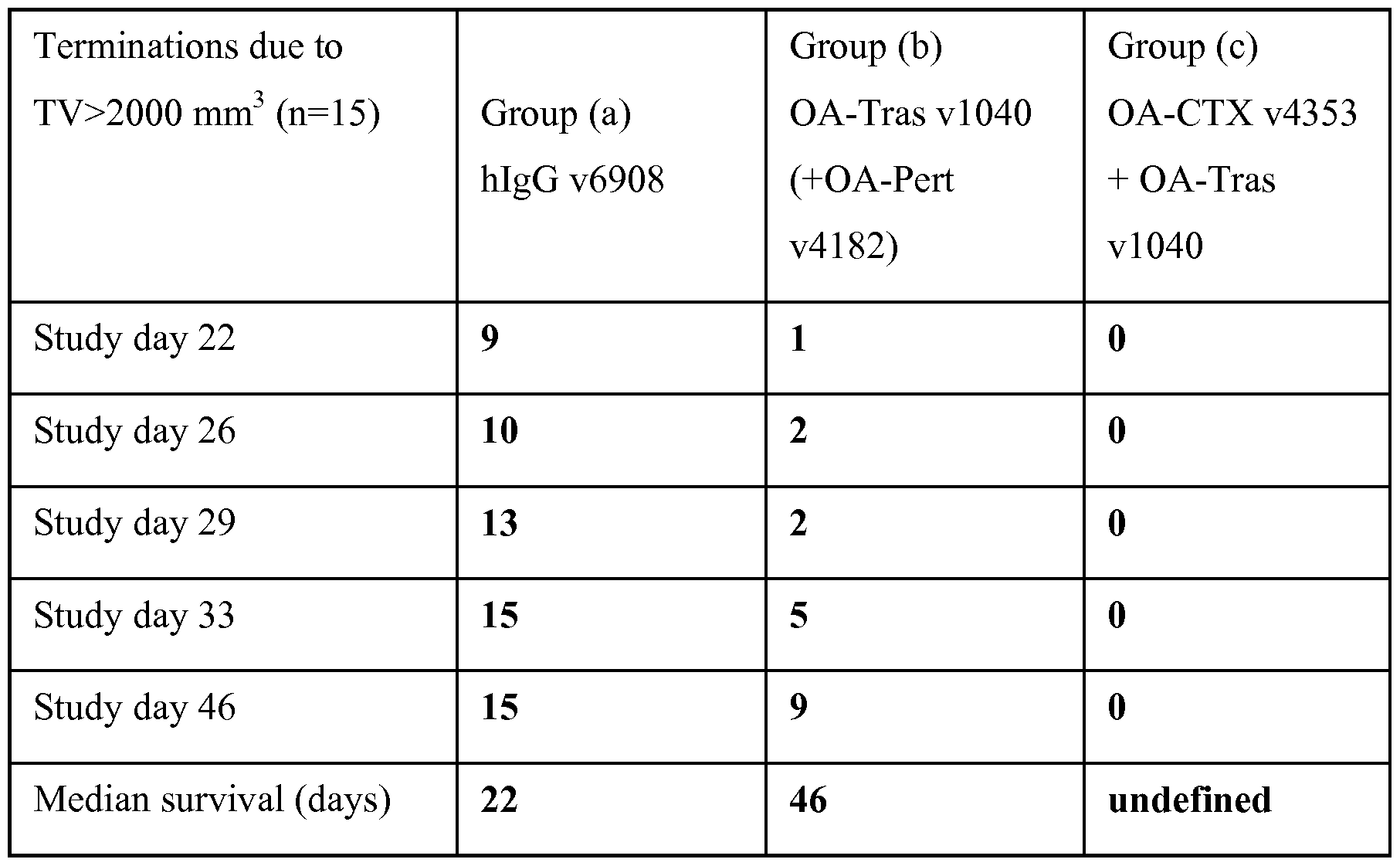


Claims
Priority Applications (8)
| Application Number | Priority Date | Filing Date | Title |
|---|---|---|---|
| KR1020167015484A KR20160083949A (en) | 2013-11-13 | 2014-11-13 | Monovalent antigen binding constructs targeting egfr and/or her2 and uses thereof |
| US15/036,174 US10273303B2 (en) | 2013-11-13 | 2014-11-13 | Monovalent antigen binding constructs targeting EGFR and/or HER2 and uses thereof |
| EP14862987.6A EP3068892A4 (en) | 2013-11-13 | 2014-11-13 | Monovalent antigen binding constructs targeting egfr and/or her2 and uses thereof |
| CN201480068379.8A CN105940113A (en) | 2013-11-13 | 2014-11-13 | Monovalent antigen binding constructs targeting egfr and/or her2 and uses thereof |
| AU2014348552A AU2014348552A1 (en) | 2013-11-13 | 2014-11-13 | Monovalent antigen binding constructs targeting EGFR and/or HER2 and uses thereof |
| CA2930307A CA2930307A1 (en) | 2013-11-13 | 2014-11-13 | Monovalent antigen binding constructs targeting egfr and/or her2 and uses thereof |
| JP2016530162A JP2016538283A (en) | 2013-11-13 | 2014-11-13 | Monovalent antigen binding constructs targeting EGFR and / or HER2 and uses thereof |
| MX2016006301A MX2016006301A (en) | 2013-11-13 | 2014-11-13 | Monovalent antigen binding constructs targeting egfr and/or her2 and uses thereof. |
Applications Claiming Priority (2)
| Application Number | Priority Date | Filing Date | Title |
|---|---|---|---|
| US201361903825P | 2013-11-13 | 2013-11-13 | |
| US61/903,825 | 2013-11-13 |
Publications (1)
| Publication Number | Publication Date |
|---|---|
| WO2015073721A1 true WO2015073721A1 (en) | 2015-05-21 |
Family
ID=53058030
Family Applications (1)
| Application Number | Title | Priority Date | Filing Date |
|---|---|---|---|
| PCT/US2014/065546 WO2015073721A1 (en) | 2013-11-13 | 2014-11-13 | Monovalent antigen binding constructs targeting egfr and/or her2 and uses thereof |
Country Status (9)
| Country | Link |
|---|---|
| US (1) | US10273303B2 (en) |
| EP (1) | EP3068892A4 (en) |
| JP (1) | JP2016538283A (en) |
| KR (1) | KR20160083949A (en) |
| CN (1) | CN105940113A (en) |
| AU (1) | AU2014348552A1 (en) |
| CA (1) | CA2930307A1 (en) |
| MX (1) | MX2016006301A (en) |
| WO (1) | WO2015073721A1 (en) |
Cited By (4)
| Publication number | Priority date | Publication date | Assignee | Title |
|---|---|---|---|---|
| RU2798981C2 (en) * | 2017-08-18 | 2023-06-30 | Байли-Байо (Чэнду) Фармасьютикал Ко., Лтд. | Antibody conjugate with non-natural amatoxin |
| EP3988575A4 (en) * | 2019-06-20 | 2023-08-16 | Chimagen Biosciences, Ltd. | Covalent multi-specific antibody |
| US11773172B2 (en) | 2018-03-19 | 2023-10-03 | WuXi Biologics Ireland Limited | Anti-EGFR antibody polypeptide |
| WO2023242372A1 (en) * | 2022-06-15 | 2023-12-21 | argenx BV | Fcrn/hsa binding molecules and methods of use |
Families Citing this family (7)
| Publication number | Priority date | Publication date | Assignee | Title |
|---|---|---|---|---|
| US9499634B2 (en) | 2012-06-25 | 2016-11-22 | Zymeworks Inc. | Process and methods for efficient manufacturing of highly pure asymmetric antibodies in mammalian cells |
| CN106699890B (en) * | 2016-10-31 | 2021-05-07 | 上海大学 | Gold nanoparticle-based artificial antibody targeting Epidermal Growth Factor Receptor (EGFR) and preparation method thereof |
| WO2018129255A1 (en) * | 2017-01-06 | 2018-07-12 | Momenta Pharmaceuticals, Inc. | Compositions and methods related to engineered fc constructs |
| EP3601368A4 (en) * | 2017-03-31 | 2021-03-31 | Zymeworks Inc. | Tumor antigen presentation inducer constructs and uses thereof |
| CN107325184A (en) * | 2017-08-08 | 2017-11-07 | 安徽大学 | The bispecific antibody of a kind of targeting EGFR and HER2 and its application |
| WO2020191434A1 (en) * | 2019-03-22 | 2020-10-01 | Olivia Newton-John Cancer Research Institute | Anti-her2 binding molecules |
| IL297662A (en) * | 2020-04-30 | 2022-12-01 | Chia Tai Tianqing Pharmaceutical Group Co Ltd | Antigen binding constructs targeting her2 and uses thereof |
Citations (59)
| Publication number | Priority date | Publication date | Assignee | Title |
|---|---|---|---|---|
| US4179337A (en) | 1973-07-20 | 1979-12-18 | Davis Frank F | Non-immunogenic polypeptides |
| US4256746A (en) | 1978-11-14 | 1981-03-17 | Takeda Chemical Industries | Dechloromaytansinoids, their pharmaceutical compositions and method of use |
| US4294757A (en) | 1979-01-31 | 1981-10-13 | Takeda Chemical Industries, Ltd | 20-O-Acylmaytansinoids |
| US4307016A (en) | 1978-03-24 | 1981-12-22 | Takeda Chemical Industries, Ltd. | Demethyl maytansinoids |
| US4313946A (en) | 1981-01-27 | 1982-02-02 | The United States Of America As Represented By The Secretary Of Agriculture | Chemotherapeutically active maytansinoids from Trewia nudiflora |
| US4315929A (en) | 1981-01-27 | 1982-02-16 | The United States Of America As Represented By The Secretary Of Agriculture | Method of controlling the European corn borer with trewiasine |
| US4322348A (en) | 1979-06-05 | 1982-03-30 | Takeda Chemical Industries, Ltd. | Maytansinoids |
| US4331598A (en) | 1979-09-19 | 1982-05-25 | Takeda Chemical Industries, Ltd. | Maytansinoids |
| US4362663A (en) | 1979-09-21 | 1982-12-07 | Takeda Chemical Industries, Ltd. | Maytansinoid compound |
| US4364866A (en) | 1979-09-21 | 1982-12-21 | Takeda Chemical Industries, Ltd. | Maytansinoids |
| US4371533A (en) | 1980-10-08 | 1983-02-01 | Takeda Chemical Industries, Ltd. | 4,5-Deoxymaytansinoids, their use and pharmaceutical compositions thereof |
| US4424219A (en) | 1981-05-20 | 1984-01-03 | Takeda Chemical Industries, Ltd. | 9-Thiomaytansinoids and their pharmaceutical compositions and use |
| US4450254A (en) | 1980-11-03 | 1984-05-22 | Standard Oil Company | Impact improvement of high nitrile resins |
| US4816567A (en) | 1983-04-08 | 1989-03-28 | Genentech, Inc. | Recombinant immunoglobin preparations |
| US4980286A (en) | 1985-07-05 | 1990-12-25 | Whitehead Institute For Biomedical Research | In vivo introduction and expression of foreign genetic material in epithelial cells |
| US5208020A (en) | 1989-10-25 | 1993-05-04 | Immunogen Inc. | Cytotoxic agents comprising maytansinoids and their therapeutic use |
| WO1993016185A2 (en) | 1992-02-06 | 1993-08-19 | Creative Biomolecules, Inc. | Biosynthetic binding protein for cancer marker |
| WO1993021319A1 (en) | 1992-04-08 | 1993-10-28 | Cetus Oncology Corporation | HUMANIZED C-erbB-2 SPECIFIC ANTIBODIES |
| US5362852A (en) | 1991-09-27 | 1994-11-08 | Pfizer Inc. | Modified peptide derivatives conjugated at 2-hydroxyethylamine moieties |
| US5500362A (en) | 1987-01-08 | 1996-03-19 | Xoma Corporation | Chimeric antibody with specificity to human B cell surface antigen |
| WO1996027011A1 (en) | 1995-03-01 | 1996-09-06 | Genentech, Inc. | A method for making heteromultimeric polypeptides |
| US5558864A (en) | 1991-03-06 | 1996-09-24 | Merck Patent Gesellschaft Mit Beschrankter Haftung | Humanized and chimeric anti-epidermal growth factor receptor monoclonal antibodies |
| US5571894A (en) | 1991-02-05 | 1996-11-05 | Ciba-Geigy Corporation | Recombinant antibodies specific for a growth factor receptor |
| US5587458A (en) | 1991-10-07 | 1996-12-24 | Aronex Pharmaceuticals, Inc. | Anti-erbB-2 antibodies, combinations thereof, and therapeutic and diagnostic uses thereof |
| US5641869A (en) | 1991-05-24 | 1997-06-24 | Genentech, Inc. | Method for purifying heregulin |
| US5648237A (en) | 1991-09-19 | 1997-07-15 | Genentech, Inc. | Expression of functional antibody fragments |
| US5789199A (en) | 1994-11-03 | 1998-08-04 | Genentech, Inc. | Process for bacterial production of polypeptides |
| US5840523A (en) | 1995-03-01 | 1998-11-24 | Genetech, Inc. | Methods and compositions for secretion of heterologous polypeptides |
| US5891996A (en) | 1972-09-17 | 1999-04-06 | Centro De Inmunologia Molecular | Humanized and chimeric monoclonal antibodies that recognize epidermal growth factor receptor (EGF-R); diagnostic and therapeutic use |
| US5959177A (en) | 1989-10-27 | 1999-09-28 | The Scripps Research Institute | Transgenic plants expressing assembled secretory antibodies |
| US6040498A (en) | 1998-08-11 | 2000-03-21 | North Caroline State University | Genetically engineered duckweed |
| US6214345B1 (en) | 1993-05-14 | 2001-04-10 | Bristol-Myers Squibb Co. | Lysosomal enzyme-cleavable antitumor drug conjugates |
| WO2001024763A2 (en) | 1999-10-01 | 2001-04-12 | Immunogen, Inc. | Compositions and methods for treating cancer using immunoconjugates and chemotherapeutic agents |
| US6420548B1 (en) | 1999-10-04 | 2002-07-16 | Medicago Inc. | Method for regulating transcription of foreign genes |
| US6441163B1 (en) | 2001-05-31 | 2002-08-27 | Immunogen, Inc. | Methods for preparation of cytotoxic conjugates of maytansinoids and cell binding agents |
| WO2002088172A2 (en) | 2001-04-30 | 2002-11-07 | Seattle Genetics, Inc. | Pentapeptide compounds and uses related thereto |
| WO2003026577A2 (en) | 2001-09-24 | 2003-04-03 | Seattle Genetics, Inc. | P-amidobenzylethers in drug delivery agents |
| US20030096743A1 (en) | 2001-09-24 | 2003-05-22 | Seattle Genetics, Inc. | p-Amidobenzylethers in drug delivery agents |
| WO2003043583A2 (en) | 2001-11-20 | 2003-05-30 | Seattle Genetics, Inc. | Treatment of immunological disorders using anti-cd30 antibodies |
| WO2003068144A2 (en) | 2001-12-21 | 2003-08-21 | Immunogen, Inc. | Cytotoxic agents |
| US20040039176A1 (en) | 2002-08-16 | 2004-02-26 | Immunogen, Inc. | Cross-linkers with high reactivity and solubility and their use in the preparation of conjugates for targeted delivery of small molecule drugs |
| WO2004032828A2 (en) | 2002-07-31 | 2004-04-22 | Seattle Genetics, Inc. | Anti-cd20 antibody-drug conjugates for the treatment of cancer and immune disorders |
| US20040235840A1 (en) | 2003-05-20 | 2004-11-25 | Immunogen, Inc. | Cytotoxic agents comprising new maytansinoids |
| US6884874B2 (en) | 2000-09-12 | 2005-04-26 | Smithkline Beecham Corporation | Process and intermediates |
| WO2005037992A2 (en) | 2003-10-10 | 2005-04-28 | Immunogen, Inc. | Method of targeting specific cell populations using cell-binding agent maytansinoid conjugates linked via a non-cleavable linker, said conjugates, and methods of making said conjugates |
| US20060018899A1 (en) | 2004-07-22 | 2006-01-26 | Genentech, Inc. | HER2 antibody composition |
| WO2006105338A2 (en) | 2005-03-31 | 2006-10-05 | Xencor, Inc. | Fc VARIANTS WITH OPTIMIZED PROPERTIES |
| US7125978B1 (en) | 1999-10-04 | 2006-10-24 | Medicago Inc. | Promoter for regulating expression of foreign genes |
| US20090148905A1 (en) * | 2007-11-30 | 2009-06-11 | Claire Ashman | Antigen-binding constructs |
| US20090202536A1 (en) | 2004-06-01 | 2009-08-13 | Genentech, Inc. | Antibody-drug conjugates and methods |
| US7678890B2 (en) * | 2005-07-22 | 2010-03-16 | Five Prime Therapeutics, Inc. | Compositions and methods of treating disease with FGFR fusion proteins |
| US20110212087A1 (en) | 2009-11-30 | 2011-09-01 | William Strohl | Antibody Fc Mutants with Ablated Effector Functions |
| WO2011120135A1 (en) | 2010-03-29 | 2011-10-06 | Zymeworks, Inc. | Antibodies with enhanced or suppressed effector function |
| US8097436B2 (en) | 2005-02-07 | 2012-01-17 | Roche Glycart Ag | Antigen binding molecules that bind EGFR, vectors encoding same, and uses thereof |
| US20120149876A1 (en) * | 2010-11-05 | 2012-06-14 | Zymeworks Inc. | Stable Heterodimeric Antibody Design with Mutations in the Fc Domain |
| US20120225058A1 (en) | 2004-10-21 | 2012-09-06 | Xencor, Inc. | Novel immunoglobulin insertions, deletions, and substitutions |
| US20120251531A1 (en) | 2011-03-29 | 2012-10-04 | Genentech, Inc. | ANTIBODY Fc VARIANTS |
| US8409572B2 (en) | 2000-04-12 | 2013-04-02 | Laboratoire Francais Du Fractionnement Et Des Biotechnologies | Monoclonal antibodies with enhanced ADCC function |
| US8624003B2 (en) | 2009-06-03 | 2014-01-07 | Immunogen, Inc. | Methods for preparation of antibody-maytansinoid conjugates |
Family Cites Families (22)
| Publication number | Priority date | Publication date | Assignee | Title |
|---|---|---|---|---|
| US7951917B1 (en) | 1997-05-02 | 2011-05-31 | Genentech, Inc. | Method for making multispecific antibodies having heteromultimeric and common components |
| US6737056B1 (en) | 1999-01-15 | 2004-05-18 | Genentech, Inc. | Polypeptide variants with altered effector function |
| AU2003217912A1 (en) | 2002-03-01 | 2003-09-16 | Xencor | Antibody optimization |
| PL374586A1 (en) | 2002-10-10 | 2005-10-31 | Merck Patent Gmbh | Bispecific anti-erb-b antibodies and their use in tumor therapy |
| US20060074225A1 (en) | 2004-09-14 | 2006-04-06 | Xencor, Inc. | Monomeric immunoglobulin Fc domains |
| AR056142A1 (en) * | 2005-10-21 | 2007-09-19 | Amgen Inc | METHODS TO GENERATE THE MONOVALENT IGG ANTIBODY |
| JP2009522316A (en) | 2006-01-04 | 2009-06-11 | メルク パテント ゲゼルシャフト ミット ベシュレンクテル ハフツング | Combination therapy using anti-EGFR antibody and anti-HER2 antibody |
| CA2588106A1 (en) * | 2007-05-18 | 2008-11-18 | Nrc - Institute For Biological Sciences | Single-domain antibodies and heavy chain antibody against egfr and uses thereof |
| EP2235064B1 (en) | 2008-01-07 | 2015-11-25 | Amgen Inc. | Method for making antibody fc-heterodimeric molecules using electrostatic steering effects |
| US9493578B2 (en) * | 2009-09-02 | 2016-11-15 | Xencor, Inc. | Compositions and methods for simultaneous bivalent and monovalent co-engagement of antigens |
| JP2013511281A (en) | 2009-11-23 | 2013-04-04 | アムジェン インコーポレイテッド | Monomeric antibody Fc |
| AR080793A1 (en) * | 2010-03-26 | 2012-05-09 | Roche Glycart Ag | BISPECIFIC ANTIBODIES |
| AU2012222252B2 (en) | 2011-02-25 | 2016-08-25 | Chugai Seiyaku Kabushiki Kaisha | FcgammaRIIb-specific Fc antibody |
| TWI687439B (en) | 2011-06-30 | 2020-03-11 | 中外製藥股份有限公司 | Heterodimerized polypeptide |
| US20130078250A1 (en) * | 2011-08-23 | 2013-03-28 | Oliver Ast | Bispecific t cell activating antigen binding molecules |
| CN103857700A (en) * | 2011-08-26 | 2014-06-11 | 梅里麦克制药股份有限公司 | Tandem FC bispecific antibodies |
| KR102052774B1 (en) * | 2011-11-04 | 2019-12-04 | 자임워크스 인코포레이티드 | Stable heterodimeric antibody design with mutations in the fc domain |
| CA2856411A1 (en) | 2011-11-21 | 2013-05-30 | Immunogen, Inc. | Method of treatment of tumors that are resistant to egfr therapies by egfr antibody cytotoxic agent conjugate |
| EP2847224A4 (en) * | 2012-05-10 | 2016-04-27 | Zymeworks Inc | Single-arm monovalent antibody constructs and uses thereof |
| JP6351572B2 (en) * | 2012-05-10 | 2018-07-04 | ザイムワークス,インコーポレイテッド | Heteromultimeric constructs of immunoglobulin heavy chains with mutations in the Fc domain |
| KR20150036606A (en) * | 2012-07-13 | 2015-04-07 | 자임워크스 인코포레이티드 | Bispecific asymmetric heterodimers comprising anti-cd3 constructs |
| CA2892623C (en) * | 2012-11-27 | 2020-08-11 | Ajou University Industry-Academic Cooperation Foundation | Ch3 domain variant pair inducing formation of heterodimer of heavy chain constant region of antibody at high efficiency, method for preparing same, and use thereof |
-
2014
- 2014-11-13 EP EP14862987.6A patent/EP3068892A4/en not_active Withdrawn
- 2014-11-13 CA CA2930307A patent/CA2930307A1/en not_active Abandoned
- 2014-11-13 MX MX2016006301A patent/MX2016006301A/en unknown
- 2014-11-13 AU AU2014348552A patent/AU2014348552A1/en not_active Abandoned
- 2014-11-13 KR KR1020167015484A patent/KR20160083949A/en not_active Application Discontinuation
- 2014-11-13 JP JP2016530162A patent/JP2016538283A/en active Pending
- 2014-11-13 CN CN201480068379.8A patent/CN105940113A/en active Pending
- 2014-11-13 US US15/036,174 patent/US10273303B2/en not_active Expired - Fee Related
- 2014-11-13 WO PCT/US2014/065546 patent/WO2015073721A1/en active Application Filing
Patent Citations (67)
| Publication number | Priority date | Publication date | Assignee | Title |
|---|---|---|---|---|
| US5891996A (en) | 1972-09-17 | 1999-04-06 | Centro De Inmunologia Molecular | Humanized and chimeric monoclonal antibodies that recognize epidermal growth factor receptor (EGF-R); diagnostic and therapeutic use |
| US4179337A (en) | 1973-07-20 | 1979-12-18 | Davis Frank F | Non-immunogenic polypeptides |
| US4307016A (en) | 1978-03-24 | 1981-12-22 | Takeda Chemical Industries, Ltd. | Demethyl maytansinoids |
| US4361650A (en) | 1978-03-24 | 1982-11-30 | Takeda Chemical Industries, Ltd. | Fermentation process of preparing demethyl maytansinoids |
| US4256746A (en) | 1978-11-14 | 1981-03-17 | Takeda Chemical Industries | Dechloromaytansinoids, their pharmaceutical compositions and method of use |
| US4294757A (en) | 1979-01-31 | 1981-10-13 | Takeda Chemical Industries, Ltd | 20-O-Acylmaytansinoids |
| US4322348A (en) | 1979-06-05 | 1982-03-30 | Takeda Chemical Industries, Ltd. | Maytansinoids |
| US4331598A (en) | 1979-09-19 | 1982-05-25 | Takeda Chemical Industries, Ltd. | Maytansinoids |
| US4362663A (en) | 1979-09-21 | 1982-12-07 | Takeda Chemical Industries, Ltd. | Maytansinoid compound |
| US4364866A (en) | 1979-09-21 | 1982-12-21 | Takeda Chemical Industries, Ltd. | Maytansinoids |
| US4371533A (en) | 1980-10-08 | 1983-02-01 | Takeda Chemical Industries, Ltd. | 4,5-Deoxymaytansinoids, their use and pharmaceutical compositions thereof |
| US4450254A (en) | 1980-11-03 | 1984-05-22 | Standard Oil Company | Impact improvement of high nitrile resins |
| US4315929A (en) | 1981-01-27 | 1982-02-16 | The United States Of America As Represented By The Secretary Of Agriculture | Method of controlling the European corn borer with trewiasine |
| US4313946A (en) | 1981-01-27 | 1982-02-02 | The United States Of America As Represented By The Secretary Of Agriculture | Chemotherapeutically active maytansinoids from Trewia nudiflora |
| US4424219A (en) | 1981-05-20 | 1984-01-03 | Takeda Chemical Industries, Ltd. | 9-Thiomaytansinoids and their pharmaceutical compositions and use |
| US4816567A (en) | 1983-04-08 | 1989-03-28 | Genentech, Inc. | Recombinant immunoglobin preparations |
| US4980286A (en) | 1985-07-05 | 1990-12-25 | Whitehead Institute For Biomedical Research | In vivo introduction and expression of foreign genetic material in epithelial cells |
| US5500362A (en) | 1987-01-08 | 1996-03-19 | Xoma Corporation | Chimeric antibody with specificity to human B cell surface antigen |
| US5208020A (en) | 1989-10-25 | 1993-05-04 | Immunogen Inc. | Cytotoxic agents comprising maytansinoids and their therapeutic use |
| US5416064A (en) | 1989-10-25 | 1995-05-16 | Immunogen, Inc. | Cytotoxic agents comprising maytansinoids and their therapeutic use |
| US5959177A (en) | 1989-10-27 | 1999-09-28 | The Scripps Research Institute | Transgenic plants expressing assembled secretory antibodies |
| US6417429B1 (en) | 1989-10-27 | 2002-07-09 | The Scripps Research Institute | Transgenic plants expressing assembled secretory antibodies |
| US5571894A (en) | 1991-02-05 | 1996-11-05 | Ciba-Geigy Corporation | Recombinant antibodies specific for a growth factor receptor |
| US5558864A (en) | 1991-03-06 | 1996-09-24 | Merck Patent Gesellschaft Mit Beschrankter Haftung | Humanized and chimeric anti-epidermal growth factor receptor monoclonal antibodies |
| US5641869A (en) | 1991-05-24 | 1997-06-24 | Genentech, Inc. | Method for purifying heregulin |
| US5648237A (en) | 1991-09-19 | 1997-07-15 | Genentech, Inc. | Expression of functional antibody fragments |
| US5362852A (en) | 1991-09-27 | 1994-11-08 | Pfizer Inc. | Modified peptide derivatives conjugated at 2-hydroxyethylamine moieties |
| US5587458A (en) | 1991-10-07 | 1996-12-24 | Aronex Pharmaceuticals, Inc. | Anti-erbB-2 antibodies, combinations thereof, and therapeutic and diagnostic uses thereof |
| WO1993016185A2 (en) | 1992-02-06 | 1993-08-19 | Creative Biomolecules, Inc. | Biosynthetic binding protein for cancer marker |
| WO1993021319A1 (en) | 1992-04-08 | 1993-10-28 | Cetus Oncology Corporation | HUMANIZED C-erbB-2 SPECIFIC ANTIBODIES |
| US6214345B1 (en) | 1993-05-14 | 2001-04-10 | Bristol-Myers Squibb Co. | Lysosomal enzyme-cleavable antitumor drug conjugates |
| US5789199A (en) | 1994-11-03 | 1998-08-04 | Genentech, Inc. | Process for bacterial production of polypeptides |
| WO1996027011A1 (en) | 1995-03-01 | 1996-09-06 | Genentech, Inc. | A method for making heteromultimeric polypeptides |
| US5840523A (en) | 1995-03-01 | 1998-11-24 | Genetech, Inc. | Methods and compositions for secretion of heterologous polypeptides |
| US6040498A (en) | 1998-08-11 | 2000-03-21 | North Caroline State University | Genetically engineered duckweed |
| WO2001024763A2 (en) | 1999-10-01 | 2001-04-12 | Immunogen, Inc. | Compositions and methods for treating cancer using immunoconjugates and chemotherapeutic agents |
| US6420548B1 (en) | 1999-10-04 | 2002-07-16 | Medicago Inc. | Method for regulating transcription of foreign genes |
| US7125978B1 (en) | 1999-10-04 | 2006-10-24 | Medicago Inc. | Promoter for regulating expression of foreign genes |
| US8409572B2 (en) | 2000-04-12 | 2013-04-02 | Laboratoire Francais Du Fractionnement Et Des Biotechnologies | Monoclonal antibodies with enhanced ADCC function |
| US6884874B2 (en) | 2000-09-12 | 2005-04-26 | Smithkline Beecham Corporation | Process and intermediates |
| WO2002088172A2 (en) | 2001-04-30 | 2002-11-07 | Seattle Genetics, Inc. | Pentapeptide compounds and uses related thereto |
| US6441163B1 (en) | 2001-05-31 | 2002-08-27 | Immunogen, Inc. | Methods for preparation of cytotoxic conjugates of maytansinoids and cell binding agents |
| US20030096743A1 (en) | 2001-09-24 | 2003-05-22 | Seattle Genetics, Inc. | p-Amidobenzylethers in drug delivery agents |
| US20030130189A1 (en) | 2001-09-24 | 2003-07-10 | Senter Peter D. | P-amidobenzylethers in drug delivery agents |
| WO2003026577A2 (en) | 2001-09-24 | 2003-04-03 | Seattle Genetics, Inc. | P-amidobenzylethers in drug delivery agents |
| WO2003043583A2 (en) | 2001-11-20 | 2003-05-30 | Seattle Genetics, Inc. | Treatment of immunological disorders using anti-cd30 antibodies |
| US20040001838A1 (en) | 2001-12-21 | 2004-01-01 | Immunogen, Inc. | Cytotoxic agents bearing a reactive polyethylene glycol moiety, cytotoxic conjugates comprising polyethylene glycol linking groups, and methods of making and using the same |
| WO2003068144A2 (en) | 2001-12-21 | 2003-08-21 | Immunogen, Inc. | Cytotoxic agents |
| WO2004032828A2 (en) | 2002-07-31 | 2004-04-22 | Seattle Genetics, Inc. | Anti-cd20 antibody-drug conjugates for the treatment of cancer and immune disorders |
| US20040039176A1 (en) | 2002-08-16 | 2004-02-26 | Immunogen, Inc. | Cross-linkers with high reactivity and solubility and their use in the preparation of conjugates for targeted delivery of small molecule drugs |
| WO2004016801A2 (en) | 2002-08-16 | 2004-02-26 | Immunogen, Inc. | Cross-linkers with high reactivity and solubility and their use in the preparation of conjugates for targeted delivery of small molecule drugs |
| US20040235840A1 (en) | 2003-05-20 | 2004-11-25 | Immunogen, Inc. | Cytotoxic agents comprising new maytansinoids |
| US8163888B2 (en) | 2003-10-10 | 2012-04-24 | Immunogen, Inc. | Method of targeting specific cell populations using cell-binding agent maytansinoid conjugates linked via a non-cleavable linker, said conjugates and methods of making said conjugates |
| WO2005037992A2 (en) | 2003-10-10 | 2005-04-28 | Immunogen, Inc. | Method of targeting specific cell populations using cell-binding agent maytansinoid conjugates linked via a non-cleavable linker, said conjugates, and methods of making said conjugates |
| US20090202536A1 (en) | 2004-06-01 | 2009-08-13 | Genentech, Inc. | Antibody-drug conjugates and methods |
| US20060018899A1 (en) | 2004-07-22 | 2006-01-26 | Genentech, Inc. | HER2 antibody composition |
| US20120225058A1 (en) | 2004-10-21 | 2012-09-06 | Xencor, Inc. | Novel immunoglobulin insertions, deletions, and substitutions |
| US8097436B2 (en) | 2005-02-07 | 2012-01-17 | Roche Glycart Ag | Antigen binding molecules that bind EGFR, vectors encoding same, and uses thereof |
| WO2006105338A2 (en) | 2005-03-31 | 2006-10-05 | Xencor, Inc. | Fc VARIANTS WITH OPTIMIZED PROPERTIES |
| US7678890B2 (en) * | 2005-07-22 | 2010-03-16 | Five Prime Therapeutics, Inc. | Compositions and methods of treating disease with FGFR fusion proteins |
| US20090148905A1 (en) * | 2007-11-30 | 2009-06-11 | Claire Ashman | Antigen-binding constructs |
| US8624003B2 (en) | 2009-06-03 | 2014-01-07 | Immunogen, Inc. | Methods for preparation of antibody-maytansinoid conjugates |
| US20110212087A1 (en) | 2009-11-30 | 2011-09-01 | William Strohl | Antibody Fc Mutants with Ablated Effector Functions |
| WO2011120134A1 (en) | 2010-03-29 | 2011-10-06 | Zymeworks, Inc. | Antibodies with enhanced or suppressed effector function |
| WO2011120135A1 (en) | 2010-03-29 | 2011-10-06 | Zymeworks, Inc. | Antibodies with enhanced or suppressed effector function |
| US20120149876A1 (en) * | 2010-11-05 | 2012-06-14 | Zymeworks Inc. | Stable Heterodimeric Antibody Design with Mutations in the Fc Domain |
| US20120251531A1 (en) | 2011-03-29 | 2012-10-04 | Genentech, Inc. | ANTIBODY Fc VARIANTS |
Non-Patent Citations (188)
| Title |
|---|
| "Antibodies, A Laboratory Manual", 1988, COLD SPRING HARBOR LABORATORY |
| "Applications Handbook and Catalog", 2003, pages: 467 - 498 |
| "Controlled Drug Bioavailability, Drug Product Design and Performance", 1984, WILEY |
| "Current Protocols in Protein Science", JOHN WILEY & SONS, INC, article "Chapter 15: Chemical Modifications of Proteins" |
| "Medical Applications of Controlled Release", 1974, CRC PRES |
| "POST-TRANSLATIONAL COVALENT MODIFICATION OF PROTEINS", 1983, ACADEMIC PRESS, pages: 1 - 12 |
| "Remington's Pharmaceutical Sciences. 18th Ed.", 1990, MACK PUBLISHING COMPANY |
| A.L. LEHNINGER: "Biochemistry", WORTH PUBLISHERS, INC. |
| ALTSCHUL ET AL., J. MOL. BIOL., vol. 215, 1990, pages 403 - 410 |
| ALTSCHUL ET AL., NUC. ACIDS RES., vol. 25, 1997, pages 3389 - 3402 |
| ANIDO ET AL., CLIN CANCER RES, vol. 9, 2003, pages 1274 - 1283 |
| ATALAY ET AL., ANN. ONCOLOGY, vol. 14, 2003, pages 1346 - 1363 |
| AUSUBEL ET AL., CURRENT PROTOCOLS IN MOLECULAR BIOLOGY, 1995 |
| BATZER ET AL., NUCLEIC ACID RES., vol. 19, 1991, pages 5081 |
| BELL ET AL., CANCER LETT., vol. 289, no. 1, 1 March 2010 (2010-03-01), pages 81 - 90 |
| BIRTALAN ET AL., JMB, vol. 377, 2008, pages 1518 - 1528 |
| BLOOD, vol. 118, no. 15, 27 July 2011 (2011-07-27), pages 4159 - 68 |
| BOLAND ET AL.: "Nimotuzumab: a novel anti-EGFR monoclonal antibody that retains anti-EGFR activity while minimizing skin toxicity.", EXPERT OPIN BIOL THER, vol. 9, no. 9, September 2009 (2009-09-01), pages 1199 - 1206, XP055344425 * |
| BOONE ET AL., ONCOLOGY, vol. 72, 2007, pages 152 - 159 |
| BOWLES J.A. ET AL: "Anti-CD20 monoclonal antibody with enhanced affinity for CD 16 activates NK cells at lower concentrations and more effectively than rituximab", BLOOD, vol. 108, no. 8, 6 July 2006 (2006-07-06), pages 2648 - 2654 |
| BUCHWALD ET AL., SURGERY, vol. 88, 1980, pages 507 |
| BUNN ET AL., CLIN CANCER RES, vol. 7, 2001, pages 3239 - 3250 |
| CAPEL ET AL., IMMUNOMETHODS, vol. 4, 1994, pages 25 - 34 |
| CAREY; SUNDBERG: "Advanced Organic Chemistry", vol. A, B, 1992, PLENUM PRESS |
| CARRAWAY ET AL., NATURE, vol. 387, 1997, pages 512 - 516 |
| CHARI ET AL., CANCER RESEARCH, vol. 52, 1992, pages 127 - 131 |
| CHARLTON: "Methods in Molecular Biology", vol. 248, 2003, HUMANA PRESS, pages: 245 - 254 |
| CHO ET AL., NATURE, vol. 421, 2003, pages 756 - 760 |
| CHU S.Y. ET AL., MOL IMMUNOL, vol. 45, no. 15, September 2008 (2008-09-01), pages 3926 - 3933 |
| CLYNES ET AL., PNAS (USA, vol. 95, 1998, pages 652 - 656 |
| COOK ET AL., MOL. CELL. BIOL., vol. 11, 1991, pages 2547 - 2557 |
| CREIGHTON: "Proteins: Structures and Molecular Principles", 1983, W. H. FREEMAN & CO. |
| CREIGHTON: "Proteins: Structures and Molecular Properties", December 1993, W H FREEMAN & CO. |
| DAERON, ANNU. REV. IMMUNOL., vol. 15, 1997, pages 203 - 234 |
| DAVIS, JH. ET AL., PROT ENG DES SEL, vol. 23, no. 4, 2010, pages 195 - 202 |
| DE HAAS ET AL., J. LAB. CLIN. MED., vol. 126, 1995, pages 330 - 341 |
| DERER ET AL.: "IMPACT OF EPIDERMAL GROWTH FACTOR RECEPTOR", JOURNAL OF IMMUNOLOGY, vol. 189, no. 11, (E, 1 December 2012 (2012-12-01), pages 5230 - 5239, XP055256902, DOI: GFR) CELL SURFACE EXPRESSION LEVELS ON * |
| DESJARLAIS J.R. ET AL: "Modulation of antibody effector function", EXP CELL RES, vol. 317, no. 9, 15 May 2011 (2011-05-15), pages 1278 - 1285 |
| DRAGOWSKA ET AL., BMC CANCER, vol. 11, 2011, pages 420 |
| DURING ET AL., ANN. NEUROL., vol. 25, 1989, pages 351 |
| DUROCHER, Y. ET AL: "High-level and high-throughput recombinant protein production by transient transfection of suspension-growing CHO cells", NUCLEIC ACIDS RESEARCH, vol. 30, 2002, pages E9 |
| E. SCHRODER ET AL: "The Peptides", vol. 1, 1965, ACADEMIC PRESS, pages: 76 - 136 |
| FALLS ET AL., CELL, vol. 72, 1993, pages 801 - 815 |
| FERRARA C. ET AL: "Unique carbohydrate-carbohydrate interactions are required for high affinity binding between FcgammaRIII and antibodies lacking core fucose", PROC NATL ACAD SCI U S A., vol. 108, no. 31, 2 August 2011 (2011-08-02), pages 12669 - 12674 |
| FIELDS, G. ET AL: "Int. J. Peptide Protein Res.", vol. 35, 1990, article "Solid phase peptide synthesis utilizing 9-fluoroenylmethoxycarbonyl amino acids", pages: 161 - 214 |
| FRANKLIN ET AL., CANCER CELL, vol. 5, 2004, pages 317 - 328 |
| GABORIT ET AL., J BIOL CHEM., vol. 286, no. 13, 1 April 2011 (2011-04-01), pages 11337 - 11345 |
| GARRETT ET AL., MOL. CELL, vol. 11, 2003, pages 495 - 505 |
| GAZZANO-SANTORO ET AL., J. IMMUNOL. METHODS, vol. 202, 1996, pages 163 |
| GEISER ET AL.: "Macromolecular Sequencing and Synthesis", 1988, ALAN R. LISS, INC., article "Automation of solid-phase peptide synthesis", pages: 199 - 218 |
| GEOGHEGAN; STROH, BIOCONJUGATE CHEM., vol. 3, 1992, pages 138 - 146 |
| GERNGROSS, NAT. BIOTECH., vol. 22, 2004, pages 1409 - 1414 |
| GERSTNER ET AL., JMB, vol. 321, 2002, pages 851 - 862 |
| GETZ ET AL., BIOCHEM., vol. 273, 1999, pages 73 - 80 |
| GHETIE ET AL., ANNU REV IMMUNOL, vol. 18, 2000, pages 739 - 766 |
| GOODSON, MEDICAL APPLICATIONS OF CONTROLLED RELEASE, vol. 2, 1984, pages 115 - 138 |
| GRAHAM ET AL., J. GEN VIROL., vol. 36, 1977, pages 59 |
| GREG T. HERMANSON: "Bioconjugate Techniques", 2013, ACADEMIC PRESS |
| GUNASEKARAN K. ET AL., J BIOL CHEM., vol. 285, 2010, pages 19637 - 19646 |
| GUYER ET AL., J. IMMUNOL., vol. 117, 1976, pages 587 |
| HAMEL ET AL., MOLECULAR PHARMACOLOGY, vol. 47, 1995, pages 965 - 976 |
| HAMEL ET AL., THE JOURNAL OF BIOLOGICAL CHEMISTRY, vol. 265, no. 28, 1990, pages 17141 - 17149 |
| HARARI ET AL., ONCOGENE, vol. 18, 1999, pages 2681 - 2689 |
| HARLOW; LANE: "Antibodies, A Laboratory Manual", 1988, COLD SPRING HARBOR PUBLICATIONS |
| HARMSEN M.M. ET AL: "Properties, production, and applications of camelid single-domain antibody fragments", APPL. MICROBIOL BIOTECHNOL., vol. 77, no. 1, 2007, pages 13 - 22 |
| HEIDER K.H. ET AL: "A novel Fc-engineered monoclonal antibody to CD37 with enhanced ADCC and high proapoptotic activity for treatment of B-cell malignancies", BLOOD, vol. 118, no. 15, 27 July 2011 (2011-07-27), pages 4159 - 4168 |
| HENIKOFF; HENIKOFF, PROC. NATL. ACAD. SCI. USA, vol. 89, 1992, pages 10915 |
| HERBST; SHIN, CANCER, vol. 94, 2002, pages 1593 - 1611 |
| HERMANSON, G. T.: "Bioconjugate Techniques", 1996, ACADEMIC PRESS, pages: 234 - 242 |
| HIGASHIYAMA ET AL., SCIENCE, vol. 251, 1991, pages 936 - 939 |
| HO ET AL., J. BIOL. CHEM., vol. 270, 1995, pages 14523 - 14532 |
| HOLMES ET AL., SCIENCE, vol. 256, 1992, pages 1205 - 1210 |
| HOWARD ET AL., J. NEUROSURG, vol. 71, 1989, pages 105 |
| HUNKAPILLER ET AL., NATURE, vol. 310, 1984, pages 105 - 111 |
| ISSEL ET AL., CAN. TREATMENT. REV, vol. 5, 1978, pages 199 - 207 |
| JANEWAY ET AL.: "Immuno Biology: the immune system in health and disease", 1999, ELSEVIER SCIENCE LTD. |
| JOLIOT ET AL., PROC. NATL. ACAD. SCI. USA, vol. 88, 1991, pages 1864 - 1868 |
| JONES ET AL., NATURE, vol. 321, 1986, pages 522 - 525 |
| JUNGHANS ET AL., CANCER RES., vol. 50, 1990, pages 1495 |
| JUNGHANS: "Cruel antibody fictions! Cellular antigen enumeration by 'saturation' binding.", IMMUNOL TODAY SEPTEMBER, vol. 20, no. 9, 1999, pages 401 - 406, XP004177375 * |
| KABAT ET AL.: "Sequences of Proteins of Immunological Interest", 1991, NATIONAL INSTITUTES OF HEALTH |
| KANNAN ET AL., J. BIOL. CHEM., vol. 272, no. 6, 1997, pages 3330 - 3335 |
| KARLIN; ALTSCHUL, PROC. NATL. ACAD. SCI. USA, vol. 90, 1993, pages 5873 - 5787 |
| KELLEY ET AL., BIOCHEM, vol. 32, no. 27, 1993, pages 6828 - 6835 |
| KIM ET AL., J. IMMUNOL., vol. 24, 1994, pages 249 |
| KIMURA ET AL., NATURE, vol. 348, 1990, pages 257 - 260 |
| KING ET AL., TETRAHEDRON LETTERS, vol. 43, 2002, pages 1987 - 1990 |
| KOMURASAKI ET AL., ONCOGENE, vol. 15, 1997, pages 2841 - 2848 |
| KURAI ET AL., CLIN CANCER RES, vol. 13, no. 5, 2007, pages 1552 - 1561 |
| LABRIJN A.F. ET AL, PROC NATL ACAD SCI U S A., vol. 110, no. 13, 26 March 2013 (2013-03-26), pages 5145 - 5150 |
| LANGER, SCIENCE, vol. 249, 1990, pages 1527 - 1533 |
| LAZAR G.A. ET AL., PROC NATL ACAD SCI USA., vol. 103, no. 11, 14 March 2006 (2006-03-14), pages 4005 - 4010 |
| LAZAR G.A. ET AL: "Engineered antibody Fc variants with enhanced effector function", PROC NATL ACAD SCI USA., vol. 103, no. 11, 6 March 2006 (2006-03-06), pages 4005 - 4010 |
| LEVY ET AL., SCIENCE, vol. 228, 1985, pages 190 |
| LI ET AL., CANCER CELL, vol. 7, no. 4, April 2005 (2005-04-01), pages 301 - 311 |
| LI ET AL., JBC, vol. 285, no. 6, 2010, pages 3865 - 3871 |
| LI ET AL., NAT. BIOTECH., vol. 24, 2006, pages 210 - 215 |
| LIVINGSTON, R. J. ET AL.: "NIEHS-SNPs, environmental genome project", NIEHS ES15478, 2004 |
| LOPEZ-BERESTEIN: "LIPOSOMES IN THE THERAPY OF INFECTIOUS DISEASE AND CANCER", pages: 317 - 327 |
| LU Y ET AL, J IMMUNOL METHODS, vol. 365, no. 1-2, 28 February 2011 (2011-02-28), pages 132 - 141 |
| LU Y ET AL: "Identification of IgG(l) variants with increased affinity to FcyRIIIa and unaltered affinity to FcyRI and FcRn: comparison of soluble receptor-based and cell-based binding assays", J IMMUNOL METHODS, vol. 365, no. 1-2, 23 December 2010 (2010-12-23), pages 132 - 141 |
| MARCHIONNI ET AL., NATURE, vol. 362, 1993, pages 312 - 318 |
| MARQUARDT ET AL., SCIENCE, vol. 223, 1984, pages 1079 - 1082 |
| MATHER ET AL., ANNALS N.Y. ACAD. SCI., vol. 383, 1982, pages 44 - 68 |
| MATHER, BIOL. REPROD, vol. 23, 1980, pages 243 - 251 |
| MCDONAGH ET AL., MOL CANCER THER., vol. 11, no. 3, March 2012 (2012-03-01), pages 582 - 593 |
| MENDELSOHN; BASELGA, ONCOGENE, vol. 19, 2000, pages 6550 - 6565 |
| MIZUSHIMA T. ET AL: "Structural basis for improved efficacy of therapeutic antibodies on defucosylation of their Fc glycans", GENES CELLS, vol. 16, no. 11, November 2011 (2011-11-01), pages 1071 - 1080 |
| MODJTAHEDI ET AL., BR. J. CANCER, vol. 73, 1996, pages 228 - 235 |
| MOORE G.L. ET AL: "Engineered Fc variant antibodies with enhanced ability to recruit complement and mediate effector functions", MABS, vol. 2, no. 2, March 2010 (2010-03-01), pages 181 - 189 |
| MULLER ET AL., ANAL. CHEM, vol. 78, 2006, pages 4390 - 4397 |
| NAKAMURA ET AL., CANCER LETTERS, vol. 230, 2005, pages 33 - 46 |
| NEEDLEMAN; WUNSCH, J. MOL. BIOL., vol. 48, 1970, pages 443 |
| NEVE ET AL., CANCER CELL, vol. 10, no. 6, December 2006 (2006-12-01), pages 515 - 527 |
| NORDSTROM J.L. ET AL., BREAST CANCER RES., vol. 13, no. 6, 30 November 2011 (2011-11-30), pages R123 |
| NORDSTROM J.L. ET AL: "Anti-tumor activity and toxicokinetics analysis of MGAH22, an anti-HER2 monoclonal antibody with enhanced Fc-gamma receptor binding properties", BREAST CANCER RES., vol. 13, no. 6, 30 November 2011 (2011-11-30), pages R123 |
| OHTSUKA ET AL., J. BIOL. CHEM., vol. 260, 1985, pages 2605 - 2608 |
| PEARSON; LIPMAN, PROC. NAT'L. ACAD. SCI. USA, vol. 85, 1988, pages 2444 |
| PELES ET AL., CELL, vol. 69, 1992, pages 205 - 216 |
| PIRKER ET AL., LANCET, vol. 13, pages 33 - 42 |
| PLOWMAN ET AL., PROC. NATL. ACAD. SCI., vol. 90, 1993, pages 1746 - 1750 |
| PLUCKTHUN: "The Pharmacology of Monoclonal Antibodies", vol. 113, 1994, SPRINGER-VERLAG, pages: 269 - 315 |
| PRANG, BRITISH JOURNAL OF CANCER RESEARCH, vol. 92, 2005, pages 342 - 349 |
| PRESTA, CURR. OP. STRUCT. BIOL., vol. 2, 1992, pages 593 - 596 |
| RAGHAVAN ET AL., ANNU REV CELL DEV BIOL, vol. 12, 1996, pages 181 - 220 |
| RANGER; PEPPAS, J., MACROMOL. SCI. REV. MACROMOL. CHEM., vol. 23, 1983, pages 61 |
| RAO ET AL., ONCOGENE, vol. 31, 2012, pages 2888 - 2898 |
| RATTAN ET AL., ANN. N.Y. ACAD. SCI., vol. 663, 1992, pages 48 - 62 |
| RAVETCH; KINET, ANNU. REV. IMMUNOL, vol. 9, 1991, pages 457 - 492 |
| RAYMOND C. ET AL.: "A simplified polyethylenimine-mediated transfection process for large-scale and high-throughput applications", METHODS, vol. 55, no. 1, 2011, pages 44 - 51 |
| REMILLARD ET AL., SCIENCE, vol. 189, 1975, pages 1002 - 1005 |
| RICHARDS J.O. ET AL: "Optimization of antibody binding to FcgammaRIIa enhances macrophage phagocytosis of tumor cells", MOL CANCER THER., vol. 7, no. 8, August 2008 (2008-08-01), pages 2517 - 2527 |
| RIECHMANN ET AL., NATURE, vol. 332, 1988, pages 323 - 329 |
| ROSSOLINI ET AL., MOL. CELL. PROBES, vol. 8, 1994, pages 91 - 98 |
| RUSCHOFF, ARCH PATHOL LAB MED, vol. 137, 2013, pages 1255 - 1261 |
| S. COLOWICK AND N. KAPLAN: "Methods In Enzymology", ACADEMIC PRESS, INC |
| SAMBROOK ET AL.: "Molecular Cloning: A Laboratory Manual", 1989 |
| SASADA ET AL., BIOCHEM. BIOPHYS. RES. COMMUN., vol. 190, 1993, pages 1173 |
| SAUDEK ET AL., N. ENGL. J. MED., vol. 321, 1989, pages 574 |
| SAVAGE ET AL., J. BIOL. CHEM., vol. 247, 1972, pages 7612 - 7621 |
| SCHAEFER ET AL., ONCOGENE, vol. 15, 1997, pages 1385 - 1394 |
| SCHMIDT, M. ET AL.: "Kinetics of anti-carcinoembryonic antigen antibody internalization: effects of affinity, bivalency, and stability", CANCER IMMUNOL IMMUNOTHER, vol. 57, 2008, pages 1879 - 1890 |
| SCHNEIDER S. ET AL: "Atomic resolution model of the antibody Fc interaction with the complement Clq component", MOL IMMUNOL., vol. 51, no. 1, May 2012 (2012-05-01), pages 66 - 72 |
| SCOPES: "Protein Purification: Principles and Practice", 1994, SPRINGER-VERLAG |
| See also references of EP3068892A4 |
| SEFTON, CRC CRIT. REF. BIOMED. ENG., vol. 14, 1987, pages 201 |
| SEIFTER ET AL., METH. ENZYMOL., vol. 182, 1990, pages 626 - 646 |
| SEMBA ET AL., PNAS (USA), vol. 82, 1985, pages 6497 - 6501 |
| SHIELDS R.L. ET AL: "High resolution mapping of the binding site on human IgGl for Fc gamma RI, Fc gamma RII, Fc gamma RIII, and FcRn and design of IgGl variants with improved binding to the Fc gamma R", J BIOL CHEM., vol. 276, no. 9, 2 March 2001 (2001-03-02), pages 6591 - 6604 |
| SHIELDS RL; NAMENUK AK; HONG K ET AL., J BIOL CHEM., vol. 276, no. 9, 2 March 2001 (2001-03-02), pages 6591 - 6604 |
| SHING ET AL., SCIENCE, vol. 259, 1993, pages 1604 - 1607 |
| SHOYAB ET AL., SCIENCE, vol. 243, 1989, pages 1074 - 1076 |
| SMITH; WATERMAN, ADV. APPL. MATH., vol. 2, 1970, pages 482C |
| SPANGLER ET AL., PNAS, vol. 107, no. 30, 2010, pages 13252 - 13257 |
| STAVENHAGEN J.B. ET AL., CANCER RES., vol. 67, no. 18, 15 September 2007 (2007-09-15), pages 8882 - 8890 |
| STAVENHAGEN J.B. ET AL: "Fc optimization of therapeutic antibodies enhances their ability to kill tumor cells in vitro and controls tumor expansion in vivo via low-affinity activating Fcgamma receptors", CANCER RES., vol. 67, no. 18, 15 September 2007 (2007-09-15), pages 8882 - 8890 |
| STEWART R. ET AL., PROTEIN ENG DES SEL., vol. 24, no. 9, September 2011 (2011-09-01), pages 671 - 678 |
| STEWART R. ET AL: "A variant human IgGl-Fc mediates improved ADCC", PROTEIN ENG DES SEL., vol. 24, no. 9, 18 May 2011 (2011-05-18), pages 671 - 678 |
| STROHL, W.R. ET AL: "Therapeutic Antibody Engineering", 2012, WOODHEAD PUBLISHING, article "Antibody Fc engineering for optimal antibody performance", pages: 225 - 249 |
| STROHL, WR, CURR OPIN BIOTECH, vol. 20, 2009, pages 685 - 691 |
| STROP ET AL., J. MOL. BIOL., vol. 420, 2012, pages 204 - 219 |
| SUBIK ET AL., BREAST CANCER, vol. 4, 20 May 2010 (2010-05-20), pages 35 - 41 |
| SUBIK ET AL., BREAST CANCER: BASIC CLINICAL RESEARCH, vol. 4, 2010, pages 35 - 41 |
| SUN ET AL., BIOORGANIC & MEDICINAL CHEMISTRY LETTERS, vol. 12, 2002, pages 2213 - 2215 |
| SUN ET AL., BIOORGANIC & MEDICINAL CHEMISTRY, vol. 11, 2003, pages 1761 - 1768 |
| T. E. CREIGHTON: "PROTEINS--STRUCTURE AND MOLECULAR PROPERTIES", 1993, W. H. FREEMAN AND COMPANY |
| T.E. CREIGHTON: "Proteins: Structures and Molecular Properties", 1993, W.H. FREEMAN AND COMPANY |
| TANNER ET AL., MOL CANCER THER, vol. 3, 2004, pages 1585 - 1592 |
| TIJSSEN: "Overview of principles of hybridization and the strategy of nucleic acid assays", LABORATORY TECHNIQUES IN BIOCHEMISTRY AND MOLECULAR BIOLOGY-HYBRIDIZATION WITH NUCLEIC PROBES, 1993 |
| TOKI ET AL., J. ORG. CHEM., vol. 67, 2002, pages 1866 - 1872 |
| TOYODA ET AL., J. BIOL. CHEM., vol. 270, 1995, pages 7495 - 7500 |
| TREAT ET AL.: "Liposomes in the Therapy of Infectious Disease and Cancer", 1989, LISS, pages: 353 - 365 |
| TSAO; HERBST, SIGNAL, vol. 4, 2003, pages 4 - 9 |
| TSE ET AL., CANCER TREAT REV., vol. 38, no. 2, April 2012 (2012-04-01), pages 133 - 142 |
| ULRICH, A. ET AL., NATURE, vol. 309, 1984, pages 418 - 425 |
| URLAUB ET AL., PROC. NATL. ACAD. SCI. USA, vol. 77, 1980, pages 4216 |
| VAJDOS ET AL., JMB, vol. 320, 2002, pages 415 - 428 |
| VON HORSTEN ET AL., GLYCOBIOLOGY, vol. 20, no. 12, 2010, pages 1607 - 1618 |
| VON HORSTEN ET AL., GLYCOBIOLOGY, vol. 20, no. 12, December 2010 (2010-12-01), pages 1607 - 1618 |
| WILLIAM R. ET AL: "Therapeutic Antibody Engineering", October 2012, WOODHEAD PUBLISHING SERIES IN BIOMEDICINE NO 11 |
| WONG: "Chemistry of Protein Conjugation and Cross-linking", 1991, CRC PRESS, INC. |
| WU; WU, J. BIOL. CHEM., vol. 262, 1987, pages 4429 - 4432 |
| XU ET AL., MOL CANCER THER, vol. 4, 2005, pages 435 - 442 |
| YAMAMOTO ET AL., NATURE, vol. 319, 1986, pages 230 - 234 |
| YANG ET AL., CRIT. REV. ONCOL./HEMATOL., vol. 38, 2001, pages 17 - 23 |
| YAZAKI; WU: "Methods in Molecular Biology", vol. 248, 2003, HUMANA PRESS, pages: 255 - 268 |
| YU ET AL., PNAS, vol. 99, 2002, pages 7968 - 7973 |
| ZHANG ET AL., PROC. NATL. ACAD. SCI., vol. 94, 1997, pages 9562 - 9567 |
Cited By (5)
| Publication number | Priority date | Publication date | Assignee | Title |
|---|---|---|---|---|
| RU2798981C2 (en) * | 2017-08-18 | 2023-06-30 | Байли-Байо (Чэнду) Фармасьютикал Ко., Лтд. | Antibody conjugate with non-natural amatoxin |
| US11773172B2 (en) | 2018-03-19 | 2023-10-03 | WuXi Biologics Ireland Limited | Anti-EGFR antibody polypeptide |
| EP3988575A4 (en) * | 2019-06-20 | 2023-08-16 | Chimagen Biosciences, Ltd. | Covalent multi-specific antibody |
| WO2023242372A1 (en) * | 2022-06-15 | 2023-12-21 | argenx BV | Fcrn/hsa binding molecules and methods of use |
| WO2023242362A1 (en) * | 2022-06-15 | 2023-12-21 | argenx BV | Fcrn/antigen-binding molecules and methods of use |
Also Published As
| Publication number | Publication date |
|---|---|
| JP2016538283A (en) | 2016-12-08 |
| EP3068892A4 (en) | 2017-05-31 |
| KR20160083949A (en) | 2016-07-12 |
| MX2016006301A (en) | 2016-12-16 |
| US10273303B2 (en) | 2019-04-30 |
| CN105940113A (en) | 2016-09-14 |
| CA2930307A1 (en) | 2015-05-21 |
| US20160289328A1 (en) | 2016-10-06 |
| AU2014348552A1 (en) | 2016-06-02 |
| EP3068892A1 (en) | 2016-09-21 |
Similar Documents
| Publication | Publication Date | Title |
|---|---|---|
| JP6727379B2 (en) | Bispecific antigen-binding constructs targeting HER2 | |
| US10239951B2 (en) | Bispecific HER2 and HER3 antigen binding constructs | |
| US10273303B2 (en) | Monovalent antigen binding constructs targeting EGFR and/or HER2 and uses thereof | |
| US20210189001A1 (en) | Methods of using bispecific antigen-binding constructs targeting her2 | |
| US20210269553A1 (en) | Methods using monovalent antigen binding constructs targeting her2 | |
| US20200297862A1 (en) | Methods of using bispecific antigen-binding constructs targeting her2 |
Legal Events
| Date | Code | Title | Description |
|---|---|---|---|
| 121 | Ep: the epo has been informed by wipo that ep was designated in this application |
Ref document number: 14862987 Country of ref document: EP Kind code of ref document: A1 |
|
| ENP | Entry into the national phase |
Ref document number: 2930307 Country of ref document: CA |
|
| ENP | Entry into the national phase |
Ref document number: 2016530162 Country of ref document: JP Kind code of ref document: A |
|
| WWE | Wipo information: entry into national phase |
Ref document number: 15036174 Country of ref document: US |
|
| NENP | Non-entry into the national phase |
Ref country code: DE |
|
| WWE | Wipo information: entry into national phase |
Ref document number: MX/A/2016/006301 Country of ref document: MX |
|
| ENP | Entry into the national phase |
Ref document number: 2014348552 Country of ref document: AU Date of ref document: 20141113 Kind code of ref document: A |
|
| ENP | Entry into the national phase |
Ref document number: 20167015484 Country of ref document: KR Kind code of ref document: A |
|
| REEP | Request for entry into the european phase |
Ref document number: 2014862987 Country of ref document: EP |
|
| WWE | Wipo information: entry into national phase |
Ref document number: 2014862987 Country of ref document: EP |
|
| REG | Reference to national code |
Ref country code: BR Ref legal event code: B01A Ref document number: 112016010805 Country of ref document: BR |
|
| ENP | Entry into the national phase |
Ref document number: 112016010805 Country of ref document: BR Kind code of ref document: A2 Effective date: 20160512 |