WO2010120561A1 - Anti-fcrh5 antibodies and immunoconjugates and methods of use - Google Patents
Anti-fcrh5 antibodies and immunoconjugates and methods of use Download PDFInfo
- Publication number
- WO2010120561A1 WO2010120561A1 PCT/US2010/029516 US2010029516W WO2010120561A1 WO 2010120561 A1 WO2010120561 A1 WO 2010120561A1 US 2010029516 W US2010029516 W US 2010029516W WO 2010120561 A1 WO2010120561 A1 WO 2010120561A1
- Authority
- WO
- WIPO (PCT)
- Prior art keywords
- antibody
- fcrh5
- cell
- seq
- sequence
- Prior art date
Links
- 238000000034 method Methods 0.000 title claims abstract description 148
- 229940127121 immunoconjugate Drugs 0.000 title claims description 24
- 101000846908 Homo sapiens Fc receptor-like protein 5 Proteins 0.000 claims abstract description 240
- 102100031507 Fc receptor-like protein 5 Human genes 0.000 claims abstract description 220
- 241000124008 Mammalia Species 0.000 claims abstract description 31
- 239000000203 mixture Substances 0.000 claims abstract description 30
- 210000004027 cell Anatomy 0.000 claims description 192
- 108090000765 processed proteins & peptides Proteins 0.000 claims description 175
- 102000004196 processed proteins & peptides Human genes 0.000 claims description 168
- 229920001184 polypeptide Polymers 0.000 claims description 167
- 206010028980 Neoplasm Diseases 0.000 claims description 129
- -1 p- aminobenzyloxycarbonyl Chemical group 0.000 claims description 125
- 125000003275 alpha amino acid group Chemical group 0.000 claims description 107
- 208000015914 Non-Hodgkin lymphomas Diseases 0.000 claims description 104
- 230000027455 binding Effects 0.000 claims description 101
- 208000037265 diseases, disorders, signs and symptoms Diseases 0.000 claims description 78
- 201000011510 cancer Diseases 0.000 claims description 75
- 239000003814 drug Substances 0.000 claims description 66
- 210000003719 b-lymphocyte Anatomy 0.000 claims description 57
- 229940127089 cytotoxic agent Drugs 0.000 claims description 57
- 235000001014 amino acid Nutrition 0.000 claims description 51
- 239000000611 antibody drug conjugate Substances 0.000 claims description 49
- 229940049595 antibody-drug conjugate Drugs 0.000 claims description 49
- 208000035475 disorder Diseases 0.000 claims description 46
- 230000012010 growth Effects 0.000 claims description 44
- 235000018417 cysteine Nutrition 0.000 claims description 42
- XUJNEKJLAYXESH-UHFFFAOYSA-N cysteine Natural products SCC(N)C(O)=O XUJNEKJLAYXESH-UHFFFAOYSA-N 0.000 claims description 40
- XUJNEKJLAYXESH-REOHCLBHSA-N L-Cysteine Chemical compound SC[C@H](N)C(O)=O XUJNEKJLAYXESH-REOHCLBHSA-N 0.000 claims description 38
- 108090000623 proteins and genes Proteins 0.000 claims description 38
- 230000002401 inhibitory effect Effects 0.000 claims description 37
- 239000002254 cytotoxic agent Substances 0.000 claims description 36
- 231100000599 cytotoxic agent Toxicity 0.000 claims description 36
- 239000003795 chemical substances by application Substances 0.000 claims description 35
- 229940079593 drug Drugs 0.000 claims description 34
- 150000001413 amino acids Chemical class 0.000 claims description 32
- 150000001875 compounds Chemical class 0.000 claims description 32
- 102000004169 proteins and genes Human genes 0.000 claims description 31
- 208000010839 B-cell chronic lymphocytic leukemia Diseases 0.000 claims description 30
- AOJJSUZBOXZQNB-TZSSRYMLSA-N Doxorubicin Chemical compound O([C@H]1C[C@@](O)(CC=2C(O)=C3C(=O)C=4C=CC=C(C=4C(=O)C3=C(O)C=21)OC)C(=O)CO)[C@H]1C[C@H](N)[C@H](O)[C@H](C)O1 AOJJSUZBOXZQNB-TZSSRYMLSA-N 0.000 claims description 30
- 235000018102 proteins Nutrition 0.000 claims description 30
- 229940124597 therapeutic agent Drugs 0.000 claims description 29
- 239000002246 antineoplastic agent Substances 0.000 claims description 27
- 239000000523 sample Substances 0.000 claims description 27
- 230000000694 effects Effects 0.000 claims description 26
- 239000012634 fragment Substances 0.000 claims description 26
- 230000002062 proliferating effect Effects 0.000 claims description 24
- 241001529936 Murinae Species 0.000 claims description 23
- NKANXQFJJICGDU-QPLCGJKRSA-N Tamoxifen Chemical compound C=1C=CC=CC=1C(/CC)=C(C=1C=CC(OCCN(C)C)=CC=1)/C1=CC=CC=C1 NKANXQFJJICGDU-QPLCGJKRSA-N 0.000 claims description 23
- 230000001472 cytotoxic effect Effects 0.000 claims description 23
- NFGXHKASABOEEW-UHFFFAOYSA-N 1-methylethyl 11-methoxy-3,7,11-trimethyl-2,4-dodecadienoate Chemical compound COC(C)(C)CCCC(C)CC=CC(C)=CC(=O)OC(C)C NFGXHKASABOEEW-UHFFFAOYSA-N 0.000 claims description 22
- 208000024893 Acute lymphoblastic leukemia Diseases 0.000 claims description 22
- 125000000539 amino acid group Chemical group 0.000 claims description 22
- 231100000433 cytotoxic Toxicity 0.000 claims description 21
- 201000009277 hairy cell leukemia Diseases 0.000 claims description 21
- 239000013598 vector Substances 0.000 claims description 21
- 208000014697 Acute lymphocytic leukaemia Diseases 0.000 claims description 20
- 208000006664 Precursor Cell Lymphoblastic Leukemia-Lymphoma Diseases 0.000 claims description 20
- 102000044694 human FCRL5 Human genes 0.000 claims description 20
- 102000040430 polynucleotide Human genes 0.000 claims description 20
- 108091033319 polynucleotide Proteins 0.000 claims description 20
- 239000002157 polynucleotide Substances 0.000 claims description 20
- 102000008394 Immunoglobulin Fragments Human genes 0.000 claims description 19
- 108010021625 Immunoglobulin Fragments Proteins 0.000 claims description 19
- 206010025323 Lymphomas Diseases 0.000 claims description 19
- 239000012636 effector Substances 0.000 claims description 19
- 125000005647 linker group Chemical group 0.000 claims description 19
- 208000032839 leukemia Diseases 0.000 claims description 18
- 208000031422 Lymphocytic Chronic B-Cell Leukemia Diseases 0.000 claims description 17
- GXJABQQUPOEUTA-RDJZCZTQSA-N bortezomib Chemical compound C([C@@H](C(=O)N[C@@H](CC(C)C)B(O)O)NC(=O)C=1N=CC=NC=1)C1=CC=CC=C1 GXJABQQUPOEUTA-RDJZCZTQSA-N 0.000 claims description 17
- 230000006870 function Effects 0.000 claims description 17
- 102000004127 Cytokines Human genes 0.000 claims description 16
- 108090000695 Cytokines Proteins 0.000 claims description 16
- 208000025205 Mantle-Cell Lymphoma Diseases 0.000 claims description 16
- 230000010056 antibody-dependent cellular cytotoxicity Effects 0.000 claims description 16
- 230000014509 gene expression Effects 0.000 claims description 16
- GOTYRUGSSMKFNF-UHFFFAOYSA-N lenalidomide Chemical compound C1C=2C(N)=CC=CC=2C(=O)N1C1CCC(=O)NC1=O GOTYRUGSSMKFNF-UHFFFAOYSA-N 0.000 claims description 16
- 238000003556 assay Methods 0.000 claims description 15
- 125000000623 heterocyclic group Chemical group 0.000 claims description 15
- 238000004519 manufacturing process Methods 0.000 claims description 15
- 238000006467 substitution reaction Methods 0.000 claims description 15
- FBOZXECLQNJBKD-ZDUSSCGKSA-N L-methotrexate Chemical compound C=1N=C2N=C(N)N=C(N)C2=NC=1CN(C)C1=CC=C(C(=O)N[C@@H](CCC(O)=O)C(O)=O)C=C1 FBOZXECLQNJBKD-ZDUSSCGKSA-N 0.000 claims description 14
- 206010035226 Plasma cell myeloma Diseases 0.000 claims description 14
- 229960004679 doxorubicin Drugs 0.000 claims description 14
- 229960000485 methotrexate Drugs 0.000 claims description 14
- RCINICONZNJXQF-MZXODVADSA-N taxol Chemical compound O([C@@H]1[C@@]2(C[C@@H](C(C)=C(C2(C)C)[C@H](C([C@]2(C)[C@@H](O)C[C@H]3OC[C@]3([C@H]21)OC(C)=O)=O)OC(=O)C)OC(=O)[C@H](O)[C@@H](NC(=O)C=1C=CC=CC=1)C=1C=CC=CC=1)O)C(=O)C1=CC=CC=C1 RCINICONZNJXQF-MZXODVADSA-N 0.000 claims description 13
- ZDZOTLJHXYCWBA-VCVYQWHSSA-N N-debenzoyl-N-(tert-butoxycarbonyl)-10-deacetyltaxol Chemical compound O([C@H]1[C@H]2[C@@](C([C@H](O)C3=C(C)[C@@H](OC(=O)[C@H](O)[C@@H](NC(=O)OC(C)(C)C)C=4C=CC=CC=4)C[C@]1(O)C3(C)C)=O)(C)[C@@H](O)C[C@H]1OC[C@]12OC(=O)C)C(=O)C1=CC=CC=C1 ZDZOTLJHXYCWBA-VCVYQWHSSA-N 0.000 claims description 12
- 229930012538 Paclitaxel Natural products 0.000 claims description 12
- 230000004048 modification Effects 0.000 claims description 12
- 238000012986 modification Methods 0.000 claims description 12
- 229960001592 paclitaxel Drugs 0.000 claims description 12
- 229960004528 vincristine Drugs 0.000 claims description 12
- OGWKCGZFUXNPDA-UHFFFAOYSA-N vincristine Natural products C1C(CC)(O)CC(CC2(C(=O)OC)C=3C(=CC4=C(C56C(C(C(OC(C)=O)C7(CC)C=CCN(C67)CC5)(O)C(=O)OC)N4C=O)C=3)OC)CN1CCC1=C2NC2=CC=CC=C12 OGWKCGZFUXNPDA-UHFFFAOYSA-N 0.000 claims description 12
- OGWKCGZFUXNPDA-XQKSVPLYSA-N vincristine Chemical compound C([N@]1C[C@@H](C[C@]2(C(=O)OC)C=3C(=CC4=C([C@]56[C@H]([C@@]([C@H](OC(C)=O)[C@]7(CC)C=CCN([C@H]67)CC5)(O)C(=O)OC)N4C=O)C=3)OC)C[C@@](C1)(O)CC)CC1=C2NC2=CC=CC=C12 OGWKCGZFUXNPDA-XQKSVPLYSA-N 0.000 claims description 12
- JXLYSJRDGCGARV-WWYNWVTFSA-N Vinblastine Natural products O=C(O[C@H]1[C@](O)(C(=O)OC)[C@@H]2N(C)c3c(cc(c(OC)c3)[C@]3(C(=O)OC)c4[nH]c5c(c4CCN4C[C@](O)(CC)C[C@H](C3)C4)cccc5)[C@@]32[C@H]2[C@@]1(CC)C=CCN2CC3)C JXLYSJRDGCGARV-WWYNWVTFSA-N 0.000 claims description 11
- 229960003668 docetaxel Drugs 0.000 claims description 11
- 229960001603 tamoxifen Drugs 0.000 claims description 11
- 229960003048 vinblastine Drugs 0.000 claims description 11
- 230000001419 dependent effect Effects 0.000 claims description 10
- 229960002949 fluorouracil Drugs 0.000 claims description 10
- HPJKCIUCZWXJDR-UHFFFAOYSA-N letrozole Chemical compound C1=CC(C#N)=CC=C1C(N1N=CN=C1)C1=CC=C(C#N)C=C1 HPJKCIUCZWXJDR-UHFFFAOYSA-N 0.000 claims description 10
- 230000008569 process Effects 0.000 claims description 10
- BFYIZQONLCFLEV-DAELLWKTSA-N Aromasine Chemical compound O=C1C=C[C@]2(C)[C@H]3CC[C@](C)(C(CC4)=O)[C@@H]4[C@@H]3CC(=C)C2=C1 BFYIZQONLCFLEV-DAELLWKTSA-N 0.000 claims description 9
- 102000004190 Enzymes Human genes 0.000 claims description 9
- 108090000790 Enzymes Proteins 0.000 claims description 9
- 102000001706 Immunoglobulin Fab Fragments Human genes 0.000 claims description 9
- 108010054477 Immunoglobulin Fab Fragments Proteins 0.000 claims description 9
- 208000021161 Plasma cell disease Diseases 0.000 claims description 9
- 239000004037 angiogenesis inhibitor Substances 0.000 claims description 9
- 229960000397 bevacizumab Drugs 0.000 claims description 9
- 229960001467 bortezomib Drugs 0.000 claims description 9
- 229960004562 carboplatin Drugs 0.000 claims description 9
- 230000004663 cell proliferation Effects 0.000 claims description 9
- 229960005395 cetuximab Drugs 0.000 claims description 9
- DQLATGHUWYMOKM-UHFFFAOYSA-L cisplatin Chemical compound N[Pt](N)(Cl)Cl DQLATGHUWYMOKM-UHFFFAOYSA-L 0.000 claims description 9
- 229960004316 cisplatin Drugs 0.000 claims description 9
- 239000003246 corticosteroid Substances 0.000 claims description 9
- SDUQYLNIPVEERB-QPPQHZFASA-N gemcitabine Chemical compound O=C1N=C(N)C=CN1[C@H]1C(F)(F)[C@H](O)[C@@H](CO)O1 SDUQYLNIPVEERB-QPPQHZFASA-N 0.000 claims description 9
- 229960005277 gemcitabine Drugs 0.000 claims description 9
- 229960004768 irinotecan Drugs 0.000 claims description 9
- 229960004942 lenalidomide Drugs 0.000 claims description 9
- 229960003881 letrozole Drugs 0.000 claims description 9
- XOFYZVNMUHMLCC-ZPOLXVRWSA-N prednisone Chemical compound O=C1C=C[C@]2(C)[C@H]3C(=O)C[C@](C)([C@@](CC4)(O)C(=O)CO)[C@@H]4[C@@H]3CCC2=C1 XOFYZVNMUHMLCC-ZPOLXVRWSA-N 0.000 claims description 9
- 229960004618 prednisone Drugs 0.000 claims description 9
- 150000003254 radicals Chemical class 0.000 claims description 9
- 238000001959 radiotherapy Methods 0.000 claims description 9
- 239000003053 toxin Substances 0.000 claims description 9
- 231100000765 toxin Toxicity 0.000 claims description 9
- 108700012359 toxins Proteins 0.000 claims description 9
- 229960000575 trastuzumab Drugs 0.000 claims description 9
- GHASVSINZRGABV-UHFFFAOYSA-N Fluorouracil Chemical compound FC1=CNC(=O)NC1=O GHASVSINZRGABV-UHFFFAOYSA-N 0.000 claims description 8
- VWUXBMIQPBEWFH-WCCTWKNTSA-N Fulvestrant Chemical compound OC1=CC=C2[C@H]3CC[C@](C)([C@H](CC4)O)[C@@H]4[C@@H]3[C@H](CCCCCCCCCS(=O)CCCC(F)(F)C(F)(F)F)CC2=C1 VWUXBMIQPBEWFH-WCCTWKNTSA-N 0.000 claims description 8
- 208000034578 Multiple myelomas Diseases 0.000 claims description 8
- 230000000202 analgesic effect Effects 0.000 claims description 8
- 230000003474 anti-emetic effect Effects 0.000 claims description 8
- 239000002111 antiemetic agent Substances 0.000 claims description 8
- 108010044540 auristatin Proteins 0.000 claims description 8
- 238000009566 cancer vaccine Methods 0.000 claims description 8
- 229940022399 cancer vaccine Drugs 0.000 claims description 8
- 229960000255 exemestane Drugs 0.000 claims description 8
- 229960002258 fulvestrant Drugs 0.000 claims description 8
- 239000003018 immunosuppressive agent Substances 0.000 claims description 8
- 229940125721 immunosuppressive agent Drugs 0.000 claims description 8
- 238000001727 in vivo Methods 0.000 claims description 8
- 239000003446 ligand Substances 0.000 claims description 8
- 229960005079 pemetrexed Drugs 0.000 claims description 8
- QOFFJEBXNKRSPX-ZDUSSCGKSA-N pemetrexed Chemical compound C1=N[C]2NC(N)=NC(=O)C2=C1CCC1=CC=C(C(=O)N[C@@H](CCC(O)=O)C(O)=O)C=C1 QOFFJEBXNKRSPX-ZDUSSCGKSA-N 0.000 claims description 8
- 229940002612 prodrug Drugs 0.000 claims description 8
- 239000000651 prodrug Substances 0.000 claims description 8
- 229940099039 velcade Drugs 0.000 claims description 8
- 208000007452 Plasmacytoma Diseases 0.000 claims description 7
- 238000001514 detection method Methods 0.000 claims description 7
- 229960003957 dexamethasone Drugs 0.000 claims description 7
- UREBDLICKHMUKA-CXSFZGCWSA-N dexamethasone Chemical compound C1CC2=CC(=O)C=C[C@]2(C)[C@]2(F)[C@@H]1[C@@H]1C[C@@H](C)[C@@](C(=O)CO)(O)[C@@]1(C)C[C@@H]2O UREBDLICKHMUKA-CXSFZGCWSA-N 0.000 claims description 7
- 239000008194 pharmaceutical composition Substances 0.000 claims description 7
- 230000002285 radioactive effect Effects 0.000 claims description 7
- 229940120975 revlimid Drugs 0.000 claims description 7
- 229960002066 vinorelbine Drugs 0.000 claims description 7
- 208000017604 Hodgkin disease Diseases 0.000 claims description 6
- 208000010747 Hodgkins lymphoma Diseases 0.000 claims description 6
- 239000003242 anti bacterial agent Substances 0.000 claims description 6
- 239000003153 chemical reaction reagent Substances 0.000 claims description 6
- 125000000151 cysteine group Chemical group N[C@@H](CS)C(=O)* 0.000 claims description 6
- 238000000338 in vitro Methods 0.000 claims description 6
- 201000000050 myeloid neoplasm Diseases 0.000 claims description 6
- 238000003127 radioimmunoassay Methods 0.000 claims description 6
- 239000007787 solid Substances 0.000 claims description 6
- 208000021519 Hodgkin lymphoma Diseases 0.000 claims description 5
- 210000004978 chinese hamster ovary cell Anatomy 0.000 claims description 5
- 238000003780 insertion Methods 0.000 claims description 5
- 230000037431 insertion Effects 0.000 claims description 5
- 230000003389 potentiating effect Effects 0.000 claims description 5
- 208000028564 B-cell non-Hodgkin lymphoma Diseases 0.000 claims description 4
- YBBLVLTVTVSKRW-UHFFFAOYSA-N anastrozole Chemical compound N#CC(C)(C)C1=CC(C(C)(C#N)C)=CC(CN2N=CN=C2)=C1 YBBLVLTVTVSKRW-UHFFFAOYSA-N 0.000 claims description 4
- 208000032852 chronic lymphocytic leukemia Diseases 0.000 claims description 4
- 239000003085 diluting agent Substances 0.000 claims description 4
- 230000002018 overexpression Effects 0.000 claims description 4
- 239000000546 pharmaceutical excipient Substances 0.000 claims description 4
- 230000035755 proliferation Effects 0.000 claims description 4
- 230000009885 systemic effect Effects 0.000 claims description 4
- DJQYYYCQOZMCRC-UHFFFAOYSA-N 2-aminopropane-1,3-dithiol Chemical compound SCC(N)CS DJQYYYCQOZMCRC-UHFFFAOYSA-N 0.000 claims description 3
- 241000588724 Escherichia coli Species 0.000 claims description 3
- IEDXPSOJFSVCKU-HOKPPMCLSA-N [4-[[(2S)-5-(carbamoylamino)-2-[[(2S)-2-[6-(2,5-dioxopyrrolidin-1-yl)hexanoylamino]-3-methylbutanoyl]amino]pentanoyl]amino]phenyl]methyl N-[(2S)-1-[[(2S)-1-[[(3R,4S,5S)-1-[(2S)-2-[(1R,2R)-3-[[(1S,2R)-1-hydroxy-1-phenylpropan-2-yl]amino]-1-methoxy-2-methyl-3-oxopropyl]pyrrolidin-1-yl]-3-methoxy-5-methyl-1-oxoheptan-4-yl]-methylamino]-3-methyl-1-oxobutan-2-yl]amino]-3-methyl-1-oxobutan-2-yl]-N-methylcarbamate Chemical compound CC[C@H](C)[C@@H]([C@@H](CC(=O)N1CCC[C@H]1[C@H](OC)[C@@H](C)C(=O)N[C@H](C)[C@@H](O)c1ccccc1)OC)N(C)C(=O)[C@@H](NC(=O)[C@H](C(C)C)N(C)C(=O)OCc1ccc(NC(=O)[C@H](CCCNC(N)=O)NC(=O)[C@@H](NC(=O)CCCCCN2C(=O)CCC2=O)C(C)C)cc1)C(C)C IEDXPSOJFSVCKU-HOKPPMCLSA-N 0.000 claims description 3
- 206010002022 amyloidosis Diseases 0.000 claims description 3
- 229960002932 anastrozole Drugs 0.000 claims description 3
- 230000003115 biocidal effect Effects 0.000 claims description 3
- 239000012472 biological sample Substances 0.000 claims description 3
- 125000004452 carbocyclyl group Chemical group 0.000 claims description 3
- 238000012258 culturing Methods 0.000 claims description 3
- 238000012217 deletion Methods 0.000 claims description 3
- 230000037430 deletion Effects 0.000 claims description 3
- 230000001293 nucleolytic effect Effects 0.000 claims description 3
- 230000000269 nucleophilic effect Effects 0.000 claims description 3
- 230000009257 reactivity Effects 0.000 claims description 3
- 230000004565 tumor cell growth Effects 0.000 claims description 3
- 208000023761 AL amyloidosis Diseases 0.000 claims description 2
- 241000894006 Bacteria Species 0.000 claims description 2
- 208000002774 Paraproteinemias Diseases 0.000 claims description 2
- 210000004748 cultured cell Anatomy 0.000 claims description 2
- 239000003937 drug carrier Substances 0.000 claims description 2
- 210000003527 eukaryotic cell Anatomy 0.000 claims description 2
- 208000025750 heavy chain disease Diseases 0.000 claims description 2
- 230000017066 negative regulation of growth Effects 0.000 claims description 2
- 210000001672 ovary Anatomy 0.000 claims description 2
- 125000001997 phenyl group Chemical group [H]C1=C([H])C([H])=C(*)C([H])=C1[H] 0.000 claims description 2
- 208000031223 plasma cell leukemia Diseases 0.000 claims description 2
- MLDQJTXFUGDVEO-UHFFFAOYSA-N BAY-43-9006 Chemical compound C1=NC(C(=O)NC)=CC(OC=2C=CC(NC(=O)NC=3C=C(C(Cl)=CC=3)C(F)(F)F)=CC=2)=C1 MLDQJTXFUGDVEO-UHFFFAOYSA-N 0.000 claims 7
- 239000005517 L01XE01 - Imatinib Substances 0.000 claims 7
- 239000005551 L01XE03 - Erlotinib Substances 0.000 claims 7
- 239000005511 L01XE05 - Sorafenib Substances 0.000 claims 7
- 239000002136 L01XE07 - Lapatinib Substances 0.000 claims 7
- 190000008236 carboplatin Chemical compound 0.000 claims 7
- 239000000430 cytokine receptor antagonist Substances 0.000 claims 7
- AAKJLRGGTJKAMG-UHFFFAOYSA-N erlotinib Chemical compound C=12C=C(OCCOC)C(OCCOC)=CC2=NC=NC=1NC1=CC=CC(C#C)=C1 AAKJLRGGTJKAMG-UHFFFAOYSA-N 0.000 claims 7
- 229960001433 erlotinib Drugs 0.000 claims 7
- 229960002411 imatinib Drugs 0.000 claims 7
- KTUFNOKKBVMGRW-UHFFFAOYSA-N imatinib Chemical compound C1CN(C)CCN1CC1=CC=C(C(=O)NC=2C=C(NC=3N=C(C=CN=3)C=3C=NC=CC=3)C(C)=CC=2)C=C1 KTUFNOKKBVMGRW-UHFFFAOYSA-N 0.000 claims 7
- 229960004891 lapatinib Drugs 0.000 claims 7
- BCFGMOOMADDAQU-UHFFFAOYSA-N lapatinib Chemical compound O1C(CNCCS(=O)(=O)C)=CC=C1C1=CC=C(N=CN=C2NC=3C=C(Cl)C(OCC=4C=C(F)C=CC=4)=CC=3)C2=C1 BCFGMOOMADDAQU-UHFFFAOYSA-N 0.000 claims 7
- 229960003787 sorafenib Drugs 0.000 claims 7
- JXLYSJRDGCGARV-XQKSVPLYSA-N vincaleukoblastine Chemical compound C([C@@H](C[C@]1(C(=O)OC)C=2C(=CC3=C([C@]45[C@H]([C@@]([C@H](OC(C)=O)[C@]6(CC)C=CCN([C@H]56)CC4)(O)C(=O)OC)N3C)C=2)OC)C[C@@](C2)(O)CC)N2CCC2=C1NC1=CC=CC=C21 JXLYSJRDGCGARV-XQKSVPLYSA-N 0.000 claims 7
- UWKQSNNFCGGAFS-XIFFEERXSA-N irinotecan Chemical compound C1=C2C(CC)=C3CN(C(C4=C([C@@](C(=O)OC4)(O)CC)C=4)=O)C=4C3=NC2=CC=C1OC(=O)N(CC1)CCC1N1CCCCC1 UWKQSNNFCGGAFS-XIFFEERXSA-N 0.000 claims 6
- YBJHBAHKTGYVGT-ZKWXMUAHSA-N (+)-Biotin Chemical compound N1C(=O)N[C@@H]2[C@H](CCCCC(=O)O)SC[C@@H]21 YBJHBAHKTGYVGT-ZKWXMUAHSA-N 0.000 claims 4
- GBABOYUKABKIAF-GHYRFKGUSA-N vinorelbine Chemical compound C1N(CC=2C3=CC=CC=C3NC=22)CC(CC)=C[C@H]1C[C@]2(C(=O)OC)C1=CC([C@]23[C@H]([C@]([C@H](OC(C)=O)[C@]4(CC)C=CCN([C@H]34)CC2)(O)C(=O)OC)N2C)=C2C=C1OC GBABOYUKABKIAF-GHYRFKGUSA-N 0.000 claims 4
- 239000007850 fluorescent dye Substances 0.000 claims 3
- 239000012070 reactive reagent Substances 0.000 claims 3
- JJAHTWIKCUJRDK-UHFFFAOYSA-N succinimidyl 4-(N-maleimidomethyl)cyclohexane-1-carboxylate Chemical compound C1CC(CN2C(C=CC2=O)=O)CCC1C(=O)ON1C(=O)CCC1=O JJAHTWIKCUJRDK-UHFFFAOYSA-N 0.000 claims 3
- JSHOVKSMJRQOGY-UHFFFAOYSA-N (2,5-dioxopyrrolidin-1-yl) 4-(pyridin-2-yldisulfanyl)butanoate Chemical compound O=C1CCC(=O)N1OC(=O)CCCSSC1=CC=CC=N1 JSHOVKSMJRQOGY-UHFFFAOYSA-N 0.000 claims 2
- BQWBEDSJTMWJAE-UHFFFAOYSA-N (2,5-dioxopyrrolidin-1-yl) 4-[(2-iodoacetyl)amino]benzoate Chemical compound C1=CC(NC(=O)CI)=CC=C1C(=O)ON1C(=O)CCC1=O BQWBEDSJTMWJAE-UHFFFAOYSA-N 0.000 claims 2
- MFRNYXJJRJQHNW-DEMKXPNLSA-N (2s)-2-[[(2r,3r)-3-methoxy-3-[(2s)-1-[(3r,4s,5s)-3-methoxy-5-methyl-4-[methyl-[(2s)-3-methyl-2-[[(2s)-3-methyl-2-(methylamino)butanoyl]amino]butanoyl]amino]heptanoyl]pyrrolidin-2-yl]-2-methylpropanoyl]amino]-3-phenylpropanoic acid Chemical compound CN[C@@H](C(C)C)C(=O)N[C@@H](C(C)C)C(=O)N(C)[C@@H]([C@@H](C)CC)[C@H](OC)CC(=O)N1CCC[C@H]1[C@H](OC)[C@@H](C)C(=O)N[C@H](C(O)=O)CC1=CC=CC=C1 MFRNYXJJRJQHNW-DEMKXPNLSA-N 0.000 claims 2
- 102000009027 Albumins Human genes 0.000 claims 2
- 108010088751 Albumins Proteins 0.000 claims 2
- 229960002685 biotin Drugs 0.000 claims 2
- 235000020958 biotin Nutrition 0.000 claims 2
- 239000011616 biotin Substances 0.000 claims 2
- 239000003638 chemical reducing agent Substances 0.000 claims 2
- 229960002173 citrulline Drugs 0.000 claims 2
- 239000007800 oxidant agent Substances 0.000 claims 2
- VOLGAXAGEUPBDM-UHFFFAOYSA-N $l^{1}-oxidanylethane Chemical compound CC[O] VOLGAXAGEUPBDM-UHFFFAOYSA-N 0.000 claims 1
- 125000004169 (C1-C6) alkyl group Chemical group 0.000 claims 1
- WMZYAQQKYLKWGI-UHFFFAOYSA-N 1-hydroxypyrrolidine-2,5-dione 4-(pyridin-2-yldisulfanyl)butanoic acid Chemical compound ON1C(=O)CCC1=O.OC(=O)CCCSSC1=CC=CC=N1 WMZYAQQKYLKWGI-UHFFFAOYSA-N 0.000 claims 1
- VILCJCGEZXAXTO-UHFFFAOYSA-N 2,2,2-tetramine Chemical compound NCCNCCNCCN VILCJCGEZXAXTO-UHFFFAOYSA-N 0.000 claims 1
- LDGWQMRUWMSZIU-LQDDAWAPSA-M 2,3-bis[(z)-octadec-9-enoxy]propyl-trimethylazanium;chloride Chemical compound [Cl-].CCCCCCCC\C=C/CCCCCCCCOCC(C[N+](C)(C)C)OCCCCCCCC\C=C/CCCCCCCC LDGWQMRUWMSZIU-LQDDAWAPSA-M 0.000 claims 1
- OMNVYXHOSHNURL-WPRPVWTQSA-N Ala-Phe Chemical compound C[C@H](N)C(=O)N[C@H](C(O)=O)CC1=CC=CC=C1 OMNVYXHOSHNURL-WPRPVWTQSA-N 0.000 claims 1
- 241000699802 Cricetulus griseus Species 0.000 claims 1
- SBJKKFFYIZUCET-JLAZNSOCSA-N Dehydro-L-ascorbic acid Chemical compound OC[C@H](O)[C@H]1OC(=O)C(=O)C1=O SBJKKFFYIZUCET-JLAZNSOCSA-N 0.000 claims 1
- SBJKKFFYIZUCET-UHFFFAOYSA-N Dehydroascorbic acid Natural products OCC(O)C1OC(=O)C(=O)C1=O SBJKKFFYIZUCET-UHFFFAOYSA-N 0.000 claims 1
- RHGKLRLOHDJJDR-BYPYZUCNSA-N L-citrulline Chemical group NC(=O)NCCC[C@H]([NH3+])C([O-])=O RHGKLRLOHDJJDR-BYPYZUCNSA-N 0.000 claims 1
- KZSNJWFQEVHDMF-BYPYZUCNSA-N L-valine Chemical compound CC(C)[C@H](N)C(O)=O KZSNJWFQEVHDMF-BYPYZUCNSA-N 0.000 claims 1
- QPCDCPDFJACHGM-UHFFFAOYSA-N N,N-bis{2-[bis(carboxymethyl)amino]ethyl}glycine Chemical compound OC(=O)CN(CC(O)=O)CCN(CC(=O)O)CCN(CC(O)=O)CC(O)=O QPCDCPDFJACHGM-UHFFFAOYSA-N 0.000 claims 1
- RHGKLRLOHDJJDR-UHFFFAOYSA-N Ndelta-carbamoyl-DL-ornithine Natural products OC(=O)C(N)CCCNC(N)=O RHGKLRLOHDJJDR-UHFFFAOYSA-N 0.000 claims 1
- 108010004729 Phycoerythrin Proteins 0.000 claims 1
- PZBFGYYEXUXCOF-UHFFFAOYSA-N TCEP Chemical group OC(=O)CCP(CCC(O)=O)CCC(O)=O PZBFGYYEXUXCOF-UHFFFAOYSA-N 0.000 claims 1
- WDLRUFUQRNWCPK-UHFFFAOYSA-N Tetraxetan Chemical compound OC(=O)CN1CCN(CC(O)=O)CCN(CC(O)=O)CCN(CC(O)=O)CC1 WDLRUFUQRNWCPK-UHFFFAOYSA-N 0.000 claims 1
- KZSNJWFQEVHDMF-UHFFFAOYSA-N Valine Natural products CC(C)C(N)C(O)=O KZSNJWFQEVHDMF-UHFFFAOYSA-N 0.000 claims 1
- 108010011559 alanylphenylalanine Proteins 0.000 claims 1
- 125000001797 benzyl group Chemical group [H]C1=C([H])C([H])=C(C([H])=C1[H])C([H])([H])* 0.000 claims 1
- 239000006143 cell culture medium Substances 0.000 claims 1
- 235000013477 citrulline Nutrition 0.000 claims 1
- 239000010949 copper Substances 0.000 claims 1
- 229910000365 copper sulfate Inorganic materials 0.000 claims 1
- ARUVKPQLZAKDPS-UHFFFAOYSA-L copper(II) sulfate Chemical group [Cu+2].[O-][S+2]([O-])([O-])[O-] ARUVKPQLZAKDPS-UHFFFAOYSA-L 0.000 claims 1
- 125000001295 dansyl group Chemical group [H]C1=C([H])C(N(C([H])([H])[H])C([H])([H])[H])=C2C([H])=C([H])C([H])=C(C2=C1[H])S(*)(=O)=O 0.000 claims 1
- 235000020960 dehydroascorbic acid Nutrition 0.000 claims 1
- 239000011615 dehydroascorbic acid Substances 0.000 claims 1
- BJAJDJDODCWPNS-UHFFFAOYSA-N dotp Chemical compound O=C1N2CCOC2=NC2=C1SC=C2 BJAJDJDODCWPNS-UHFFFAOYSA-N 0.000 claims 1
- GNBHRKFJIUUOQI-UHFFFAOYSA-N fluorescein Chemical compound O1C(=O)C2=CC=CC=C2C21C1=CC=C(O)C=C1OC1=CC(O)=CC=C21 GNBHRKFJIUUOQI-UHFFFAOYSA-N 0.000 claims 1
- PYWVYCXTNDRMGF-UHFFFAOYSA-N rhodamine B Chemical compound [Cl-].C=12C=CC(=[N+](CC)CC)C=C2OC2=CC(N(CC)CC)=CC=C2C=1C1=CC=CC=C1C(O)=O PYWVYCXTNDRMGF-UHFFFAOYSA-N 0.000 claims 1
- 125000006850 spacer group Chemical group 0.000 claims 1
- 125000000446 sulfanediyl group Chemical group *S* 0.000 claims 1
- MPLHNVLQVRSVEE-UHFFFAOYSA-N texas red Chemical compound [O-]S(=O)(=O)C1=CC(S(Cl)(=O)=O)=CC=C1C(C1=CC=2CCCN3CCCC(C=23)=C1O1)=C2C1=C(CCC1)C3=[N+]1CCCC3=C2 MPLHNVLQVRSVEE-UHFFFAOYSA-N 0.000 claims 1
- 150000003573 thiols Chemical class 0.000 claims 1
- ANRHNWWPFJCPAZ-UHFFFAOYSA-M thionine Chemical compound [Cl-].C1=CC(N)=CC2=[S+]C3=CC(N)=CC=C3N=C21 ANRHNWWPFJCPAZ-UHFFFAOYSA-M 0.000 claims 1
- NQPDZGIKBAWPEJ-UHFFFAOYSA-N valeric acid Chemical compound CCCCC(O)=O NQPDZGIKBAWPEJ-UHFFFAOYSA-N 0.000 claims 1
- 239000004474 valine Substances 0.000 claims 1
- 238000011282 treatment Methods 0.000 abstract description 30
- 201000005787 hematologic cancer Diseases 0.000 abstract description 5
- 208000024200 hematopoietic and lymphoid system neoplasm Diseases 0.000 abstract description 5
- 239000000427 antigen Substances 0.000 description 81
- 108091007433 antigens Proteins 0.000 description 79
- 102000036639 antigens Human genes 0.000 description 79
- 150000007523 nucleic acids Chemical class 0.000 description 62
- 102000039446 nucleic acids Human genes 0.000 description 38
- 108020004707 nucleic acids Proteins 0.000 description 38
- 229940024606 amino acid Drugs 0.000 description 32
- 201000010099 disease Diseases 0.000 description 32
- 230000001363 autoimmune Effects 0.000 description 29
- 125000003729 nucleotide group Chemical group 0.000 description 26
- 108091028043 Nucleic acid sequence Proteins 0.000 description 25
- 239000002773 nucleotide Substances 0.000 description 23
- 108060003951 Immunoglobulin Proteins 0.000 description 22
- 102000018358 immunoglobulin Human genes 0.000 description 22
- 210000001519 tissue Anatomy 0.000 description 19
- 108020004414 DNA Proteins 0.000 description 18
- 102000009109 Fc receptors Human genes 0.000 description 17
- 108010087819 Fc receptors Proteins 0.000 description 17
- 108010076504 Protein Sorting Signals Proteins 0.000 description 17
- 230000001225 therapeutic effect Effects 0.000 description 17
- 230000001684 chronic effect Effects 0.000 description 16
- 210000001744 T-lymphocyte Anatomy 0.000 description 15
- 208000011580 syndromic disease Diseases 0.000 description 15
- 108091007491 NSP3 Papain-like protease domains Proteins 0.000 description 14
- 210000004369 blood Anatomy 0.000 description 14
- 239000008280 blood Substances 0.000 description 14
- 125000004432 carbon atom Chemical group C* 0.000 description 14
- 102000005962 receptors Human genes 0.000 description 14
- 108020003175 receptors Proteins 0.000 description 14
- 241001465754 Metazoa Species 0.000 description 13
- 241000894007 species Species 0.000 description 13
- 208000023275 Autoimmune disease Diseases 0.000 description 12
- 230000001154 acute effect Effects 0.000 description 12
- 239000005557 antagonist Substances 0.000 description 12
- 125000003118 aryl group Chemical group 0.000 description 12
- 210000004881 tumor cell Anatomy 0.000 description 12
- 206010018364 Glomerulonephritis Diseases 0.000 description 11
- 208000010668 atopic eczema Diseases 0.000 description 11
- 238000006243 chemical reaction Methods 0.000 description 11
- 230000001404 mediated effect Effects 0.000 description 11
- 239000012528 membrane Substances 0.000 description 11
- 108091026890 Coding region Proteins 0.000 description 10
- 230000004071 biological effect Effects 0.000 description 10
- 206010025135 lupus erythematosus Diseases 0.000 description 10
- 210000004379 membrane Anatomy 0.000 description 10
- 210000004180 plasmocyte Anatomy 0.000 description 10
- IJGRMHOSHXDMSA-UHFFFAOYSA-N Atomic nitrogen Chemical compound N#N IJGRMHOSHXDMSA-UHFFFAOYSA-N 0.000 description 9
- 230000000172 allergic effect Effects 0.000 description 9
- 206010003246 arthritis Diseases 0.000 description 9
- 230000001900 immune effect Effects 0.000 description 9
- 238000012413 Fluorescence activated cell sorting analysis Methods 0.000 description 8
- ZHNUHDYFZUAESO-UHFFFAOYSA-N Formamide Chemical compound NC=O ZHNUHDYFZUAESO-UHFFFAOYSA-N 0.000 description 8
- 239000000243 solution Substances 0.000 description 8
- 238000012360 testing method Methods 0.000 description 8
- 239000002253 acid Substances 0.000 description 7
- 239000000556 agonist Substances 0.000 description 7
- 230000004540 complement-dependent cytotoxicity Effects 0.000 description 7
- 230000000875 corresponding effect Effects 0.000 description 7
- 229940088598 enzyme Drugs 0.000 description 7
- 238000009396 hybridization Methods 0.000 description 7
- 239000000543 intermediate Substances 0.000 description 7
- 230000003834 intracellular effect Effects 0.000 description 7
- 210000000265 leukocyte Anatomy 0.000 description 7
- 230000036210 malignancy Effects 0.000 description 7
- 210000000056 organ Anatomy 0.000 description 7
- 238000002360 preparation method Methods 0.000 description 7
- 230000004044 response Effects 0.000 description 7
- 229960004641 rituximab Drugs 0.000 description 7
- 210000002966 serum Anatomy 0.000 description 7
- 235000000346 sugar Nutrition 0.000 description 7
- 201000000596 systemic lupus erythematosus Diseases 0.000 description 7
- 108020004705 Codon Proteins 0.000 description 6
- 208000015943 Coeliac disease Diseases 0.000 description 6
- 206010061218 Inflammation Diseases 0.000 description 6
- 206010027476 Metastases Diseases 0.000 description 6
- 241000699670 Mus sp. Species 0.000 description 6
- 206010034277 Pemphigoid Diseases 0.000 description 6
- RWRDLPDLKQPQOW-UHFFFAOYSA-N Pyrrolidine Chemical group C1CCNC1 RWRDLPDLKQPQOW-UHFFFAOYSA-N 0.000 description 6
- FAPWRFPIFSIZLT-UHFFFAOYSA-M Sodium chloride Chemical compound [Na+].[Cl-] FAPWRFPIFSIZLT-UHFFFAOYSA-M 0.000 description 6
- 206010047115 Vasculitis Diseases 0.000 description 6
- 241000251539 Vertebrata <Metazoa> Species 0.000 description 6
- 230000004075 alteration Effects 0.000 description 6
- 230000000890 antigenic effect Effects 0.000 description 6
- 230000001580 bacterial effect Effects 0.000 description 6
- 230000015572 biosynthetic process Effects 0.000 description 6
- 230000037396 body weight Effects 0.000 description 6
- 210000004899 c-terminal region Anatomy 0.000 description 6
- 230000030833 cell death Effects 0.000 description 6
- 125000001072 heteroaryl group Chemical group 0.000 description 6
- RAXXELZNTBOGNW-UHFFFAOYSA-N imidazole Natural products C1=CNC=N1 RAXXELZNTBOGNW-UHFFFAOYSA-N 0.000 description 6
- 230000004054 inflammatory process Effects 0.000 description 6
- 230000003211 malignant effect Effects 0.000 description 6
- 125000002496 methyl group Chemical group [H]C([H])([H])* 0.000 description 6
- 229910052757 nitrogen Inorganic materials 0.000 description 6
- 238000011275 oncology therapy Methods 0.000 description 6
- 230000009467 reduction Effects 0.000 description 6
- 206010039073 rheumatoid arthritis Diseases 0.000 description 6
- 229920006395 saturated elastomer Polymers 0.000 description 6
- 230000009870 specific binding Effects 0.000 description 6
- 239000000126 substance Substances 0.000 description 6
- 125000001424 substituent group Chemical group 0.000 description 6
- 208000031212 Autoimmune polyendocrinopathy Diseases 0.000 description 5
- 102100022005 B-lymphocyte antigen CD20 Human genes 0.000 description 5
- 101000897405 Homo sapiens B-lymphocyte antigen CD20 Proteins 0.000 description 5
- 206010020751 Hypersensitivity Diseases 0.000 description 5
- 108091034117 Oligonucleotide Proteins 0.000 description 5
- 201000004681 Psoriasis Diseases 0.000 description 5
- 208000024780 Urticaria Diseases 0.000 description 5
- 206010046851 Uveitis Diseases 0.000 description 5
- 238000004458 analytical method Methods 0.000 description 5
- 208000006673 asthma Diseases 0.000 description 5
- HXCHCVDVKSCDHU-LULTVBGHSA-N calicheamicin Chemical compound C1[C@H](OC)[C@@H](NCC)CO[C@H]1O[C@H]1[C@H](O[C@@H]2C\3=C(NC(=O)OC)C(=O)C[C@](C/3=C/CSSSC)(O)C#C\C=C/C#C2)O[C@H](C)[C@@H](NO[C@@H]2O[C@H](C)[C@@H](SC(=O)C=3C(=C(OC)C(O[C@H]4[C@@H]([C@H](OC)[C@@H](O)[C@H](C)O4)O)=C(I)C=3C)OC)[C@@H](O)C2)[C@@H]1O HXCHCVDVKSCDHU-LULTVBGHSA-N 0.000 description 5
- 229930195731 calicheamicin Natural products 0.000 description 5
- 230000001413 cellular effect Effects 0.000 description 5
- 206010009887 colitis Diseases 0.000 description 5
- 238000002648 combination therapy Methods 0.000 description 5
- 230000000295 complement effect Effects 0.000 description 5
- 238000004590 computer program Methods 0.000 description 5
- 230000021615 conjugation Effects 0.000 description 5
- 230000001086 cytosolic effect Effects 0.000 description 5
- 239000000824 cytostatic agent Substances 0.000 description 5
- 230000006378 damage Effects 0.000 description 5
- 238000010494 dissociation reaction Methods 0.000 description 5
- 230000005593 dissociations Effects 0.000 description 5
- 230000003325 follicular Effects 0.000 description 5
- 201000003444 follicular lymphoma Diseases 0.000 description 5
- 210000003958 hematopoietic stem cell Anatomy 0.000 description 5
- 230000001965 increasing effect Effects 0.000 description 5
- 208000027866 inflammatory disease Diseases 0.000 description 5
- 230000002757 inflammatory effect Effects 0.000 description 5
- 230000005764 inhibitory process Effects 0.000 description 5
- 210000004698 lymphocyte Anatomy 0.000 description 5
- 210000003563 lymphoid tissue Anatomy 0.000 description 5
- 230000009401 metastasis Effects 0.000 description 5
- 201000006417 multiple sclerosis Diseases 0.000 description 5
- 125000004123 n-propyl group Chemical group [H]C([H])([H])C([H])([H])C([H])([H])* 0.000 description 5
- 210000003819 peripheral blood mononuclear cell Anatomy 0.000 description 5
- 230000002093 peripheral effect Effects 0.000 description 5
- 238000003752 polymerase chain reaction Methods 0.000 description 5
- 238000000746 purification Methods 0.000 description 5
- 239000001509 sodium citrate Substances 0.000 description 5
- 229960005486 vaccine Drugs 0.000 description 5
- STQGQHZAVUOBTE-UHFFFAOYSA-N 7-Cyan-hept-2t-en-4,6-diinsaeure Natural products C1=2C(O)=C3C(=O)C=4C(OC)=CC=CC=4C(=O)C3=C(O)C=2CC(O)(C(C)=O)CC1OC1CC(N)C(O)C(C)O1 STQGQHZAVUOBTE-UHFFFAOYSA-N 0.000 description 4
- 108091008875 B cell receptors Proteins 0.000 description 4
- 208000036170 B-Cell Marginal Zone Lymphoma Diseases 0.000 description 4
- 208000011691 Burkitt lymphomas Diseases 0.000 description 4
- 206010008609 Cholangitis sclerosing Diseases 0.000 description 4
- 208000006344 Churg-Strauss Syndrome Diseases 0.000 description 4
- 108010047041 Complementarity Determining Regions Proteins 0.000 description 4
- 206010011878 Deafness Diseases 0.000 description 4
- 201000004624 Dermatitis Diseases 0.000 description 4
- 208000018428 Eosinophilic granulomatosis with polyangiitis Diseases 0.000 description 4
- 208000024869 Goodpasture syndrome Diseases 0.000 description 4
- 101001012157 Homo sapiens Receptor tyrosine-protein kinase erbB-2 Proteins 0.000 description 4
- 208000010159 IgA glomerulonephritis Diseases 0.000 description 4
- 206010021263 IgA nephropathy Diseases 0.000 description 4
- 108010073807 IgG Receptors Proteins 0.000 description 4
- 208000022559 Inflammatory bowel disease Diseases 0.000 description 4
- 208000029523 Interstitial Lung disease Diseases 0.000 description 4
- 208000031671 Large B-Cell Diffuse Lymphoma Diseases 0.000 description 4
- 102100029185 Low affinity immunoglobulin gamma Fc region receptor III-B Human genes 0.000 description 4
- NWIBSHFKIJFRCO-WUDYKRTCSA-N Mytomycin Chemical compound C1N2C(C(C(C)=C(N)C3=O)=O)=C3[C@@H](COC(N)=O)[C@@]2(OC)[C@@H]2[C@H]1N2 NWIBSHFKIJFRCO-WUDYKRTCSA-N 0.000 description 4
- 206010029164 Nephrotic syndrome Diseases 0.000 description 4
- 201000011152 Pemphigus Diseases 0.000 description 4
- 241000288906 Primates Species 0.000 description 4
- KAESVJOAVNADME-UHFFFAOYSA-N Pyrrole Chemical group C=1C=CNC=1 KAESVJOAVNADME-UHFFFAOYSA-N 0.000 description 4
- 102100030086 Receptor tyrosine-protein kinase erbB-2 Human genes 0.000 description 4
- 108020004511 Recombinant DNA Proteins 0.000 description 4
- YTPLMLYBLZKORZ-UHFFFAOYSA-N Thiophene Chemical group C=1C=CSC=1 YTPLMLYBLZKORZ-UHFFFAOYSA-N 0.000 description 4
- 208000003441 Transfusion reaction Diseases 0.000 description 4
- 206010067584 Type 1 diabetes mellitus Diseases 0.000 description 4
- 208000026935 allergic disease Diseases 0.000 description 4
- 230000008901 benefit Effects 0.000 description 4
- 229910052799 carbon Inorganic materials 0.000 description 4
- 239000002458 cell surface marker Substances 0.000 description 4
- 238000002591 computed tomography Methods 0.000 description 4
- 201000003278 cryoglobulinemia Diseases 0.000 description 4
- 230000001085 cytostatic effect Effects 0.000 description 4
- STQGQHZAVUOBTE-VGBVRHCVSA-N daunorubicin Chemical compound O([C@H]1C[C@@](O)(CC=2C(O)=C3C(=O)C=4C=CC=C(C=4C(=O)C3=C(O)C=21)OC)C(C)=O)[C@H]1C[C@H](N)[C@H](O)[C@H](C)O1 STQGQHZAVUOBTE-VGBVRHCVSA-N 0.000 description 4
- 206010012601 diabetes mellitus Diseases 0.000 description 4
- LOKCTEFSRHRXRJ-UHFFFAOYSA-I dipotassium trisodium dihydrogen phosphate hydrogen phosphate dichloride Chemical compound P(=O)(O)(O)[O-].[K+].P(=O)(O)([O-])[O-].[Na+].[Na+].[Cl-].[K+].[Cl-].[Na+] LOKCTEFSRHRXRJ-UHFFFAOYSA-I 0.000 description 4
- 206010014599 encephalitis Diseases 0.000 description 4
- 238000009472 formulation Methods 0.000 description 4
- 238000013467 fragmentation Methods 0.000 description 4
- 238000006062 fragmentation reaction Methods 0.000 description 4
- 230000009036 growth inhibition Effects 0.000 description 4
- 230000036541 health Effects 0.000 description 4
- 208000016354 hearing loss disease Diseases 0.000 description 4
- 229940022353 herceptin Drugs 0.000 description 4
- 125000005842 heteroatom Chemical group 0.000 description 4
- 239000008241 heterogeneous mixture Substances 0.000 description 4
- 229940088597 hormone Drugs 0.000 description 4
- 239000005556 hormone Substances 0.000 description 4
- 230000028993 immune response Effects 0.000 description 4
- 229940072221 immunoglobulins Drugs 0.000 description 4
- 230000008595 infiltration Effects 0.000 description 4
- 238000001764 infiltration Methods 0.000 description 4
- 239000003112 inhibitor Substances 0.000 description 4
- 230000003993 interaction Effects 0.000 description 4
- GURKHSYORGJETM-WAQYZQTGSA-N irinotecan hydrochloride (anhydrous) Chemical compound Cl.C1=C2C(CC)=C3CN(C(C4=C([C@@](C(=O)OC4)(O)CC)C=4)=O)C=4C3=NC2=CC=C1OC(=O)N(CC1)CCC1N1CCCCC1 GURKHSYORGJETM-WAQYZQTGSA-N 0.000 description 4
- 210000001165 lymph node Anatomy 0.000 description 4
- 239000002207 metabolite Substances 0.000 description 4
- 210000001616 monocyte Anatomy 0.000 description 4
- 125000004108 n-butyl group Chemical group [H]C([H])([H])C([H])([H])C([H])([H])C([H])([H])* 0.000 description 4
- 230000009826 neoplastic cell growth Effects 0.000 description 4
- 230000001613 neoplastic effect Effects 0.000 description 4
- 201000008383 nephritis Diseases 0.000 description 4
- 210000000440 neutrophil Anatomy 0.000 description 4
- 239000002953 phosphate buffered saline Substances 0.000 description 4
- 239000013612 plasmid Substances 0.000 description 4
- XJMOSONTPMZWPB-UHFFFAOYSA-M propidium iodide Chemical compound [I-].[I-].C12=CC(N)=CC=C2C2=CC=C(N)C=C2[N+](CCC[N+](C)(CC)CC)=C1C1=CC=CC=C1 XJMOSONTPMZWPB-UHFFFAOYSA-M 0.000 description 4
- 230000005180 public health Effects 0.000 description 4
- 238000012552 review Methods 0.000 description 4
- 125000006413 ring segment Chemical group 0.000 description 4
- 150000003839 salts Chemical class 0.000 description 4
- 208000010157 sclerosing cholangitis Diseases 0.000 description 4
- 208000017520 skin disease Diseases 0.000 description 4
- 239000011780 sodium chloride Substances 0.000 description 4
- NLJMYIDDQXHKNR-UHFFFAOYSA-K sodium citrate Chemical compound O.O.[Na+].[Na+].[Na+].[O-]C(=O)CC(O)(CC([O-])=O)C([O-])=O NLJMYIDDQXHKNR-UHFFFAOYSA-K 0.000 description 4
- 239000007790 solid phase Substances 0.000 description 4
- 229910052717 sulfur Chemical group 0.000 description 4
- 208000024891 symptom Diseases 0.000 description 4
- 125000003396 thiol group Chemical class [H]S* 0.000 description 4
- 206010043778 thyroiditis Diseases 0.000 description 4
- WYWHKKSPHMUBEB-UHFFFAOYSA-N tioguanine Chemical compound N1C(N)=NC(=S)C2=C1N=CN2 WYWHKKSPHMUBEB-UHFFFAOYSA-N 0.000 description 4
- 230000005945 translocation Effects 0.000 description 4
- HOFQVRTUGATRFI-XQKSVPLYSA-N vinblastine Chemical compound C([C@@H](C[C@]1(C(=O)OC)C=2C(=CC3=C([C@]45[C@H]([C@@]([C@H](OC(C)=O)[C@]6(CC)C=CCN([C@H]56)CC4)(O)C(=O)OC)N3C)C=2)OC)C[C@@](C2)(O)CC)N2CCC2=C1N=C1[C]2C=CC=C1 HOFQVRTUGATRFI-XQKSVPLYSA-N 0.000 description 4
- 238000005406 washing Methods 0.000 description 4
- WOWDZACBATWTAU-FEFUEGSOSA-N (2s)-2-[[(2s)-2-(dimethylamino)-3-methylbutanoyl]amino]-n-[(3r,4s,5s)-1-[(2s)-2-[(1r,2r)-3-[[(1s,2r)-1-hydroxy-1-phenylpropan-2-yl]amino]-1-methoxy-2-methyl-3-oxopropyl]pyrrolidin-1-yl]-3-methoxy-5-methyl-1-oxoheptan-4-yl]-n,3-dimethylbutanamide Chemical compound CC(C)[C@H](N(C)C)C(=O)N[C@@H](C(C)C)C(=O)N(C)[C@@H]([C@@H](C)CC)[C@H](OC)CC(=O)N1CCC[C@H]1[C@H](OC)[C@@H](C)C(=O)N[C@H](C)[C@@H](O)C1=CC=CC=C1 WOWDZACBATWTAU-FEFUEGSOSA-N 0.000 description 3
- 206010001052 Acute respiratory distress syndrome Diseases 0.000 description 3
- 208000032671 Allergic granulomatous angiitis Diseases 0.000 description 3
- 208000024827 Alzheimer disease Diseases 0.000 description 3
- 102000000412 Annexin Human genes 0.000 description 3
- 108050008874 Annexin Proteins 0.000 description 3
- 208000002267 Anti-neutrophil cytoplasmic antibody-associated vasculitis Diseases 0.000 description 3
- 206010003827 Autoimmune hepatitis Diseases 0.000 description 3
- 208000003950 B-cell lymphoma Diseases 0.000 description 3
- 208000009137 Behcet syndrome Diseases 0.000 description 3
- UHOVQNZJYSORNB-UHFFFAOYSA-N Benzene Chemical compound C1=CC=CC=C1 UHOVQNZJYSORNB-UHFFFAOYSA-N 0.000 description 3
- 208000031648 Body Weight Changes Diseases 0.000 description 3
- 241000283690 Bos taurus Species 0.000 description 3
- 206010006187 Breast cancer Diseases 0.000 description 3
- 241000282472 Canis lupus familiaris Species 0.000 description 3
- OKTJSMMVPCPJKN-UHFFFAOYSA-N Carbon Chemical compound [C] OKTJSMMVPCPJKN-UHFFFAOYSA-N 0.000 description 3
- 239000004215 Carbon black (E152) Substances 0.000 description 3
- 201000009030 Carcinoma Diseases 0.000 description 3
- 102000000844 Cell Surface Receptors Human genes 0.000 description 3
- 108010001857 Cell Surface Receptors Proteins 0.000 description 3
- 241000282693 Cercopithecidae Species 0.000 description 3
- 208000002691 Choroiditis Diseases 0.000 description 3
- 206010009900 Colitis ulcerative Diseases 0.000 description 3
- 206010009944 Colon cancer Diseases 0.000 description 3
- 208000011231 Crohn disease Diseases 0.000 description 3
- 206010011715 Cyclitis Diseases 0.000 description 3
- UHDGCWIWMRVCDJ-CCXZUQQUSA-N Cytarabine Chemical compound O=C1N=C(N)C=CN1[C@H]1[C@@H](O)[C@H](O)[C@@H](CO)O1 UHDGCWIWMRVCDJ-CCXZUQQUSA-N 0.000 description 3
- 206010012442 Dermatitis contact Diseases 0.000 description 3
- 241000283086 Equidae Species 0.000 description 3
- 241000282326 Felis catus Species 0.000 description 3
- YLQBMQCUIZJEEH-UHFFFAOYSA-N Furan Chemical group C=1C=COC=1 YLQBMQCUIZJEEH-UHFFFAOYSA-N 0.000 description 3
- 208000030836 Hashimoto thyroiditis Diseases 0.000 description 3
- 208000035186 Hemolytic Autoimmune Anemia Diseases 0.000 description 3
- 241000282412 Homo Species 0.000 description 3
- 101000878605 Homo sapiens Low affinity immunoglobulin epsilon Fc receptor Proteins 0.000 description 3
- 206010021245 Idiopathic thrombocytopenic purpura Diseases 0.000 description 3
- 102000006496 Immunoglobulin Heavy Chains Human genes 0.000 description 3
- 108010019476 Immunoglobulin Heavy Chains Proteins 0.000 description 3
- 108010067060 Immunoglobulin Variable Region Proteins 0.000 description 3
- 102000017727 Immunoglobulin Variable Region Human genes 0.000 description 3
- SIKJAQJRHWYJAI-UHFFFAOYSA-N Indole Chemical compound C1=CC=C2NC=CC2=C1 SIKJAQJRHWYJAI-UHFFFAOYSA-N 0.000 description 3
- 102100020880 Kit ligand Human genes 0.000 description 3
- 102100038007 Low affinity immunoglobulin epsilon Fc receptor Human genes 0.000 description 3
- 206010025280 Lymphocytosis Diseases 0.000 description 3
- 239000004472 Lysine Substances 0.000 description 3
- KDXKERNSBIXSRK-UHFFFAOYSA-N Lysine Natural products NCCCCC(N)C(O)=O KDXKERNSBIXSRK-UHFFFAOYSA-N 0.000 description 3
- 201000009906 Meningitis Diseases 0.000 description 3
- 201000002481 Myositis Diseases 0.000 description 3
- 208000005225 Opsoclonus-Myoclonus Syndrome Diseases 0.000 description 3
- 208000031845 Pernicious anaemia Diseases 0.000 description 3
- 206010036105 Polyneuropathy Diseases 0.000 description 3
- 241000700159 Rattus Species 0.000 description 3
- 208000012322 Raynaud phenomenon Diseases 0.000 description 3
- 206010063837 Reperfusion injury Diseases 0.000 description 3
- 230000018199 S phase Effects 0.000 description 3
- 206010039705 Scleritis Diseases 0.000 description 3
- 208000034189 Sclerosis Diseases 0.000 description 3
- 208000021386 Sjogren Syndrome Diseases 0.000 description 3
- 108010039445 Stem Cell Factor Proteins 0.000 description 3
- 201000009594 Systemic Scleroderma Diseases 0.000 description 3
- 206010042953 Systemic sclerosis Diseases 0.000 description 3
- 208000031981 Thrombocytopenic Idiopathic Purpura Diseases 0.000 description 3
- 201000006704 Ulcerative Colitis Diseases 0.000 description 3
- 206010047124 Vasculitis necrotising Diseases 0.000 description 3
- 208000033559 Waldenström macroglobulinemia Diseases 0.000 description 3
- 150000007513 acids Chemical class 0.000 description 3
- 230000004913 activation Effects 0.000 description 3
- 230000007815 allergy Effects 0.000 description 3
- 206010002026 amyotrophic lateral sclerosis Diseases 0.000 description 3
- 208000007502 anemia Diseases 0.000 description 3
- 229940088710 antibiotic agent Drugs 0.000 description 3
- 239000000074 antisense oligonucleotide Substances 0.000 description 3
- 238000012230 antisense oligonucleotides Methods 0.000 description 3
- 230000006907 apoptotic process Effects 0.000 description 3
- 125000004429 atom Chemical group 0.000 description 3
- 201000008937 atopic dermatitis Diseases 0.000 description 3
- 201000000448 autoimmune hemolytic anemia Diseases 0.000 description 3
- 201000003710 autoimmune thrombocytopenic purpura Diseases 0.000 description 3
- 125000002619 bicyclic group Chemical group 0.000 description 3
- 210000001772 blood platelet Anatomy 0.000 description 3
- 230000004579 body weight change Effects 0.000 description 3
- 210000001185 bone marrow Anatomy 0.000 description 3
- 150000001720 carbohydrates Chemical class 0.000 description 3
- 239000000969 carrier Substances 0.000 description 3
- 238000004113 cell culture Methods 0.000 description 3
- 210000003169 central nervous system Anatomy 0.000 description 3
- 230000008859 change Effects 0.000 description 3
- 230000002759 chromosomal effect Effects 0.000 description 3
- 238000003776 cleavage reaction Methods 0.000 description 3
- 239000002299 complementary DNA Substances 0.000 description 3
- 239000000356 contaminant Substances 0.000 description 3
- 239000013068 control sample Substances 0.000 description 3
- 230000009260 cross reactivity Effects 0.000 description 3
- 230000034994 death Effects 0.000 description 3
- 206010012818 diffuse large B-cell lymphoma Diseases 0.000 description 3
- 150000002019 disulfides Chemical class 0.000 description 3
- 201000002491 encephalomyelitis Diseases 0.000 description 3
- 238000005516 engineering process Methods 0.000 description 3
- 239000003623 enhancer Substances 0.000 description 3
- 210000003979 eosinophil Anatomy 0.000 description 3
- 210000003743 erythrocyte Anatomy 0.000 description 3
- 125000001495 ethyl group Chemical group [H]C([H])([H])C([H])([H])* 0.000 description 3
- VJJPUSNTGOMMGY-MRVIYFEKSA-N etoposide Chemical compound COC1=C(O)C(OC)=CC([C@@H]2C3=CC=4OCOC=4C=C3[C@@H](O[C@H]3[C@@H]([C@@H](O)[C@@H]4O[C@H](C)OC[C@H]4O3)O)[C@@H]3[C@@H]2C(OC3)=O)=C1 VJJPUSNTGOMMGY-MRVIYFEKSA-N 0.000 description 3
- 201000001155 extrinsic allergic alveolitis Diseases 0.000 description 3
- 210000001280 germinal center Anatomy 0.000 description 3
- 239000003102 growth factor Substances 0.000 description 3
- 230000010370 hearing loss Effects 0.000 description 3
- 231100000888 hearing loss Toxicity 0.000 description 3
- 208000006454 hepatitis Diseases 0.000 description 3
- 229930195733 hydrocarbon Natural products 0.000 description 3
- 125000002887 hydroxy group Chemical group [H]O* 0.000 description 3
- 208000022098 hypersensitivity pneumonitis Diseases 0.000 description 3
- 230000001976 improved effect Effects 0.000 description 3
- 230000006872 improvement Effects 0.000 description 3
- 238000011534 incubation Methods 0.000 description 3
- 125000000959 isobutyl group Chemical group [H]C([H])([H])C([H])(C([H])([H])[H])C([H])([H])* 0.000 description 3
- 150000002632 lipids Chemical class 0.000 description 3
- 210000004185 liver Anatomy 0.000 description 3
- 208000014018 liver neoplasm Diseases 0.000 description 3
- 125000003588 lysine group Chemical group [H]N([H])C([H])([H])C([H])([H])C([H])([H])C([H])([H])C([H])(N([H])[H])C(*)=O 0.000 description 3
- 201000007924 marginal zone B-cell lymphoma Diseases 0.000 description 3
- 208000021937 marginal zone lymphoma Diseases 0.000 description 3
- 238000005259 measurement Methods 0.000 description 3
- 210000001806 memory b lymphocyte Anatomy 0.000 description 3
- GLVAUDGFNGKCSF-UHFFFAOYSA-N mercaptopurine Chemical compound S=C1NC=NC2=C1NC=N2 GLVAUDGFNGKCSF-UHFFFAOYSA-N 0.000 description 3
- 230000004060 metabolic process Effects 0.000 description 3
- 229910052751 metal Inorganic materials 0.000 description 3
- 239000002184 metal Substances 0.000 description 3
- 150000002739 metals Chemical class 0.000 description 3
- KKZJGLLVHKMTCM-UHFFFAOYSA-N mitoxantrone Chemical compound O=C1C2=C(O)C=CC(O)=C2C(=O)C2=C1C(NCCNCCO)=CC=C2NCCNCCO KKZJGLLVHKMTCM-UHFFFAOYSA-N 0.000 description 3
- 238000010369 molecular cloning Methods 0.000 description 3
- 210000004877 mucosa Anatomy 0.000 description 3
- 206010028417 myasthenia gravis Diseases 0.000 description 3
- 210000000822 natural killer cell Anatomy 0.000 description 3
- 108010068617 neonatal Fc receptor Proteins 0.000 description 3
- 208000008795 neuromyelitis optica Diseases 0.000 description 3
- QJGQUHMNIGDVPM-UHFFFAOYSA-N nitrogen group Chemical group [N] QJGQUHMNIGDVPM-UHFFFAOYSA-N 0.000 description 3
- 229910052760 oxygen Inorganic materials 0.000 description 3
- 208000012111 paraneoplastic syndrome Diseases 0.000 description 3
- 208000033808 peripheral neuropathy Diseases 0.000 description 3
- 229910052698 phosphorus Inorganic materials 0.000 description 3
- 229920000642 polymer Polymers 0.000 description 3
- 208000005987 polymyositis Diseases 0.000 description 3
- 230000007824 polyneuropathy Effects 0.000 description 3
- 201000000742 primary sclerosing cholangitis Diseases 0.000 description 3
- 230000000069 prophylactic effect Effects 0.000 description 3
- 238000011321 prophylaxis Methods 0.000 description 3
- 208000009954 pyoderma gangrenosum Diseases 0.000 description 3
- 230000005855 radiation Effects 0.000 description 3
- 208000002574 reactive arthritis Diseases 0.000 description 3
- 238000011160 research Methods 0.000 description 3
- 230000007017 scission Effects 0.000 description 3
- 125000002914 sec-butyl group Chemical group [H]C([H])([H])C([H])([H])C([H])(*)C([H])([H])[H] 0.000 description 3
- 150000008163 sugars Chemical class 0.000 description 3
- 238000002198 surface plasmon resonance spectroscopy Methods 0.000 description 3
- 125000000999 tert-butyl group Chemical group [H]C([H])([H])C(*)(C([H])([H])[H])C([H])([H])[H] 0.000 description 3
- 238000002560 therapeutic procedure Methods 0.000 description 3
- 238000004448 titration Methods 0.000 description 3
- GBABOYUKABKIAF-IELIFDKJSA-N vinorelbine Chemical compound C1N(CC=2C3=CC=CC=C3NC=22)CC(CC)=C[C@H]1C[C@]2(C(=O)OC)C1=CC([C@]23[C@H]([C@@]([C@H](OC(C)=O)[C@]4(CC)C=CCN([C@H]34)CC2)(O)C(=O)OC)N2C)=C2C=C1OC GBABOYUKABKIAF-IELIFDKJSA-N 0.000 description 3
- 108091032973 (ribonucleotides)n+m Proteins 0.000 description 2
- TZCPCKNHXULUIY-RGULYWFUSA-N 1,2-distearoyl-sn-glycero-3-phosphoserine Chemical compound CCCCCCCCCCCCCCCCCC(=O)OC[C@H](COP(O)(=O)OC[C@H](N)C(O)=O)OC(=O)CCCCCCCCCCCCCCCCC TZCPCKNHXULUIY-RGULYWFUSA-N 0.000 description 2
- HZAXFHJVJLSVMW-UHFFFAOYSA-N 2-Aminoethan-1-ol Chemical compound NCCO HZAXFHJVJLSVMW-UHFFFAOYSA-N 0.000 description 2
- 125000004922 2-methyl-3-pentyl group Chemical group CC(C)C(CC)* 0.000 description 2
- NDMPLJNOPCLANR-UHFFFAOYSA-N 3,4-dihydroxy-15-(4-hydroxy-18-methoxycarbonyl-5,18-seco-ibogamin-18-yl)-16-methoxy-1-methyl-6,7-didehydro-aspidospermidine-3-carboxylic acid methyl ester Natural products C1C(CC)(O)CC(CC2(C(=O)OC)C=3C(=CC4=C(C56C(C(C(O)C7(CC)C=CCN(C67)CC5)(O)C(=O)OC)N4C)C=3)OC)CN1CCC1=C2NC2=CC=CC=C12 NDMPLJNOPCLANR-UHFFFAOYSA-N 0.000 description 2
- 125000004919 3-methyl-2-pentyl group Chemical group CC(C(C)*)CC 0.000 description 2
- 125000004921 3-methyl-3-pentyl group Chemical group CC(CC)(CC)* 0.000 description 2
- AOJJSUZBOXZQNB-VTZDEGQISA-N 4'-epidoxorubicin Chemical compound O([C@H]1C[C@@](O)(CC=2C(O)=C3C(=O)C=4C=CC=C(C=4C(=O)C3=C(O)C=21)OC)C(=O)CO)[C@H]1C[C@H](N)[C@@H](O)[C@H](C)O1 AOJJSUZBOXZQNB-VTZDEGQISA-N 0.000 description 2
- QFVHZQCOUORWEI-UHFFFAOYSA-N 4-[(4-anilino-5-sulfonaphthalen-1-yl)diazenyl]-5-hydroxynaphthalene-2,7-disulfonic acid Chemical compound C=12C(O)=CC(S(O)(=O)=O)=CC2=CC(S(O)(=O)=O)=CC=1N=NC(C1=CC=CC(=C11)S(O)(=O)=O)=CC=C1NC1=CC=CC=C1 QFVHZQCOUORWEI-UHFFFAOYSA-N 0.000 description 2
- UJOBWOGCFQCDNV-UHFFFAOYSA-N 9H-carbazole Chemical compound C1=CC=C2C3=CC=CC=C3NC2=C1 UJOBWOGCFQCDNV-UHFFFAOYSA-N 0.000 description 2
- 208000030507 AIDS Diseases 0.000 description 2
- 208000026872 Addison Disease Diseases 0.000 description 2
- 206010001889 Alveolitis Diseases 0.000 description 2
- 208000035939 Alveolitis allergic Diseases 0.000 description 2
- 206010002198 Anaphylactic reaction Diseases 0.000 description 2
- 108020000948 Antisense Oligonucleotides Proteins 0.000 description 2
- CIWBSHSKHKDKBQ-JLAZNSOCSA-N Ascorbic acid Chemical compound OC[C@H](O)[C@H]1OC(=O)C(O)=C1O CIWBSHSKHKDKBQ-JLAZNSOCSA-N 0.000 description 2
- 201000002909 Aspergillosis Diseases 0.000 description 2
- 208000036641 Aspergillus infections Diseases 0.000 description 2
- 201000001320 Atherosclerosis Diseases 0.000 description 2
- 206010063836 Atrioventricular septal defect Diseases 0.000 description 2
- 208000004300 Atrophic Gastritis Diseases 0.000 description 2
- 241000972773 Aulopiformes Species 0.000 description 2
- 208000032116 Autoimmune Experimental Encephalomyelitis Diseases 0.000 description 2
- 208000006373 Bell palsy Diseases 0.000 description 2
- 208000008439 Biliary Liver Cirrhosis Diseases 0.000 description 2
- 208000033222 Biliary cirrhosis primary Diseases 0.000 description 2
- 206010005003 Bladder cancer Diseases 0.000 description 2
- 108010006654 Bleomycin Proteins 0.000 description 2
- 108091003079 Bovine Serum Albumin Proteins 0.000 description 2
- 208000026310 Breast neoplasm Diseases 0.000 description 2
- 102000017420 CD3 protein, epsilon/gamma/delta subunit Human genes 0.000 description 2
- 108050005493 CD3 protein, epsilon/gamma/delta subunit Proteins 0.000 description 2
- GAGWJHPBXLXJQN-UORFTKCHSA-N Capecitabine Chemical compound C1=C(F)C(NC(=O)OCCCCC)=NC(=O)N1[C@H]1[C@H](O)[C@H](O)[C@@H](C)O1 GAGWJHPBXLXJQN-UORFTKCHSA-N 0.000 description 2
- 108090000994 Catalytic RNA Proteins 0.000 description 2
- 102000053642 Catalytic RNA Human genes 0.000 description 2
- 208000031976 Channelopathies Diseases 0.000 description 2
- 208000031404 Chromosome Aberrations Diseases 0.000 description 2
- 206010008909 Chronic Hepatitis Diseases 0.000 description 2
- 208000001333 Colorectal Neoplasms Diseases 0.000 description 2
- 102100032768 Complement receptor type 2 Human genes 0.000 description 2
- 108091035707 Consensus sequence Proteins 0.000 description 2
- CMSMOCZEIVJLDB-UHFFFAOYSA-N Cyclophosphamide Chemical compound ClCCN(CCCl)P1(=O)NCCCO1 CMSMOCZEIVJLDB-UHFFFAOYSA-N 0.000 description 2
- 108010092160 Dactinomycin Proteins 0.000 description 2
- WEAHRLBPCANXCN-UHFFFAOYSA-N Daunomycin Natural products CCC1(O)CC(OC2CC(N)C(O)C(C)O2)c3cc4C(=O)c5c(OC)cccc5C(=O)c4c(O)c3C1 WEAHRLBPCANXCN-UHFFFAOYSA-N 0.000 description 2
- 208000006313 Delayed Hypersensitivity Diseases 0.000 description 2
- 208000016192 Demyelinating disease Diseases 0.000 description 2
- 206010012434 Dermatitis allergic Diseases 0.000 description 2
- 206010012438 Dermatitis atopic Diseases 0.000 description 2
- 206010051392 Diapedesis Diseases 0.000 description 2
- KCXVZYZYPLLWCC-UHFFFAOYSA-N EDTA Chemical compound OC(=O)CN(CC(O)=O)CCN(CC(O)=O)CC(O)=O KCXVZYZYPLLWCC-UHFFFAOYSA-N 0.000 description 2
- 241000196324 Embryophyta Species 0.000 description 2
- 206010014950 Eosinophilia Diseases 0.000 description 2
- HTIJFSOGRVMCQR-UHFFFAOYSA-N Epirubicin Natural products COc1cccc2C(=O)c3c(O)c4CC(O)(CC(OC5CC(N)C(=O)C(C)O5)c4c(O)c3C(=O)c12)C(=O)CO HTIJFSOGRVMCQR-UHFFFAOYSA-N 0.000 description 2
- 102000003951 Erythropoietin Human genes 0.000 description 2
- 108090000394 Erythropoietin Proteins 0.000 description 2
- 108010021468 Fc gamma receptor IIA Proteins 0.000 description 2
- 108010021472 Fc gamma receptor IIB Proteins 0.000 description 2
- 102000012673 Follicle Stimulating Hormone Human genes 0.000 description 2
- 108010079345 Follicle Stimulating Hormone Proteins 0.000 description 2
- 208000018522 Gastrointestinal disease Diseases 0.000 description 2
- ZWZWYGMENQVNFU-UHFFFAOYSA-N Glycerophosphorylserin Natural products OC(=O)C(N)COP(O)(=O)OCC(O)CO ZWZWYGMENQVNFU-UHFFFAOYSA-N 0.000 description 2
- DHMQDGOQFOQNFH-UHFFFAOYSA-N Glycine Chemical compound NCC(O)=O DHMQDGOQFOQNFH-UHFFFAOYSA-N 0.000 description 2
- 102000003886 Glycoproteins Human genes 0.000 description 2
- 108090000288 Glycoproteins Proteins 0.000 description 2
- 201000005569 Gout Diseases 0.000 description 2
- 206010072579 Granulomatosis with polyangiitis Diseases 0.000 description 2
- 208000015023 Graves' disease Diseases 0.000 description 2
- 208000002250 Hematologic Neoplasms Diseases 0.000 description 2
- 206010019939 Herpes gestationis Diseases 0.000 description 2
- 101000840258 Homo sapiens Immunoglobulin J chain Proteins 0.000 description 2
- 108010000521 Human Growth Hormone Proteins 0.000 description 2
- 102000002265 Human Growth Hormone Human genes 0.000 description 2
- 239000000854 Human Growth Hormone Substances 0.000 description 2
- 241000725303 Human immunodeficiency virus Species 0.000 description 2
- 206010020649 Hyperkeratosis Diseases 0.000 description 2
- 208000000038 Hypoparathyroidism Diseases 0.000 description 2
- 201000009794 Idiopathic Pulmonary Fibrosis Diseases 0.000 description 2
- DGAQECJNVWCQMB-PUAWFVPOSA-M Ilexoside XXIX Chemical compound C[C@@H]1CC[C@@]2(CC[C@@]3(C(=CC[C@H]4[C@]3(CC[C@@H]5[C@@]4(CC[C@@H](C5(C)C)OS(=O)(=O)[O-])C)C)[C@@H]2[C@]1(C)O)C)C(=O)O[C@H]6[C@@H]([C@H]([C@@H]([C@H](O6)CO)O)O)O.[Na+] DGAQECJNVWCQMB-PUAWFVPOSA-M 0.000 description 2
- 102100029571 Immunoglobulin J chain Human genes 0.000 description 2
- 108010063738 Interleukins Proteins 0.000 description 2
- 102000015696 Interleukins Human genes 0.000 description 2
- 206010022941 Iridocyclitis Diseases 0.000 description 2
- OUYCCCASQSFEME-QMMMGPOBSA-N L-tyrosine Chemical compound OC(=O)[C@@H](N)CC1=CC=C(O)C=C1 OUYCCCASQSFEME-QMMMGPOBSA-N 0.000 description 2
- 201000010743 Lambert-Eaton myasthenic syndrome Diseases 0.000 description 2
- 201000001779 Leukocyte adhesion deficiency Diseases 0.000 description 2
- 208000005777 Lupus Nephritis Diseases 0.000 description 2
- 102000009151 Luteinizing Hormone Human genes 0.000 description 2
- 108010073521 Luteinizing Hormone Proteins 0.000 description 2
- 208000028018 Lymphocytic leukaemia Diseases 0.000 description 2
- 230000027311 M phase Effects 0.000 description 2
- 101710085938 Matrix protein Proteins 0.000 description 2
- 101710127721 Membrane protein Proteins 0.000 description 2
- 102000029749 Microtubule Human genes 0.000 description 2
- 108091022875 Microtubule Proteins 0.000 description 2
- 208000003250 Mixed connective tissue disease Diseases 0.000 description 2
- 208000010190 Monoclonal Gammopathy of Undetermined Significance Diseases 0.000 description 2
- YNAVUWVOSKDBBP-UHFFFAOYSA-N Morpholine Chemical group C1COCCN1 YNAVUWVOSKDBBP-UHFFFAOYSA-N 0.000 description 2
- 241000699666 Mus <mouse, genus> Species 0.000 description 2
- 206010028424 Myasthenic syndrome Diseases 0.000 description 2
- 206010028665 Myxoedema Diseases 0.000 description 2
- IMNFDUFMRHMDMM-UHFFFAOYSA-N N-Heptane Chemical compound CCCCCCC IMNFDUFMRHMDMM-UHFFFAOYSA-N 0.000 description 2
- UFWIBTONFRDIAS-UHFFFAOYSA-N Naphthalene Chemical class C1=CC=CC2=CC=CC=C21 UFWIBTONFRDIAS-UHFFFAOYSA-N 0.000 description 2
- 241000283973 Oryctolagus cuniculus Species 0.000 description 2
- 229910019142 PO4 Inorganic materials 0.000 description 2
- 206010033645 Pancreatitis Diseases 0.000 description 2
- 206010033661 Pancytopenia Diseases 0.000 description 2
- 208000018737 Parkinson disease Diseases 0.000 description 2
- 208000008223 Pemphigoid Gestationis Diseases 0.000 description 2
- 241000721454 Pemphigus Species 0.000 description 2
- 206010057249 Phagocytosis Diseases 0.000 description 2
- GLUUGHFHXGJENI-UHFFFAOYSA-N Piperazine Chemical compound C1CNCCN1 GLUUGHFHXGJENI-UHFFFAOYSA-N 0.000 description 2
- NQRYJNQNLNOLGT-UHFFFAOYSA-N Piperidine Chemical compound C1CCNCC1 NQRYJNQNLNOLGT-UHFFFAOYSA-N 0.000 description 2
- 206010065159 Polychondritis Diseases 0.000 description 2
- 239000002202 Polyethylene glycol Substances 0.000 description 2
- 229920001213 Polysorbate 20 Polymers 0.000 description 2
- 208000003971 Posterior uveitis Diseases 0.000 description 2
- 208000012654 Primary biliary cholangitis Diseases 0.000 description 2
- 108010029485 Protein Isoforms Proteins 0.000 description 2
- 102000001708 Protein Isoforms Human genes 0.000 description 2
- 102100024924 Protein kinase C alpha type Human genes 0.000 description 2
- 101710109947 Protein kinase C alpha type Proteins 0.000 description 2
- 201000001263 Psoriatic Arthritis Diseases 0.000 description 2
- 208000036824 Psoriatic arthropathy Diseases 0.000 description 2
- WTKZEGDFNFYCGP-UHFFFAOYSA-N Pyrazole Chemical compound C=1C=NNC=1 WTKZEGDFNFYCGP-UHFFFAOYSA-N 0.000 description 2
- JUJWROOIHBZHMG-UHFFFAOYSA-N Pyridine Chemical group C1=CC=NC=C1 JUJWROOIHBZHMG-UHFFFAOYSA-N 0.000 description 2
- SMWDFEZZVXVKRB-UHFFFAOYSA-N Quinoline Chemical compound N1=CC=CC2=CC=CC=C21 SMWDFEZZVXVKRB-UHFFFAOYSA-N 0.000 description 2
- 208000003782 Raynaud disease Diseases 0.000 description 2
- 208000033464 Reiter syndrome Diseases 0.000 description 2
- 208000007400 Relapsing-Remitting Multiple Sclerosis Diseases 0.000 description 2
- 208000013616 Respiratory Distress Syndrome Diseases 0.000 description 2
- 208000025747 Rheumatic disease Diseases 0.000 description 2
- 206010039085 Rhinitis allergic Diseases 0.000 description 2
- 241000283984 Rodentia Species 0.000 description 2
- 206010039710 Scleroderma Diseases 0.000 description 2
- 206010040047 Sepsis Diseases 0.000 description 2
- BQCADISMDOOEFD-UHFFFAOYSA-N Silver Chemical compound [Ag] BQCADISMDOOEFD-UHFFFAOYSA-N 0.000 description 2
- 108010003723 Single-Domain Antibodies Proteins 0.000 description 2
- CDBYLPFSWZWCQE-UHFFFAOYSA-L Sodium Carbonate Chemical compound [Na+].[Na+].[O-]C([O-])=O CDBYLPFSWZWCQE-UHFFFAOYSA-L 0.000 description 2
- DBMJMQXJHONAFJ-UHFFFAOYSA-M Sodium laurylsulphate Chemical compound [Na+].CCCCCCCCCCCCOS([O-])(=O)=O DBMJMQXJHONAFJ-UHFFFAOYSA-M 0.000 description 2
- 206010042033 Stevens-Johnson syndrome Diseases 0.000 description 2
- 206010072148 Stiff-Person syndrome Diseases 0.000 description 2
- NINIDFKCEFEMDL-UHFFFAOYSA-N Sulfur Chemical group [S] NINIDFKCEFEMDL-UHFFFAOYSA-N 0.000 description 2
- 206010051379 Systemic Inflammatory Response Syndrome Diseases 0.000 description 2
- 108091008874 T cell receptors Proteins 0.000 description 2
- 102000016266 T-Cell Antigen Receptors Human genes 0.000 description 2
- 229940123237 Taxane Drugs 0.000 description 2
- 241001116498 Taxus baccata Species 0.000 description 2
- WYURNTSHIVDZCO-UHFFFAOYSA-N Tetrahydrofuran Chemical group C1CCOC1 WYURNTSHIVDZCO-UHFFFAOYSA-N 0.000 description 2
- FOCVUCIESVLUNU-UHFFFAOYSA-N Thiotepa Chemical compound C1CN1P(N1CC1)(=S)N1CC1 FOCVUCIESVLUNU-UHFFFAOYSA-N 0.000 description 2
- 102000036693 Thrombopoietin Human genes 0.000 description 2
- 108010041111 Thrombopoietin Proteins 0.000 description 2
- 201000007023 Thrombotic Thrombocytopenic Purpura Diseases 0.000 description 2
- 102000011923 Thyrotropin Human genes 0.000 description 2
- 108010061174 Thyrotropin Proteins 0.000 description 2
- 101710183280 Topoisomerase Proteins 0.000 description 2
- 108010009583 Transforming Growth Factors Proteins 0.000 description 2
- 102000009618 Transforming Growth Factors Human genes 0.000 description 2
- 102000004243 Tubulin Human genes 0.000 description 2
- 108090000704 Tubulin Proteins 0.000 description 2
- 108060008682 Tumor Necrosis Factor Proteins 0.000 description 2
- 102000000852 Tumor Necrosis Factor-alpha Human genes 0.000 description 2
- 102100036922 Tumor necrosis factor ligand superfamily member 13B Human genes 0.000 description 2
- 208000007097 Urinary Bladder Neoplasms Diseases 0.000 description 2
- 102000005789 Vascular Endothelial Growth Factors Human genes 0.000 description 2
- 108010019530 Vascular Endothelial Growth Factors Proteins 0.000 description 2
- 230000002159 abnormal effect Effects 0.000 description 2
- 230000005856 abnormality Effects 0.000 description 2
- DZBUGLKDJFMEHC-UHFFFAOYSA-N acridine Chemical compound C1=CC=CC2=CC3=CC=CC=C3N=C21 DZBUGLKDJFMEHC-UHFFFAOYSA-N 0.000 description 2
- RJURFGZVJUQBHK-UHFFFAOYSA-N actinomycin D Natural products CC1OC(=O)C(C(C)C)N(C)C(=O)CN(C)C(=O)C2CCCN2C(=O)C(C(C)C)NC(=O)C1NC(=O)C1=C(N)C(=O)C(C)=C2OC(C(C)=CC=C3C(=O)NC4C(=O)NC(C(N5CCCC5C(=O)N(C)CC(=O)N(C)C(C(C)C)C(=O)OC4C)=O)C(C)C)=C3N=C21 RJURFGZVJUQBHK-UHFFFAOYSA-N 0.000 description 2
- 230000003213 activating effect Effects 0.000 description 2
- 229960000548 alemtuzumab Drugs 0.000 description 2
- 125000000217 alkyl group Chemical group 0.000 description 2
- 201000010105 allergic rhinitis Diseases 0.000 description 2
- 150000001412 amines Chemical class 0.000 description 2
- ROBVIMPUHSLWNV-UHFFFAOYSA-N aminoglutethimide Chemical compound C=1C=C(N)C=CC=1C1(CC)CCC(=O)NC1=O ROBVIMPUHSLWNV-UHFFFAOYSA-N 0.000 description 2
- 229960003437 aminoglutethimide Drugs 0.000 description 2
- 230000036783 anaphylactic response Effects 0.000 description 2
- 208000003455 anaphylaxis Diseases 0.000 description 2
- 201000004612 anterior uveitis Diseases 0.000 description 2
- MWPLVEDNUUSJAV-UHFFFAOYSA-N anthracene Chemical class C1=CC=CC2=CC3=CC=CC=C3C=C21 MWPLVEDNUUSJAV-UHFFFAOYSA-N 0.000 description 2
- 238000011230 antibody-based therapy Methods 0.000 description 2
- 229940041181 antineoplastic drug Drugs 0.000 description 2
- 208000002399 aphthous stomatitis Diseases 0.000 description 2
- QVGXLLKOCUKJST-UHFFFAOYSA-N atomic oxygen Chemical group [O] QVGXLLKOCUKJST-UHFFFAOYSA-N 0.000 description 2
- 208000027625 autoimmune inner ear disease Diseases 0.000 description 2
- 208000010928 autoimmune thyroid disease Diseases 0.000 description 2
- HONIICLYMWZJFZ-UHFFFAOYSA-N azetidine Chemical compound C1CNC1 HONIICLYMWZJFZ-UHFFFAOYSA-N 0.000 description 2
- 125000004069 aziridinyl group Chemical group 0.000 description 2
- 208000002479 balanitis Diseases 0.000 description 2
- 210000003651 basophil Anatomy 0.000 description 2
- OYVAGSVQBOHSSS-UAPAGMARSA-O bleomycin A2 Chemical class N([C@H](C(=O)N[C@H](C)[C@@H](O)[C@H](C)C(=O)N[C@@H]([C@H](O)C)C(=O)NCCC=1SC=C(N=1)C=1SC=C(N=1)C(=O)NCCC[S+](C)C)[C@@H](O[C@H]1[C@H]([C@@H](O)[C@H](O)[C@H](CO)O1)O[C@@H]1[C@H]([C@@H](OC(N)=O)[C@H](O)[C@@H](CO)O1)O)C=1N=CNC=1)C(=O)C1=NC([C@H](CC(N)=O)NC[C@H](N)C(N)=O)=NC(N)=C1C OYVAGSVQBOHSSS-UAPAGMARSA-O 0.000 description 2
- 230000000903 blocking effect Effects 0.000 description 2
- 229940098773 bovine serum albumin Drugs 0.000 description 2
- 239000000872 buffer Substances 0.000 description 2
- 235000014633 carbohydrates Nutrition 0.000 description 2
- CREMABGTGYGIQB-UHFFFAOYSA-N carbon carbon Chemical compound C.C CREMABGTGYGIQB-UHFFFAOYSA-N 0.000 description 2
- 239000011203 carbon fibre reinforced carbon Substances 0.000 description 2
- YAYRGNWWLMLWJE-UHFFFAOYSA-L carboplatin Chemical compound O=C1O[Pt](N)(N)OC(=O)C11CCC1 YAYRGNWWLMLWJE-UHFFFAOYSA-L 0.000 description 2
- 210000002711 centrocyte Anatomy 0.000 description 2
- 239000002738 chelating agent Substances 0.000 description 2
- 210000000038 chest Anatomy 0.000 description 2
- 229960004630 chlorambucil Drugs 0.000 description 2
- JCKYGMPEJWAADB-UHFFFAOYSA-N chlorambucil Chemical compound OC(=O)CCCC1=CC=C(N(CCCl)CCCl)C=C1 JCKYGMPEJWAADB-UHFFFAOYSA-N 0.000 description 2
- 208000016644 chronic atrophic gastritis Diseases 0.000 description 2
- 208000025302 chronic primary adrenal insufficiency Diseases 0.000 description 2
- 208000024376 chronic urticaria Diseases 0.000 description 2
- 230000024203 complement activation Effects 0.000 description 2
- 208000010247 contact dermatitis Diseases 0.000 description 2
- 208000004921 cutaneous lupus erythematosus Diseases 0.000 description 2
- 125000004122 cyclic group Chemical group 0.000 description 2
- 125000000753 cycloalkyl group Chemical group 0.000 description 2
- OPTASPLRGRRNAP-UHFFFAOYSA-N cytosine Chemical compound NC=1C=CNC(=O)N=1 OPTASPLRGRRNAP-UHFFFAOYSA-N 0.000 description 2
- 229960000975 daunorubicin Drugs 0.000 description 2
- 201000001981 dermatomyositis Diseases 0.000 description 2
- 229960000633 dextran sulfate Drugs 0.000 description 2
- 239000000539 dimer Substances 0.000 description 2
- ZUOUZKKEUPVFJK-UHFFFAOYSA-N diphenyl Chemical class C1=CC=CC=C1C1=CC=CC=C1 ZUOUZKKEUPVFJK-UHFFFAOYSA-N 0.000 description 2
- 238000009826 distribution Methods 0.000 description 2
- AMRJKAQTDDKMCE-UHFFFAOYSA-N dolastatin Chemical compound CC(C)C(N(C)C)C(=O)NC(C(C)C)C(=O)N(C)C(C(C)C)C(OC)CC(=O)N1CCCC1C(OC)C(C)C(=O)NC(C=1SC=CN=1)CC1=CC=CC=C1 AMRJKAQTDDKMCE-UHFFFAOYSA-N 0.000 description 2
- 229930188854 dolastatin Natural products 0.000 description 2
- 231100000673 dose–response relationship Toxicity 0.000 description 2
- 230000003828 downregulation Effects 0.000 description 2
- 238000001211 electron capture detection Methods 0.000 description 2
- 230000002124 endocrine Effects 0.000 description 2
- 208000030172 endocrine system disease Diseases 0.000 description 2
- 206010014801 endophthalmitis Diseases 0.000 description 2
- 230000002255 enzymatic effect Effects 0.000 description 2
- 229960001904 epirubicin Drugs 0.000 description 2
- 229940105423 erythropoietin Drugs 0.000 description 2
- 229960005420 etoposide Drugs 0.000 description 2
- 239000013604 expression vector Substances 0.000 description 2
- 238000001943 fluorescence-activated cell sorting Methods 0.000 description 2
- 238000002509 fluorescent in situ hybridization Methods 0.000 description 2
- 201000005206 focal segmental glomerulosclerosis Diseases 0.000 description 2
- 231100000854 focal segmental glomerulosclerosis Toxicity 0.000 description 2
- OVBPIULPVIDEAO-LBPRGKRZSA-N folic acid Chemical compound C=1N=C2NC(N)=NC(=O)C2=NC=1CNC1=CC=C(C(=O)N[C@@H](CCC(O)=O)C(O)=O)C=C1 OVBPIULPVIDEAO-LBPRGKRZSA-N 0.000 description 2
- 229940028334 follicle stimulating hormone Drugs 0.000 description 2
- 125000000524 functional group Chemical group 0.000 description 2
- CHPZKNULDCNCBW-UHFFFAOYSA-N gallium nitrate Chemical compound [Ga+3].[O-][N+]([O-])=O.[O-][N+]([O-])=O.[O-][N+]([O-])=O CHPZKNULDCNCBW-UHFFFAOYSA-N 0.000 description 2
- 230000002496 gastric effect Effects 0.000 description 2
- 150000004676 glycans Chemical class 0.000 description 2
- 208000014951 hematologic disease Diseases 0.000 description 2
- 208000019691 hematopoietic and lymphoid cell neoplasm Diseases 0.000 description 2
- 208000007475 hemolytic anemia Diseases 0.000 description 2
- 108010037896 heparin-binding hemagglutinin Proteins 0.000 description 2
- 230000002440 hepatic effect Effects 0.000 description 2
- 231100000283 hepatitis Toxicity 0.000 description 2
- 206010073071 hepatocellular carcinoma Diseases 0.000 description 2
- 230000004727 humoral immunity Effects 0.000 description 2
- 210000004408 hybridoma Anatomy 0.000 description 2
- 125000004435 hydrogen atom Chemical group [H]* 0.000 description 2
- 208000003532 hypothyroidism Diseases 0.000 description 2
- 229960001101 ifosfamide Drugs 0.000 description 2
- HOMGKSMUEGBAAB-UHFFFAOYSA-N ifosfamide Chemical compound ClCCNP1(=O)OCCCN1CCCl HOMGKSMUEGBAAB-UHFFFAOYSA-N 0.000 description 2
- MTNDZQHUAFNZQY-UHFFFAOYSA-N imidazoline Chemical group C1CN=CN1 MTNDZQHUAFNZQY-UHFFFAOYSA-N 0.000 description 2
- 239000012642 immune effector Substances 0.000 description 2
- 229940121354 immunomodulator Drugs 0.000 description 2
- 238000011065 in-situ storage Methods 0.000 description 2
- 238000002347 injection Methods 0.000 description 2
- 239000007924 injection Substances 0.000 description 2
- 208000014674 injury Diseases 0.000 description 2
- NOESYZHRGYRDHS-UHFFFAOYSA-N insulin Chemical compound N1C(=O)C(NC(=O)C(CCC(N)=O)NC(=O)C(CCC(O)=O)NC(=O)C(C(C)C)NC(=O)C(NC(=O)CN)C(C)CC)CSSCC(C(NC(CO)C(=O)NC(CC(C)C)C(=O)NC(CC=2C=CC(O)=CC=2)C(=O)NC(CCC(N)=O)C(=O)NC(CC(C)C)C(=O)NC(CCC(O)=O)C(=O)NC(CC(N)=O)C(=O)NC(CC=2C=CC(O)=CC=2)C(=O)NC(CSSCC(NC(=O)C(C(C)C)NC(=O)C(CC(C)C)NC(=O)C(CC=2C=CC(O)=CC=2)NC(=O)C(CC(C)C)NC(=O)C(C)NC(=O)C(CCC(O)=O)NC(=O)C(C(C)C)NC(=O)C(CC(C)C)NC(=O)C(CC=2NC=NC=2)NC(=O)C(CO)NC(=O)CNC2=O)C(=O)NCC(=O)NC(CCC(O)=O)C(=O)NC(CCCNC(N)=N)C(=O)NCC(=O)NC(CC=3C=CC=CC=3)C(=O)NC(CC=3C=CC=CC=3)C(=O)NC(CC=3C=CC(O)=CC=3)C(=O)NC(C(C)O)C(=O)N3C(CCC3)C(=O)NC(CCCCN)C(=O)NC(C)C(O)=O)C(=O)NC(CC(N)=O)C(O)=O)=O)NC(=O)C(C(C)CC)NC(=O)C(CO)NC(=O)C(C(C)O)NC(=O)C1CSSCC2NC(=O)C(CC(C)C)NC(=O)C(NC(=O)C(CCC(N)=O)NC(=O)C(CC(N)=O)NC(=O)C(NC(=O)C(N)CC=1C=CC=CC=1)C(C)C)CC1=CN=CN1 NOESYZHRGYRDHS-UHFFFAOYSA-N 0.000 description 2
- 229940047122 interleukins Drugs 0.000 description 2
- 208000036971 interstitial lung disease 2 Diseases 0.000 description 2
- 125000001449 isopropyl group Chemical group [H]C([H])([H])C([H])(*)C([H])([H])[H] 0.000 description 2
- 230000000366 juvenile effect Effects 0.000 description 2
- 201000002364 leukopenia Diseases 0.000 description 2
- 239000002502 liposome Substances 0.000 description 2
- 201000007270 liver cancer Diseases 0.000 description 2
- 210000004072 lung Anatomy 0.000 description 2
- 229940040129 luteinizing hormone Drugs 0.000 description 2
- 210000004324 lymphatic system Anatomy 0.000 description 2
- 230000000527 lymphocytic effect Effects 0.000 description 2
- 208000003747 lymphoid leukemia Diseases 0.000 description 2
- 201000007919 lymphoplasmacytic lymphoma Diseases 0.000 description 2
- 239000000463 material Substances 0.000 description 2
- 210000003519 mature b lymphocyte Anatomy 0.000 description 2
- 229960004961 mechlorethamine Drugs 0.000 description 2
- HAWPXGHAZFHHAD-UHFFFAOYSA-N mechlorethamine Chemical compound ClCCN(C)CCCl HAWPXGHAZFHHAD-UHFFFAOYSA-N 0.000 description 2
- SGDBTWWWUNNDEQ-LBPRGKRZSA-N melphalan Chemical compound OC(=O)[C@@H](N)CC1=CC=C(N(CCCl)CCCl)C=C1 SGDBTWWWUNNDEQ-LBPRGKRZSA-N 0.000 description 2
- 229960001924 melphalan Drugs 0.000 description 2
- 201000008350 membranous glomerulonephritis Diseases 0.000 description 2
- 229960001428 mercaptopurine Drugs 0.000 description 2
- 125000001360 methionine group Chemical group N[C@@H](CCSC)C(=O)* 0.000 description 2
- 206010063344 microscopic polyangiitis Diseases 0.000 description 2
- 210000004688 microtubule Anatomy 0.000 description 2
- 229960004857 mitomycin Drugs 0.000 description 2
- 229960001156 mitoxantrone Drugs 0.000 description 2
- 238000012544 monitoring process Methods 0.000 description 2
- 201000005328 monoclonal gammopathy of uncertain significance Diseases 0.000 description 2
- 208000037890 multiple organ injury Diseases 0.000 description 2
- 230000035772 mutation Effects 0.000 description 2
- 201000005962 mycosis fungoides Diseases 0.000 description 2
- 208000003786 myxedema Diseases 0.000 description 2
- 125000001280 n-hexyl group Chemical group C(CCCCC)* 0.000 description 2
- 125000000740 n-pentyl group Chemical group [H]C([H])([H])C([H])([H])C([H])([H])C([H])([H])C([H])([H])* 0.000 description 2
- 201000001119 neuropathy Diseases 0.000 description 2
- 230000007823 neuropathy Effects 0.000 description 2
- 239000002777 nucleoside Substances 0.000 description 2
- 208000005963 oophoritis Diseases 0.000 description 2
- 201000005737 orchitis Diseases 0.000 description 2
- 210000004789 organ system Anatomy 0.000 description 2
- 201000008482 osteoarthritis Diseases 0.000 description 2
- DWAFYCQODLXJNR-BNTLRKBRSA-L oxaliplatin Chemical compound O1C(=O)C(=O)O[Pt]11N[C@@H]2CCCC[C@H]2N1 DWAFYCQODLXJNR-BNTLRKBRSA-L 0.000 description 2
- 229960001756 oxaliplatin Drugs 0.000 description 2
- 239000001301 oxygen Chemical group 0.000 description 2
- 229960001972 panitumumab Drugs 0.000 description 2
- 230000001575 pathological effect Effects 0.000 description 2
- 230000037361 pathway Effects 0.000 description 2
- 201000001976 pemphigus vulgaris Diseases 0.000 description 2
- 229960002087 pertuzumab Drugs 0.000 description 2
- 238000002823 phage display Methods 0.000 description 2
- 230000008782 phagocytosis Effects 0.000 description 2
- NBIIXXVUZAFLBC-UHFFFAOYSA-K phosphate Chemical compound [O-]P([O-])([O-])=O NBIIXXVUZAFLBC-UHFFFAOYSA-K 0.000 description 2
- 239000010452 phosphate Substances 0.000 description 2
- BASFCYQUMIYNBI-UHFFFAOYSA-N platinum Chemical compound [Pt] BASFCYQUMIYNBI-UHFFFAOYSA-N 0.000 description 2
- 201000006292 polyarteritis nodosa Diseases 0.000 description 2
- 229920001223 polyethylene glycol Polymers 0.000 description 2
- 239000000256 polyoxyethylene sorbitan monolaurate Substances 0.000 description 2
- 235000010486 polyoxyethylene sorbitan monolaurate Nutrition 0.000 description 2
- 229920001282 polysaccharide Polymers 0.000 description 2
- 239000005017 polysaccharide Substances 0.000 description 2
- 239000001267 polyvinylpyrrolidone Substances 0.000 description 2
- 229920000036 polyvinylpyrrolidone Polymers 0.000 description 2
- 235000013855 polyvinylpyrrolidone Nutrition 0.000 description 2
- OXCMYAYHXIHQOA-UHFFFAOYSA-N potassium;[2-butyl-5-chloro-3-[[4-[2-(1,2,4-triaza-3-azanidacyclopenta-1,4-dien-5-yl)phenyl]phenyl]methyl]imidazol-4-yl]methanol Chemical compound [K+].CCCCC1=NC(Cl)=C(CO)N1CC1=CC=C(C=2C(=CC=CC=2)C2=N[N-]N=N2)C=C1 OXCMYAYHXIHQOA-UHFFFAOYSA-N 0.000 description 2
- 125000002924 primary amino group Chemical group [H]N([H])* 0.000 description 2
- 206010063401 primary progressive multiple sclerosis Diseases 0.000 description 2
- 239000000047 product Substances 0.000 description 2
- 125000006239 protecting group Chemical group 0.000 description 2
- 238000000159 protein binding assay Methods 0.000 description 2
- 231100000654 protein toxin Toxicity 0.000 description 2
- ZCCUUQDIBDJBTK-UHFFFAOYSA-N psoralen Chemical compound C1=C2OC(=O)C=CC2=CC2=C1OC=C2 ZCCUUQDIBDJBTK-UHFFFAOYSA-N 0.000 description 2
- 208000005069 pulmonary fibrosis Diseases 0.000 description 2
- 229960004622 raloxifene Drugs 0.000 description 2
- GZUITABIAKMVPG-UHFFFAOYSA-N raloxifene Chemical compound C1=CC(O)=CC=C1C1=C(C(=O)C=2C=CC(OCCN3CCCCC3)=CC=2)C2=CC=C(O)C=C2S1 GZUITABIAKMVPG-UHFFFAOYSA-N 0.000 description 2
- 230000008707 rearrangement Effects 0.000 description 2
- 238000010188 recombinant method Methods 0.000 description 2
- 230000002829 reductive effect Effects 0.000 description 2
- 230000010076 replication Effects 0.000 description 2
- 108091092562 ribozyme Proteins 0.000 description 2
- 235000019515 salmon Nutrition 0.000 description 2
- 201000000306 sarcoidosis Diseases 0.000 description 2
- 230000003248 secreting effect Effects 0.000 description 2
- 230000028327 secretion Effects 0.000 description 2
- 229940095743 selective estrogen receptor modulator Drugs 0.000 description 2
- 239000000333 selective estrogen receptor modulator Substances 0.000 description 2
- 238000013207 serial dilution Methods 0.000 description 2
- 229910052709 silver Inorganic materials 0.000 description 2
- 239000004332 silver Substances 0.000 description 2
- 201000009890 sinusitis Diseases 0.000 description 2
- 150000003384 small molecules Chemical class 0.000 description 2
- 239000011734 sodium Substances 0.000 description 2
- 229910052708 sodium Inorganic materials 0.000 description 2
- 238000002415 sodium dodecyl sulfate polyacrylamide gel electrophoresis Methods 0.000 description 2
- 239000001488 sodium phosphate Substances 0.000 description 2
- 229910000162 sodium phosphate Inorganic materials 0.000 description 2
- 238000009987 spinning Methods 0.000 description 2
- 210000000952 spleen Anatomy 0.000 description 2
- 206010041823 squamous cell carcinoma Diseases 0.000 description 2
- PVYJZLYGTZKPJE-UHFFFAOYSA-N streptonigrin Chemical compound C=1C=C2C(=O)C(OC)=C(N)C(=O)C2=NC=1C(C=1N)=NC(C(O)=O)=C(C)C=1C1=CC=C(OC)C(OC)=C1O PVYJZLYGTZKPJE-UHFFFAOYSA-N 0.000 description 2
- 239000000758 substrate Substances 0.000 description 2
- 239000011593 sulfur Chemical group 0.000 description 2
- 238000003786 synthesis reaction Methods 0.000 description 2
- 230000008685 targeting Effects 0.000 description 2
- RCINICONZNJXQF-XAZOAEDWSA-N taxol® Chemical compound O([C@@H]1[C@@]2(CC(C(C)=C(C2(C)C)[C@H](C([C@]2(C)[C@@H](O)C[C@H]3OC[C@]3(C21)OC(C)=O)=O)OC(=O)C)OC(=O)[C@H](O)[C@@H](NC(=O)C=1C=CC=CC=1)C=1C=CC=CC=1)O)C(=O)C1=CC=CC=C1 RCINICONZNJXQF-XAZOAEDWSA-N 0.000 description 2
- 229960001196 thiotepa Drugs 0.000 description 2
- 206010043554 thrombocytopenia Diseases 0.000 description 2
- 210000001541 thymus gland Anatomy 0.000 description 2
- 229960003087 tioguanine Drugs 0.000 description 2
- 229960005267 tositumomab Drugs 0.000 description 2
- 230000001988 toxicity Effects 0.000 description 2
- 231100000419 toxicity Toxicity 0.000 description 2
- 230000035897 transcription Effects 0.000 description 2
- 238000013518 transcription Methods 0.000 description 2
- 230000014616 translation Effects 0.000 description 2
- 238000013519 translation Methods 0.000 description 2
- 238000011269 treatment regimen Methods 0.000 description 2
- RYFMWSXOAZQYPI-UHFFFAOYSA-K trisodium phosphate Chemical compound [Na+].[Na+].[Na+].[O-]P([O-])([O-])=O RYFMWSXOAZQYPI-UHFFFAOYSA-K 0.000 description 2
- 230000004614 tumor growth Effects 0.000 description 2
- 208000001072 type 2 diabetes mellitus Diseases 0.000 description 2
- OUYCCCASQSFEME-UHFFFAOYSA-N tyrosine Natural products OC(=O)C(N)CC1=CC=C(O)C=C1 OUYCCCASQSFEME-UHFFFAOYSA-N 0.000 description 2
- 201000005112 urinary bladder cancer Diseases 0.000 description 2
- 229960004355 vindesine Drugs 0.000 description 2
- UGGWPQSBPIFKDZ-KOTLKJBCSA-N vindesine Chemical compound C([C@@H](C[C@]1(C(=O)OC)C=2C(=CC3=C([C@]45[C@H]([C@@]([C@H](O)[C@]6(CC)C=CCN([C@H]56)CC4)(O)C(N)=O)N3C)C=2)OC)C[C@@](C2)(O)CC)N2CCC2=C1N=C1[C]2C=CC=C1 UGGWPQSBPIFKDZ-KOTLKJBCSA-N 0.000 description 2
- 125000000391 vinyl group Chemical group [H]C([*])=C([H])[H] 0.000 description 2
- NNJPGOLRFBJNIW-HNNXBMFYSA-N (-)-demecolcine Chemical compound C1=C(OC)C(=O)C=C2[C@@H](NC)CCC3=CC(OC)=C(OC)C(OC)=C3C2=C1 NNJPGOLRFBJNIW-HNNXBMFYSA-N 0.000 description 1
- WDQLRUYAYXDIFW-RWKIJVEZSA-N (2r,3r,4s,5r,6r)-4-[(2s,3r,4s,5r,6r)-3,5-dihydroxy-4-[(2r,3r,4s,5s,6r)-3,4,5-trihydroxy-6-(hydroxymethyl)oxan-2-yl]oxy-6-[[(2r,3r,4s,5s,6r)-3,4,5-trihydroxy-6-(hydroxymethyl)oxan-2-yl]oxymethyl]oxan-2-yl]oxy-6-(hydroxymethyl)oxane-2,3,5-triol Chemical compound O[C@@H]1[C@@H](CO)O[C@@H](O)[C@H](O)[C@H]1O[C@H]1[C@H](O)[C@@H](O[C@H]2[C@@H]([C@@H](O)[C@H](O)[C@@H](CO)O2)O)[C@H](O)[C@@H](CO[C@H]2[C@@H]([C@@H](O)[C@H](O)[C@@H](CO)O2)O)O1 WDQLRUYAYXDIFW-RWKIJVEZSA-N 0.000 description 1
- YXTKHLHCVFUPPT-YYFJYKOTSA-N (2s)-2-[[4-[(2-amino-5-formyl-4-oxo-1,6,7,8-tetrahydropteridin-6-yl)methylamino]benzoyl]amino]pentanedioic acid;(1r,2r)-1,2-dimethanidylcyclohexane;5-fluoro-1h-pyrimidine-2,4-dione;oxalic acid;platinum(2+) Chemical compound [Pt+2].OC(=O)C(O)=O.[CH2-][C@@H]1CCCC[C@H]1[CH2-].FC1=CNC(=O)NC1=O.C1NC=2NC(N)=NC(=O)C=2N(C=O)C1CNC1=CC=C(C(=O)N[C@@H](CCC(O)=O)C(O)=O)C=C1 YXTKHLHCVFUPPT-YYFJYKOTSA-N 0.000 description 1
- TVIRNGFXQVMMGB-OFWIHYRESA-N (3s,6r,10r,13e,16s)-16-[(2r,3r,4s)-4-chloro-3-hydroxy-4-phenylbutan-2-yl]-10-[(3-chloro-4-methoxyphenyl)methyl]-6-methyl-3-(2-methylpropyl)-1,4-dioxa-8,11-diazacyclohexadec-13-ene-2,5,9,12-tetrone Chemical compound C1=C(Cl)C(OC)=CC=C1C[C@@H]1C(=O)NC[C@@H](C)C(=O)O[C@@H](CC(C)C)C(=O)O[C@H]([C@H](C)[C@@H](O)[C@@H](Cl)C=2C=CC=CC=2)C/C=C/C(=O)N1 TVIRNGFXQVMMGB-OFWIHYRESA-N 0.000 description 1
- OMJKFYKNWZZKTK-POHAHGRESA-N (5z)-5-(dimethylaminohydrazinylidene)imidazole-4-carboxamide Chemical compound CN(C)N\N=C1/N=CN=C1C(N)=O OMJKFYKNWZZKTK-POHAHGRESA-N 0.000 description 1
- AESVUZLWRXEGEX-DKCAWCKPSA-N (7S,9R)-7-[(2S,4R,5R,6R)-4-amino-5-hydroxy-6-methyloxan-2-yl]oxy-6,9,11-trihydroxy-9-(2-hydroxyacetyl)-4-methoxy-8,10-dihydro-7H-tetracene-5,12-dione iron(3+) Chemical compound [Fe+3].COc1cccc2C(=O)c3c(O)c4C[C@@](O)(C[C@H](O[C@@H]5C[C@@H](N)[C@@H](O)[C@@H](C)O5)c4c(O)c3C(=O)c12)C(=O)CO AESVUZLWRXEGEX-DKCAWCKPSA-N 0.000 description 1
- JXVAMODRWBNUSF-KZQKBALLSA-N (7s,9r,10r)-7-[(2r,4s,5s,6s)-5-[[(2s,4as,5as,7s,9s,9ar,10ar)-2,9-dimethyl-3-oxo-4,4a,5a,6,7,9,9a,10a-octahydrodipyrano[4,2-a:4',3'-e][1,4]dioxin-7-yl]oxy]-4-(dimethylamino)-6-methyloxan-2-yl]oxy-10-[(2s,4s,5s,6s)-4-(dimethylamino)-5-hydroxy-6-methyloxan-2 Chemical compound O([C@@H]1C2=C(O)C=3C(=O)C4=CC=CC(O)=C4C(=O)C=3C(O)=C2[C@@H](O[C@@H]2O[C@@H](C)[C@@H](O[C@@H]3O[C@@H](C)[C@H]4O[C@@H]5O[C@@H](C)C(=O)C[C@@H]5O[C@H]4C3)[C@H](C2)N(C)C)C[C@]1(O)CC)[C@H]1C[C@H](N(C)C)[C@H](O)[C@H](C)O1 JXVAMODRWBNUSF-KZQKBALLSA-N 0.000 description 1
- INAUWOVKEZHHDM-PEDBPRJASA-N (7s,9s)-6,9,11-trihydroxy-9-(2-hydroxyacetyl)-7-[(2r,4s,5s,6s)-5-hydroxy-6-methyl-4-morpholin-4-yloxan-2-yl]oxy-4-methoxy-8,10-dihydro-7h-tetracene-5,12-dione;hydrochloride Chemical compound Cl.N1([C@H]2C[C@@H](O[C@@H](C)[C@H]2O)O[C@H]2C[C@@](O)(CC=3C(O)=C4C(=O)C=5C=CC=C(C=5C(=O)C4=C(O)C=32)OC)C(=O)CO)CCOCC1 INAUWOVKEZHHDM-PEDBPRJASA-N 0.000 description 1
- RCFNNLSZHVHCEK-IMHLAKCZSA-N (7s,9s)-7-(4-amino-6-methyloxan-2-yl)oxy-6,9,11-trihydroxy-9-(2-hydroxyacetyl)-4-methoxy-8,10-dihydro-7h-tetracene-5,12-dione;hydrochloride Chemical compound [Cl-].O([C@H]1C[C@@](O)(CC=2C(O)=C3C(=O)C=4C=CC=C(C=4C(=O)C3=C(O)C=21)OC)C(=O)CO)C1CC([NH3+])CC(C)O1 RCFNNLSZHVHCEK-IMHLAKCZSA-N 0.000 description 1
- NOPNWHSMQOXAEI-PUCKCBAPSA-N (7s,9s)-7-[(2r,4s,5s,6s)-4-(2,3-dihydropyrrol-1-yl)-5-hydroxy-6-methyloxan-2-yl]oxy-6,9,11-trihydroxy-9-(2-hydroxyacetyl)-4-methoxy-8,10-dihydro-7h-tetracene-5,12-dione Chemical compound N1([C@H]2C[C@@H](O[C@@H](C)[C@H]2O)O[C@H]2C[C@@](O)(CC=3C(O)=C4C(=O)C=5C=CC=C(C=5C(=O)C4=C(O)C=32)OC)C(=O)CO)CCC=C1 NOPNWHSMQOXAEI-PUCKCBAPSA-N 0.000 description 1
- FPVKHBSQESCIEP-UHFFFAOYSA-N (8S)-3-(2-deoxy-beta-D-erythro-pentofuranosyl)-3,6,7,8-tetrahydroimidazo[4,5-d][1,3]diazepin-8-ol Natural products C1C(O)C(CO)OC1N1C(NC=NCC2O)=C2N=C1 FPVKHBSQESCIEP-UHFFFAOYSA-N 0.000 description 1
- LKJPYSCBVHEWIU-KRWDZBQOSA-N (R)-bicalutamide Chemical compound C([C@@](O)(C)C(=O)NC=1C=C(C(C#N)=CC=1)C(F)(F)F)S(=O)(=O)C1=CC=C(F)C=C1 LKJPYSCBVHEWIU-KRWDZBQOSA-N 0.000 description 1
- AGNGYMCLFWQVGX-AGFFZDDWSA-N (e)-1-[(2s)-2-amino-2-carboxyethoxy]-2-diazonioethenolate Chemical compound OC(=O)[C@@H](N)CO\C([O-])=C\[N+]#N AGNGYMCLFWQVGX-AGFFZDDWSA-N 0.000 description 1
- KEIFWROAQVVDBN-UHFFFAOYSA-N 1,2-dihydronaphthalene Chemical class C1=CC=C2C=CCCC2=C1 KEIFWROAQVVDBN-UHFFFAOYSA-N 0.000 description 1
- FONKWHRXTPJODV-DNQXCXABSA-N 1,3-bis[2-[(8s)-8-(chloromethyl)-4-hydroxy-1-methyl-7,8-dihydro-3h-pyrrolo[3,2-e]indole-6-carbonyl]-1h-indol-5-yl]urea Chemical compound C1([C@H](CCl)CN2C(=O)C=3NC4=CC=C(C=C4C=3)NC(=O)NC=3C=C4C=C(NC4=CC=3)C(=O)N3C4=CC(O)=C5NC=C(C5=C4[C@H](CCl)C3)C)=C2C=C(O)C2=C1C(C)=CN2 FONKWHRXTPJODV-DNQXCXABSA-N 0.000 description 1
- WNXJIVFYUVYPPR-UHFFFAOYSA-N 1,3-dioxolane Chemical compound C1COCO1 WNXJIVFYUVYPPR-UHFFFAOYSA-N 0.000 description 1
- JPRPJUMQRZTTED-UHFFFAOYSA-N 1,3-dioxolanyl Chemical group [CH]1OCCO1 JPRPJUMQRZTTED-UHFFFAOYSA-N 0.000 description 1
- MYBLAOJMRYYKMS-RTRLPJTCSA-N 1-(2-chloroethyl)-1-nitroso-3-[(3r,4r,5s,6r)-2,4,5-trihydroxy-6-(hydroxymethyl)oxan-3-yl]urea Chemical compound OC[C@H]1OC(O)[C@H](NC(=O)N(CCCl)N=O)[C@@H](O)[C@@H]1O MYBLAOJMRYYKMS-RTRLPJTCSA-N 0.000 description 1
- DGHHQBMTXTWTJV-BQAIUKQQSA-N 119413-54-6 Chemical compound Cl.C1=C(O)C(CN(C)C)=C2C=C(CN3C4=CC5=C(C3=O)COC(=O)[C@]5(O)CC)C4=NC2=C1 DGHHQBMTXTWTJV-BQAIUKQQSA-N 0.000 description 1
- BAXOFTOLAUCFNW-UHFFFAOYSA-N 1H-indazole Chemical compound C1=CC=C2C=NNC2=C1 BAXOFTOLAUCFNW-UHFFFAOYSA-N 0.000 description 1
- YBYIRNPNPLQARY-UHFFFAOYSA-N 1H-indene Chemical class C1=CC=C2CC=CC2=C1 YBYIRNPNPLQARY-UHFFFAOYSA-N 0.000 description 1
- BTOTXLJHDSNXMW-POYBYMJQSA-N 2,3-dideoxyuridine Chemical compound O1[C@H](CO)CC[C@@H]1N1C(=O)NC(=O)C=C1 BTOTXLJHDSNXMW-POYBYMJQSA-N 0.000 description 1
- JKTCBAGSMQIFNL-UHFFFAOYSA-N 2,3-dihydrofuran Chemical compound C1CC=CO1 JKTCBAGSMQIFNL-UHFFFAOYSA-N 0.000 description 1
- BOMZMNZEXMAQQW-UHFFFAOYSA-N 2,5,11-trimethyl-6h-pyrido[4,3-b]carbazol-2-ium-9-ol;acetate Chemical compound CC([O-])=O.C[N+]1=CC=C2C(C)=C(NC=3C4=CC(O)=CC=3)C4=C(C)C2=C1 BOMZMNZEXMAQQW-UHFFFAOYSA-N 0.000 description 1
- FFMBYMANYCDCMK-UHFFFAOYSA-N 2,5-dihydro-1h-imidazole Chemical group C1NCN=C1 FFMBYMANYCDCMK-UHFFFAOYSA-N 0.000 description 1
- UEJJHQNACJXSKW-UHFFFAOYSA-N 2-(2,6-dioxopiperidin-3-yl)-1H-isoindole-1,3(2H)-dione Chemical compound O=C1C2=CC=CC=C2C(=O)N1C1CCC(=O)NC1=O UEJJHQNACJXSKW-UHFFFAOYSA-N 0.000 description 1
- HIXDQWDOVZUNNA-UHFFFAOYSA-N 2-(3,4-dimethoxyphenyl)-5-hydroxy-7-methoxychromen-4-one Chemical compound C=1C(OC)=CC(O)=C(C(C=2)=O)C=1OC=2C1=CC=C(OC)C(OC)=C1 HIXDQWDOVZUNNA-UHFFFAOYSA-N 0.000 description 1
- BGFTWECWAICPDG-UHFFFAOYSA-N 2-[bis(4-chlorophenyl)methyl]-4-n-[3-[bis(4-chlorophenyl)methyl]-4-(dimethylamino)phenyl]-1-n,1-n-dimethylbenzene-1,4-diamine Chemical compound C1=C(C(C=2C=CC(Cl)=CC=2)C=2C=CC(Cl)=CC=2)C(N(C)C)=CC=C1NC(C=1)=CC=C(N(C)C)C=1C(C=1C=CC(Cl)=CC=1)C1=CC=C(Cl)C=C1 BGFTWECWAICPDG-UHFFFAOYSA-N 0.000 description 1
- QCXJFISCRQIYID-IAEPZHFASA-N 2-amino-1-n-[(3s,6s,7r,10s,16s)-3-[(2s)-butan-2-yl]-7,11,14-trimethyl-2,5,9,12,15-pentaoxo-10-propan-2-yl-8-oxa-1,4,11,14-tetrazabicyclo[14.3.0]nonadecan-6-yl]-4,6-dimethyl-3-oxo-9-n-[(3s,6s,7r,10s,16s)-7,11,14-trimethyl-2,5,9,12,15-pentaoxo-3,10-di(propa Chemical compound C[C@H]1OC(=O)[C@H](C(C)C)N(C)C(=O)CN(C)C(=O)[C@@H]2CCCN2C(=O)[C@H](C(C)C)NC(=O)[C@H]1NC(=O)C1=C(N=C2C(C(=O)N[C@@H]3C(=O)N[C@H](C(N4CCC[C@H]4C(=O)N(C)CC(=O)N(C)[C@@H](C(C)C)C(=O)O[C@@H]3C)=O)[C@@H](C)CC)=C(N)C(=O)C(C)=C2O2)C2=C(C)C=C1 QCXJFISCRQIYID-IAEPZHFASA-N 0.000 description 1
- SCVJRXQHFJXZFZ-KVQBGUIXSA-N 2-amino-9-[(2r,4s,5r)-4-hydroxy-5-(hydroxymethyl)oxolan-2-yl]-3h-purine-6-thione Chemical compound C1=2NC(N)=NC(=S)C=2N=CN1[C@H]1C[C@H](O)[C@@H](CO)O1 SCVJRXQHFJXZFZ-KVQBGUIXSA-N 0.000 description 1
- VNBAOSVONFJBKP-UHFFFAOYSA-N 2-chloro-n,n-bis(2-chloroethyl)propan-1-amine;hydrochloride Chemical compound Cl.CC(Cl)CN(CCCl)CCCl VNBAOSVONFJBKP-UHFFFAOYSA-N 0.000 description 1
- 125000004398 2-methyl-2-butyl group Chemical group CC(C)(CC)* 0.000 description 1
- 125000004493 2-methylbut-1-yl group Chemical group CC(C*)CC 0.000 description 1
- 125000003903 2-propenyl group Chemical group [H]C([*])([H])C([H])=C([H])[H] 0.000 description 1
- 125000001494 2-propynyl group Chemical group [H]C#CC([H])([H])* 0.000 description 1
- RSEBUVRVKCANEP-UHFFFAOYSA-N 2-pyrroline Chemical group C1CC=CN1 RSEBUVRVKCANEP-UHFFFAOYSA-N 0.000 description 1
- XMTQQYYKAHVGBJ-UHFFFAOYSA-N 3-(3,4-DICHLOROPHENYL)-1,1-DIMETHYLUREA Chemical compound CN(C)C(=O)NC1=CC=C(Cl)C(Cl)=C1 XMTQQYYKAHVGBJ-UHFFFAOYSA-N 0.000 description 1
- PWMYMKOUNYTVQN-UHFFFAOYSA-N 3-(8,8-diethyl-2-aza-8-germaspiro[4.5]decan-2-yl)-n,n-dimethylpropan-1-amine Chemical compound C1C[Ge](CC)(CC)CCC11CN(CCCN(C)C)CC1 PWMYMKOUNYTVQN-UHFFFAOYSA-N 0.000 description 1
- 125000004917 3-methyl-2-butyl group Chemical group CC(C(C)*)C 0.000 description 1
- JVQIKJMSUIMUDI-UHFFFAOYSA-N 3-pyrroline Chemical group C1NCC=C1 JVQIKJMSUIMUDI-UHFFFAOYSA-N 0.000 description 1
- VXGRJERITKFWPL-UHFFFAOYSA-N 4',5'-Dihydropsoralen Natural products C1=C2OC(=O)C=CC2=CC2=C1OCC2 VXGRJERITKFWPL-UHFFFAOYSA-N 0.000 description 1
- MCGBIXXDQFWVDW-UHFFFAOYSA-N 4,5-dihydro-1h-pyrazole Chemical group C1CC=NN1 MCGBIXXDQFWVDW-UHFFFAOYSA-N 0.000 description 1
- CLPFFLWZZBQMAO-UHFFFAOYSA-N 4-(5,6,7,8-tetrahydroimidazo[1,5-a]pyridin-5-yl)benzonitrile Chemical compound C1=CC(C#N)=CC=C1C1N2C=NC=C2CCC1 CLPFFLWZZBQMAO-UHFFFAOYSA-N 0.000 description 1
- DODQJNMQWMSYGS-QPLCGJKRSA-N 4-[(z)-1-[4-[2-(dimethylamino)ethoxy]phenyl]-1-phenylbut-1-en-2-yl]phenol Chemical compound C=1C=C(O)C=CC=1C(/CC)=C(C=1C=CC(OCCN(C)C)=CC=1)/C1=CC=CC=C1 DODQJNMQWMSYGS-QPLCGJKRSA-N 0.000 description 1
- FWMNVWWHGCHHJJ-SKKKGAJSSA-N 4-amino-1-[(2r)-6-amino-2-[[(2r)-2-[[(2r)-2-[[(2r)-2-amino-3-phenylpropanoyl]amino]-3-phenylpropanoyl]amino]-4-methylpentanoyl]amino]hexanoyl]piperidine-4-carboxylic acid Chemical compound C([C@H](C(=O)N[C@H](CC(C)C)C(=O)N[C@H](CCCCN)C(=O)N1CCC(N)(CC1)C(O)=O)NC(=O)[C@H](N)CC=1C=CC=CC=1)C1=CC=CC=C1 FWMNVWWHGCHHJJ-SKKKGAJSSA-N 0.000 description 1
- 125000004920 4-methyl-2-pentyl group Chemical group CC(CC(C)*)C 0.000 description 1
- 102100033051 40S ribosomal protein S19 Human genes 0.000 description 1
- 125000001826 4H-pyranyl group Chemical group O1C(=CCC=C1)* 0.000 description 1
- 102100022464 5'-nucleotidase Human genes 0.000 description 1
- LGZKGOGODCLQHG-CYBMUJFWSA-N 5-[(2r)-2-hydroxy-2-(3,4,5-trimethoxyphenyl)ethyl]-2-methoxyphenol Chemical compound C1=C(O)C(OC)=CC=C1C[C@@H](O)C1=CC(OC)=C(OC)C(OC)=C1 LGZKGOGODCLQHG-CYBMUJFWSA-N 0.000 description 1
- IDPUKCWIGUEADI-UHFFFAOYSA-N 5-[bis(2-chloroethyl)amino]uracil Chemical compound ClCCN(CCCl)C1=CNC(=O)NC1=O IDPUKCWIGUEADI-UHFFFAOYSA-N 0.000 description 1
- NMUSYJAQQFHJEW-KVTDHHQDSA-N 5-azacytidine Chemical compound O=C1N=C(N)N=CN1[C@H]1[C@H](O)[C@H](O)[C@@H](CO)O1 NMUSYJAQQFHJEW-KVTDHHQDSA-N 0.000 description 1
- WYXSYVWAUAUWLD-SHUUEZRQSA-N 6-azauridine Chemical compound O[C@@H]1[C@H](O)[C@@H](CO)O[C@H]1N1C(=O)NC(=O)C=N1 WYXSYVWAUAUWLD-SHUUEZRQSA-N 0.000 description 1
- ZGXJTSGNIOSYLO-UHFFFAOYSA-N 88755TAZ87 Chemical compound NCC(=O)CCC(O)=O ZGXJTSGNIOSYLO-UHFFFAOYSA-N 0.000 description 1
- 208000002008 AIDS-Related Lymphoma Diseases 0.000 description 1
- 208000035657 Abasia Diseases 0.000 description 1
- 206010056508 Acquired epidermolysis bullosa Diseases 0.000 description 1
- 108010059616 Activins Proteins 0.000 description 1
- 102000005606 Activins Human genes 0.000 description 1
- 206010000748 Acute febrile neutrophilic dermatosis Diseases 0.000 description 1
- 208000031261 Acute myeloid leukaemia Diseases 0.000 description 1
- 206010001076 Acute sinusitis Diseases 0.000 description 1
- 206010001257 Adenoviral conjunctivitis Diseases 0.000 description 1
- 206010062269 Adrenalitis Diseases 0.000 description 1
- 229920000936 Agarose Polymers 0.000 description 1
- 201000010000 Agranulocytosis Diseases 0.000 description 1
- 208000035285 Allergic Seasonal Rhinitis Diseases 0.000 description 1
- 206010027654 Allergic conditions Diseases 0.000 description 1
- 206010049153 Allergic sinusitis Diseases 0.000 description 1
- 201000004384 Alopecia Diseases 0.000 description 1
- 206010001766 Alopecia totalis Diseases 0.000 description 1
- 208000024985 Alport syndrome Diseases 0.000 description 1
- 206010001881 Alveolar proteinosis Diseases 0.000 description 1
- 206010001935 American trypanosomiasis Diseases 0.000 description 1
- 102000009091 Amyloidogenic Proteins Human genes 0.000 description 1
- 108010048112 Amyloidogenic Proteins Proteins 0.000 description 1
- 206010073478 Anaplastic large-cell lymphoma Diseases 0.000 description 1
- 206010002412 Angiocentric lymphomas Diseases 0.000 description 1
- 208000028185 Angioedema Diseases 0.000 description 1
- 108090000644 Angiozyme Proteins 0.000 description 1
- 108010005853 Anti-Mullerian Hormone Proteins 0.000 description 1
- 208000003343 Antiphospholipid Syndrome Diseases 0.000 description 1
- 206010002961 Aplasia Diseases 0.000 description 1
- 208000032467 Aplastic anaemia Diseases 0.000 description 1
- 239000004475 Arginine Substances 0.000 description 1
- 108010078554 Aromatase Proteins 0.000 description 1
- 206010003210 Arteriosclerosis Diseases 0.000 description 1
- 206010003251 Arthritis climacteric Diseases 0.000 description 1
- 206010003267 Arthritis reactive Diseases 0.000 description 1
- DCXYFEDJOCDNAF-UHFFFAOYSA-N Asparagine Natural products OC(=O)C(N)CC(N)=O DCXYFEDJOCDNAF-UHFFFAOYSA-N 0.000 description 1
- 206010003487 Aspergilloma Diseases 0.000 description 1
- 206010003591 Ataxia Diseases 0.000 description 1
- 206010003594 Ataxia telangiectasia Diseases 0.000 description 1
- 208000012657 Atopic disease Diseases 0.000 description 1
- 206010003645 Atopy Diseases 0.000 description 1
- 206010003694 Atrophy Diseases 0.000 description 1
- 206010003805 Autism Diseases 0.000 description 1
- 208000020706 Autistic disease Diseases 0.000 description 1
- 206010071576 Autoimmune aplastic anaemia Diseases 0.000 description 1
- 206010064539 Autoimmune myocarditis Diseases 0.000 description 1
- 206010055128 Autoimmune neutropenia Diseases 0.000 description 1
- NOWKCMXCCJGMRR-UHFFFAOYSA-N Aziridine Chemical class C1CN1 NOWKCMXCCJGMRR-UHFFFAOYSA-N 0.000 description 1
- 108010028006 B-Cell Activating Factor Proteins 0.000 description 1
- 208000004736 B-Cell Leukemia Diseases 0.000 description 1
- 108010008014 B-Cell Maturation Antigen Proteins 0.000 description 1
- 102000006942 B-Cell Maturation Antigen Human genes 0.000 description 1
- 102100025218 B-cell differentiation antigen CD72 Human genes 0.000 description 1
- 102100038080 B-cell receptor CD22 Human genes 0.000 description 1
- 102100024222 B-lymphocyte antigen CD19 Human genes 0.000 description 1
- 208000032791 BCR-ABL1 positive chronic myelogenous leukemia Diseases 0.000 description 1
- 108010077805 Bacterial Proteins Proteins 0.000 description 1
- 206010004078 Balanoposthitis Diseases 0.000 description 1
- 208000023328 Basedow disease Diseases 0.000 description 1
- 208000027496 Behcet disease Diseases 0.000 description 1
- 102100029945 Beta-galactoside alpha-2,6-sialyltransferase 1 Human genes 0.000 description 1
- 229940122361 Bisphosphonate Drugs 0.000 description 1
- 208000033932 Blackfan-Diamond anemia Diseases 0.000 description 1
- 201000004569 Blindness Diseases 0.000 description 1
- 208000019838 Blood disease Diseases 0.000 description 1
- ZOXJGFHDIHLPTG-UHFFFAOYSA-N Boron Chemical compound [B] ZOXJGFHDIHLPTG-UHFFFAOYSA-N 0.000 description 1
- 201000006474 Brain Ischemia Diseases 0.000 description 1
- 208000014644 Brain disease Diseases 0.000 description 1
- 201000004813 Bronchopneumonia Diseases 0.000 description 1
- KGGVWMAPBXIMEM-ZRTAFWODSA-N Bullatacinone Chemical compound O1[C@@H]([C@@H](O)CCCCCCCCCC)CC[C@@H]1[C@@H]1O[C@@H]([C@H](O)CCCCCCCCCC[C@H]2OC(=O)[C@H](CC(C)=O)C2)CC1 KGGVWMAPBXIMEM-ZRTAFWODSA-N 0.000 description 1
- KGGVWMAPBXIMEM-JQFCFGFHSA-N Bullatacinone Natural products O=C(C[C@H]1C(=O)O[C@H](CCCCCCCCCC[C@H](O)[C@@H]2O[C@@H]([C@@H]3O[C@@H]([C@@H](O)CCCCCCCCCC)CC3)CC2)C1)C KGGVWMAPBXIMEM-JQFCFGFHSA-N 0.000 description 1
- COVZYZSDYWQREU-UHFFFAOYSA-N Busulfan Chemical compound CS(=O)(=O)OCCCCOS(C)(=O)=O COVZYZSDYWQREU-UHFFFAOYSA-N 0.000 description 1
- 102100025074 C-C chemokine receptor-like 2 Human genes 0.000 description 1
- 102100031658 C-X-C chemokine receptor type 5 Human genes 0.000 description 1
- 102100024217 CAMPATH-1 antigen Human genes 0.000 description 1
- 101150013553 CD40 gene Proteins 0.000 description 1
- 201000007155 CD40 ligand deficiency Diseases 0.000 description 1
- 108010065524 CD52 Antigen Proteins 0.000 description 1
- 102100027221 CD81 antigen Human genes 0.000 description 1
- 102100027217 CD82 antigen Human genes 0.000 description 1
- 102100035793 CD83 antigen Human genes 0.000 description 1
- 201000002829 CREST Syndrome Diseases 0.000 description 1
- 208000004434 Calcinosis Diseases 0.000 description 1
- OYPRJOBELJOOCE-UHFFFAOYSA-N Calcium Chemical compound [Ca] OYPRJOBELJOOCE-UHFFFAOYSA-N 0.000 description 1
- KLWPJMFMVPTNCC-UHFFFAOYSA-N Camptothecin Natural products CCC1(O)C(=O)OCC2=C1C=C3C4Nc5ccccc5C=C4CN3C2=O KLWPJMFMVPTNCC-UHFFFAOYSA-N 0.000 description 1
- GAGWJHPBXLXJQN-UHFFFAOYSA-N Capecitabine Natural products C1=C(F)C(NC(=O)OCCCCC)=NC(=O)N1C1C(O)C(O)C(C)O1 GAGWJHPBXLXJQN-UHFFFAOYSA-N 0.000 description 1
- 208000020119 Caplan syndrome Diseases 0.000 description 1
- 241000283707 Capra Species 0.000 description 1
- SHHKQEUPHAENFK-UHFFFAOYSA-N Carboquone Chemical compound O=C1C(C)=C(N2CC2)C(=O)C(C(COC(N)=O)OC)=C1N1CC1 SHHKQEUPHAENFK-UHFFFAOYSA-N 0.000 description 1
- 208000031229 Cardiomyopathies Diseases 0.000 description 1
- AOCCBINRVIKJHY-UHFFFAOYSA-N Carmofur Chemical compound CCCCCCNC(=O)N1C=C(F)C(=O)NC1=O AOCCBINRVIKJHY-UHFFFAOYSA-N 0.000 description 1
- 208000025985 Central nervous system inflammatory disease Diseases 0.000 description 1
- 208000018152 Cerebral disease Diseases 0.000 description 1
- 206010008120 Cerebral ischaemia Diseases 0.000 description 1
- 206010008342 Cervix carcinoma Diseases 0.000 description 1
- 208000024699 Chagas disease Diseases 0.000 description 1
- 108010012236 Chemokines Proteins 0.000 description 1
- 102000019034 Chemokines Human genes 0.000 description 1
- 101100004180 Chironomus tentans BR3 gene Proteins 0.000 description 1
- JWBOIMRXGHLCPP-UHFFFAOYSA-N Chloditan Chemical compound C=1C=CC=C(Cl)C=1C(C(Cl)Cl)C1=CC=C(Cl)C=C1 JWBOIMRXGHLCPP-UHFFFAOYSA-N 0.000 description 1
- XCDXSSFOJZZGQC-UHFFFAOYSA-N Chlornaphazine Chemical compound C1=CC=CC2=CC(N(CCCl)CCCl)=CC=C21 XCDXSSFOJZZGQC-UHFFFAOYSA-N 0.000 description 1
- 206010008748 Chorea Diseases 0.000 description 1
- 102100021809 Chorionic somatomammotropin hormone 1 Human genes 0.000 description 1
- 206010008805 Chromosomal abnormalities Diseases 0.000 description 1
- 206010008874 Chronic Fatigue Syndrome Diseases 0.000 description 1
- 208000008818 Chronic Mucocutaneous Candidiasis Diseases 0.000 description 1
- 208000006545 Chronic Obstructive Pulmonary Disease Diseases 0.000 description 1
- 208000030939 Chronic inflammatory demyelinating polyneuropathy Diseases 0.000 description 1
- 208000010833 Chronic myeloid leukaemia Diseases 0.000 description 1
- 206010009137 Chronic sinusitis Diseases 0.000 description 1
- KRKNYBCHXYNGOX-UHFFFAOYSA-K Citrate Chemical compound [O-]C(=O)CC(O)(CC([O-])=O)C([O-])=O KRKNYBCHXYNGOX-UHFFFAOYSA-K 0.000 description 1
- 102100026735 Coagulation factor VIII Human genes 0.000 description 1
- 208000010007 Cogan syndrome Diseases 0.000 description 1
- 208000027932 Collagen disease Diseases 0.000 description 1
- 102000007644 Colony-Stimulating Factors Human genes 0.000 description 1
- 108010071942 Colony-Stimulating Factors Proteins 0.000 description 1
- 206010053138 Congenital aplastic anaemia Diseases 0.000 description 1
- 206010010619 Congenital rubella infection Diseases 0.000 description 1
- 206010056370 Congestive cardiomyopathy Diseases 0.000 description 1
- 206010010741 Conjunctivitis Diseases 0.000 description 1
- 101150073133 Cpt1a gene Proteins 0.000 description 1
- 208000019707 Cryoglobulinemic vasculitis Diseases 0.000 description 1
- 229930188224 Cryptophycin Natural products 0.000 description 1
- 208000014311 Cushing syndrome Diseases 0.000 description 1
- 206010011686 Cutaneous vasculitis Diseases 0.000 description 1
- 201000003883 Cystic fibrosis Diseases 0.000 description 1
- FBPFZTCFMRRESA-FSIIMWSLSA-N D-Glucitol Natural products OC[C@H](O)[C@H](O)[C@@H](O)[C@H](O)CO FBPFZTCFMRRESA-FSIIMWSLSA-N 0.000 description 1
- FBPFZTCFMRRESA-KVTDHHQDSA-N D-Mannitol Chemical compound OC[C@@H](O)[C@@H](O)[C@H](O)[C@H](O)CO FBPFZTCFMRRESA-KVTDHHQDSA-N 0.000 description 1
- FBPFZTCFMRRESA-JGWLITMVSA-N D-glucitol Chemical compound OC[C@H](O)[C@@H](O)[C@H](O)[C@H](O)CO FBPFZTCFMRRESA-JGWLITMVSA-N 0.000 description 1
- WQZGKKKJIJFFOK-QTVWNMPRSA-N D-mannopyranose Chemical compound OC[C@H]1OC(O)[C@@H](O)[C@@H](O)[C@@H]1O WQZGKKKJIJFFOK-QTVWNMPRSA-N 0.000 description 1
- HMFHBZSHGGEWLO-SOOFDHNKSA-N D-ribofuranose Chemical compound OC[C@H]1OC(O)[C@H](O)[C@@H]1O HMFHBZSHGGEWLO-SOOFDHNKSA-N 0.000 description 1
- XUIIKFGFIJCVMT-GFCCVEGCSA-N D-thyroxine Chemical compound IC1=CC(C[C@@H](N)C(O)=O)=CC(I)=C1OC1=CC(I)=C(O)C(I)=C1 XUIIKFGFIJCVMT-GFCCVEGCSA-N 0.000 description 1
- 102000053602 DNA Human genes 0.000 description 1
- 239000012624 DNA alkylating agent Substances 0.000 description 1
- 230000004544 DNA amplification Effects 0.000 description 1
- 229940124087 DNA topoisomerase II inhibitor Drugs 0.000 description 1
- 230000004568 DNA-binding Effects 0.000 description 1
- 102000016928 DNA-directed DNA polymerase Human genes 0.000 description 1
- 108010014303 DNA-directed DNA polymerase Proteins 0.000 description 1
- 102000004163 DNA-directed RNA polymerases Human genes 0.000 description 1
- 108090000626 DNA-directed RNA polymerases Proteins 0.000 description 1
- NNJPGOLRFBJNIW-UHFFFAOYSA-N Demecolcine Natural products C1=C(OC)C(=O)C=C2C(NC)CCC3=CC(OC)=C(OC)C(OC)=C3C2=C1 NNJPGOLRFBJNIW-UHFFFAOYSA-N 0.000 description 1
- 208000001490 Dengue Diseases 0.000 description 1
- 206010012310 Dengue fever Diseases 0.000 description 1
- 108010002156 Depsipeptides Proteins 0.000 description 1
- 206010012455 Dermatitis exfoliative Diseases 0.000 description 1
- 206010012468 Dermatitis herpetiformis Diseases 0.000 description 1
- 206010048768 Dermatosis Diseases 0.000 description 1
- 239000004375 Dextrin Substances 0.000 description 1
- 229920001353 Dextrin Polymers 0.000 description 1
- 208000007342 Diabetic Nephropathies Diseases 0.000 description 1
- 206010012689 Diabetic retinopathy Diseases 0.000 description 1
- AUGQEEXBDZWUJY-ZLJUKNTDSA-N Diacetoxyscirpenol Chemical compound C([C@]12[C@]3(C)[C@H](OC(C)=O)[C@@H](O)[C@H]1O[C@@H]1C=C(C)CC[C@@]13COC(=O)C)O2 AUGQEEXBDZWUJY-ZLJUKNTDSA-N 0.000 description 1
- AUGQEEXBDZWUJY-UHFFFAOYSA-N Diacetoxyscirpenol Natural products CC(=O)OCC12CCC(C)=CC1OC1C(O)C(OC(C)=O)C2(C)C11CO1 AUGQEEXBDZWUJY-UHFFFAOYSA-N 0.000 description 1
- 201000004449 Diamond-Blackfan anemia Diseases 0.000 description 1
- 201000003066 Diffuse Scleroderma Diseases 0.000 description 1
- BWGNESOTFCXPMA-UHFFFAOYSA-N Dihydrogen disulfide Chemical compound SS BWGNESOTFCXPMA-UHFFFAOYSA-N 0.000 description 1
- 201000010046 Dilated cardiomyopathy Diseases 0.000 description 1
- 108010053187 Diphtheria Toxin Proteins 0.000 description 1
- 102000016607 Diphtheria Toxin Human genes 0.000 description 1
- 208000006926 Discoid Lupus Erythematosus Diseases 0.000 description 1
- 206010061818 Disease progression Diseases 0.000 description 1
- 206010013700 Drug hypersensitivity Diseases 0.000 description 1
- 208000003556 Dry Eye Syndromes Diseases 0.000 description 1
- 206010013774 Dry eye Diseases 0.000 description 1
- 208000001708 Dupuytren contracture Diseases 0.000 description 1
- 229930193152 Dynemicin Natural products 0.000 description 1
- 208000005373 Dyshidrotic Eczema Diseases 0.000 description 1
- 108010024212 E-Selectin Proteins 0.000 description 1
- 102100023471 E-selectin Human genes 0.000 description 1
- 208000005235 Echovirus Infections Diseases 0.000 description 1
- 206010014190 Eczema asteatotic Diseases 0.000 description 1
- 206010014201 Eczema nummular Diseases 0.000 description 1
- 208000032274 Encephalopathy Diseases 0.000 description 1
- 208000017701 Endocrine disease Diseases 0.000 description 1
- 206010060742 Endocrine ophthalmopathy Diseases 0.000 description 1
- 206010014733 Endometrial cancer Diseases 0.000 description 1
- 206010014759 Endometrial neoplasm Diseases 0.000 description 1
- 201000009273 Endometriosis Diseases 0.000 description 1
- 208000037487 Endotoxemia Diseases 0.000 description 1
- AFMYMMXSQGUCBK-UHFFFAOYSA-N Endynamicin A Natural products C1#CC=CC#CC2NC(C=3C(=O)C4=C(O)C=CC(O)=C4C(=O)C=3C(O)=C3)=C3C34OC32C(C)C(C(O)=O)=C(OC)C41 AFMYMMXSQGUCBK-UHFFFAOYSA-N 0.000 description 1
- SAMRUMKYXPVKPA-VFKOLLTISA-N Enocitabine Chemical compound O=C1N=C(NC(=O)CCCCCCCCCCCCCCCCCCCCC)C=CN1[C@H]1[C@@H](O)[C@H](O)[C@@H](CO)O1 SAMRUMKYXPVKPA-VFKOLLTISA-N 0.000 description 1
- 208000004232 Enteritis Diseases 0.000 description 1
- 206010014952 Eosinophilia myalgia syndrome Diseases 0.000 description 1
- 206010014954 Eosinophilic fasciitis Diseases 0.000 description 1
- 206010015084 Episcleritis Diseases 0.000 description 1
- OBMLHUPNRURLOK-XGRAFVIBSA-N Epitiostanol Chemical compound C1[C@@H]2S[C@@H]2C[C@]2(C)[C@H]3CC[C@](C)([C@H](CC4)O)[C@@H]4[C@@H]3CC[C@H]21 OBMLHUPNRURLOK-XGRAFVIBSA-N 0.000 description 1
- 206010015108 Epstein-Barr virus infection Diseases 0.000 description 1
- 206010015150 Erythema Diseases 0.000 description 1
- 206010015153 Erythema annulare Diseases 0.000 description 1
- 206010055035 Erythema dyschromicum perstans Diseases 0.000 description 1
- 206010015218 Erythema multiforme Diseases 0.000 description 1
- 206010015226 Erythema nodosum Diseases 0.000 description 1
- 206010015251 Erythroblastosis foetalis Diseases 0.000 description 1
- 208000030644 Esophageal Motility disease Diseases 0.000 description 1
- 229930189413 Esperamicin Natural products 0.000 description 1
- JOYRKODLDBILNP-UHFFFAOYSA-N Ethyl urethane Chemical compound CCOC(N)=O JOYRKODLDBILNP-UHFFFAOYSA-N 0.000 description 1
- 208000004332 Evans syndrome Diseases 0.000 description 1
- 208000009386 Experimental Arthritis Diseases 0.000 description 1
- 108050001049 Extracellular proteins Proteins 0.000 description 1
- 206010061850 Extranodal marginal zone B-cell lymphoma (MALT type) Diseases 0.000 description 1
- 201000003542 Factor VIII deficiency Diseases 0.000 description 1
- 102100027285 Fanconi anemia group B protein Human genes 0.000 description 1
- 208000027445 Farmer Lung Diseases 0.000 description 1
- 102100031511 Fc receptor-like protein 2 Human genes 0.000 description 1
- 102100031512 Fc receptor-like protein 3 Human genes 0.000 description 1
- 102100031513 Fc receptor-like protein 4 Human genes 0.000 description 1
- 102100031508 Fc receptor-like protein 6 Human genes 0.000 description 1
- 208000028387 Felty syndrome Diseases 0.000 description 1
- 102000018233 Fibroblast Growth Factor Human genes 0.000 description 1
- 108050007372 Fibroblast Growth Factor Proteins 0.000 description 1
- 208000001640 Fibromyalgia Diseases 0.000 description 1
- 206010016654 Fibrosis Diseases 0.000 description 1
- 229920001917 Ficoll Polymers 0.000 description 1
- 208000004262 Food Hypersensitivity Diseases 0.000 description 1
- 206010016952 Food poisoning Diseases 0.000 description 1
- 208000019331 Foodborne disease Diseases 0.000 description 1
- 208000036495 Gastritis atrophic Diseases 0.000 description 1
- 206010064147 Gastrointestinal inflammation Diseases 0.000 description 1
- 206010017993 Gastrointestinal neoplasms Diseases 0.000 description 1
- 108010010803 Gelatin Proteins 0.000 description 1
- JRZJKWGQFNTSRN-UHFFFAOYSA-N Geldanamycin Natural products C1C(C)CC(OC)C(O)C(C)C=C(C)C(OC(N)=O)C(OC)CCC=C(C)C(=O)NC2=CC(=O)C(OC)=C1C2=O JRZJKWGQFNTSRN-UHFFFAOYSA-N 0.000 description 1
- 208000034951 Genetic Translocation Diseases 0.000 description 1
- 208000007465 Giant cell arteritis Diseases 0.000 description 1
- 208000032612 Glial tumor Diseases 0.000 description 1
- 206010018338 Glioma Diseases 0.000 description 1
- 206010018366 Glomerulonephritis acute Diseases 0.000 description 1
- 206010018367 Glomerulonephritis chronic Diseases 0.000 description 1
- 206010018372 Glomerulonephritis membranous Diseases 0.000 description 1
- WQZGKKKJIJFFOK-GASJEMHNSA-N Glucose Natural products OC[C@H]1OC(O)[C@H](O)[C@@H](O)[C@@H]1O WQZGKKKJIJFFOK-GASJEMHNSA-N 0.000 description 1
- 239000004471 Glycine Substances 0.000 description 1
- 102000006771 Gonadotropins Human genes 0.000 description 1
- 108010086677 Gonadotropins Proteins 0.000 description 1
- 108010069236 Goserelin Proteins 0.000 description 1
- BLCLNMBMMGCOAS-URPVMXJPSA-N Goserelin Chemical compound C([C@@H](C(=O)N[C@H](COC(C)(C)C)C(=O)N[C@@H](CC(C)C)C(=O)N[C@@H](CCCN=C(N)N)C(=O)N1[C@@H](CCC1)C(=O)NNC(N)=O)NC(=O)[C@H](CO)NC(=O)[C@H](CC=1C2=CC=CC=C2NC=1)NC(=O)[C@H](CC=1NC=NC=1)NC(=O)[C@H]1NC(=O)CC1)C1=CC=C(O)C=C1 BLCLNMBMMGCOAS-URPVMXJPSA-N 0.000 description 1
- 206010018634 Gouty Arthritis Diseases 0.000 description 1
- 208000009329 Graft vs Host Disease Diseases 0.000 description 1
- 206010018691 Granuloma Diseases 0.000 description 1
- 201000005708 Granuloma Annulare Diseases 0.000 description 1
- 208000003807 Graves Disease Diseases 0.000 description 1
- 208000003084 Graves Ophthalmopathy Diseases 0.000 description 1
- 108010051696 Growth Hormone Proteins 0.000 description 1
- 208000035895 Guillain-Barré syndrome Diseases 0.000 description 1
- 102100030595 HLA class II histocompatibility antigen gamma chain Human genes 0.000 description 1
- 102100031546 HLA class II histocompatibility antigen, DO beta chain Human genes 0.000 description 1
- 208000008899 Habitual abortion Diseases 0.000 description 1
- 206010018910 Haemolysis Diseases 0.000 description 1
- 208000031071 Hamman-Rich Syndrome Diseases 0.000 description 1
- 208000001204 Hashimoto Disease Diseases 0.000 description 1
- 208000016621 Hearing disease Diseases 0.000 description 1
- 208000018565 Hemochromatosis Diseases 0.000 description 1
- 208000009292 Hemophilia A Diseases 0.000 description 1
- 208000032843 Hemorrhage Diseases 0.000 description 1
- 201000004331 Henoch-Schoenlein purpura Diseases 0.000 description 1
- 206010019617 Henoch-Schonlein purpura Diseases 0.000 description 1
- 206010062506 Heparin-induced thrombocytopenia Diseases 0.000 description 1
- 206010019755 Hepatitis chronic active Diseases 0.000 description 1
- 206010019860 Hereditary angioedema Diseases 0.000 description 1
- 208000007514 Herpes zoster Diseases 0.000 description 1
- 241000238631 Hexapoda Species 0.000 description 1
- 101000678236 Homo sapiens 5'-nucleotidase Proteins 0.000 description 1
- 101000934359 Homo sapiens B-cell differentiation antigen CD72 Proteins 0.000 description 1
- 101000884305 Homo sapiens B-cell receptor CD22 Proteins 0.000 description 1
- 101000980825 Homo sapiens B-lymphocyte antigen CD19 Proteins 0.000 description 1
- 101000863864 Homo sapiens Beta-galactoside alpha-2,6-sialyltransferase 1 Proteins 0.000 description 1
- 101000716068 Homo sapiens C-C chemokine receptor type 6 Proteins 0.000 description 1
- 101000922405 Homo sapiens C-X-C chemokine receptor type 5 Proteins 0.000 description 1
- 101000914479 Homo sapiens CD81 antigen Proteins 0.000 description 1
- 101000914469 Homo sapiens CD82 antigen Proteins 0.000 description 1
- 101000946856 Homo sapiens CD83 antigen Proteins 0.000 description 1
- 101100275686 Homo sapiens CR2 gene Proteins 0.000 description 1
- 101000911390 Homo sapiens Coagulation factor VIII Proteins 0.000 description 1
- 101000941929 Homo sapiens Complement receptor type 2 Proteins 0.000 description 1
- 101000846911 Homo sapiens Fc receptor-like protein 2 Proteins 0.000 description 1
- 101000846910 Homo sapiens Fc receptor-like protein 3 Proteins 0.000 description 1
- 101000846909 Homo sapiens Fc receptor-like protein 4 Proteins 0.000 description 1
- 101000846906 Homo sapiens Fc receptor-like protein 6 Proteins 0.000 description 1
- 101001082627 Homo sapiens HLA class II histocompatibility antigen gamma chain Proteins 0.000 description 1
- 101000866281 Homo sapiens HLA class II histocompatibility antigen, DO beta chain Proteins 0.000 description 1
- 101001017833 Homo sapiens Leucine-rich repeat-containing protein 4 Proteins 0.000 description 1
- 101000777628 Homo sapiens Leukocyte antigen CD37 Proteins 0.000 description 1
- 101000984196 Homo sapiens Leukocyte immunoglobulin-like receptor subfamily A member 5 Proteins 0.000 description 1
- 101000984190 Homo sapiens Leukocyte immunoglobulin-like receptor subfamily B member 1 Proteins 0.000 description 1
- 101000980823 Homo sapiens Leukocyte surface antigen CD53 Proteins 0.000 description 1
- 101000884271 Homo sapiens Signal transducer CD24 Proteins 0.000 description 1
- 101100425948 Homo sapiens TNFRSF13C gene Proteins 0.000 description 1
- 101000851376 Homo sapiens Tumor necrosis factor receptor superfamily member 8 Proteins 0.000 description 1
- VSNHCAURESNICA-UHFFFAOYSA-N Hydroxyurea Chemical compound NC(=O)NO VSNHCAURESNICA-UHFFFAOYSA-N 0.000 description 1
- 208000004454 Hyperalgesia Diseases 0.000 description 1
- 208000035154 Hyperesthesia Diseases 0.000 description 1
- 208000019758 Hypergammaglobulinemia Diseases 0.000 description 1
- 206010020850 Hyperthyroidism Diseases 0.000 description 1
- 206010058359 Hypogonadism Diseases 0.000 description 1
- 206010021067 Hypopituitarism Diseases 0.000 description 1
- MPBVHIBUJCELCL-UHFFFAOYSA-N Ibandronate Chemical compound CCCCCN(C)CCC(O)(P(O)(O)=O)P(O)(O)=O MPBVHIBUJCELCL-UHFFFAOYSA-N 0.000 description 1
- XDXDZDZNSLXDNA-TZNDIEGXSA-N Idarubicin Chemical compound C1[C@H](N)[C@H](O)[C@H](C)O[C@H]1O[C@@H]1C2=C(O)C(C(=O)C3=CC=CC=C3C3=O)=C3C(O)=C2C[C@@](O)(C(C)=O)C1 XDXDZDZNSLXDNA-TZNDIEGXSA-N 0.000 description 1
- XDXDZDZNSLXDNA-UHFFFAOYSA-N Idarubicin Natural products C1C(N)C(O)C(C)OC1OC1C2=C(O)C(C(=O)C3=CC=CC=C3C3=O)=C3C(O)=C2CC(O)(C(C)=O)C1 XDXDZDZNSLXDNA-UHFFFAOYSA-N 0.000 description 1
- 208000016300 Idiopathic chronic eosinophilic pneumonia Diseases 0.000 description 1
- 208000031814 IgA Vasculitis Diseases 0.000 description 1
- 102100026120 IgG receptor FcRn large subunit p51 Human genes 0.000 description 1
- 101710177940 IgG receptor FcRn large subunit p51 Proteins 0.000 description 1
- 108010009817 Immunoglobulin Constant Regions Proteins 0.000 description 1
- 102000009786 Immunoglobulin Constant Regions Human genes 0.000 description 1
- 108010091135 Immunoglobulin Fc Fragments Proteins 0.000 description 1
- 102000018071 Immunoglobulin Fc Fragments Human genes 0.000 description 1
- 208000004575 Infectious Arthritis Diseases 0.000 description 1
- 208000027601 Inner ear disease Diseases 0.000 description 1
- 102000004877 Insulin Human genes 0.000 description 1
- 108090001061 Insulin Proteins 0.000 description 1
- 108090000723 Insulin-Like Growth Factor I Proteins 0.000 description 1
- 108090001117 Insulin-Like Growth Factor II Proteins 0.000 description 1
- 102100037852 Insulin-like growth factor I Human genes 0.000 description 1
- 102100025947 Insulin-like growth factor II Human genes 0.000 description 1
- 102000005755 Intercellular Signaling Peptides and Proteins Human genes 0.000 description 1
- 108010070716 Intercellular Signaling Peptides and Proteins Proteins 0.000 description 1
- 102000006992 Interferon-alpha Human genes 0.000 description 1
- 108010047761 Interferon-alpha Proteins 0.000 description 1
- 102000003996 Interferon-beta Human genes 0.000 description 1
- 108090000467 Interferon-beta Proteins 0.000 description 1
- 102000008070 Interferon-gamma Human genes 0.000 description 1
- 108010074328 Interferon-gamma Proteins 0.000 description 1
- 102000014150 Interferons Human genes 0.000 description 1
- 108010050904 Interferons Proteins 0.000 description 1
- 108010002350 Interleukin-2 Proteins 0.000 description 1
- 108010002386 Interleukin-3 Proteins 0.000 description 1
- 108090000978 Interleukin-4 Proteins 0.000 description 1
- 108010002616 Interleukin-5 Proteins 0.000 description 1
- 108090001007 Interleukin-8 Proteins 0.000 description 1
- 108010002335 Interleukin-9 Proteins 0.000 description 1
- 206010022557 Intermediate uveitis Diseases 0.000 description 1
- 208000000209 Isaacs syndrome Diseases 0.000 description 1
- 208000009388 Job Syndrome Diseases 0.000 description 1
- 206010059176 Juvenile idiopathic arthritis Diseases 0.000 description 1
- 208000011200 Kawasaki disease Diseases 0.000 description 1
- 206010023335 Keratitis interstitial Diseases 0.000 description 1
- 208000009319 Keratoconjunctivitis Sicca Diseases 0.000 description 1
- 208000001126 Keratosis Diseases 0.000 description 1
- 208000008839 Kidney Neoplasms Diseases 0.000 description 1
- ODKSFYDXXFIFQN-BYPYZUCNSA-P L-argininium(2+) Chemical compound NC(=[NH2+])NCCC[C@H]([NH3+])C(O)=O ODKSFYDXXFIFQN-BYPYZUCNSA-P 0.000 description 1
- DCXYFEDJOCDNAF-REOHCLBHSA-N L-asparagine Chemical compound OC(=O)[C@@H](N)CC(N)=O DCXYFEDJOCDNAF-REOHCLBHSA-N 0.000 description 1
- ZDXPYRJPNDTMRX-VKHMYHEASA-N L-glutamine Chemical compound OC(=O)[C@@H](N)CCC(N)=O ZDXPYRJPNDTMRX-VKHMYHEASA-N 0.000 description 1
- KDXKERNSBIXSRK-YFKPBYRVSA-N L-lysine Chemical compound NCCCC[C@H](N)C(O)=O KDXKERNSBIXSRK-YFKPBYRVSA-N 0.000 description 1
- 208000017119 Labyrinth disease Diseases 0.000 description 1
- 208000001913 Lamellar ichthyosis Diseases 0.000 description 1
- 229920006063 Lamide® Polymers 0.000 description 1
- 208000006404 Large Granular Lymphocytic Leukemia Diseases 0.000 description 1
- 208000032004 Large-Cell Anaplastic Lymphoma Diseases 0.000 description 1
- 208000004554 Leishmaniasis Diseases 0.000 description 1
- 229920001491 Lentinan Polymers 0.000 description 1
- 206010024229 Leprosy Diseases 0.000 description 1
- 102100033304 Leucine-rich repeat-containing protein 4 Human genes 0.000 description 1
- 102100031586 Leukocyte antigen CD37 Human genes 0.000 description 1
- 102100025574 Leukocyte immunoglobulin-like receptor subfamily A member 5 Human genes 0.000 description 1
- 102100024221 Leukocyte surface antigen CD53 Human genes 0.000 description 1
- 208000034624 Leukocytoclastic Cutaneous Vasculitis Diseases 0.000 description 1
- 208000032514 Leukocytoclastic vasculitis Diseases 0.000 description 1
- 108010000817 Leuprolide Proteins 0.000 description 1
- 208000007820 Lichen Sclerosus et Atrophicus Diseases 0.000 description 1
- 206010024434 Lichen sclerosus Diseases 0.000 description 1
- 206010024436 Lichen spinulosus Diseases 0.000 description 1
- 208000001244 Linear IgA Bullous Dermatosis Diseases 0.000 description 1
- 208000012309 Linear IgA disease Diseases 0.000 description 1
- 208000004883 Lipoid Nephrosis Diseases 0.000 description 1
- 201000009324 Loeffler syndrome Diseases 0.000 description 1
- GQYIWUVLTXOXAJ-UHFFFAOYSA-N Lomustine Chemical compound ClCCN(N=O)C(=O)NC1CCCCC1 GQYIWUVLTXOXAJ-UHFFFAOYSA-N 0.000 description 1
- 102100029204 Low affinity immunoglobulin gamma Fc region receptor II-a Human genes 0.000 description 1
- 102100029205 Low affinity immunoglobulin gamma Fc region receptor II-b Human genes 0.000 description 1
- 208000019693 Lung disease Diseases 0.000 description 1
- 206010025102 Lung infiltration Diseases 0.000 description 1
- 208000016604 Lyme disease Diseases 0.000 description 1
- 108010074338 Lymphokines Proteins 0.000 description 1
- 102000008072 Lymphokines Human genes 0.000 description 1
- 206010025312 Lymphoma AIDS related Diseases 0.000 description 1
- 208000030289 Lymphoproliferative disease Diseases 0.000 description 1
- 102000004083 Lymphotoxin-alpha Human genes 0.000 description 1
- 108090000542 Lymphotoxin-alpha Proteins 0.000 description 1
- 201000003791 MALT lymphoma Diseases 0.000 description 1
- 229930195725 Mannitol Natural products 0.000 description 1
- VJRAUFKOOPNFIQ-UHFFFAOYSA-N Marcellomycin Natural products C12=C(O)C=3C(=O)C4=C(O)C=CC(O)=C4C(=O)C=3C=C2C(C(=O)OC)C(CC)(O)CC1OC(OC1C)CC(N(C)C)C1OC(OC1C)CC(O)C1OC1CC(O)C(O)C(C)O1 VJRAUFKOOPNFIQ-UHFFFAOYSA-N 0.000 description 1
- 229930126263 Maytansine Natural products 0.000 description 1
- IVDYZAAPOLNZKG-KWHRADDSSA-N Mepitiostane Chemical compound O([C@@H]1[C@]2(CC[C@@H]3[C@@]4(C)C[C@H]5S[C@H]5C[C@@H]4CC[C@H]3[C@@H]2CC1)C)C1(OC)CCCC1 IVDYZAAPOLNZKG-KWHRADDSSA-N 0.000 description 1
- 208000019695 Migraine disease Diseases 0.000 description 1
- 206010049567 Miller Fisher syndrome Diseases 0.000 description 1
- VFKZTMPDYBFSTM-KVTDHHQDSA-N Mitobronitol Chemical compound BrC[C@@H](O)[C@@H](O)[C@H](O)[C@H](O)CBr VFKZTMPDYBFSTM-KVTDHHQDSA-N 0.000 description 1
- 229930192392 Mitomycin Natural products 0.000 description 1
- 108010050619 Monokines Proteins 0.000 description 1
- 102000013967 Monokines Human genes 0.000 description 1
- 206010028080 Mucocutaneous candidiasis Diseases 0.000 description 1
- 208000034486 Multi-organ failure Diseases 0.000 description 1
- 208000010718 Multiple Organ Failure Diseases 0.000 description 1
- 208000005647 Mumps Diseases 0.000 description 1
- 241000699660 Mus musculus Species 0.000 description 1
- 101100275687 Mus musculus Cr2 gene Proteins 0.000 description 1
- 101000846907 Mus musculus Fc receptor-like protein 5 Proteins 0.000 description 1
- 208000021642 Muscular disease Diseases 0.000 description 1
- 208000000112 Myalgia Diseases 0.000 description 1
- 208000003926 Myelitis Diseases 0.000 description 1
- 201000003793 Myelodysplastic syndrome Diseases 0.000 description 1
- 208000033761 Myelogenous Chronic BCR-ABL Positive Leukemia Diseases 0.000 description 1
- 208000033776 Myeloid Acute Leukemia Diseases 0.000 description 1
- 208000009525 Myocarditis Diseases 0.000 description 1
- NQTADLQHYWFPDB-UHFFFAOYSA-N N-Hydroxysuccinimide Chemical compound ON1C(=O)CCC1=O NQTADLQHYWFPDB-UHFFFAOYSA-N 0.000 description 1
- OVBPIULPVIDEAO-UHFFFAOYSA-N N-Pteroyl-L-glutaminsaeure Natural products C=1N=C2NC(N)=NC(=O)C2=NC=1CNC1=CC=C(C(=O)NC(CCC(O)=O)C(O)=O)C=C1 OVBPIULPVIDEAO-UHFFFAOYSA-N 0.000 description 1
- 206010051606 Necrotising colitis Diseases 0.000 description 1
- 206010065673 Nephritic syndrome Diseases 0.000 description 1
- 108010025020 Nerve Growth Factor Proteins 0.000 description 1
- 102000007072 Nerve Growth Factors Human genes 0.000 description 1
- 208000028389 Nerve injury Diseases 0.000 description 1
- 208000012902 Nervous system disease Diseases 0.000 description 1
- 206010029240 Neuritis Diseases 0.000 description 1
- 201000009053 Neurodermatitis Diseases 0.000 description 1
- 208000036110 Neuroinflammatory disease Diseases 0.000 description 1
- 208000025966 Neurological disease Diseases 0.000 description 1
- 206010072359 Neuromyotonia Diseases 0.000 description 1
- SYNHCENRCUAUNM-UHFFFAOYSA-N Nitrogen mustard N-oxide hydrochloride Chemical compound Cl.ClCC[N+]([O-])(C)CCCl SYNHCENRCUAUNM-UHFFFAOYSA-N 0.000 description 1
- 206010029461 Nodal marginal zone B-cell lymphomas Diseases 0.000 description 1
- KGTDRFCXGRULNK-UHFFFAOYSA-N Nogalamycin Natural products COC1C(OC)(C)C(OC)C(C)OC1OC1C2=C(O)C(C(=O)C3=C(O)C=C4C5(C)OC(C(C(C5O)N(C)C)O)OC4=C3C3=O)=C3C=C2C(C(=O)OC)C(C)(O)C1 KGTDRFCXGRULNK-UHFFFAOYSA-N 0.000 description 1
- 238000000636 Northern blotting Methods 0.000 description 1
- 101710163270 Nuclease Proteins 0.000 description 1
- 206010029888 Obliterative bronchiolitis Diseases 0.000 description 1
- 229930187135 Olivomycin Natural products 0.000 description 1
- 102000043276 Oncogene Human genes 0.000 description 1
- 108700020796 Oncogene Proteins 0.000 description 1
- 241001420836 Ophthalmitis Species 0.000 description 1
- 208000003435 Optic Neuritis Diseases 0.000 description 1
- 101710160107 Outer membrane protein A Proteins 0.000 description 1
- 206010033128 Ovarian cancer Diseases 0.000 description 1
- 206010061535 Ovarian neoplasm Diseases 0.000 description 1
- ZCQWOFVYLHDMMC-UHFFFAOYSA-N Oxazole Chemical compound C1=COC=N1 ZCQWOFVYLHDMMC-UHFFFAOYSA-N 0.000 description 1
- 102100037603 P2X purinoceptor 5 Human genes 0.000 description 1
- 101710189969 P2X purinoceptor 5 Proteins 0.000 description 1
- 208000002193 Pain Diseases 0.000 description 1
- 241000609499 Palicourea Species 0.000 description 1
- VREZDOWOLGNDPW-ALTGWBOUSA-N Pancratistatin Chemical compound C1=C2[C@H]3[C@@H](O)[C@H](O)[C@@H](O)[C@@H](O)[C@@H]3NC(=O)C2=C(O)C2=C1OCO2 VREZDOWOLGNDPW-ALTGWBOUSA-N 0.000 description 1
- VREZDOWOLGNDPW-MYVCAWNPSA-N Pancratistatin Natural products O=C1N[C@H]2[C@H](O)[C@H](O)[C@H](O)[C@H](O)[C@@H]2c2c1c(O)c1OCOc1c2 VREZDOWOLGNDPW-MYVCAWNPSA-N 0.000 description 1
- 206010061902 Pancreatic neoplasm Diseases 0.000 description 1
- 108090000526 Papain Proteins 0.000 description 1
- 102000015094 Paraproteins Human genes 0.000 description 1
- 108010064255 Paraproteins Proteins 0.000 description 1
- 208000030852 Parasitic disease Diseases 0.000 description 1
- 102000003982 Parathyroid hormone Human genes 0.000 description 1
- 108090000445 Parathyroid hormone Proteins 0.000 description 1
- 208000008071 Parvoviridae Infections Diseases 0.000 description 1
- 206010057343 Parvovirus infection Diseases 0.000 description 1
- 241001494479 Pecora Species 0.000 description 1
- 208000026433 Pemphigus erythematosus Diseases 0.000 description 1
- 208000027086 Pemphigus foliaceus Diseases 0.000 description 1
- 108010057150 Peplomycin Proteins 0.000 description 1
- 102000057297 Pepsin A Human genes 0.000 description 1
- 108090000284 Pepsin A Proteins 0.000 description 1
- 208000008469 Peptic Ulcer Diseases 0.000 description 1
- 102000015731 Peptide Hormones Human genes 0.000 description 1
- 108010038988 Peptide Hormones Proteins 0.000 description 1
- 206010034620 Peripheral sensory neuropathy Diseases 0.000 description 1
- NBIIXXVUZAFLBC-UHFFFAOYSA-N Phosphoric acid Chemical group OP(O)(O)=O NBIIXXVUZAFLBC-UHFFFAOYSA-N 0.000 description 1
- OAICVXFJPJFONN-UHFFFAOYSA-N Phosphorus Chemical group [P] OAICVXFJPJFONN-UHFFFAOYSA-N 0.000 description 1
- KMSKQZKKOZQFFG-HSUXVGOQSA-N Pirarubicin Chemical compound O([C@H]1[C@@H](N)C[C@@H](O[C@H]1C)O[C@H]1C[C@@](O)(CC=2C(O)=C3C(=O)C=4C=CC=C(C=4C(=O)C3=C(O)C=21)OC)C(=O)CO)[C@H]1CCCCO1 KMSKQZKKOZQFFG-HSUXVGOQSA-N 0.000 description 1
- 108010003044 Placental Lactogen Proteins 0.000 description 1
- 239000000381 Placental Lactogen Substances 0.000 description 1
- 108010064851 Plant Proteins Proteins 0.000 description 1
- 206010035664 Pneumonia Diseases 0.000 description 1
- 206010035742 Pneumonitis Diseases 0.000 description 1
- 206010036030 Polyarthritis Diseases 0.000 description 1
- 208000007048 Polymyalgia Rheumatica Diseases 0.000 description 1
- 239000004793 Polystyrene Substances 0.000 description 1
- 239000004372 Polyvinyl alcohol Substances 0.000 description 1
- 206010036242 Post vaccination syndrome Diseases 0.000 description 1
- 206010036297 Postpartum hypopituitarism Diseases 0.000 description 1
- 206010036524 Precursor B-lymphoblastic lymphomas Diseases 0.000 description 1
- HFVNWDWLWUCIHC-GUPDPFMOSA-N Prednimustine Chemical compound O=C([C@@]1(O)CC[C@H]2[C@H]3[C@@H]([C@]4(C=CC(=O)C=C4CC3)C)[C@@H](O)C[C@@]21C)COC(=O)CCCC1=CC=C(N(CCCl)CCCl)C=C1 HFVNWDWLWUCIHC-GUPDPFMOSA-N 0.000 description 1
- 206010036631 Presenile dementia Diseases 0.000 description 1
- 206010065857 Primary Effusion Lymphoma Diseases 0.000 description 1
- 208000002500 Primary Ovarian Insufficiency Diseases 0.000 description 1
- 206010036697 Primary hypothyroidism Diseases 0.000 description 1
- 206010036711 Primary mediastinal large B-cell lymphomas Diseases 0.000 description 1
- 108010076181 Proinsulin Proteins 0.000 description 1
- 102000003946 Prolactin Human genes 0.000 description 1
- 108010057464 Prolactin Proteins 0.000 description 1
- 206010060862 Prostate cancer Diseases 0.000 description 1
- 208000000236 Prostatic Neoplasms Diseases 0.000 description 1
- 239000004365 Protease Substances 0.000 description 1
- 102000052575 Proto-Oncogene Human genes 0.000 description 1
- 108700020978 Proto-Oncogene Proteins 0.000 description 1
- 208000003251 Pruritus Diseases 0.000 description 1
- 206010037575 Pustular psoriasis Diseases 0.000 description 1
- 206010037596 Pyelonephritis Diseases 0.000 description 1
- CZPWVGJYEJSRLH-UHFFFAOYSA-N Pyrimidine Chemical compound C1=CN=CN=C1 CZPWVGJYEJSRLH-UHFFFAOYSA-N 0.000 description 1
- 238000011529 RT qPCR Methods 0.000 description 1
- 206010071141 Rasmussen encephalitis Diseases 0.000 description 1
- 208000004160 Rasmussen subacute encephalitis Diseases 0.000 description 1
- 208000021329 Refractory celiac disease Diseases 0.000 description 1
- 108090000103 Relaxin Proteins 0.000 description 1
- 102000003743 Relaxin Human genes 0.000 description 1
- 206010038389 Renal cancer Diseases 0.000 description 1
- 206010038422 Renal cortical necrosis Diseases 0.000 description 1
- 206010063897 Renal ischaemia Diseases 0.000 description 1
- 206010038748 Restrictive cardiomyopathy Diseases 0.000 description 1
- OWPCHSCAPHNHAV-UHFFFAOYSA-N Rhizoxin Natural products C1C(O)C2(C)OC2C=CC(C)C(OC(=O)C2)CC2CC2OC2C(=O)OC1C(C)C(OC)C(C)=CC=CC(C)=CC1=COC(C)=N1 OWPCHSCAPHNHAV-UHFFFAOYSA-N 0.000 description 1
- 108091028664 Ribonucleotide Proteins 0.000 description 1
- PYMYPHUHKUWMLA-LMVFSUKVSA-N Ribose Natural products OC[C@@H](O)[C@@H](O)[C@@H](O)C=O PYMYPHUHKUWMLA-LMVFSUKVSA-N 0.000 description 1
- 102220492414 Ribulose-phosphate 3-epimerase_H35A_mutation Human genes 0.000 description 1
- 108010039491 Ricin Proteins 0.000 description 1
- NSFWWJIQIKBZMJ-YKNYLIOZSA-N Roridin A Chemical compound C([C@]12[C@]3(C)[C@H]4C[C@H]1O[C@@H]1C=C(C)CC[C@@]13COC(=O)[C@@H](O)[C@H](C)CCO[C@H](\C=C\C=C/C(=O)O4)[C@H](O)C)O2 NSFWWJIQIKBZMJ-YKNYLIOZSA-N 0.000 description 1
- 241001303601 Rosacea Species 0.000 description 1
- 241000710799 Rubella virus Species 0.000 description 1
- 206010061934 Salivary gland cancer Diseases 0.000 description 1
- 206010039491 Sarcoma Diseases 0.000 description 1
- 206010039793 Seborrhoeic dermatitis Diseases 0.000 description 1
- 206010040070 Septic Shock Diseases 0.000 description 1
- 108010071390 Serum Albumin Proteins 0.000 description 1
- 102000007562 Serum Albumin Human genes 0.000 description 1
- 208000032384 Severe immune-mediated enteropathy Diseases 0.000 description 1
- 208000009359 Sezary Syndrome Diseases 0.000 description 1
- 208000021388 Sezary disease Diseases 0.000 description 1
- 201000009895 Sheehan syndrome Diseases 0.000 description 1
- 102100038081 Signal transducer CD24 Human genes 0.000 description 1
- 201000010001 Silicosis Diseases 0.000 description 1
- 229920000519 Sizofiran Polymers 0.000 description 1
- 206010041067 Small cell lung cancer Diseases 0.000 description 1
- VMHLLURERBWHNL-UHFFFAOYSA-M Sodium acetate Chemical compound [Na+].CC([O-])=O VMHLLURERBWHNL-UHFFFAOYSA-M 0.000 description 1
- 102100038803 Somatotropin Human genes 0.000 description 1
- 238000002105 Southern blotting Methods 0.000 description 1
- 208000006045 Spondylarthropathies Diseases 0.000 description 1
- 201000002661 Spondylitis Diseases 0.000 description 1
- 231100000168 Stevens-Johnson syndrome Toxicity 0.000 description 1
- 206010042342 Subcorneal pustular dermatosis Diseases 0.000 description 1
- 206010061373 Sudden Hearing Loss Diseases 0.000 description 1
- 241000282887 Suidae Species 0.000 description 1
- 208000010265 Sweet syndrome Diseases 0.000 description 1
- 208000027522 Sydenham chorea Diseases 0.000 description 1
- 206010042742 Sympathetic ophthalmia Diseases 0.000 description 1
- 230000006044 T cell activation Effects 0.000 description 1
- 230000024932 T cell mediated immunity Effects 0.000 description 1
- BXFOFFBJRFZBQZ-QYWOHJEZSA-N T-2 toxin Chemical compound C([C@@]12[C@]3(C)[C@H](OC(C)=O)[C@@H](O)[C@H]1O[C@H]1[C@]3(COC(C)=O)C[C@@H](C(=C1)C)OC(=O)CC(C)C)O2 BXFOFFBJRFZBQZ-QYWOHJEZSA-N 0.000 description 1
- 201000008717 T-cell large granular lymphocyte leukemia Diseases 0.000 description 1
- 206010042971 T-cell lymphoma Diseases 0.000 description 1
- 206010043189 Telangiectasia Diseases 0.000 description 1
- 241000534944 Thia Species 0.000 description 1
- FZWLAAWBMGSTSO-UHFFFAOYSA-N Thiazole Chemical compound C1=CSC=N1 FZWLAAWBMGSTSO-UHFFFAOYSA-N 0.000 description 1
- RYYWUUFWQRZTIU-UHFFFAOYSA-N Thiophosphoric acid Chemical class OP(O)(S)=O RYYWUUFWQRZTIU-UHFFFAOYSA-N 0.000 description 1
- 206010043561 Thrombocytopenic purpura Diseases 0.000 description 1
- 201000009365 Thymic carcinoma Diseases 0.000 description 1
- 208000024770 Thyroid neoplasm Diseases 0.000 description 1
- 206010043781 Thyroiditis chronic Diseases 0.000 description 1
- 206010043784 Thyroiditis subacute Diseases 0.000 description 1
- 239000000317 Topoisomerase II Inhibitor Substances 0.000 description 1
- 206010044223 Toxic epidermal necrolysis Diseases 0.000 description 1
- 231100000087 Toxic epidermal necrolysis Toxicity 0.000 description 1
- 206010044248 Toxic shock syndrome Diseases 0.000 description 1
- 231100000650 Toxic shock syndrome Toxicity 0.000 description 1
- 206010044314 Tracheobronchitis Diseases 0.000 description 1
- 102000004887 Transforming Growth Factor beta Human genes 0.000 description 1
- 108090001012 Transforming Growth Factor beta Proteins 0.000 description 1
- 102400001320 Transforming growth factor alpha Human genes 0.000 description 1
- 101800004564 Transforming growth factor alpha Proteins 0.000 description 1
- SHGAZHPCJJPHSC-NWVFGJFESA-N Tretinoin Chemical compound OC(=O)/C=C(\C)/C=C/C=C(C)C=CC1=C(C)CCCC1(C)C SHGAZHPCJJPHSC-NWVFGJFESA-N 0.000 description 1
- GLNADSQYFUSGOU-GPTZEZBUSA-J Trypan blue Chemical compound [Na+].[Na+].[Na+].[Na+].C1=C(S([O-])(=O)=O)C=C2C=C(S([O-])(=O)=O)C(/N=N/C3=CC=C(C=C3C)C=3C=C(C(=CC=3)\N=N\C=3C(=CC4=CC(=CC(N)=C4C=3O)S([O-])(=O)=O)S([O-])(=O)=O)C)=C(O)C2=C1N GLNADSQYFUSGOU-GPTZEZBUSA-J 0.000 description 1
- 101710181056 Tumor necrosis factor ligand superfamily member 13B Proteins 0.000 description 1
- 102100029690 Tumor necrosis factor receptor superfamily member 13C Human genes 0.000 description 1
- 102100040245 Tumor necrosis factor receptor superfamily member 5 Human genes 0.000 description 1
- 102100036857 Tumor necrosis factor receptor superfamily member 8 Human genes 0.000 description 1
- 208000006391 Type 1 Hyper-IgM Immunodeficiency Syndrome Diseases 0.000 description 1
- 206010070517 Type 2 lepra reaction Diseases 0.000 description 1
- 206010053614 Type III immune complex mediated reaction Diseases 0.000 description 1
- 208000006105 Uterine Cervical Neoplasms Diseases 0.000 description 1
- 208000036826 VIIth nerve paralysis Diseases 0.000 description 1
- 108010073929 Vascular Endothelial Growth Factor A Proteins 0.000 description 1
- 206010054880 Vascular insufficiency Diseases 0.000 description 1
- 206010047112 Vasculitides Diseases 0.000 description 1
- 102100026383 Vasopressin-neurophysin 2-copeptin Human genes 0.000 description 1
- 208000014926 Vesiculobullous Skin disease Diseases 0.000 description 1
- 241000863480 Vinca Species 0.000 description 1
- 229940122803 Vinca alkaloid Drugs 0.000 description 1
- 206010047642 Vitiligo Diseases 0.000 description 1
- 206010047741 Vulval cancer Diseases 0.000 description 1
- 208000006110 Wiskott-Aldrich syndrome Diseases 0.000 description 1
- 208000027418 Wounds and injury Diseases 0.000 description 1
- 201000001696 X-linked hyper IgM syndrome Diseases 0.000 description 1
- SPJCRMJCFSJKDE-ZWBUGVOYSA-N [(3s,8s,9s,10r,13r,14s,17r)-10,13-dimethyl-17-[(2r)-6-methylheptan-2-yl]-2,3,4,7,8,9,11,12,14,15,16,17-dodecahydro-1h-cyclopenta[a]phenanthren-3-yl] 2-[4-[bis(2-chloroethyl)amino]phenyl]acetate Chemical compound O([C@@H]1CC2=CC[C@H]3[C@@H]4CC[C@@H]([C@]4(CC[C@@H]3[C@@]2(C)CC1)C)[C@H](C)CCCC(C)C)C(=O)CC1=CC=C(N(CCCl)CCCl)C=C1 SPJCRMJCFSJKDE-ZWBUGVOYSA-N 0.000 description 1
- IFJUINDAXYAPTO-UUBSBJJBSA-N [(8r,9s,13s,14s,17s)-17-[2-[4-[4-[bis(2-chloroethyl)amino]phenyl]butanoyloxy]acetyl]oxy-13-methyl-6,7,8,9,11,12,14,15,16,17-decahydrocyclopenta[a]phenanthren-3-yl] benzoate Chemical compound C([C@@H]1[C@@H](C2=CC=3)CC[C@]4([C@H]1CC[C@@H]4OC(=O)COC(=O)CCCC=1C=CC(=CC=1)N(CCCl)CCCl)C)CC2=CC=3OC(=O)C1=CC=CC=C1 IFJUINDAXYAPTO-UUBSBJJBSA-N 0.000 description 1
- IHGLINDYFMDHJG-UHFFFAOYSA-N [2-(4-methoxyphenyl)-3,4-dihydronaphthalen-1-yl]-[4-(2-pyrrolidin-1-ylethoxy)phenyl]methanone Chemical compound C1=CC(OC)=CC=C1C(CCC1=CC=CC=C11)=C1C(=O)C(C=C1)=CC=C1OCCN1CCCC1 IHGLINDYFMDHJG-UHFFFAOYSA-N 0.000 description 1
- XZSRRNFBEIOBDA-CFNBKWCHSA-N [2-[(2s,4s)-4-[(2r,4s,5s,6s)-4-amino-5-hydroxy-6-methyloxan-2-yl]oxy-2,5,12-trihydroxy-7-methoxy-6,11-dioxo-3,4-dihydro-1h-tetracen-2-yl]-2-oxoethyl] 2,2-diethoxyacetate Chemical compound O([C@H]1C[C@](CC2=C(O)C=3C(=O)C4=CC=CC(OC)=C4C(=O)C=3C(O)=C21)(O)C(=O)COC(=O)C(OCC)OCC)[C@H]1C[C@H](N)[C@H](O)[C@H](C)O1 XZSRRNFBEIOBDA-CFNBKWCHSA-N 0.000 description 1
- JLCPHMBAVCMARE-UHFFFAOYSA-N [3-[[3-[[3-[[3-[[3-[[3-[[3-[[3-[[3-[[3-[[3-[[5-(2-amino-6-oxo-1H-purin-9-yl)-3-[[3-[[3-[[3-[[3-[[3-[[5-(2-amino-6-oxo-1H-purin-9-yl)-3-[[5-(2-amino-6-oxo-1H-purin-9-yl)-3-hydroxyoxolan-2-yl]methoxy-hydroxyphosphoryl]oxyoxolan-2-yl]methoxy-hydroxyphosphoryl]oxy-5-(5-methyl-2,4-dioxopyrimidin-1-yl)oxolan-2-yl]methoxy-hydroxyphosphoryl]oxy-5-(6-aminopurin-9-yl)oxolan-2-yl]methoxy-hydroxyphosphoryl]oxy-5-(6-aminopurin-9-yl)oxolan-2-yl]methoxy-hydroxyphosphoryl]oxy-5-(6-aminopurin-9-yl)oxolan-2-yl]methoxy-hydroxyphosphoryl]oxy-5-(6-aminopurin-9-yl)oxolan-2-yl]methoxy-hydroxyphosphoryl]oxyoxolan-2-yl]methoxy-hydroxyphosphoryl]oxy-5-(5-methyl-2,4-dioxopyrimidin-1-yl)oxolan-2-yl]methoxy-hydroxyphosphoryl]oxy-5-(4-amino-2-oxopyrimidin-1-yl)oxolan-2-yl]methoxy-hydroxyphosphoryl]oxy-5-(5-methyl-2,4-dioxopyrimidin-1-yl)oxolan-2-yl]methoxy-hydroxyphosphoryl]oxy-5-(5-methyl-2,4-dioxopyrimidin-1-yl)oxolan-2-yl]methoxy-hydroxyphosphoryl]oxy-5-(6-aminopurin-9-yl)oxolan-2-yl]methoxy-hydroxyphosphoryl]oxy-5-(6-aminopurin-9-yl)oxolan-2-yl]methoxy-hydroxyphosphoryl]oxy-5-(4-amino-2-oxopyrimidin-1-yl)oxolan-2-yl]methoxy-hydroxyphosphoryl]oxy-5-(4-amino-2-oxopyrimidin-1-yl)oxolan-2-yl]methoxy-hydroxyphosphoryl]oxy-5-(4-amino-2-oxopyrimidin-1-yl)oxolan-2-yl]methoxy-hydroxyphosphoryl]oxy-5-(6-aminopurin-9-yl)oxolan-2-yl]methoxy-hydroxyphosphoryl]oxy-5-(4-amino-2-oxopyrimidin-1-yl)oxolan-2-yl]methyl [5-(6-aminopurin-9-yl)-2-(hydroxymethyl)oxolan-3-yl] hydrogen phosphate Polymers Cc1cn(C2CC(OP(O)(=O)OCC3OC(CC3OP(O)(=O)OCC3OC(CC3O)n3cnc4c3nc(N)[nH]c4=O)n3cnc4c3nc(N)[nH]c4=O)C(COP(O)(=O)OC3CC(OC3COP(O)(=O)OC3CC(OC3COP(O)(=O)OC3CC(OC3COP(O)(=O)OC3CC(OC3COP(O)(=O)OC3CC(OC3COP(O)(=O)OC3CC(OC3COP(O)(=O)OC3CC(OC3COP(O)(=O)OC3CC(OC3COP(O)(=O)OC3CC(OC3COP(O)(=O)OC3CC(OC3COP(O)(=O)OC3CC(OC3COP(O)(=O)OC3CC(OC3COP(O)(=O)OC3CC(OC3COP(O)(=O)OC3CC(OC3COP(O)(=O)OC3CC(OC3COP(O)(=O)OC3CC(OC3COP(O)(=O)OC3CC(OC3CO)n3cnc4c(N)ncnc34)n3ccc(N)nc3=O)n3cnc4c(N)ncnc34)n3ccc(N)nc3=O)n3ccc(N)nc3=O)n3ccc(N)nc3=O)n3cnc4c(N)ncnc34)n3cnc4c(N)ncnc34)n3cc(C)c(=O)[nH]c3=O)n3cc(C)c(=O)[nH]c3=O)n3ccc(N)nc3=O)n3cc(C)c(=O)[nH]c3=O)n3cnc4c3nc(N)[nH]c4=O)n3cnc4c(N)ncnc34)n3cnc4c(N)ncnc34)n3cnc4c(N)ncnc34)n3cnc4c(N)ncnc34)O2)c(=O)[nH]c1=O JLCPHMBAVCMARE-UHFFFAOYSA-N 0.000 description 1
- AIWRTTMUVOZGPW-HSPKUQOVSA-N abarelix Chemical compound C([C@@H](C(=O)N[C@H](CC(N)=O)C(=O)N[C@@H](CC(C)C)C(=O)N[C@@H](CCCCNC(C)C)C(=O)N1[C@@H](CCC1)C(=O)N[C@H](C)C(N)=O)N(C)C(=O)[C@H](CO)NC(=O)[C@@H](CC=1C=NC=CC=1)NC(=O)[C@@H](CC=1C=CC(Cl)=CC=1)NC(=O)[C@@H](CC=1C=C2C=CC=CC2=CC=1)NC(C)=O)C1=CC=C(O)C=C1 AIWRTTMUVOZGPW-HSPKUQOVSA-N 0.000 description 1
- 108010023617 abarelix Proteins 0.000 description 1
- 229960002184 abarelix Drugs 0.000 description 1
- 210000001015 abdomen Anatomy 0.000 description 1
- 230000035508 accumulation Effects 0.000 description 1
- 238000009825 accumulation Methods 0.000 description 1
- ZOZKYEHVNDEUCO-XUTVFYLZSA-N aceglatone Chemical compound O1C(=O)[C@H](OC(C)=O)[C@@H]2OC(=O)[C@@H](OC(=O)C)[C@@H]21 ZOZKYEHVNDEUCO-XUTVFYLZSA-N 0.000 description 1
- 229950002684 aceglatone Drugs 0.000 description 1
- 229930183665 actinomycin Natural products 0.000 description 1
- RJURFGZVJUQBHK-IIXSONLDSA-N actinomycin D Chemical compound C[C@H]1OC(=O)[C@H](C(C)C)N(C)C(=O)CN(C)C(=O)[C@@H]2CCCN2C(=O)[C@@H](C(C)C)NC(=O)[C@H]1NC(=O)C1=C(N)C(=O)C(C)=C2OC(C(C)=CC=C3C(=O)N[C@@H]4C(=O)N[C@@H](C(N5CCC[C@H]5C(=O)N(C)CC(=O)N(C)[C@@H](C(C)C)C(=O)O[C@@H]4C)=O)C(C)C)=C3N=C21 RJURFGZVJUQBHK-IIXSONLDSA-N 0.000 description 1
- 230000009471 action Effects 0.000 description 1
- 239000004480 active ingredient Substances 0.000 description 1
- 239000000488 activin Substances 0.000 description 1
- 208000037855 acute anterior uveitis Diseases 0.000 description 1
- 231100000851 acute glomerulonephritis Toxicity 0.000 description 1
- 201000004073 acute interstitial pneumonia Diseases 0.000 description 1
- 125000002015 acyclic group Chemical group 0.000 description 1
- 238000011374 additional therapy Methods 0.000 description 1
- 210000004100 adrenal gland Anatomy 0.000 description 1
- 229940009456 adriamycin Drugs 0.000 description 1
- 208000011341 adult acute respiratory distress syndrome Diseases 0.000 description 1
- 201000000028 adult respiratory distress syndrome Diseases 0.000 description 1
- 238000001042 affinity chromatography Methods 0.000 description 1
- 230000009824 affinity maturation Effects 0.000 description 1
- 108700025316 aldesleukin Proteins 0.000 description 1
- 229940045714 alkyl sulfonate alkylating agent Drugs 0.000 description 1
- 150000008052 alkyl sulfonates Chemical class 0.000 description 1
- 229940100198 alkylating agent Drugs 0.000 description 1
- 239000002168 alkylating agent Substances 0.000 description 1
- 125000002947 alkylene group Chemical group 0.000 description 1
- 208000002029 allergic contact dermatitis Diseases 0.000 description 1
- 208000030961 allergic reaction Diseases 0.000 description 1
- 201000010435 allergic urticaria Diseases 0.000 description 1
- 231100000360 alopecia Toxicity 0.000 description 1
- 208000004631 alopecia areata Diseases 0.000 description 1
- HMFHBZSHGGEWLO-UHFFFAOYSA-N alpha-D-Furanose-Ribose Natural products OCC1OC(O)C(O)C1O HMFHBZSHGGEWLO-UHFFFAOYSA-N 0.000 description 1
- HSFWRNGVRCDJHI-UHFFFAOYSA-N alpha-acetylene Natural products C#C HSFWRNGVRCDJHI-UHFFFAOYSA-N 0.000 description 1
- 229940059260 amidate Drugs 0.000 description 1
- 150000001408 amides Chemical class 0.000 description 1
- 125000003277 amino group Chemical group 0.000 description 1
- 229960002749 aminolevulinic acid Drugs 0.000 description 1
- 229960001220 amsacrine Drugs 0.000 description 1
- XCPGHVQEEXUHNC-UHFFFAOYSA-N amsacrine Chemical compound COC1=CC(NS(C)(=O)=O)=CC=C1NC1=C(C=CC=C2)C2=NC2=CC=CC=C12 XCPGHVQEEXUHNC-UHFFFAOYSA-N 0.000 description 1
- BBDAGFIXKZCXAH-CCXZUQQUSA-N ancitabine Chemical compound N=C1C=CN2[C@@H]3O[C@H](CO)[C@@H](O)[C@@H]3OC2=N1 BBDAGFIXKZCXAH-CCXZUQQUSA-N 0.000 description 1
- 229950000242 ancitabine Drugs 0.000 description 1
- 239000003098 androgen Substances 0.000 description 1
- 229940030486 androgens Drugs 0.000 description 1
- 230000033115 angiogenesis Effects 0.000 description 1
- 239000002870 angiogenesis inducing agent Substances 0.000 description 1
- 210000004102 animal cell Anatomy 0.000 description 1
- 238000010171 animal model Methods 0.000 description 1
- 238000000137 annealing Methods 0.000 description 1
- 230000003042 antagnostic effect Effects 0.000 description 1
- 239000003817 anthracycline antibiotic agent Substances 0.000 description 1
- 230000002280 anti-androgenic effect Effects 0.000 description 1
- 230000001093 anti-cancer Effects 0.000 description 1
- 229940046836 anti-estrogen Drugs 0.000 description 1
- 230000001833 anti-estrogenic effect Effects 0.000 description 1
- 230000000340 anti-metabolite Effects 0.000 description 1
- 239000000868 anti-mullerian hormone Substances 0.000 description 1
- 230000000259 anti-tumor effect Effects 0.000 description 1
- 239000000051 antiandrogen Substances 0.000 description 1
- 229940030495 antiandrogen sex hormone and modulator of the genital system Drugs 0.000 description 1
- 239000013059 antihormonal agent Substances 0.000 description 1
- 229940100197 antimetabolite Drugs 0.000 description 1
- 239000002256 antimetabolite Substances 0.000 description 1
- 229940045687 antimetabolites folic acid analogs Drugs 0.000 description 1
- 239000003963 antioxidant agent Substances 0.000 description 1
- 235000006708 antioxidants Nutrition 0.000 description 1
- 230000001640 apoptogenic effect Effects 0.000 description 1
- 238000003782 apoptosis assay Methods 0.000 description 1
- 238000013459 approach Methods 0.000 description 1
- 150000008209 arabinosides Chemical class 0.000 description 1
- ODKSFYDXXFIFQN-UHFFFAOYSA-N arginine Natural products OC(=O)C(N)CCCNC(N)=N ODKSFYDXXFIFQN-UHFFFAOYSA-N 0.000 description 1
- 229940078010 arimidex Drugs 0.000 description 1
- 229940087620 aromasin Drugs 0.000 description 1
- 239000003886 aromatase inhibitor Substances 0.000 description 1
- 229940046844 aromatase inhibitors Drugs 0.000 description 1
- 206010003119 arrhythmia Diseases 0.000 description 1
- 230000006793 arrhythmia Effects 0.000 description 1
- 208000011775 arteriosclerosis disease Diseases 0.000 description 1
- 206010063452 arteriosclerotic retinopathy Diseases 0.000 description 1
- 206010003230 arteritis Diseases 0.000 description 1
- 201000009361 ascariasis Diseases 0.000 description 1
- 235000010323 ascorbic acid Nutrition 0.000 description 1
- 229960005070 ascorbic acid Drugs 0.000 description 1
- 239000011668 ascorbic acid Substances 0.000 description 1
- 235000009582 asparagine Nutrition 0.000 description 1
- 229960001230 asparagine Drugs 0.000 description 1
- 210000001130 astrocyte Anatomy 0.000 description 1
- 230000001977 ataxic effect Effects 0.000 description 1
- 230000037444 atrophy Effects 0.000 description 1
- 208000001974 autoimmune enteropathy Diseases 0.000 description 1
- 201000005000 autoimmune gastritis Diseases 0.000 description 1
- 201000004982 autoimmune uveitis Diseases 0.000 description 1
- 230000005784 autoimmunity Effects 0.000 description 1
- 201000000751 autosomal recessive congenital ichthyosis Diseases 0.000 description 1
- 229940120638 avastin Drugs 0.000 description 1
- 229960002756 azacitidine Drugs 0.000 description 1
- 229950011321 azaserine Drugs 0.000 description 1
- 150000001541 aziridines Chemical class 0.000 description 1
- 239000002585 base Substances 0.000 description 1
- 230000009286 beneficial effect Effects 0.000 description 1
- 150000001555 benzenes Chemical class 0.000 description 1
- 125000003785 benzimidazolyl group Chemical group N1=C(NC2=C1C=CC=C2)* 0.000 description 1
- 125000000499 benzofuranyl group Chemical group O1C(=CC2=C1C=CC=C2)* 0.000 description 1
- 125000004601 benzofurazanyl group Chemical group N1=C2C(=NO1)C(=CC=C2)* 0.000 description 1
- 125000001164 benzothiazolyl group Chemical group S1C(=NC2=C1C=CC=C2)* 0.000 description 1
- 125000004196 benzothienyl group Chemical group S1C(=CC2=C1C=CC=C2)* 0.000 description 1
- 125000004541 benzoxazolyl group Chemical group O1C(=NC2=C1C=CC=C2)* 0.000 description 1
- WQZGKKKJIJFFOK-VFUOTHLCSA-N beta-D-glucose Chemical compound OC[C@H]1O[C@@H](O)[C@H](O)[C@@H](O)[C@@H]1O WQZGKKKJIJFFOK-VFUOTHLCSA-N 0.000 description 1
- 229960000997 bicalutamide Drugs 0.000 description 1
- GNTFBMAGLFYMMZ-UHFFFAOYSA-N bicyclo[3.2.2]nonane Chemical compound C1CC2CCC1CCC2 GNTFBMAGLFYMMZ-UHFFFAOYSA-N 0.000 description 1
- 239000013060 biological fluid Substances 0.000 description 1
- 230000008827 biological function Effects 0.000 description 1
- 238000001574 biopsy Methods 0.000 description 1
- 239000004305 biphenyl Chemical class 0.000 description 1
- 235000010290 biphenyl Nutrition 0.000 description 1
- 208000015440 bird fancier lung Diseases 0.000 description 1
- 229950008548 bisantrene Drugs 0.000 description 1
- 150000004663 bisphosphonates Chemical class 0.000 description 1
- 229950006844 bizelesin Drugs 0.000 description 1
- 229960001561 bleomycin Drugs 0.000 description 1
- 230000036765 blood level Effects 0.000 description 1
- 230000036772 blood pressure Effects 0.000 description 1
- 210000000988 bone and bone Anatomy 0.000 description 1
- 238000007469 bone scintigraphy Methods 0.000 description 1
- 229910052796 boron Inorganic materials 0.000 description 1
- 108010006025 bovine growth hormone Proteins 0.000 description 1
- 230000008993 bowel inflammation Effects 0.000 description 1
- 210000000133 brain stem Anatomy 0.000 description 1
- 201000003848 bronchiolitis obliterans Diseases 0.000 description 1
- 208000023367 bronchiolitis obliterans with obstructive pulmonary disease Diseases 0.000 description 1
- 206010006451 bronchitis Diseases 0.000 description 1
- 229960005520 bryostatin Drugs 0.000 description 1
- MJQUEDHRCUIRLF-TVIXENOKSA-N bryostatin 1 Chemical compound C([C@@H]1CC(/[C@@H]([C@@](C(C)(C)/C=C/2)(O)O1)OC(=O)/C=C/C=C/CCC)=C\C(=O)OC)[C@H]([C@@H](C)O)OC(=O)C[C@H](O)C[C@@H](O1)C[C@H](OC(C)=O)C(C)(C)[C@]1(O)C[C@@H]1C\C(=C\C(=O)OC)C[C@H]\2O1 MJQUEDHRCUIRLF-TVIXENOKSA-N 0.000 description 1
- MUIWQCKLQMOUAT-AKUNNTHJSA-N bryostatin 20 Natural products COC(=O)C=C1C[C@@]2(C)C[C@]3(O)O[C@](C)(C[C@@H](O)CC(=O)O[C@](C)(C[C@@]4(C)O[C@](O)(CC5=CC(=O)O[C@]45C)C(C)(C)C=C[C@@](C)(C1)O2)[C@@H](C)O)C[C@H](OC(=O)C(C)(C)C)C3(C)C MUIWQCKLQMOUAT-AKUNNTHJSA-N 0.000 description 1
- 239000008366 buffered solution Substances 0.000 description 1
- 208000000594 bullous pemphigoid Diseases 0.000 description 1
- 229960002092 busulfan Drugs 0.000 description 1
- 108700002839 cactinomycin Proteins 0.000 description 1
- 229950009908 cactinomycin Drugs 0.000 description 1
- 239000011575 calcium Substances 0.000 description 1
- 229910052791 calcium Inorganic materials 0.000 description 1
- 238000004422 calculation algorithm Methods 0.000 description 1
- 238000004364 calculation method Methods 0.000 description 1
- 229950009823 calusterone Drugs 0.000 description 1
- IVFYLRMMHVYGJH-PVPPCFLZSA-N calusterone Chemical compound C1C[C@]2(C)[C@](O)(C)CC[C@H]2[C@@H]2[C@@H](C)CC3=CC(=O)CC[C@]3(C)[C@H]21 IVFYLRMMHVYGJH-PVPPCFLZSA-N 0.000 description 1
- 229940112129 campath Drugs 0.000 description 1
- 229940088954 camptosar Drugs 0.000 description 1
- 229940127093 camptothecin Drugs 0.000 description 1
- VSJKWCGYPAHWDS-FQEVSTJZSA-N camptothecin Chemical compound C1=CC=C2C=C(CN3C4=CC5=C(C3=O)COC(=O)[C@]5(O)CC)C4=NC2=C1 VSJKWCGYPAHWDS-FQEVSTJZSA-N 0.000 description 1
- 229960004117 capecitabine Drugs 0.000 description 1
- 150000004657 carbamic acid derivatives Chemical class 0.000 description 1
- 235000013877 carbamide Nutrition 0.000 description 1
- 125000002837 carbocyclic group Chemical group 0.000 description 1
- 150000001721 carbon Chemical group 0.000 description 1
- 229960002115 carboquone Drugs 0.000 description 1
- 230000002612 cardiopulmonary effect Effects 0.000 description 1
- XREUEWVEMYWFFA-CSKJXFQVSA-N carminomycin Chemical compound C1[C@H](N)[C@H](O)[C@H](C)O[C@H]1O[C@@H]1C2=C(O)C(C(=O)C3=C(O)C=CC=C3C3=O)=C3C(O)=C2C[C@@](O)(C(C)=O)C1 XREUEWVEMYWFFA-CSKJXFQVSA-N 0.000 description 1
- 229930188550 carminomycin Natural products 0.000 description 1
- XREUEWVEMYWFFA-UHFFFAOYSA-N carminomycin I Natural products C1C(N)C(O)C(C)OC1OC1C2=C(O)C(C(=O)C3=C(O)C=CC=C3C3=O)=C3C(O)=C2CC(O)(C(C)=O)C1 XREUEWVEMYWFFA-UHFFFAOYSA-N 0.000 description 1
- 229960003261 carmofur Drugs 0.000 description 1
- 229950001725 carubicin Drugs 0.000 description 1
- BBZDXMBRAFTCAA-AREMUKBSSA-N carzelesin Chemical compound C1=2NC=C(C)C=2C([C@H](CCl)CN2C(=O)C=3NC4=CC=C(C=C4C=3)NC(=O)C3=CC4=CC=C(C=C4O3)N(CC)CC)=C2C=C1OC(=O)NC1=CC=CC=C1 BBZDXMBRAFTCAA-AREMUKBSSA-N 0.000 description 1
- 229950007509 carzelesin Drugs 0.000 description 1
- 108010047060 carzinophilin Proteins 0.000 description 1
- 230000022131 cell cycle Effects 0.000 description 1
- 230000006369 cell cycle progression Effects 0.000 description 1
- 230000032823 cell division Effects 0.000 description 1
- 230000010261 cell growth Effects 0.000 description 1
- 230000022534 cell killing Effects 0.000 description 1
- 230000011748 cell maturation Effects 0.000 description 1
- 210000000170 cell membrane Anatomy 0.000 description 1
- 208000010353 central nervous system vasculitis Diseases 0.000 description 1
- 210000003588 centroblast Anatomy 0.000 description 1
- 208000025434 cerebellar degeneration Diseases 0.000 description 1
- 230000002490 cerebral effect Effects 0.000 description 1
- 206010008118 cerebral infarction Diseases 0.000 description 1
- 201000008191 cerebritis Diseases 0.000 description 1
- 201000010881 cervical cancer Diseases 0.000 description 1
- 238000012412 chemical coupling Methods 0.000 description 1
- 238000002512 chemotherapy Methods 0.000 description 1
- 229950008249 chlornaphazine Drugs 0.000 description 1
- 229960001480 chlorozotocin Drugs 0.000 description 1
- 201000004709 chorioretinitis Diseases 0.000 description 1
- 210000000349 chromosome Anatomy 0.000 description 1
- 201000009323 chronic eosinophilic pneumonia Diseases 0.000 description 1
- 208000030949 chronic idiopathic urticaria Diseases 0.000 description 1
- 201000005795 chronic inflammatory demyelinating polyneuritis Diseases 0.000 description 1
- 230000012085 chronic inflammatory response Effects 0.000 description 1
- 208000027157 chronic rhinosinusitis Diseases 0.000 description 1
- 206010072757 chronic spontaneous urticaria Diseases 0.000 description 1
- 125000000259 cinnolinyl group Chemical group N1=NC(=CC2=CC=CC=C12)* 0.000 description 1
- 230000007882 cirrhosis Effects 0.000 description 1
- 208000019425 cirrhosis of liver Diseases 0.000 description 1
- ACSIXWWBWUQEHA-UHFFFAOYSA-N clodronic acid Chemical compound OP(O)(=O)C(Cl)(Cl)P(O)(O)=O ACSIXWWBWUQEHA-UHFFFAOYSA-N 0.000 description 1
- 229960002286 clodronic acid Drugs 0.000 description 1
- 238000010367 cloning Methods 0.000 description 1
- 208000008609 collagenous colitis Diseases 0.000 description 1
- 208000029742 colonic neoplasm Diseases 0.000 description 1
- 229940047120 colony stimulating factors Drugs 0.000 description 1
- LGZKGOGODCLQHG-UHFFFAOYSA-N combretastatin Natural products C1=C(O)C(OC)=CC=C1CC(O)C1=CC(OC)=C(OC)C(OC)=C1 LGZKGOGODCLQHG-UHFFFAOYSA-N 0.000 description 1
- 238000012875 competitive assay Methods 0.000 description 1
- 230000004154 complement system Effects 0.000 description 1
- 238000009833 condensation Methods 0.000 description 1
- 230000005494 condensation Effects 0.000 description 1
- 235000009508 confectionery Nutrition 0.000 description 1
- 239000000562 conjugate Substances 0.000 description 1
- 239000005289 controlled pore glass Substances 0.000 description 1
- 230000001276 controlling effect Effects 0.000 description 1
- 238000007796 conventional method Methods 0.000 description 1
- 208000029078 coronary artery disease Diseases 0.000 description 1
- 210000003792 cranial nerve Anatomy 0.000 description 1
- 238000004132 cross linking Methods 0.000 description 1
- 230000005574 cross-species transmission Effects 0.000 description 1
- 108010090203 cryptophycin 8 Proteins 0.000 description 1
- 239000013078 crystal Substances 0.000 description 1
- 239000012228 culture supernatant Substances 0.000 description 1
- 125000001995 cyclobutyl group Chemical group [H]C1([H])C([H])([H])C([H])(*)C1([H])[H] 0.000 description 1
- 125000000582 cycloheptyl group Chemical group [H]C1([H])C([H])([H])C([H])([H])C([H])([H])C([H])(*)C([H])([H])C1([H])[H] 0.000 description 1
- 125000003678 cyclohexadienyl group Chemical group C1(=CC=CCC1)* 0.000 description 1
- 125000000113 cyclohexyl group Chemical group [H]C1([H])C([H])([H])C([H])([H])C([H])(*)C([H])([H])C1([H])[H] 0.000 description 1
- 125000006547 cyclononyl group Chemical group [H]C1([H])C([H])([H])C([H])([H])C([H])([H])C([H])([H])C([H])(*)C([H])([H])C([H])([H])C1([H])[H] 0.000 description 1
- 125000000640 cyclooctyl group Chemical group [H]C1([H])C([H])([H])C([H])([H])C([H])([H])C([H])(*)C([H])([H])C([H])([H])C1([H])[H] 0.000 description 1
- 125000001511 cyclopentyl group Chemical group [H]C1([H])C([H])([H])C([H])([H])C([H])(*)C1([H])[H] 0.000 description 1
- 229960004397 cyclophosphamide Drugs 0.000 description 1
- 125000001559 cyclopropyl group Chemical group [H]C1([H])C([H])([H])C1([H])* 0.000 description 1
- 238000002574 cystoscopy Methods 0.000 description 1
- 229960000684 cytarabine Drugs 0.000 description 1
- 230000009089 cytolysis Effects 0.000 description 1
- 208000024389 cytopenia Diseases 0.000 description 1
- 229940104302 cytosine Drugs 0.000 description 1
- 210000001151 cytotoxic T lymphocyte Anatomy 0.000 description 1
- 230000003013 cytotoxicity Effects 0.000 description 1
- 231100000135 cytotoxicity Toxicity 0.000 description 1
- 239000002619 cytotoxin Substances 0.000 description 1
- 229960003901 dacarbazine Drugs 0.000 description 1
- 229960000640 dactinomycin Drugs 0.000 description 1
- 231100000895 deafness Toxicity 0.000 description 1
- 230000007423 decrease Effects 0.000 description 1
- 230000003247 decreasing effect Effects 0.000 description 1
- 230000007812 deficiency Effects 0.000 description 1
- 229960005052 demecolcine Drugs 0.000 description 1
- 230000008716 dendritic activation Effects 0.000 description 1
- 210000004443 dendritic cell Anatomy 0.000 description 1
- 208000025729 dengue disease Diseases 0.000 description 1
- 239000005547 deoxyribonucleotide Substances 0.000 description 1
- 125000002637 deoxyribonucleotide group Chemical group 0.000 description 1
- 230000002074 deregulated effect Effects 0.000 description 1
- 229950003913 detorubicin Drugs 0.000 description 1
- 230000001627 detrimental effect Effects 0.000 description 1
- 238000011161 development Methods 0.000 description 1
- 230000018109 developmental process Effects 0.000 description 1
- 235000019425 dextrin Nutrition 0.000 description 1
- 201000010064 diabetes insipidus Diseases 0.000 description 1
- 208000033679 diabetic kidney disease Diseases 0.000 description 1
- 238000003745 diagnosis Methods 0.000 description 1
- 229940039227 diagnostic agent Drugs 0.000 description 1
- 239000000032 diagnostic agent Substances 0.000 description 1
- 125000002576 diazepinyl group Chemical group N1N=C(C=CC=C1)* 0.000 description 1
- WVYXNIXAMZOZFK-UHFFFAOYSA-N diaziquone Chemical compound O=C1C(NC(=O)OCC)=C(N2CC2)C(=O)C(NC(=O)OCC)=C1N1CC1 WVYXNIXAMZOZFK-UHFFFAOYSA-N 0.000 description 1
- 229950002389 diaziquone Drugs 0.000 description 1
- 230000004069 differentiation Effects 0.000 description 1
- 238000009792 diffusion process Methods 0.000 description 1
- 230000029087 digestion Effects 0.000 description 1
- 125000004852 dihydrofuranyl group Chemical group O1C(CC=C1)* 0.000 description 1
- 125000005043 dihydropyranyl group Chemical group O1C(CCC=C1)* 0.000 description 1
- 125000005057 dihydrothienyl group Chemical group S1C(CC=C1)* 0.000 description 1
- 230000010339 dilation Effects 0.000 description 1
- 230000003292 diminished effect Effects 0.000 description 1
- 125000000532 dioxanyl group Chemical group 0.000 description 1
- 230000005750 disease progression Effects 0.000 description 1
- 208000032625 disorder of ear Diseases 0.000 description 1
- 125000002228 disulfide group Chemical group 0.000 description 1
- 125000005883 dithianyl group Chemical group 0.000 description 1
- 125000005411 dithiolanyl group Chemical group S1SC(CC1)* 0.000 description 1
- NAGJZTKCGNOGPW-UHFFFAOYSA-N dithiophosphoric acid Chemical class OP(O)(S)=S NAGJZTKCGNOGPW-UHFFFAOYSA-N 0.000 description 1
- VSJKWCGYPAHWDS-UHFFFAOYSA-N dl-camptothecin Natural products C1=CC=C2C=C(CN3C4=CC5=C(C3=O)COC(=O)C5(O)CC)C4=NC2=C1 VSJKWCGYPAHWDS-UHFFFAOYSA-N 0.000 description 1
- 239000003534 dna topoisomerase inhibitor Substances 0.000 description 1
- 239000002552 dosage form Substances 0.000 description 1
- ZWAOHEXOSAUJHY-ZIYNGMLESA-N doxifluridine Chemical compound O[C@@H]1[C@H](O)[C@@H](C)O[C@H]1N1C(=O)NC(=O)C(F)=C1 ZWAOHEXOSAUJHY-ZIYNGMLESA-N 0.000 description 1
- 229950005454 doxifluridine Drugs 0.000 description 1
- NOTIQUSPUUHHEH-UXOVVSIBSA-N dromostanolone propionate Chemical compound C([C@@H]1CC2)C(=O)[C@H](C)C[C@]1(C)[C@@H]1[C@@H]2[C@@H]2CC[C@H](OC(=O)CC)[C@@]2(C)CC1 NOTIQUSPUUHHEH-UXOVVSIBSA-N 0.000 description 1
- 229950004683 drostanolone propionate Drugs 0.000 description 1
- 229960005501 duocarmycin Drugs 0.000 description 1
- VQNATVDKACXKTF-XELLLNAOSA-N duocarmycin Chemical compound COC1=C(OC)C(OC)=C2NC(C(=O)N3C4=CC(=O)C5=C([C@@]64C[C@@H]6C3)C=C(N5)C(=O)OC)=CC2=C1 VQNATVDKACXKTF-XELLLNAOSA-N 0.000 description 1
- 229930184221 duocarmycin Natural products 0.000 description 1
- AFMYMMXSQGUCBK-AKMKHHNQSA-N dynemicin a Chemical compound C1#C\C=C/C#C[C@@H]2NC(C=3C(=O)C4=C(O)C=CC(O)=C4C(=O)C=3C(O)=C3)=C3[C@@]34O[C@]32[C@@H](C)C(C(O)=O)=C(OC)[C@H]41 AFMYMMXSQGUCBK-AKMKHHNQSA-N 0.000 description 1
- 201000011191 dyskinesia of esophagus Diseases 0.000 description 1
- 210000005069 ears Anatomy 0.000 description 1
- FSIRXIHZBIXHKT-MHTVFEQDSA-N edatrexate Chemical compound C=1N=C2N=C(N)N=C(N)C2=NC=1CC(CC)C1=CC=C(C(=O)N[C@@H](CCC(O)=O)C(O)=O)C=C1 FSIRXIHZBIXHKT-MHTVFEQDSA-N 0.000 description 1
- 229950006700 edatrexate Drugs 0.000 description 1
- 230000000235 effect on cancer Effects 0.000 description 1
- 230000002526 effect on cardiovascular system Effects 0.000 description 1
- 229950000549 elliptinium acetate Drugs 0.000 description 1
- 206010014665 endocarditis Diseases 0.000 description 1
- 210000000750 endocrine system Anatomy 0.000 description 1
- 201000003914 endometrial carcinoma Diseases 0.000 description 1
- 230000002357 endometrial effect Effects 0.000 description 1
- 201000010048 endomyocardial fibrosis Diseases 0.000 description 1
- 210000002472 endoplasmic reticulum Anatomy 0.000 description 1
- 229950011487 enocitabine Drugs 0.000 description 1
- 230000002327 eosinophilic effect Effects 0.000 description 1
- 208000021373 epidemic keratoconjunctivitis Diseases 0.000 description 1
- 102000052116 epidermal growth factor receptor activity proteins Human genes 0.000 description 1
- 108700015053 epidermal growth factor receptor activity proteins Proteins 0.000 description 1
- 201000011114 epidermolysis bullosa acquisita Diseases 0.000 description 1
- 206010015037 epilepsy Diseases 0.000 description 1
- 229950002973 epitiostanol Drugs 0.000 description 1
- 229930013356 epothilone Natural products 0.000 description 1
- 150000003883 epothilone derivatives Chemical class 0.000 description 1
- 229940082789 erbitux Drugs 0.000 description 1
- 231100000321 erythema Toxicity 0.000 description 1
- ITSGNOIFAJAQHJ-BMFNZSJVSA-N esorubicin Chemical compound O([C@H]1C[C@@](O)(CC=2C(O)=C3C(=O)C=4C=CC=C(C=4C(=O)C3=C(O)C=21)OC)C(=O)CO)[C@H]1C[C@H](N)C[C@H](C)O1 ITSGNOIFAJAQHJ-BMFNZSJVSA-N 0.000 description 1
- 229950002017 esorubicin Drugs 0.000 description 1
- 150000002148 esters Chemical class 0.000 description 1
- 229960001842 estramustine Drugs 0.000 description 1
- FRPJXPJMRWBBIH-RBRWEJTLSA-N estramustine Chemical compound ClCCN(CCCl)C(=O)OC1=CC=C2[C@H]3CC[C@](C)([C@H](CC4)O)[C@@H]4[C@@H]3CCC2=C1 FRPJXPJMRWBBIH-RBRWEJTLSA-N 0.000 description 1
- 229940011871 estrogen Drugs 0.000 description 1
- 239000000262 estrogen Substances 0.000 description 1
- 239000000328 estrogen antagonist Substances 0.000 description 1
- RTZKZFJDLAIYFH-UHFFFAOYSA-N ether Substances CCOCC RTZKZFJDLAIYFH-UHFFFAOYSA-N 0.000 description 1
- 201000009320 ethmoid sinusitis Diseases 0.000 description 1
- QSRLNKCNOLVZIR-KRWDZBQOSA-N ethyl (2s)-2-[[2-[4-[bis(2-chloroethyl)amino]phenyl]acetyl]amino]-4-methylsulfanylbutanoate Chemical compound CCOC(=O)[C@H](CCSC)NC(=O)CC1=CC=C(N(CCCl)CCCl)C=C1 QSRLNKCNOLVZIR-KRWDZBQOSA-N 0.000 description 1
- 125000005469 ethylenyl group Chemical group 0.000 description 1
- 125000002534 ethynyl group Chemical group [H]C#C* 0.000 description 1
- NPUKDXXFDDZOKR-LLVKDONJSA-N etomidate Chemical compound CCOC(=O)C1=CN=CN1[C@H](C)C1=CC=CC=C1 NPUKDXXFDDZOKR-LLVKDONJSA-N 0.000 description 1
- 238000011156 evaluation Methods 0.000 description 1
- 230000005284 excitation Effects 0.000 description 1
- 208000004526 exfoliative dermatitis Diseases 0.000 description 1
- 201000006569 extramedullary plasmacytoma Diseases 0.000 description 1
- 208000030533 eye disease Diseases 0.000 description 1
- 229950011548 fadrozole Drugs 0.000 description 1
- 208000022195 farmer lung disease Diseases 0.000 description 1
- 229940087476 femara Drugs 0.000 description 1
- 230000001605 fetal effect Effects 0.000 description 1
- 208000001031 fetal erythroblastosis Diseases 0.000 description 1
- 210000003754 fetus Anatomy 0.000 description 1
- 229940126864 fibroblast growth factor Drugs 0.000 description 1
- 235000019688 fish Nutrition 0.000 description 1
- ODKNJVUHOIMIIZ-RRKCRQDMSA-N floxuridine Chemical compound C1[C@H](O)[C@@H](CO)O[C@H]1N1C(=O)NC(=O)C(F)=C1 ODKNJVUHOIMIIZ-RRKCRQDMSA-N 0.000 description 1
- 229960000961 floxuridine Drugs 0.000 description 1
- 229960000390 fludarabine Drugs 0.000 description 1
- GIUYCYHIANZCFB-FJFJXFQQSA-N fludarabine phosphate Chemical compound C1=NC=2C(N)=NC(F)=NC=2N1[C@@H]1O[C@H](COP(O)(O)=O)[C@@H](O)[C@@H]1O GIUYCYHIANZCFB-FJFJXFQQSA-N 0.000 description 1
- 229960002074 flutamide Drugs 0.000 description 1
- MKXKFYHWDHIYRV-UHFFFAOYSA-N flutamide Chemical compound CC(C)C(=O)NC1=CC=C([N+]([O-])=O)C(C(F)(F)F)=C1 MKXKFYHWDHIYRV-UHFFFAOYSA-N 0.000 description 1
- 229960000304 folic acid Drugs 0.000 description 1
- 235000019152 folic acid Nutrition 0.000 description 1
- 239000011724 folic acid Substances 0.000 description 1
- 150000002224 folic acids Chemical class 0.000 description 1
- 235000020932 food allergy Nutrition 0.000 description 1
- YAKWPXVTIGTRJH-UHFFFAOYSA-N fotemustine Chemical compound CCOP(=O)(OCC)C(C)NC(=O)N(CCCl)N=O YAKWPXVTIGTRJH-UHFFFAOYSA-N 0.000 description 1
- 229960004783 fotemustine Drugs 0.000 description 1
- 201000006916 frontal sinusitis Diseases 0.000 description 1
- 230000005714 functional activity Effects 0.000 description 1
- 230000002538 fungal effect Effects 0.000 description 1
- 125000004612 furopyridinyl group Chemical group O1C(=CC2=C1C=CC=N2)* 0.000 description 1
- ZZUFCTLCJUWOSV-UHFFFAOYSA-N furosemide Chemical compound C1=C(Cl)C(S(=O)(=O)N)=CC(C(O)=O)=C1NCC1=CC=CO1 ZZUFCTLCJUWOSV-UHFFFAOYSA-N 0.000 description 1
- 125000002541 furyl group Chemical group 0.000 description 1
- 230000004927 fusion Effects 0.000 description 1
- 229940044658 gallium nitrate Drugs 0.000 description 1
- 239000008273 gelatin Substances 0.000 description 1
- 229920000159 gelatin Polymers 0.000 description 1
- 235000019322 gelatine Nutrition 0.000 description 1
- 235000011852 gelatine desserts Nutrition 0.000 description 1
- QTQAWLPCGQOSGP-GBTDJJJQSA-N geldanamycin Chemical compound N1C(=O)\C(C)=C/C=C\[C@@H](OC)[C@H](OC(N)=O)\C(C)=C/[C@@H](C)[C@@H](O)[C@H](OC)C[C@@H](C)CC2=C(OC)C(=O)C=C1C2=O QTQAWLPCGQOSGP-GBTDJJJQSA-N 0.000 description 1
- 229960003297 gemtuzumab ozogamicin Drugs 0.000 description 1
- 229940020967 gemzar Drugs 0.000 description 1
- 238000001415 gene therapy Methods 0.000 description 1
- 231100000722 genetic damage Toxicity 0.000 description 1
- 230000002068 genetic effect Effects 0.000 description 1
- 238000010353 genetic engineering Methods 0.000 description 1
- 230000000762 glandular Effects 0.000 description 1
- 239000011521 glass Substances 0.000 description 1
- 230000002518 glial effect Effects 0.000 description 1
- 239000008103 glucose Substances 0.000 description 1
- ZDXPYRJPNDTMRX-UHFFFAOYSA-N glutamine Natural products OC(=O)C(N)CCC(N)=O ZDXPYRJPNDTMRX-UHFFFAOYSA-N 0.000 description 1
- 229930182470 glycoside Natural products 0.000 description 1
- 230000002710 gonadal effect Effects 0.000 description 1
- 239000002622 gonadotropin Substances 0.000 description 1
- 229960002913 goserelin Drugs 0.000 description 1
- 208000024908 graft versus host disease Diseases 0.000 description 1
- 210000000224 granular leucocyte Anatomy 0.000 description 1
- 210000003714 granulocyte Anatomy 0.000 description 1
- 239000000122 growth hormone Substances 0.000 description 1
- 230000009422 growth inhibiting effect Effects 0.000 description 1
- 210000004837 gut-associated lymphoid tissue Anatomy 0.000 description 1
- 229940093915 gynecological organic acid Drugs 0.000 description 1
- 230000003394 haemopoietic effect Effects 0.000 description 1
- 201000010536 head and neck cancer Diseases 0.000 description 1
- 208000014829 head and neck neoplasm Diseases 0.000 description 1
- 208000019622 heart disease Diseases 0.000 description 1
- 230000002489 hematologic effect Effects 0.000 description 1
- 210000000777 hematopoietic system Anatomy 0.000 description 1
- 201000001505 hemoglobinuria Diseases 0.000 description 1
- 230000008588 hemolysis Effects 0.000 description 1
- 208000003215 hereditary nephritis Diseases 0.000 description 1
- 150000002391 heterocyclic compounds Chemical class 0.000 description 1
- 210000005260 human cell Anatomy 0.000 description 1
- 150000007857 hydrazones Chemical class 0.000 description 1
- 230000007062 hydrolysis Effects 0.000 description 1
- 238000006460 hydrolysis reaction Methods 0.000 description 1
- 229920001477 hydrophilic polymer Polymers 0.000 description 1
- 230000002209 hydrophobic effect Effects 0.000 description 1
- 229960001330 hydroxycarbamide Drugs 0.000 description 1
- 208000014796 hyper-IgE recurrent infection syndrome 1 Diseases 0.000 description 1
- 206010051040 hyper-IgE syndrome Diseases 0.000 description 1
- 208000026095 hyper-IgM syndrome type 1 Diseases 0.000 description 1
- 206010020718 hyperplasia Diseases 0.000 description 1
- 230000003463 hyperproliferative effect Effects 0.000 description 1
- 230000009610 hypersensitivity Effects 0.000 description 1
- 201000006362 hypersensitivity vasculitis Diseases 0.000 description 1
- 230000002267 hypothalamic effect Effects 0.000 description 1
- 230000002989 hypothyroidism Effects 0.000 description 1
- 229940015872 ibandronate Drugs 0.000 description 1
- 206010021198 ichthyosis Diseases 0.000 description 1
- 229960000908 idarubicin Drugs 0.000 description 1
- 208000013397 idiopathic acute eosinophilic pneumonia Diseases 0.000 description 1
- 238000003384 imaging method Methods 0.000 description 1
- 125000002632 imidazolidinyl group Chemical group 0.000 description 1
- 125000002883 imidazolyl group Chemical group 0.000 description 1
- 125000004857 imidazopyridinyl group Chemical group N1C(=NC2=C1C=CC=N2)* 0.000 description 1
- 210000002861 immature t-cell Anatomy 0.000 description 1
- 230000016178 immune complex formation Effects 0.000 description 1
- 230000008105 immune reaction Effects 0.000 description 1
- 208000026278 immune system disease Diseases 0.000 description 1
- 208000015446 immunoglobulin a vasculitis Diseases 0.000 description 1
- 238000003364 immunohistochemistry Methods 0.000 description 1
- 230000001506 immunosuppresive effect Effects 0.000 description 1
- 238000009169 immunotherapy Methods 0.000 description 1
- DBIGHPPNXATHOF-UHFFFAOYSA-N improsulfan Chemical compound CS(=O)(=O)OCCCNCCCOS(C)(=O)=O DBIGHPPNXATHOF-UHFFFAOYSA-N 0.000 description 1
- 229950008097 improsulfan Drugs 0.000 description 1
- 238000005462 in vivo assay Methods 0.000 description 1
- 125000003392 indanyl group Chemical class C1(CCC2=CC=CC=C12)* 0.000 description 1
- 125000003453 indazolyl group Chemical group N1N=C(C2=C1C=CC=C2)* 0.000 description 1
- 125000003454 indenyl group Chemical class C1(C=CC2=CC=CC=C12)* 0.000 description 1
- PZOUSPYUWWUPPK-UHFFFAOYSA-N indole Natural products CC1=CC=CC2=C1C=CN2 PZOUSPYUWWUPPK-UHFFFAOYSA-N 0.000 description 1
- RKJUIXBNRJVNHR-UHFFFAOYSA-N indolenine Natural products C1=CC=C2CC=NC2=C1 RKJUIXBNRJVNHR-UHFFFAOYSA-N 0.000 description 1
- 125000003387 indolinyl group Chemical group N1(CCC2=CC=CC=C12)* 0.000 description 1
- 125000003406 indolizinyl group Chemical group C=1(C=CN2C=CC=CC12)* 0.000 description 1
- 125000001041 indolyl group Chemical group 0.000 description 1
- 230000006698 induction Effects 0.000 description 1
- 230000001939 inductive effect Effects 0.000 description 1
- 208000015181 infectious disease Diseases 0.000 description 1
- 201000006747 infectious mononucleosis Diseases 0.000 description 1
- 208000000509 infertility Diseases 0.000 description 1
- 230000036512 infertility Effects 0.000 description 1
- 231100000535 infertility Toxicity 0.000 description 1
- 208000030603 inherited susceptibility to asthma Diseases 0.000 description 1
- 239000000893 inhibin Substances 0.000 description 1
- ZPNFWUPYTFPOJU-LPYSRVMUSA-N iniprol Chemical compound C([C@H]1C(=O)NCC(=O)NCC(=O)N[C@H]2CSSC[C@H]3C(=O)N[C@@H](CCCCN)C(=O)N[C@@H](C)C(=O)N[C@@H](CCCNC(N)=N)C(=O)N[C@H](C(N[C@H](C(=O)N[C@@H](CCCNC(N)=N)C(=O)N[C@@H](CC=4C=CC(O)=CC=4)C(=O)N[C@@H](CC=4C=CC=CC=4)C(=O)N[C@@H](CC=4C=CC(O)=CC=4)C(=O)N[C@@H](CC(N)=O)C(=O)N[C@@H](C)C(=O)N[C@@H](CCCCN)C(=O)N[C@@H](C)C(=O)NCC(=O)N[C@@H](CC(C)C)C(=O)N[C@@H](CSSC[C@H](NC(=O)[C@H](CC(O)=O)NC(=O)[C@H](CCC(O)=O)NC(=O)[C@H](C)NC(=O)[C@H](CO)NC(=O)[C@H](CCCCN)NC(=O)[C@H](CC=4C=CC=CC=4)NC(=O)[C@H](CC(N)=O)NC(=O)[C@H](CC(N)=O)NC(=O)[C@H](CCCNC(N)=N)NC(=O)[C@H](CCCCN)NC(=O)[C@H](C)NC(=O)[C@H](CCCNC(N)=N)NC2=O)C(=O)N[C@@H](CCSC)C(=O)N[C@@H](CCCNC(N)=N)C(=O)N[C@@H]([C@@H](C)O)C(=O)N[C@@H](CSSC[C@H](NC(=O)[C@H](CC=2C=CC=CC=2)NC(=O)[C@H](CC(O)=O)NC(=O)[C@H]2N(CCC2)C(=O)[C@@H](N)CCCNC(N)=N)C(=O)N[C@@H](CC(C)C)C(=O)N[C@@H](CCC(O)=O)C(=O)N2[C@@H](CCC2)C(=O)N2[C@@H](CCC2)C(=O)N[C@@H](CC=2C=CC(O)=CC=2)C(=O)N[C@@H]([C@@H](C)O)C(=O)NCC(=O)N2[C@@H](CCC2)C(=O)N3)C(=O)NCC(=O)NCC(=O)N[C@@H](C)C(O)=O)C(=O)N[C@@H](CCC(N)=O)C(=O)N[C@H](C(=O)N[C@@H](CC=2C=CC=CC=2)C(=O)N[C@H](C(=O)N1)C(C)C)[C@@H](C)O)[C@@H](C)CC)=O)[C@@H](C)CC)C1=CC=C(O)C=C1 ZPNFWUPYTFPOJU-LPYSRVMUSA-N 0.000 description 1
- 229940125396 insulin Drugs 0.000 description 1
- 102000006495 integrins Human genes 0.000 description 1
- 108010044426 integrins Proteins 0.000 description 1
- 239000000138 intercalating agent Substances 0.000 description 1
- 229940047124 interferons Drugs 0.000 description 1
- 201000006904 interstitial keratitis Diseases 0.000 description 1
- 230000007154 intracellular accumulation Effects 0.000 description 1
- 238000001990 intravenous administration Methods 0.000 description 1
- 230000009545 invasion Effects 0.000 description 1
- 201000004614 iritis Diseases 0.000 description 1
- 208000001875 irritant dermatitis Diseases 0.000 description 1
- 208000028867 ischemia Diseases 0.000 description 1
- 208000012947 ischemia reperfusion injury Diseases 0.000 description 1
- 230000000302 ischemic effect Effects 0.000 description 1
- GWVMLCQWXVFZCN-UHFFFAOYSA-N isoindoline Chemical compound C1=CC=C2CNCC2=C1 GWVMLCQWXVFZCN-UHFFFAOYSA-N 0.000 description 1
- 125000000904 isoindolyl group Chemical group C=1(NC=C2C=CC=CC12)* 0.000 description 1
- 125000002183 isoquinolinyl group Chemical group C1(=NC=CC2=CC=CC=C12)* 0.000 description 1
- ZLTPDFXIESTBQG-UHFFFAOYSA-N isothiazole Chemical compound C=1C=NSC=1 ZLTPDFXIESTBQG-UHFFFAOYSA-N 0.000 description 1
- 125000001786 isothiazolyl group Chemical group 0.000 description 1
- CTAPFRYPJLPFDF-UHFFFAOYSA-N isoxazole Chemical compound C=1C=NOC=1 CTAPFRYPJLPFDF-UHFFFAOYSA-N 0.000 description 1
- 125000000842 isoxazolyl group Chemical group 0.000 description 1
- 208000018937 joint inflammation Diseases 0.000 description 1
- 201000002215 juvenile rheumatoid arthritis Diseases 0.000 description 1
- 206010023332 keratitis Diseases 0.000 description 1
- 210000003734 kidney Anatomy 0.000 description 1
- 201000010982 kidney cancer Diseases 0.000 description 1
- 208000005430 kidney cortex necrosis Diseases 0.000 description 1
- 208000017169 kidney disease Diseases 0.000 description 1
- 230000002147 killing effect Effects 0.000 description 1
- 229940115286 lentinan Drugs 0.000 description 1
- 230000003902 lesion Effects 0.000 description 1
- 231100001022 leukopenia Toxicity 0.000 description 1
- GFIJNRVAKGFPGQ-LIJARHBVSA-N leuprolide Chemical compound CCNC(=O)[C@@H]1CCCN1C(=O)[C@H](CCCNC(N)=N)NC(=O)[C@H](CC(C)C)NC(=O)[C@@H](CC(C)C)NC(=O)[C@@H](NC(=O)[C@H](CO)NC(=O)[C@H](CC=1C2=CC=CC=C2NC=1)NC(=O)[C@H](CC=1N=CNC=1)NC(=O)[C@H]1NC(=O)CC1)CC1=CC=C(O)C=C1 GFIJNRVAKGFPGQ-LIJARHBVSA-N 0.000 description 1
- 229960004338 leuprorelin Drugs 0.000 description 1
- 206010024428 lichen nitidus Diseases 0.000 description 1
- 201000011486 lichen planus Diseases 0.000 description 1
- 230000002197 limbic effect Effects 0.000 description 1
- 230000000670 limiting effect Effects 0.000 description 1
- 208000029631 linear IgA Dermatosis Diseases 0.000 description 1
- 208000019423 liver disease Diseases 0.000 description 1
- 229960002247 lomustine Drugs 0.000 description 1
- YROQEQPFUCPDCP-UHFFFAOYSA-N losoxantrone Chemical compound OCCNCCN1N=C2C3=CC=CC(O)=C3C(=O)C3=C2C1=CC=C3NCCNCCO YROQEQPFUCPDCP-UHFFFAOYSA-N 0.000 description 1
- 229950008745 losoxantrone Drugs 0.000 description 1
- 201000005249 lung adenocarcinoma Diseases 0.000 description 1
- 201000003265 lymphadenitis Diseases 0.000 description 1
- 210000005210 lymphoid organ Anatomy 0.000 description 1
- 208000006116 lymphomatoid granulomatosis Diseases 0.000 description 1
- 150000002671 lyxoses Chemical class 0.000 description 1
- 201000000564 macroglobulinemia Diseases 0.000 description 1
- 210000002540 macrophage Anatomy 0.000 description 1
- 238000002595 magnetic resonance imaging Methods 0.000 description 1
- 201000004792 malaria Diseases 0.000 description 1
- 208000015486 malignant pancreatic neoplasm Diseases 0.000 description 1
- 239000000594 mannitol Substances 0.000 description 1
- 235000010355 mannitol Nutrition 0.000 description 1
- MQXVYODZCMMZEM-ZYUZMQFOSA-N mannomustine Chemical compound ClCCNC[C@@H](O)[C@@H](O)[C@H](O)[C@H](O)CNCCCl MQXVYODZCMMZEM-ZYUZMQFOSA-N 0.000 description 1
- 229950008612 mannomustine Drugs 0.000 description 1
- 210000003826 marginal zone b cell Anatomy 0.000 description 1
- 208000008585 mastocytosis Diseases 0.000 description 1
- 230000008774 maternal effect Effects 0.000 description 1
- 239000011159 matrix material Substances 0.000 description 1
- 201000008836 maxillary sinusitis Diseases 0.000 description 1
- WKPWGQKGSOKKOO-RSFHAFMBSA-N maytansine Chemical compound CO[C@@H]([C@@]1(O)C[C@](OC(=O)N1)([C@H]([C@@H]1O[C@@]1(C)[C@@H](OC(=O)[C@H](C)N(C)C(C)=O)CC(=O)N1C)C)[H])\C=C\C=C(C)\CC2=CC(OC)=C(Cl)C1=C2 WKPWGQKGSOKKOO-RSFHAFMBSA-N 0.000 description 1
- 230000007246 mechanism Effects 0.000 description 1
- 230000010534 mechanism of action Effects 0.000 description 1
- RQZAXGRLVPAYTJ-GQFGMJRRSA-N megestrol acetate Chemical compound C1=C(C)C2=CC(=O)CC[C@]2(C)[C@@H]2[C@@H]1[C@@H]1CC[C@@](C(C)=O)(OC(=O)C)[C@@]1(C)CC2 RQZAXGRLVPAYTJ-GQFGMJRRSA-N 0.000 description 1
- 229960004296 megestrol acetate Drugs 0.000 description 1
- 238000002844 melting Methods 0.000 description 1
- 230000008018 melting Effects 0.000 description 1
- 231100000855 membranous nephropathy Toxicity 0.000 description 1
- 229950009246 mepitiostane Drugs 0.000 description 1
- 108020004999 messenger RNA Proteins 0.000 description 1
- VJRAUFKOOPNFIQ-TVEKBUMESA-N methyl (1r,2r,4s)-4-[(2r,4s,5s,6s)-5-[(2s,4s,5s,6s)-5-[(2s,4s,5s,6s)-4,5-dihydroxy-6-methyloxan-2-yl]oxy-4-hydroxy-6-methyloxan-2-yl]oxy-4-(dimethylamino)-6-methyloxan-2-yl]oxy-2-ethyl-2,5,7,10-tetrahydroxy-6,11-dioxo-3,4-dihydro-1h-tetracene-1-carboxylat Chemical compound O([C@H]1[C@@H](O)C[C@@H](O[C@H]1C)O[C@H]1[C@H](C[C@@H](O[C@H]1C)O[C@H]1C[C@]([C@@H](C2=CC=3C(=O)C4=C(O)C=CC(O)=C4C(=O)C=3C(O)=C21)C(=O)OC)(O)CC)N(C)C)[C@H]1C[C@H](O)[C@H](O)[C@H](C)O1 VJRAUFKOOPNFIQ-TVEKBUMESA-N 0.000 description 1
- QRMNENFZDDYDEF-GOSISDBHSA-N methyl (8s)-8-(bromomethyl)-2-methyl-4-(4-methylpiperazine-1-carbonyl)oxy-6-(5,6,7-trimethoxy-1h-indole-2-carbonyl)-7,8-dihydro-3h-pyrrolo[3,2-e]indole-1-carboxylate Chemical compound C1([C@H](CBr)CN(C1=C1)C(=O)C=2NC3=C(OC)C(OC)=C(OC)C=C3C=2)=C2C(C(=O)OC)=C(C)NC2=C1OC(=O)N1CCN(C)CC1 QRMNENFZDDYDEF-GOSISDBHSA-N 0.000 description 1
- CAAULPUQFIIOTL-UHFFFAOYSA-N methyl dihydrogen phosphate Chemical class COP(O)(O)=O CAAULPUQFIIOTL-UHFFFAOYSA-N 0.000 description 1
- HPNSFSBZBAHARI-UHFFFAOYSA-N micophenolic acid Natural products OC1=C(CC=C(C)CCC(O)=O)C(OC)=C(C)C2=C1C(=O)OC2 HPNSFSBZBAHARI-UHFFFAOYSA-N 0.000 description 1
- 244000005700 microbiome Species 0.000 description 1
- 208000008275 microscopic colitis Diseases 0.000 description 1
- 206010027599 migraine Diseases 0.000 description 1
- 231100000324 minimal toxicity Toxicity 0.000 description 1
- 229960005485 mitobronitol Drugs 0.000 description 1
- MXWHMTNPTTVWDM-NXOFHUPFSA-N mitoguazone Chemical compound NC(N)=N\N=C(/C)\C=N\N=C(N)N MXWHMTNPTTVWDM-NXOFHUPFSA-N 0.000 description 1
- 229960003539 mitoguazone Drugs 0.000 description 1
- VFKZTMPDYBFSTM-GUCUJZIJSA-N mitolactol Chemical compound BrC[C@H](O)[C@@H](O)[C@@H](O)[C@H](O)CBr VFKZTMPDYBFSTM-GUCUJZIJSA-N 0.000 description 1
- 229950010913 mitolactol Drugs 0.000 description 1
- 229960000350 mitotane Drugs 0.000 description 1
- 239000003607 modifier Substances 0.000 description 1
- 125000001620 monocyclic carbocycle group Chemical group 0.000 description 1
- 125000002950 monocyclic group Chemical group 0.000 description 1
- 108010093470 monomethyl auristatin E Proteins 0.000 description 1
- 150000002772 monosaccharides Chemical class 0.000 description 1
- 208000001725 mucocutaneous lymph node syndrome Diseases 0.000 description 1
- 210000004400 mucous membrane Anatomy 0.000 description 1
- 206010065579 multifocal motor neuropathy Diseases 0.000 description 1
- 208000016638 multiple benign circumferential skin creases on limbs Diseases 0.000 description 1
- 208000029744 multiple organ dysfunction syndrome Diseases 0.000 description 1
- 208000010805 mumps infectious disease Diseases 0.000 description 1
- 208000029766 myalgic encephalomeyelitis/chronic fatigue syndrome Diseases 0.000 description 1
- 229960000951 mycophenolic acid Drugs 0.000 description 1
- HPNSFSBZBAHARI-RUDMXATFSA-N mycophenolic acid Chemical compound OC1=C(C\C=C(/C)CCC(O)=O)C(OC)=C(C)C2=C1C(=O)OC2 HPNSFSBZBAHARI-RUDMXATFSA-N 0.000 description 1
- 210000001167 myeloblast Anatomy 0.000 description 1
- 208000025113 myeloid leukemia Diseases 0.000 description 1
- 230000002107 myocardial effect Effects 0.000 description 1
- NJSMWLQOCQIOPE-OCHFTUDZSA-N n-[(e)-[10-[(e)-(4,5-dihydro-1h-imidazol-2-ylhydrazinylidene)methyl]anthracen-9-yl]methylideneamino]-4,5-dihydro-1h-imidazol-2-amine Chemical compound N1CCN=C1N\N=C\C(C1=CC=CC=C11)=C(C=CC=C2)C2=C1\C=N\NC1=NCCN1 NJSMWLQOCQIOPE-OCHFTUDZSA-N 0.000 description 1
- YOHYSYJDKVYCJI-UHFFFAOYSA-N n-[3-[[6-[3-(trifluoromethyl)anilino]pyrimidin-4-yl]amino]phenyl]cyclopropanecarboxamide Chemical compound FC(F)(F)C1=CC=CC(NC=2N=CN=C(NC=3C=C(NC(=O)C4CC4)C=CC=3)C=2)=C1 YOHYSYJDKVYCJI-UHFFFAOYSA-N 0.000 description 1
- 210000004160 naive b lymphocyte Anatomy 0.000 description 1
- 239000002105 nanoparticle Substances 0.000 description 1
- 125000004593 naphthyridinyl group Chemical group N1=C(C=CC2=CC=CN=C12)* 0.000 description 1
- 201000003631 narcolepsy Diseases 0.000 description 1
- 201000009240 nasopharyngitis Diseases 0.000 description 1
- 229930014626 natural product Natural products 0.000 description 1
- 229940086322 navelbine Drugs 0.000 description 1
- 208000004995 necrotizing enterocolitis Diseases 0.000 description 1
- 230000027498 negative regulation of mitosis Effects 0.000 description 1
- 208000009928 nephrosis Diseases 0.000 description 1
- 231100001027 nephrosis Toxicity 0.000 description 1
- 230000008764 nerve damage Effects 0.000 description 1
- 230000001537 neural effect Effects 0.000 description 1
- 230000002232 neuromuscular Effects 0.000 description 1
- 230000007935 neutral effect Effects 0.000 description 1
- 230000003472 neutralizing effect Effects 0.000 description 1
- 229960002653 nilutamide Drugs 0.000 description 1
- XWXYUMMDTVBTOU-UHFFFAOYSA-N nilutamide Chemical compound O=C1C(C)(C)NC(=O)N1C1=CC=C([N+]([O-])=O)C(C(F)(F)F)=C1 XWXYUMMDTVBTOU-UHFFFAOYSA-N 0.000 description 1
- VFEDRRNHLBGPNN-UHFFFAOYSA-N nimustine Chemical compound CC1=NC=C(CNC(=O)N(CCCl)N=O)C(N)=N1 VFEDRRNHLBGPNN-UHFFFAOYSA-N 0.000 description 1
- 229960001420 nimustine Drugs 0.000 description 1
- 201000006039 nodal marginal zone lymphoma Diseases 0.000 description 1
- KGTDRFCXGRULNK-JYOBTZKQSA-N nogalamycin Chemical compound CO[C@@H]1[C@@](OC)(C)[C@@H](OC)[C@H](C)O[C@H]1O[C@@H]1C2=C(O)C(C(=O)C3=C(O)C=C4[C@@]5(C)O[C@H]([C@H]([C@@H]([C@H]5O)N(C)C)O)OC4=C3C3=O)=C3C=C2[C@@H](C(=O)OC)[C@@](C)(O)C1 KGTDRFCXGRULNK-JYOBTZKQSA-N 0.000 description 1
- 229950009266 nogalamycin Drugs 0.000 description 1
- 229940085033 nolvadex Drugs 0.000 description 1
- 208000002154 non-small cell lung carcinoma Diseases 0.000 description 1
- 239000002736 nonionic surfactant Substances 0.000 description 1
- 231100000252 nontoxic Toxicity 0.000 description 1
- 230000003000 nontoxic effect Effects 0.000 description 1
- 150000003833 nucleoside derivatives Chemical class 0.000 description 1
- TVMXDCGIABBOFY-UHFFFAOYSA-N octane Chemical compound CCCCCCCC TVMXDCGIABBOFY-UHFFFAOYSA-N 0.000 description 1
- 208000028780 ocular motility disease Diseases 0.000 description 1
- 238000002515 oligonucleotide synthesis Methods 0.000 description 1
- CZDBNBLGZNWKMC-MWQNXGTOSA-N olivomycin Chemical class O([C@@H]1C[C@@H](O[C@H](C)[C@@H]1O)OC=1C=C2C=C3C[C@H]([C@@H](C(=O)C3=C(O)C2=C(O)C=1)O[C@H]1O[C@@H](C)[C@H](O)[C@@H](OC2O[C@@H](C)[C@H](O)[C@@H](O)C2)C1)[C@H](OC)C(=O)[C@@H](O)[C@@H](C)O)[C@H]1C[C@H](O)[C@H](OC)[C@H](C)O1 CZDBNBLGZNWKMC-MWQNXGTOSA-N 0.000 description 1
- 230000004768 organ dysfunction Effects 0.000 description 1
- 150000007524 organic acids Chemical class 0.000 description 1
- 235000005985 organic acids Nutrition 0.000 description 1
- 230000002138 osteoinductive effect Effects 0.000 description 1
- 125000001715 oxadiazolyl group Chemical group 0.000 description 1
- 125000002971 oxazolyl group Chemical group 0.000 description 1
- 125000003551 oxepanyl group Chemical group 0.000 description 1
- 125000003566 oxetanyl group Chemical group 0.000 description 1
- 230000003647 oxidation Effects 0.000 description 1
- 238000007254 oxidation reaction Methods 0.000 description 1
- 230000001590 oxidative effect Effects 0.000 description 1
- NOOLISFMXDJSKH-UHFFFAOYSA-N p-menthan-3-ol Chemical compound CC(C)C1CCC(C)CC1O NOOLISFMXDJSKH-UHFFFAOYSA-N 0.000 description 1
- 210000002741 palatine tonsil Anatomy 0.000 description 1
- VREZDOWOLGNDPW-UHFFFAOYSA-N pancratistatine Natural products C1=C2C3C(O)C(O)C(O)C(O)C3NC(=O)C2=C(O)C2=C1OCO2 VREZDOWOLGNDPW-UHFFFAOYSA-N 0.000 description 1
- 201000002528 pancreatic cancer Diseases 0.000 description 1
- 208000008443 pancreatic carcinoma Diseases 0.000 description 1
- 208000010403 panophthalmitis Diseases 0.000 description 1
- 229940055729 papain Drugs 0.000 description 1
- 235000019834 papain Nutrition 0.000 description 1
- 206010057056 paraneoplastic pemphigus Diseases 0.000 description 1
- 239000000199 parathyroid hormone Substances 0.000 description 1
- 229960001319 parathyroid hormone Drugs 0.000 description 1
- 239000002245 particle Substances 0.000 description 1
- 230000008506 pathogenesis Effects 0.000 description 1
- 230000001717 pathogenic effect Effects 0.000 description 1
- 231100000255 pathogenic effect Toxicity 0.000 description 1
- 230000007170 pathology Effects 0.000 description 1
- 239000013610 patient sample Substances 0.000 description 1
- 210000004197 pelvis Anatomy 0.000 description 1
- 125000003538 pentan-3-yl group Chemical group [H]C([H])([H])C([H])([H])C([H])(*)C([H])([H])C([H])([H])[H] 0.000 description 1
- 229960002340 pentostatin Drugs 0.000 description 1
- FPVKHBSQESCIEP-JQCXWYLXSA-N pentostatin Chemical compound C1[C@H](O)[C@@H](CO)O[C@H]1N1C(N=CNC[C@H]2O)=C2N=C1 FPVKHBSQESCIEP-JQCXWYLXSA-N 0.000 description 1
- QIMGFXOHTOXMQP-GFAGFCTOSA-N peplomycin Chemical compound N([C@H](C(=O)N[C@H](C)[C@@H](O)[C@H](C)C(=O)N[C@@H]([C@H](O)C)C(=O)NCCC=1SC=C(N=1)C=1SC=C(N=1)C(=O)NCCCN[C@@H](C)C=1C=CC=CC=1)[C@@H](O[C@H]1[C@H]([C@@H](O)[C@H](O)[C@H](CO)O1)O[C@@H]1[C@H]([C@@H](OC(N)=O)[C@H](O)[C@@H](CO)O1)O)C=1NC=NC=1)C(=O)C1=NC([C@H](CC(N)=O)NC[C@H](N)C(N)=O)=NC(N)=C1C QIMGFXOHTOXMQP-GFAGFCTOSA-N 0.000 description 1
- 229950003180 peplomycin Drugs 0.000 description 1
- 229940111202 pepsin Drugs 0.000 description 1
- 208000011906 peptic ulcer disease Diseases 0.000 description 1
- 239000000813 peptide hormone Substances 0.000 description 1
- 201000006195 perinatal necrotizing enterocolitis Diseases 0.000 description 1
- 230000000737 periodic effect Effects 0.000 description 1
- 208000029308 periodic paralysis Diseases 0.000 description 1
- 210000001322 periplasm Anatomy 0.000 description 1
- 201000002628 peritoneum cancer Diseases 0.000 description 1
- 210000001986 peyer's patch Anatomy 0.000 description 1
- 239000012071 phase Substances 0.000 description 1
- 125000002467 phosphate group Chemical group [H]OP(=O)(O[H])O[*] 0.000 description 1
- 150000004713 phosphodiesters Chemical class 0.000 description 1
- 150000003904 phospholipids Chemical class 0.000 description 1
- 239000011574 phosphorus Chemical group 0.000 description 1
- 125000004592 phthalazinyl group Chemical group C1(=NN=CC2=CC=CC=C12)* 0.000 description 1
- 230000004962 physiological condition Effects 0.000 description 1
- 229960000952 pipobroman Drugs 0.000 description 1
- NJBFOOCLYDNZJN-UHFFFAOYSA-N pipobroman Chemical compound BrCCC(=O)N1CCN(C(=O)CCBr)CC1 NJBFOOCLYDNZJN-UHFFFAOYSA-N 0.000 description 1
- NUKCGLDCWQXYOQ-UHFFFAOYSA-N piposulfan Chemical compound CS(=O)(=O)OCCC(=O)N1CCN(C(=O)CCOS(C)(=O)=O)CC1 NUKCGLDCWQXYOQ-UHFFFAOYSA-N 0.000 description 1
- 229950001100 piposulfan Drugs 0.000 description 1
- 229960001221 pirarubicin Drugs 0.000 description 1
- 235000021118 plant-derived protein Nutrition 0.000 description 1
- 230000036470 plasma concentration Effects 0.000 description 1
- 229910052697 platinum Inorganic materials 0.000 description 1
- 150000003057 platinum Chemical class 0.000 description 1
- 231100000614 poison Toxicity 0.000 description 1
- 229920001983 poloxamer Polymers 0.000 description 1
- 230000008488 polyadenylation Effects 0.000 description 1
- 208000030428 polyarticular arthritis Diseases 0.000 description 1
- 208000006473 polyradiculopathy Diseases 0.000 description 1
- 229920001296 polysiloxane Polymers 0.000 description 1
- 229920002223 polystyrene Polymers 0.000 description 1
- 229920002451 polyvinyl alcohol Polymers 0.000 description 1
- 201000011461 pre-eclampsia Diseases 0.000 description 1
- 239000002243 precursor Substances 0.000 description 1
- 208000017426 precursor B-cell acute lymphoblastic leukemia Diseases 0.000 description 1
- 229960004694 prednimustine Drugs 0.000 description 1
- 230000035935 pregnancy Effects 0.000 description 1
- 206010036601 premature menopause Diseases 0.000 description 1
- 208000017942 premature ovarian failure 1 Diseases 0.000 description 1
- 201000009395 primary hyperaldosteronism Diseases 0.000 description 1
- 210000001948 pro-b lymphocyte Anatomy 0.000 description 1
- CPTBDICYNRMXFX-UHFFFAOYSA-N procarbazine Chemical compound CNNCC1=CC=C(C(=O)NC(C)C)C=C1 CPTBDICYNRMXFX-UHFFFAOYSA-N 0.000 description 1
- 229960000624 procarbazine Drugs 0.000 description 1
- 238000012545 processing Methods 0.000 description 1
- 230000005522 programmed cell death Effects 0.000 description 1
- 230000000750 progressive effect Effects 0.000 description 1
- 229940097325 prolactin Drugs 0.000 description 1
- 229940087463 proleukin Drugs 0.000 description 1
- 108010087851 prorelaxin Proteins 0.000 description 1
- 210000002307 prostate Anatomy 0.000 description 1
- 208000023958 prostate neoplasm Diseases 0.000 description 1
- 239000013636 protein dimer Substances 0.000 description 1
- 230000006337 proteolytic cleavage Effects 0.000 description 1
- 125000001042 pteridinyl group Chemical group N1=C(N=CC2=NC=CN=C12)* 0.000 description 1
- WOLQREOUPKZMEX-UHFFFAOYSA-N pteroyltriglutamic acid Chemical compound C=1N=C2NC(N)=NC(=O)C2=NC=1CNC1=CC=C(C(=O)NC(CCC(=O)NC(CCC(=O)NC(CCC(O)=O)C(O)=O)C(O)=O)C(O)=O)C=C1 WOLQREOUPKZMEX-UHFFFAOYSA-N 0.000 description 1
- 201000003489 pulmonary alveolar proteinosis Diseases 0.000 description 1
- 230000002685 pulmonary effect Effects 0.000 description 1
- 201000009732 pulmonary eosinophilia Diseases 0.000 description 1
- 150000003212 purines Chemical class 0.000 description 1
- 125000000561 purinyl group Chemical group N1=C(N=C2N=CNC2=C1)* 0.000 description 1
- 201000004537 pyelitis Diseases 0.000 description 1
- 125000003373 pyrazinyl group Chemical group 0.000 description 1
- DNXIASIHZYFFRO-UHFFFAOYSA-N pyrazoline Chemical group C1CN=NC1 DNXIASIHZYFFRO-UHFFFAOYSA-N 0.000 description 1
- 125000002755 pyrazolinyl group Chemical group 0.000 description 1
- 125000003226 pyrazolyl group Chemical group 0.000 description 1
- PBMFSQRYOILNGV-UHFFFAOYSA-N pyridazine Chemical group C1=CC=NN=C1 PBMFSQRYOILNGV-UHFFFAOYSA-N 0.000 description 1
- 125000002098 pyridazinyl group Chemical group 0.000 description 1
- MQDVUDAZJMZQMF-UHFFFAOYSA-N pyridin-2-ylurea Chemical compound NC(=O)NC1=CC=CC=N1 MQDVUDAZJMZQMF-UHFFFAOYSA-N 0.000 description 1
- UMJSCPRVCHMLSP-UHFFFAOYSA-N pyridine Natural products COC1=CC=CN=C1 UMJSCPRVCHMLSP-UHFFFAOYSA-N 0.000 description 1
- 125000004076 pyridyl group Chemical group 0.000 description 1
- 150000003230 pyrimidines Chemical class 0.000 description 1
- 125000000714 pyrimidinyl group Chemical group 0.000 description 1
- 125000000168 pyrrolyl group Chemical group 0.000 description 1
- 238000010791 quenching Methods 0.000 description 1
- 230000000171 quenching effect Effects 0.000 description 1
- 125000002294 quinazolinyl group Chemical group N1=C(N=CC2=CC=CC=C12)* 0.000 description 1
- 125000002943 quinolinyl group Chemical group N1=C(C=CC2=CC=CC=C12)* 0.000 description 1
- 125000001567 quinoxalinyl group Chemical group N1=C(C=NC2=CC=CC=C12)* 0.000 description 1
- 206010061928 radiculitis Diseases 0.000 description 1
- 238000000163 radioactive labelling Methods 0.000 description 1
- 238000003156 radioimmunoprecipitation Methods 0.000 description 1
- 238000002708 random mutagenesis Methods 0.000 description 1
- BMKDZUISNHGIBY-UHFFFAOYSA-N razoxane Chemical compound C1C(=O)NC(=O)CN1C(C)CN1CC(=O)NC(=O)C1 BMKDZUISNHGIBY-UHFFFAOYSA-N 0.000 description 1
- 229960000460 razoxane Drugs 0.000 description 1
- 239000000376 reactant Substances 0.000 description 1
- 238000003259 recombinant expression Methods 0.000 description 1
- 230000022983 regulation of cell cycle Effects 0.000 description 1
- 208000009169 relapsing polychondritis Diseases 0.000 description 1
- 230000003578 releasing effect Effects 0.000 description 1
- 210000005227 renal system Anatomy 0.000 description 1
- 230000010410 reperfusion Effects 0.000 description 1
- 230000002207 retinal effect Effects 0.000 description 1
- 229930002330 retinoic acid Natural products 0.000 description 1
- 201000003068 rheumatic fever Diseases 0.000 description 1
- OWPCHSCAPHNHAV-LMONGJCWSA-N rhizoxin Chemical compound C/C([C@H](OC)[C@@H](C)[C@@H]1C[C@H](O)[C@]2(C)O[C@@H]2/C=C/[C@@H](C)[C@]2([H])OC(=O)C[C@@](C2)(C[C@@H]2O[C@H]2C(=O)O1)[H])=C\C=C\C(\C)=C\C1=COC(C)=N1 OWPCHSCAPHNHAV-LMONGJCWSA-N 0.000 description 1
- 239000002336 ribonucleotide Substances 0.000 description 1
- 125000002652 ribonucleotide group Chemical group 0.000 description 1
- 229950004892 rodorubicin Drugs 0.000 description 1
- IMUQLZLGWJSVMV-UOBFQKKOSA-N roridin A Natural products CC(O)C1OCCC(C)C(O)C(=O)OCC2CC(=CC3OC4CC(OC(=O)C=C/C=C/1)C(C)(C23)C45CO5)C IMUQLZLGWJSVMV-UOBFQKKOSA-N 0.000 description 1
- 201000004700 rosacea Diseases 0.000 description 1
- VHXNKPBCCMUMSW-FQEVSTJZSA-N rubitecan Chemical compound C1=CC([N+]([O-])=O)=C2C=C(CN3C4=CC5=C(C3=O)COC(=O)[C@]5(O)CC)C4=NC2=C1 VHXNKPBCCMUMSW-FQEVSTJZSA-N 0.000 description 1
- 201000003804 salivary gland carcinoma Diseases 0.000 description 1
- 201000004409 schistosomiasis Diseases 0.000 description 1
- 210000003786 sclera Anatomy 0.000 description 1
- 208000008742 seborrheic dermatitis Diseases 0.000 description 1
- 150000003341 sedoheptuloses Chemical class 0.000 description 1
- 201000005572 sensory peripheral neuropathy Diseases 0.000 description 1
- 201000001223 septic arthritis Diseases 0.000 description 1
- 208000013223 septicemia Diseases 0.000 description 1
- 201000006476 shipyard eye Diseases 0.000 description 1
- 238000004904 shortening Methods 0.000 description 1
- 230000019491 signal transduction Effects 0.000 description 1
- 229950001403 sizofiran Drugs 0.000 description 1
- 208000000587 small cell lung carcinoma Diseases 0.000 description 1
- 239000001632 sodium acetate Substances 0.000 description 1
- 235000017281 sodium acetate Nutrition 0.000 description 1
- 229910000029 sodium carbonate Inorganic materials 0.000 description 1
- FQENQNTWSFEDLI-UHFFFAOYSA-J sodium diphosphate Chemical compound [Na+].[Na+].[Na+].[Na+].[O-]P([O-])(=O)OP([O-])([O-])=O FQENQNTWSFEDLI-UHFFFAOYSA-J 0.000 description 1
- 239000012064 sodium phosphate buffer Substances 0.000 description 1
- 229940048086 sodium pyrophosphate Drugs 0.000 description 1
- 210000004872 soft tissue Anatomy 0.000 description 1
- 239000002904 solvent Substances 0.000 description 1
- 239000000600 sorbitol Substances 0.000 description 1
- 201000006923 sphenoid sinusitis Diseases 0.000 description 1
- 125000003003 spiro group Chemical group 0.000 description 1
- 229950006315 spirogermanium Drugs 0.000 description 1
- 206010062113 splenic marginal zone lymphoma Diseases 0.000 description 1
- 201000005671 spondyloarthropathy Diseases 0.000 description 1
- ICXJVZHDZFXYQC-UHFFFAOYSA-N spongistatin 1 Natural products OC1C(O2)(O)CC(O)C(C)C2CCCC=CC(O2)CC(O)CC2(O2)CC(OC)CC2CC(=O)C(C)C(OC(C)=O)C(C)C(=C)CC(O2)CC(C)(O)CC2(O2)CC(OC(C)=O)CC2CC(=O)OC2C(O)C(CC(=C)CC(O)C=CC(Cl)=C)OC1C2C ICXJVZHDZFXYQC-UHFFFAOYSA-N 0.000 description 1
- 208000017572 squamous cell neoplasm Diseases 0.000 description 1
- 239000003381 stabilizer Substances 0.000 description 1
- 238000011476 stem cell transplantation Methods 0.000 description 1
- 230000004936 stimulating effect Effects 0.000 description 1
- 238000003756 stirring Methods 0.000 description 1
- ZSJLQEPLLKMAKR-GKHCUFPYSA-N streptozocin Chemical compound O=NN(C)C(=O)N[C@H]1[C@@H](O)O[C@H](CO)[C@@H](O)[C@@H]1O ZSJLQEPLLKMAKR-GKHCUFPYSA-N 0.000 description 1
- 229960001052 streptozocin Drugs 0.000 description 1
- 201000007497 subacute thyroiditis Diseases 0.000 description 1
- 150000005846 sugar alcohols Chemical class 0.000 description 1
- PXQLVRUNWNTZOS-UHFFFAOYSA-N sulfanyl Chemical compound [SH] PXQLVRUNWNTZOS-UHFFFAOYSA-N 0.000 description 1
- 230000001629 suppression Effects 0.000 description 1
- 239000004094 surface-active agent Substances 0.000 description 1
- 238000001356 surgical procedure Methods 0.000 description 1
- 230000004083 survival effect Effects 0.000 description 1
- 201000004595 synovitis Diseases 0.000 description 1
- 238000010189 synthetic method Methods 0.000 description 1
- 238000007910 systemic administration Methods 0.000 description 1
- 229940063683 taxotere Drugs 0.000 description 1
- 208000009056 telangiectasis Diseases 0.000 description 1
- 206010043207 temporal arteritis Diseases 0.000 description 1
- NRUKOCRGYNPUPR-QBPJDGROSA-N teniposide Chemical compound COC1=C(O)C(OC)=CC([C@@H]2C3=CC=4OCOC=4C=C3[C@@H](O[C@H]3[C@@H]([C@@H](O)[C@@H]4O[C@@H](OC[C@H]4O3)C=3SC=CC=3)O)[C@@H]3[C@@H]2C(OC3)=O)=C1 NRUKOCRGYNPUPR-QBPJDGROSA-N 0.000 description 1
- 229960001278 teniposide Drugs 0.000 description 1
- 210000001550 testis Anatomy 0.000 description 1
- 229960005353 testolactone Drugs 0.000 description 1
- BPEWUONYVDABNZ-DZBHQSCQSA-N testolactone Chemical compound O=C1C=C[C@]2(C)[C@H]3CC[C@](C)(OC(=O)CC4)[C@@H]4[C@@H]3CCC2=C1 BPEWUONYVDABNZ-DZBHQSCQSA-N 0.000 description 1
- 125000003718 tetrahydrofuranyl group Chemical group 0.000 description 1
- 125000001412 tetrahydropyranyl group Chemical group 0.000 description 1
- 125000005958 tetrahydrothienyl group Chemical group 0.000 description 1
- 125000004632 tetrahydrothiopyranyl group Chemical group S1C(CCCC1)* 0.000 description 1
- 235000019818 tetrasodium diphosphate Nutrition 0.000 description 1
- 239000001577 tetrasodium phosphonato phosphate Substances 0.000 description 1
- 125000003831 tetrazolyl group Chemical group 0.000 description 1
- ZRKFYGHZFMAOKI-QMGMOQQFSA-N tgfbeta Chemical compound C([C@H](NC(=O)[C@H](C(C)C)NC(=O)CNC(=O)[C@H](CCC(O)=O)NC(=O)[C@H](CCCNC(N)=N)NC(=O)[C@H](CC(N)=O)NC(=O)[C@H](CC(C)C)NC(=O)[C@H]([C@@H](C)O)NC(=O)[C@H](CCC(O)=O)NC(=O)[C@H]([C@@H](C)O)NC(=O)[C@H](CC(C)C)NC(=O)CNC(=O)[C@H](C)NC(=O)[C@H](CO)NC(=O)[C@H](CCC(N)=O)NC(=O)[C@@H](NC(=O)[C@H](C)NC(=O)[C@H](C)NC(=O)[C@@H](NC(=O)[C@H](CC(C)C)NC(=O)[C@@H](N)CCSC)C(C)C)[C@@H](C)CC)C(=O)N[C@@H]([C@@H](C)O)C(=O)N[C@@H](C(C)C)C(=O)N[C@@H](CC=1C=CC=CC=1)C(=O)N[C@@H](C)C(=O)N1[C@@H](CCC1)C(=O)N[C@@H]([C@@H](C)O)C(=O)N[C@@H](CC(N)=O)C(=O)N[C@@H](CCC(O)=O)C(=O)N[C@@H](C)C(=O)N[C@@H](CC=1C=CC=CC=1)C(=O)N[C@@H](CCCNC(N)=N)C(=O)N[C@@H](C)C(=O)N[C@@H](CC(C)C)C(=O)N1[C@@H](CCC1)C(=O)N1[C@@H](CCC1)C(=O)N[C@@H](CCCNC(N)=N)C(=O)N[C@@H](CCC(O)=O)C(=O)N[C@@H](CCCNC(N)=N)C(=O)N[C@@H](CO)C(=O)N[C@@H](CCCNC(N)=N)C(=O)N[C@@H](CC(C)C)C(=O)N[C@@H](CC(C)C)C(O)=O)C1=CC=C(O)C=C1 ZRKFYGHZFMAOKI-QMGMOQQFSA-N 0.000 description 1
- 229960003433 thalidomide Drugs 0.000 description 1
- 231100001274 therapeutic index Toxicity 0.000 description 1
- 125000001113 thiadiazolyl group Chemical group 0.000 description 1
- 125000001544 thienyl group Chemical group 0.000 description 1
- 125000001583 thiepanyl group Chemical group 0.000 description 1
- 229930192474 thiophene Chemical group 0.000 description 1
- 125000005503 thioxanyl group Chemical group 0.000 description 1
- 208000008732 thymoma Diseases 0.000 description 1
- 201000002510 thyroid cancer Diseases 0.000 description 1
- 210000001685 thyroid gland Anatomy 0.000 description 1
- 208000005057 thyrotoxicosis Diseases 0.000 description 1
- 229940034208 thyroxine Drugs 0.000 description 1
- XUIIKFGFIJCVMT-UHFFFAOYSA-N thyroxine-binding globulin Natural products IC1=CC(CC([NH3+])C([O-])=O)=CC(I)=C1OC1=CC(I)=C(O)C(I)=C1 XUIIKFGFIJCVMT-UHFFFAOYSA-N 0.000 description 1
- YFTWHEBLORWGNI-UHFFFAOYSA-N tiamiprine Chemical compound CN1C=NC([N+]([O-])=O)=C1SC1=NC(N)=NC2=C1NC=N2 YFTWHEBLORWGNI-UHFFFAOYSA-N 0.000 description 1
- 229950011457 tiamiprine Drugs 0.000 description 1
- 208000037816 tissue injury Diseases 0.000 description 1
- 229940044693 topoisomerase inhibitor Drugs 0.000 description 1
- 229960000303 topotecan Drugs 0.000 description 1
- 229960005026 toremifene Drugs 0.000 description 1
- 231100000331 toxic Toxicity 0.000 description 1
- 231100000167 toxic agent Toxicity 0.000 description 1
- 230000002588 toxic effect Effects 0.000 description 1
- 239000003440 toxic substance Substances 0.000 description 1
- 238000012546 transfer Methods 0.000 description 1
- 230000009261 transgenic effect Effects 0.000 description 1
- 238000011830 transgenic mouse model Methods 0.000 description 1
- 230000001052 transient effect Effects 0.000 description 1
- 208000016367 transient hypogammaglobulinemia of infancy Diseases 0.000 description 1
- 230000032258 transport Effects 0.000 description 1
- 238000010323 transrectal needle biopsy Methods 0.000 description 1
- 230000008733 trauma Effects 0.000 description 1
- IUCJMVBFZDHPDX-UHFFFAOYSA-N tretamine Chemical compound C1CN1C1=NC(N2CC2)=NC(N2CC2)=N1 IUCJMVBFZDHPDX-UHFFFAOYSA-N 0.000 description 1
- 229950001353 tretamine Drugs 0.000 description 1
- 229960001727 tretinoin Drugs 0.000 description 1
- 125000004306 triazinyl group Chemical group 0.000 description 1
- PXSOHRWMIRDKMP-UHFFFAOYSA-N triaziquone Chemical compound O=C1C(N2CC2)=C(N2CC2)C(=O)C=C1N1CC1 PXSOHRWMIRDKMP-UHFFFAOYSA-N 0.000 description 1
- 229960004560 triaziquone Drugs 0.000 description 1
- 125000001425 triazolyl group Chemical group 0.000 description 1
- 229930013292 trichothecene Natural products 0.000 description 1
- 150000003327 trichothecene derivatives Chemical class 0.000 description 1
- KVJXBPDAXMEYOA-CXANFOAXSA-N trilostane Chemical compound OC1=C(C#N)C[C@]2(C)[C@H]3CC[C@](C)([C@H](CC4)O)[C@@H]4[C@@H]3CC[C@@]32O[C@@H]31 KVJXBPDAXMEYOA-CXANFOAXSA-N 0.000 description 1
- 229960001670 trilostane Drugs 0.000 description 1
- NOYPYLRCIDNJJB-UHFFFAOYSA-N trimetrexate Chemical compound COC1=C(OC)C(OC)=CC(NCC=2C(=C3C(N)=NC(N)=NC3=CC=2)C)=C1 NOYPYLRCIDNJJB-UHFFFAOYSA-N 0.000 description 1
- 229960001099 trimetrexate Drugs 0.000 description 1
- 229950000212 trioxifene Drugs 0.000 description 1
- HRXKRNGNAMMEHJ-UHFFFAOYSA-K trisodium citrate Chemical compound [Na+].[Na+].[Na+].[O-]C(=O)CC(O)(CC([O-])=O)C([O-])=O HRXKRNGNAMMEHJ-UHFFFAOYSA-K 0.000 description 1
- 229940038773 trisodium citrate Drugs 0.000 description 1
- 229960000875 trofosfamide Drugs 0.000 description 1
- UMKFEPPTGMDVMI-UHFFFAOYSA-N trofosfamide Chemical compound ClCCN(CCCl)P1(=O)OCCCN1CCCl UMKFEPPTGMDVMI-UHFFFAOYSA-N 0.000 description 1
- 201000002311 trypanosomiasis Diseases 0.000 description 1
- 201000008827 tuberculosis Diseases 0.000 description 1
- 208000029729 tumor suppressor gene on chromosome 11 Diseases 0.000 description 1
- 230000002476 tumorcidal effect Effects 0.000 description 1
- 208000035408 type 1 diabetes mellitus 1 Diseases 0.000 description 1
- 238000002604 ultrasonography Methods 0.000 description 1
- 238000011144 upstream manufacturing Methods 0.000 description 1
- 229960001055 uracil mustard Drugs 0.000 description 1
- 230000002485 urinary effect Effects 0.000 description 1
- 210000002700 urine Anatomy 0.000 description 1
- 206010046766 uterine cancer Diseases 0.000 description 1
- 208000012991 uterine carcinoma Diseases 0.000 description 1
- 210000001745 uvea Anatomy 0.000 description 1
- 230000006492 vascular dysfunction Effects 0.000 description 1
- 208000023577 vascular insufficiency disease Diseases 0.000 description 1
- CILBMBUYJCWATM-PYGJLNRPSA-N vinorelbine ditartrate Chemical compound OC(=O)[C@H](O)[C@@H](O)C(O)=O.OC(=O)[C@H](O)[C@@H](O)C(O)=O.C1N(CC=2C3=CC=CC=C3NC=22)CC(CC)=C[C@H]1C[C@]2(C(=O)OC)C1=CC([C@]23[C@H]([C@@]([C@H](OC(C)=O)[C@]4(CC)C=CCN([C@H]34)CC2)(O)C(=O)OC)N2C)=C2C=C1OC CILBMBUYJCWATM-PYGJLNRPSA-N 0.000 description 1
- 229920002554 vinyl polymer Polymers 0.000 description 1
- 230000009385 viral infection Effects 0.000 description 1
- 239000013603 viral vector Substances 0.000 description 1
- 230000003612 virological effect Effects 0.000 description 1
- 238000012800 visualization Methods 0.000 description 1
- 229960001771 vorozole Drugs 0.000 description 1
- XLMPPFTZALNBFS-INIZCTEOSA-N vorozole Chemical compound C1([C@@H](C2=CC=C3N=NN(C3=C2)C)N2N=CN=C2)=CC=C(Cl)C=C1 XLMPPFTZALNBFS-INIZCTEOSA-N 0.000 description 1
- 201000005102 vulva cancer Diseases 0.000 description 1
- 229940053867 xeloda Drugs 0.000 description 1
- 150000003742 xyloses Chemical class 0.000 description 1
- FBTUMDXHSRTGRV-ALTNURHMSA-N zorubicin Chemical compound O([C@H]1C[C@@](O)(CC=2C(O)=C3C(=O)C=4C=CC=C(C=4C(=O)C3=C(O)C=21)OC)C(\C)=N\NC(=O)C=1C=CC=CC=1)[C@H]1C[C@H](N)[C@H](O)[C@H](C)O1 FBTUMDXHSRTGRV-ALTNURHMSA-N 0.000 description 1
- 229960000641 zorubicin Drugs 0.000 description 1
Classifications
-
- C—CHEMISTRY; METALLURGY
- C07—ORGANIC CHEMISTRY
- C07K—PEPTIDES
- C07K16/00—Immunoglobulins [IGs], e.g. monoclonal or polyclonal antibodies
- C07K16/18—Immunoglobulins [IGs], e.g. monoclonal or polyclonal antibodies against material from animals or humans
- C07K16/28—Immunoglobulins [IGs], e.g. monoclonal or polyclonal antibodies against material from animals or humans against receptors, cell surface antigens or cell surface determinants
- C07K16/2803—Immunoglobulins [IGs], e.g. monoclonal or polyclonal antibodies against material from animals or humans against receptors, cell surface antigens or cell surface determinants against the immunoglobulin superfamily
-
- G—PHYSICS
- G01—MEASURING; TESTING
- G01N—INVESTIGATING OR ANALYSING MATERIALS BY DETERMINING THEIR CHEMICAL OR PHYSICAL PROPERTIES
- G01N33/00—Investigating or analysing materials by specific methods not covered by groups G01N1/00 - G01N31/00
- G01N33/48—Biological material, e.g. blood, urine; Haemocytometers
- G01N33/50—Chemical analysis of biological material, e.g. blood, urine; Testing involving biospecific ligand binding methods; Immunological testing
- G01N33/68—Chemical analysis of biological material, e.g. blood, urine; Testing involving biospecific ligand binding methods; Immunological testing involving proteins, peptides or amino acids
- G01N33/6854—Immunoglobulins
-
- A—HUMAN NECESSITIES
- A61—MEDICAL OR VETERINARY SCIENCE; HYGIENE
- A61K—PREPARATIONS FOR MEDICAL, DENTAL OR TOILETRY PURPOSES
- A61K31/00—Medicinal preparations containing organic active ingredients
- A61K31/13—Amines
- A61K31/135—Amines having aromatic rings, e.g. ketamine, nortriptyline
- A61K31/138—Aryloxyalkylamines, e.g. propranolol, tamoxifen, phenoxybenzamine
-
- A—HUMAN NECESSITIES
- A61—MEDICAL OR VETERINARY SCIENCE; HYGIENE
- A61K—PREPARATIONS FOR MEDICAL, DENTAL OR TOILETRY PURPOSES
- A61K31/00—Medicinal preparations containing organic active ingredients
- A61K31/33—Heterocyclic compounds
- A61K31/395—Heterocyclic compounds having nitrogen as a ring hetero atom, e.g. guanethidine or rifamycins
- A61K31/41—Heterocyclic compounds having nitrogen as a ring hetero atom, e.g. guanethidine or rifamycins having five-membered rings with two or more ring hetero atoms, at least one of which being nitrogen, e.g. tetrazole
- A61K31/4196—1,2,4-Triazoles
-
- A—HUMAN NECESSITIES
- A61—MEDICAL OR VETERINARY SCIENCE; HYGIENE
- A61K—PREPARATIONS FOR MEDICAL, DENTAL OR TOILETRY PURPOSES
- A61K31/00—Medicinal preparations containing organic active ingredients
- A61K31/33—Heterocyclic compounds
- A61K31/395—Heterocyclic compounds having nitrogen as a ring hetero atom, e.g. guanethidine or rifamycins
- A61K31/435—Heterocyclic compounds having nitrogen as a ring hetero atom, e.g. guanethidine or rifamycins having six-membered rings with one nitrogen as the only ring hetero atom
- A61K31/44—Non condensed pyridines; Hydrogenated derivatives thereof
- A61K31/445—Non condensed piperidines, e.g. piperocaine
- A61K31/4523—Non condensed piperidines, e.g. piperocaine containing further heterocyclic ring systems
- A61K31/454—Non condensed piperidines, e.g. piperocaine containing further heterocyclic ring systems containing a five-membered ring with nitrogen as a ring hetero atom, e.g. pimozide, domperidone
-
- A—HUMAN NECESSITIES
- A61—MEDICAL OR VETERINARY SCIENCE; HYGIENE
- A61K—PREPARATIONS FOR MEDICAL, DENTAL OR TOILETRY PURPOSES
- A61K31/00—Medicinal preparations containing organic active ingredients
- A61K31/33—Heterocyclic compounds
- A61K31/395—Heterocyclic compounds having nitrogen as a ring hetero atom, e.g. guanethidine or rifamycins
- A61K31/435—Heterocyclic compounds having nitrogen as a ring hetero atom, e.g. guanethidine or rifamycins having six-membered rings with one nitrogen as the only ring hetero atom
- A61K31/47—Quinolines; Isoquinolines
- A61K31/4738—Quinolines; Isoquinolines ortho- or peri-condensed with heterocyclic ring systems
- A61K31/4745—Quinolines; Isoquinolines ortho- or peri-condensed with heterocyclic ring systems condensed with ring systems having nitrogen as a ring hetero atom, e.g. phenantrolines
-
- A—HUMAN NECESSITIES
- A61—MEDICAL OR VETERINARY SCIENCE; HYGIENE
- A61K—PREPARATIONS FOR MEDICAL, DENTAL OR TOILETRY PURPOSES
- A61K31/00—Medicinal preparations containing organic active ingredients
- A61K31/33—Heterocyclic compounds
- A61K31/395—Heterocyclic compounds having nitrogen as a ring hetero atom, e.g. guanethidine or rifamycins
- A61K31/495—Heterocyclic compounds having nitrogen as a ring hetero atom, e.g. guanethidine or rifamycins having six-membered rings with two or more nitrogen atoms as the only ring heteroatoms, e.g. piperazine or tetrazines
- A61K31/505—Pyrimidines; Hydrogenated pyrimidines, e.g. trimethoprim
- A61K31/517—Pyrimidines; Hydrogenated pyrimidines, e.g. trimethoprim ortho- or peri-condensed with carbocyclic ring systems, e.g. quinazoline, perimidine
-
- A—HUMAN NECESSITIES
- A61—MEDICAL OR VETERINARY SCIENCE; HYGIENE
- A61K—PREPARATIONS FOR MEDICAL, DENTAL OR TOILETRY PURPOSES
- A61K31/00—Medicinal preparations containing organic active ingredients
- A61K31/56—Compounds containing cyclopenta[a]hydrophenanthrene ring systems; Derivatives thereof, e.g. steroids
- A61K31/565—Compounds containing cyclopenta[a]hydrophenanthrene ring systems; Derivatives thereof, e.g. steroids not substituted in position 17 beta by a carbon atom, e.g. estrane, estradiol
- A61K31/568—Compounds containing cyclopenta[a]hydrophenanthrene ring systems; Derivatives thereof, e.g. steroids not substituted in position 17 beta by a carbon atom, e.g. estrane, estradiol substituted in positions 10 and 13 by a chain having at least one carbon atom, e.g. androstanes, e.g. testosterone
- A61K31/5685—Compounds containing cyclopenta[a]hydrophenanthrene ring systems; Derivatives thereof, e.g. steroids not substituted in position 17 beta by a carbon atom, e.g. estrane, estradiol substituted in positions 10 and 13 by a chain having at least one carbon atom, e.g. androstanes, e.g. testosterone having an oxo group in position 17, e.g. androsterone
-
- A—HUMAN NECESSITIES
- A61—MEDICAL OR VETERINARY SCIENCE; HYGIENE
- A61K—PREPARATIONS FOR MEDICAL, DENTAL OR TOILETRY PURPOSES
- A61K31/00—Medicinal preparations containing organic active ingredients
- A61K31/69—Boron compounds
-
- A—HUMAN NECESSITIES
- A61—MEDICAL OR VETERINARY SCIENCE; HYGIENE
- A61K—PREPARATIONS FOR MEDICAL, DENTAL OR TOILETRY PURPOSES
- A61K33/00—Medicinal preparations containing inorganic active ingredients
- A61K33/24—Heavy metals; Compounds thereof
- A61K33/243—Platinum; Compounds thereof
-
- A—HUMAN NECESSITIES
- A61—MEDICAL OR VETERINARY SCIENCE; HYGIENE
- A61K—PREPARATIONS FOR MEDICAL, DENTAL OR TOILETRY PURPOSES
- A61K38/00—Medicinal preparations containing peptides
- A61K38/16—Peptides having more than 20 amino acids; Gastrins; Somatostatins; Melanotropins; Derivatives thereof
- A61K38/17—Peptides having more than 20 amino acids; Gastrins; Somatostatins; Melanotropins; Derivatives thereof from animals; from humans
- A61K38/19—Cytokines; Lymphokines; Interferons
-
- A—HUMAN NECESSITIES
- A61—MEDICAL OR VETERINARY SCIENCE; HYGIENE
- A61K—PREPARATIONS FOR MEDICAL, DENTAL OR TOILETRY PURPOSES
- A61K39/00—Medicinal preparations containing antigens or antibodies
- A61K39/0005—Vertebrate antigens
- A61K39/0011—Cancer antigens
- A61K39/001102—Receptors, cell surface antigens or cell surface determinants
-
- A—HUMAN NECESSITIES
- A61—MEDICAL OR VETERINARY SCIENCE; HYGIENE
- A61K—PREPARATIONS FOR MEDICAL, DENTAL OR TOILETRY PURPOSES
- A61K39/00—Medicinal preparations containing antigens or antibodies
- A61K39/395—Antibodies; Immunoglobulins; Immune serum, e.g. antilymphocytic serum
- A61K39/39533—Antibodies; Immunoglobulins; Immune serum, e.g. antilymphocytic serum against materials from animals
- A61K39/39558—Antibodies; Immunoglobulins; Immune serum, e.g. antilymphocytic serum against materials from animals against tumor tissues, cells, antigens
-
- A—HUMAN NECESSITIES
- A61—MEDICAL OR VETERINARY SCIENCE; HYGIENE
- A61K—PREPARATIONS FOR MEDICAL, DENTAL OR TOILETRY PURPOSES
- A61K47/00—Medicinal preparations characterised by the non-active ingredients used, e.g. carriers or inert additives; Targeting or modifying agents chemically bound to the active ingredient
- A61K47/50—Medicinal preparations characterised by the non-active ingredients used, e.g. carriers or inert additives; Targeting or modifying agents chemically bound to the active ingredient the non-active ingredient being chemically bound to the active ingredient, e.g. polymer-drug conjugates
- A61K47/51—Medicinal preparations characterised by the non-active ingredients used, e.g. carriers or inert additives; Targeting or modifying agents chemically bound to the active ingredient the non-active ingredient being chemically bound to the active ingredient, e.g. polymer-drug conjugates the non-active ingredient being a modifying agent
- A61K47/68—Medicinal preparations characterised by the non-active ingredients used, e.g. carriers or inert additives; Targeting or modifying agents chemically bound to the active ingredient the non-active ingredient being chemically bound to the active ingredient, e.g. polymer-drug conjugates the non-active ingredient being a modifying agent the modifying agent being an antibody, an immunoglobulin or a fragment thereof, e.g. an Fc-fragment
- A61K47/6801—Drug-antibody or immunoglobulin conjugates defined by the pharmacologically or therapeutically active agent
- A61K47/6803—Drugs conjugated to an antibody or immunoglobulin, e.g. cisplatin-antibody conjugates
-
- A—HUMAN NECESSITIES
- A61—MEDICAL OR VETERINARY SCIENCE; HYGIENE
- A61K—PREPARATIONS FOR MEDICAL, DENTAL OR TOILETRY PURPOSES
- A61K47/00—Medicinal preparations characterised by the non-active ingredients used, e.g. carriers or inert additives; Targeting or modifying agents chemically bound to the active ingredient
- A61K47/50—Medicinal preparations characterised by the non-active ingredients used, e.g. carriers or inert additives; Targeting or modifying agents chemically bound to the active ingredient the non-active ingredient being chemically bound to the active ingredient, e.g. polymer-drug conjugates
- A61K47/51—Medicinal preparations characterised by the non-active ingredients used, e.g. carriers or inert additives; Targeting or modifying agents chemically bound to the active ingredient the non-active ingredient being chemically bound to the active ingredient, e.g. polymer-drug conjugates the non-active ingredient being a modifying agent
- A61K47/68—Medicinal preparations characterised by the non-active ingredients used, e.g. carriers or inert additives; Targeting or modifying agents chemically bound to the active ingredient the non-active ingredient being chemically bound to the active ingredient, e.g. polymer-drug conjugates the non-active ingredient being a modifying agent the modifying agent being an antibody, an immunoglobulin or a fragment thereof, e.g. an Fc-fragment
- A61K47/6835—Medicinal preparations characterised by the non-active ingredients used, e.g. carriers or inert additives; Targeting or modifying agents chemically bound to the active ingredient the non-active ingredient being chemically bound to the active ingredient, e.g. polymer-drug conjugates the non-active ingredient being a modifying agent the modifying agent being an antibody, an immunoglobulin or a fragment thereof, e.g. an Fc-fragment the modifying agent being an antibody or an immunoglobulin bearing at least one antigen-binding site
- A61K47/6849—Medicinal preparations characterised by the non-active ingredients used, e.g. carriers or inert additives; Targeting or modifying agents chemically bound to the active ingredient the non-active ingredient being chemically bound to the active ingredient, e.g. polymer-drug conjugates the non-active ingredient being a modifying agent the modifying agent being an antibody, an immunoglobulin or a fragment thereof, e.g. an Fc-fragment the modifying agent being an antibody or an immunoglobulin bearing at least one antigen-binding site the antibody targeting a receptor, a cell surface antigen or a cell surface determinant
-
- A—HUMAN NECESSITIES
- A61—MEDICAL OR VETERINARY SCIENCE; HYGIENE
- A61P—SPECIFIC THERAPEUTIC ACTIVITY OF CHEMICAL COMPOUNDS OR MEDICINAL PREPARATIONS
- A61P35/00—Antineoplastic agents
-
- A—HUMAN NECESSITIES
- A61—MEDICAL OR VETERINARY SCIENCE; HYGIENE
- A61P—SPECIFIC THERAPEUTIC ACTIVITY OF CHEMICAL COMPOUNDS OR MEDICINAL PREPARATIONS
- A61P35/00—Antineoplastic agents
- A61P35/02—Antineoplastic agents specific for leukemia
-
- A—HUMAN NECESSITIES
- A61—MEDICAL OR VETERINARY SCIENCE; HYGIENE
- A61P—SPECIFIC THERAPEUTIC ACTIVITY OF CHEMICAL COMPOUNDS OR MEDICINAL PREPARATIONS
- A61P37/00—Drugs for immunological or allergic disorders
-
- A—HUMAN NECESSITIES
- A61—MEDICAL OR VETERINARY SCIENCE; HYGIENE
- A61P—SPECIFIC THERAPEUTIC ACTIVITY OF CHEMICAL COMPOUNDS OR MEDICINAL PREPARATIONS
- A61P43/00—Drugs for specific purposes, not provided for in groups A61P1/00-A61P41/00
-
- C—CHEMISTRY; METALLURGY
- C07—ORGANIC CHEMISTRY
- C07K—PEPTIDES
- C07K16/00—Immunoglobulins [IGs], e.g. monoclonal or polyclonal antibodies
- C07K16/18—Immunoglobulins [IGs], e.g. monoclonal or polyclonal antibodies against material from animals or humans
- C07K16/28—Immunoglobulins [IGs], e.g. monoclonal or polyclonal antibodies against material from animals or humans against receptors, cell surface antigens or cell surface determinants
- C07K16/2803—Immunoglobulins [IGs], e.g. monoclonal or polyclonal antibodies against material from animals or humans against receptors, cell surface antigens or cell surface determinants against the immunoglobulin superfamily
- C07K16/283—Immunoglobulins [IGs], e.g. monoclonal or polyclonal antibodies against material from animals or humans against receptors, cell surface antigens or cell surface determinants against the immunoglobulin superfamily against Fc-receptors, e.g. CD16, CD32, CD64
-
- G—PHYSICS
- G01—MEASURING; TESTING
- G01N—INVESTIGATING OR ANALYSING MATERIALS BY DETERMINING THEIR CHEMICAL OR PHYSICAL PROPERTIES
- G01N33/00—Investigating or analysing materials by specific methods not covered by groups G01N1/00 - G01N31/00
- G01N33/48—Biological material, e.g. blood, urine; Haemocytometers
- G01N33/50—Chemical analysis of biological material, e.g. blood, urine; Testing involving biospecific ligand binding methods; Immunological testing
- G01N33/53—Immunoassay; Biospecific binding assay; Materials therefor
- G01N33/574—Immunoassay; Biospecific binding assay; Materials therefor for cancer
-
- G—PHYSICS
- G01—MEASURING; TESTING
- G01N—INVESTIGATING OR ANALYSING MATERIALS BY DETERMINING THEIR CHEMICAL OR PHYSICAL PROPERTIES
- G01N33/00—Investigating or analysing materials by specific methods not covered by groups G01N1/00 - G01N31/00
- G01N33/48—Biological material, e.g. blood, urine; Haemocytometers
- G01N33/50—Chemical analysis of biological material, e.g. blood, urine; Testing involving biospecific ligand binding methods; Immunological testing
- G01N33/68—Chemical analysis of biological material, e.g. blood, urine; Testing involving biospecific ligand binding methods; Immunological testing involving proteins, peptides or amino acids
- G01N33/6854—Immunoglobulins
- G01N33/6857—Antibody fragments
-
- A—HUMAN NECESSITIES
- A61—MEDICAL OR VETERINARY SCIENCE; HYGIENE
- A61K—PREPARATIONS FOR MEDICAL, DENTAL OR TOILETRY PURPOSES
- A61K39/00—Medicinal preparations containing antigens or antibodies
- A61K2039/505—Medicinal preparations containing antigens or antibodies comprising antibodies
-
- A—HUMAN NECESSITIES
- A61—MEDICAL OR VETERINARY SCIENCE; HYGIENE
- A61K—PREPARATIONS FOR MEDICAL, DENTAL OR TOILETRY PURPOSES
- A61K39/00—Medicinal preparations containing antigens or antibodies
- A61K2039/80—Vaccine for a specifically defined cancer
- A61K2039/804—Blood cells [leukemia, lymphoma]
-
- C—CHEMISTRY; METALLURGY
- C07—ORGANIC CHEMISTRY
- C07K—PEPTIDES
- C07K2317/00—Immunoglobulins specific features
- C07K2317/20—Immunoglobulins specific features characterized by taxonomic origin
- C07K2317/24—Immunoglobulins specific features characterized by taxonomic origin containing regions, domains or residues from different species, e.g. chimeric, humanized or veneered
-
- C—CHEMISTRY; METALLURGY
- C07—ORGANIC CHEMISTRY
- C07K—PEPTIDES
- C07K2317/00—Immunoglobulins specific features
- C07K2317/50—Immunoglobulins specific features characterized by immunoglobulin fragments
- C07K2317/52—Constant or Fc region; Isotype
- C07K2317/522—CH1 domain
-
- C—CHEMISTRY; METALLURGY
- C07—ORGANIC CHEMISTRY
- C07K—PEPTIDES
- C07K2317/00—Immunoglobulins specific features
- C07K2317/50—Immunoglobulins specific features characterized by immunoglobulin fragments
- C07K2317/56—Immunoglobulins specific features characterized by immunoglobulin fragments variable (Fv) region, i.e. VH and/or VL
- C07K2317/565—Complementarity determining region [CDR]
-
- C—CHEMISTRY; METALLURGY
- C07—ORGANIC CHEMISTRY
- C07K—PEPTIDES
- C07K2317/00—Immunoglobulins specific features
- C07K2317/70—Immunoglobulins specific features characterized by effect upon binding to a cell or to an antigen
- C07K2317/73—Inducing cell death, e.g. apoptosis, necrosis or inhibition of cell proliferation
-
- Y—GENERAL TAGGING OF NEW TECHNOLOGICAL DEVELOPMENTS; GENERAL TAGGING OF CROSS-SECTIONAL TECHNOLOGIES SPANNING OVER SEVERAL SECTIONS OF THE IPC; TECHNICAL SUBJECTS COVERED BY FORMER USPC CROSS-REFERENCE ART COLLECTIONS [XRACs] AND DIGESTS
- Y02—TECHNOLOGIES OR APPLICATIONS FOR MITIGATION OR ADAPTATION AGAINST CLIMATE CHANGE
- Y02A—TECHNOLOGIES FOR ADAPTATION TO CLIMATE CHANGE
- Y02A50/00—TECHNOLOGIES FOR ADAPTATION TO CLIMATE CHANGE in human health protection, e.g. against extreme weather
- Y02A50/30—Against vector-borne diseases, e.g. mosquito-borne, fly-borne, tick-borne or waterborne diseases whose impact is exacerbated by climate change
Definitions
- the present invention is directed to compositions of matter useful for the treatment of hematopoietic tumor in mammals and to methods of using those compositions of matter for the same,
- Cancer is characterized by the increase in the number of abnormal, or neoplastic, cells derived from a norma] tissue which proliferate to form a tumor mass, the invasion of adjacent tissues by these neoplastic tumor cells, and the generation of malignant cells which eventually spread via the blood or lymphatic system to regional lymph nodes and to distant sites via a process called metastasis In a cancerous state, ⁇ cell proliferates under conditions in which normal cells would not grow. Cancer manifests itself in a wide variety of forms, characterized by different degrees of invasiveness and aggressiveness.
- lymphocytes which can be found in blood and lymphatic tissue and are critical for immune response are categorized into two main classes of lymphocytes: B lymphocytes (B cells) and T lymphocytes ( T cells), which mediate humoral and cell mediated immunity, respectively.
- B cells mature within the bone marrow and leave the marrow expressing an antigen-binding antibody on their cell surface.
- a naive B cell first encounters the antigen for which its membrane-bound antibody is specific, the cell begins to divide rapidly and its progeny differentiate into memory B cells and effector cells called "plasma cells".
- Memory B cells have a longer life span and continue to express membrane-bound antibody with the same specificity as the original parent cell.
- Plasma cells do not produce membrane-bound antibody but instead produce the antibody in a form that can be secreted. Secreted antibodies are the major effector molecule of humoral immunity.
- T cells mature within the thymus which provides an environment for the proliferation and differentiation of immature T cells.
- T cell maturation the T cells undergo the gene rearrangements that produce the T-cell receptor and the positive and negative selection which helps determine the cell-surface phenotype of the mature T cell.
- Characteristic cell surface markers of mature T cells are the CD3: T-cell receptor complex and one of the coreceptors, CD4 or CD8.
- transmembrane or otherwise membrane-associated polypeptides that are specifically expressed on the surface of one or more particular type(s) of cancer cell as compared to on one or more normal non-cancerous cell(s). Often, such membrane-associated polypeptides are more abundantly expressed on the surface of the cancer cells as compared to on the surface of the non-cancerous cell..
- the identification of such tumor-associated cell surface antigen polypeptides has given rise to the ability to specifically target cancer cells for destruction via antibody-based therapies. In this regard, it ts noted that antibody-based therapy has proved very effective in the treatment of certain cancers.
- HERCEPTIN® and RITUXAN® are antibodies that have been used successfully to treat breast cancer and non-Hodgkin's lymphoma, respectively. More specifically, HERCEPTIN® is a recombinant DNA-derived humanized monoclonal antibody that selectively binds to the extracellular domain of the human epidermal growth factor receptor 2 (HER2) proto- oncogene. HER2 protein overexpression is observed in 25-30% of primary breast cancers.
- RITUXAN® is a genetically engineered chimeric murine/human monoclonal antibody directed against the CD20 antigen found on the surface of normal and malignant B lymphocytes. Both these antibodies are recombinantly produced in CHO cells.
- non- membrane-associated polypeptides that are specifically produced by one or more particular type(s) of cancer cell(s) as compared to by one or more particular type(s) of non-cancerous normal cell(s), (2) polypeptides that are produced by cancer cells at an expression level that is significantly higher than that of one or more normal non-cancerous cell(s), or (3) polypeptides whose expression is specifically limited to only a single (or very limited number of different) tissue type(s) in both the cancerous and non-cancerous state (e.g., normal prostate and prostate tumor tissue).
- tissue type(s) in both the cancerous and non-cancerous state e.g., normal prostate and prostate tumor tissue.
- Such polypeptides may remain intracellularly located or may be secreted by the cancer cell.
- polypeptides may be expressed not by the cancer cell itself, but rather by cells which produce and/or secrete polypeptides having a potentiating or growth-enhancing effect on cancer cells.
- secreted polypeptides are often proteins that provide cancer cells with a growth advantage over normal cells and include such things as, for example, angiogenic factors, cellular adhesion factors, growth factors, and the like. Identification of antagonists of such non-membrane associated polypeptides would be expected to serve as effective therapeutic agents for the treatment of such cancers. Furthermore, identification of the expression pattern of such polypeptides would be useful for the diagnosis of particular cancers in mammals.
- polypeptides, cell membrane-associated, secreted or intracellular polypeptides whose expression is specifically limited to only a single (or very limited number of different) tissue type(s), hematopoietic tissues, in both a cancerous and non-cancerous state, and to use those polypeptides, and their encoding nucleic acids, to produce compositions of matter useful in the therapeutic treatment and/or detection of hematopoietic cancer in mammals.
- chromosome Iq21 Abnormalities of chromosome Iq21 are common in B cell malignancies, including B cell lymphoma and myeloma, but the genes targeted by these aberrations are largely unknown. Chromosomal abnormalities involving band Iq21-q23 are among the most frequent genetic lesions in both B cell non-Hodgkins lymphoma and multiple myeloma. Among non-Hodgkin's lymphoma subtypes, translocation breakpoints at 1 q21-q23, including translocations and duplications, have been reported, often as the single chromosomal abnormality in follicular and diffuse large B cell lymphoma (DLCL) in marginal-zone B cell lymphoma and in Burlcitt lymphoma.
- DLCL diffuse large B cell lymphoma
- IRTA immunoglobulin superfamily receptor translocation associated
- FcRH5 (or IRTA2) is a cell surface receptor with homologies to the Fc receptor families, It is normally expressed in mature B cells, and has a different distribution in peripheral lymphoid organs than FcRH4 (IRTAl). IRTAl is expressed in marginal zone B cells, while IRTA2 is also expressed in germinal center centrocytes and in immunoblasts. ERTA2 expression is deregulated in multiple myeloma and Burkitt lymphoma cell lines with Iq21 abnormalities (see Milter et al., Blood 99:2662-2669, 2002).
- the present invention satisfies this and other needs.
- the present invention provides anti-FcRH5 antibodies that overcome the limitations of current therapeutic compositions as well as offer additional advantages that will be apparent from the detailed description below.
- ADC antibody-drug conjugates
- cytotoxic or cytostatic agents i.e. drugs to kill or inhibit tumor cells in the treatment of cancer
- Drug moieties used in antibody drug conjugates include bacterial protein toxins such as diphtheria toxin, plant protein toxins such as ricin, small molecules such as auristatins, geldanamycin (Mandler et al (2000) J. of the Nat. Cancer Inst.
- auristatin peptides auristatin E (AE) and monomethylauristatin (MMAE), synthetic analogs of dolastatin (WO 02/088172), have been conjugated as drug moieties to; (i) chimeric monoclonal antibodies cBR96 (specific to Lewis Y on carcinomas); (ii) cAC 10 which is specific to CD30 on hematological malignancies (Klussman, et al (2004), Bioconjugate Chemistry 15(4):765-773; Doronina et al (2003) Nature Biotechnology 21 (7):778-784; Francisco et al (2003) Blood 102(4): 1458-1465: US 2004/0018194; (iii) anti-CD20 antibodies such as rituxan (WO 04/032828) for the treatment of CD20-expressing cancers and immune disorders; (iv) anti-Ept»B2R antibody 2H9 for treatment of colorectal cancer (Mao et al (2004) Cancer Research 64(3):781
- auristatin E is disclosed in US 5767237 and US 6124431. Monomethyl auristatin E conjugated to monoclonal antibodies are disclosed in SenteT et al, Proceedings of the American Association for Cancer Research. Volume 45, Abstract Number 623, presented March 28, 2004. Auristatin analogs MMAE and MMAF have been conjugated to various antibodies (US 2005/0238649).
- Analytical and preparative methods may be inadequate to separate and characterize the antibody-drug conjugate species molecules within the heterogeneous mixture resulting from a conjugation reaction.
- Antibodies are large, complex and structurally diverse biomolecules, often with many reactive functional groups. Their reactivities with linker reagents and drug-linker intermediates are dependent on factors such as pH, concentration, salt concentration, and co-solvents. Furthermore, the multistep conjugation process may be nonreproducible due to difficulties in controlling the reaction conditions and characterizing reactants and intermediates.
- Cysteine thiols are reactive at neutral pH, unlike most amines which are protonated and less nucleophilic near pH 7. Since free thiol (RSH 1 sulfhydryl) groups are relatively reactive, proteins with cysteine residues often exist in their oxidized form as disulride-lLnked oligomers or have internally bridged disulfide groups. Extracellular proteins generally do not have free thiols (Gartnan, 1997, Non-Radioactive Labelling: A Practical Approach, Academic Press, London, at page 55). Antibody cysteine thiol groups are generally more reactive, i.e. more nucleophilic, towards electrophllic conjugation reagents than antibody amine or hydroxyl groups.
- Cysteine residues have been introduced into proteins by genetic engineering techniques to form covalent attachments to ligands or to form new intramolecular disulfide bonds (Better et al (1994) J. Biol. Chem. 13:9644-9650; Bernhard et al (1994) Bioco ⁇ jugate Chem. 5: 126-132; Greenwood et al (1994) Therapeutic Immunology 1 :247-255; Tu et al ( 1999) Proc. NaU. Acad. Sci USA 96:4862-4867; Kanno et al (2000) J. of Biotechnology, 76:207-214; Chmura et al (2001) Proc. NaL Acad. Sci.
- both Cys thiol groups are unavailable for active site participation and interactions.
- the protein may be rendered inactive or non-specific, by misfolding or loss of tertiary structure (Zhang et al (2002) Anal. Biochem. 31 1 : 1-9). Cysteine-engineered antibodies have been designed as FAB antibody fragments (thioFab) and expressed as full-length. IgG monoclonal (thioMab) antibodies (Junutula, J.R. et al (2008) J Immunol Methods 332:41-52; US 2007/0092940, the contents of which are incorporated by reference).
- ThioFab and ThioMab antibodies have been conjugated through linkers at the newly introduced cysteine thiols with thiol-reactive linker reagents and drug-linker reagents io prepare antibody drug conjugates (Thio ADC).
- the invention provides anti-FcRH5 antibodies or functional fragments thereof, and their method of use in the treatment of hematopoietic tumors,
- the invention provides an antibody which binds, preferably specifically, to any of the above or below described polypeptides.
- the antibody is a monoclonal antibody, antibody fragment, including Fab, Fab', F(ab') 2 , and Fv fragment, diabody, single domain antibody, chimeric antibody, humanized antibody, single-chain antibody or antibody that competitively inhibits the binding of an anti-FcRH5 polypeptide antibody to its respective antigenic epitope.
- Antibodies of the present invention may optionally be conjugated to a growth inhibitory agent or cytotoxic agent such as a toxin, including, for example, an auristatin, a maytansinoid, a dolostatin derivative or a calicheamicin, an antibiotic, a radioactive isotope, a nucleolytic enzyme, or the like.
- a growth inhibitory agent or cytotoxic agent such as a toxin, including, for example, an auristatin, a maytansinoid, a dolostatin derivative or a calicheamicin, an antibiotic, a radioactive isotope, a nucleolytic enzyme, or the like.
- the antibodies of the present invention may optionally be produced in CHO cells or bacterial cells and preferably induce death of a cell to which they bind.
- the antibodies of the present invention may be detectably labeled, attached to a solid support, or the like.
- the invention provides a humanized anti-FcRH5 antibody wherein the monovalent affinity (e.g affinity of the antibody as a Fab fragment to FcRH5) or affinity in its bivalent form of the antibody to FcRH5 (e g, affinity of the antibody as an IgG fragment to FcRH5) is substantially the same as, lower than, or greater than, the monovalent affinity or affinity in its bivalent form, respectively, of a murine antibody (e.g. affinity of the murine antibody as a Fab fragment or as an IgG fragment to FcRH5) or a chimeric antibody (e.g.
- the monovalent affinity e.g affinity of the antibody as a Fab fragment to FcRH5
- affinity in its bivalent form of the antibody to FcRH5 e.g, affinity of the antibody as an IgG fragment to FcRH5
- a murine antibody e.g. affinity of the murine antibody as a Fab fragment or as an IgG fragment to FcRH5
- affinity of the chimeric antibody as a Fab fragment or as an IgG fragment to FcRH5) comprising, consisting or consisting essentially of a light chain and heavy chain variable domain sequence as depicted in Figure 9 (SEQ IO NO: 19) and Figure 10 (SEQ ID N0:21).
- the invention provides a humanized anti-FcRH5 antibody wherein the affinity of the antibody in its bivalent form to FcRH5 (e.g., affinity of the antibody as an IgG to FcRH5) is 0.4 nM, 0,2 nM or 0 5 nM.
- an antibody that binds to FcRH5 comprises at least one, two, three, four, five or six HVRs selected trom the group consisting of: (i) HVR-Ll comprising the sequence KASQDVSTAVA (SEQ ID NO: 26) (ii) HVR-L2 comprising the sequence SASYRYT (SEQ ED NO: 27) (iii) HVR-L3 comprising the sequence QQHFSSPRT (SEQ ED NO: 28) (iv) HVR-Hl comprising the sequence GFTFSSY AVS (SEQ ED NO: 35) (V) HVR-H2 comprising the sequence ATISSGGSLTFYLDSVR (SEQ ID NO: 36); and (vi) HVR-H3 comprising the sequence PIFDYYALDY (SEQ ID NO: 37)
- the HVR-H2 has the sequence ot SEQ ID NO: 36
- the antibody comprises human K subgroup 1 consensus framework sequence
- the antibody comprises heavy chain human subgroup III consensus framework sequence. In another embodiment, the antibody comprises a light chain variable domain having at least 90% amino acid sequence identity to an amino acid sequence selected from SEQ ID NO: 19. In one embodiment, the antibody comprises a heavy chain variable domain having at least 90% amino acid sequence identity to an amino acid sequence selected from SEQ ID NO: 21. In another embodiment, the antibody comprises a light chain variable domain having at least 90% amino acid sequence identity to an amino acid sequence selected from SEQ ID NO: 19.
- the antibody comprises a heavy chain variable domain having at least 90% amino acid sequence identity to an amino acid sequence selected from SEQ ID NO: 21 , In one embodiment, the antibody comprises a heavy chain variable domain comprising one, two, three or four framework amino acid sequences selected from SEQ ID NOs: 31 , 32, 33 and 34. In other embodiments, the antibody comprises a light chain variable domain comprising one, two, three or four framework amino acid sequences selected from SEQ ID NOs: 22, 23, 24 and 25. In another embodiment, the antibody comprises a heavy chain variable domain comprising one, two, three or four framework amino acid sequences having at least 90% amino acid sequence identity to an amino acid selected from SEQ ID NOs: 31, 32, 33 and 34. In other embodiments, the antibody comprises a light chain variable domain comprising one, two, three or four framework amino acid sequences having at least 90% amino acid sequence identity to an amino acid selected from selected from SEQ ID NOs: 22. 23, 24 and 25.
- an antibody that binds to FcRH5 comprises at least one variant HVR wherein the variant HVR sequence comprises modification of at least one residue of the sequence depicted in SEQ ID NOs: 26, 27, 28, 35, 36 or 37, In one other embodiment, the modification is substitution, insertion or deletion.
- At least a portion of the framework sequence is a human consensus framework sequence.
- the invention provides a humanized anti-FcRH5 antibody wherein monovalent affinity of the antibody to human FcRH5 is substantially the same as monovalent affinity of a murine antibody comprising a light chain and heavy chain variable sequence as depicted in Figure 9 (SEQ ID NO: 19) and Figure 10 (SEQ ID NO: 21).
- the invention provides a humanized anti-FcRH5 antibody wherein monovalent affinity of the antibody to human FcRH5 is at least 1, 2, 3, 4, 5, 6, 7, 8, 9 or 10-fold greater than monovalent affinity of a murine or chimeric antibody comprising a light chain and heavy chain variable sequence as depicted in Figure 9 (SEQ ID NO: 19) and Figure 10 (SEQ ID NO: 21).
- the invention provides a humanized anti-FcRH5 antibody wherein monovalent affinity of the antibody to human FcRH5 is at least 1 , 2, 3, 4, 5, 6, 7, 8, 9, 10, 1 1, 12, 13, 14, 15, 16, 17, 18, 19, 20, 25, 30, 35, 40, 45, 50, 55 or 60-fold lower than monovalent affinity of a murine or chimeric antibody comprising a light chain and heavy chain variable sequence as depicted in Figure 9 (SEQ ID NO: 18) and Figure 10 (SEQ ID NO: 20).
- the invention provides a humanized anti-FcRH5 antibody wherein the affinity of the antibody in its bivalent form to human FcRH5 is substantially the same as the affinity of a murine antibody in its bivalent form and comprising a light chain and heavy chain variable sequence as depicted in Figure 9 (SEQ ID NO: 1 8) and Figure 10 (SEQ ID NO: 20).
- the invention provides a humanized anti-FcRH5 antibody the affinity of the antibody in its bivalent form to human FcRH5 is at least 1, 2, 3, 4, 5, 6, 7, 8, 9 or 10-fold greater than the affinity of a murine or chimeric antibody in its bivalent form and comprising a light chain and heavy chain variable sequence as depicted in Figure 9 (SEQ ID NO: 18) and Figure 10 (SEQ ED NO: 20).
- the invention provides humanized anti-FcRH5 antibody wherein the affinity of the antibody in its bivalent form to human FcRH5 is at least 1 , 2, 3, 4, 5, 6, 7, 8, 9, 10, 1 1 , 12, 13, 14, 15, 16, 17, 18, 19, 20, 25, 30, 35, 40, 45, 50, 55 or 60-fold lower than the affinity of a murine or chimeric antibody in its bivalent form comprising a light chain and heavy chain variable sequence as depicted in Figure 9 (SEQ ID NO: 18) and Figure 10 (SEQ ID NO: 20).
- the invention provides a humanized anti-FcRH5 antibody wherein the affinity of the antibody in ics bivalent form to human FcRH5 is 0.4 nM. In one embodiment, the affinity of the antibody in its bivalent form to human FcRH5 is 0.4 nM +/- .04.
- the invention provides a humanized a ⁇ ti-FcRH5 antibody wherein the affinity of the antibody Ln its bivalent form to human FcRH5 is 0.2 nM. In one embodiment, the affinity of the antibody Ln its bivalent form to human FcRH5 is 0.2 nM +/- .02.
- the invention provides a humanized anti-FcRH5 antibody wherein the affinity of the antibody in its bivalent form to human FcRH5 is 0.5 nM. In one embodiment, the affinity of the antibody in its bivalent form to human FcRH5 is 0.5 nM +/- .01. In all aspects, the binding affinity is expressed as a Kd value. In another aspect, the binding affinity is measured by Biacore or radioimmunoassay.
- the invention provides a humanized anti- FcRH5 antibody wherein the humanized antibody when conjugated to a cytotoxic agent inhibits tumor cell growth.
- the invention provides a humanized anti- FcRH5 antibody wherein the humanized antibody when conjugated to a cytotoxic agent inhibits tumor cell growth,
- both the humanized antibody and the chimeric antibody are monovalent or bivalent. In another embodiment, both the humanized antibody and chimeric antibody comprise a single Fab region linked to an Fc region.
- the invention provides an antibody comprising a heavy chain variable domain comprising HVRl-HC, HVR2-HC and/or HVR3-HC sequence depicted in Figure 1 1 (SEQ ID NOs: 35-37).
- the variable domain comprises FRl -HC, FR2-HC, FR3-HC and/or FR4-HC sequence depicted in Figure 1 1 (SEQ ID NOs: 31-34).
- the antibody comprises CHl and/or Fc sequence depicted in Figure 1 1 (SEQ ID NO: 38 and/or 39).
- the invention provides an antibody comprising a light chain variable domain comprising HVRl-LC, HVR2-LC and/or HVR3-LC sequence depicted in Figure 11 (SEQ ID NOs: 26-28).
- the variable domain comprises FRl-LC, FR2-LC, FR3-LC and/or FR4-LC sequence depicted in Figure 11 (SEQ ID NOs: 22-25).
- the antibody comprises CLl sequence depicted Ln Figure U (SEQ ID NO: 29).
- the invention provides a polypeptide comprising the sequence depicted in Figure 10 (SEQ ID NO: 21 ). In another aspect, the invention provides a polypeptide comprising the sequence depicted in Figure 9 (SEQ ID NO: 19).
- the invention provides an antibody made by the process of (a) culturing a cell expressing an antibody comprising a heavy chain variable domain described herein and a light chain variable domain described herein; and (b) isolating the antibody from said cultured cell.
- the invention provides an antibody comprising a heavy chain variable domain described herein and a light chain variable domain described herein.
- the antibody is monovalent and comprises an Fc region.
- the invention provides a polynucleotide encoding an antibody described herein.
- the invention provides a vector comprising the polynucleotide.
- the invention provides a host cell comprising the vector.
- the invention includes a cysteine engineered anti-FcRH5 antibody comprising one or more free cysteine amino acids and a sequence selected from SEQ ID NOs: 41-44.
- the cysteine engineered anti-FcRH5 antibody may bind to a FcRH5 polypeptide.
- the cysteine engineered anti-FcRH5 antibody may be prepared by a process comprising replacing one or more amino acid residues of a parent anti-FcRH5 antibody by cysteine,
- the invention includes a cysteine engineered anti-FcRH5 antibody comprising one or more free cysteine amino acids wherein the cysteine engineered anti-FcRH5 antibody binds to a FcRH5 polypeptide and is prepared by a process comprising replacing one or more amino acid residues of a parent anti-FcRH5 antibody by cysteine wherein the parent antibody comprises at least one HVR sequence selected from:
- HVR-Ll comprising the sequence KASQDVSTAVA (SEQ ID NO: 26)
- HVR-L2 comprising the sequence SASYRYT (SEQ ID NO: 27)
- HVR-L3 comprising the sequence QQHFSSPRT (SEQ ID NO: 28)
- HVR-Hl comprising the sequence GFTFSSYAVS (SEQ ID NO: 35)
- HVR-H2 comprising the sequence ATISSGGSLTFYLDSVR (SEQ ID NO: 36); and (vi) HVR-H3 comprising the sequence PIPDYYALDY (SEQ ID NO: 37).
- the cysteine engineered anti-FcRH5 antibody may be a monoclonal antibody, antibody fragment, chimeric antibody, humanized antibody, single-chain antibody or antibody that competitively inhibits the binding of an anti- FcRH5 polypeptide antibody to its respective antigenic epitope.
- Antibodies of the present invention may optionally be conjugated to a growth inhibitory agent or cytotoxic agent such as a toxin, including, for example, an auristatin or maytansinoid,
- the antibodies of the present invention may optionally be produced in CHO cells or bacterial cells and preferably inhibit the growth or proliferation of or induce the death of a cell to which they bind.
- the antibodies of the present invention may be detectably labeled, attached to a solid support, or the like.
- the invention provides methods for making an antibody of the invention.
- the invention provides a method of making a FcRH5 antibody (which, as defined herein includes full length and fragments thereof), said method comprising expressing in a suitable host cell a recombinant vector of the invention encoding said antibody (or fragment thereof), and recovering said antibody.
- the invention provides a bispecific antibody capable of binding to a first ceil that expresses FcRH5 and to a second cell that expresses a cell-surface target antigen.
- the second cell is a T cell.
- the cell-surface target antigen is CD3.
- the bispecific antibody is a protruberance-into-c ⁇ viry antibody.
- the bispecific antibody is aglycosylated.
- the bispecific antibody is produced in an Escherichia coli host cell.
- the bispecific antibody lacks one or more Fc effector functions.
- the bispecific antibody lacks ADCC activity.
- the invention is a pharmaceutical formulation comprising an antibody of the invention or an antibody-drug conjugate of the invention, and a pharmaceutically acceptable diluent, carrier or excipient.
- the invention provides an article of manufacture comprising a container; and a composition contained within the container, wherein the composition comprises one or more FcRH5 antibodies of the invention.
- the invention provides a kit comprising a first container comprising a composition comprising one or more FcRH5 antibodies of the invention; and a second container comprising a buffer.
- the invention provides use of a FcRH5 antibody of the invention in the preparation of a medicament for the therapeutic and/or prophylactic treatment of a disease, such as a cancer, a tumor and/or a cell proliferative disorder.
- the invention provides use of an article of manufacture of the invention in the preparation of a medicament for the therapeutic and/or prophylactic treatment of a disease, such as a cancer, a tumor and/or a cell proliferative disorder.
- a disease such as a cancer, a tumor and/or a cell proliferative disorder.
- the invention provides use of a kit of the invention in the preparation of a medicament for the therapeutic and/or prophylactic treatment of a disease, such as a cancer, a tumor and/or a cell proliferative disorder.
- the invention provides a method of inhibiting the growth of a cell that expresses FcRH5, said method comprising contacting said cell with an antibody of the invention thereby causing an inhibition of growth of said cell.
- the antibody is conjugated to a cytotoxic agent.
- the antibody is conjugated to a growth inhibitory agent.
- the invention provides a method of therapeutically treating a mammal having a cancerous tumor comprising a eel! that expresses FcRH5, said method comprising administering to said mammal a therapeutically effective amount of an antibody of the invention, thereby effectively treating said mammal.
- the antibody is conjugated to a cytotoxic agent.
- the antibody is conjugated to a growth inhibitory agent.
- the invention provides a method for treating or preventing a cell proliferative disorder associated with increased expression of FcRH5, said method comprising administering to a subject in need of such treatment an effective amount of an antibody of the invention, thereby effectively treating or preventing said cell proliferative disorder.
- said proliferative disorder Ls cancer.
- the antibody is conjugated to a cytotoxic agent. In one embodiment, the antibody is conjugated to a growth inhibitory agent.
- the invention provides a method for inhibiting the growth of a cell, wherein growth of said cell is at least in part dependent upon a growth potentiating effect of FcRH5, said method comprising contacting said cell with an effective amount of an antibody of the invention, thereby inhibiting the growth of said cell.
- the antibody is conjugated to a cytotoxic agent.
- the antibody is conjugated to a growth inhibitory agent.
- the invention provides a method of therapeutically treating a tumor in a mammal, wherein the growth of said tumor is at least in part dependent upon a growth potentiating effect of FcRH5, said method comprising contacting said cell with an effective amount of an antibody of the invention, thereby effectively treating said tumor,
- the antibody is conjugated to a cytotoxic agent.
- the antibody is conjugated to a growth inhibitory agent.
- the invention provides a method of treating cancer comprising administering to a patient the pharmaceutical formulation comprising an immunconjugate described herein, acceptable diluent, carrier or excipient.
- the invention provides a method of inhibiting B cell proliferation comprising exposing a cell to an immunoconjugate comprising an antibody of the invention under conditions permissive for binding of the immunoconjugate to FcRH5.
- the methods of the present invention can further comprise additional treatment steps.
- a method further comprises a step wherein a targeted cell and/or tissue (e.g., a cancer cell) is exposed to radiation treatment or a chemotherapeutic agent.
- the invention provides methods comprising administration of an effective amount of an anti- FcRH5 antibody in combination with an effective amount of another therapeutic agent (such as an anti-angiogenesis agent, another antibody, a chemotherapeutic agent, a cytotoxic agent, an immunosuppressive agent, a prodrug, a cytokine, cytotoxic radiotherapy, a corticosteroid, an anti-emetic, a cancer vaccine, an analgesic, or a growth inhibitory agent),
- anti-FcRH5 antibodies or immunoconjugates are used in combinations with an anticancer agent or an anti-angiogenic agent to treat various neoplastic or non-neoplastic conditions
- the anti-FcRH5 antibodies are used in combination with Velcade® (bortezomib) , RevUmid® (lenalidomidc) , tamoxifen, letrozole, exemeslane, anastrozole, irinotecan, cetux
- the combination therapy of the invention can be combined with additional therapeutic agents, such as chemotherapeutic agents, or additional therapies such as radiotherapy or surgery.
- additional therapeutic agents such as chemotherapeutic agents, or additional therapies such as radiotherapy or surgery.
- chemotherapeutic agents can be used in the combination therapy of the invention, Preferably those chemotherapeutic agents that are standard for the treatment of the specific indications will be used.
- Dosage or frequency of each therapeutic agent to be used in the combination is preferably the same as, or less than, the dosage or frequency of the corresponding agent when used without the other agent(s).
- the invention provides any of the anti-FcRH5 antibodies described herein, wherein the ami- FcRH5 antibody comprises a detectable label
- the invention provides a method of determining the presence of FcRH5 in a sample suspected of containing FcRH5, said method comprising exposing said sample to an antibody of the invention, and determining binding of said antibody to FcRH5 in said sample wherein binding of said antibody to FcRH5 in said sample is indicative of the presence of said protein in said sample.
- the invention provides a method of diagnosing a cell proliferative disorder associated with an increase in cells, such as B cells, expressing FcRH5 is provided, the method comprising contacting a test cells in a biological sample with any of the above antibodies; determining the level of antibody bound to test cells in the sample by detecting binding of the antibody to FcRH5; and comparing the level of antibody bound to cells in a control sample, wherein the level of antibody bound is normalized to the number of FcRH5-expressing cells in the test and control samples, and wherein a higher level of antibody bound in the test sample as compared to the control sample indicates the presence of a cell proliferative disorder associated with cells expressing FcRH5.
- the invention provides a method of detecting soluble FcRH5 in blood or serum, the method comprising contacting a test sample of blood or serum from a mammal suspected of experiencing a B cell proliferative disorder with an anti-FcRH5 antibody of the invention and detecting a increase in soluble FcRH5 in the test sample relative to a control sample of blood or serum from a normal mammal.
- the invention provides a method of binding an antibody of the invention to a cell that expresses FcRH5, said method comprising contacting said cell with an antibody of the invention.
- the antibody is conjugated to a cytotoxic agent.
- the antibody is conjugated to a growth inhibitory agent.
- Figure 1 depicts chimeric anti-human FcRH5 7Dl 1 light chain (DNA).
- Figure 2 depicts chimeric anti-human FcRH5 7Dl 1 light chain (amino acid).
- Figure 3 depicts chimeric anti-human FcRH5 7Dl 1 heavy chain (DNA).
- Figure 4 depicts chimeric anti-human FcRH5 7Dl 1 heavy chain (amino acid).
- Figure S depicts chimeric anti-human FcRH5 (mAbl OA8) light chain (DNA).
- Figure 6 depicts chimeric anti-human FcRH5 (mAblOA8) light chain (amino acid).
- Figure 7 depicts chimeric anti-human FcRH5 (mAblOA8) heavy chain (DNA).
- Figure 8 depicts chimeric anti -human FcRH5 (mAbl OA ⁇ ) heavy chain (amino acid).
- Figure 9 shows the alignment of variable light chains for murine I 0A8 and humanized 10A8vl with human consensus VL kappa L (huKI) domain (SEQ ID NO: 64).
- Figure 10 shows the alignment of variable heavy chains for murine 10A8 and humanized lOA ⁇ vl with human subgroup consensus VH (hum) domain (SEQ ID NO: 65 ).
- Figure 1 1 shows amino actd sequences of an antibody of the invention (Hul0A8 vl)
- Figure 12 shows amino acid se quences of cysteine engineered polypeptide chains of a thio-Mab of the invention.
- Figure 13 shows amino acid sequences of cysteine engineered polypeptide chains of a thio-Mab of the invention.
- Figure 14 shows crossreactivity of FcRH5 antibodies.
- Figure 15 shows crossreactivity of FcRH5 antibodies.
- Figure 16 shows crossreactivity of FcRH5 antibodies.
- FIG. 17 shows FACS analysis of FcRH5 antibodies.
- Figure 18 shows FcRH5 antibody titration.
- Figure 19 shows FcRH5 antibody titration.
- Figure 20 shows FACS analysis of a thio-Mab of the invention.
- Figure 21 shows FACS analysis of a thio-Mab of the invention.
- Figure 22 shows FACS analysis of a thio-Mab of the invention.
- Figure 23 shows FACS analysis of a thio-Mab of the invention.
- Figure 24 shows FACS analysis of a conjugated antibody of the invention.
- Figure 25 shows FACS analysis of a conjugated antibody of the invention.
- Figure 26 shows the effect of antibodies of the invention on mean tumor volume.
- Figure 27 shows the effect of antibodies of the invention on mean body weight.
- Figure 28 shows the effect of antibodies of the invention on mean tumor volume.
- Figure 29 shows the effect of antibodies of the invention on mean body weight.
- Figure 30a shows the effect of antibody conjugates of the invention on mean tumor volume.
- Figure 30b shows the effect of antibody conjugates of the invention on mean body weight.
- Figure 31 shows ⁇ single compound dose response analysis.
- Figure 32 shows a single compound dose response analysis
- Figure 33 shows a nucleotide sequence (SEQ ID NO: 58) of a PRO820 cDNA, wherein SEQ ID NO: 58 is a clone designated herein as "DNA56041- 1416" (also referred here in as "FcRH5").
- the nucleotide sequence encodes for
- Figure 34 shows the amino acid sequence (SEQ ID NO- 59) derived from the coding sequence of SEQ ID NO: 59 shown in Figure 33. (See Goddard et al. U.S. Patent No. 7491529).
- Figure 35 A-B shows a nucleotide sequence (SEQ ID NO:602) of a PRO52387 cDNA, wherein SEQ ID NO: 60 is a clone designated herein as "DNA257845" (also referred here in as '"FcRH5")
- SEQ ID NO: 60 is a clone designated herein as "DNA257845" (also referred here in as '"FcRH5")
- the nucleotide sequence encodes for
- Figure 36 shows the amino acid sequence (SEQ ED NO: 61 ) derived from the coding sequence ⁇ l ' SEQ ID NO: 61 shown in Figure 36A-B (See Chang et al. U.S. Published Patent Application No. 20060251662).
- Figure 37 shows the nucleotide sequence (SEQ ID NO:62) of a PRO314992 cDNA, wherein SEQ ID NO: 62 is a clone designated herein as "DNA676969" from cynornologous monkey (also referred to herein as "cyno FcRH5").
- Figure 33 shows the amino acid sequence (SEQ ED NO: 63) derived from the coding sequence of SEQ ID NO: 63 shown in Figure 37.
- Figure 39 shows the effect of a combined administration of an antibody immunoconjugate and at least one chemotherapeutic agent on mean tumor volume in mice.
- Figure 40 shows the percent body weight change for animals in each dosage group.
- Figure 41 shows the effect of a combined administration of an antibody immunoconjugate and at least one chemotherapeutic agent on mean tumor volume Ln mice.
- Figure 42 shows the percent body weight change for animals in each dosage group.
- Figure 43 shows the effect of a combined administration of an antibody immunoconjugate and at least one chemotherapeutic agent on mean tumor volume in mice.
- Figure 44 shows the percent body weight change for animals in each dosage group.
- Figure 45 shows the effect of antibody conjugates of the invention on mean tumor volume.
- Figure 46 shows the effect of antibody conjugates of the invention on mean body weight.
- Figure 47 shows the effect of antibody conjugates of the invention on mean tumor volume
- Figure 48 shows the effect of antibody conjugates of the invention on mean body weight.
- the invention provides methods, compositions, kits and articles of manufacture for identifying compositions useful for the treatment of hematopoietic tumor in mammals and to methods of using those compositions of matter for the same.
- B-cell surface marker or "B-cdl surface antigen” herein is an antigen expressed on the surface of a B cell that can be targeted with an antagonist that binds thereto, including but not limited to, antibodies to a B-cell surface antigen or a soluble form a B-cell surface antigen capable of antagonizing binding of a ligand to the naturally occurring B-cell antigen.
- Exemplary B-cell surface markers include the CD IO, CD19, CD20, CD21 , CD22, CD23, CD24, CD37, CD40, CD53, CD72, CD73, CD74, CDw75, CDw76, CD77, CDw78.
- B-cell surface markers include RP105. FcRH2, B-cell CR2, CCR6, P2X5, HLA-DOB. CXCR5, FCER2, BR3, BAFF, BLyS, Btig, NAG14, SLGC16270, FcRHl, IRTA2, ATWD578, FcRH3, IRTAl , FcRH6, BCMA, and 239287.
- the B-cell surface marker of particular interest is preferentially expressed on B cells compared to other non-B-cell tissues of a mammal and may be expressed on both precursor B cells and mature B cells.
- FcRH5 refers to any native FcRH5 from any vertebrate source , including mammals such as primates (e.g. humans, cynomologus monkey (cyno)) and rodents (.e.g., mice and rate ), unless otherwise indicated.
- FcRH5 Human FcRH5 is also referred herein to as “TAH018” or “PRO85143” (SEQ ID NO: 2) and encoded by the nucleotide sequence (SEQ ID NO: I ) also referred herein to as “DNA340394". Cynomologus FcRH5 is also referred herein to as “cyno FcRH5".
- FcRH5 encompasses "full-length,” unprocessed FcRH5 as well as any form of FcRH5 that results from processing in (he cell.
- the term also encompasses naturally occurring variants of FcRH5, e.g., splice variants, allelic variants and isoforms.
- the FcRH5 polypeptides described herein may be isolated from a variety of sources, such as from human tissue types or from another source, or prepared by recombinant or synthetic methods.
- a "native sequence FcRH5 polypeptide” comprises a polypeptide having the same amino acid sequence as the corresponding FcRH5 polypeptide derived from nature. Such native sequence FcRH5 polypeptides can be isolated from nature or can be produced by recombinant or synthetic means.
- native sequence FcRH5 polypeptide specifically encompasses naturally-occurring truncated or secreted forms of the specific RRHS polypeptide (e.g., an extracellular domain sequence), naturally-occurring variant forms (e.g., alternatively spliced forms) and naturally-occurring allelic variants of the polypeptide.
- the native sequence FcRH5 polypeptides disclosed herein are mature or full-length native sequence polypeptides comprising the full-length amino acids sequences shown in the accompanying figures. Start and stop codons (if Indicated) are shown in bold font and underlined in the figures.
- Nucleic acid residues indicated as "N” in the accompanying figures are any nucleic acid residue.
- the FcRH5 polypeptides disclosed in the accompanying figures are shown to begin with methionine residues designated herein as amino acid position 1 in the figures, it is conceivable and possible that other methionine residues located either upstream or downstream from the amino acid position 1 in the figures may be employed as the starting amino acid residue for the FcRH5 polypeptides.
- Murine anti-FcRH5 monoclonal antibody may be purchased from commercial sources.
- chlOA8 or “chMAFcRH5 ( I 0A8)” or “chFcRH5 (10A8)” or “chimeric MAFcRH5 (10A8) antibody” is used herein to specifically refer to chimeric anti-human FcRH5 antibody wherein the chimeric ⁇ nti-FcRH5 antibody comprises the light chain of SEQ ID NO: 15 ( Figure 6).
- the light chain of SEQ ID NO: 15 further comprises a variable domain shown in Figure 9 and the light chain constant domain of human IgGl .
- the chimeric anti-FcRH5 ( 10A8) antibody further comprises the heavy chain of SEQ ID NO: 17 ( Figure 8).
- the heavy chain of SEQ ID NO: 17 further comprises a variable domain shown in Figure 10 and the heavy chain constant domain of human IgGl .
- "ch7Dl 1" or “chMAFcRH5 (7Dl I)” or “chFcRH5 (7Dl 1 )” or “chimeric MAFcRH5 (7Dl 1 ) antibody” is used herein to specifically refer to chimeric anti-human FcRH5 antibody wherein the chimeric anti-FcRH5 antibody comprises the light chain ⁇ f SEQ ED NO 1 1 ( Figure 2), which comprises the light chain constant domain of human IgG 1.
- the chimeric anti-FcRH5 (7Dl 1) antibody further comprises the heavy chain of SEQ ID NO: 13 ( Figure 4), which comprises the heavy chain constant domain of human IgG I
- anti-cynoFcRH5 or "anti-cyno FcRH5" is used herein to refer to antibodies that bind to cyno FcRH5.
- 10A8- graft or "lOA ⁇ -grafted 'humanized' antibody” or “hu10A8 graft” is used herein to specifically refer to the graft generated by grafting the hypervariable regions from murine I 0A8 anti-FcRH5 antibody (mulOA8) into the acceptor human consensus VL kappa I (huKI) and human subgroup HI consensus VH (hu1H) (Carter et al., Proc. Natl Acad. ScL USA, 89:4285 (1992)) (See Example 1 and Figures 9 (SEQ ID NO: 19) and 10 (SEQ ID NO: 21)).
- "hulOA ⁇ ” or “hulOA ⁇ vl” is used herein to specifically refer to humanized 10A8 antibody.
- a “modification" of an amino acid residue/position refers to a change of a primary amino acid sequence as compared to a starting amino acid sequence, wherein the change results from a sequence alteration involving said amino acid residue/positions.
- typical modifications include substitution of the residue (or at said position) with another amino acid (e.g., a conservative or non-conservative substitution), insertion of one or more (generally fewer than 5 or 3) amino acids adjacent to said residue/position, and deletion of said residue/position.
- An “amino acid substitution”, or variation thereof refers to the replacement of an existing amino acid residue in a predetermined (starting) amino acid sequence with a different amino acid residue.
- the modification results in alteration in at least one physicobiochemical activity of the variant polypeptide compared to a polypeptide comprising the starting (or "wild type") amino acid sequence.
- a physicobiochemical activity that Is altered can be binding affinity, binding capability and/or binding effect upon a target molecule.
- antibody is used in the broadest sense and specifically covers, for example, single anti-FcRH5 monoclonal antibodies (including agonist, antagonist, neutralizing antibodies, full length or intact monoclonal antibodies), anti-FcRH5 antibody compositions with polyepitopic specificity, polyclonal antibodies, multivalent antibodies, multispecific antibodies (e g., bispectfk antibodies so long as they exhibit the desired biological activity), formed from at least two intact antibodies, single chain anti-FcRH5 antibodies, and fragments of anti- FcRH5 antibodies (see below), including Fab, Fab', F(ab') 2 and Fv fragments, diabodies, single domain antibodies (sdAbs), as long as they exhibit the desired biological or immunological activity.
- immunoglobulin Ig
- An antibody can be human, humanized and/or affinity matured.
- a ⁇ ti-FcRH5 antibody or "an antibody that binds to FcRH5" refers to an antibody that is capable of binding FcRH5 with sufficient affinity such that the antibody is useful as a diagnostic and/or therapeutic agent in targeting FcRH5
- the extent of binding of an anti-FcRH5 antibody to an unrelated, ⁇ on- FcRH5 protein is less than about 10% of the binding of the antibody to FcRH5 as measured, e.g., by a radioimmunoassay (RIA).
- an antibody that binds Io FcRH5 has a dissociation constant (Kd) of ⁇ 1 ⁇ M, ⁇ 100 nM, ⁇ 10 nM, ⁇ 1 nM, or ⁇ 0.1 ⁇ M
- Kd dissociation constant
- anti- FcRH5 antibody binds to an epitope of FcRH5 that is conserved among FcRH5 from different species.
- an "isolated antibody” is one which has been identified and separated and/or recovered from a component of its natural environment. Contaminant components of its natural environment arc materials which would interfere with therapeutic uses for the antibody, and may include enzymes, hormones, and other proteinaceous or nonproteinaceous solutes.
- the antibody will be purified (1 ) to greater than 95% by weight of antibody as determined by the Lowry method, and most preferably more than 99% by weight, (2) to a degree sufficient to obtain at least IS residues of N-terminal or internal amino acid sequence by use of a spinning cup sequenator, or (3) to homogeneity by SDS-PAGE under reducing or nonreducing conditions using Coomassie blue or, preferably, silver stain.
- Isolated antibody includes the antibody in situ within recombinant cells since at least one component of the antibody's natural environment will not be present. Ordinarily, however, isolated antibody will be prepared by at least one purification step.
- the basic 4-chain antibody unit is a heterotetrameric glycoprotein composed of two identical light (L) chains and two identical heavy (H) chains (an IgM antibody consists of S of the basic heterotetramer unit along with an additional polypeptide called J chain, and therefore contain 10 antigen binding sites, while secreted IgA antibodies can polymerize to form polyvalent assemblages comprising 2-5 of the basic 4-chain units along with J chain).
- the 4-chain unit is generally about 150,000 daltons.
- Each L chain is linked to a H chain by one covalent disulfide bond, while the two H chains are linked to each other by one or more disulfide bonds depending on the H chain isotype.
- Each H and L chain also has regularly spaced intrachain disulfide bridges.
- Each H chain has at the N-cermi ⁇ us, a variable domain (VR) followed by three constant domains (C H ) for each of the ⁇ and ⁇ chains and four C H domains for ⁇ and ⁇ isotypes.
- Each L chain has at the N-terminus, a variable domain (V 1 J followed by a constant domain (CO at its other end.
- the V L is aligned with the V H and the C L is aligned with the first constant domain of the heavy chain (CHO- Particular amino acid residues are believed to form an interface between the light chain and heavy chain variable domains.
- the pairing of a V H and V L together forms a single antigen-binding site.
- immunoglobulins can be assigned to different classes or isotypes. There are five classes of immunoglobulins: IgA, IgD, IgE, IgG, and IgM, having heavy chains designated o, ⁇ , ⁇ , ⁇ , and ⁇ , respectively.
- the ⁇ and ⁇ classes are further divided into subclasses on the basis of relatively minor differences in C H sequence and function, e.g., humans express the following subclasses: IgG l, IgG2, IgG3, IgG4, IgAl 1 and IgA2.
- variable region refers to the amino- terminal domains of the heavy or light chain of the antibody.
- variable domain of the heavy chain may be referred to as "VM.”
- variable domain of the light chain may be referred to as "VL.” These domains are generally the most variable parts of an antibody and contain the antigen-binding sites.
- variable refers to the fact that certain segments of the variable domains differ extensively in sequence among antibodies, The V domain mediates antigen binding and defines specificity of a particular antibody for its particular antigen. However, the variability is not evenly distributed across the 1 10-amino acid span of the variable domains. Instead, the V regions consist of relatively invariant stretches called framework regions (FRs) of 15-30 amino acids separated by shorter regions of extreme variability called “hypervariable regions” that are each 9- 12 amino acids long,
- FRs framework regions
- hypervariable regions that are each 9- 12 amino acids long
- the variable domains of native heavy and light chains each comprise four FRs, largely adopting a ⁇ -sheet configuration, connected by three hypervariable regions, which form loops connecting, and in some cases forming part of, the ⁇ -sheet structure.
- the hypervariable regions in each chain are held together in close proximity by the FRs and, with the hypervariable regions from the other chain, contribute to die formation of the antigen-binding site of antibodies (see Kabat et al., Sequences of Proteins of Immunological Interest. 5th Ed. Public Health Service, National Institutes of Health, Bethesda, MD. ( 1991)).
- the constant domains are not involved directly in binding an antibody to an antigen, but exhibit various effector functions, such as participation of the antibody in antibody dependent cellular cytotoxicity (AX)CC),
- an “intact” antibody is one which comprises an antigen-binding site as well as a C L and at least heavy chain constant domains, C H 1.
- the constant domains may be native sequence constant domains ⁇ e.g. human native sequence constant domains) or amino acid sequence variant (hereof.
- the intact antibody has one or more effector functions.
- naked antibody for the purposes herein is an antibody that is not conjugated to a cytotoxic moiety or radiolabel.
- Antibody fragments comprise a portion of an intact antibody, preferably the antigen binding or variable region of the intact antibody.
- antibody fragments include Fab, Fab', F(ab')_. and Fv fragments; diabodies; linear antibodies (see U.S. Patent No. 5,641 ,870, Example 2; Zapata et al., Protein Ene. 8( 10): 1057- 1062 [1995]); single-chain antibody molecules; and tnultispecific antibodies formed from antibody fragments.
- an antibody fragment comprises an antigen binding site of the intact antibody and thus retains the ability to bind antigen
- Papain digestion of antibodies produces two identical antigen-binding fragments, called “Fab” fragments, and a residual "Fc” fragment, a designation reflecting the ability to crystallize readily.
- the Fab fragment consists of an entire L chain along with the variable region domain of the H chain (V H ). and the first constant domain of one heavy chain (C 11 1). Each Fab fragment is monovalent with respect to antigen binding, i.e., it has a single antigen- binding site.
- Fab' fragments differ from Fab fragments by having additional few residues at the carboxy terminus of the CHI domain including one or more cysteines from the antibody hinge region.
- Fab'-SH is the designation herein for Fab' in which the cysteine residue ⁇ ) of the constant domains bear a free thiol group.
- F(ab'> 2 antibody fragments originally were produced as pairs of Fab' fragments which have hinge cysteines between them. Other chemical couplings of antibody fragments are also known.
- the Fc fragment comprises the carboxy-termlnal portions of both H chains held together by disulfides.
- the effector functions of antibodies are determined by sequences in the Fc region, which region is also the part recognized by Fc receptors (FcR) found on certain types of cells.
- Fv is the minimum antibody fragment which contains a complete antigen-recognition and -binding site. This fragment consists of a dimer of one heavy- and one light-chain variable region domain in tight, non-covalent association.
- scFv single-chain Fv
- one heavy- and one light-chain variable domain can be covalently linked by a flexible peptide linker such that the light and heavy chains can associate in a "dimeric" structure analogous to that in a two-chain Fv species. From the folding of these two domains emanate six hypervariable loops (3 loops each from the H and L chain) that contribute the amino acid residues for antigen binding and confer antigen binding specificity to the antibody.
- six hypervariable loops (3 loops each from the H and L chain) that contribute the amino acid residues for antigen binding and confer antigen binding specificity to the antibody.
- a single variable domain or half of an Fv comprising only three CDRs specific for an antigen
- Single-chain Fv also abbreviated as “sFv” or “scFv” are antibody fragments that comprise the V H and Vi. antibody domains connected into a single polypeptide chain.
- the sFv polypeptide further comprises a polypeptide linker between the V H and V L domains which enables the sFv to form the desired structure for antigen binding.
- diabodies refers to antibody fragments with two antigen-binding sites, which fragments comprise a heavy-chain variable domain (VH) connected to a light-chain variable domain (VL) in the same polypeptide chain (VH-VL).
- VH heavy-chain variable domain
- VL light-chain variable domain
- the small antibody fragments are prepared by constructing sFv fragments (see preceding paragraph) with short linkers (about S-IO residues) between the V H and V L domains such that inter-chain but not intra-chain pairing of the V domains is achieved, resulting in a bivalent fragment, i.e., fragment having two antigen-binding sites.
- Diabodies may be bivalent or bispecific.
- Bispecific diabodies are heterodimers of two "crossover" sFv fragments in which the Vu and V L domains of the two antibodies are present on different polypeptide chains.
- Diabodies are described more fully in, for example, EP 404,097; WO 93/1 1 161 ; Hudson et at., Nat. Med. 9: 129-134 (2003); and Holiinger et al., Proc. Natl. Acad. Set. USA, 90:6444-6448 (1993).
- Triabodies and tetrabodies are also described in Hudson et al., Nat. Med. 9: 129- 134 (2003).
- monoclonal antibody refers to an antibody obtained from a population of substantially homogeneous antibodies, i.e., the individual antibodies comprising the population are identical except for possible naturally occurring mutations that may be present Ln minor amounts. Monoclonal antibodies are highly specific, being directed against a single antigenic site. Furthermore, in contrast to polyclonal antibody preparations which include different antibodies directed against different determinants (epitopes), each monoclonal antibody is directed against a single determinant on the antigen.
- the monoclonal antibodies are advantageous in that they may be synthesized u ⁇ contami ⁇ ated by other antibodies,
- the modifier "monoclonal” is not to be construed as requiring production of the antibody by any particular method,
- the monoclonal antibodies useful in the present invention may be prepared by the hybridoma methodology first described by Kohler et al., Nature. 256:495 ( 1975), or may be made using recombinant DNA methods in bacterial, eukaryotic animal or plant cells (see, e.g., U.S. Patent No. 4,816,567),
- the "monoclonal antibodies” may also be isolated from phage antibody libraries using the techniques described in Clackson et al., Nature. 352:624-628 (1991) and Marks et al., £ MoI. Bio!.. 222:581-597 ( 1991 ), for example.
- the monoclonal antibodies herein include "chimeric" antibodies in which a portion of the heavy and/or light chain is identical with or homologous to corresponding sequences in antibodies derived from a particular species or belonging to a particular antibody class or subclass, while the remainder of the chain(s) is identical with or homologous to corresponding sequences in antibodies derived from another species or belonging to another antibody class or subclass, as well as fragments of such antibodies, so long as they exhibit the desired biological activity (see U.S. Patent No. 4.816,567; and Morrison et al., Proc. Natl. Acad. Sci. USA. 81 :6851-6855 (1984)).
- Chimeric antibodies of interest herein include "primatized" antibodies comprising variable domain antigen-binding sequences derived from a non-human primate ⁇ e.g. Old World Monkey, Ape etc), and human constant region sequences.
- non-human (e.g.. rodent) antibodies are chimeric antibodies that contain minimal sequence derived from the non-human antibody.
- humanized antibodies are human immunoglobulins (recipient antibody) in which residues from a hypervariable region of the recipient are replaced by residues from a hypervariable region of a non-human species (donor antibody) such as mouse, rat. rabbit or non- human primate having the desired antibody specificity, affinity, and capability.
- donor antibody such as mouse, rat. rabbit or non- human primate having the desired antibody specificity, affinity, and capability.
- framework region (FR) residues of the human immunoglobulin are replaced by corresponding non-human residues.
- humanized antibodies may comprise residues that are not found in the recipient antibody or in the donor antibody. These modifications are made to further refine antibody performance.
- the humanized antibody will comprise substantially all of at least one, and typically two, variable domains, in which all or substantially all of the hypervariable loops correspond to those of a non-human immunoglobulin and all or substantially all of the FRs are those of a human immunoglobulin sequence.
- the humanized antibody optionally also will comprise at least a portion of an immunoglobulin constant region (Fc), typically that of a human immunoglobulin.
- Fc immunoglobulin constant region
- Thio when used herein to refer to an antibody refers to a cysteine-engineered antibody while “hu” when used herein to refer to an antibody refers to a humanized antibody.
- a "human antibody” is one which possesses an amino acid sequence which corresponds to that of an antibody produced by a human and/or has been made using any of ' the techniques for making human antibodies as disclosed herein. This definition of a human antibody specifically excludes a humanized antibody comprising non- human antigen-binding residues.
- Human antibodies can be produced using various techniques known in the art, including phage-display libraries. Hoogenboom and Winter, / MoI. Biol., 227:381 (1991 ); Marks et al., J. MoI. Biol., 222:581 (1991), Also available for the preparation of human monoclonal antibodies are methods described in Cole et aL, Monoclonal Antibodies and Cancer Therapy, Alan R.
- Human antibodies can be prepared by administering the antigen to a transgenic animal that has been modified to produce such antibodies in response to antigenic challenge, but whose endogenous loci have been disabled, e.g., immunized xenomice (see, e.g., U.S. Pat. Nos. 6,075, 181 and 6, 150,584 regarding XENOMOUSETM technology). See also, for example, Li et aL, Proc. Natl. Acad. Sc ⁇ USA, 103:3557-3562 (2006) regarding human antibodies generated via a human B-cell hybridoma technology.
- hypervariable region when used herein refers to the regions of an antibody variable domain which are hypervariable in sequence and/or form structurally defined loops.
- antibodies comprise six hypervariable regions; three in the VH (Hl , H2, H3), and three in the VL (Ll, L2, L3).
- a number of hypervariable region delineations are in use and are encompassed herein.
- the Kabat Complementarity Determining Regions are based on sequence variability and are the most commonly used (Kabat et aL. Sequences of Proteins of Immunological Interest, 5th Ed.
- Chothia refers instead to the location of the structural loops (Chothia and Lesk / MoL Biol. 196:901 -917 ( 1987)).
- the end of the Chothia CDR-Hl loop when numbered using the Kabat numbering convention varies between H32 and H34 depending on the length of the loop (this is because the Kabat numbering scheme places the insertions at H35A and H35B; if neither 35A nor 35B is present, the loop ends at 32; if only 35A is present, the loop ends at 33; if both 35 A and 35B are present, the loop ends at 34).
- the AbM hypervariable regions represent a compromise between the Kabat CDRs and Chothia structural loops, and are used by Oxford Molecular's AbM antibody modeling software.
- the "contact" hypervariable regions are based on an analysis of the available complex crystal structures. The residues from each of these hypervariable regions are noted below.
- Hypervariable regions may comprise "extended hypervariable regions" as follows: 24-36 or 24-34 (L I ),
- variable domain residues are numbered according to Kabat et al, supra for each of these definitions.
- Framework or "FR” residues are those variable domain residues other than the hypervariable region residues herein defined.
- variable domain residue numbering as in Kabat or "amino acid position numbering as in Kabat”, and variations thereof, refers to the numbering system used for heavy chain variable domains or light chain variable domains of the compilation of antibodies in Kabat et al.. Sequences of Proteins of Immunological Interest, 5th Ed. Public Health Service, National Institutes of Health, Bethesda, MD. (1991). Using this numbering system, the actual linear amino acid sequence may contain fewer or additional amino acids corresponding to a shortening of, or insertion into, a FR or CDR of the variable domain.
- a heavy chain variable domain may include a single amino acid insert (residue 52a according to Kabat) after residue 52 of H2 and inserted residues (e.g. residues 82a, 82b, and 82c, etc according to Kabat) after heavy chain FR residue 82.
- the Kabat numbering of residues may be determined for a given antibody by alignment at regions of homology of the sequence of the antibody with a "standard" Kabat numbered sequence.
- the Kabat numbering system is generally used when referring to a residue in the variable domain (approximately residues 1- 107 of the light chain and residues 1-1 13 of the heavy chain) (e.g, Kabat et al , Sequences of Immunological Interest. 5th Ed. Public Health Service, National Institutes of Health, Bethesda, Md. (1991)).
- the "EU numbering system” or "EU index” is generally used when referring to a residue in an immunoglobulin heavy chain constant region (e.g., the EU index reported in Kabat et al, supra).
- the "EU index as in Kabat” refers to the residue numbering of the human IgGl EU antibody.
- references to residue numbers in the variable domain of antibodies means residue numbering by the Kabat numbering system. Unless stated otherwise herein, references to residue numbers in the constant domain of antibodies means residue numbering by the EU numbering system (e.g., see United States Provisional Application No. 60/640,323, Figures for EU numbering).
- affinity matured antibody is one with one or more alterations in one or more HVRs thereof which result in an improvement in the affinity of the antibody for antigen, compared to a parent antibody which does not possess those alteration(s).
- Preferred affinity matured antibodies will have nanomolar or even picomolar affinities for the target antigen.
- Affinity matured antibodies are produced by procedures known in the art. Marks et al. Biotechnology 10:779-783 ( 1992) describes affinity maturation by VH and VL domain shuffling. Random mutagenesis of HVR and/or framework residues is described by: Barbas et al. Proc Mat. Acad. Sci, USA 91 :3809- 3813 ( 1994); Schier et al.
- a “'blocking” antibody or an “antagonist” antibody is one which inhibits or reduces biological activity of the antigen it binds.
- Preferred blocking antibodies or antagonist antibodies substantially or completely inhibit the biological activity of the antigen.
- An "agonist antibody”, as used herein, is an antibody which mimics at least one of the functional activities of a polypeptide of interest.
- a "species-dependent antibody,” e.g., a mammalian anti-human IgE antibody, is an antibody which has a stronger binding affinity for an antigen from a first mammalian species than it has for a homologue of that antigen from a second mammalian species.
- the species-dependent antibody "bind specifically" to a human antigen (i.e., has a binding affinity (Kd) value of no more than about 1 x 10 '7 KL preferably no more than about I x 10 -8 and most preferably no more than about 1 x I0 -9 M) but has a binding affinity for a homologue of the antigen from a second non-human mammalian species which is at least about 50 fold, or at least about SOO fold, or at least about 1000 fold, weaker than its binding affinity for the human antigen.
- the species-dependent antibody can be of any of the various types of antibodies as defined above, but preferably is a humanized or human antibody.
- Binding affinity generally refers to the strength of the sum total of ⁇ oncovalent interactions between a single binding site of a molecule (e.g., an antibody) and its binding partner (e.g., an antigen). Unless indicated otherwise, as used herein, "binding affinity” refers to intrinsic binding affinity which reflects a 1 : 1 interaction between members of a binding pair (e.g., antibody and antigen).
- the affinity of a molecule X for its partner Y can generally be represented by the dissociation constant (Kd). Affinity can be measured by common methods known in the art, including those described herein.
- binding affinity refers to a stronger binding between a molecule and its binding partner.
- Or better when used herein refers to a stronger binding, represented by a smaller numerical Kd value.
- an antibody which has an affinity for an antigen of ".6 nM or better” the antibody's affinity for the antigen is ⁇ 6 nM, i.e. .59 nM, .58 nM, .57 nM etc. or any value less than .6 nM.
- the "Kd" or "Kd value” according to this invention is measured by a radiolabeled antigen binding assay (RIA) performed with the Fab version of an antibody of interest and its antigen as described by the following assay that measures solution binding affinity of Fabs for antigen by equilibrating Fab with a minimal concentration of ( 125 I)- labeled antigen in the presence of a titration series of unlabeled antigen, then capturing bound antigen with an anti-Fab antibody-coated plate (Chen, et al,, (1999) J. MoI Biol 293:865-881 ).
- RIA radiolabeled antigen binding assay
- microtiter plates (Dynex) are coated overnight with 5 ⁇ g/ml of a capturing a ⁇ ti- Fab antibody (Cappel Labs) in 50 mM sodium carbonate (pH 9.6), and subsequently blocked with 2% (w/v) bovine serum albumin in PBS for two to five hours at room temperature (approximately 23°C).
- a capturing a ⁇ ti- Fab antibody Cappel Labs
- bovine serum albumin in PBS for two to five hours at room temperature (approximately 23°C).
- the Kd or Kd value is measured by using surface plasmon resonance assays using a BIAcoreTM-2000 or a BIAcoreTM-3000 (BIAcore, Inc., Piscataway. NJ) at 25C with immobilized antigen CMS chips at ⁇ 10 response units (RU).
- carboxymethylated dextran biosensor chips (CMS, BIAcore Inc.) are activated with N-ethyl- ⁇ T- (3-dime ⁇ ylaminopropyl)-carbodiimide hydrochloride (EDC) and N- hydroxysuccinimide (NHS) according to the supplier's instructions.
- EDC N-ethyl- ⁇ T- (3-dime ⁇ ylaminopropyl)-carbodiimide hydrochloride
- NHS N- hydroxysuccinimide
- Antigen is diluted with 1OmM sodium acetate, pH 4.S, into Sug/ml ( ⁇ 0.2uM) before injection at a flow rate of Sul/minute to achieve approximately 10 response units (RU) of coupled protein.
- LM ethanolamine is injected to block u ⁇ reacted groups.
- an "on-rate” or “rate of association” or “association rate” or “Ic 011 " according to this invention can also be determined with the same surface plasmon resonance technique described above using a BIAcoreTM-2000 or a BIAcoreTM-3000 (BIAcore, Inc., Piscataway, NJ) as described above.
- substantially similar denotes a sufficiently high degree of similarity between two numeric values (generally one associated with an antibody of the invention and the other associated with a reference/comparator antibody) such that one of skill in the art would consider the difference between the two values to be of little or no biological and/or statistical significance within the context of the biological characteristic measured by said values (e.g., Kd values).
- the difference between said two values is preferably less than about 50%, preferably less than about 40%, preferably less than about 30%, preferably less than about 20%, preferably less than about 10% as a function of the value for the reference/comparator antibody.
- the phrase "substantially reduced,” or “substantially different”, as used herein, denotes a sufficiently high degree of difference between two numeric values (generally one associated with an antibody of the invention and the other associated with a reference/comparator antibody) such that one of skill in the art would consider the difference between the two values to be of statistical significance within the context of the biological characteristic measured by said values (e.g., Kd values, HAMA response).
- the difference between said two values is preferably greater than about 10%. preferably greater than about 20%, preferably greater than about 30%, preferably greater than about 40%, preferably greater than about 50% as a function of the value for the reference/comparator antibody.
- An "antigen" is a predetermined antigen to which an antibody can selectively bind.
- the target antigen may be polypeptide, carbohydrate, nucleic acid, lipid, hapten or other naturally occurring or synthetic compound.
- the target antigen is a polypeptide.
- acceptor human framework for the purposes herein is a framework comprising the amino acid sequence of a VL or VH framework derived from a human immunoglobulin framework, or from a human consensus framework.
- An acceptor human framework "derived from” a human immunoglobulin framework or human consensus framework may comprise the same amino acid sequence thereof, or may contain pre-existing amino acid sequence changes. Where pre-existing amino acid changes are present, preferably no more than S and preferably 4 or less, or 3 or less, pre-existing amino acid changes are present.
- VH preferably those changes are only at three, two or one of positions 71H, 73H and 78H; for instance, the amino acid residues at those positions may be 71 A, 73T and/or 78A.
- the VL acceptor human framework is identical in sequence to the VL human immunoglobulin framework sequence or human consensus framework sequence.
- a "human consensus framework” is a framework which represents the most commonly occurring amino acid residue in a selection of human immunoglobulin VL or VH framework sequences.
- the selection of human immunoglobulin VL or VH sequences is from a subgroup of variable domain sequences, Generally, the subgroup of sequences is a subgroup as in Kabat et al. In one embodiment, for the VL, the subgroup is subgroup kappa I as in Kabat et al. In one embodiment, for the VH, the subgroup is subgroup III as in Kabat et al
- VH subgroup HI consensus framework comprises the consensus sequence obtained from the amino acid sequences in variable heavy subgroup HI of Kabat et al.
- the VH subgroup in consensus framework amino acid sequence comprises at least a portion or all of each of the following sequences: EVQLVEvSGGGLVQPGGSLRLSCAAS (SEQ ID NO: 49)-Hl- WVRQ APGKGLEWV (SEQ ED NO: 50)-H2- RF ⁇ SRDNSKNTLYLQMNSLRAEDTAVYYC (SEQ ID NO: 51)-H3-WGQGTLVTVSS (SEQ ID NO: 52).
- VL subgroup I consensus framework comprises the consensus sequence obtained from the amino acid sequences in variable light kappa subgroup I of Kabat et al
- the VL subgroup I consensus framework amino acid sequence comprises at least a portion or all of each of the following sequences: DIQMTQSPSSLSASVGDRVTITC (SEQ ID NO: 45)-Ll -WYQQKPGKAPKLUY (SEQ ID NO: 46)-L2- GVPSRFSGSGSGTDFTLTISSLQPEDFATYYC (SEQ BD NO: 47)-L3-FGQGTKVEIKR (SEQ ID NO: 48).
- An "'unmodified human framework” is a human framework which has the same amino acid sequence as the acceptor human framework, e.g. lacking human to non-human amino acid substitution(s) in the acceptor human framework.
- altered hypervariable region for the purposes herein is a hypervariable region comprising one or more (e.g. one to about 16) amino acid substitutions) therein.
- hypervariable region for the purposes herein is a hypervariable region having the same amino acid sequence as a non-human antibody from which it was derived, i.e. one which lacks one or more amino acid substitutions therein.
- an antibody "which binds" an antigen of interest e.g. a tumor-associated polypeptide antigen target
- an antigen of interest e.g. a tumor-associated polypeptide antigen target
- an antigen of interest e.g. a tumor-associated polypeptide antigen target
- the extent of binding of the antibody to a "non-target" protein will be less than about 10% of the binding of the antibody to its particular target protein as determined by fluorescence activated cell sorting (FACS) analysis or radioimmunoprecipitation (RIA).
- the term “specific binding” or “specifically binds to” or is “specific for” a particular polypeptide or an epitope on a particular polypeptide target means binding that is measurably different from a non-specific interaction.
- Specific binding can be measured, for example, by determining binding of a molecule compared to binding of a control molecule, which generally is a molecule of similar structure that does not have binding activity.
- specific binding can be determined by competition with a control molecule that is similar to the target, for example, an excess of non- labeled target. In this case, specific binding is indicated if the binding of the labeled target to a probe is competitively inhibited by excess unlabeled target.
- telomere binding or “specifically binds to" or is "specific for" a particular polypeptide or an epitope on a particular polypeptide target as used herein can be exhibited, for example, by a molecule having a Kd for the target of at least about 1O -4 M, alternatively at least about 10 '5 M, alternatively at least about I O -6 M, alternatively at least about 10 '7 M, alternatively at least about 10 -B M. alternatively at least about 10-' M, alternatively at least about 10 '10 M, alternatively at least about ! 0 ' ⁇ M, alternatively at least about 10 '12 M, or greater.
- the term "specific binding” refers to binding where a molecule binds to a particular polypeptide or epitope on a particular polypeptide without substantially binding to any other polypeptide or polypeptide epitope.
- An antibody that "inhibits the growth of tumor cells expressing a FcRH5 polypeptide” or a “growth inhibitory” antibody is one which results in measurable growth inhibition of cancer cells expressing or overexpresslng the appropriate FcRH5 polypeptide.
- the FcRH5 polypeptide may be a transmembrane polypeptide expressed on the surface of a cancer cell or may be a polypeptide that is produced and secreted by a cancer cell.
- Preferred growth inhibitory a ⁇ ti-FcRH5 antibodies inhibit growth of FcRH5 -expressing tumor cells by greater than 20%, preferably from about 20% to about 50%, and even more preferably, by greater than 50% (e.g., from about 50% to about 100%) as compared to the appropriate control, the control typically being tumor cells not treated with the antibody being tested.
- growth inhibition can be measured at an antibody concentration of about 0.1 to 30 ⁇ g/ml or about 0.5 nM to 200 nM in cell culture, where the growth inhibition is determined 1 - 10 days after exposure of the tumor cells to the antibody.
- the antibody Ls growth inhibitory in vivo if administration of the anti-FcRH5 antibody at about I ⁇ g/kg to about 100 mg/kg body weight results in reduction in tumor size or tumor cell proliferation within about 5 days to 3 months from the first administration of the antibody, preferably within about 5 to 30 days.
- an antibody which "induces apoptosis" is one which induces programmed cell death as determined by binding of annex in V, fragmentation of DNA, cell shrinkage, dilation of endoplasmic reticulum, cell fragmentation, and/or formation of membrane vesicles (called apoptotic bodies).
- the cell is usually one which overexpresses a FcREiS polypeptide.
- the ceil is a tumor cell, e.g., a hematopoietic cell, such as a B cell, T cell, basophil, eosinophil, neutrophil, monocyte, platelet or erythrocyte.
- a hematopoietic cell such as a B cell, T cell, basophil, eosinophil, neutrophil, monocyte, platelet or erythrocyte.
- phosphatidyl serine (PS) translocation can be measured by annexin binding; DNA fragmentation can be evaluated through DNA laddering; and nuclear/chror ⁇ atin condensation along with DNA fragmentation can be evaluated by any increase in hypodiploid cells.
- the antibody which induces apoptosis is one which results in about 2 to 50 fold, preferably about S to 50 fold, and most preferably about 10 to 50 fold, induction of annexin binding relative to untreated cell in an annexin binding assay.
- An antibody which "induces cell death” is one which causes a viable cell to become nonviable.
- the cell is one which expresses a FcRH5 polypeptide and is of a cell type which specifically expresses or overexpresses a
- the cell may be cancerous or normal cells of the particular cell type.
- the FcRH5 polypeptide may be a transmembrane polypeptide expressed on the surface of a cancer cell or may be a polypeptide that is produced and secreted by a cancer cell.
- the cell may be a cancer ceil, e.g., a B cell or T cell.
- Cell death in vitro may be determined in the absence of complement and immune effector cells to distinguish cell death induced by antibody-dependent cell-mediated cytotoxicity (ADCC) or complement dependent cytotoxicity (CDC).
- ADCC antibody-dependent cell-mediated cytotoxicity
- CDC complement dependent cytotoxicity
- the assay for cell death may be performed using heat inactivated serum (i.e., in the absence of complement) and in the absence of immune effector cells.
- Preferred cell death-inducing antibodies are those which induce PI uptake in the PI uptake assay in BT474 cells.
- Antibody effector functions refer to those biological activities attributable to the Fc region (a native sequence Fc region or amino acid sequence variant Fc region) of an antibody, and vary with the antibody isotype. Examples of antibody effector functions include: C Iq binding and complement dependent cytotoxicity; Fc receptor binding; antibody-dependent cell-mediated cytotoxicity (ADCC); phagocytosis; down regulation of cell surface receptors (e.g., B eel] receptor); and B eel! activation.
- Fc region herein is used to define a C-tertninal region of an immunoglobulin heavy chain, including native sequence Fc regions and variant Fc regions.
- the human IgG heavy chain Fc region is usually defined to stretch from an amino acid residue at position Cys226, or from Pro230, to the carboxyl-terminus thereof.
- the C-terminal lysine (residue 447 according to the EU numbering system) of the Fc region may be removed, for example, during production or purification of the antibody, or by recombinantly engineering the nucleic acid encoding a heavy chain of the antibody. Accordingly, a composition of intact antibodies may comprise antibody populations with all K447 residues removed, antibody populations with no K447 residues removed, and antibody populations having a mixture of antibodies with and without the K447 residue,
- a “functional Fc region” possesses an “effector function” of a native sequence Fc region.
- effector functions include CI q binding; CDC; Fc receptor binding; ADCC; phagocytosis; down regulation of cell surface receptors (e.g. B cell receptor; BCR), etc.
- Such effector functions generally require the Fc region to be combined with a binding domain (e.g., an antibody variable domain) and can be assessed using various assays as disclosed, for example, in definitions herein.
- a “native sequence Fc region” comprises an amino acid sequence identical to the amino acid sequence of an Fc region found in nature.
- Native sequence human Fc regions include a native sequence human IgO 1 Fc region (non-A and A allotypes); native sequence human IgG2 Fc region; native sequence human IgG3 Fc region; and native sequence human IgO4 Fc region as well as naturally occurring variants thereof.
- a “variant Fc region” comprises an amino acid sequence which differs from that of a native sequence Fc region by virtue of at least one amino acid modification, preferably one or more amino acid substirution(s),
- the variant Fc region has at least one amino acid substitution compared to a native sequence Fc region or to the Fc region of a parent polypeptide, e.g. from about one to about ten amino acid substitutions, and preferably from about one to about five amino acid substitutions in a native sequence Fc region or in the Fc region of the parent polypeptide.
- the variant Fc region herein will preferably possess at least about 80% homology with a native sequence Fc region and/or with an Fc region of a parent polypeptide, and most preferably at least about 90% homology therewith, more preferably at least about 95% homology therewith.
- ADCC antibody-dependent cell-mediated cytotoxicity
- FcRs Fc receptors
- cytotoxic cells e.g., Natural Killer (NK) ceils, neutrophils, and macrophages
- the antibodies "arm" the cytotoxic cells and are absolutely required for such killing.
- the primary cells for mediating ADCC NK cells, express Fc ⁇ RIII only, whereas monocytes express Fc ⁇ RI, Fe ⁇ RII and Fc ⁇ RIII.
- FcR expression on hematopoietic cells is summarized in Table 3 on page 464 of Ravetch and Kinet. An ⁇ u. Rev., Immunol. 9:457-92 ( 1991 ).
- an in vitro ADCC assay such as that described in US Patent No. 5,500,362 or 5,821 ,337 may be performed.
- PBMC peripheral blood mononuclear cells
- NK Natural Killer
- ADCC activity of the molecule of interest may be assessed in vivo, e.g., in a animal model such as that disclosed in Clyntes et al. (USA) 95:632-656 ( 1998).
- Fc receptor or “FcR” describes a receptor that binds to the Fc region of an antibody.
- the preferred FcR is a native sequence human FcR.
- a preferred FcR is one which binds an IgG antibody (a gamma receptor) and includes receptors of the Fc ⁇ RI, Fc ⁇ RII and Fc ⁇ RIII subclasses, including allelic variants and alternatively spliced forms of these receptors.
- Fc ⁇ RII receptors include Fc ⁇ RIIA (an “activating receptor”) and Fc ⁇ RIIB (an “inhibiting receptor”), which have similar amino acid sequences that differ primarily in the cytoplasmic domains thereof.
- Activating receptor Fc ⁇ RIIA contains an immunoreceptor tyrosine-based activation motif (ITAM) in its cytoplasmic domain.
- Inhibiting receptor Fc ⁇ RIIB contains an immunoreceptor tyrosine-based inhibition motif (ITIM) in its cytoplasmic domain, (see review M. in Da ⁇ ron, Annu. Rev. Immunol. 15:203-234 (1997)).
- FcRs are reviewed Ln Ravetch and Kinet, Annu. Rev. Immunol. 9:457-492 (1991); Capel et al., Immunomethods 4:25-34 (1994); and de Haas et al., J. Lab. Clin. Med.
- FcR FcR
- FcRn neonatal receptor
- Binding to human FcRn in vivo and serum half life of human FcRn high affinity binding polypeptides can be assayed, e.g., in transgenic mice or transfected human cell lines expressing human FcRn, or in primates to which the polypeptides with a variant Fc region are administered.
- WO 2000/42072 (Presta) describes antibody variants with improved or diminished binding to FcRs. See also, e.g.. Shields et al J. Biol. Chem. 9(2):659I-6604 (2001).
- Human effector cells are leukocytes which express one or more FcRs and perform effector functions. Preferably, the cells express at least Fc ⁇ RIII and perform ADCC effector function. Examples of human leukocytes which mediate ADCC include peripheral blood mononuclear cells (PBMC), natural killer (NK) cells, monocytes, cytotoxic T cells and neutrophils; with PBMCs and NK cells being preferred.
- PBMC peripheral blood mononuclear cells
- NK natural killer cells
- monocytes cytotoxic T cells and neutrophils
- the effector cells may be isolated from a native source, e.g., from blood.
- “Complement dependent cytotoxicity” or “CDC” refers to the lysis of a target cell in the presence of complement. Activation of the classical complement pathway is initiated by the binding of the first component of the complement system (CIq) to antibodies (of the appropriate subclass) which are bound to their cognate antigen.
- CIq first component of the complement system
- a CDC assay e.g., as described in Gazzano-Santoro et al., J. Immunol. Methods 202: 163 (1996), may be performed.
- Polypeptide variants with altered Fc region amino acid sequences may be performed.
- Fc region-comprising antibody refers to an antibody that comprises an Fc region.
- the C- terminal lysine (residue 447 according to the EU numbering system) of the Fc region may be removed, for example. during purification of the antibody or by recombinant engineering of the nucleic acid encoding the antibody.
- composition comprising an antibody having an Fc region according to this invention can comprise an antibody with K447, with all K447 removed, or a mixture of antibodies with and without the K447 residue.
- the FcRH5 polypeptide "extracellular domain” or “ECD” refers to a form of the FcRH5 polypeptide which is essentially free of the transmembrane and cytoplasmic domains. Ordinarily, a FcRH5 polypeptide ECD will have less than 1 % of such transmembrane and/or cytoplasmic domains and preferably, will have less than 0.5% of such domains. It will be understood that any transmembrane domains identified for the FcRH5 polypeptides of the present invention are identified pursuant to criteria routinely employed in the art for identifying that type of hydrophobic domain.
- transmembrane domain may vary but most likely by no more than about S amino acids at either end of the domain as initially identified herein.
- an extracellular domain of a FcRH5 polypeptide may contain from about 5 or fewer amino acids on either side of the transmembrane domain/extracellular domain boundary as identified in the Examples or specification and such polypeptides, with or without the associated signal peptide, and nucleic acid encoding them, are contemplated by the present invention,
- the approximate location of the "signal peptides" of the PcRHS polypeptide disclosed herein may be shown in the present specification and/or the accompanying figures. It is noted, however, that the C-terminal boundary of a signal peptide may vary, but most likely by no more than about 5 amino acids on either side of the signal peptide C-terminal boundary as initially identified herein, wherein the C-terminal boundary of the signal peptide may be identified pursuant to criteria routinely employed in the art for identifying that type of amino acid sequence element (e.g., Nielsen et al., Prot. Eng. 10: 1-6 (1997) and von Heinje et al., Nucl. Acids. Res. 14:4683- 4690 ( 1986) ⁇ .
- type of amino acid sequence element e.g., Nielsen et al., Prot. Eng. 10: 1-6 (1997) and von Heinje et al., Nucl. Acids. Res. 14:4683- 4690 ( 1986) ⁇ .
- cleavage of a signal sequence from a secreted polypeptide is not entirely uniform, resulting in more than one secreted species.
- These mature polypeptides, where the signal peptide is cleaved within no more than about 5 amino acids on either side of the C-terminal boundary of the signal peptide as identified herein, and the polynucleotides encoding them, are contemplated by the present invention.
- FcRH5 polypeptide variant means a FcRH5 polypeptide, preferably an active FcRH5 polypeptide, as defined herein having at least about 80% amino acid sequence identity with a full-length native sequence FcRH5 polypeptide sequence as disclosed herein, a FcRH5 polypeptide sequence lacking the signal peptide as disclosed herein, an extracellular domain of a FcRH5 polypeptide, with or without the signal peptide, as disclosed herein or any other fragment of a full-length FcRH5 polypeptide sequence as disclosed herein (such as those encoded by a nucleic acid that represents only a portion of the complete coding sequence for a full-length FcRH5 polypeptide).
- FcRH5 polypeptide variants include, for instance, FcRH5 polypeptides wherein one or more amino acid residues are added, or deleted, at the N- or C-terminus of the full-length native amino acid sequence.
- a FcRH5 polypeptide variant will have at least about 80% amino acid sequence identity, alternatively at least about 81%, 82%, 83%, 84%, 85%, 86%, 87%, 88%, 89%, 90%, 91%, 92%, 93%, 94%, 95%.
- FcRH5 variant polypeptides are at least about 10 amino acids in length, alternatively at least about 20, 30, 40, 50, 60, 70, 80, 90, 100, 1 10, 120, 130, 140, 150, 160, 170, 180, 190, 200, 210, 220, 230, 240, 250, 260, 270, 280, 290, 300, 310, 320, 330, 340, 350, 360, 370, 380, 390, 400, 410, 420, 430, 440, 450, 460, 470, 480, 490, 500, 5 10, 520, 530, 540, 550. 560, 570, 580. 590, 600 amino acids in length, or more.
- FcRH5 variant polypeptides will have no more than one conservative amino acid substitution as compared to the native FcRH5 polypeptide sequence, alternatively no more than 2, 3, 4, S, 6, 7, 8, 9, or 10 conservative amino acid substitution as compared to the native FcRH5 polypeptide sequence,
- Percent (%) amino acid sequence identity with respect to a peptide or polypeptide sequence, i.e. FcRH5 polypeptide sequences identified herein, is defined as the percentage of amino acid residues in a candidate sequence that are identical with the amino acid residues in the specific peptide or polypeptide sequence, i.e. FcRH5 polypeptide sequence, after aligning the sequences and introducing gaps, if necessary, to achieve the maximum percent .sequence identity, and not considering any conservative substitutions as pan of the sequence identity.
- Alignment for purposes of determining percent amino acid sequence identity can be achieved in various ways that are within the skill in the art, for instance, using publicly available computer software such as BLAST, BL ⁇ ST-2, ALIGN or Megalign (DNASTAR) software. Those skilled in the art can determine appropriate parameters for measuring alignment, including any algorithms needed to achieve maximal alignment over the full length of the sequences being compared. For purposes herein, however, % amino acid sequence identity values are generated using the sequence comparison computer program ALIGN- 2, wherein the complete source code for the ALIGN-2 program is provided in Table 1 below.
- the ALIGN-2 sequence comparison computer program was authored by Genentech, Inc. and the source code shown in Table 1 below has been filed with user documentation in the U.S. Copyright Office, Washington D.C., 20559, where it is registered under U.S. Copyright Registration No. TXUS 10087, The ALIGN-2 program is publicly available through Genentech, Inc., South San Francisco,
- ALIGN-2 program should be compiled for use on a UNIX operating system, preferably digital UNIX V4.0D. All sequence comparison parameters are set by the ALIGN-2 program and do not vary.
- % amino acid sequence identity of a given amino acid sequence A to, with, or against a given amino acid sequence B is calculated as follows:
- FcRH5 variant polynucleotide or "FcRH5 variant nucleic acid sequence” means a nucleic acid molecule which encodes a FcRH5 polypeptide, preferably an active FcRH5 polypeptide, as defined herein and which has at least about 80% nucleic acid sequence identity with a nucleotide acid sequence encoding a full-length native sequence FcRH5 polypeptide sequence as disclosed herein, a full-length native sequence FcRH5 polypeptide sequence lacking the signal peptide as disclosed herein, an extracellular domain of a FcRH5 polypeptide, with or without the signal peptide, as disclosed herein or any other fragment of a full-length FcRH5 polypeptide sequence as disclosed herein (such as those encoded by a nucleic acid that represents only a portion of the complete coding sequence for a full-length FcRH5 polypeptide).
- a FcRH5 variant polynucleotide will have at least about 80% nucleic acid sequence identity, alternatively at least about 81%, 82%, 83%, 84%, 85%, 86%, 87%, 88%, 89%, 90%, 91 %.
- nucleic acid sequence identity with a nucleic acid sequence encoding a full-length native sequence FcRH5 polypeptide sequence as disclosed herein, a full-length native .sequence FcRH5 polypeptide sequence lacking the signal peptide as disclosed herein, an extracellular domain of a FcRH5 polypeptide, with or without the signal sequence, as disclosed herein or any other fragment of a full- length FcRH5 polypeptide sequence as disclosed herein. Variants do not encompass the native nucleotide sequence,
- FcRH5 variant polynucleotides are at least about S nucleotides in length, alternatively at least about 6, 7. 8, 9, 10, I I, 12, 13, 14, 15, 16, 17, 18, 19, 20, 21 , 22, 23, 24, 25, 26, 27, 28, 29, 30, 35, 40, 45. 50, 55, 60, 65, 70, 75. 80. 85, 90. 95, 100, 105, 1 10, 1 15, 120, 125, 130, 135, 140, 145, 150, 155. 160, 165, 170, 175, 180, 185, 190, 195, 200, 210, 220, 230, 240, 250, 260, 270, 280, 290, 300, 310, 320, 330, 340, 350, 360, 370, 380.
- Percent (%) nucleic acid sequence identity with respect to FcRH5-encoding nucleic acid sequences identified herein is defined as the percentage of nucleotides in a candidate sequence that are identical with the nucleotides in the FcRH5 nucleic acid sequence of interest, after aligning the sequences and introducing gaps, if necessary, to achieve the maximum percent sequence identity. Alignment for purposes of determining percent nucleic acid sequence identity can be achieved in various ways that are within the skill in the art, for instance, using publicly available computer software such as BLAST, BLAST-2, ALIGN or Megalign (DNASTAR) software.
- % nucleic acid sequence identity values are generated using the sequence comparison computer program ALIGN-2, wherein the complete source code for the ALIGN-2 program is provided Ln Table 1 below.
- the ALIGN-2 sequence comparison computer program was authored by Genentech, Inc. and the source code shown in Table 1 below has been filed with user documentation in the U.S. Copyright Office, Washington D.C., 20559, where it is registered under U.S. Copyright Registration No. TXU510087.
- the ALIGN-2 program is publicly available through Genentech, Inc., South San Francisco, California or may be compiled from the source code provided in Table 1 below.
- the ALIGN-2 program should be compiled for use on a UNIX operating system, preferably digital UNIX V4.0D. All sequence comparison parameters are set by the ALIGN-2 program and do not vary.
- the % nucleic acid sequence identity of a given nucleic acid sequence C to, with, or against a given nucleic acid sequence D is calculated as follows:
- FcRH5 variant polynucleotides are nucleic acid molecules that encode a FcRH5 polypeptide and which are capable of hybridizing, preferably under stringent hybridization and wash conditions, to nucleotide sequences encoding a full-length FcRH5 polypeptide as disclosed herein.
- FcRH5 variant polypeptides may be those that are encoded by a FcRH5 variant polynucleotide.
- full-length coding region when used in reference to a nucleic acid encoding a FcRH5 polypeptide refers to the sequence of nucleotides which encode the full-length FcRH5 polypeptide of the invention
- full-length coding region when ured in reference to an ATCC deposited nucleic acid refers to the FcRH5 polypeptide-encoding portion of the cDN A that is inserted into the vector deposited with the ATCC (which is often shown between start and stop codons, inclusive thereof, in the accompanying figures (start and stop codons are bolded and underlined in the figures).
- isolated when used to describe the various FcRH ⁇ polypeptides disclosed herein, means polypeptide that has been identified and separated and/or recovered from a component of its natural environment. Contaminant components of its natural environment are materials that would typically interfere with therapeutic uses for the polypeptide, and may include enzymes, hormones, and other protetnaceous or non-proteinaceous solutes. Io preferred embodiments, the polypeptide will be purified (!
- Isolated polypeptide includes polypeptide in situ within recombinant cells, since at least one component of the FcRH5 polypeptide natural environment will not be present. Ordinarily, however, isolated polypeptide will be prepared by at least one purification step.
- An “isolated” FcRH5 potypeptide-encoding nucleic acid or other polypeptide-encoding nucleic acid is a nucleic acid molecule that is identified and separated from at least one contaminant nucleic acid molecule with which it is ordinarily associated in the natural source of the polypeptide-encoding nucleic acid.
- An isolated polypeptide-encoding nucleic acid molecule is other than in the form or setting in which it is found in nature. Isolated polypeptide-encoding nucleic acid molecules therefore are distinguished from the specific polypeptide- encoding nucleic acid molecule as it exists in natural cells.
- an isolated polypeptide-encoding nucleic acid molecule includes polypeptide-encoding nucleic acid molecules contained in cells that ordinarily express the polypeptide where, for example, the nucleic acid molecule is in a chromosomal location different from that of natural cells.
- control sequences refers to DNA sequences necessary for the expression of an operably linked coding sequence in a particular host organism.
- the control sequences that are suitable for prokaryotes include a promoter, optionally an operator sequence, and a ribosome binding site.
- Eukaryotic cells are known to utilize promoters, polyadenylation signals, and enhancers.
- Nucleic acid is "operably linked" when it is placed into a functional relationship with another nucleic acid sequence.
- DNA for a presequence or secretory leader is operably linked to DNA for a polypeptide if it is expressed as a preprotein that participates in the secretion of the polypeptide;
- a promoter or enhancer is operably linked to a coding sequence if it affects the transcription of the sequence; or
- a ribosome binding site is operably linked to a coding sequence if it is positioned so as to facilitate translation.
- "operably linked” means that the DNA sequences being linked are contiguous, and, in the case of a secretory leader, contiguous and in reading phase.
- “Stringent conditions” or “high stringency conditions”, as defined herein, may be identified by those that: (1 ) employ low ionic strength and high temperature for washing, for example 0.015 M sodium chIoride/0.0015 M sodium citrate/0.1% sodium dodecyl sulfate at 50EC: (2) employ during hybridization a denaturing agent, such as formamide, for example, 50% (v/v) formamide with 0.1% bovine serum albumin/0.1 % Ficoll/0.1 % polyvinylpyrrolidone/50mM sodium phosphate buffer at pH 6.5 with 750 mM sodium chloride, 75 mM sodium citrate at 42EC; or (3) overnight hybridization in a solution that employs 50% formamide, 5 x SSC (0,75 M NaCl 1 0.075 M sodium citrate), 50 mM sodium phosphate (pH 6.8), 0.1 % sodium pyrophosphate, 5 x Denhardt's solution, sonicated salmon sperm DNA (50 ⁇ g/
- Modely stringent conditions may be identified as described by Sambroolc et al., Molecular Cloning: A Laboratory Manual. New York: Cold Spring Harbor Press, 1989, and include the use of washing solution and hybridization conditions (e.g., temperature, ionic strength and %SDS) less stringent that those described above.
- washing solution and hybridization conditions e.g., temperature, ionic strength and %SDS
- moderately stringent conditions is overnight incubation at 37EC in a solution comprising: 20% formamide, 5 x SSC (150 mM NaCl, 15 mM trisodium citrate), 50 mM sodium phosphate (pH 7.6), 5 x Denhardt's solution, 10% dextran sulfate, and 20 mg/ml denatured sheared salmon sperm DNA, followed by washing the filters in 1 x SSC at about 37-50EC.
- the skilled artisan will recognize how to adjust the temperature, ionic strength, etc. as necessary to accommodate factors such as probe length and Che like.
- epitope tagged when used herein refers to a chimeric polypeptide comprising a FcRH5 polypeptide or anti- FcRH5 antibody fused to a "tag polypeptide".
- the tag polypeptide has enough residues to provide an epitope against which an antibody can be made, yet is short enough such that it does not interfere with activity of the polypeptide to which it is fused.
- the tag polypeptide preferably also is fairly unique so that the antibody does not substantially cross-react with other epitopes.
- Suitable tag polypeptides generally have at least six amino acid residues and usually between about 8 and 50 amino acid residues (preferably, between about 10 and 20 amino acid residues).
- “Active” or “activity” for the purposes herein refers to form(s) of a FcRH5 polypeptide which retain a biological and/or an immunological activity of native or natural ly-occurrlng FcRH5, wherein "biological” activity refers to a biological function (either inhibitory or stimulatory) caused by a native or naturally-occurring FcRH5 other than the ability to induce the production of an antibody against an antigenic epitope possessed by a native or naturally-occurring FcRH5 and an "immunological” activity refers to the ability to induce the production of an antibody against an antigenic epitope possessed by a native or naturally-occurring FcRH5,
- antagonist is used in the broadest sense, and includes any molecule that partially or fully blocks, inhibits, or neutralizes a biological activity of a native FcRH5 polypeptide.
- agonist is used in the broadest sense and includes any molecule that mimics a biological activity of a native FcRH5 polypeptide.
- Suitable agonist or antagonist molecules specifically include agonist or antagonist antibodies or antibody fragments, fragments or amino acid sequence variants of native FcRH5 polypeptides, peptides, antisense oligonucleotides, small organic molecules, etc.
- Methods for identifying agonists or antagonists of a FcRH5 polypeptide may comprise contacting a FcRH5 polypeptide, with a candidate agonist or antagonist molecule and measuring a detectable change in one or more biological activities normally associated with the FcRH5 polypeptide.
- “Purified” means that a molecule is present in a sample at a concentration of at least 95% by weight, or at least 98% by weight of the sample in which it is contained,
- nucleic acid molecule is a nucleic acid molecule that is separated from at least one other nucleic acid molecule with which it is ordinarily associated, for example, in its natural environment,
- An isolated nucleic acid molecule further includes a nucleic acid molecule contained in cells that ordinarily express the nucleic acid molecule, but the nucleic acid molecule is present extrachromasomally or at a chromosomal location that is different from its natural chromosomal location.
- vector is intended to refer to a nucleic acid molecule capable of transporting another nucleic acid to which it has been linked.
- plasmid refers to a circular double stranded DNA loop into which additional DNA segments may be ligated.
- phage vector Another type of vector is a viral vector, wherein additional DNA segments may be ligated into the viral genome.
- viral vector is capable of autonomous replication in a host cell into which they are introduced (e.g., bacterial vectors having a bacterial origin of replication and episomal mammalian vectors).
- vectors e.g., non-episomal mammalian vectors
- vectors can be integrated into the genome of a host cell upon introduction into the host cell, and thereby are replicated along with the host genome.
- certain vectors are capable of directing the expression of genes to which they are operatively linked.
- Such vectors are referred to herein as "recombinant expression vectors” (or simply, “recombinant vectors”).
- expression vectors of utility in recombinant DNA techniques are often in the form of plasmids.
- plasmid and “vector” may be used interchangeably as the plasmid is the most commonly used form of vector.
- Polynucleotide or “nucleic acid,” as used interchangeably herein, refer to polymers of nucleotides of any length, and include DNA and RNA.
- the nucleotides can be deoxyribonucleotides, ribonucleotides, modified nucleotides or bases, and/or their analogs, or any substrate that can be incorporated into a polymer by DNA or RNA polymerase, or by a synthetic reaction,
- a polynucleotide may comprise modified nucleotides, such as methylated nucleotides and their analogs. If present, modification to the nucleotide structure may be imparted before or after assembly of the polymer.
- the sequence of nucleotides may be interrupted by ⁇ on- ⁇ ucleotide components.
- a polynucleotide may be further modified after synthesis, such as by conjugation with a label, Other types of modifications include, for example, "caps", substitution of one or more of the naturally occurring nucleotides with an analog, internucleotide modifications such as, for example, those with uncharged linkages (e.g., methyl phosphorates, phosphotriesters, phosphoamidates, carbamates, etc.) and with charged linkages (e.g., phosphorothioates, phosphorodithioates, etc.), those containing pendant moieties, such as, for example, proteins (e.g., nucleases, toxins, antibodies, signal peptides, ply-L-lysine, etc.), those with intercalators (e.g., acridine, psoralen, etc.), those containing chelators (e.g., metals
- any of the hydroxyl groups ordinarily present in the sugars may be replaced, for example, by phosphorate groups, phosphate groups, protected by standard protecting groups, or activated to prepare additional linkages to additional nucleotides, or may be conjugated to solid or semi-solid supports.
- the 5' and 3 1 terminal OH can be phosphorylated or substituted with amines or organic capping group moieties of from 1 to 20 carbon atoms.
- Other hydroxyls may also be derivatized to standard protecting groups.
- Polynucleotides can also contain analogous forms of ribose or deoxyribose sugars that are generally known in the art, including, for example, 2'-O-methyl-, 2'-0-allyl, 2'-fluoro- or 2'-azido-ribose, carbocyclic sugar analogs, .alpha.-anomeric sugars, epimeric sugars such as ⁇ rabinose, xyloses or lyxoses, pyranose sugars, furanose sugars, sedoheptuloses, acyclic analogs and abasic nucleoside analogs such as methyl riboside.
- One or more phosphodiester linkages may be replaced by alternative linking groups.
- linking groups include, but are not limited to, embodiments wherein phosphate is replaced by P(O)S ("thioate”), P(S)S (' • dithioate”), "(O)NR.sub.2 ("amidate"), P(O)R, P(O)OR', CO or CH.sub.2 (“formacetaJ"), in which each R or R 1 is independently H or substituted or unsubstituted alkyl ( 1 -20 C.) optionally containing an ether (-0-) linkage, aryl, aikenyl, cycloalkyl, cycloalkeny! or uraldyl. Not all linkages in a polynucleotide need be identical, The preceding description applies to all polynucleotides referred to herein, including RNA and DNA.
- Oligonucleotide generally refers to short, generally single stranded, generally synthetic polynucleotides that are generally, but not necessarily, less than about 2(X) nucleotides in length.
- oligonucleotide and “polynucleotide” are not mutually exclusive. The description above for polynucleotides is equally and fully applicable to oligonucleotides.
- cancer and “cancerous” refer to or describe the physiological condition in mammals that is typically characterized by unregulated cell growth
- examples of cancer include, but are not limited to, hematopoietic cancers or blood-related cancers, such as lymphoma, leukemia, myeloma or lymphoid malignancies, but also cancers of the spleen and cancers of the lymph nodes and also carcinoma, blastema and sarcoma.
- B-cell associated cancers including for example, high, intermediate and low grade lymphomas (including B cell lymphomas such as, for example, mucosa-associated- lymphoid tissue B cell lymphoma and non-Hodgkin's lymphoma (NHL), mantle cell lymphoma, Burkitt's lymphoma, small lymphocytic lymphoma, marginal 2one lymphoma, diffuse large cell lymphoma, follicular lymphoma, and Hodgkin's lymphoma and T cell lymphomas) and Ieukemias (including secondary leukemia, chronic lymphocytic leukemia (CLL), such as B cell leukemia (CD5+ B lymphocytes), myeloid leukemia, such as acute myeloid leukemia, chronic myeloid leukemia, lymphoid leukemia, such as acute lymphoblastic leukemia (ALL) and myelodysplasia), and other hematological and/or B cell lymphomas (
- cancerous B cell proliferative disorders selected from the following: lymphoma, non-Hodgkins lymphoma (NHL), aggressive NHL, relapsed aggressive NHL, relapsed indolent NHL, refractory NHL, refractory indolent NHL, chronic lymphocytic leukemia (CLL), small lymphocytic lymphoma, leukemia, hairy cell leukemia (HCL), acute lymphocytic leukemia (ALL), and mantle cell lymphoma.
- NHL non-Hodgkins lymphoma
- aggressive NHL relapsed aggressive NHL
- relapsed indolent NHL refractory NHL
- refractory indolent NHL refractory indolent NHL
- CLL chronic lymphocytic leukemia
- small lymphocytic lymphoma small lymphocytic lymphoma
- leukemia hairy cell leukemia
- HCL hairy cell leukemia
- ALL acute lymphocytic le
- B-cell cancers include as follows: marginal zone B-cell lymphoma origins in memory B-cell s in marginal zone, follicular lymphoma and diffuse large B-cell lymphoma originates in centrocytes in the light zone of germinal centers, chronic lymphocytic leukemia and small lymphocytic leukemia originates In B 1 cells (CD5+), mantle cell lymphoma originates in naive B-cells in the mantle zone and Burkitt's lymphoma originates in centroblasts in the dark zone of germinal centers.
- Tissues which include hematopoietic cells referred herein to as ''hematopoietic cell tissues include thymus and bone marrow and peripheral lymphoid tissues, such as spleen, lymph nodes, lymphoid tissues associated with mucosa, such as the gut-associated lymphoid tissues, tonsils, Peyer's patches and appendix and lymphoid tissues associated with other mucosa, for example, the bronchial linings.
- cancers include squamous cell cancer, small-cell lung cancer, non-small cell lung cancer, adenocarcinoma of the lung, squamous carcinoma of the lung, cancer of the peritoneum, hepatocellular cancer, gastrointestinal cancer, pancreatic cancer, glioma, cervical cancer, ovarian cancer, liver cancer, bladder cancer, hepatoma, breast cancer, colon cancer, colorectal cancer, endometrial or uterine carcinoma, salivary gland carcinoma, kidney cancer, liver cancer, prostate cancer, vulval cancer, thyroid cancer, hepatic carcinoma, leukemia and other lymphoproliferative disorders, and various types of head and neck cancer.
- B -cell malignancy herein includes non-Hodgkin's lymphoma (NHL), including low grade/follicular NHL, small lymphocytic (SL) NHL, intermediate grade/follicular NHL, intermediate grade diffuse NHL, high grade immunoblastic NHL, high grade lymphoblastic NHL, high grade small non-cleaved cell NHL, bulky disease NHL, mantle cell lymphoma, AIDS-related lymphoma, and Waldenstrom's Macroglobulinemia, non-Hodgkin's lymphoma (NHL), lymphocyte predominant Hodgkin's disease (LPHD), small lymphocytic lymphoma (SLL), chronic lymphocytic leukemia (CLL), indolent NHL including relapsed indolent NHL and rituximab-rcfractory indolent NHL: leukemia, including acute lymphoblastic leukemia (ALL), chronic lymphocytic leukemia (CLL), Hairy cell leukemia, chronic myeukemia
- Such malignancies may be treated with antibodies directed against B-cell surface markers, such as FcRH5, Such diseases are contemplated herein to be treated by the administration of an antibody directed against a B cell surface marker, such as FcRH5, and includes the administration of an unconjugated ("naked") antibody or an antibody conjugated to a cytotoxic agent as disclosed herein. Such diseases are also contemplated herein to be treated by combination therapy including an anti-FcRH5 antibody or anti-FcRH5 antibody drug conjugate of the invention in combination with another antibody or antibody drug conjugate, another cy toxic agent, radiation or other treatment administered simultaneously or in series.
- an anti-FcRH5 antibody of the invention is administered in combination with an anti-CD20 antibody, immunoglobulin, or CD20 binding fragment thereof, either together or sequentially.
- the anti-CD20 antibody may be a naked antibody or an antibody drug conjugate.
- the anti-FcRH5 antibody is an antibody of the present invention and the anti-CD20 antibody is Rituxan® (rituximab).
- Hodgkin's lymphomas can generally be distinguished from non-Hodgkin's lymphomas by the presence of Reed-Stemberg cells in Hodgkin's lymphomas and the absence of said cells in non- Hodgkin's lymphomas.
- Examples of non-Hodgkin's lymphomas encompassed by the term as used herein include any that would be identified as such by one skilled in the art (e.g., an oncologist or pathologist) in accordance with classification schemes known in the art, such as the Revised European-American Lymphoma (REAL) scheme as described in Color Atlas of Clinical Hematology (3rd edition), A. Victor Hoffbrand and John E. Pettit (eds.) (Harcourt Publishers Ltd., 2000).
- RRL Revised European-American Lymphoma
- relapsed or refractory NHL front line low grade NHL, Stage HI/TV NHL, chemotherapy resistant NHL, precursor B lymphoblastic leukemia and/or lymphoma, small lymphocytic lymphoma, B cell chronic lymphocytic leukemia and/or prolymphocyte leukemia and/or small lymphocytic lymphoma, B-celi prolymphocyte lymphoma, immiinocytoma and/or lymphoplasmacytic lymphoma, lymphoplasmacytic lymphoma, marginal zone B cell lymphoma, splenic marginal zone lymphoma, extranodal marginal zone - MALT lymphoma, nodal marginal zone lymphoma, hairy ceil Leukemia, plasmacytoma and/or plasma cell myeloma, low grade/follicular lymphoma, intermediate grade/follicular NHL
- Plasma cells disorders result from the uncontrolled division or multiplication of a plasma cell clone.
- Plasma cell arise from activated B lymphocytes (i.e., B cells).
- B cells activated B lymphocytes
- Each B cell produces a unique receptor, known as the B cell receptor, arrayed on its cell surface that is specific for a foreign substance, i.e., antigen.
- B cell receptor binds its cognate antigen
- rhe cell expressing the receptor is activated to re-enter the cell cycle, producing many clonal copies of itself.
- the clones mature into plasma cells that reside principally in Che bone marrow and that are specialized to produce copies of the B cell receptor that are released into the blood stream as antibodies.
- the plasma cell or the parent B cell suffers genetic damage resulting in suppression of or insensitivity to the normal restraints on cell division and/or activity.
- Daughter plasma cells derived from such cells are malignant in that they may divide unchecked and/or generate excess amount of the same immunoglobulin (antibody).
- the immunoglobulin produced is incomplete or has an incorrect conformation that can result in accumulation of the protein (also known as monoclonal protein, M protein, paraprotein or amyloid protein, dependent on the specific disorder) in the serum, tissues or organs (especially the kidneys), leading to organ dysfunction and/or failure.
- Plasma cell disorders include monoclonal gammopathies of undetermined significance (MGUS), multiple myeloma (MM), macro globulinemia, heavy chain diseases, and systemic light-chain amyloidosis (AL), which are differentiated based on the proliferative nature of the clone, the extent of marrow involvement, and the type of M protein expressed.
- Additional plasma cell disorders are solitary plasmacytoma, extramedullary plasmacytoma, multiple solitary plasmacytomas, plasma cell leukaemia, Waldenstrom's macroglobulinaemia, B-cell non-Hodgkin lymphomas, B -cell chronic lymphocytic leukaemia.
- FcRHJ IRT A2
- B cells B cells
- plasma cells multiple myeloma cells
- multiple myeloma cells including cells from MM patient samples
- the molecule is an excellent target for therapy of tumors in mammals, including plasma cell disorders, such as those described herein (i.e. multiple myeloma) and diseases associated with antibody-secretion, such as allergy or autoimmune diseases.
- a “disorder” is any condition that would benefit from treatment with a substance/molecule or method of the invention. This includes chronic and acute disorders or diseases including those pathological conditions which predispose the mammal to the disorder in question.
- disorders to be treated herein include cancerous conditions such as malignant and benign tumors; non-leukemias and lymphoid malignancies; neuronal, glial, astrocyte, hypothalamic and other glandular, macrophagal, epithelial, stromal and blastocoelic disorders; and inflammatory, immunologic and other angiogenesis-related disorders.
- disorders further include cancerous conditions such as B cell proliferative disorders and/or B cell tumors, e.g., lymphoma, non-Hodgkins lymphoma (NHL), aggressive NKL, relapsed aggressive NHL, relapsed indolent NHL 1 refractory NHL 1 refractory indolent NHL, chronic lymphocytic leukemia (CLL), small lymphocytic lymphoma, leukemia, hairy cell leukemia (HCL), acute lymphocytic leukemia (ALL), and mantle cell lymphoma.
- B cell proliferative disorders and/or B cell tumors e.g., lymphoma, non-Hodgkins lymphoma (NHL), aggressive NKL, relapsed aggressive NHL, relapsed indolent NHL 1 refractory NHL 1 refractory indolent NHL, chronic lymphocytic leukemia (CLL), small lymphocytic lymphoma, leukemia, hair
- cell proliferative disorder and “proliferative disorder” refer to disorders that are associated with some degree of abnormal cell proliferation.
- the cell proliferative disorder is cancer.
- 'Tumor refers to all neoplastic cell growth and proliferation, whether malignant or benign, and all precancerous and cancerous cells and tissues.
- An "autoimmune disease” herein is a disease or disorder arising from and directed against an individual's own tissues or organs or a co- segregate or manifestation thereof or resulting condition therefrom.
- B-cell mediated autoimmune disease it is believed that B cells demonstrate a pathogenic effect in human autoimmune diseases through a multitude of mechanistic pathways, including autoantibody production, immune complex formation, dendritic and T-cell activation, cytokine synthesis, direct chemokine release, and providing a nidus for ectopic neo-lymphogenesis. Each of these pathways may participate to different degrees in the pathology of autoimmune diseases.
- ''Autoimmune disease can be an organ-specific disease (i.e., the immune response is specifically directed against an organ system such as the endocrine system, the hematopoietic system, the skin, the cardiopulmonary system, the gastrointestinal and liver systems, the renal system, the thyroid, the ears, the neuromuscular system, the central nervous system, etc.) or a systemic disease which can affect multiple organ systems (for example, systemic lupus erythematosus (SLE), rheumatoid arthritis, polymyositis, etc.).
- organ system such as the endocrine system, the hematopoietic system, the skin, the cardiopulmonary system, the gastrointestinal and liver systems, the renal system, the thyroid, the ears, the neuromuscular system, the central nervous system, etc.
- a systemic disease which can affect multiple organ systems (for example, systemic lupus erythematosus (SLE), rheumatoid arthritis, polymyositis,
- Preferred such diseases include autoimmune rheumatologic disorders (such as, for example, rheumatoid arthritis, Sjogren's syndrome, scleroderma, lupus such as SLE and lupus nephritis, polymyositis/dermatomyositis, cryoglobulinemia, an ti -phospholipid antibody syndrome, and psoriatic arthritis), autoimmune gastrointestinal and liver disorders (such as, for example, inflammatory bowel diseases (e.g., ulcerative colitis and Crohn's disease), autoimmune gastritis and pernicious anemia, autoimmune hepatitis, primary biliary cirrhosis, primary sclerosing cholangitis, and celiac disease), vasculitis (such as, for example, ANCA-negative vasculitis and ANCA-associated vasculitis, including Churg-Strauss vasculitis, Wegener's granulomatosis, and microscopic poly
- autoimmune diseases include, but are not limited to, arthritis (acute and chronic, rheumatoid arthritis including juvenile- onset rheumatoid arthritis and stages such as rheumatoid synovitis, gout or gouty arthritis, acute immunological arthritis, chronic inflammatory arthritis, degenerative arthritis, type II collagen- induced arthritis, infectious arthritis, Lyme arthritis, proliferative arthritis, psoriatic arthritis, Still's disease, vertebral arthritis, osteoarthritis, arthritis chronica progrediente, arthritis deformans, polyarthritis chronica primaria, reactive arthritis, menopausal arthritis, estrogen-depletion arthritis, and ankylosing sp ⁇ ndyhtis/rheumatotd spondylitis), autoimmune lymphoproiiferative disease, inflammatory hyperproliferative skin diseases, psoriasis such as plaque psoriasis, gutatte p
- PPMS primary progressive MS
- RRMS relapsing remitting MS
- progressive systemic sclerosis atherosclerosis, arteriosclerosis, sclerosis disseminata, ataxic sclerosis, neuromyelitis optica (NMO), inflammatory bowel disease (IBD) (for example, Crohn's disease, autoimmune-mediated gastrointestinal diseases, gastrointestinal inflammation, colitis such as ulcerative colitis, colitis ulcerosa, microscopic colitis, collagenous colitis, colitis polyposa, necrotizing enterocolitis, and transmural colitis, and autoimmune inflammatory bowel disease), bowel inflammation, pyoderma gangrenosum, erythema nodosum, primary sclerosing cholangitis, respiratory distress syndrome, including adult or acute respiratory distress syndrome (ARDS), meningitis, inflammation of all or part of the uvea, ulceris, choroiditis, an autoimmune hematological disorder, graft-versus-host disease, angioedem
- Type I diabetes mellitus including pediatric IDDM, adult onset diabetes mellitus (Type II diabetes), autoimmune diabetes, idiopathic diabetes insipidus, diabetic retinopathy, diabetic nephropathy, diabetic colitis, diabetic large-artery disorder, immune responses associated with acute and delayed hypersensitivity mediated by cytokines and T- lymphocytes, tuberculosis, sarcoidosis, granulomatosis including lymphomatoid granulomatosis, agranulocytosis, vasculitides (including large-vessel vasculitis such as polymyalgia rheumatica and giant-cell (Takayasu's) arteritis, medium-vessel vasculitis such us Kawasaki's disease and polyarteritis no
- Coombs positive anemia Diamond Blackfan anemia, hemolytic anemia or immune hemolytic anemia including autoimmune hemolytic anemia (A1HA), pernicious anemia (anemia perniciosa), Addison's disease, pure red cell anemia or aplasia (PRCA), Factor VITl deficiency, hemophilia A, autoimmune neutropenia(s), cytopenias such as pancytopenia, leukopenia, diseases involving leukocyte diapedesis.
- A1HA autoimmune hemolytic anemia
- pernicious anemia anemia perniciosa
- Addison's disease pure red cell anemia or aplasia
- Factor VITl deficiency hemophilia A
- autoimmune neutropenia(s) such as pancytopenia, leukopenia, diseases involving leukocyte diapedesis.
- CNS inflammatory disorders Alzheimer's disease, Parkinson's disease, multiple organ injury syndrome such as those secondary to septicemia, trauma or hemorrhage, antigen-antibody complex- mediated diseases, anti-glomerular basement membrane disease, anti-phospholipid antibody syndrome, motoneuritis, allergic neuritis, Behcet's disease/syndrome, Castteman's syndrome, Goodpasture's syndrome, Raynaud's syndrome, Sjogren's syndrome, Stevens -Johnson syndrome, pemphigoid or pemphigus such as pemphigoid bullous, cicatricial (mucous membrane) pemphigoid, skin pemphigoid, pemphigus vulgaris, paraneoplastic pemphigus, pemphigus foliaceus, pemphigus mucus-membrane pemphigoid, and pemphigus erythematosus, epidermolysis bullosa acquisita, ocular inflammation
- oophoritis primary myxedema, autoimmune atrophic gastritis, rheumatic diseases, mixed connective tissue disease, nephrotic syndrome, insulitis, polyendocrine failure, autoimmune polyglandular syndromes, including polyglandular syndrome type I, adult-onset idiopathic hypoparathyroidism (AO1H), cardiomyopathy such as dilated cardiomyopathy, epidermolisis bullosa acquisita (EBA), hemochromatosis, myocarditis, nephrotic syndrome, primary sclerosing cholangitis, purulent or nonpurulent sinusitis, acute or chronic sinusitis, ethmoid, frontal, maxillary, or sphenoid sinusitis, allergic sinusitis, an eosinophil-relatcd disorder such as eosinophilia, pulmonary infiltration eosinophilia, eosinophilia-myalgia syndrome, Loff
- Such diseases are contemplated herein to be treated by the administration of an antibody which binds to a B cell surface marker, such as PcRHS, and includes the administration of an unconjugated ("naked") antibody or an antibody conjugated to a cytotoxic agent as disclosed herein.
- Such diseases are also contemplated herein to be treated by combination therapy including an anti- FcRH5 antibody or and- FcRH5 antibody drug conjugate of the invention in combination with another antibody or antibody drug conjugate, another cytoxic agent, radiation or other treatment administered simultaneously or in series.
- 'Treating or “treatment” or “alleviation” refers to both therapeutic treatment and prophylactic or preventative measures, wherein the object is to prevent or slow down (lessen) the targeted pathologic condition or disorder.
- Those in need of treatment include those already with the disorder as well as those prone to have the disorder or those in whom the disorder is to be prevented.
- a subject or mammal is successfully "treated" for a FcRH5 polypeptide-expressing cancer if, after receiving a therapeutic amount of an anti- FcRH5 antibody according to the methods of the present invention, the patient shows observable and/or measurable reduction in or absence of one or more of the following: reduction in the number of cancer cells or absence of the cancer cells; reduction in the tumor size; inhibition (i.e., slow to some extent and preferably stop) of cancer cell infiltration into peripheral organs including the spread of cancer into soft tissue and bone; inhibition (i.e., slow to some extent and preferably stop) of tumor metastasis; inhibition, to some extent, of tumor growth; and/or relief to some extent, one or more of the symptoms associated with the specific cancer; reduced morbidity and mortality, and improvement in quality of life issues.
- the ami- FcRH5 antibody may prevent growth and/or kill existing cancer cells, it may be cytostatic and/or cytotoxic. Reduction of these signs or symptoms may also be felt by die patient.
- TTP time to disease progression
- RR response rate
- Metastasis can be determined by staging tests and by bone scan and tests for calcium level and other enzymes to determine spread to the bane.
- CT scans can also be done to look for spread to the pelvis and lymph nodes in the area, Chest X-rays and measurement of liver enzyme levels by known methods are used to look for metastasis to the lungs and liver, respectively.
- Other routine methods for monitoring the disease include transrectal ultrasonography (TRUS) and transrectal needle biopsy (TRNB).
- bladder cancer which is a more localized cancer
- methods to determine progress of disease include urinary cytologic evaluation by cystoscopy, monitoring for presence of blood in the urine, visualization of the urothelial tract by sonography or an intravenous pyelograr ⁇ , computed tomography (CT) and magnetic resonance imaging (MRl).
- CT computed tomography
- MRl magnetic resonance imaging
- the presence of distant metastases can be assessed by CT of the abdomen, chest x-rays, or radionuclide imaging of the skeleton.
- “Chronic” administration refers to administration of the agent(s) in a continuous mode as opposed to an acute mode, so as to maintain the initial therapeutic effect (activity) for an extended period of time.
- Intermittent administration is treatment that is not consecutively done without interruption, but rather is cyclic in nature.
- an “individual” is a vertebrate.
- the vertebrate is a mammal.
- Mammals include, but are not limited to, farm animals (such as cows), sport animals, pets (such as cats, dogs, and horses), primates, mice and rats.
- a mammal is a human.
- “Mammal” for purposes of the treatment of, alleviating the symptoms of a cancer refers to any animal classified as a mammal, including humans, domestic and farm animals, and zoo, sports, or pet animals, such as dogs, cats, cattle, horses, sheep, pigs, goats, rabbits, etc.
- the mammal is human.
- Administration "in combination with” one or more further therapeutic agents includes simultaneous (concurrent) and consecutive administration in any order,
- Carriers as used herein include pharmaceutically acceptable carriers, excipients, or stabilizers which are nontoxic to the cell or mammal being exposed thereto at the dosages and concentrations employed. Often the physiologically acceptable carrier is an aqueous pH buffered solution.
- physiologically acceptable carriers include buffers such as phosphate, citrate, and other organic acids; antioxidants including ascorbic acid; low molecular weight (less than about 10 residues) polypeptide; proteins, such as serum albumin, gelatin, or immunoglobulins; hydrophilic polymers such as polyvinylpyrrolidone; amino acids such as glycine, glutamine, asparagine, arginine or lysine; monosaccharides, di saccharides, and other carbohydrates including glucose, mannose, or dextrins; chelating agents such as EDTA; sugar alcohols such as mannitol or sorbitol; salt-forming counterions such as sodium; and/or nonionic surfactants such as T WEEN®, polyethylene glycol (PEG), and PLURONICS®.
- buffers such as phosphate, citrate, and other organic acids
- antioxidants including ascorbic acid
- proteins such as serum albumin,
- solid phase or “solid support” is meant a non-aqueous matrix to which an antibody of the present invention can adhere or attach.
- solid phases encompassed herein include those formed partially or entirely of glass (e.g., controlled pore glass), polysaccharides (e.g., agarose), poly aery lamides, polystyrene, polyvinyl alcohol and silicones.
- the solid phase can comprise the well of an assay plate; in others it is a purification column (e.g., an affinity chromatography column). This term also includes a discontinuous solid phase of discrete particles, such as those described in U.S. Patent No, 4,275, 149.
- a “liposome” is a small vesicle composed of various types of lipids, phospholipids and/or surfactant which is useful for delivery of a drug (such as on FcRH5 antibody) to a mammal.
- the components of the liposome are commonly arranged in a bilayer formation, similar to the lipid arrangement of biological membranes.
- a “small” molecule or “small” organic molecule is defined herein to have a molecular weight below about 500 Daltons.
- An "individual,” “subject,'' or “patient” is a vertebrate.
- the vertebrate is a mammal, mammals include, but are not limited to, farm animals (such as cows), sport animals, pets (such as cats, dogs, and horses), primates, mice and rats.
- a mammal is human.
- pharmaceutical formulation refers to a preparation which is in such form as to permit the biological activity of the active ingredient to be effective, and which contains no additional components which are unacceptably toxic to a subject to which the formulation would be administered.
- Such formulation may be sterile,
- a "sterile" formulation is aseptic of free from all living microorganisms and their spores.
- Aa "effective amount” of an antibody as disclosed herein is an amount sufficient to carry out a specifically stated purpose.
- An “effective amount” may be determined empirically and in a routine manner, in relation to the stated purpose.
- the term "therapeutically effective amount” refers to an amount of an antibody or other drug effective to "treat” a disease or disorder in a subject or mammal.
- the therapeutically effective amount of the drug may reduce the number of cancer cells; reduce the tumor size; inhibit (i.e., slow to some extent and preferably stop) cancer cell infiltration into peripheral organs; inhibit (i.e., slow to some extent and preferably stop) tumor metastasis; inhibit, to some extent, rumor growth; and/or relieve to some extent one or more of the symptoms associated with the cancer, See the definition herein of "treating”.
- the drug may prevent growth and/or kill existing cancer cells, it may be cytostatic and/or cytotoxic.
- a “prophy tactically effective amount” refers to an amount effective, at dosages and for periods of time necessary, to achieve the desired prophylactic result. Typically but not necessarily, since a prophylactic dose is used in subjects prior to or at an earlier stage of disease, the prophylactically effective amount will be less than the therapeutically effective amount.
- a “growth inhibitory amount" of an anti- FcRH5 antibody is an amount capable of inhibiting the growth of a cell, especially tumor, e.g., cancer cell, either in vitro or in vivo.
- a “growth inhibitory amount" of an anti- FcRH.) antibody for purposes of inhibiting neoplastic cell growth may be determined empirically and in a routine manner.
- a "cytotoxic amount” of an anti- FcRH5 antibody is an amount capable of causing the destruction of a cell, especially tumor, e.g., cancer cell, either in vitro or in vivo.
- a "cytotoxic amount" of an anti- FcRH5 antibody for purposes of inhibiting neoplastic cell growth may be determined empirically and in a routine manner.
- a “FcRH5 -expressing cell” is a cell which expresses an endogenous or transfected FcRH5 polypeptide either on the cell surface or in a secreted form.
- a “Fc RH5 -expressing cancer” is a cancer comprising cells that have a FcRH5 polypeptide present on the cell surface or that produce and secrete a FcRH5 polypeptide.
- a “FcRH5- expressing cancer” optionally produces sufficient levels of FcRH5 polypeptide on the surface of cells thereof, such that an anti- FcRH5 antibody can bind thereto and have a therapeutic effect with respect to the cancer.
- a "FcRH5-expressing cancer” optionally produces and secretes sufficient levels of FcRH5 polypeptide, such that an anti- FcRH5 antibody antagonist can bind thereto and have a therapeutic effect with respect to the cancer.
- the antagonist may be an antisense oligonucleotide which reduces, inhibits or prevents production and secretion of the secreted FcRH5 polypeptide by tumor cells.
- FcRH5 polypeptide “overexpresses" a FcRH5 polypeptide is one which has significantly higher levels of FcRH5 polypeptide at the cell surface thereof, or produces and secretes, compared to a noncancerous cell of the same tissue type. Such overexpressicn may be caused by gene amplification or by increased transcription or translation. FcRH5 polypeptide overexpression may be determined in a detection or prognostic assay by evaluating increased levels of the FcRH5 protein present on the surface of a cell, or secreted by rhe cell (e.g..).
- FISH fluorescent in situ hybridization using a nucleic acid based probe corresponding to a FcRH5- encoding nucleic acid or the complement thereof;
- FISH fluorescent in situ hybridization
- Southern blotting Northern blotting
- PCR polymerase chain reaction
- RT- PCR real time quantitative PCR
- the term "immun ⁇ adhesi ⁇ ” designates antibody-like molecules which combine the binding specificity of a heterologous protein (an “adhesin”) with the effector functions of immunoglobulin constant domains, Structurally, the immunoadhesins comprise a fusion of an amino acid sequence with the desired binding specificity which is other than the antigen recognition and binding site of an antibody (i.e., is “heterologous"), and an immunoglobulin constant domain sequence.
- the adhesin part of an immunoadb.es in molecule typically is a contiguous amino acid sequence comprising at least the binding site of a receptor or a ligand.
- the immunoglobulin constant domain sequence in the immunoadhesin may be obtained from any immunoglobulin, such as IgG- 1 , IgG-2. IgG-3, or IgG-4 subtypes, IgA (including IgA- 1 and IgA- 2), IgE, IgD or IgM.
- immunoglobulin such as IgG- 1 , IgG-2. IgG-3, or IgG-4 subtypes, IgA (including IgA- 1 and IgA- 2), IgE, IgD or IgM.
- label when used herein refers to a detectable compound or composition which is conjugated directly or indirectly to the antibody so as to generate a "labeled” antibody.
- the label may be detectable by itself (e.g. radioisotope labels or fluorescent labels) or, in the case of an enzymatic label, may catalyze chemical alteration of a substrate compound or composition which is detectable.
- cytotoxic agent as used herein refers to a substance that inhibits or prevents the function of cells and/or causes destruction of cells.
- radioactive isotopes e.g., At 211 , 1 13 ' , I 125 , Y 90 , Re 186 , Re 188 , Sm 153 , Bi 212 , P 32 and radioactive isotopes of Lu
- chemotherapeutic agents e.g., At 211 , 1 13 ' , I 125 , Y 90 , Re 186 , Re 188 , Sm 153 , Bi 212 , P 32 and radioactive isotopes of Lu
- chemotherapeutic agents e.g.
- methotrexate adriamicin, vinca alkaloids (vincristine, vinblastine, etoposide), doxorubicin, melphalan, mitomycin C, chlorambucil, daunorubicin or other intercalating agents, enzymes and fragments thereof such as nuclcolytic enzymes, antibiotics, and toxins such as small molecule toxins or enzymatically active toxins of bacterial, fungal, plant or animal origin, including fragments and/or variants thereof, and the various antitumor or anticancer agents disclosed below. Other cytotoxic agents are described below.
- a tumoricidal agent causes destruction of tumor cells.
- a “toxin” is any substance capable of having a detrimental effect on the growth or proliferation of a cell.
- a “chemotherapeutic agent” is a chemical compound useful in the treatment of cancer, regardless of mechanism of action. Examples of chemotherapeutic agents include is a chemical compound useful in the treatment of cancer, Examples of chemotherapeutic agents include alkylating agents such as thiotepa and CYTOXAN® cyclosphosphamide; alkyl sulfonates such as busulfan, improsulfan and piposulfan; aziridines such as benzodopa, carboquone, meturedopa.
- ethylenimines and methylamelami ⁇ es including ultretamine, triethylenemelamine, trietylenephosphoramide, triethiylenethiophosphoramide and trimethylolomelamine; acetogenins (especially builatacin and bullatacinone); a camptothecin (including the synthetic analogue topotecan); bryostatin; caJlystatin; CC- 1065 (including its adozeiesin, carzelesin and bizelesin synthetic analogues); cryptophycins (particularly cryptophyci ⁇ 1 and cryptophycin 8); dolastatin; duocarmycin (including the synthetic analogues, KW-2189 and CB l-TMl): ⁇ leutherobin: pancratistatin; a sarcodictyln; spongistatin; nitrogen mustards such as chlorambucil, chlornaphazine, cholophosp
- calicheamicin especially calicheamicin gamma II and calicheamicin omegall (.see, e.g., Agnew, Chem ImL Ed. Engl., 33: 183-186 (1994)); dynemicin, including dynemicin A; bisphosphonates, such as clodronate; an esperatnicin; as well as neocarzi ⁇ ostatin chromophore and related chromoprotein enediyne a ⁇ tiobiotic chromophores), aclacinomysins, actinomycin, authramycin, azaserine.
- dynemicin including dynemicin A
- bisphosphonates such as clodronate
- an esperatnicin as well as neocarzi ⁇ ostatin chromophore and related chromoprotein enediyne a ⁇ tiobiotic chromophores
- bleomycins cactinomycin, carabicin, carminomycin, carzinophilin, chro ⁇ omycinis, dactinomycin, daunorubicin, detorubicin, 6-diazo-S-oxo-L-norleucine, ADRIAMYCIN® doxorubicin (including morpholino-doxorubicin, cyanomorpholino-doxorubicln, 2-pyrrolino-doxorubicin and deoxydoxorubicin), epirubicin, esorubicin, idarubicin, marcellomycin, mitomycins such as mitomycin C, mycophenolic acid, nogalamycin, olivomycins, peplomycin, potfiromycin, puromyci ⁇ , quelamycin, rodorubicin, streptonigrin, streptozocin, tubercidi ⁇ , ubeni ⁇ wx, zi ⁇ ostatin, zorubicin; anti-metabolites
- anti-hormonal agents that act to regulate or inhibit hormone action on tumors
- anti-estrogens and selective estrogen receptor modulators SERMs
- SERMs selective estrogen receptor modulators
- tamoxifen including NOLVADEX® tamoxifen
- raloxifene including NOLVADEX® tamoxifen
- droloxife ⁇ e 4-hydroxytamoxifen
- trioxifene keoxifene
- aromatase inhibitors that inhibit the enzyme aromatase.
- estrogen production in the adrenal glands such as, for example, 4(5)- imidazoles, aminoglutethimide, MEGASE® megestrol acetate, AROMASIN® exemestane, formestanie, fadrozole.
- RIVISOR® vorozole FEMARA® letrozole, and ARIMIDEX® a ⁇ astrozole
- anti-androgens such as flutamide, nilutamide, bicalutamide, leuprolide, and goserelin
- troxaciUibine a 1 ,3-dioxolane nucleoside cytosine unalog
- antisense oligonucleotides particularly those which inhibit expression of genes in signaling pathways implicated in abherant cell proliferation, such as, for example, PKC-alpha, Raf and H-Ras
- ribozymes such as a VEGF expression inhibitor (e.g., ANGIOZYME® ribozyme) and a HER2 expression inhibitor
- vaccines such as gene therapy vaccines, for example, ALLOVECTIN® vaccine, LEUVECTIN® vaccine, and VAXID® vaccine
- PROLEUKIN® rIL-2 LURTOTEC AN® topoisomerase
- Alemtuzumab (Campath), bevacizumab (AVASTIN®, Genentech); cetuximab (ERBITUX®, Imclone); panitumumab (VECTIBIX®, Amgen), rituximab (RITUXAN ⁇ , Genentech/Bioge ⁇ zic), pertuzumab (OMNITARGTM. 2C4, Genentech), trastuzumab (HERCEPTIN®. Genentech). tositumomab (Bexxar, Corixia), and the antibody drug conjugate, gemtuzumab ozogamicin (M YLOT ARG®, Wyeth).
- a “growth inhibitory agent” when used herein refers to a compound or composition which inhibits growth of a cell, especially a FcRH5-expressing cancer cell, either in vitro or in vivo.
- the growth inhibitory agent may be one which significantly reduces the percentage of FcRH5-expressing cells in S phase.
- growth inhibitory agents include agents that block cell cycle progression (at a place other than S phase), such as agents that induce G 1 arrest and M-phase arrest.
- Classical M-phase blockers include the vincas (vincristine and vinblastine), taxanes, and topoisomerase II inhibitors such as doxorubicin, epirubicin, daunorubicia, etoposide, and bleomycin.
- DNA alkylating agents such as tamoxifen, prednisone, dacarbazine, mechlorethamine, cisplati ⁇ , methotrexate, 5-fluoro uracil, and ara-C
- DNA alkylating agents such as tamoxifen, prednisone, dacarbazine, mechlorethamine, cisplati ⁇ , methotrexate, 5-fluoro uracil, and ara-C
- DNA alkylating agents such as tamoxifen, prednisone, dacarbazine, mechlorethamine, cisplati ⁇ , methotrexate, 5-fluoro uracil, and ara-C
- Docetaxel (TAX0TERE®, Rhone-Poulenc Rorer), derived from the European yew, is a semisynthetic analogue of paclitaxel (TAXOL®, Bristol-Myers Squibb). Paclitaxel and docetaxel promote the assembly of microtubules from tubulin dimers and stabilize microtubules by preventing depolymerization, which results in the inhibition of mitosis in ceils, "Doxorubicin” is an anthracycline antibiotic.
- doxorubicin is (8S-cis)- 10-[(3- amino-2,3,6-trideoxy- ⁇ -L-lyxo-hexapyranosyl)oxy]-7,8,9,10-tetrahydio-6,8,l l-trihydroxy-8-(hydroxyace ⁇ yl)-I- methoxy-5, 12-naphthacenedione,
- cytokine is a generic term for proteins released by one cell population which act on another cell as intercellular mediators.
- cytokines are lymphokines, monokines, and traditional polypeptide hormones. Included among the cytokines are growth hormone such as human growth hormone, N-methionyl human growth hormone, and bovine growth hormone; parathyroid hormone; thyroxine; insulin; proinsulin; relaxin; prorelaxin; glycoprotein hormones such as follicle stimulating hormone (FSH), thyroid stimulating hormone (TSH), and luteinizing hormone (LH); hepatic growth factor; fibroblast growth factor; prolactin; placental lactogen; tumor necrosis factor- ⁇ and - ⁇ ; mullerian-inhibiting substance; mouse gonadotropin-associated peptide; inhibin; activin; vascular endothelial growth factor; integrin; thrombopoietin (TPO); nerve growth factors such as NGF- ⁇ ; platelet- growth factor;
- cytokine includes proteins from natural sources or from recombinant cell culture and biologically active equivalents of the native sequence cyto
- package insert is used to refer to instructions customarily included in commercial packages of therapeutic products, that contain information about the indications, usage, dosage, administration, contraindications and/or warnings concerning the use of such therapeutic products.
- Intracellular metabolite refers to a compound resulting from a metabolic process or reaction inside a cell on an antibody-drug conjugate (ADC).
- the metabolic process or reaction may be an enzymatic process, such as proteolytic cleavage of a peptide Linker of the ADC, or hydrolysis of a functional group such as a hydrazone, ester, or amide.
- Intracellular metabolites include, but are not limited to, antibodies and free drug which have undergone intracellular cleavage after entry, diffusion, uptake or transport into a cell.
- intracellularly cleaved and intracellular cleavage refer to a metabolic process or reaction inside a cell on an antibody-drug conjugate (ADC) whereby the covalent attachment, i.e. linker, between the drug moiety (D) and the antibody (Ab) is broken, resulting in the free drug dissociated from the antibody inside the cell.
- ADC antibody-drug conjugate
- the cleaved moieties of the ADC are thus intracellular metabolites.
- Bioavailability refers to the systemic availability (Le., blood/plasma levels) of a given amount of drug administered to a patient
- Bioavailability is an absolute term that indicates measurement of both the time (rate) and total amount (extent) of drug that reaches the general circulation from an administered dosage form.
- cytotoxic activity refers to a cell-killing, cytostatic or growth inhibitory effect of an ADC or an intracellular metabolite of an ADC
- Cytotoxic activity may be expressed as the IC 50 value, which is the concentration (molar or mass) per unit volume at which half the ceils survive.
- alky refers to a saturated linear or branched-chain monovalent hydrocarbon radical of one to twelve carbon atoms (C 1 -Ci 2 ), wherein the alkyl radical may be optionally substituted independently with one or more substituents described below.
- an alkyl radical is one to eight carbon atoms (C 1 -Ci), or one to six carbon atoms (C 1 -C 1S ).
- Example, of alkyl groups include, but are not limited to, methyl (Me, -CH 3 ), ethyl (Et, -CHjCH 3 ). 1-propyl (n-Pr, n-propyi, -CH 2 CH 2 CH 3 ).
- alkynyl refers to a linear or branched monovalent hydrocarbon radical of two to eight carbon atoms (C 2 -C 8 ) with at least one site of unsaturation. i e , a carbon-carbon, sp triple bond, wherein the aikynyl radical may be optionally substituted independently with one or more subs ⁇ tuents described herein Examples include, but are not limited to, ethynyl (-CsCH), propyiiyl (propargyl, -CH 1 C-CH), and the like.
- Carbocyole refers to a monovalent non- aromatic, saturated or partially unsaturated ring having 3 to 12 carbon atoms (C 3 -Ci 2 ) as a monocyclic ⁇ ng or 7 to 12 carbon atoms as a bicyclic ⁇ ng.
- Bicyclic carbocycles having 7 to 12 atoms can be arranged, tor example, as a bicyclo [4.51, [5-5], [5,6] or [6,6] system, and bicyclic carbocycles having 9 or 10 ⁇ ng atoms can be arranged as a bicyclo [5,6] or [6,6] system, or as bridged systems such as bicyclo[2.2. l]heptane, bicyclo [2 2 2] octane and bicyclo[3.2.2]nonane.
- monocyclic carbocycles include, but are not limited to, cyclopropyl, cyclobutyl, cyclopentyl, l-cyclopent- l-enyl, l-cyclopent-2-enyl, l -cyclopent-3-enyl, cyclohexyl, 1-cyclohex- I-enyl, 1- cyclohex-2-enyl, l-cyclohex-3-enyl, cyclohexadienyl, cycloheptyl, cyclooctyl, cyclononyl, cyclodecyl, cycloundecyl, cyclododecyl, and the like.
- Aryl means a monovalent aromatic hydrocarbon radical of 6-20 carbon atoms (C 6 -C 1O ) derived by the removal of one hydrogen atom from a .ingle carbon atom of a parent aromatic ring system
- Some aryl groups are represented in the exemplary structures as "Ar”.
- Aryl includes bicychc radicals comprising an aromatic ring fused to a saturated, partially unsaturated ring, or aromatic carbocyciic ring.
- Typical aryl groups include, but are not limited to, radicals derived from benzene (phenyl), substituted benzenes, naphthalene, anthracene, biphenyl, indenyl, indanyl, 1,2-dihydronaphthalene, 1.2,3,4-tetrahydronaphthyl, and the like
- Aryl groups are optionally substituted independently with one or more substituents described herein.
- heterocycle refers to a saturated or a partially unsaturated (i.e , having one or more double and/or triple bonds within the ring) carbocyciic radical of 3 to 20 ring atoms in which at least one ring atom is a heteroatom selected from nitrogen, oxygen, phosphorus and sulfur, the remaining ring atoms being C, where one or more ring atoms is optionally substituted independently with one or more substituents described below
- a heterocycle may be a monocycle having 3 to 7 ring members (2 to 6 carbon atoms and 1 to 4 heteroatoms selected from N, O 1 P, and S) or a bicycle having 7 to IO ring members (4 to 9 carbon atoms and 1 to 6 heteroatoms selected from N, O, P, and S), for example a bicyclo [4,5], [5,5], [5,6], or [6,6] system.
- Heterocycles are described in Paquette, Leo A ; "Principles of Modern Heterocyclic Chemistry” (W A. Benjamin, New York, 1968), particularly Chapters 1 , 3, 4, 6, 7, and 9, “The Chemistry of Heterocyclic Compounds, A se ⁇ es of Monographs” (John Wiley & Sons, New York, 1950 to present), in particular Volumes 13, 14, 16, 19, and 28; and J Am Chera. Soc. (1960) 82 5566.
- Heterocyclyl also includes radicals where heterocycle radical, are fused with a saturated, partially unsaturated ring, or aromatic carbocyciic or heterocyclic ring.
- heterocyclic rings include, but are not limited to, pyrrolidmyl, tetrahydrofuranyl. dihydrofuranyl, tetrahydrothienyl, tetrahydropyranyl, dihjdropyranyl, tetrahydrothiopyranyl, piperidino, morpholino, thiomorphoiino, thioxanyl, ptperazinyl, homopiperazmyl, azetidmyl, oxetanyl, thietanyt, homopipe ⁇ dinyl, oxepanyl, thiepanyl, oxazepmyl, diazepinyl, thiazepi ⁇ yl, 2-pyrrolinyl, 3-pyrrolinyi, indolinyl, 2H- pyranyl, 4H-pyranyl, dioxanyl, 1 ,3-dioxolanyl, pyrazoliny
- heteroaryl refers to a monovalent aromatic radical of 5-, 6-, or 7-membered rings, and includes fused ring systems (at least one of which is aromatic) of 5-20 atoms, containing one or more heteroatoms independently selected from nitrogen, oxygen, and sulfur.
- heteroaryl groups are pyridinyl (including, for example, 2-hydroxypyridinyl), imidazolyl, imidazopyridinyl, pyrimidinyl (including, for example, 4- hydroxypyrimidinyl), pyrazolyl, triazolyl, pyrazinyl, tetrazolyl, furyl, thienyl, isoxazolyl, thia/olyl, oxazolyl, isothiazolyl, pyrrolyl, quinolinyl, isoquinolinyl, indolyl, benzimidazolyl, benzofuranyl, cinnolinyl, indazolyl, indolizinyl, phthalazinyl, pyridazinyl, triazinyl, isoindolyl, pteridinyl, purinyl, oxadiazolyl, triazolyi, thladiazolyl, thiadia
- the heterocycle or heteroaryl groups may be carbon (carbon-linked), or nitrogen (nitrogen-linked) bonded where such is possible.
- carbon bonded heterocycles or heteroaryls ore bonded at position 2, 3, 4, 5, or 6 of a pyridine, position 3, 4, 5, or 6 of a pyridazine, position 2, 4, 5.
- a pyrimidine position 2, 3, S, or 6 of a pyrazi ⁇ e, position 2, 3, 4, or 5 of a furan, tetrahydrofuran, thiofuran, thiophene, pyrrole or tetrahydropyrrole, position 2, 4, or S of an oxazole, imidazole or thiazole, position 3, 4, or S of an isoxazole, pyrazole, or isothiazole, position 2 or 3 of an aziridine, position 2, 3, or 4 of an azetidine, position 2, 3, 4, S, 6, 7, or 8 of a quinoline or position 1 , 3, 4, S, 6, 7, or 8 of an Lsoquinoltne.
- nitrogen bonded heterocycles or heteroaryls are bonded at position 1 of an aziridine, azetidine, pyrrole, pyrrolidine, 2-pyrroline, 3-pyrroline, imidazole, imidazoline, 2-imidazoline, 3-imidazoline, pyrazole, pyrazoline, 2-pyrazoline, 3-pyrazoli ⁇ e, piperidine, piperazine, indole, indoline, 1H- indazole, position 2 of a isoindoie, or isoindoline, position 4 of a morpholine, and position 9 of a carbazole, or (J- carboline.
- Alkylene refers to a saturated, branched or straight chain or cyclic hydrocarbon radical of 1-18 carbon atoms, and having two monovalent radical centers derived by the removal of two hydrogen atoms from the same or two different carbon atoms of a parent alk ⁇ ne.
- Typical alkylene radicals include, but are not limited to: methylene (-CH 2 -) 1,2-ethyl (-CH 2 CHr), 1,3-propyl (-CH 2 CH 2 CHj-), 1,4-butyl (-CH 2 CH 1 CH 2 CH 2 -), and the like.
- a "C 1 -CiO alkylene” is a straight chain, saturated hydrocarbon group of the formula -(CH 2 )
- Examples of a C 1 -C 10 alkylene include methylene, ethylene, propylene, butylene, pentylene, hexylene, heptylene, ocytylene, nonylene and decalene.
- Alkynylene refers to an unsaturated, branched or straight chain or cyclic hydrocarbon radical of 2- 18 carbon atoms, and having two monovalent radical centers derived by the removal of two hydrogen atoms from the same or two different carbon atoms of a parent alkyne.
- Typical alkynylene radicals include, but are not limited to: acetylene (-OC-), propargyl (-CH 2 CM:-), and 4-penty ⁇ yl (-CHICH 2 CH 2 CSC-).
- An ''arylene is an aryl group which has two covalent bonds and can be in the ortho, meta, or para configurations as shown in the following structures: in which the phenyl group can be uns instituted or substituted with up to four groups including, but not limited to, - Ci-C 8 alkyl, -O-(C r C 8 atkyl), -aryl, -C(O)R', -OC(O)R', -C(O)OR', -C(O)NH 2 , -C(O)NHR', -C(O)N(R 1 ⁇ - NHC(O)R', -S(OhR'.
- each R' is independently selected from H, -Ci-C 5 alkyl and aryl.
- Arylalkyl refers to an acyclic alkyl radical in which one of the hydrogen atoms bonded to a carbon atom, typically a terminal or sp 3 carbon atom, is replaced with an aryl radical.
- Typical arylalkyl groups include, but are not limited to, benzyl, 2-phenyleth ⁇ n- l-yl, 2-phenylethen- l-yl, naphthylmethyl, 2-naphthylethan- l-yl, 2-naphthylethen- 1-yl, naphthobenzyl, 2-naphthophenyletha ⁇ -l-yl and the like.
- the arylalkyl group comprises 6 to 20 carbon atoms, e.g. the alkyl moiety, including alkanyl, alkenyl or alkynyl groups, of the arylalkyl group is 1 to 6 carbon atoms and the aryl moiety is 5 to 14 carbon atoms.
- Heteroarylaikyl refers to an acyclic alkyl radical in which one of the hydrogen atoms bonded to a carbon atom, typically a terminal or sp 3 carbon atom, is replaced with a heteroaryl radical.
- Typical heteroarylalkyl groups include, but are not limited to, 2-benzimidazolylmethyl, 2-furylethyI, and the like.
- the heteroarylalkyl group comprises 6 to 20 carbon atoms, e.g.
- the alkyl moiety, including alkanyl, alkenyl or alkynyl groups, of the heteroarylaikyl group is 1 to 6 carbon atoms and the heteroaryl moiety is 5 to 14 carbon atoms and 1 to 3 heteroatoms selected from N, O, P, and S.
- the heteroaryl moiety of the heteroarylalkyl group may be a monocycle having 3 to 7 ring members (2 to 6 carbon atoms or a bicycle having 7 to 10 ring members (4 to 9 carbon atoms and 1 to 3 heteroatoms selected from N, O, P, and S), for example: a bicyclo [4,5], [5,5], [5,6], or [6,6] system.
- prodrug refers to a precursor or derivative form of a pharmaceutically active substance that is less cytotoxic to tumor cells compared to the parent drug and is capable of being enzymatically activated or converted into the more active parent form. See, e.g., Wilman, "Prodrugs in Cancer Chemotherapy” Biochemical Society Transactions, 14, pp. 375-382, 615th Meeting Harbor (1986) and Stella et al., “Prodrugs: A Chemical Approach to Targeted Drug Delivery,” Directed Drug Delivery, Borchardt et al., (ed,), pp. 247-267, Humana Press ( 1985).
- the prodrugs of this invention include, but are not limited to, phosphate- containing prodrugs, thiophosphate-containing prodrugs, sulfate-containing prodrugs, peptide-containing prodrugs, D-amino acid-modified prodrugs, glycosylated prodrugs, ⁇ -lactam-containing prodrugs, optionally substituted phenoxyaceiamide-containing prodrugs or optionally substituted phenylacetamide-containing prodrugs, 5- fluorocytosine and other 5-fluorouridine prodrugs which can be converted into the more active cytotoxic free drug.
- cytotoxic drugs that can be derivatized into a prodrug form for use in this invention include, but are not limited to, those chemotherapeutic agents described above.
- radiation therapy is meant the use of directed gamma rays or beta rays to induce sufficient damage to a cell so as to limit its ability to function normally or to destroy the cell altogether. It will be appreciated that there will be many ways known in the art to determine the dosage and duration of treatment Typical treatments ore given as a one time administration and typical dosages range from 10 to 200 units (Grays) per day.
- a “metabolite” is a product produced through metabolism in the body of a specified compound or salt thereof. Metabolites of a compound may be identified using routine techniques known in the art and their activities determined using tests such as those described herein. Such products may result for example from the oxidation, reduction, hydrolysis, amidation, deamidatio ⁇ , esterificatton. deesterification, enzymatic cleavage, and the like, of the administered compound. Accordingly, the invention includes metabolites of compounds of the invention, including compounds produced by a process comprising contacting a compound of this invention with a mammal for ⁇ period of time sufficient to yield a metabolic product thereof.
- a “liposome” is a small vesicle composed of various types of lipids, phospholipids and/or surfactant which is useful for delivery of a drug to a mammal.
- the components of the liposome are commonly arranged in a bi layer formation, similar to the lipid arrangement of biological membranes.
- Linker refers to a chemical moiety comprising a covalent bond or a chain of atoms that covalently attaches an antibody to a drug moiety.
- linkers include a divalent radical such as an alkyldiyl, an aryldiyl, a heteroaryldiyl, moieties such as: -(CR 2 ) B O(CR 2 ) O -, repeating units of alkyloxy (e.g. polyethylenoxy, PEG, polymethyleneoxy) and alkylamino (e.g. polyethyleneamino, JeffamineTM); and diacid ester and amides including succinate, succlnamide, diglycolate, malonate, and caproamide.
- chiral refers to molecules which have the property of non-superimposability of the mirror image partner, while the term “achiral” refers to molecules which are superimposable on their mirror image partner.
- stereoisomers refers to compounds which have identical chemical constitution, but differ with regard to the arrangement of the atoms or groups in space.
- d and 1 or (+) and (-) are employed to designate the sign of rotation of plane-polarized light by the compound, with (-) or 1 meaning that the compound is levorotatory.
- a compound prefixed with (+) or d is dextrorotatory.
- these stereoisomers are identical except that they are mirror images of one another.
- a specific stereoisomer may also be referred to as an enantiomer, and a mixture of such isomers is often called an enantiomeric mixture.
- a 50:50 mixture of enantiomers is referred to as a racemic mixture or a racemate, which may occur where there has been no stereoselection or stereospecificity in a chemical reaction or process.
- racemic mixture and “racemate” refer to an equimolar mixture of two enantiomeric species, devoid of optical activity
- tautomer or “tautomeric form” refers to structural isomers of different energies which are interconvertible via a low energy barrier.
- proton tautomers also known as prototropic tautomers
- Valence tautomers include interconversions by reorganization of some of the bonding electrons.
- phrases "pharmaceutically acceptable salt” as used herein, refers to pharmaceutically acceptable organic or inorganic salts of a compound of the invention.
- Exemplary salts include, but are not limited, to sulfate, citrate, acetate, oxalate, chloride, bromide, iodide, nitrate, bi sulfate, phosphate, acid phosphate, iso ⁇ icotinate, lactate, salicylate, acid citrate, tartrate, oleate, tannate, pantothenate, bi tartrate, ascorbate, succinate, maleate, ge ⁇ tisinatc, fumarate, gluconate, glucuronate, saccharate, formate, benzoate, glutamate, methanesulfonace "mesylate", ethanesulfonate, benzenesulfonate, p-toluenesulfonate, and pamoate (i.e., l,
- a pharmaceutically acceptable salt may involve the inclusion of another molecule such as an acetate ion, a succinate ion or other counter ion.
- the counter ion may be any organic or inorganic moiety that stabilizes the charge on the parent compound.
- a pharmaceutically acceptable salt may have more than one charged atom in its structure. Instances where multiple charged atoms are part of the pharmaceutically acceptable salt can have multiple counter ions. Hence, a pharmaceutically acceptable salt can have one or more charged atoms and/or one or more counter ion.
- the desired pharmaceutically acceptable salt may be prepared by any suitable method available in the art, for example, treatment of the free base with an inorganic acid, such as hydrochloric acid, hydrobromic acid, sulfuric acid, nitric acid, methanesulfonic acid, phosphoric acid and the like, or with an organic acid, such as acetic acid, trifluoroacetic acid, maleic acid, succinic acid, mandelic acid, fumaric acid, malo ⁇ ic acid, pyruvic acid, oxalic acid, glycol ic acid, salicylic acid, a pyranosidyl acid, such as glucuronic acid or galacturonic acid, an alpha hydroxy acid, such as citric acid or tartaric acid, an amino acid, such as aspartic acid or glutamic acid, an aromatic acid, such as benzoic acid or cinnamic acid, a sulfonic acid, such as p- toluenesulf
- an inorganic acid such as hydro
- the desired pharmaceutically acceptable salt may be prepared by any suitable method, for example, treatment of the free acid with an inorganic or organic base, such as an amine (primary, secondary or tertiary), an alkali metal hydroxide or alkaline earth metal hydroxide, or the like.
- suitable salts include, but are not limited to, organic salts derived from amino acids, such as glycine and arginine, ammonia, primary, secondary, and tertiary amines, and cyclic amines, such as piperidine, morpholine and piperazine, and inorganic salts derived from sodium, calcium, potassium, magnesium, manganese, iron, copper, zinc, aluminum and lithium.
- phrases "pharmaceutically acceptable” indicates that the substance or composition must be compatible chemically and/or toxico logically, with the other ingredients comprising a formulation, and/or the mammal being treated therewith.
- a “solvate” refers to an association or complex of one or more solvent molecules and a compound of the invention.
- solvents that form solvates include, but are not limited to, water, isopropanol, ethanol, methanol, DMSO, ethyl acetate, acetic acid, and ethanolamine.
- hydrate refers to the complex where the solvent molecule is water.
- protecting group refers to a substituent that is commonly employed to block or protect a particular functionality while reacting other functional groups on the compound.
- an “amino- protecting group” is a substituent attached to an amino group that blocks or protects the amino functionality in the compound.
- Suitable amino-protecting groups include acetyl, trifluoroacetyl, t-butoxycarbonyt (BOC). benzyloxycarbonyl (CBZ) and 9-fluore ⁇ yimethylenoxycarbonyl (Fmoc).
- a "hydroxy-protecting group” refers to a substituent of a hydroxy group that blocks or protects the hydroxy functionality. Suitable protecting groups include acetyl and siiyl.
- a “carboxy-proteccing group” refers to a substituent of the carboxy group that blocks or protects the carboxy functionality.
- Common carboxy-protec ting groups include phenyl.ulfonylethyl, cyanoethyl, 2-(trimethyKilyl)ethyl, 2-(crimethylsilyl)ethoxymethyl, 2-(p-toiuenesulfonyi)ethyl. 2-(p- nitrophenyi.ulfenyl)ethyl, 2-(diphenylphosphino)-ethyl, nitrocthyl and the like, For a general description of protecting groups and their use, see T. W.
- leaving group refers to a functional group that can be substituted by another functional group. Certain leaving groups are well known in the art, and examples include, but are not limited to, a halide (e.g., chloride, bromide, iodide), methanesulfo ⁇ yl (mesyl). p-toluenesulfonyl (tosyl), trifluoromethylsulfonyl (triflate), and trifluoromethylsuifonate, Abbreviations
- a halide e.g., chloride, bromide, iodide
- methanesulfo ⁇ yl mesyl
- p-toluenesulfonyl tosyl
- triflate trifluoromethylsuifonate
- Val'Cit or "vc” valine-citrulline (an exemplary dipeptide in a protease cteavable linker)
- Citrulline 2-amino-5-ureido pentanoic acid
- PAB p-aminobenzyloxycarbonyl (an example of a "self immolative" linker component)
- Me-Val-Cit N-methyl- valine-citrulline (wherein the linker peptide bond has been modified to prevent its cleavage by cathepsin B)
- MC(PEG)6-OH a maleimidocaproyl- polyethylene glycol (can be attached to antibody cysteines), CYTOTOXIC DRUOS: MMAE a mono-methyl auristatin E (MW 718)
- MMAF variant of auristatin E (MMAE) with a phenylalanine at the C-terminus of the drug (MW 731.5)
- MMAF-DMAEA MMAF with DMAEA (dunethylaminoethylamine) in an amide linkage to the C- terminal phenylalanine (MW 801.S)
- DM3 N(2')-deacetyl-N2-(4-mercapto-l-oxopenty])-maytansine
- AE is auristatin E
- Boc is jV-(f-butoxycarbonyl)
- cit is citrulline
- dap is dolaproine
- DCC is l ⁇ -dicyclohexylcarbodiimide
- DCM is dichloromethane
- DEA diethylamine
- DEAD is diethylazodicarboxylate
- DEPC diethylphosphorylcyanidate
- DIAD diisopropyiazodicarboxylate
- DIEA is N, N- diisopropylethylamine
- dil is dolaisoleucine
- DMA is dimethylacetamide
- DMAP is 4-dimethylaminopyridine
- DME is ethyleneglycol dimethyl ether (or 1,2-dirnethoxyethane)
- DMF is ⁇ W-dimethylformamide
- DMSO is dtmethylsulfoxide
- doe is dolaphenine
- dov is /V.
- MeOH is methanol
- Mtr is 4-anisyldiphenylmethyl (or 4-methoxytrityl)
- nor is (75, 2 ⁇ M+)- norephedrine
- PBS is phosphate-buffered saline (pH 7.4)
- PEG is polyethylene glycol
- Ph is phenyl
- Pnp is p- nitrophenyl
- MC is 6-maleimidocaproyl
- phe is L-phenylalanine
- PyBrop is bromo fri j-pyrrolidino phosphonium hexafluorophosphate
- SEC is size-exclusion chromatography
- Su succinirnide
- TFA is trifluoroacetic acid
- TLC thin layer chromatography
- UV is ultraviolet
- val valine.
- a “free cysteine amino acid” refers to a cysteine amino acid residue which has been engineered into a parent antibody, has a thiol functional group (-SH), and is not paired as an intramolecular or intermolecular disulfide bridge.
- thiol reactivity value is a quantitative characterization of the reactivity of free cysteine amino acids.
- the thiol reactivity value is the percentage of a free cysteine amino acid in a cysteine engineered antibody which reacts with a thiol-reactive reagent, and converted to a maximum value of 1 ,
- a free cysteine amino acid on a cysteine engineered antibody which reacts in 100% yield with a thiol-reactive reagent, such as a biotin-maleimide reagent, to form a biotin-labelled antibody has a thiol reactivity value of 1.0.
- Another cysteine amino acid engineered into the same or different parent antibody which reacts in 30% yield with a thiol-reactive reagent has a thiol reactivity value of 0,8.
- Another cysteine amino acid engineered into the same oi different parent antibody which fails totally to react with a thiol-reactive reagent has a thiol reactivity value of 0. Determination of the thiol reactivity value of a particular cysteine may be conducted by ELISA assay, mass spectroscopy, liquid chromatography, autoradiography, or other quantitative analytical tests.
- a "parent antibody” is an antibody comprising an amino acid sequence from which one or more amino acid re sidues are replaced by one or more cysteine residues.
- the parent antibody may comprise a native or wild type sequence.
- the parent antibody may have pre-existing amino acid sequence modifications (such as additions, deletions and/or substitutions) relative to other native, wild type, or modified forms of an antibody.
- a parent antibody may be directed against a target antigen of interest, e.g. a biologically important polypeptide.
- Antibodies directed against nonpolypeptide antigens are also contemplated.
- the invention provides anti- FcRH5 antibodies or functional fragments thereof, and their method of use in the treatment of hematopoietic tumors.
- the invention provides an antibody which binds, preferably specifically, to any of the above or below described polypeptides.
- the antibody is a monoclonal antibody, antibody fragment, including Fab, Fab', F(ab% and Fv fragment, diabody, single domain antibody, chimeric antibody, humanized antibody, single-chain antibody or antibody that competitively inhibits the binding of an anti-FcRH5 polypeptide antibody to its respective antigenic epitope.
- Antibodies of the present invention may optionally be conjugated to a growth inhibitory agent or cytotoxic agent such as a toxin, including, for example, an auristatin, a maytansinoid, a dolostatin derivative or a calicheamicin, an antibiotic, a radioactive isotope, a micleolytic enzyme, or the like.
- a growth inhibitory agent or cytotoxic agent such as a toxin, including, for example, an auristatin, a maytansinoid, a dolostatin derivative or a calicheamicin, an antibiotic, a radioactive isotope, a micleolytic enzyme, or the like.
- the antibodies of the present invention may optionally be produced in CHO cells or bacterial cells and preferably induce death of a cell to which they bind.
- the antibodies of the present invention may be detectably labeled, attached to a solid support, or the like.
- the invention provides a humanized a ⁇ ti-FcRH5 antibody wherein the monovalent affinity of the antibody to FcRH5 (e.g. affinity of the antibody as a Fab fragment to FcRH5) is substantially the same as the monovalent affinity of a murine antibody (e.g. affinity of the murine antibody as a Fab fragment to FcRH5) or a chimeric antibody (e.g. affinity of the chimeric antibody as a Fab fragment to FcRH5), comprising, consisting or consisting essentially of a light chain and heavy chain variable domain sequence as depicted in Figure 9 (SEQ ID NO: 18) and Figure 10 (SEQ ID NO: 20),
- the invention provides a humanized anti-FcRH5 antibody wherein the monovalent affinity of the antibody to FcRH5 (e.g., affinity of the antibody as a Fab fragment to FcRH5) is lower, for example at least 1 , 2. 3, 4, 5, 6, 7, 8, 9, 10, 1 L, 12, 13, 14, 15, 16, 17, 18, 19, 20, 25, 30, 35, 40, 45, 50, 55 or 60-fold lower, than the monovalent affinity of a murine antibody (e.g., affinity of the murine antibody as a Fab fragment to FcRH5) or a chimeric antibody (e.g.
- the invention provides a humanized anti-FcRH5 antibody wherein the monovalent affinity of the antibody to FcRH5 (e.g., affinity of the antibody as a Fab fragment to FcRH5) is greater, for example at least 1 , 2, 3, 4, 5, 6, 7, 8, 9 or 10-fold greater, than the monovalent affinity of a murine antibody (e.g., affinity of the murine antibody as a Fab fragment to FcRH5) or a chimeric antibody (e.g.
- affinity of the chimeric antibody as a Fab fragment to FcRH5) comprising, consisting or consisting essentially of a light chain and heavy chain variable domain sequence as depicted in Figure 9 (SEQ ID NO: 18) and Figure 10 (SEQ ID NO: 20).
- the invention provides a humanized anti-FcKHS antibody wherein the affinity of the antibody in its bivalent form to FcRH5 (e.g. affinity of the antibody as an IgG to FcRH5) is substantially the same as the affinity of a murine antibody (e.g. affinity of the antibody as an IgG to FcRH5) or a chimeric antibody (e.g. affinity of the chimeric antibody as a Fab fragment to FcRH5) in its bivalent form, comprising, consisting or consisting essentially of a light chain and heavy chain variable domain sequence as depicted in Figure 9 (SEQ ID NO: 18) and Figure 10 (SEQ ID NO: 20).
- FcRH5 e.g. affinity of the antibody as an IgG to FcRH5
- a murine antibody e.g. affinity of the antibody as an IgG to FcRH5
- a chimeric antibody e.g. affinity of the chimeric antibody as a Fab fragment to FcRH
- the invention provides a humanized anti-FcRH5 antibody wherein the affinity of the antibody in its bivalent form to FcRH5 (e.g. affinity of the antibody as an IgG to FcRH5) is lower, for example at least 1 , 2, 3. 4, 5. 6, 7, 8, 9. 10. 1 1, 12. 13, 14, 15. 16, 17, 18, 19, 20, 25, 30, 35, 40, 45, 50, 55 or 60-fold lower, as the affinity of a murine antibody (e.g. affinity of the antibody as an IgG to FcRH5) or a chimeric antibody (e.g.
- the invention provides a humanized anti-FcRH5 antibody wherein the affinity of the antibody in its bivalent form to FcRH5 (e.g. affinity of the antibody as an IgG to FcRH5) is greater, for example at least 1, 2, 3, 4, 5, 6, 7, 8, 9 or 10-fold greater, than the affinity of a murine antibody (e.g. affinity of the antibody as an IgG to FcRH5) or a chimeric antibody (e.g.
- affinity of the chimeric antibody as an IgG fragment to FcRH5) in its bivalent form comprising, consisting or consisting essentially of a light chain and heavy chain variable domain sequence as depicted in Figure 9 (SEQ ID NO: 18) and Figure 10 (SEQ TD NO: 20).
- the invention provides a humanized anti -FcRH5 antibody wherein the affinity of the antibody in its bivalent form to FcRH5 (e.g., affinity of the antibody as an IgG to FcRH5) is 0.4 Nm.
- the invention provides a humanized anti-FcRH5 antibody wherein the affinity of the antibody in its bivalent form to FcRH5 (e.g., affinity of the antibody as an IgG to FcRH5) is 0.4 nM +/- 0.04.
- the invention provides a humanized anti-FcRH5 antibody wherein the affinity of the antibody in its bivalent form to FcRH5 (e.g., affinity of the antibody as an IgG to FcRH5) is 0.3 nM or better.
- the invention provides a humanized anti-FcRH5 antibody wherein the affinity of the antibody in its bivalent form to FcRH5 (e.g.. affinity of the antibody as an IgG to FcRH5) is 0.32 nM or better.
- the invention provides a humanized anti-FcRH5 antibody wherein the affinity of the antibody in its bivalent form to FcRH5 (e.g., affinity of the antibody as an IgG to FcRH5) is 0.36 nM or better.
- the invention provides a humanized anti-FcRH5 antibody wherein the affinity of the antibody in its bivalent form to FcRH5 (e.g., affinity of the antibody as an IgG to FcRH5) is 0.4 nM or better.
- the invention provides a humanized anti-FcRH5 antibody wherein the affinity of the antibody in its bivalent form to FcRH5 (e.g., affinity of the antibody as an IgG to FcRH5) is 0.44 nM or better.
- the invention provides a humanized anti- FcRH5 antibody wherein the affinity of the antibody in its bivalent form to FcRH5 (e.g.. affinity of the antibody as an IgG to FcRH5) is 0.48 nM or better.
- the invention provides a humanized anti-FcRH5 antibody wherein the affinity of the antibody in its bivalent form to FcRH5 (e.g., affinity of the antibody as an IgG to FcRH5) is 0.5 nM OT better, In another aspect, the invention provides a humanized anti-FcRH5 antibody wherein the affinity of the antibody in its bivalent form to FcRH5 (e.g., affinity of the antibody as an IgG to FcRH5) is between 0.3 nM and 0.S nM.
- the invention provides a humanized anti-FcRH5 antibody wherein the affinity of the antibody in its bivalent form to FcRH5 (e.g., affinity of the antibody as an IgG to FcRH5) is between 0.32 nM and 0.48 nM.
- the invention provides a humanized anti-FcRH5 antibody wherein the affinity of the antibody in its bivalent form to FcRH5 (e.g., affinity of the antibody as an IgG to FcRH5) is between 0.36 nM and 0.44 nM.
- the invention provides a humanized anti-FcRH5 antibody wherein the affinity of the antibody in its bivalent form to FcRH5 (e.g., affinity of the antibody as an IgG to FcRH5) is 0.2 nM.
- the invention provides a humanized anti-FcRH5 antibody wherein the affinity of the antibody in its bivalent form to FcRH5 (e.g., affinity of the antibody as an IgG to FcRH5) is 0.2 nM +/- 0.02.
- the invention provides a humanized anti-FcRH5 antibody wherein the affinity of the antibody in its bivalent form to FcRH5 (e.g., affinity of the antibody as an IgG to FcRH5) is 0.1 nM or better.
- the invention provides a humanized anti-FcRH5 antibody wherein the affinity of the antibody in its bivalent form to FcRH5 (e.g., affinity of the antibody as an IgG to FcRH5) is 0.12 nM or better.
- the invention provides a humanized anti-FcRH5 antibody wherein the affinity of the antibody in its bivalent form to FcRH5 (e.g., affinity of the antibody as an IgG to FcRH5) is 0.14 nM or better.
- the invention provides a humanized anti-FcRH5 antibody wherein the affinity of the antibody in its bivalent form to FcRH5 (e.g., affinity of the antibody as an IgG to FcRH5) is 0.16 nM or belter, In another aspect, the invention provides a humanized anti-FcRH5 antibody wherein the affinity of the antibody in its bivalent form to FcRH5 (e.g., affinity of the antibody as an IgG to FcRH5) is 0.18 nM or better.
- the invention provides a humanized anti- FcRH5 antibody wherein the affinity of the antibody in its bivalent form to FcRH5 (e.g., affinity of the antibody as an IgG to FcRH5) is 0.2 nM or better.
- the invention provides a humanized anti-FcRH5 antibody wherein the affinity of the antibody in its bivalent form to FcRH5 (e.g., affinity of the antibody as an IgG to FcRH5) is 0.22 nM or better.
- the invention provides a humanized anti-FcRH5 antibody wherein the affinity of the antibody in its bivalent form to FcRH5 (e.g., affinity of the antibody as an IgG to FcRH5) is 0.24 nM or better.
- the invention provides a humanized anti-FcRH5 antibody wherein the affinity of the antibody in its bivalent form to FcRH5 (e.g., affinity of the antibody as an IgG to FcRH5) is 0.26 nM or better, In another aspect, the invention provides a humanized anti-FcRH5 antibody wherein the affinity of the antibody in its bivalent form to FcRH5 (e.g., affinity of the antibody as an IgG to FcRH5) is 0.28 nM or better.
- the invention provides a humanized anti-FcRH5 antibody wherein the affinity of the antibody in its bivalent form to FcRH5 (e.g., affinity of the antibody as an IgG to FcRH5) is 0.30 nM or better.
- the invention provides a humanized anti-FcRH5 antibody wherein the affinity of the antibody in its bivalent form to FcRH5 (e.g., affinity of the antibody as an IgG to FcRH5) is between 0.1 nM and 0.3 nM.
- the invention provides a humanized anti-FcRH5 antibody wherein the affinity of the antibody in its bivalent form to FcRH5 (e.g., affinity of the antibody as an IgG to FcRH5) is between 0.12 nM and 0.28 nM.
- the invention provides a humanized anti-FcRH5 antibody wherein the affinity of the antibody in its bivalent form to FcRH5 (e.g., affinity of the antibody as an IgG to FcRH5) is between 0.14 nM and 0.26 nM.
- the invention provides a humanized anti-FcRH5 antibody wherein the affinity of the antibody in its bivalent form to FcRH5 (e.g., affinity of the antibody as an IgG to FcRH5) is between 0.16 nM and 0.24 nM, In another aspect, the invention provides a humanized anti-FcRH5 antibody wherein the affinity of the antibody in its bivalent form to FcRH5 (e.g., affinity of the antibody as an IgG to FcRH5) is between 0.18 nM and 0.22 nM In another aspect, the invention provides a humanized anti-FcRH5 antibody wherein the affinity of the antibody in its bivalent form to FcRH5 (e.g., affinity of the antibody as an IgG to FcRH5) is 0.5 nM.
- the invention provides a humanized anti-FcRH5 antibody wherein the affinity of the antibody in its bivalent form to FcRH5 (e.g., affinity of the antibody as an IgG to FcRH5) is 0.5 nM +/- 0.1.
- the invention provides a humanized anti-FcRH5 antibody wherein the affinity of the antibody in its bivalent form to FcRH5 (e.g., affinity of the antibody as an IgG to FcRH5) is 0.4 nM or better.
- the invention provides a humanized anti-FcRH5 antibody wherein the affinity of the antibody in its bivalent form to FcRH5 (e.g., affinity of the antibody as an IgG to FcRH5) is 0.5 nM or better.
- the invention provides a humanized anti-FcRH5 antibody wherein the affinity of the antibody in its bivalent form to FcRH5 (e.g., affinity of the antibody as an IgG to FcRH5) is 0.6 nM or better.
- the invention provides a humanized anti-FcRH5 antibody wherein the affinity of the antibody in its bivalent form to FcRH5 (e.g., affinity of the antibody as un IgG to FcRH5) is 0,7 nM or better.
- the invention provides a humanized anti-FcRH5 antibody wherein the affinity of the antibody in its bivalent form to FcRKS (e.g., affinity of the antibody as an IgG to FcRH5) is between 0,3 nM and 0,7 nM.
- the invention provides a humanized anti-FcRH5 antibody wherein the affinity of the antibody in its bivalent form to FcRH5 (e.g., affinity of the antibody as an IgG to FcRH5) is between 0,4 nM and 0,6 nM.
- the invention provides a humanized anti-FcRH5 antibody wherein the affinity of the antibody in its bivalent form to FcRH5 (e.g., affinity of the antibody as an IgG to FcRH5) is between 0.5 nM and 0.55 nM.
- the monovalent affinity of the murine antibody to FcRH5 is substantially the same as the binding affinity of a Fab fragment comprising variable domain sequences of Figure 9 (SEQ ID NO: 18) and Figure 10 (SEQ ED NO: 20).
- binding affinity of a ligand to its receptor can be determined using any of a variety of assays, and expressed in terms of a variety of quantitative values. Accordingly, in one embodiment, the binding affinity is expressed as Kd values and reflects intrinsic binding affinity (e.g., with minimized avidity effects). Generally and preferably, binding affinity is measured in vitro, whether in a cell-free or cell-associated setting.
- fold difference in binding affinity can be quantified in terms of the ratio of the monovalent binding affinity value of a humanized antibody (e.g., in Fab form) and the monovalent binding affinity value of a reference/comparator antibody (e.g., in Fab form) (eg,, a murine antibody having donor hypervariable region sequences), wherein the binding affinity values are determined under similar assay conditions.
- the fold difference in binding affinity is determined as the ratio of the Kd values of the humanized antibody in Fab form and said reference/comparator Fab antibody.
- an antibody of the invention (A) has an affinity that is "3-fold lower” than the affinity of a reference antibody (M), then if the Kd value for A is 3x, the Kd value of M would be Ix, and the ratio of Kd of A to Kd of M would be 3: 1.
- an antibody of the invention (C) has an affinity that is "3-fold greater” than the affinity of a reference antibody (R)
- the Kd value for C is Ix
- the Kd value of R would be 3x
- the ratio of Kd of C to Kd of R would be 1:3.
- Any of a number of assays known in the art. including those described herein, can be used to obtain binding affinity measurements, including, for example, Biacore. radioimmunoassay (RIA) and ELISA.
- an antibody that binds to FcRH5 comprises: (a) at least one, two, three, four, five or six HVRs selected from the group consisting of:
- HVR-Ll comprising the sequence KASQDVST AVA (SEQ LD NO: 26)
- HVR-L2 comprising the sequence SASYRYT (SEQ ID NO: 27)
- HVR-L3 comprising the sequence QQHFSSPRT (SEQ ID NO: 28)
- HVR-Hl comprising the sequence GFTFSSYAVS (SEQ ID NO: 35)
- HVR-H2 comprising the sequence ATISSGGSLTFYLDSVR (SEQ ID NO: 36) and
- HVR-H3 comprising the sequence PIPDYYALDY (SEQ ID NO: 37).
- HVR-Ll of an antibody of the invention comprises the sequence of SEQ ID NO: 26.
- HVR- L2 of an antibody of the invention comprises the sequence of SEQ ED NO: 27.
- HVR-L3 of an antibody of the invention comprises the sequence of SEQ ID NO: 28.
- HVR-Hl of an antibody of the invention comprises the sequence of SEQ ID NO: 35.
- HVR-H2 of an antibody of the invention comprises the sequence of SEQ ID NO: 36.
- HVR-H3 of an antibody of the invention comprises the sequence of SEQ ID NO: 37.
- an antibody of the invention comprising these sequences (in combination as described herein) is humanized or human.
- an antibody that binds to FcRH5 comprises:
- HVR-Ll comprising the sequence KASQDVSTAVA (SEQ ID NO: 26)
- HVR-L2 comprising the sequence SASYRYT (SEQ ID NO: 27)
- HVR-L3 comprising the sequence QQHFSSPRT (SEQ ID NO: 28)
- HVR-Hl comprising the sequence GFTFSSYAVS (SEQ ID NO: 35)
- HVR-H2 comprising the sequence ATISSGGSLTFYLDSVR (SEQ ID NO: 36) and (vi) HVR-H3 comprising the sequence PIPDYYALDY (SEQ ID NO: 37);
- HVR-L l of an antibody of the invention comprises the sequence of SEQ ID NO: 26.
- HVR-L2 of an antibody of the invention comprises the sequence of SEQ ID NO: 27.
- HVR-L3 of an antibody of the invention comprises the sequence of SEQ ID NO: 28.
- HVR-Hl of an antibody of the invention comprises the sequence of SEQ ID NO: 35.
- HVR-H2 of an antibody of the invention comprises the sequence of SEQ ID NO: 36.
- HVR-H3 of an antibody of the invention comprises the sequence of SEQ ID NO: 37
- an antibody of the invention comprising these sequences (in combination as described herein) is humanized or human
- the invention provides an antibody comprising one, two, three, four, five or six HVRs, wherein each HVR comprises, consists or consists essentially of a sequence selected from the group consisting of SEQ ID NOs: 26, 27, 28, 35, 36 or 37, and wherein SEQ ID NO: 26 corresponds to an HVR-Ll , SEQ ED NO: 27 corresponds to HVR-L2, SEQ ID NO: 28 corresponds to an HVR-L3, SEQ ID NO: 35 corresponds to an HVR-Hl, SEQ EO NO: 36 corresponds to an HVR-H2, and SEQ ID NO: 37 corresponds to an HVR-H3.
- an antibody of the invention comprises HVR-Ll , HVR-L2, HVR-L3, HVR-Hl .
- Variant HVRs in an antibody of an invention can have modifications of one or more residues within the HVR.
- a therapeutic agent for use in a host subject preferably elicits little to no immunogenic response against the ugent in said subject.
- the invention provides such an agent.
- the invention provides a humanized antibody that elicits and/or is expected to elicit a human anti-mouse antibody response (HAMA) at a substantially reduced level compared to an antibody comprising the sequence of SEQ CD NO: 18 & 20 in a host subject.
- the invention provides a humanized antibody that elicits and/or is expected to elicit minimal or no human anti-mouse antibody response (HAMA).
- an antibody of the invention elicits anti-mouse antibody response that is at or less than a clinically-acceptable level.
- a humanized antibody of the invention may comprise one or more human and/or human consensus non- hypervariable region (e.g., framework) sequences in its heavy and/or light chain variable domain, In some embodiments, one or more additional modifications are present within the human and/or human consensus non- hypervariable region sequences.
- the heavy chain variable domain of an antibody of the invention comprises a human consensus framework sequence, which in one embodiment is the subgroup III consensus framework sequence.
- an antibody of the invention comprises a variant subgroup ⁇ i consensus framework sequence modified at least one amino acid position.
- the light chain variable domain of an antibody ⁇ f the invention comprises a human consensus framework sequence, which in one embodiment is the id consensus framework sequence.
- an antibody of the invention comprises a variant ⁇ l consensus framework sequenced modified at least one amino acid position,
- the amino acid position/boundary delineating a hypervariable region of an antibody can vary, depending on the context and the various definitions known in the art (as described below). Some positions within a variable domain may be viewed as hybrid hypervariable positions in that these positions can be deemed to be within a hypervariable region under one set of criteria while being deemed to be outside a hypervariable region under a different set of criteria. One or more of these positions can also be found in extended hypervariable regions (as further defined below).
- the invention provides antibodies comprising modifications in these hybrid hypervariable positions.
- these hypervariable positions include one or more positions 26-30, 33-35B , 47-49, 57-65 , 93 , 94 and 101 - 102 in a heavy chain variable domain.
- these hybrid hypervariable positions include one or more of positions 24-29, 35-36, 46-49, 56 and 97 in a light chain variable domain.
- an antibody of the invention comprises a human variant human subgroup consensus framework sequence modified at one or more hybrid hypervariable positions.
- an antibody of the invention comprises a heavy chain variable domain comprising a variant human subgroup DI consensus framework sequence modified at one or more of positions 26-30, 33-35, 48-49, 58, 60-63, 93 and 101.
- an antibody of the invention comprises a light chain variable domain comprising a variant human kappa subgroup I consensus framework sequenced modified at one or more of positions 24, 27-29, 56 and 97.
- an antibody of the invention comprises a heavy chain variable domain comprising a variant human subgroup III consensus framework sequence modified at 1 , 2, 3, 4, 5, 6, 7, 8, 9, 10, U 1 12. 13, 14, 15. 16 or all of positions 26-30, 33-35, 48-49, 58. 60-63, 93 and 101.
- an antibody of the invention comprises a light chain variable domain comprising a variant human kappa subgroup I consensus framework sequence modified at 1 , 2, 3, 4, 5 or all of positions 24, 27-29, 56 and 97.
- An antibody of the invention can comprise any suitable human or human consensus light chain framework sequences, provided the antibody exhibits the desired biological characteristics (e.g., a desired binding affinity).
- an antibody of the invention comprises at least a portion (or all) of the framework sequence of human K light chain.
- an antibody of the invention comprises at least a portion (or all) of human K subgroup I framework consensus sequence.
- an antibody of the invention comprises a heavy and/or light chain variable domain comprising framework sequence depicted in one of Figures 9- 1 1.
- an antibody of the invention is a humanized anti-FcRH5 antibody conjugated to a cytotoxic agent.
- the humanized anti-FcRH5 antibody conjugated to a cytotoxic agent inhibits tumor progression in xenografts.
- both the humanized antibody and chimeric antibody are monovalent.
- both the humanized and chimeric antibody comprise a single Fab region linked to an Fc region.
- the reference chimeric antibody comprises variable domain sequences depicted in Figures 2 (SEQ ID NO: 1 1 ) and Figures 4 (SEQ ED NO: 13) linked to a human Fc region.
- the human Fc region is that of an IgG (e.g., IgGl , 2, 3 or 4).
- the invention provides an antibody comprising a heavy chain variable domain comprising the HVRl-HC, HVR2-HC and/or HVR3-HC sequence depicted in Figures 9-11.
- the variable domain comprises FRl-HC, FR2-HC, FR3-HC and/or FR4-HC sequence depicted in Figures 9-1 1.
- the antibody comprises CHl and/or Fc sequence depicted in Figure 1 1.
- an antibody of the invention comprises a heavy chain variable domain comprising the HVRI -HC, HVR2-HC and/or HVR3-HC sequence Figures 10- 1 1, and the FRl-HC, FR2-HC, FR3-HC and/or FR4-HC sequence Figures 10- 1 1.
- an antibody of the invention comprises a heavy chain variable domain comprising the HVRl- HC, HVR2-HC and/or HVR3-HC sequence Figures 10- 1 1 , and the CHl and/or Fc sequence depicted in Figure 1 1.
- an antibody of the invention comprises a heavy chain variable domain comprising the HVRl- HC, HVR2-HC and/or HVR3-HC sequence Figures 10-1 1 , and the FRl-HC, FR2-HC, FR3-HC and/or FR4-HC sequence Figures 10- 1 1 , and the CH! and/or Fc ( Figure 1 1 ).
- the invention provides an antibody comprising a light chain variable domain comprising HVRl-LC, HVR2-LC and/or HVR3-LC sequence depicted in Figures 9 and U.
- the variable domain comprises FRJ -LC, FR2-LC, FR3-LC and/or FR4-LC sequence depicted in Figures 9 and L 1.
- the antibody comprises CL 1 sequence depicted in Figure 1 1.
- the antibody of the invention comprises a light chain variable domain comprising the HVRl-LC, HVR2-LC and/or HVR3-LC sequence, and the FRl-LC 1 FR2-LC, FR3-LC and/or FR4-LC sequence depicted in Figures 9 and 1 1.
- an antibody of the invention comprises a light chain variable domain comprising the HVRl-LC, HVR2-LC and/or HVR3-LC sequence, and the CLl sequence depicted in Figures 9 and I l .
- an antibody of the invention comprises a light chain variable domain comprising the HVRl-LC, HVR2-LC and/or HVR3-LC sequence, and the FRl-LC, FR2-LC, FR3-LC and/or FR4-LC sequence depicted in Figures 9 and 1 1 , and the CLl sequence depicted in Figure 1 1.
- the antibodies of the invention include cysteine engineered antibodies where one or more amino acids of a parent antibody are replaced with a free cysteine amino acid as disclosed in WO2006/034488; US 2007/0092940 (herein incorporated by reference in its entirety). Any form of anti-FcRH5 antibody may be so engineered, i.e. mutated.
- a parent Fab antibody fragment may be engineered to form a cysteine engineered Fab, referred to herein as "ThioFab.”
- a parent monoclonal antibody may be engineered to form a "ThioMab.” It should be noted that a single site mutation yields a single engineered cysteine residue in a ThioFab, while a single site mutation yields two engineered cysteine residues in a ThioMab. due to the dimeric nature of the IgG antibody.
- the cysteine engineered anti-FcRH5 antibodies of the invention include monoclonaJ antibodies, humanized or chimeric monoclonal antibodies., and antigen-binding fragments of antibodies, fusion polypeptides and analogs that preferentially bind cell-associated FcRH5 polypeptides.
- a cysteine engineered antibody may alternatively comprise an antibody comprising a cysteine at a position disclosed herein in the antibody or Fab, resulting from the sequence design and/or selection of the antibody, without necessarily altering a parent antibody, such as by phage display antibody design and selection or through de novo design of light chain and/or heavy chain framework sequences and constant regions.
- a cysteine engineered antibody comprises one or more free cysteine amino acids having a thiol reactivity value in the ranges of 0,6 to 1.0; 0.7 to 1.0 or 0.8 to 1.0.
- a free cysteine amino acid is a cysteine residue which has been engineered into the parent antibody and is not part of a disulfide bridge.
- Cysteine engineered antibodies are useful for attachment of cytotoxic and/or imaging compounds at the site of the engineered cysteine through, for example, a maleimide or haloacetyl.
- the micleophilic reactivity of the thiol functionality of a Cys residue to a maleimide group is about 1000 times higher compared to any other amino acid functionality in a protein, such as amino group of lysine residues or the N-terminal amino group.
- Thiol specific functionality in iodoacetyl and maleimide reagents may react with amine groups, but higher pH O9.0) and longer reaction times are required (Garman, 1997, Non-Radioactive Labelling: A Practical Approach, Academic Press, London).
- a cysteine engineered anti-FcRH5 antibody of the invention comprises an engineered cysteine at a suitable position, where the position is numbered according to Kabat et al. in the light chain (see Kabat et al (1991) Sequences of Proteins of Immunological Interest, 5th Ed. Public Health Service, National Institutes of Health, Bethesda, MD) and according to EU numbering in the heavy chain (including the Fc region) (see Kabat et al. (1991), supra).
- the invention includes a cysteine engineered anti-FcRH5 antibody comprises one or more free cysteine amino acids wherein the cysteine engineered anti-FcRH5 antibody binds to a FcRH5 polypeptide and is prepared by a process comprising replacing one or more amino acid residues of a parent anti-FcRH5 antibody by cysteine wherein the parent antibody comprises at least one HVR sequence described herein.
- the invention concerns a cysteine engineered anii-FcRH5 antibody, comprising an amino acid sequence having at least about 80% amino acid sequence identity, alternatively at least about 81%, 82%, 83%, 84%, 85%, 86%, 87%, 88%, 89%, 90%, 91%. 92%, 93%, 94%, 95%, 96%, 97%, 98%, 99% or 100% amino acid sequence identity, to a cysteine engineered antibody having a full-length amino acid sequence as disclosed herein, or a cysteine engineered antibody amino acid sequence lacking the signal peptide as disclosed herein.
- the invention concerns an isolated cysteine engineered ami-FcRH5 antibody comprising an amino acid sequence that is encoded by a nucleotide sequence that hybridizes to the complement of a ONA molecule encoding (a) a cysteine engineered antibody having a full-length amino acid sequence as disclosed herein, (b) a cysteine engineered antibody amino acid sequence lacking the signal peptide as disclosed herein, (c) an extracellular domain of a transmembrane cysteine engineered antibody protein, with or without the signal peptide, as disclosed herein, (d) an amino acid sequence encoded by any of the nucleic acid sequences disclosed herein or (e) any other specifically defined fragment of a full-length cysteine engineered antibody amino acid sequence as disclosed herein.
- the invention provides an isolated cysteine engineered anti-FcRH5 antibody without the N-terminal signal sequence and/or without the initiating methionine and is encoded by a nucleotide sequence Chat encodes such an amino acid sequence as described in.
- Processes for producing the same are also herein described, wherein those processes comprise culturing a host cell comprising a vector which comprises the appropriate encoding nucleic acid molecule under conditions suitable for expression of the cysteine engineered antibody and recovering the cysteine engineered antibody from the cell culture.
- Another aspect of the invention provides an isolated cysteine engineered anti-FcRH5 antibody which is either transmembrane domain-deleted or transmembrane domain-inactivated.
- Processes for producing the same are also herein described, wherein those processes comprise culturing a host cell comprising a vector which comprises the appropriate encoding nucleic acid molecule under conditions suitable for expression of the cysteine engineered antibody and recovering the cysteine engineered antibody from the cell culture.
- the invention provides isolated anti-FcRH5 chimeric cysteine engineered antibodies comprising any of the herein described cysteine engineered antibody fused to a heterologous (no ⁇ -FcRH5) polypeptide.
- chimeric molecules comprise any of the herein described cysteine engineered antibodies fused to a heterologous polypeptide such as, for example, an epitope tag sequence or a Fc region of an immunoglobulin.
- the cysteine engineered anti-FcRH5 antibody may be a monoclonal antibody, antibody fragment, chimeric antibody, humanized antibody, single-chain antibody or antibody that competitively inhibits the binding of an anli- FcRH5 polypeptide antibody to its respective antigenic epitope.
- Antibodies of the present invention may optionally be conjugated to a growth inhibitory agent or cytotoxic agent such as a toxin, including, for example, an auristatin. a maytansinoid, a dolostatin derivative or a calicheamicin, an antibiotic, a radioactive isotope, a nucleolytic enzyme, or the like.
- the antibodies of the present invention may optionally be produced in CHO cells or bacterial cells and preferably inhibit the growth or proliferation of or induce the death of a cell to which they bind.
- the antibodies of the present invention may be detectably labeled, attached to a solid support, or the like.
- the invention provides vectors comprising DNA encoding any of the herein described anti-FcRH5 antibodies and anti-FcRH5 cysteine engineered antibodies.
- Host cells comprising any such vector are also provided.
- the host cells may be CHO cells, E. coli cells, or yeast cells.
- a process for producing any of the herein described polypeptides is further provided and comprises culturing host cells under conditions suitable for expression of the desired polypeptide and recovering the desired polypeptide from the cell culture.
- Cysteine engineered antibodies may be useful in the treatment of cancer and include antibodies specific for cell surface and transmembrane receptors, and tumor-associated antigens (TAA).
- Such antibodies may be used as naked antibodies (unconjugated to a drug or label moiety) or as antibody-drug conjugates (ADC), Cysteine engineered antibodies of the invention may be site-specifically and efficiently coupled with a thiol-reactive reagent.
- the thiol-reactive reagent may be a multifunctional linker reagent, a capture label reagent, a fluorophore reagent, or a drug-linker intermediate.
- the cysteine engineered antibody may be labeled with a detectable label, immobilized on a solid phase support and/or conjugated with a drug moiety.
- Thiol reactivity may be generalized to any antibody where substitution of amino acids with reactive cysteine amino acids may be made within the ranges in the light chain selected from amino acid ranges: L10-L20, L105-L1 15, L109-L1 19, Ll 16-L126, L 122-L132, L163-L173, L200-L2I0; and within the ranges in the heavy chain selected from amino acid ranges: Hl-HlO, H18-H28, H79- H89, H107-H1 17, H109-H1 19, Hl 1 1 -Hl 21 , and in the Fc region within the ranges selected from H270-H280,
- H366-H376 H391-401, where the numbering of amino acid positions begins at position 1 of the Kabat numbering system (Kabat et al. ( 1991 ) Sequences of Proteins of Immunological Interest, 5th Ed. Public Health Service, National Institutes of Health, Bethesda, MD) and continues sequentially thereafter as disclosed in WO2006034488; US 2007/0092940.
- Thiol reactivity may also be generalized to certain domains of an antibody, such as the light chain constant domain (CL) and heavy chain constant domains, CHl , CH2 and CH3.
- Cysteine replacements resulting in thiol reactivity values of 0.6 and higher may be made in the heavy chain constant domains ⁇ , ⁇ , ⁇ , 7, and ⁇ of intact antibodies IgA, IgD, IgE, IgG, and IgM, respectively, including the IgG subclasses: IgGl , IgG2, IgG3, IgG4, IgA 1 and IgA2.
- IgGl immunoglobulfin, antigen IgA 1 and IgA2.
- cysteine engineered antibodies of the invention preferably retain the antigen binding capability of their wild type, parent antibody counterparts.
- cysteine engineered antibodies are capable of binding, preferably specifically, to antigens.
- antigens include, for example, tumor-associated antigens (TAA), cell surface receptor proteins and other cell surface molecules, transmembrane proteins, signalling proteins, cell survival regulatory factors, cell proliferation regulatory factors, molecules associated with (for e.g., known or suspected to contribute functionally to) tissue development or differentiation, lymphokines, cytokines, molecules involved in cell cycle regulation, molecules involved in vasculo genesis and molecules associated with (for e.g., known or suspected to contribute functionally to) angiogenesis.
- TAA tumor-associated antigens
- cell surface receptor proteins and other cell surface molecules include, for example, tumor-associated antigens (TAA), cell surface receptor proteins and other cell surface molecules, transmembrane proteins, signalling proteins, cell survival regulatory factors, cell proliferation regulatory factors, molecules associated with (for e.g., known
- the tumor-associated antigen may be a cluster differentiation factor (i.e., a CD protein, including but not limited to FcRH5).
- Cysteine engineered anti-FcRH5 antibodies of the invention retain the antigen binding ability of their parent anti-FcRH5 antibody counterparts.
- cysteine engineered anti-FcRH5 antibodies of the invention are capable of binding, preferably specifically, to FcRH5 antigens including human anti-FcRH5 isoforms beta and/or alpha, including when such antigens are expressed on the surface of cells, including, without limitation, B cells.
- antibodies of the invention may be conjugated with any label moiety which can be covalently attached to the antibody through a reactive moiety, an activated moiety, or a reactive cysteine thiol group (Singh et al (2002) Anal. Biochem. 304.147-15; Harlow E. and Lane, D. (1999) Using Antibodies: A Laboratory Manual, Cold Springs Harbor Laboratory Press, Cold Spring Harbor, NY; Lundblad RJ-. (1991 ) Chemical Reagents tor Protein Modification, 2nd ed CRC Press, Boca Raton, FL).
- the attached label may function to: (i) provide a detectable signal; (ii) interact with a second label to modify the detectable signal provided by the first or second label, e.g. to give FRET (fluorescence resonance energy transfer); (iii) stabilize interactions or increase affinity of binding, with antigen or liga ⁇ d, (iv) affect mobility, e.g, electrophoretic mobility or cell -permeability, by charge, hydrophobic ity, shape, or other physical parameters, or (v) provide a capture moiety, to modulate ligand affinity, antibody/antigen binding, or ionic complexation
- Labelled cysteine engineered antibodies may be useful in diagnostic assays, e.g., for detecting expression of an antigen of interest in specific cells, tissues, or serum
- the antibody will typically be labeled with a detectable moiety.
- Radioisotopes radioisotopes (radionuclides), such as 3 H. ' 1 C, U C, 18 F, 32 P, 35 S, "Cu, 68 Ga. 86 Y, 59 Tc, 111 In, 123 1, 124 1, 125 I,
- Radioisotope labelled antibodies are usefuJ in receptor targeted imaging experiments.
- the antibody can be labeled with ligand reagents that bind, chelate or otherwise complex a radioisotope metal where the reagent is reactive with the engineered cysteine thiol of the antibody, using the techniques described in Current Protocols in Immunology, Volumes 1 and 2, Coligen et al, Ed. Wiley-Interscience, New York, NY, Pubs. ( 1991 ).
- Chelating ligands which may complex a mecal ion include DOTA, DOTP. DOTMA, DTPA and TETA (Macrocyclics, Dallas. TX).
- Radionuclides can be targeted via complexation with the a ⁇ tibody- drug conjugates of the invention (Wu et al (2005) Nature Biotechnology 23(9): 1 137- 1 146).
- Linker reagents such as DOTA-maleimide (4-maleimidobutyramidobenzyl-DOTA) can be prepared by the reaction of aminobenzyl-DOTA with 4-male ⁇ midobuty ⁇ c acid (Fluka) activated with, isopropylchloroformate (Aldrich), following the procedure of Axworthy et al (2000) Proc. Natl. Acad. Sci. USA 97(4).1802- 1807) DOTA- maleimide reagents react with the tree cysteine amino acids of the cysteine engineered antibodies, and provide a metal e ⁇ mplexing ligand on the antibody (Lewis et al (1998) Bioco ⁇ j.
- DOTA-maleimide 4-maleimidobutyramidobenzyl-DOTA
- Chem 9:72-86 Chelating linker labelling reagents such as DOTA-NHS ( 1 ,4,7, IO-tetraazacyc]ododeeane- l,4,7, 10-tetra!icetic acid mono (N- hydroxysuccinimide ester) are commercially available (Macrocyclics, Dallas, TX). Receptor target imaging with radionuclide labelled antibodies can provide a marker ot pathway activation by detection and quantitation of progressive accumulation of antibodies in tumor tissue (Albert et al ( 1998) Bioorg. Med Chem Lett. 8: 1207- 1210). The conjugated radio-metals may remain intracellular following lysosomal degradation.
- DOTA-NHS 1 ,4,7, IO-tetraazacyc]ododeeane- l,4,7, 10-tetra!icetic acid mono (N- hydroxysuccinimide ester)
- Fluorescent labels such as rare earth chelates (europium chelates), fluorescein types including FITC, 5- carboxyfluorescein, 6-carboxy fluorescein, rhodamine types including TAMRA; dansyl; Lissamine; cyanines; phycoerythrins; Texas Red; and analogs thereof.
- the fluorescent labels can be conjugated to antibodies using the techniques disclosed in Current Protocol, in Immunology, supra, for example.
- Fluorescent dyes and fluorescent label reagents include those which are commercially available from Invitrogen/Molecular Probes (Eugene, OR) and Pierce Biotechnology, Inc. (Rockford, IL).
- the enzyme generally catalyzes a chemical alteration of a chromogenic substrate that can be measured using various techniques.
- the enzyme may catalyze a color change in a substrate, which can be measured spectrophotometrically.
- the enzyme may alter the fluorescence or chemiluminescence of the substrate. Techniques for quantifying a change in fluorescence are described above.
- the chemiluminescent substrate becomes electronically excited by a chemical reaction and may then emit light which can be measured (using a chemiluminometer, for example) or donates energy to a fluorescent acceptor.
- enzymatic labels include luciferases (e.g., firefly luc if erase and bacterial luciferase; US 4737456), luciferin, 2,3-dihydrophthalazi ⁇ ediones, malate dehydrogenase, urease, peroxidase such as horseradish peroxidase (HRP), alkaline phosphatase (AP), ⁇ -galactosidase, glucoamylase, lysozyme, saccharide oxidases (e.g., glucose oxidase, galactose oxidase, and giucose-6-phosphate dehydrogenase), heterocyclic oxidases (such as uricase and xanthine oxidase), lactoperoxidase, microperoxidase, and Che like.
- luciferases e.g., firefly luc if erase and bacterial luciferase;
- enzyme-substrate combinations include, for example* (0 Horseradish peroxidase (HRP) with hydrogen peroxidase as a substrate, wherein the hydrogen peroxidase oxidizes a dye precursor (e.g., orthophenyiene diamine (OPD) or 3,3',5,5'-tetramethylbenzidine hydrochloride (TMB)), (ii) alkaline phosphatase (AP) with para-nitrophenyl phosphate as chromoge ⁇ ic substrate; and (iii) (J-D-galactosidase ( ⁇ -D-Gal) with a chromogenic substrate (e.g., p-nitrophenyl- ⁇ -D- galactosidase) or fluorogenic substrate 4-methylumbeliiferyl- ⁇ -D-galactosidase.
- HRP Horseradish peroxidase
- OPD orthophenyiene diamine
- TMB 3,3',5,5'-tetramethylbenzidine
- a label may be indirectly conjugated with an amino acid side chain, an activated amino acid side chain, a cysteine engineered antibody, and the like.
- the antibody can be conjugated with biotin and any of the three broad categories of labels mentioned above can be conjugated with avidin or streptavidi ⁇ or vice versa, Biotin binds selectively to streptavidin and thus, the label can be conjugated with the antibody in this indirect manner.
- the polypeptide variant is conjugated with a small hapten (e.g., digoxin) and one of the different types of labels mentioned above is conjugated with an anti-hapten polypeptide variant (e.g., anti-digoxin antibody).
- the antibody of the present invention may be employed in any known assay method, such as ELISA, competitive binding assays, direct and indirect sandwich assays, and immunoprecipitation assays (Zola. (1987) Monoclonal Antibodies: A Manual of Techniques, pp. ) 47- 158, CRC Press, Inc.).
- a detection label may be useful for localizing, visualizing, and quantitating a binding or recognition event.
- the labelled antibodies of the invention can detect cell-surface receptors.
- Another use for detectably labelled antibodies is a method of bead-based immunocapture comprising conjugating a bead with a fluorescent labelled antibody and detecting a fluorescence signal upon binding of a ligand. Similar binding detection methodologies utilize the surface plasrnon resonance (SPR) effect to measure and detect antibody-antigen interactions.
- SPR surface plasrnon resonance
- Detection label such as fluorescent dyes and chemiluminescent dyes (Briggs et al ( 1997) "Synthesis of Functional ised Fluorescent Dyes and Their Coupling to Amines and Amino Acids," J, Chem. Soc, Perkin- Trans. 1 : 1051-1058) provide a detectable signal and are generally applicable for labelling antibodies, preferably with the following properties: (i) the labelled antibody should produce a very high signal with low background so that small quantities of antibodies can be sensitively detected in both cell-free and cell-based assays; and (ii) the labelled antibody should be photostat) Ie so that the fluorescent signal may be observed, monitored and recorded without significant photo bleaching.
- the labels preferably (iii) have good water-solubility to achieve effective conjugate concentration and detection sensitivity and (iv) are non-toxic to living cells so as not to disrupt the normal metabolic processes of the cells or cause premature cell death.
- Direct quantification of cellular fluorescence intensity and enumeration of fluorescently Labelled events may be conducted on an system (FMAT® 8) 00 HTS System, Applied Biosystems, Foster City, Calif.) that automates mix-and-read, non-radioactive assays with live cells or beads (Miraglia, "Homogeneous cell- and bead-based assays for high throughput screening using fluorometric microvolume assay technology", ( 1999) J. of Biomolecular Screening 4: 193-204).
- labelled antibodies also include cell surface receptor binding assays, iiunmunocapture assays, fluorescence linked immunosorbent assays (FIJSA), caspase-cleavage (Zheng, "Caspase-3 controls both cytoplasmic and nuclear events associated with Fas- mediated apoptosts in vivo", ( 1998) Proc. Natl. Acad. Sd. USA 95:618-23; US 6372907), apoptosis (Vermes, "A novel assay for apoptosis. Flow cytometric detection of phosphatidylserine expression on early apoptotic cells using fluorescein labelled Annexin V ( J 995) J. Immunol.
- Fluorometric microvolume assay technology can be used to identify the up or down regulation by a molecule that is targeted to the cell surface (Swartzman, "A homogeneous and multiplexed immunoassay for high-throughput screening using fluorometric microvolume assay technology", (1999) Anal. Biochem. 271 : 143-51 ).
- Labelled antibodies of the invention are useful as imaging biomarkers and probes by the various methods and techniques of biomedical and molecular imaging such as: (i) MRI (magnetic resonance imaging); (ii) MicroCT (computerized tomography); (iii) SPECT (single photon emission computed tomography); (iv) PET (positron emission tomography) Chen et al (2004) Bioconjugate Chem. 15:41-49; (v) bioluminescence; (vi) fluorescence; and (vii) ultrasound.
- Imm ⁇ nosci ⁇ tigraphy is an imaging procedure in which antibodies labeled with radioactive substances are administered to an animal or human patient and a picture is taken of sites in the body where the antibody localizes (US 6528624).
- Imaging biomarkers may be objectively measured and evaluated as an indicator of normal biological processes, pathogenic processes, or pharmacological responses to a therapeutic intervention.
- Biomarkers may be of several types: Type 0 are natural history markers of a disease and correlate longitudinally with known clinical indices, e.g. MRI assessment of synovial inflammation in rheumatoid arthritis; Type I markers capture the effect of an intervention in accordance with a mechanism-of-action, even though the mechanism may not be associated with clinical outcome; Type II markers function as surrogate endpoints where the change in, or signal from, the bi ⁇ marker predicts a clinical benefit to "validate" the targeted response, such as measured bone erosion in rheumatoid arthritis by CT.
- Imaging biomarkers thus can provide pharmacodynamic (PD) therapeutic information about: (i) expression of a target protein, (ii) binding of a therapeutic to the target protein, i.e. selectivity, and (iii) clearance and half-life pharmacokinetic data.
- Advantages of in vivo imaging biomarkers relative to lab- based biomarkers include: non-invasive treatment, quantifiable, whole body assessment, repetitive dosing and assessment, i.e. multiple time points, and potentially transferable effects from preclinical (small animal) to clinical (human) results. For some applications, bioimaging supplants or minimizes the number of animal experiments in preclinical studies.
- Reporter groups are typically fluorescent dyes that are excited by light at a certain wavelength and transfer energy to an acceptor, or quencher, group, with the appropriate Stokes shift for emission at maximal brightness.
- Fluorescent dyes include molecules with extended ⁇ romaticity, such as fluorescein and rhodamine, and their derivatives.
- the fluorescent reporter may be partially or significantly quenched by rhe quencher moiety in an intact peptide. Upon cleavage of the peptide by a peptidase or protease, a detectable increase in fluorescence may be measured ⁇ Knight, C. ( 1995) "Fluori metric Assays of Proteolytic Enzymes", Methods in Enzymology, Academic Press, 248: 18-34).
- the labelled antibodies of the invention may also be used as an affinity purification agent.
- the labelled antibody is immobilized on a solid phase such a Sephadex resin or filter paper, using methods well known in the art.
- the immobilized antibody is contacted with a sample containing the antigen to be purified, and thereafter the support is washed with a suitable solvent that will remove substantially all the material in the sample except the antigen to be purified, which is bound to the immobilized polypeptide variant. Finally, the support is washed with another suitable solvent, such as glycine buffer, pH 5.0, that will release the antigen from the polypeptide variant.
- Labelling reagents typically bear reactive functionality which may react (i) directly with a cysteine thiol of a cysteine engineered antibody to form the labelled antibody, (ii) with a linker reagent to form a linker-label intermediate, or ( ⁇ ii) with a linker antibody to form the labelled antibody.
- Reactive functionality of labelling reagents include: maleimide, haloacetyl, iodoacetamide succinimidyl ester (e.g.
- An exemplary reactive functional group is N-hydroxysucci ⁇ imidyl ester (NHS) of a carboxyl group substituent of a detectable label, e.g. biotin or a fluorescent dye.
- the NHS ester of the label may be preformed, isolated, purified, and/or characterized, or it may be formed in situ and reacted with a nucleophilic group of an antibody.
- the carboxyl form of the label is activated by reacting with some combination of a carbodiimide reagent, e.g.
- a uranium reagent e.g, TSTU (O- (N-Succinimidyl)-N,N,N' JJ'-tetramethyluronium tetrafluoroborate, HBTU (O-benzotriazol-l -yl)-N,N,N l ,N'- tetramethyluronium hexafluorophosphate), or HATU (0-(7-azabenzotriazol-l-yl)-N,N ⁇ ',N'-tetrar ⁇ ethyluroniuin hexafluorophosphate), an activator, such as l-hydroxybenzotriazole (HOBt), and N-hydroxysuccinimide to give the NHS ester of the label.
- TSTU O- (N-Succinimidyl)-N,N,N' JJ'-tetramethyluronium tetrafluoroborate
- HBTU O-benzotri
- the label and the antibody may be coupled by in situ activation of the label and reaction with the antibody to form the label-antibody conjugate in one step.
- Other activating and coupling reagents include TBTU (2-(lH-benzotriazo-NylH-l > 3,3-tetramethyluronium hexafluorophosphate), TFFH (N f N ⁇ N",N""-tetramethyluronium 2-fluoro-hexafluorophosphate), PyBOP ⁇ benzotriazole- 1 -yl-oxy-tris-pyrrolidino- phosphonium hexafluorophosphate, EEOQ (2-ethoxy- l-ethoxycarbonyl- l,2-dihydro-quinoline), DCC (dicyclohexylcarbodiimide); DIPCDI (diisopropylcarbodiimide), MSNT (I-(mesitylene-2-sulfonyl)-3-nitro-
- Albumin binding peptide-Fab compounds of the invention In one aspect, the antibody of the invention is fused to an albumin binding protein. Plasma-protein binding can be an effective means of improving the pharmacokinetic properties of short lived molecules. Albumin is the most abundant protein in plasma. Serum albumin binding peptides (ABP) can alter the pharmacodynamics of fused active domain proteins, including alteration of tissue uptake, penetration, and diffusion. These pharmacodynamic parameters can be modulated by specific selection of the appropriate serum albumin binding peptide sequence (US 20040001827). A series of albumin binding peptides were identified by phage display screening (Dennis et al.
- Albumin Binding (ABP)-Fabs are engineered by fusing an albumin binding peptide to the C-terminus of Fab heavy chain in [ : 1 stoichiometric ratio (1 ABP / I Fab). It was shown that association of these ABP-Fabs with albumin increased antibody half iife by more than 25 fold in rabbits and mice. The above described reactive Cys residues cm therefore be introduced in these ABP-Fabs and used for site-specific conjugation with cytotoxic drugs followed by in vivo animal studies.
- albumin binding peptide sequences include, but are not limited to the amino acid sequences listed in SEQ ID NOS: 53-57: CDKTHTGGGSQRLMEDICLPRWGCLWEDDF SEQ ID NO: 53
- the invention provides itnmunoconjugates, or antibody-drug conjugates (ADC), comprising an antibody conjugated to a cytotoxic agent such as a chemotherapeutic agent, a drug, a growth inhibitory agent, a toxin (e.g., an enzymatically active toxin of bacterial, fungal, plant, or animal origin, or fragments thereof), or a radioactive isotope (i.e., a radioconjugate).
- a cytotoxic agent such as a chemotherapeutic agent, a drug, a growth inhibitory agent, a toxin (e.g., an enzymatically active toxin of bacterial, fungal, plant, or animal origin, or fragments thereof), or a radioactive isotope (i.e., a radioconjugate).
- a cytotoxic agent such as a chemotherapeutic agent, a drug, a growth inhibitory agent, a toxin (e.g., an
- a FcRH5 antibody of the invention binds to the same epitope on FcRH5 bound by another FcRH5 antibody.
- a FcRH5 antibody of the invention binds to an epitope on FcRH5 distinct from an epitope bound by another FcRH5 antibody.
- an antibody of the invention specifically binds to FcRH5 of a fust animal species, and does not specifically bind to FcRH5 of a second animal species.
- the first animal species is human and/or primate (e.g., cynomoigus monkey), and the second animal species is murine (e.g., mouse) and/or canine.
- the first animal species is human.
- the first animal species is primate, for example cynomoigus monkey
- the second animal species is murine, for example mouse.
- the second animal species is canine.
- the invention provides compositions comprising one or more antibodies of the invention and a carrier.
- the carrier is pharmaceutically acceptable.
- the invention provides nucleic acids encoding a FcRH5 antibody of the invention. In one aspect, the invention provides vectors comprising a nucleic acid of the invention.
- the invention provides host cells comprising a nucleic acid or a vector of the invention.
- a vector can be of any type, for example a recombinant vector such as an expression vector. Any of a variety of host cells can be used.
- a host cell is a prokaryotic cell, for example, E coli.
- a host cell is a eukaryotic cell, for example a mammalian cell such as Chinese Hamster Ovary (CHO) cell.
- the invention provides methods for making an antibody of the invention.
- the invention provides a method of making a FcRH5 antibody (which, as defined herein includes full length and fragments thereof), said method comprising expressing in a suitable host cell a recombinant vector of the invention encoding said antibody (or fragment thereof), and recovering said antibody.
- the invention provides an article of manufacture comprising a container; and a composition contained within the container, wherein the composition comprises one or more FcRH5 antibodies of the invention.
- the composition comprises a nucleic acid of the invention.
- a composition comprising an antibody further comprises a farrier, which in some embodiments is pharmaceutically acceptable.
- an article of manufacture of the invention further comprises instructions for administering the composition (e.g., the antibody) to a subject.
- the invention provides a kit comprising a first container comprising a composition comprising one or more FcRH5 antibodies of the invention: and a second container comprising a buffer.
- the buffer Ls pharmaceutically acceptable.
- a composition comprising an antagonist antibody further comprises a carrier, which in some embodiments is pharmaceutically acceptable.
- a kit further comprises instructions for administering the composition (e.g., the antibody) to a subject.
- the invention provides use of a FcRH5 antibody of the invention in the preparation of a medicament for the therapeutic and/or prophylactic treatment of a disease, such as. a cancer, a tumor and/or a ceil proliferative disorder.
- a disease such as. a cancer, a tumor and/or a ceil proliferative disorder.
- cancer, tumor and/or cell proliferative disorder is selected from lymphoma, non-Hodgkins lymphoma (NHL), aggressive NHL, relapsed aggressive NHL, relapsed indolent NHL, refractory NHL, refractory indolent NHL, chronic lymphocytic leukemia (CLL), small lymphocytic lymphoma, leukemia, hairy cell leukemia (HCL), acute lymphocytic leukemia (ALL), and mantle cell lymphoma.
- NHL non-Hodgkins lymphoma
- aggressive NHL relapsed aggressive NHL
- relapsed indolent NHL refractory NHL
- refractory indolent NHL refractory indolent NHL
- CLL chronic lymphocytic leukemia
- small lymphocytic lymphoma small lymphocytic lymphoma
- leukemia hairy cell leukemia
- HCL hairy cell leukemia
- ALL acute lymphocytic
- the invention provides use of a nucleic acid of the invention in the preparation of a medicament for the therapeutic and/or prophylactic treatment of a disease, such as a cancer, a tumor and/or a cell proliferative disorder.
- cancer, tumor and/or cell proliferative disorder is selected from lymphoma, non-Hodgkins lymphoma (NHL), aggressive NHL, relapsed aggressive NHL, relapsed indolent NHL, refractory NHL, refractory indolent NHL, chronic lymphocytic leukemia (CLL), small lymphocytic lymphoma, leukemia, hairy cell leukemia (HCL), acute lymphocytic leukemia (ALL), and mantle ceil lymphoma.
- NHL non-Hodgkins lymphoma
- aggressive NHL relapsed aggressive NHL
- relapsed indolent NHL refractory NHL
- refractory indolent NHL chronic lymphocytic leukemia (CLL) small
- the invention provides use of an expression vector of the invention in the preparation of a medicament for the therapeutic and/or prophylactic treatment of a disease, such as a cancer, a tumor and/or a cell proliferative disorder.
- cancer, tumor and/or cell proliferative disorder is selected from lymphoma, non-Hodgkins lymphoma (NHL) 1 aggressive NHL, relapsed aggressive NHL, relapsed indolent NHL, refractory NHL, refractory indolent NHL, chronic lymphocytic leukemia (CLL), small lymphocytic lymphoma, leukemia, hairy cell leukemia (HCL), acute lymphocytic leukemia (ALL), and mantle cell lymphoma.
- NHL non-Hodgkins lymphoma
- CLL chronic lymphocytic leukemia
- CLL small lymphocytic lymphoma
- leukemia hairy cell leukemia
- ALL acute lymphocytic leukemia
- the invention provides use of a host cell of the invention in the preparation of a medicament for the therapeutic and/or prophylactic treatment of a disease, such as a cancer, a tumor and/or a cell proliferative disorder
- cancer, tumor and/or cell proliferative disorder is selected from lymphoma, non- Hodgkins lymphoma (NHL), aggressive NHL, relapsed aggressive NHL, relapsed indolent NHL, refractory NHL, refractory indolent NHL, chronic lymphocytic leukemia (CLL), small lymphocytic lymphoma, leukemia, hairy cell leukemia (HCL), acute lymphocytic leukemia (ALL), and mantle cell lymphoma.
- NHL non- Hodgkins lymphoma
- aggressive NHL relapsed aggressive NHL
- relapsed indolent NHL refractory NHL
- refractory indolent NHL chronic lymphocytic leukemia (CLL)
- the invention provides use of an article of manufacture of the invention in. the preparation of a medicament for the therapeutic and/or prophylactic treatment of a disease, such as a cancer, a tumor and/or a cell proliferative disorder.
- cancer, tumor and/or cell proliferative disorder is .selected from lymphoma, non-Hodgkins lymphoma (NHL), aggressive NHL, relapsed aggressive NHL, relapsed indolent NHL, refractory NHL, refractory indolent NHL, chronic lymphocytic leukemia (CLL), small lymphocytic lymphoma, leukemia, hairy ceil leukemia (HCL), acute lymphocytic leukemia (ALL), and mantle cell lymphoma.
- NHL non-Hodgkins lymphoma
- aggressive NHL relapsed aggressive NHL
- relapsed indolent NHL refractory NHL
- refractory indolent NHL chronic lymphocytic leukemia (CLL
- the invention provides use of a kit of the invention in the preparation of a medicament for the therapeutic and/or prophylactic treatment of a disease, such as a cancer, a tumor and/or a cell proliferative disorder
- a disease such as a cancer, a tumor and/or a cell proliferative disorder
- cancer, tumor and/or cell proliferative disorder is selected from lymphoma, non- Hodgkins lymphoma (NHL), aggresbive NHL, relapsed aggressive NHL, relapied indolent NHL.
- NHL non- Hodgkins lymphoma
- refractory NHl refractory indolent NHL, chronic lymphocytic leukemia (CLL), small lymphocytic lymphoma, leukemia, hairy cell leukemia (HCL), acute lymphocytic leukemia (ALL), and mantle cell lymphoma.
- CLL chronic lymphocytic leukemia
- HCL hairy cell leukemia
- ALL acute lymphocytic leukemia
- mantle cell lymphoma mantle cell lymphoma.
- the invention provides a method of inhibiting the growth of a cell that expresses FcRH5, said method comprising contacting said cell with an antibody of the invention thereby causing an inhibition of growth of said cell,
- the antibody is conjugated to a cytotoxic agent.
- the antibody is conjugated to a growth inhibitory agent.
- the invention provides a method of therapeutically treating a mammal having a cancerous tumor comprising a cell that expresses FcRH5, said method comprising administering to said mammal a therapeutically effective amount of an antibody of the invention, thereby effectively treating said mammal,
- the antibody is conjugated to a cytotoxic agent.
- the antibody is conjugated to a growth inhibitory agent,
- the invention provides a method for treating or preventing a cell proliferative disorder associated with increased expression of FcRH5, said method comprising administering to a subject in need of such treatment an effective amount of an antibody of the invention, thereby effectively treating or preventing said cell proliferative disorder,
- said proliferative disorder is cancer.
- the antibody is conjugated to a cytotoxic agent.
- the antibody is conjugated to a growth inhibitory agent.
- the invention provides a method for inhibiting the growth of a cell, wherein growth of said cell is at least in part dependent upon a growth potentiating effect of FcRH5, said method comprising contacting said cell with an effective amount of an antibody of the invention, thereby inhibiting the g ⁇ owth of said cell.
- the antibody is conjugated to a cytotoxic agent.
- the antibody is conjugated to a growth inhibitory agent.
- the invention provides a method of therapeutically treating a tumor in a mammal, wherein the growth of said tumor is at least in part dependent upon a growth potentiating effect of FcRH5, said method comprising contacting said cell with an effective amount of an antibody of the invention, thereby effectively treating said tumor.
- the antibody is conjugated to a cytotoxic agent
- the antibody is conjugated to a growth inhibitory agent
- the invention provides a method of treating cancer comprising administering to a patient the pharmaceutical formulation comprising an immunoconjugate described herein, acceptable diluent, carrier or excipient.
- the cancer is selected from the lymphoma, non-Hodgkins lymphoma (NHL) 1 aggressive NHL, relapsed aggressive NHL, relapsed indolent NHL, refractory NHL, refractory indolent NHL, chronic lymphocytic leukemia (CLL), small lymphocytic lymphoma, leukemia, hairy cell leukemia (HCL). acute lymphocytic leukemia (ALL) and mantle cell lymphoma.
- the patient is administered ⁇ cytotoxic agent in combination with the antibody-drug conjugate compound.
- the invention provides a method of inhibiting B cell proliferation comprising exposing a cell to an immunoconjugate comprising an antibody of the invention under conditions permissive for binding of the immunoconjugate to FcRH5.
- the B cell proliferation is selected from lymphoma, ⁇ on- Hodgki ⁇ s lymphoma (NHL), aggressive NHL, relapsed aggressive NHL, relapsed indolent NHL, refractory NHL, refractory indolent NHL, chronic lymphocytic leukemia (CLL), small lymphocytic lymphoma, leukemia, hairy cell leukemia (HCL), acute lymphocytic leukemia (ALL) and mantle cell lymphoma.
- NHL ⁇ on- Hodgki ⁇ s lymphoma
- aggressive NHL relapsed aggressive NHL
- relapsed indolent NHL refractory NHL
- refractory indolent NHL chronic lymphocytic leukemia (CLL)
- the B cell is a xenograft
- the exposing takes place in vitro.
- the exposing taxes place in vivo.
- the invention provides a method of determining the presence of FcRH5 in a sample suspected of containing FcRH5, said method comprising exposing said sample to an antibody of the invention, and determining binding of said antibody to FcRH5 in said sample wherein binding of said antibody to FcRH5 in said sample is indicative of the presence of said protein in .aid sample.
- the sample is a biological sample.
- the biological sample comprises B cells
- the biological sample is from a mamma] experiencing or suspected of experiencing a B cell disorder and/or a B cell proliferative disorder including, but not limited to, lymphoma, non-Hodgkm's lymphoma (NfHL), aggressive NHL, relapsed aggressive NHL, relapsed indolent NHL 1 refractory NHL, refractory indolent NHL, chronic lymphocytic leukemia (CLL), small lymphocytic lymphoma, leukemia, hairy cell leukemia (HCL), acute lymphocytic leukemia (ALL) and mantle cell lymphoma.
- NfHL non-Hodgkm's lymphoma
- aggressive NHL relapsed aggressive NHL
- relapsed indolent NHL 1 refractory NHL refractory indolent NHL
- CLL chronic lymphocytic leukemia
- small lymphocytic lymphoma small lymphocytic lymphoma
- a method of diagnosing a cell proliferative disorder associated with an increase in cells, such as B cells, expressing FcRH5 comprising contacting a test cells in a biological sample with any of the above antibodies; determining the level of antibody bound to test cells in the sample by detecting binding of the antibody to FcRH5; and comparing the level of antibody bound to cells in a control sample, wherein the level of antibody bound is normalized to the number of FcRH5-expressing ceils in the test and control samples, and wherein a higher level of antibody bound in the test sample as compared to the control sample indicates the presence of a cell proliferative disorder associated with cells expressing FcRH5
- a method of detecting soluble FcRH5 in blood or serum comprising contacting a test sample of blood or serum from a mammal suspected of experiencing a B cell proliferative disorder with an anti- FcRH5 antibody of the invention and detecting a increase in soluble FcRH5 in the test sample relative to a control sample of blood or serum from a normal mammal
- the method of detecting is useful as. a method of diagnosing a B eel! proliferative disorder associated with an increase in soluble FcRH5 in blood or serum of a mammal
- a method of binding an antibody of the invention to a cell that expresses FcRH5 comprising contacting said cell with an antibody of the invention
- the antibody is conjugated to a cytotoxic agent
- the antibody is conjugated to a growth inhibitory agent.
- a cell that is targeted in a method of the invention is a hematopoietic cell.
- a hematopoietic cell can be one selected from the group consisting of a lymphocyte, leukocyte, platelet, erythrocyte and natural killer cell.
- a cell that is targeted in a method of the invention is a B cell or T cell
- a cell that is targeted in a method of the invention is a cancer cell.
- a cancer cell can be one selected from the group co ⁇ sisting of a lymphoma cell, leukemia cell, or myeloma cell.
- Methods of the invention can further comprise additional treatment steps.
- a method further comprise!) a step wherein a targeted cell and/or tissue (e.g , a cancer cell) is exposed to radiation treatment or a chemo therapeutic agent.
- a targeted cell and/or tissue e.g , a cancer cell
- a cell that is targeted e.g , a cancer cell
- FcRH5 is expressed as compared to a cell that does not express FcRH5
- the targeted eel! is a cancer cell in which FcRH5 expression is enhanced as compared to a normal non cancer cell of the same tissue type
- ⁇ method of the invention causes the death of a targeted ceil
- the invention provides vectors comprising DNA encoding any of the herein described antibodies.
- Host cell comprising any such vector are also provided.
- the host cells may be CHO celts, E, coli cells, or yeast cells.
- a process for producing any of the herein described antibodies is further provided and comprises culturing host cells under conditions suitable for expression of the desired antibody and recovering the desired antibody from the cell culture.
- the invention concerns a composition of matter comprising an anti-FcRH5 antibody as described herein, in combination with a carrier.
- the carrier is a pharmaceutically acceptable carrier.
- Another aspect of the present invention is directed to the use of an anti-FcRH5 polypeptide antibody as described herein, for the preparation of a medicament useful in the treatment of a condition which is responsive to the anti-FcRH5 polypeptide antibody.
- compositions comprising a mixture of antibody-drug compounds of Formula I where the average drug loading per antibody is about 2 to about S, or about 3 to about 4.
- Another aspect of the invention is a pharmaceutical composition including a Formula I ADC compound, a mixture of Formula I ADC compounds, or a pharmaceutically acceptable salt or solvate thereof, and a pharmaceutically acceptable diluent, carrier, or excipient
- Another aspect provides a pharmaceutical combination comprising a Formula I ADC compound and a second compound having anticancer properties or other therapeutic effects.
- Another aspect is a method for killing or inhibiting the proliferation of tumor cells or cancer cells comprising treating the cells with an amount of an antibody-drug conjugate of Formula I, or a pharmaceutically acceptable salt or solvate thereof, being effective to kill or inhibit the proliferation of the tumor cells or cancer cells.
- Another aspect is a method of treating cancer comprising administering to a patient a therapeutically effective amount of a pharmaceutical composition including a Formula I ADC.
- kits comprising an antibody-drug conjugate, a container, and a package insert or label indicating a treatment.
- An aspect of the invention is a method for making a Formula I antibody drug conjugate compound comprising the steps of: (a) reacting an engineered cysteine group of the cysteine engineered antibody with a linker reagent to form antibody-linker intermediate Ab-L; and (b) reacting Ab-L with an activated drug moiety D; whereby the antibody-drug conjugate is formed; or comprising the steps of: (c) reacting a nucleophilic group of a drug moiety with a linker reagent to form drug-linker intermediate D-L; and (d) reacting D-L with an engineered cysteine group of the cysteine engineered antibody; whereby the antibody-drug conjugate is formed.
- An aspect of the invention is an assay for detecting cancer cells comprising: (a) exposing cells to a cysteine engineered anti-FcRH5 antibody-drug conjugate; and (b) determining the extent of binding of the cysteine engineered anti-FcRH5 antibody-drug conjugate compound to the cells.
- anii-FcRH5 Antibodies In one embodiment, the present invention provides anti-FcRH5 antibodies which may find use herein as therapeutic agents. Exemplary antibodies include polyclonal, monoclonal, humanized, bispecific, and heteroco ⁇ jugate antibodies.
- Polyclonal antibodies are preferably raised in animals by multiple subcutaneous (sc) or intraperitoneal (ip) injections of the relevant antigen and an adjuvant. It may be useful to conjugate the relevant antigen (especially when synthetic peptides are used) to a protein that is immunogenic in the species to be immunized.
- the antigen can be conjugated to keyhole limpet hemocyani ⁇ (KLH), serum albumin, bovine thyroglobulin, or soybean trypsin inhibitor, using u bi functional or derivatizing agent, e g., maleimidobenzoyl suJfosucci ⁇ imide ester (conjugation through cysteine residues), N-hydroxysuccinimide (through lysine residues), glutaraldehyde, succinic anhydride, SOCIi, or R 1 NaCeNR, where R and R 1 are different alky] groups.
- Animals are immunized against the antigen, immunogenic conjugates, or derivatives by combining, e.g.,
- Conjugates also can be made Ln recombinant cell culture as protein fusions. Also, aggregating agents such as alum are suitably used to enhance the immune response.
- Monoclonal antibodies may be made using the hybridoma method first described by Kotiler et al., Nature. 256:493 (1975), or may be made by recombinant DNA methods (U.S. Patent No. 4,816,567).
- a mouse or other appropriate host animal such as a hamster, is immunized as described above to elicit lymphocytes that produce or are capable of producing antibodies that will specifically bind to the protein used for immunization.
- lymphocytes may be immunized in vitro.
- lymphocytes are isolated and then fused with a myeloma cell line using a suitable fusing agent, such as polyethylene glycol, to form a hybridoma cell (Goding, Monoclonal Antibodies: Principles and Practice. pp,59- 103 (Academic Press, 1986)).
- a suitable fusing agent such as polyethylene glycol
- the hybndoma ceils thus prepared are seeded and grown in a suitable culture medium which medium preferably contains one or more substances that inhibit the growth or survival of the unfused, parental myeloma cells (also referred to as fusion partner).
- a suitable culture medium which medium preferably contains one or more substances that inhibit the growth or survival of the unfused, parental myeloma cells (also referred to as fusion partner).
- the parental myeloma cells lack the enzyme hypoxanthine guanine phosphoribosyl transferase (HGPRT or HPRT)
- HGPRT hypoxanthine guanine phosphoribosyl transferase
- HAT medium thymidine
- Preferred fusion partner myeloma cells are those that fuse efficiently, support stable high-level production of antibody by the selected antibody-producing cells, and are sensitive to a selective medium Chat selects against the unfused parental cells.
- Preferred myeloma cell lines are murine myeloma lines, such as those derived from MOPC- 21 and MPC- 1 1 mouse tumors available from the SaIk Institute Cell Distribution Center, San Diego, California USA, and SP- 2 and derivatives e g., X63-Ag8-653 cells available from the American Type Culture Collection, Manassas, Virginia, USA.
- Human myeloma and mouse-human heteromyeloma cell lines also have been described for the production of human monoclonal antibodies (Kozbor, J. Immunol.. 133:3001 (1984); and Brodeur et al., Monoclonal Antibody Production Techniques And Applications, pp. 51-63 (Marcel Dekker, Inc., New York, 1987))
- Culture medium in which hybridoma cells are growing is assayed for production of monoclonal antibodies directed against the antigen.
- the binding specificity of monoclonal antibodies produced by hybridoma cells is determined by immunoprecipitation or by an in vitro binding assay, such as radioimmunoassay (RIA) or enzyme-linked immunosorbent assay (ELISA).
- RIA radioimmunoassay
- ELISA enzyme-linked immunosorbent assay
- the binding affinity of the monoclonal antibody can. for example, be determined by the Scatchard analysis described in Munson et al.. Anal, Biochem,. 107:220 ( 1980)
- the clones may be subctoned by limiting dilution procedures and grown by standard methods (Goding, Monoclonal Antibodies: Principles and Practice, pp.59-103 (Academic Press, 1986)). Suitable culture media for this purpose include, for example, D-MEM or RPMI- 1640 medium.
- the hybridoma cells may be grown in vivo as ascites tumors in an animal e.g,, by i.p. injection of the cells into mice.
- the monoclonal antibodies secreted by the subclones are suitably separated from the culture medium, ascites fluid, or serum by conventional antibody purification procedures such as, for example, affinity chromatography (e.g., using protein A or protein G-Sepharose) or ion-exchange chromatography, hydroxylapatite chromatography, gel electrophoresis, dialysis, etc.
- affinity chromatography e.g., using protein A or protein G-Sepharose
- ion-exchange chromatography e.g., ion-exchange chromatography
- hydroxylapatite chromatography hydroxylapatite chromatography
- gel electrophoresis hydroxylapatite chromatography
- dialysis etc.
- DNA encoding the monoclonal antibodies is readily isolated and sequence ⁇ using conventional procedures (e.g., by using oligonucleotide probes that are capable of binding specifically to genes encoding the heavy and light chains of murine antibodies).
- the hybridoma cells serve as a preferred source of such DNA.
- the DNA may be placed into expression vectors, which are then transfected into host cells such as E. coli cells, simian COS cells, Chinese Hamster Ovary (CHO) cells, or myeloma cells that do not otherwise produce antibody protein, to obtain the synthesis of monoclonal antibodies in the recombinant host cells.
- host cells such as E. coli cells, simian COS cells, Chinese Hamster Ovary (CHO) cells, or myeloma cells that do not otherwise produce antibody protein.
- Review articles on recombinant expression in bacteria of DNA encoding the antibody include Skerra et al., Curr. Opinion in Immunol,. 5:256-262 C
- monoclonal antibodies or antibody fragments can be isolated from antibody phage libraries generated using the techniques described in McCafferty et al., Nature. 348:552-554 (1990). Clackson et a!.. Nature. 352:624-628 (1991 ) and Marks et al,, J. MoI. Biol.. 222:581 -597 (1991) describe the isolation of murine and human antibodies, respectively, using phage libraries. Subsequent publications describe the production of high affinity (nM range) human antibodies by chain shuffling (Marks et al., Bio/Technology.
- the DNA that encodes the antibody may be modified to produce chimeric or fusion antibody polypeptides, for example, by substituting human heavy chain and light chain constant domain (C H and C L ) sequences for the homologous murine sequences (U.S, Patent No. 4,816,567; and Morrison, et al., Proc. Natl Acad. Sci. USA. 81:685 L (1984)), or by fusing the immunoglobulin coding sequence with ail or part of the coding sequence for a ⁇ on-immunoglobulin polypeptide (heterologous polypeptide).
- C H and C L human heavy chain and light chain constant domain
- the non-immunoglobulin polypeptide sequences can substitute for the constant domains of an antibody, or they are substituted for the variable domains of one antigen- combining site of an antibody to create a chimeric bivalent antibody comprising one antigen-combining site having specificity for an antigen and another antigen-combining site having specificity for a different antigen,
- the anti-FcRHi antibodies of the invention may further comprise humanized antibodies or human antibodies.
- Humanized forms of non-human (e.g.. murine) antibodies are chimeric immunoglobulins, immunoglobulin chains or fragments thereof (such as Fv, Fab, Fab 1 , F(ab') 2 or other antigen-binding subsequences of antibodies) which contain minimal sequence derived from non-human immunoglobulin,
- Humanized antibodies include human immunoglobulins (recipient antibody) in which residues from a complementary determining region (CDR) of the recipient are replaced by residues from a CDR of a non-human species (donor antibody) such as mouse, rat or rabbit having the desired specificity, affinity and capacity.
- CDR complementary determining region
- Fv framework residues of the human immunoglobulin are replaced by corresponding non-human residues.
- Humanized antibodies may also comprise residues which are found neither in the recipient antibody nor in the imported CDR or framework sequences.
- the humanized antibody will comprise substantially all of at least one, and typically two, variable domains, in which all or substantially all of the CDR regions correspond to those of a non-human immunoglobulin and all or substantially all of the FR regions are those of a human immunoglobulin consensus sequence.
- the humanized antibody optimally also will comprise at least a portion of an immunoglobulin constant region (Fc), typically that of a human immunoglobulin [Jones et al., Nature. 321:522-525 ( 1986); Riechmann et al., Nature. 332:323-329 ( 1988); and Presta, Curr. OD. Struct. Biol.. 2:593-596 ( 1992)].
- Fc immunoglobulin constant region
- a humanized antibody has one or more amino acid residues introduced into it from a source which is non-human. These non- human amino acid residues are often referred to as "import" residues, which are typically taken from an "import” variable domain.
- Humanization can be essentially performed following the method of Winter and co-workers [Jones et al.. Nature. 211:522-525 ( 1986); Riechmann et al., Naiserg, 232:323-327 (1988); Verhoeyen et al., Science. 239: 1534-1536 (1988)], by substituting rodent CDRs or CDR sequences for the corresponding sequences of a human antibody.
- humanized antibodies are chimeric antibodies (U.S. Patent No. 4,816,567), wherein substantially less than an intact human variable domain has been substituted by the corresponding sequence from a non-human species.
- humanized antibodies are typically human antibodies in which some CDR residues and possibly some FR residues are substituted by residues from analogous sites in rodent antibodies.
- HAMA response human anti-mouse antibody
- Reduction or elimination of a HAMA response is a significant aspect of clinical development of suitable therapeutic agents. See, e.g., Khaxzaeli et al., J. Natl. Cancer Inst. (1988), 80:937; Jaffers et al., Transplantation ( 1986), 41:572; Shawler et al., J. Immunol. ( 1985), 135: 1530; Sears et al., J. Biol. Response Mod. (1984), 3: 138; Miller et al..
- the invention provides antibodies that are humanized such that HAMA response is reduced or eliminated. Variants of these antibodies can further be obtained using routine methods known in the art, some of which are further described below. According to the so-called "best-fit" method, the sequence of the variable domain of a rodent antibody is screened against the entire library of known human variable domain sequences.
- the human V domain sequence which is closest to that of the rodent is identified and the human framework region (FR) within it accepted for the humanized antibody (Sims et al., J. Immunol. 151 :2296 ( 1993); Chothia et al., J. MoI. Biol.. 196:901 (1987)).
- Another method uses a particular framework region derived from the consensus sequence of all human antibodies of a particular subgroup of light or heavy chains. The same framework may be used for several different humanized antibodies (Carter et al., Proc. Natl, Acad. Sci. USA. 89:4285 (1992); Presta et ⁇ l., J. Immunol. 151 :2623 ( 1993)).
- an amino acid sequence from an antibody as described herein can serve as a starting (parent) sequence for diversification of the framework and/or hypervariable sequence(s).
- a selected framework sequence to which a starting hypervariable sequence is linked is referred to herein as an acceptor human framework.
- the acceptor human frameworks may be from, or derived from, a human immunoglobulin (the VL and/or VH regions thereof), preferably the acceptor human frameworks are from, or derived from, a human consensus framework sequence as such frameworks have been demonstrated to have minimal, or no, immunogenicity in human patient?.
- human consensus frameworks herein are from, or derived from, VH subgroup III and/or VL kappa subgroup I consensus framework sequences.
- acceptor may be identical in sequence to the human framework sequence selected, whether that be from a human immunoglobulin or a human consensus framework
- the present invention contemplates that the acceptor sequence may comprise pre-existing amino acid substitutions relative to the human immunoglobulin sequence or human consensus framework sequence. These pre-existing substitutions ore preferably minimal; usually four, three, two or one amino acid differences only relative to the human immunoglobulin sequence or consensus framework sequence.
- Hypervariable region residues of the non-human antibody are incorporated into the VL and/or VH acceptor human frameworks.
- the extended hypervariable region residues as follows are incorporated: 24-34 (Ll ), 50-56 (L2) and 89-97 (L3), 26-35B (Hl ), 50- 65, 47-65 or 49-65 (H2) and 93- 102, 94- 102, or 95-102 (H3).
- nucleic acid encoding the desired amino acid sequence can be generated by mutating nucleic acid encoding the mouse variable domain sequence so that the framework residues thereof are changed to acceptor human framework residues, or by mutating nucleic acid encoding the human variable domain sequence so that the hypervariable domain residues are changed to non-human residues, or by synthesizing nucleic acid encoding the desired sequence, etc.
- hypervariable region-grafted variants were generated by Kunkel mutagenesis of nucleic acid encoding the human acceptor sequences, using a separate oligonucleotide for each hypervariable region.
- Phage(mid) display (also referred to herein as phage display in some contexts) can be used as a convenient and fast method for generating and screening many different potential variant antibodies in a library generated by sequence randomization. However, other methods for making and screening altered antibodies are available to the skilled person,
- Phage(mid) display technology has provided a powerful tool for generating and selecting novel proteins which bind to a ligand, such as an antigen. Using the techniques of phage(mid) display allows the generation of large libraries of protein variants which can be rapidly sorted for those sequences that bind to a target molecule with high affinity.
- Nucleic acids encoding variant polypeptides are generally fused to a nucleic acid sequence encoding a viral coat protein, such as the gene IH protein or the gene VIII protein.
- Monovalent phagemid display systems where the nucleic acid sequence encoding the protein or polypeptide is fused to a nucleic acid sequence encoding a portion of the gene III protein have been developed. (Bass, S., Proteins, 8:309 (1990); Lowman and Wells,
- Libraries of antibodies or antigen binding polypeptides have been prepared in a number of ways including by altering a single gene by inserting random DNA sequences or by cloning a family of related genes. Methods for displaying antibodies or antigen binding fragments using phage(mid) display have been described in U.S. Patent Nfos. 5,750,373, 5,733,743, 5,837,242, 5,969,108, 6, 172,197, 5,580,717. and 5,658,727. The library is then screened for expression of antibodies or antigen binding proteins with the desired characteristics.
- the sequence of oligonucleotides includes one or more of the designed codon sets for the hypervariable region residues to be altered.
- a codon set is a set of different nucleotide triplet sequences used to encode desired variant amino acids. Codon sets can be represented using symbols to designate particular nucleotides or equimolar mixtures of nucleotides as shown in below according to the IUB code.
- D can be nucleotides A or G or T; V can be A or G or C; and K can be G or T.
- This codon set can present 18 different codons and can encode amino dcids Ala, Trp, Tyr, Lys, Thr, Asn, Lys, Ser, Arg, Asp, GIu, GIy, and Cys.
- Oligonucleotide or primer sets can be synthesized using standard methods.
- a set of oligonucleotides can be synthesized, for example, by solid phase synthesis, containing sequences that represent all possible combinations of nucleotide triplets provided by the codon set and that will encode the desired group of amino acids.
- oligonucleotides with selected nucleotide "degeneracy" at certain positions is well known in that art.
- Such sets of nucleotides having certain codon sets can be synthesized using commercial nucleic acid synthesizers (available from, for example, Applied Biosystems, Foster City, CA), or can be obtained commercially (for example, from Life Technologies, Rockvbook. MD). Therefore, a set of oligonucleotides synthesized having a particular codon set will typically include a plurality of oligonucleotides with different sequences, the differences established by the codon set within the overall sequence.
- Oligonucleotides, as used according to the invention have sequences that allow for hybridization to a variable domain nucleic acid template and also can include restriction enzyme sites for cloning purposes,
- nucleic acid sequences encoding variant amino acids can be created by oligonucleotide- mediated mutagenesis. This technique is well known in the art as described by Zoller et al. Nucleic Acids Res 10:6487-6504( 1987). Briefly, nucleic acid sequences encoding variant amino acids are created by hybridizing an oligonucleotide set encoding the desired codon sets to a DNA template, where the template is the single-stranded form of the plasmid containing a variable region nucleic acid template sequence. After hybridization, DNA polymerase is used to synthesize an entire second complementary strand of the template that will thus incorporate the oligonucleotide primer, and will contain the codon sets as provided by the oligonucleotide set.
- oligonucleotides of at least 25 nucleotides in length are used.
- An optimal oligonucleotide will have 12 to 15 nucleotides that aie completely complementary to the template on either side of the nucleotide(s) coding for the mutation(s). This ensures that the oligonucleotide will hybridize properly to the single-stranded ONA template molecule.
- the oligonucleotides are readily synthesized using techniques known in the art such as that described by Crea et al., Proc. Natl Acad. ScL USA, 75:5765 (1978).
- the DNA template is generated by those vectors that are either derived from bacteriophage M 13 vectors (the commercially available Ml 3mpl8 and Ml 3mpl9 vectors are suitable), or those vectors that contain a single- stranded phage origin of replication as described by Viera et al, Meth, EnzymoL, 153:3 ( 1987).
- the DNA that is to be mutated can be inserted into one of these vectors in order to generate single-stranded template. Production of the single-stranded template is described in sections 4.21-4.41 of Sambrook et ai, above.
- the oligonucleotide is hybridized to the single stranded template under suitable hybridization conditions.
- a DNA polymerizing enzyme usually T7 DNA polymerase or the Klenow fragment of DNA polymerase I, is then added to synthesize the complementary strand of the template using the oligonucleotide as a primer for synthesis.
- a heteroduplex molecule is thus formed such that one strand of DNA encodes the mutated form of gene 1 , and the other strand (the original template) encodes the native, unaltered sequence of gene 1.
- This heteroduplex molecule is then transformed into a suitable host cell, usually a prokaryote such as E. coli JMl O I .
- the method described immediately above may be modified such that a homoduplex molecule is created wherein both strands of the plasmid contain the mutation(s).
- the modifications are as follows: The single stranded oligonucleotide is annealed to the single- stranded template as described above.
- dCTP-(aS) deoxyriboadenosine
- dGTP deoxyriboguanosine
- dTT deoxyribothymidine
- the template strand of the double-stranded heteroduplex is nicked with an appropriate restriction enzyme
- the template strand can be digested with ExoHI nuclease or another appropriate nuclease past the region that contains the site(s) to be mutagenized.
- the reaction is then stopped to leave a molecule that is only partially single-stranded.
- a complete double-stranded DNA homoduplex is then formed using DNA polymerase in the presence of all four deoxyribonucleotide triphosphates, ATP 1 and DNA ligase. This homoduplex molecule can then be transformed into a suitable host cell.
- the sequence of the oligonucleotide set is of sufficient length to hybridise to the template nucleic acid and muy also, but does not necessarily, contain restriction sites.
- the DNA template can be generated by those vectors that are either derived from bacteriophage M 13 vectors or vectors that contain a single- stranded phage origin of replication a*, described by Viera et al. Meth. Enzymol . 153:3 (1987). Thus, the DNA that is to be mutated must be inserted into one of these vectors in order to generate single-stranded template. Production of the single-stranded template is described in sections 4.21-4.41 of Sambrook et at, supra.
- antigen binding may be restored during humanizatio ⁇ of antibodies through the selection of repaired hypervariable regions (See Application No. 1 1/061 ,841, filed February 18, 2005),
- the method includes incorporating non-human hypervariable regions onto an acceptor framework and further introducing one or more amino acid substitutions in one or more hypervariable regions without modifying the acceptor framework sequence.
- the introduction of one or more amino acid substitutions may be accompanied by modifications in the acceptor framework sequence.
- a library can be generated by providing upstream and downstream oligonucleotide sets, each set having a plurality of oligonucleotides with different sequences, the different sequences established by the codo ⁇ sets provided within the sequence of the oligonucleotides.
- the upstream and downstream oligonucleotide sets, along with a variable domain template nucleic acid sequence, can be used in a polymerase chain reaction to generate a "library" of PCR products.
- the PCR products can be referred to as "nucleic acid cassettes", as they can be fused with other related or unrelated nucleic acid sequences, for example, viral coat proteins and dimerization domains, using established molecular biology techniques.
- the sequence of the PCR primers includes one or more of the designed codon sets for the solvent accessible and highly diverse positions in a hypervariable region, As described above, a codon set is a set of different nucleotide triplet sequences used to encode desired variant amino acids.
- Antibody selectants that meet the desired criteria, as selected through appropriate screening/selection steps can be isolated and cloned using standard recombinant techniques.
- humanized antibodies are prepared by a process of analysis of the parental sequences and various conceptual humanized products using three-dimensional models of the parental and humanized sequences.
- Three-dimensional immunoglobulin models are commonly available and are familiar to those skilled in the art.
- Computer programs are available which illustrate and display probable three-dimensional conformational structures of selected candidate immunoglobulin sequences. Inspection of these displays permits analysis of the likely role of the residues in the functioning of the candidate immunoglobulin sequence, i.e., the analysis of residues that influence the ability of the candidate immunoglobulin to bind its antigen.
- FR residues can be selected and combined from the recipient and import sequences so chat the desired antibody characteristic, such as increased affinity for the target antigen(s), is achieved.
- the hypervariable region residues are directly and most substantially involved in influencing antigen binding.
- the humanized antibody may be an antibody fragment, such as a Fab, which is optionally conjugated with one or more cytotoxic agent(s) in order to generate an immunoconjugate.
- the humanized antibody may be an intact antibody, such as an intact IgGl antibody.
- human antibodies can be generated.
- transgenic animals e.g., mice
- transgenic animals e.g., mice
- JH antibody heavy-chain joining region
- transfer of the human germ-line immunoglobulin gene array into such germ-line mutant mice will result in the production of human antibodies upon antigen challenge See, e.g , Jakoboviti et al., Proc Natl, Acad Sci. USA.
- phage display technology (McCafferty et al., Nature 348 552-553 [ 1990J) can be used to produce human antibodies and antibody fragments in vitro, from immunoglobulin variable (V) domain gene repertoires from unimmunized donors
- V domain genes are cloned in-frame into either a major or minor coat protein gene of a filamentous bacteriophage, such as Ml 3 or fd, and displayed as functional antibody fragments on the surface of the phage particle.
- the filamentous particle contains a single-stranded DNA copy of the phage genome, selections based on the functional properties of the antibody also result in selection of the gene encoding the antibody exhibiting those properties
- the phage mimics some of the properties of the B-cell.
- Phage display can be performed in a variety of formats, reviewed in, e g., Johnson, Kevin S and Chiswell, David J., Current Opinion in Structural Biology 3 564-571 (1993) Several sources of V- gene segments can be used for phage display Clackson et al.. Nature.
- human antibodies may also be generated by in vitro activated B cells (see U S Patents 5,567,610 and 5,229,275).
- Antibody fragments can be isolated from the antibody phage libraries discussed above Alternatively, Fab'-SH fragments can be directly recovered from E coli and chemically coupled to form F(ab') 2 fragments (Carter et al., Bio/Technoloev 10 163-167 ( 1992)) According to another approach, F(ab )2 fragments can be isolated directly from recombinant host cell culture Fab and F(ab')2 fragment with increased in vivo half-life comprising a salvage receptor binding epitope residues are described in U.S.
- the antibody of choice is a single chain Fv fragment (scFv) See WO 93/16185, U S. Patent No. 5,571 ,894, and U S. Patent No. 5487,458 Fv and sFv are the only species with intact combining sites that are devoid of constant regions: thus, they are suitable for reduced nonspecific binding during in vivo use.
- sFv fusion proteins may be constructed to yield fusion of an effector protein at either the amino or the carboxy terminus of an sFv See Antibody Engineering, ed. Borrebaeck, supra
- the antibody fragment may also be a 'linear antibody", e.g , as described in U S. Patent 5,641 ,870 for example Such linear antibody fragments may be monospecific or bispecific.
- Bispecific antibodies are antibodies that have binding specificities for at least two different epitopes. Exemplary bispecific antibodies may bind to two different epitopes of a FcRH5 protein as described herein. Other such antibodies may combine a FcRH5 binding site with a binding site for another protein. Alternatively, an anti- FcRH5 arm may be combined with an arm which binds to a triggering molecule on a leukocyte such as a T-cell receptor molecule (e.g.
- Bispecific antibodies may also be used to localize cytotoxic agents to cells which express FcRH5. These antibodies possess a FcRH5-binding arm and on arm which binds the cytotoxic agent (e.g., saporin, anti-interferon- ⁇ , vinca alkaloid, ricin A chain, methotrexate or radioactive isotope hapten). Bispecific antibodies can be prepared as full length antibodies or antibody fragments (e.g., F(ab'h bispecific antibodies).
- WO 96/16673 describes a bispecific anti-ErbB2/anti-Fc ⁇ RHI antibody and U.S. Patent No. 5,837,234 discloses a bispecific anti-ErbB2/anti-Fc ⁇ RI antibody. A bispecific anti-ErbB2/Fca antibody is shown in WO98/02463.
- U.S. Patent No. 5,821 ,337 and 6,407,213 teach bispecific anti-ErbB2/anti-CD3 antibodies. Additional bispecific antibodies that bind an epitope on the CD3 antigen and a second epitope have been described. See, for example, U.S. Patent Nos.
- antibody variable domains with the desired binding specificities are fused to immunoglobulin constant domain sequences.
- the fusion is with an Ig heavy chain constant domain, comprising at least part of the hinge, CH2, and CH3 regions. It is preferred to have the first heavy-chain constant region (CHO containing the site necessary for light chain bonding, present in at least one of the fusions.
- DNAs encoding the immunoglobulin heavy chain fusions and, if desired, the immunoglobulin light chain are inserted into separate expression vectors, and are co-transfected into a suitable host cell.
- the bispecific antibodies are composed of a hybrid immunoglobulin heavy chain with a first binding specificity in one arm, and a hybrid immunoglobulin heavy chain- light chain pair (providing a second binding specificity) in the other arm. It was found that this asymmetric structure facilitates the separation of the desired bispecific compound from unwanted immunoglobulin chain combinations, as the presence of an immunoglobulin light chain in only one half of the bispecific molecule provides tor a facile way of separation. This approach is disclosed in WO 94/04690. For further details of generating bispecific antibodies see, for example, Suresh et aJ.. Methods in Enzvmologv 121 :210 ( 1986).
- the interface between a pair of antibody molecules can be engineered to maximize the percentage of heterodimers which are recovered from recombinant cell culture.
- the preferred interface comprises at least a part of the CH3 domain.
- one or more small amino acid side chains from the interface of the first antibody molecule are replaced with larger side chains (e.g., tyrosine or tryptophan).
- Compensatory "cavities" of identical or similar size to the large side chain(s) are created on the interface of the second antibody molecule by replacing large amino acid side chains with smaller ones (e.g., alanine or threonine).
- This provides a mechanism for increasing the yield of the heterodimer over other unwanted end-products such as homodimers.
- Bispecific antibodies produced in accordance with this approach are referred to herein as "protuberance-into-cavity" antibodies.
- Bispecific antibodies include cross-linked or "heteroconjugate" antibodies.
- one of the antibodies in the heteroconjugate can be coupled to avidin, the other to biotin.
- Such antibodies have, for example, been proposed to target immune system cells to unwanted cells (U.S. Patent No. 4,676,980), and for treatment of HIV infection (WO 91 /00360, WO 92/200373, and EP 03089).
- Heteroconjugate antibodies may be made using any convenient cross-linking methods. Suitable cross-linking agents are well known in the art, and are disclosed in U.S. Patent No. 4,676,980, along with a number of cross-linking techniques.
- bispecific antibodies can be prepared using chemical linkage.
- Brennan et ai., Science 229:81 (1985) describe a procedure wherein intact antibodies are proteol ytically cleaved to generate F(ab')2 fragments. These fragments are reduced in the presence of the dithiol complexing agent, sodium arsenite. to stabilize vicinal dithiols and prevent intermolecular disulfide formation.
- the Fab' fragments generated are then converted to thionitrobenzoate (TNB) derivatives, One of the Fab'-TNB derivatives is then reconverted to the Fab'- thiol by reduction with mercaptoethylamine and is mixed with an equimolar amount of the other Fab'-TNB derivative to form the bispecific antibody.
- TAB thionitrobenzoate
- One of the Fab'-TNB derivatives is then reconverted to the Fab'- thiol by reduction with mercaptoethylamine and is mixed with an equimolar amount of the other Fab'-TNB derivative to form the bispecific antibody.
- the bispecific antibodies produced can be used as agents for the selective immobilization of enzymes.
- bispecific antibodies have been produced using leucine zippers.
- the leucine zipper peptides from the Fos and Jun proteins were linked to the Fab 1 portions of two different antibodies by gene fusion.
- the antibody homodimers were reduced at the hinge region to form monomers and then re-oxidized to form the antibody heterodimers. This method can also be utilized for the production of antibody homodimers.
- the fragments comprise a VH connected to a VL by a linker which is too short to allow pairing between the two domains on the same chain. Accordingly, the VK and VL domains of one fragment are forced to pair with the complementary VL and V ⁇ r domains of another fragment, thereby forming two antigen-binding sites.
- Another strategy for making bispecific antibody fragments by the use of single-chain Fv (sFv) dimers has also been reported. See Gruber et al,, J. Immunol,. 152:5368 (1994). Antibodies with more than two valencies are contemplated. For example, trispecific antibodies can be prepared. Tutt et aL J. Immunol. 147:60 ( 1991),
- Heteroconjugate antibodies are also within the scope of the present invention.
- Heteroconjugate antibodies are composed of two covalently joined antibodies. Such antibodies have, for example, been proposed to target immune system cells to unwanted cells (U.S. Patent No. 4,676,980], and for treatment of HIV infection (WO
- the antibodies may be prepared in vitro using known methods in synthetic protein chemistry, including those involving crosslinking agents.
- immunotoxins may be constructed using a disulfide exchange reaction or by forming a thioether bond.
- suitable reagents for this purpose include iminothiolate and methyl-4-mercaptobutyrimidate and those disclosed, for example, in U.S. Patent No. 4,676,980.
- a multivalent antibody may be internalized (and/or catabolized) faster than a bivalent antibody by a cell expressing an antigen to which the antibodies bind.
- the antibodies of the present invention can be multivalent antibodies (which are other than of the IgM class) with three or more antigen binding sites (e.g. tetravalent antibodies), which can be readily produced by recombinant expression of nucleic acid encoding the polypeptide chains of the antibody.
- the multivalent antibody can comprise a dimerization domain and three or more antigen binding sites.
- the preferred dimerization domain comprises (or consists of) an Fc region or a hinge region. In this scenario, the antibody will comprise an Fc region and three or more antigen binding sites amino-terminal to the Fc region.
- the preferred multivalent antibody herein comprises (or consists of) three to about eight, but preferably four, antigen binding sites.
- the multivalent antibody comprises at least one polypeptide chain (and preferably two polypeptide chains), wherein the polypeptide chain(s) comprise two or more variable domains,
- the polypeptide chain(s) may comprise VD l -(Xl J n - VD2-(X2)n-Fc, wherein VDl is a first variable domain, VD2 is a second variable domain, Fc is one polypeptide chain of an Fc region, Xl and X2 represent an amino acid or polypeptide, and n is 0 or I ,
- the polypeptide chain(s) may comprise: VH-CH 1 -flexible linker- VH- CHl -Fc region chain; or VH-CHl -VH-CHl -Fc region chain.
- the multivalent antibody herein preferably further comprises at least two (and preferably four) light chain variable domain polypeptides.
- the multivalent antibody herein may, for instance, comprise from about two to about eight light chain variable domain polypeptides.
- the light chain variable domain polypeptides contemplated here comprise a light chain variable domain and, optionally, further comprise a CL domain.
- ADCC antigen-dependent cell-mediated cyotoxicity
- CDC complement dependent cytotoxicity
- This may be achieved by introducing one or more amino acid substitutions in an Fc region of the antibody.
- cysteine residue(s) may be introduced in the Fc region, thereby allowing interchain disulfide bond formation in this region.
- the homodimeric antibody thus generated may have improved internalization capability and/or increased complement-mediated cell killing and antibody-dependent cellular cytotoxicity (ADCC). See Caron et al., J. EXP Med. J 76:1191- 1 195 (1992) and Shopes, B.
- Homodimeric antibodies with enhanced anti-tumor activity may also be prepared using heterobirunctional cross-linkers as described in Wolff et al., Cancer Research 53:2560-2565 (1993).
- an antibody can be engineered which has dual Fc regions and may thereby have enhanced complement lysis and ADCC capabilities. See Stevenson et al., Anti-Cancer Drug Design 3:219-230 ( 1989).
- a salvage receptor binding epitope into the antibody (especially an antibody fragment) as described in U.S. Patent 5,739,277, for example.
- the term "salvage receptor binding epitope” refers to an epitope of the Fc region of an IgG molecule (e.g., IgG], IgG 2 , IgGj, or IgG4) that is responsible for increasing the in vivo serum half-life of the IgG molecule.
- immunoconjugates (interchangeably referred to as "antibody-drug conjugates," or “ADCs") comprising an antibody conjugated to a cytotoxic agent such as a chemotherapeutic agent, a growth inhibitory agent, a toxin (e.g., an enzytnatically active toxin of bacterial, fungal, plant, or animal origin, or fragments thereof), or a radioactive isotope (i.e., a radioconjugate).
- a cytotoxic agent such as a chemotherapeutic agent, a growth inhibitory agent, a toxin (e.g., an enzytnatically active toxin of bacterial, fungal, plant, or animal origin, or fragments thereof), or a radioactive isotope (i.e., a radioconjugate).
- an Lmmunoconjugate comprises an antibody and a chemotherapeutic agent or other toxin. Chemotherapeutic agents useful in the generation of such immunoconju
- Enzymatically active toxins and fragments thereof that can be used include diphtheria A chain, nonbinding active fragments of diphtheria toxin, exotoxin A chain (from Pseudomonas aeruginosa), ricin A chain, abrin A chain, modeccin A chain, alpha-sarcin, Aleurites fordii proteins, dia ⁇ thin proteins, Phytolaca americana proteins (PAPI, PAPn, and PAP-S), momordica charantia inhibitor, curcin, crotln, sapaonaria officinalis inhibitor, gelonin, mitogellin, restrictocin, phenomycin, enomycin, and the tricothecenes.
- radionuclides are available for the production of radioconjugated antibodies. Examples include 212 Bi, 131 1, 131 In, 90 Y, and 186 Re. Conjugates of the antibody and cytotoxic agent are made using a variety of bifunctional protein-coupling agents such as N- succinimidyl-3-(2-pyridyldithiol) propionate (SPDP), iminothiolane (IT), bifunctional derivatives of imidoesters (such as dimethyl adipimidate HCL), active asters (such as disuccinimidyl suberate), aldehydes (such as glutareldehyde), bis-azido compounds (such as bis (p-azidobenzoyl) hexanediamine), bis-diazonium derivatives (such as bis-(p-diazoniumbenzoyl)-ethylenediamine).
- SPDP N- succinimidyl-3-(2-pyridyldithiol) propionate
- a ricin immunotoxin can be prepared as described in Vitetta et al.. Science. 238: 1098 ( 1987).
- Carbon- 14-labeled 1 -isothiocyanatobenzyl-3- methyldiethylene triaminepentaacetic acid (MX-DTPA) is an exemplary chelating agent for conjugation of radionudeotide to the antibody. See WO94/1 1026.
- Conjugates of an antibody and one or more small molecule toxins such as a calicheamicin, auristatin peptides, such as monomethylauristatin (MMAE) (synthetic analog of dolastatin), may tans inoids, such as DMl , a trichothenc, and CC1065, and the derivatives of these toxins that have toxin activity, are also contemplated herein.
- small toxins such as a calicheamicin, auristatin peptides, such as monomethylauristatin (MMAE) (synthetic analog of dolastatin)
- MMAE monomethylauristatin
- tans inoids such as DMl , a trichothenc, and CC1065
- An immunoconjugate (or "antibody-drug conjugate” (“ADC”)) of the invention may be of Formula I, below, wherein an antibody Is conjugated (i.e., covalently attached) to one or more drug moieties (D) through an optional linker (L).
- ADCs may include rhioMAb drug conjugates ( 1 TDC").
- the antibody may be conjugated to the drug either directly or via a linker.
- p is Lhe average number of drug moieties per antibody, which can range, e.g., from about 1 to about 20 drug moieties per antibody, and in certain embodiments, from I to about 8 drug moieties per antibody.
- the invention includes a composition comprising a mixture of antibody-dnig compounds of Formula I where the average drug loading per antibody is about 2 to about 5, or about 3 to about 4. a.
- Exemplary Linkers
- a linker may comprise one or more linker components.
- exemplary linker components include 6- maleimidocaproy! (“MC"), maleimidopropanoyl ("MF 1 ), valine-citrulline (“val-cit” or “vc”), alanine-phenylalanine (.”ala-phe”), p-aminobenzyloxycarbonyl (a "PAB 1 '), and those resulting from conjugation with linker reagents: N- Succinimidyl 4-(2-pyridylthio) pentanoate forming linker moiety 4-mercaptopentanoic acid (“SPF”), N- succinimidyl 4-(N-maleimidomethyl) cyclohexane-1 carboxylate forming linker moiety 4-((2,5- dioxopyrrolidin-l -yl)methyl)cyclohexanecarboxylic acid (“SMCC”, also referred to herein as "MCC”), 2,5- dio
- a linker may be a "cleavable linker,” facilitating release of a drug in the cell.
- an acid-labile linker e.g., hydrazo ⁇ e
- protease-scnsitive e.g., peptidase-sensitive linker
- photolabile linker e.g., dimethyl linker or disuIMde-contai ⁇ ing linker
- a linker is as shown in the following Formula II: wherein A is a stretcher unit, and a is an integer from 0 to 1 ; W is an amino acid unit, and w is an integer from 0 to
- a linker component may comprise a "stretcher unit” that links an antibody to another linker component or to a drug moiety.
- stretcher units are shown below (wherein the wavy line indicates sites of covalent attachment to an antibody):
- a linker component may comprise an amino acid unit.
- the amino acid unit allows for cleavage of the linker by a protease, thereby facilitating release of the drug from Che i ⁇ munoconjugate upon exposure to intracellular proteases, such as lysosomal enzymes. See, e.g., Doro ⁇ ina et al. (2003) Nat. Biotechnal. 21 :778-784, Exemplary amino acid units include, but are not limited to, a dipeptide, a tripeptide, a tetrapeptide, and a pentapeptide.
- Exemplary dipeptides include: valine-citrulline (vc or val-cit), alanine-phenylalanine (af or ala-phe); phenylalanine-lysine (fk or phe-lys); or N-methyl- valine-citrulline (Me-val- cit).
- Exemplary tripeptides include: glycine-valine-citrulline (gly-val-cit) and glycine-glycine-glycine (gly-gly-gly).
- An amino acid unit may comprise amino acid residues that occur naturally, as well as minor amino acids and non- naturally occurring amino acid analogs, such as citrulline.
- Amino acid units can be designed and optimized in their selectivity for enzymatic cleavage by a particular enzyme, for example, a tumor-associated protease, cathepsin B, C and D. or a plasmin protease.
- a tumor-associated protease for example, a tumor-associated protease, cathepsin B, C and D. or a plasmin protease.
- a linker component may comprise a "spacer" unit that links the antibody to a drug moiety, either directly or by way of a stretcher unit and/or an amino acid unit.
- a spacer unit may be "self- immoiative” or a "non-self-immolative.”
- a "non-self-immolative" spacer unit is one in which part or all of the spacer unit remains bound to the drug moiety upon enzymatic (e.g., proteolytic) cleavage of the ADC.
- non-self-immolative spacer units include, but are not limited to, a glycine spacer unit and a glycine-glycine spacer unit.
- peptidic spacers susceptible to sequence- specific enzymatic cleavage are also contemplated, For example, enzymatic cleavage of an ADC containing a glycine-glycine spacer unit by a tumor- cell associated protease would result in release of a glycine-glycine-drug moiety from the remainder of the ADC.
- the glycine-glycine-dmg moiety is then subjected to a separate hydrolysis step in the tumor cell, thus cleaving the glycine-glycine spacer unit from the drug moiety.
- a spacer unit of a linker comprises a p-aminobenzyl unit.
- a p- aminobenzyl alcohol is attached to an amino acid unit via an amide bond, and a carbamate, methyicarbamate, or carbonate is made between the benzyl alcohol and a cytotoxic agent. See, e.g., Hamann et al. (2005) Expert Opin. Ther. Patents (2005) 15:1087-1103.
- the spacer unit is p-ami ⁇ obenzytoxycarbonyt (PAB).
- the phenylene portion of a p-amino benzyl unit is substituted with Qm, wherein Q is -C 1 -C* alky I, -0-(CpCg alkyl), -halogen,- nitro or -cyano; and m is an integer ranging from 0-4,
- self- immolative spacer units further include, but are not limited to, aromatic compounds that are electronically similar to p-aminobenzyl alcohol (see, e.g., US 2005/0256030 Al), such as 2-aminoimidazol-5-methanol derivatives (Hay et al. ( 1999) Bioorg. Med. Chem. Lett.
- Spacers can be used that undergo cyclization upon amide bond hydrolysis, such as substituted and unsubstituted 4-aminobutyric acid amides (Rodrigues et al.. Chemistry Biology, 1995, 2, 223); appropriately substituted bicyclo[2.2.1] and bicyclo[2.2.2] ring systems (Storm, et al., J. Amer. Chem. Sac , 1972, 94, 5815); and 2-aminophenylpropionic acid amides (Amsberry, et al., J, Org. Chem., 1990, 55, 5867).
- Elimination of amine-containing drugs that are substituted at the a-position of glycine are also examples of self-immolative spacers useful in ADCs.
- a spacer unit is a branched bis(hydroxymethyl)sty ⁇ ene (BHMS) unit as depicted below, which can be used to incorporate and release multiple drugs.
- BHMS branched bis(hydroxymethyl)sty ⁇ ene
- Q is -C 1 -C 8 alkyl, -0-(C 1 -C 8 alkyl), -halogen, -nitro or -cyano;
- m is an integer ranging from 0-4;
- n is 0 or 1 ; and
- p ranges raging from 1 to about 20.
- linker L may be a dendritic type linker for covalent attachment of more than one drug moiety through a branching, multifunctional linker moiety to an antibody
- Dendritic linkers can increase the molar ratio of drug to antibody, i.e. loading, which is related to the potency of the ADC.
- a cysteine engineered antibody bears only one reactive cysteine thiol group, a multitude of drug moieties may be attached through a dendritic linker.
- Linkers components including stretcher, spacer, and amino acid units, may be synthesized by methods known in the art, such as those described in US 2005-0238649 Al .
- Exemplary Drug Moieties may be synthesized by methods known in the art, such as those described in US 2005-0238649 Al .
- an immunoconjugate comprises an antibody conjugated to one or more maytansinoid molecules
- Maytansinoids are mitototic inhibitors which act by inhibiting tubulin polymerization.
- Maytansine was first isolated from the east African shrub Mavtcnus serrata (U.S. Patent No. 38961 11 ).
- Maytansinoid drug moieties are attractive drug moieties in antibody-drug conjugates because they are: (i) relatively accessible to prepare by fermentation or chemical modification or derivatizatio ⁇ of fermentation products, (ii) amenable to derealization with functional groups suitable for conjugation through disulfide and non-disulfide linkers to antibodies, (iii) stable in plasma, and (iv) effective against a variety of tumor eel! lines.
- Maytansine compounds suitable for use as maytansinoid drug moieties are well known in the art and can be isolated from natural sources according to known methods or produced using genetic engineering and fermentation techniques (US 6790952; US 2005/0170475; Yu et al (2002) PNAS 99:7968-7973). Maytansinol and maytansinol analogues may also be prepared synthetically according to known methods.
- Exemplary maytansinoid drug moieties include those having a modified aromatic ring, such as: C- 19- dechloro (US Pat. No. 4256746) (prepared by lithium aluminum hydride reduction of ansamytocin P2); C-20- hydroxy (or C-20-demethyl) +/-C- 19-dechloro (US Pat. Nos. 4361650 and 4307016) (prepared by demethylation using Streptomyces or Actinomyces or dechlorination using LAH); and C-20-demethoxy, C-20-acyloxy (-OCOR), +/-dechloro (U.S. Pat. No. 4,294,757) (prepared by a ⁇ ylation using acyl chlorides) and those having modifications at other positions.
- C- 19- dechloro (US Pat. No. 4256746) (prepared by lithium aluminum hydride reduction of ansamytocin P2)
- Exemplary maytansinoid drug moieties also include those having modifications such as: C-9-SH (US Pat. No. 4424219) (prepared by the reaction of maytansinol with H 1 S or P 2 S 3 ); C-14-alkoxymethyl(demethoxy/CH 2 OR)(US 4331598); C- 14-hydroxymethyl or acyloxymethy! (CH 2 OH or CH 2 OAc) (US Pat. No. 4450254) (prepared from Nocardia); C- 15 -hydroxy /acyloxy (US 4364866) (prepared by the conversion of maytansinol by Streptomyces); C- 15-methoxy (US Pat. Nos.
- the C-3 position having a hydroxyl group, the C- 14 position modified with hydroxymethyl, the C- 15 position modified with a hydroxyl group and the C-20 position having a hydroxyl group are all suitable (US 5208020; US RE39151; US 6913748; US 7368565; US 2006/0167245; US 2007/0037972).
- Maytansinoid drug moieties include those having the structure:
- R may independently be H or a Q-C 6 alkyl.
- the alkylene chain attaching the amide group to the sulfur atom may be methanyi, ethanyl. or propyl, i.e., m is 1, 2, or 3 (US 633410 ; US 5208020; US 7276497; Chari et a) ( 1992) Cancer Res. 52: 127- 131 ; Uu et al ( 1996) Proc. Natl. Acad. Sci USA 93:8618-8623).
- Ail stereoisomers of the maytansinoid drug moiety are contemplated for the compounds of the invention, i.e. any combination of R and S configurations at the chiral carbons of D.
- ihe maytansinoid drug moiety will have the following stereochemistry: CH 3 O
- Exemplary embodiments of maytansinoid drug moieities include: DMl ; DM3; and DM4, having the structures:
- the antibody-drug conjugate is formed where DM4 is linked through an SPDB linker to a thiol group of the antibody (see U.S. Patents Nos. 6913748 and 7276497, the entire contents of which are incorporated by reference herein).
- Exemplary antibody-drug conjugates where DM l is linked through a BMPEO linker to a thiol group of the antibody have the structure and abbreviation:
- Antibody- maytansi ⁇ oid conjugates are prepared by chemically linking an antibody to a inaytansinoid molecule without significantly diminishing the biological activity of either the antibody or the may tans ino id molecule.
- Maytansinoids can be synthesized by known techniques or isolated from natural sources. Suitable maytansinoids are disclosed, for example, in U.S. Patent No. 5.208,020 and in the other patent, and nonp ⁇ te ⁇ t publications referred to hereinabove, such as maytansinol and maytansino!
- analogues modified in the aromatic ring or at other positions of the maytansinol molecule, such as various maytansino enters.
- There are many linking groups known in the art for making antibody-maytansinoid conjugates including, for example, those disclosed in U.S. Patent No. 5208020 or EP Patent 0 425 235 B l ; Chari et al. Cancer Research 52: 127-131 ( 1992); and US 2005/016993 Al , the disclosures of which are hereby expressly incorporated by reference.
- Antibody-maytansinoid conjugates comprising the linker component SMCC may be prepared as disclosed in US 2005/0276812 A l, "Antibody-drug conjugates and Methods.”
- the linkers comprise disulfide group., thioether groups, acid labile groups, photolabile groups, peptidase labile groups, or esterase labile groups, as disclosed in the above-identified patents. Additional linkers are described and exemplified herein.
- Conjugates of the antibody and maytansinoid may be made using a variety of bifunctionai protein coupling agents such as N-succinimidyl-3-(2-pyridyldithio) propionate (SPDP), succinimidyl-4-(N-maleimidome ⁇ iyl) cydohexane- 1-carboxylate (SMCC), iminothiolane (IT), bifunctionai derivatives of imidoesters (such as dimethyl adipimidate HCl), active esters (such as disuccinimidyl suberate), aldehydes (such as glutaraidehyde), bis-azido compounds (such as bis (p-azid ⁇ benzoyl) hexanediamine), bis-diazonium derivatives (such as bis-(p- diazoniumbenzoyD-ethylenediami ⁇ e), diisocyanates (such as toluene 2,6-diisocyanate), and
- the coupling agent is N- succinimidyl-3-(2-pyridyldithio) propionate (SPDP) (Carlsson et al., Biochem. J. 173 :723-737 ( 1978)) or N- succinimidyl-4-(2-pyridy[thio)pentanoate (SPP) to provide for a disulfide linkage.
- SPDP N- succinimidyl-3-(2-pyridyldithio) propionate
- SPP N- succinimidyl-4-(2-pyridy[thio)pentanoate
- the linker may be attached to the maytansinoid molecule at various positions, depending on the type of the link.
- an ester linkage may be formed by reaction with a hydroxy! group using conventional coupling techniques, The reaction may occur at the C-3 position having a hydroxyl group, the C- 14 position modified with hydroxymethyl, the C- 15 position modified with a hydroxyl group, and the C-20 position having a hydroxyl group.
- the linkage is formed at the C-3 position of mayta ⁇ sinol or a maytansinol analogue.
- an immunoconjugate comprises an antibody conjugated to dolastatin or a do las tat in peptidic analog or derivative, e.g., an auristatin (US Pat. Nos. 5635483; 5780588).
- auristatin US Pat. Nos. 5635483; 5780588.
- Dolastatins and auristatins have been shown to interfere with microtubule dynamics, GTP hydrolysis, and nuclear and cellular division (Woyke et al (2001) Antimicr ⁇ b. Agents and Chemother. 45(12):3580-3584) and have anticancer (US Pat. No.5663149) and antifungal activity (Pettit et al ( 1998) Antimicrob. Agents Chemother. 42:2961-2965).
- the dolastatin or auristatin drug moiety may be attached to the antibody through the N (amino) terminus or the C (carboxyl) terminus of the peptidic drug moiety (WO 02/088172).
- exemplary auristatin embodiments include the N-terminus linked monomethylauristatin drug moieties DE and DF (US 2005/0238649, disclosed in Senter et al, Proceedings of the American Association for Cancer Research, Volume 45, Abstract Number 623, presented March 28, 2004, the disclosure of which is expressly incorporated by reference in its entirety).
- a peptidic drug moiety may be selected from Formulas D f and Dp below:
- R 2 is selected from H and Ci-C 8 alkyt
- R 3 is selected from H, C 1 -Ci alky], C 3 -C 8 carbocycle, aryl, C,-C 8 alkyl-aryl, C r C g alkyl-(Ci-C 8 carbocycle),
- R 4 is selected from H, C 1 -C 8 alkyl, C 3 -C 8 carbocycle, aryl, Ci-C 8 alkyl-aryl, C 1 -Ct alkyl-(C 3 -C 8 carbocycle), CyCz heterocycle and C 1 -C 8 alkyl-(C 3 -C 8 heterocycle);
- R 5 is selected from H and methyl; or R 4 and R 5 jointly form a carbocyclic ring and have the formula -(CR 11 R 1 V wherein R a and R b are independently selected from H, Ci-Cg alkyl and Cj-C 8 carbocycle and n is selected from 2, 3, 4, 5 and 6;
- R 6 is selected from H and C 1 -C 8 alkyl
- R 7 is selected from H. C 1 -Cj alkyl, C 3 -C 8 carbocycle, aryl, C 1 -Ci alkyl-aryl, Ci-C 8 alkyl-(C 3 -C 8 carbocycle), Cj-C 8 heterocycle and Ci-C 8 alkyl-(C 3 -C 8 heterocycle); each R 8 is independently selected from H, OH, C, -C 8 alkyl, C,-C 8 carbocycle and 0-(Cj-C 8 alkyl);
- R 9 is selected from H and C i -C 8 alkyl
- R 1C is selected from aryl or C 3 -C 6 heterocycle
- Z is O, S, NH, or NR 12 , wherein R 12 is C,-C 3 alkyl;
- R 11 is selected from H, Ci-C 20 alkyl, aryl, C 3 -C 8 heterocycie, -(R 13 O) 11 -R 1 *, or -(R' 3 0) o -CH(R 15 h; m is an integer ranging from 1-1000;
- R 13 is C 2 -C 8 alkyl
- R 14 is H or Ci-C 8 alkyl; each occurrence of R ⁇ s is independently H, COOH, -(CHj) n -N(R 16 ⁇ , -(CHj) n -SO 1 H, or -(CH 2 ) n -SO r Ci- C 8 alkyl; each occurrence of R 16 is independently H, C]-C 8 alkyl, or -(CH 2 VCOOH;
- R' 8 is selected from -C(R 8 ) 2 -C ⁇ RVaryl, -C(RVC(RV(C,-C 8 heterocycle), and -C(R 8 ) 2 -C(R 8 ) 2 -(C 3 - C 8 carbocycle); and n is an integer ranging from O to 6.
- R 3 , R 4 and R 7 are independently isopropyl or sec-butyl and R 5 is -H or methyl, In an exemplary embodiment, R-' and R 4 are each isopropyl, R 5 is -H, and R 7 is sec-butyl.
- R 2 and R* are each methyl, and R 9 is -H.
- each occurrence of R 8 is -OCH 3 .
- R 3 and R 4 are each isopropyl
- R 2 and R 6 are each methyl
- R 5 is -H
- R 7 is sec- butyl
- each occurrence of R 8 is -OCHi
- R 9 is -H.
- Z is -O- or -NH-.
- R' c is aryl
- R 10 is -phenyl
- R 11 when Z is -O-, R 11 is -H, methyl or t-butyl. In one embodiment, when Z is -NH, R 11 is -CH(R I5 ) 2 . wherein R 15 is -(CH 2 J 0 -N(R 16 ) 2 , and R l ⁇ is -Ci -C 9 alkyl or -(CHj) n -COOH.
- R" is -CH(R 1 V wherein R ls is -(CH 2 ) I1 -SOJH.
- An exemplary auristalin embodiment of formula Du is MMAE, wherein the wavy line indicates the covalent attachment to a linker (L) of an antibody-drug conjugate:
- An exemplary auristatin embodiment of formula D F is MMAF, wherein the wavy line indicates the covalent attachment to a linker (L) of an antibody-drug conjugate ⁇ see US 2005/0238649 and Doronina et al. (2006) Bioconjugate Chem. 17: 1 14-124):
- exemplary embodiments include mono methyl valine compounds having phenylalanine carboxy modifications at the C-terminus of the pentapeptide auristatin drug moiety (WO 2007/008848) and monomethy 1 valine compounds having phenylalanine sidechain modifications at the C-terminus of the pentapeptide auristatin drug moiety (WO 2007/008603).
- MMAF derivatives wherein the wavy line indicates the covalent attachment to a linker (L) of an antibody-drug conjugate:
- hydrophilic groups including but not limited to, Methylene glycol esters (TEG), us shown above, can be attached to the drug moiety at R". Without being bound by any particular theory, the hydrophilic groups assist in the internalization and non-agglomeration of the drug moiety.
- TEG Methylene glycol esters
- Exemplary embodiments of ADCs of Formula I comprising an auristatin/dolastatin or derivative thereof are described in US 2005-0238649 and Doronina et al. (2006) Bioconjugate Chem. 17: 1 14- 124, which is expressly incorporated herein by reference.
- Exemplary embodiments of AOCs of Formula I comprising MMAE or MMAF and various linker components have the following structures and abbreviations (wherein "Ab” is an antibody; p is 1 to about 8, “Val-Cit” or “vc” is a valine-citrulline dipeptide; and "S” is a sulfur atom.
- Exemplary embodiments of ADCs of Formula I comprising MMAF and various linker components further include Ab-MC-PAB-MMAF and Ab-PAB-MMAF.
- immunoconjugates comprising MMAF attached to an antibody by a linker that is not proteolytically cleavable have been shown to possess activity comparable to immunoconjugates comprising MMAF attached to an antibody by a proteolytically cleavable linker. See, Doronina et al. (2006) Bioconjugate Chem. 17: 1 14-124. In such instances, drug release is believed to be effected by antibody degradation in the cell. Id.
- peptide-based drug moieties can be prepared by forming a peptide bond between two or more amino acids and/or peptide fragments.
- Such peptide bonds can be prepared, for example, according to the liquid phase synthesis method (see E. Schroder and K. Lilbke, 'The Peptides", volume 1 , pp 76-136, 1965, Academic
- Auristatin/dolastatjn drug moieties may be prepared according to the methods of: US 2005-0238649 Al ; US Pat No.5635483; US Pat. No.5780588; Pettit et al ( 1989) /.
- auristatin/dolastatin drug moieties of formula Dp such as MMAF and derivatives thereof, may be prepared using methods described in US 2005-0238649 Al and Doronina et al. (2006) Bioconjugate Chem.
- Auiistatin/dolastatin drug moieties of formula DB such as MMAE and derivatives thereof, may be prepared using methods described in Doronina et al. (2003) Nat. Biotech. 21 :778-784.
- Drug-linker moieties MC- MMAF, MC-MMAE, MC-vc-PAB-MMAF, and MC-vc-P AB-MMAE may be conveniently synthesized by routine methods, e.g., as described in Doronina et al. (2003) Nat. Biotech. 21 :778-784, and Patent Application Publication
- the immunoconjugate comprises an antibody conjugated to one or more calicheamicin molecules.
- the calicheamicin family of antibiotics are capable of producing double-stranded DNA breaks at sub-picomolar concentrations.
- conjugates of the calicheamicin family see U.S. Pat.
- Structural analogues of calicheamicLn which may be used include, but are not limited to, ⁇ , 1 ,
- 5-fluorouracil the family of agents known collectively as the LL-E33288 complex, described in US Pat. Nos.
- Enzymaticaly active toxins and fragments thereof which can be used include diphtheria A chain, nonbinding active fragments of diphtheria toxin, exotoxin A chain (from Pseudomonas aeruginosa), ricin A chain, abrin A chain, modeccin A chain, alpha-sarci ⁇ , Aleurites fordii proteins, dianthin proteins, Phytolaca americana proteins (PAPI. PAPII. and PAP-S), momordica charantia inhibitor, curcin, crotin, sapao ⁇ aria officinalis inhibitor, gelonin, mitogellin, restrictocin, phenomycin, enomycin and the tricothecenes. See, for example, WO 93/21232 published October 28, 1993.
- the present invention further contemplates an immunoeonjugate formed between an antibody and a compound with nucleolytic activity (e.g., a ribonuc lease or a DNA endonuclease such as a deoxyribonuclease; DNase).
- a compound with nucleolytic activity e.g., a ribonuc lease or a DNA endonuclease such as a deoxyribonuclease; DNase.
- an immunoconjugate may comprise a highly radioactive atom.
- radioactive Isotopes are available for the production of radioco ⁇ jugated antibodies. Examples include At 211 , 1 131 , l l2i , Y 90 , Re 186 , Re' 88 , Sm 1 ", Bi 212 , P 32 , Pb 212 and radioactive isotopes of Lu.
- the immunoconjugate When used for detection, it may comprise a radioactive atom for scintigraphic studies, for example tc" m or I 123 , or a spin label tor nuclear magnetic resonance (NMR) imaging (also known as magnetic resonance imaging, mri), such as iodine-123, iodine-131, indium- I l l , fluorine- 19, carbon- 13, nitrogen- 15, oxygen- 17, gadolinium, manganese or iron.
- NMR spin label tor nuclear magnetic resonance
- the radio- or other labels may be incorporated in the immunoconjugate in known ways.
- the peptide may be biosynthesized or may be synthesized by chemical amino acid synthesis using suitable amino acid precursors involving, for example, fluorine-19 in place of hydrogen.
- Labels such as tc 990 or I 123 , Re 18 *, Re 188 and In" 1 can be attached via a cysteine residue in the peptide.
- Yttrium-90 can be attached via a lysine residue.
- the IODOOEN method (Fraker et al (1978) Biochem. Biophys. Res. Commun. 80: 49-57 can be used to incorporate iodi ⁇ e-123. "Monoclonal Antibodies in Immunoscintigraphy" (Chatal, CRC Press 1989) describes other methods in detail.
- an immunoconjugate may comprise an antibody conjugated to a prodrug- activating enzyme that converts a prodrug (e.g., a peptidyl chemotherapeutic agent, see WO 81/01145) to an active drug, such as an anti-cancer drug.
- a prodrug e.g., a peptidyl chemotherapeutic agent, see WO 81/01145
- an active drug such as an anti-cancer drug.
- ADEPT antibody-dependent enzyme-mediated prodrug therapy
- Enzymes that may be conjugated to an antibody include, but are not limited to, alkaline phosphatases, which are useful for convening phosphate-containing prodrugs into free drugs; arylsulfatases, which are useful for converting sulfate-containing prodrugs into free drugs; cytosine deaminase, which is useful for converting non-toxic 5-fluorocytosine into the anti-cancer drug, 5-floorouracil; proteases, such as serratia protease, thermolysin, subtilisin, carboxypeptidases and cathepsins (such as cathepsi ⁇ s B and L), which are useful for converting peptide-containing prodrugs into free drugs; D-alanylcarboxypeptidases, which are useful for converting prodrugs that contain D-amino acid substituents; carbohydrate-cleaving enzymes such as ⁇ -galactosidase and neuraminidase, which are useful for converting glycosy
- Drug loading is represented by p, the average number of drug moieties per antibody in a molecule of Formula I. Drug loading may range from 1 to 20 drug moieties (D) per antibody.
- ADCs of Formula I include collections of antibodies conjugated with a range of drug moieties, from 1 to 20. The average number of drug moieties per antibody in preparations of ADC from conjugation reactions may be characterized by conventional means such as mass spectroscopy, ELISA assay, and HPLC. The quantitative distribution of ADC in terms of p may also be determined.
- separation, purification, and characterization of homogeneous ADC where p is a certain value from /VDC with other drug loadings may be achieved by means such as reverse phase HPLC or electrophoresis
- Pharmaceutical formulations of Formula I antibody-drug conjugates may thus be a heterogeneous mixture of such conjugates with antibodies linked to 1, 2, 3, 4, or more drug moieties
- p may be limited by the number of attachment sites on the antibody
- an antibody may have only one or several cysteine thiol groups, or may have only one or several sufficiently reactive thiol groups through which a linker may be attached
- higher drug loading e.g.
- the drug loading for an ADC of the invention ranges from 1 to about 8, from about 2 to about 6, or from about 3 to about 5 Indeed, it has been shown that for certain ADCs, the optimal ratio of drug moieties per antibody may be less than 8, and may be about 2 to about S. See US 2005-0238649 Al
- an antibody may contain, for example, lysine residues that do not react with the drug-linker intermediate or linker reagent, as discussed below Generally, antibodies do not contain many free and reactive cysteine thiol groups which may be linked to a drug moiety; indeed most cysteine thiol residues in antibodies exist as disulfide bridges.
- an antibody may be reduced with a reducing agent such as dithiothreitol (DTT) or tricarbonylethylphosphine (TCEP), under partial or total reducing conditions, to generate reactive cysteine thiol groups.
- DTT dithiothreitol
- TCEP tricarbonylethylphosphine
- an antibody is subjected to denaturing conditions to reveal reactive nucleophilic groups such as lysine or cysteine.
- the loading (drug/antibody ratio) of an ADC may be controlled in different ways, e.g . by (i) limiting the molar excess of drug-linker intermediate or linker reagent relative to antibody, (ii) limiting the conjugation reaction time or temperature, and (iii) partial or limiting reductive conditions for cysteine thiol modification
- the resulting product is a mixture of ADC compounds with a distribution of one or more drug moieties attached to an antibody
- the average number of drugs per antibody may be calculated from the mixture by a dual ELISA antibody assay, which is specific for antibody and specific for the drug.
- Individual ADC molecules may be identified in the mixture by mass spectroscopy and separated by HPLC, e.g hydrophobic interaction chromatography (see, e.g , McDonagh et ai (2006) Prot Engr.
- a homogeneous ADC with a single loading value may be isolated from the conjugation mixture by electrophoresis or chromatography e.
- An ADC of Formula I may be prepared by several routes employing organic chemistry reactions, conditions, and reagents known to those skilled in the art, including- (1 ) reaction of a nucleophilic group of an antibody with a bivalent linker reagent to form Ab-L via a covalent bond, followed by reaction with a drug moiety D, and (2) reaction of a nucleophilic group of a drug moiety with a bivalent linker reagent, to form D-L, via a oovalent bond, followed by reaction with a nucleophilic group of an antibody Exemplary methods for prep ⁇ ng an ADC of Formula I via the latter route are described in US 2005-0238649 Al , which is expressly incorporated herein by reference Nucleophilic groups on antibodies include, but are not limited to: (i) N-terminal amine groups, (ii) side chain amine groups, e.g.
- lysine (iii) side chain thiol groups, e.g. cysteine, and (iv) sugar hydroxyl or amino groups where the antibody is glycosylated.
- Amine, thiol, and hydroxyl groups are nucleophilic and capable of reacting to form covending bonds with electrophilic groups on linker moieties and linker reagents including: (i) active esters such as NHS esters, HOBt esters, haloformates, and acid halides; (ii) alkyl and benzyl halides such as haloacetamides; (iii) aldehydes, ketones, carboxyl, and maleimide groups.
- active esters such as NHS esters, HOBt esters, haloformates, and acid halides
- alkyl and benzyl halides such as haloacetamides
- aldehydes ketones, carboxyl, and maleimide groups.
- Certain antibodies have reducible interchain disulfides, i.e. cysteine bridges.
- Antibodies may be made reactive for conjugation with linker reagents by treatment with a reducing agent such as DTT (dithiothreitol) or tricarbonylethylphosphine (TCEP), such chat the antibody is fully or partially reduced.
- a reducing agent such as DTT (dithiothreitol) or tricarbonylethylphosphine (TCEP)
- TCEP tricarbonylethylphosphine
- Each cysteine bridge will thus form, theoretically, two reactive thiol nucleophiles.
- Additional nucleophilic groups can be introduced into antibodies through modification of lysine residues, e.g., by reacting lysine residues with 2-iminothiolane (Traut's reagent), resulting in conversion of an amine into a thiol.
- Reactive thiol groups may be
- Antibody-drug conjugates of the invention may also be produced by reaction between an electrophilic group on an antibody, such as an aldehyde or ketone carbonyl group, with a nucleophilic group on a linker reagent or drug.
- an electrophilic group on an antibody such as an aldehyde or ketone carbonyl group
- nucleophilic groups on a linker reagent or drug include, but are not limited to, hydrazide, oxime, amino, hydrazine, thiosemicarbazone, hydrazine carboxylate, and arylhydrazide.
- an antibody is modified to introduce electrophilic moieties that are capable of reacting with nucleophilic subslitue ⁇ ts on the linker reagent or drug.
- the sugars of glycosylated antibodies may be oxidized, e.g. with periodate oxidizing reagents, to form aldehyde or ketone groups which may react with the amine group of linker reagents or drug moieties.
- the resulting imine Schiff base groups may form a stable linkage, or may be reduced, e.g. by borohydride reagents to form stable amine linkages.
- reaction of the carbohydrate portion of a glycosylated antibody with either galactose oxidase or sodium meta-periodate may yield carbonyl (aldehyde and ketone) groups in the antibody that can react with appropriate groups on the drug (Hermanson, Bioconjugate Techniques).
- antibodies containing N-terminal serine or threonine residues can react with sodium meta-periodate, resulting in production of an aldehyde in place of the first amino acid (Geoghegan & Stroh, ( 1992) Bioconjugate Chem. 3: 138- 146; US 5362852).
- an aldehyde can be reacted with a drug moiety or linker nucleophile.
- Nucleophilic groups on a drug moiety include, but are not limited to: amine, thiol, hydroxyl, hydrazide, oxime, hydrazine, thiosemicarbazone, hydrazine carboxylate, and arylhydrazide groups capable of reacting to form covarri bonds with electrophilic groups on linker moieties and linker reagents including: (i) active esters such as NHS esters, HOBt esters, haloformates, and acid halides; (ii) alkyl and benzyl halides such as haloacetamides; (iii) aldehydes, ketones, carboxyl, and maleimide groups.
- active esters such as NHS esters, HOBt esters, haloformates, and acid halides
- alkyl and benzyl halides such as haloacetamides
- aldehydes ketones, carboxyl, and maleimide groups
- the compounds of the invention expressly contemplate, but are not limited to, AOC prepared with the following cross-linker reagents: BMPS, EMCS. GMBS, HBVS, LC-SMCC 1 MBS, MPBH, SBAP, SIA. SIAB,
- SMCC SMPB
- SMPH sulfo-EMCS
- sulfo-GMBS sulfo-KMUS
- sulfo-MBS sulfo-SIAB
- sulfo-SMCC sulfo-SMPB
- SVSB succinimidyl-(4-vinylsulfone)benzoate
- Immunoconjugates comprising an antibody and a cytotoxic agent may also be made using a variety of bifunctional protein coupling agents such as N-succinimidyl-3-(2-pyridyIdithio) propionate (SPDP), succinimidy!- 4-(Nf-maleimidomethyl) cyclohexane- l-carboxylate (SMCC), iminothiolane (IT), bifunctional derivatives of imidoesters (such as dimethyl adipimidate HCI), active esters (such as disiiccinimidyl suberate), aldehydes (such as glutaialdehyde), bis-azido compounds (such as bis (p-azidobenzoyl) hexanediamine), bis-diazonium derivatives (such as bis-(p-diazoniumbenzoyl)-ethylenediamine), diisocyanates (such as toluene 2,6-diisocyanate), and bis
- a ricin immunotoxin can be prepared as described in V itetta et al., S ⁇ >nc ⁇ ? 238: 1098 (1987).
- Carbon- 14-labeled l -isothiocyanatobenzyl-3- methyldiethylene triami ⁇ epentaacetic acid (MX-DTPA) is an exemplary chelating agent for conjugation of radionucleotide to the antibody. See WO94/1 1026.
- a fusion protein comprising an antibody and a cytotoxic agent may be made, e.g., by recombinant techniques or peptide synthesis.
- a recombinant DNA molecule may comprise regions encoding the antibody and cytotoxic portions of the conjugate either adjacent to one another or separated by a region encoding a linker peptide which does not destroy the desired properties of the conjugate.
- an antibody may be conjugated to a "receptor” (such as streptavidin) for utilization in tumor pre-targeting wherein the antibody-receptor conjugate is administered to the patient, followed by removal of unbound conjugate from the circulation using a clearing agent and then administration of a "ligand” (e.g., avidin) which is conjugated to a cytotoxic agent (e.g., a radionucleotide).
- a "ligand” e.g., avidin
- cytotoxic agent e.g., a radionucleotide
- DNA encoding an amino acid sequence variant of the cysteine engineered anti-FcRH5 antibodies and parent anti-FcRHS antibodies of the invention is prepared by a variety of methods which include, but are not limited to, isolation from a natural source (in the case of naturally occurring amino acid sequence variants), preparation by site-directed (or oligonucleotide- mediated) mutagenesis (Carter ( 1985) et al Nucleic Acids Res. 13:4431 -4443; Ho et al (1989) Gene (Amst.) 77:51-59; tCunkel et al (1987) Proc. Natl. Acad. Sci. USA 82:488; Liu et al (1998) J. Biol. Chem.
- Single mutations are also generated by oligonucleotide directed mutagenesis using double stranded plasmid DNA as template by PCR based mutagenesis ⁇ Sambrook and Russel, (2001) Molecular Cloning: A Laboratory Manual, 3rd edition; Zoller et al (1983) Methods Enzymol. 100:468-500; Zoller, M.J. and Smith, M. (1982) Nucl. Acids Res. 10:6487-6500).
- Variants of recombinant antibodies may be constructed also by restriction fragment manipulation or by overlap extension PCR with synthetic oligonucleotides. Mutagenic primers encode the cysteine codon replacement(s).
- Standard mutagenesis techniques can be employed to generate DNA encoding such mutant cysteine engineered antibodies (Sambrook et al Molecular Cloning, A Laboratory Manual, Coid Spring Harbor Laboratory Press, Cold Spring Harbor, N. Y., 1989; and Ausubel et al Current Protocols in Molecular Biology, Greene Publishing and Wiley- I ⁇ terscie ⁇ ce, New York, N. Y., 1993).
- Phage display technology (McCafferry et al (1990) Nature 348:552-553) can be used to produce anti- FcRH5 human antibodies and antibody fragments in vitro, from immunoglobulin variable (V) domain gene repertoires from unimmunized donors.
- antibody V domain genes are cloned in-frame into either a major or minor coat protein gene of a filamentous bacteriophage, such as Ml 3 or fd, and displayed as functional antibody fragments on the surface of the phage particle. Because the filamentous particle contains a single-stranded DNA copy of the phage genome, selections based on the functional properties of the antibody also result in selection of the gene encoding the antibody exhibiting those properties.
- the phage mimics some of the properties of the B-cell (Johnson et al (1993) Current Opinion in Structural Biology 3:564-571 ; Clackson et ⁇ i ( 199L) Nature, 352:624-628; Marks et al ( 1991 ) J. Mo!. Biol. 222:581 -597; Griffith et al ( 1993) EMBO J. 12:725- 734; US 5565332; US 5573905; US 5567610; US 5229275).
- Anti-FeRH5 antibodies may be chemically synthesized using known oligopeptide synthesis methodology or may be prepared and purified using recombinant technology.
- the appropriate amino acid sequence, or portions thereof, may be produced by direct peptide synthesis using solid-phase techniques (Stewart et al., Solid-Phase Peptide Synthesis. (1969)W.H. Freeman Co., San Francisco, CA; Merrifield, ( 1963) J. Am. Chem. Soc, 85:2149- 2154).
- In vitro protein synthesis may be performed using manual techniques or by automation.
- Automated solid phase synthesis may be accomplished, for instance, employing t-BOC or Ftnoc protected amino acids and using an Applied Biosystems Peptide Synthesizer (Foster City, CA) using manufacturer's instructions.
- Various portions of the anti-FcRH5 antibody or FcRH5 polypeptide may be chemically synthesized separately and combined using chemical or enzymatic methods to produce the desired anti-FcRH5 antibody or FcRH5 polypeptide.
- Fab'-SH fragments can be directly recovered from E, coli and chemically coupled to form F(ab')_ fragments (Carter et al ( 1992) Bio/Technology 10: 163- 167), or isolated directly from recombinant host cell culture.
- the anti-FcRH5 antibody may be a (scFv) single chain Fv fragment (WO 93/ 16185; US 5571894; US. 5587458).
- the anti-FcRH5 antibody fragment may also be a "linear antibody” (US 5641870). Such linear antibody fragments may be monospecific or bispecific.
- DNA encoding anti-FcRH5 antibodies may be obtained from a cDNA library prepared from tissue believed to possess the anti-
- human anti-FcRH5 antibody or FcRH5 polypeptide DNA can be conveniently obtained from a cDNA library prepared from human tissue.
- the anti-FcRH5 antibody-encoding gene may also be obtained from a genomic library or by known synthetic procedures (e.g., automated nucleic acid synthesis).
- the design, selection, and preparation methods of the invention enable cysteine engineered anti-FcRH5 antibodies which are reactive with electrophilic functionality. These methods further enable antibody conjugate compounds such as antibody-drug conjugate (ADC) compounds with drug molecules at designated, designed, selective sites.
- ADC antibody-drug conjugate
- Reactive cysteine residues on an antibody surface allow specifically conjugating a drug moiety through a thiol reactive group such as maleimide or haioacetyl.
- a thiol reactive group such as maleimide or haioacetyl.
- the ⁇ ucleophilic reactivity of the thiol functionality of a Cys residue to a maleimide group is about I (XX) times higher compared to any other amino acid functionality in a protein, such as amino group of lysine residues or the N-terminal amino group.
- Thiol specific functionality in iodoacetyl and maleimide reagents may react with amine groups, but higher pH (>9.0) and longer reaction times are required (German, 1997, Non-Radioactive Labelling; A Practical Approach, Academic Press, London).
- the amount of nee thiol in a protein may be estimated by the standard Ellman's assay.
- Immunoglobulin M is an example of a disulfide-li ⁇ ked pcntamer
- immunoglobulin G is an example of a protein with internal disulfide bridges bonding the subunits together.
- DTT dithiothreitol
- selenol Singh et al (2002) Anal. Biochem. 304: 147- 156
- ThLs approach may result in loss of antibody tertiary structure and antigen binding specificity.
- the PHESELECTOR Phage ELJSA for Selection of Reactive Thiols
- Assay allows for detection of reactive cysteine group, in antibodies in an ELISA phage format thereby assisting in the design of cysteine engineered antibodies (Junutula, J.R. et al. (2008) J Immunol Methods 332:41 -52; WO 2006/034488; US
- cysteine engineered antibody is coated on well surfaces, followed by incubation with phage particles, addition of HRP labeled secondary antibody, and absorbance detection. Mutant proteins displayed on phage may be screened in a rapid, robust, and high-throughput manner. Libraries of cysteine engineered antibodies can be produced and subjected to binding selection using the same approach to identify appropriately reactive sites of free Cys incorporation from random protein-phage libraries of antibodies or other proteins. This technique includes reacting cysteine mutant proteins displayed on phage with an affinity reagent or reporter group which is also thiol-reactive.
- the PHESELECTOR assay allows screening of reactive thiol groups in antibodies. Identification of the Al 21C variant by this method is exemplary. The entire Fab molecule may be effectively searched to identify more ThioFab variants with reactive thiol groups. A parameter, fractional surface accessibility, was employed to identify and quantitate the accessibility of solvent to the amino acid residues in a polypeptide.
- the surface accessibility can be expressed as the surface area (A 2 ) that can be contacted by a solvent molecule, e.g. water. The occupied space of water is approximated as a 1.4 A radius sphere.
- AREAIMOL defines the solvent accessible surface of a protein as the locus of the centre of a probe sphere (representing a solvent molecule) as it rolls over the Van der Waals surface of the protein.
- AREAIMOL calculates the solvent accessible surface area by generating surface points on an extended sphere about each atom (at a distance from the atom centre equal to the sum of the atom and probe radii), and eliminating those that lie within equivalent spheres associated with neighboring atoms.
- AREAIMOL finds the solvent accessible area of atoms in a PDB coordinate file, and summarizes the accessible area by residue, by chain and for the whole molecule. Accessible areas (or area differences) for individual atoms can be written to a pseudo-PDB output file.
- AREAIMOL assumes a single radius for each element, and only recognizes a limited number of different elements.
- AREAIMOL and SURFACE report absolute accessibilities, i.e. the number of square Angstroms (A).
- Fractional surface accessibility is calculated by reference to a standard state relevant for an amino acid within a polypeptide.
- the reference state is tripeptide Gly-X-Gly. where X is the amino acid of interest, and the reference state should be an 'extended' conformation, i.e. like those in beta-strands.
- the extended conformation maximizes the accessibility of X.
- a calculated accessible area is divided by the accessible area in a GIy-X-GIy tripeptide reference state and reports the quotient, which is the fractional accessibility. Percent accessibility is fractional accessibility multiplied by 100.
- Another exemplary algorithm for calculating surface accessibility is based on the SOLV module of the program xsae (Broger, C, F. Hoffman-LaRoche, Basel) which calculates fractional accessibility of an amino acid residue to a water sphere based on the X-ray coordinates of the polypeptide.
- the fractional surface accessibility for every amino acid in an antibody may be calculated using available crystal structure information (Eigenbrot et al. ( 1993) J MoI Biol. 229:969-995).
- DNA encoding the cysteine engineered antibodies is readily isolated and sequenced using conventional procedures (e.g., by using oligonucleotide probes that are capable of binding specifically to genes encoding the heavy and light chains of murine antibodies).
- the hybridoma cells serve as a source of such DNA. Once isolated, the DNA may be placed into expression vectors, which are then transfected into host cells such as E.
- cysteine engineered antibodies e.g. ThioFabs, with the engineered, highly reactive unpaired Cys residues, "free cysteine amino acids"
- cysteine amino acids may be produced by: (i) expression in a bacterial, e.g. E. coli, system (Skerra et al ( 1993) Curr. Opinion in Immunol.
- the engineered Cys thiol groups react with electrophilic linker reagents and drug-linker intermediates to form cysteine engineered antibody drug conjugates and other labelled cysteine engineered antibodies, Cys residues of cysteine engineered antibodies, and present in the parent antibodies, which are paired and form interchain and intrachain disulfide bonds do not have any reactive thiol groups (unless treated with a reducing agent) and do not react with electrophilic linker reagents or drug-linker intermediates.
- the newly engineered Cys residue can remain unpaired, and able to react with, i.e. conjugate to, an electrophilic linker reagent or drug-linker intermediate, such as a drug-maleimide.
- Exemplary drug-linker intermediates include: MC-MMAE, MC-MMAF, MC- vc-PAB -MMAE, and MC-vc-PAB-MMAF.
- the structure positions of the engineered Cys residues of the heavy and light chains are numbered according to a sequential numbering system. This sequential numbering system is correlated to the Kabat numbering system (Kabat et al., (1991 ) Sequences of Proteins of Immunological Interest, 5th Ed. Public Health Service, National Institutes of Health, Bethesda, MD) starting at the N-terminus, differs from the Kabat numbering scheme (bottom row) by insertions noted by a,b,c.
- the actual linear amino acid sequence may contain fewer or additional amino acids corresponding to a shortening of, or insertion into, a FR or CDR of the variable domain.
- the cysteine engineered heavy chain variant sites are identified by the sequential numbering and Kabat numbering schemes,
- cysteine engineered anti-FcRH5 antibody is prepared by a process comprising:
- cysteine engineered anti-FcRH5 antibody (b) determining the thiol reactivity of the cysteine engineered anti-FcRH5 antibody by reacting the cysteine engineered antibody with a thiol-reactive reagent.
- the cysteine engineered antibody may be more reactive than the parent antibody with the thiol-reactive reagent.
- the free cysteine amino acid residues may be located in the heavy or light chains, or in the constant or variable domains.
- Antibody fragments e.g. Fab, may also be engineered with one or more cysteine amino acids replacing amino acids of che antibody fragment, to form cysteine engineered antibody fragments.
- Another embodiment of the invention provides a method of preparing (making) a cysteine engineered anti-
- FcRR FcRR
- Step (b) determining the thiol reactivity of the cysteine engineered antibody with a thio [-reactive reagent; wherein the cysteine engineered antibody is more reactive than the parent antibody with the thiol-reactive reagent.
- Step (a) of the method of preparing a cysteine engineered antibody may comprise:
- Step (b) of the method of preparing a cysteine engineered antibody may comprise expressing the cysteine engineered antibody on a viral particle selected from a phage or a phagemid particle.
- Step (b) of the method of preparing a cysteine engineered antibody may also comprise:
- Another embodiment of the invention is a method of screening cysteine engineered antibodies with highly reactive, unpaired cysteine amino acids for thiol reactivity comprising:
- Step (d) determining the thiol reactivity of the cysteine engineered antibody with Che thiol-reactive reagent.
- Step (a) of the method of screening cysteine engineered antibodies may comprise:
- Step (b) of the method of screening cysteine engineered antibodies may comprise expressing the cysteine engineered antibody on a viral particle selected from a phage or a phagemid particle.
- Step (b) of the method of screening cysteine engineered antibodies may also comprise: (i) reacting the cysteine engineered antibody with a thiol-reactive affinity reagent to generate an affinity labelled, cysteine engineered antibody; and
- Cysteine was introduced at the heavy chain 1 18 (EU numbering) (equivalent to heavy chain position 1 18, sequential numbering) site into the full-length, chimeric parent monoclonal anti-FcRHS antibodies or at the light chain 205 (Kabat numbering) (equivalent to light chain position 209, .sequential numbering) site into the full-length, chimeric parental monoclonal anti-FcRHS antibodies by the cysteine engineering methods described herein.
- EU numbering heavy chain 1 18
- Kabat numbering Equivalent to light chain position 209, .sequential numbering
- Cysteine engineered antibodies with cysteine at heavy chain 1 18 generated were: (a) thio- hulOAR v 1 -HC(Al 18C) with heavy chain sequence (SEQ ID NO: 42) and light chain sequence (SEQ ED NO: 41 ), Figure 12.
- Cysteine engineered antibodies with cysteine at light chain 205 generated were: (a) thio-hulOA8 v l-LC9(V205C) with heavy chain sequence (SEQ ID NO: 44) and light chain sequence (SEQ ID NO: 43), Figure 13.
- cysteine engineered monoclonal antibodies were expressed in CHO (Chinese Hamster Ovary) cells by transient fermentation in media containing 1 mM cysteine.
- CHO Choinese Hamster Ovary
- Cysteine engineered anti- FcRH5 antibodies may be site- specifically and efficiently coupled with a thiol- reactive reagent.
- the thiol -reactive reagent may be a multifunctional Linker reagent, a capture, i.e. affinity, label reagent (e.g. a biotin-linker reagent), a detection label (e.g. a fluorophore reagent), a solid phase immobilization reagent (e.g. SEPHAROSETM, polystyrene, or glass), or a drug-linker intermediate.
- label reagent e.g. a biotin-linker reagent
- detection label e.g. a fluorophore reagent
- solid phase immobilization reagent e.g. SEPHAROSETM, polystyrene, or glass
- a drug-linker intermediate e.g. SEPHAROSETM, polystyrene, or glass
- reaction of a ThioFab with a biotin-linker reagent provides a biotinylated ThioFab by which the presence and reactivity of the engineered cysteine residue may be detected and measured.
- reaction of a ThioFab with a multifunctional linker reagent provides a ThioFab with a functionalized linker which may be further reacted with a drug moiety reagent or other label.
- Reaction of a ThioFab with a drug-linker intermediate provides a ThioFab drug conjugate.
- the exemplary methods described here may be applied generally to the identification and production of antibodies, and more generally, to other proteins through application of the design and screening steps described herein.
- Such an approach may be applied to the conjugation of other thiol-reactive reagents in which the reactive group is, for example, a maleimide, an iodoacetamide, a pyridyl disulfide, or other thiol-reactive conjugation partner (Haugland, 2003, Molecular Probes Handbook of Fluorescent Probes and Research Chemicals, Molecular Probes, Inc.; Brinkley, 1992, Bioconjugate Chem. 3:2; Garman, 1997, Non-Radioactive Labelling: A Practical Approach, Academic Press, London; Means ( 1990) Bioconjugate Chem. 1:2; Hermanson, G. in Bioconjugate Techniques (1996) Academic Press, San Diego, pp. 40-55, 643-671 ).
- the reactive group is, for example, a maleimide, an iodoacetamide, a pyridyl disulfide, or other thiol-reactive conjugation partner
- the thiol-reactive reagent may be a drug moiety, a fluorophore such as a fluorescent dye like fluorescein or rhodamine, a chelating agent for an imaging or radiotherapeutic metal, a peptidyl or non-peptidyl label or detection tag, or a clearance-modifying agent such as various isomers of polyethylene glycol, a peptide that binds to a third component, or another carbohydrate or lipophilic agent, d.
- a fluorophore such as a fluorescent dye like fluorescein or rhodamine
- a chelating agent for an imaging or radiotherapeutic metal a peptidyl or non-peptidyl label or detection tag
- a clearance-modifying agent such as various isomers of polyethylene glycol, a peptide that binds to a third component, or another carbohydrate or lipophilic agent, d.
- the present invention further provides methods of preventing, managing, treating or ameliorating one or more symptoms associated with a B-cell related disorder.
- the present invention provides methods of preventing, managing, treating, or ameliorating one or more symptoms associated with a ceil proliferative disorder, such as cancer, e.g., lymphoma, non-Hodgkins lymphoma (NHL), aggressive NHL, relapsed aggressive NHL, relapsed indolent NHL, refractory NHL, refractory indolent NHL, chronic lymphocytic leukemia (CLL), small lymphocytic lymphoma, leukemia, hairy cell leukemia (HCL), acute lymphocytic leukemia (ALL), and mantle cell lymphoma.
- a ceil proliferative disorder such as cancer, e.g., lymphoma, non-Hodgkins lymphoma (NHL), aggressive NHL, relapsed aggressive NHL, relapsed indolent NHL, refractory NHL
- the present invention still further provides methods for diagnosing a FcRH5 related disorder or predisposition to developing such a disorder, as well as methods for identifying antibodies, and antigen- binding fragments of antibodies, that preferentially bind B cell-associated FcRH5 polypeptides.
- Another embodiment of the present invention is directed to the use of a cysteine engineered anti- FcRH5 antibody for the preparation of a medicament useful in the treatment of a condition which is responsive to a B cell related disorder.
- TDCs Thio-antibody Drug Conjugates
- Another aspect of the invention is an antibody-drug conjugate compound comprising a cysteine engineered anti- FcRH5 antibody (Ab), and an auristatin drug moiety (D) wherein the cysteine engineered antibody is attached through one or more free cysteine amino acids by a linker moiety (L) to D; the compound having Formula I: Ab-(L-D) p I where p is 1 , 2, 3, or 4; and wherein the cysteine engineered antibody is prepared by a process comprising replacing one or more amino acid residues of a parent anti- FcEtEiS antibody by one or more free cysteine amino acids.
- Ab cysteine engineered anti- FcRH5 antibody
- D auristatin drug moiety
- L linker moiety
- compositions comprising a mixture of antibody-drug compounds of Formula I where the average drug loading per antibody is about 2 to about S, or about 3 to about 4.
- cysteine engineered anti- FcRHf antibody drug conjugates include improved safety (larger therapeutic index), improved PK parameters, the antibody inter-chain disulfide bonds are retained which may stabilize the conjugate and retain its active binding conformation, the sites of drug conjugation are defined, and the preparation of cysteine engineered antibody drug conjugates from conjugation of cysteine engineered antibodies to drug-linker reagents results in a more homogeneous product.
- Linkers linkers
- Linker means a chemical moiety comprising a covale ⁇ t bond or a chain of atoms that covalently attaches an antibody to a drug moiety.
- a linker is specified as L.
- a “Linker” (L) is a bifunctional or multifunctional moiety which can be used to link one or more Drug moieties (D) and an antibody unit (Ab) to form antibody-drug conjugates (ADC) of Formula I.
- ADC Antibody-drug conjugates (ADC) can be conveniently prepared using a Linker having reactive functionality for binding to the Drug and to the Antibody.
- a cysteine thiol of a cysteine engineered antibody (Ab) can form a bond with an electrophilic functional group of a linker reagent, a drug moiety or drug-linker intermediate,
- a Linker has a reactive site which has an electrophilic group that is reactive to a nucleophilic cysteine present on an antibody.
- the cysteine thiol of the antibody is reactive with an electrophilic group on a
- Linker and forms a covalent bond to a Linker.
- Useful electrophilic groups include, but are not limited to, maleimide and haloacetamide groups.
- Linkers include a divalent radical such as an alkyldiyl, an arylene, a heteroarylene, moieties such as: -(CIt ⁇ iiOCCR.).-, repeating units of alkyloxy (e.g. polyethylenoxy, PEG, potymethyleneoxy) and alkylamino (e.g. polyethyleneamino, JeffamineTM); and diacid ester and amides including succinate, succi ⁇ amide, diglycolate, malo ⁇ ate, and caproamide.
- alkyloxy e.g. polyethylenoxy, PEG, potymethyleneoxy
- alkylamino e.g. polyethyleneamino, JeffamineTM
- diacid ester and amides including succinate, succi ⁇ amide, diglycolate, malo ⁇ ate, and caproamide.
- Cysteine engineered antibodies react with linker reagents or drug-linker intermediates, with electrophilic functional groups such as maleimide or ⁇ -halo carbo ⁇ yl, according to the conjugation method at page 766 of KJussman, et al (2004), Bioconjugate Chemistry 15(4):765-773.
- the linker may be composed of one or more linker components.
- linker components include 6- maleimidocaproyl ("MC”), maleimidopropanoyl (“MP”), valine-citrulline (“val-eit” or “Vc”), alanine-phenylaianine (“ala-phe” or “at”), p-aminobenzyloxycarbonyl (“PAB”), N-succinimidyl 4-(2- ⁇ yridylthio) pentanoate ('"SPP”), N- succinimidy!
- linker L of an ADC has the formula: wherein:
- -A- is a Stretcher unic covalently attached to a cysteine thiol of the antibody (Ab); a is 0 or L; each -W- is independently an Amino Acid unit; w is independently an integer ranging from 0 to 12;
- -Y- is a Spacer unit covalently attached to the drug moiety; and y is 0, 1 or 2.
- Stretcher unit (-A-), when present, is capable of linking an antibody unit to an amino acid unit (-W-).
- an antibody (Ab) has a functional group that can form a bond with a functional group of a Stretcher.
- Useful functional groups that can be present on an antibody, either naturally or via chemical manipulation include, but are not limited to, sulfhydryl (-SH), amino, hydroxy!, carboxy, the anomeric hydroxy! group of a carbohydrate, and carboxy 1.
- the antibody functional groups are sulfhydryl or amino. SuI thy dry I groups can be generated by reduction of an intramolecular disulfide bond of an antibody.
- sulfhydryl groups can be generated by reaction of an amino group of a lysine moiety of an antibody using 2-iminothiol ⁇ ne (Traut's reagent) or another sulfhydryl generating reagent.
- an antibody (Ab) has a free cysteine thiol group that can form a bond with an electrophilic functional group of a Stretcher Unit.
- Exemplary stretcher units in Formula I conjugates are depicted by Formulas II and HI, wherein Ab-, -W-, -Y-, -D, w and y are as defined above, and R 17 is a divalent radical selected from (CH 2 ),, C 3 -Cj carbocyclyl, 0-(CH 2 ),, arylene, (CH 2 ) r -arylene, -arylene-(CH 2 ),-, (CH 2 Jr-(C 3 -C 8 carbocyclyl), (C 3 -C 8 carbocyclylHCH:),, C 3 -C 8 heterocyclyi, (CH 2 )HCj-C 8 heterocyclyl), -(C.,-C 3 heterocyclylHCHA-, -(CH ⁇ ACONR ⁇ CH ⁇ - -(CHjCH 2 O),- -(CH 2 CH 2 O) r -CH 2 -, -(CH 2 ) r C(O)NR b (CHjCH
- Arylene includes divalent aromatic hydrocarbon radicals of 6-20 carbon atoms derived by the removal of two hydrogen atoms from the aromatic ring system.
- Typical arylene groups include, but are not limited to, radicals derived from benzene, substituted benzene, naphthalene, anthracene, biphenyl, and the like.
- Heterocyclyl groups include a ring system in which one or more ring atoms is a heteroatom, e.g. nitrogen, oxygen, and sulfur.
- the heterocycle radical comprises 1 to 20 carbon atoms and 1 to 3 heteroatoms selected from N, O, P, and S.
- a heterocycle may be a monocycle having 3 to 7 ring members (2 to 6 carbon atoms and 1 to 3 heteroatoms selected from N, O, P, and S) or a bicycle having 7 to 10 ring members (4 to 9 carbon atoms and 1 to 3 heteroatoms selected from N. O, P, and S), for example: a bieyelo [4,5], [5,5], [5.6], or [6.6] system.
- heterocycles include by way of example and not limitation pyridyl, dihydroypyridyl, tetrahydropyridyt (piperidyl), thiazolyl, tetrahydrothiophenyl, sulfur oxidized cetrahydrothiophenyl. pyrimidinyl. furanyl, thienyl. pyrrolyl, pyrazolyl, imidazoiyl, tetrazolyi, benzofuranyl, thianaphthalenyl, indolyl, indolenyl, quinolinyl, isoquinolinyl, benz imidazoiyl, piperidinyl, 4-piperido ⁇ yl.
- pyrrolidinyl 2- ⁇ yrrolidonyl, pyrrolinyl.
- tetrahydrofuranyl bis-tetrahydrofuranyl, tetrahydropyranyl, bis-tetrahydropyranyl, tetrahydroquinolinyl, tetrahydroisoquinolinyl, decahydroquinolinyl, octahydroisoquinolinyl, azocinyl, triazinyi, 6H- l ,2,5-thiadiaziny!, 2H,6H- 1 ,5,2-dithiuzinyl, thienyl, thianthrenyl, pyranyl, isobenzofu ⁇ myl, chromenyl, xanthenyl, phenoxathinyl, 2H- pyrrolyl, is ⁇ thiazolyl, isoxazolyl, pyraziny], pyridazinyl, ind
- Carbocyclyl groups include a saturated or unsaturated ring having 3 to 7 carbon atoms as a tnonocycle or 7 to 12 carbon atoms as a bicycle.
- Monocyclic carbocycles have 3 to 6 ring atoms, still more typically S or 6 ring atoms.
- Bicyc ⁇ c carbocycles have 7 to 12 ring atoms, e.g. arranged as a bicyclo [4,5], [5,5], [5,6] or [6,6] system, or 9 or 10 ring atoms arranged as a bicyclo [5,6] or [6,6] system.
- Examples of monocyclic carbocycles include cyclopropyl, cyclobiityl, cyclopentyl, 1-cyclopent-l -enyl, l-cyclopent-2-enyI, ] -cyclopent-3-enyl, cyclohexyl, 1- cyclohex-1-enyl, l-cyclohex-2-enyl, l-cyclohex-3-enyl, cycloheptyl, and cyclooctyl.
- An illustrative Formula ⁇ Stretcher unit is derived from maleimido-caproyl (MC) wherein R 17 is -(CH 2 ) S -:
- An illustrative Stretcher unit of Formula II is derived from maleimido-propanoyl (MP) wherein R 17 is -(CH 2 ):-:
- the Stretcher unit is linked to the cysteine engineered anti-FcRH5 antibody via a disulfide bond between the engineered cysteine sulfur atom of the antibody and a sulfur atom of the Stretcher unit.
- a representative Stretcher unit of this embodiment is depicted by Formula IV, wherein R 17 , Ab-, -W-, -Y-, -D, w and y are as defined above.
- the reactive group of the Stretcher contains a thiol-reactive functional group that can form a bond with a free cysteine thiol of an antibody.
- thiol-reaction functional groups include, but are not limited to, maleimide, ⁇ -haloacetyl, activated esters such as succinimide esters, 4-nitrophenyI esters, pentafluorophenyl esters, tetrafluorophenyt esters, anhydrides, acid chlorides, sulfonyl chlorides, isocyanates and isothiocyanaies.
- Representative Stretcher units of this embodiment are depicted by Formulas Va and Vb, wherein - R 17 -, Ab-, -W-, -Y-, -D, w and y are as defined above;
- the linker may be a dendritic type linker for covalent attachment of more than one drug moiety through a branching, multifunctional linker moiety to an antibody (Sun et al ⁇ 2002) Bioorganic & Medicinal Chemistry Letters 12:2213-2215; Sun et al (2003) Bioorganic & Medicinal Chemistry 1 1 : 1761- 1768: King (2002) Tetrahedron Letters 43: 1987- 1990).
- Dendritic linkers can increase the molar ratio of drug to antibody, i.e. loading, which is related to the potency of the ADC.
- a cysteine engineered antibody bears only one reactive cysteine thiol group, a multitude of drug moieties may be attached through a dendritic linker.
- the linker may comprise amino acid residues.
- the Amino Acid unit (-W*-) when present, links the antibody (Ab) to the drug moiety (D) of the cysteine engineered antibody-drug conjugate (ADC) of the invention.
- -W ⁇ - is a dipeptide, tripeptide, tetrapeptide, pentapeptide, hexapeptide, heptapeptide, octapeptide, nonapeptide, decapeptide, undecapeptide or dodecapeptide unit.
- Amino acid residues which comprise the Amino Acid unit include those occurring naturally, as well as minor amino acids and non-naturally occurring amino acid analogs, such as citrulline.
- Each -W- unit independently has the formula denoted below in the square brackets, and w is an integer ranging from 0 to 12:
- R 19 When R 19 is other than hydrogen, the carbon atom to which R 19 is attached Ls chiral. Each carbon atom to which R 19 is attached is independently in the (S) or (R) configuration, or a racemic mixture Amino acid units may thus be enantiomerically pure, racemic, or diastereomenc.
- Exemplary ⁇ W W - Amino Acid unite include ⁇ dipeptide, a tripeptide, a tetrapeptide or a pentapeptide.
- Exemplary dipeptides include: valine-citrulhne fvc or val-cit), alanine-phenylalanine (af or a!a-phe).
- Exemplary tripeptides include, glycine- valine-dtrulline (gly-val-cit) and glycine-glycine-glycme (gly-gly-gly).
- Amino acid residues which comprise an amino acid linker component include those occurring naturally, as well as minor amino acids and non-nat ⁇ rally occurring amino acid analogs, such as citrulline.
- the Amino Acid unit can be enzymatically cleaved by one or more enzymes, including a tumor-associated protease, to liberate the Drug moiety (-D), which in one embodiment is protonated in vivo upon release to provide a Drug (D),
- Amino acid linker components can be designed and optimized in their selectivity for enzymatic cleavage by a particular enzymes, for example, a tumor-associated protease, cathepsin B, C and D, or a plasmin protease.
- a non self-immolative Spacer unit is one in which part or all of the Spacer unit remains bound to the Drug moiety after cleavage, particularly enzymatic, of an Amino Acid unit from the antibody-drug conjugate or the Drug moiety-linker.
- an ADC containing a glycine-glycine Spacer unit or a glycine Spacer unit undergoes enzymatic cleavage via a tumor-cell associated-protease, a ca ⁇ cer-cell-associated protease or a lymphocyte-associated protease, a glycine-glycine-Drug moiety or a glycine-Drug moiety is cleaved from Ab-A a -Ww-.
- an independent hydrolysis reaction takes place within the target cell, cleaving the glycine-Drug moiety bond and liberating the Drug.
- -Y y - is a p-aminobenzy I carbamoyl (PAB) unit whose phenylene portion is substituted with Q n , wherein Q is -Ci-Cs alkyl, -0-(Ci-C 8 alkyi), -halogen,- nitro or -cyano; and m is an integer ranging from 0-4.
- PAB p-aminobenzy I carbamoyl
- Exemplary embodiments of a non sclf-immolative Spacer unit are: -Gly-Gly- ; -GIy- ; -Ala-Phe- ; - Val-Cit- .
- an ADC containing a self-immolative Spacer unit can release -D.
- -Y- is a PAB group that is linked to -W w - via the amino nitrogen atom of the PAB group, and connected directly to -D via a carbonate, carbamate or ether group, where the ADC has the exemplary structure:
- Q is -Ci-C 8 alkyl, -0-(C 1 -Cs alkyl), -halogen, -nitro or -cyano; m is an integer ranging from 0-4; and p ranges from 1 to 4.
- self-immolative spacers include, but are not limited to, aromatic compounds that are electronically similar to the PAB group such as 2-aminoimidazol-5-methanol derivatives (Hay et al. (1999) Bioorg.
- Spacer units (-Y y -) are represented by Formulas X-XII:
- linker L may be a dendritic type linker for covalent attachment of more than one drug moiety through a branching, multifunctional linker moiety to an antibody
- Dendritic linkers can increase the molar ratio of drug to antibody, i.e, loading, which is related to Che potency of the ADC.
- a cysteine engineered antibody bears only one reactive cysteine thiol group, a multitude of drug moieties may be attached through a dendritic linker.
- Exemplary embodiments of branched, dendritic linkers include 2,6-bis(hydroxymethyl)-p-cresol and 2,4,6-tris(hydroxymethy!)-phenol dendrimer units (WO 2004/01993 ; Szalai et al (2003) J, Amer. Chem. Soc. 125: 15688- 15689; Shamis et al (2004) J, Amer. Chem. Soc. 126: 1726-1731 ; Amir et al (2003) Angew. Chem. Int. Ed. 42:4494-4499).
- the Spacer unit is a branched bis(hydroxymethyl)styrene (BHMS), which can be used to incorporate and release multiple drugs, having the structure:
- Exemplary embodiments of the Formula I antibody-drug conjugate compounds include XIIIa (MC), XIIIb (val-cit), XUIc (MC-vaJ-cit). and XIUd (MC-val-cit-PAB):
- Formula Ia antibody-drug conjugate compounds include XIVa-e: where X is:
- R is independently H or Ci-C 6 alkyl; and o is 1 to 12
- a Linker has a reactive functional group which has a nucleophilic group that is reactive to an electrophilic group present on an antibody.
- Useful electrophihc groups on an antibody include, but are not limited to, aldehyde and ketone carbonyl groups.
- the heteroatom of a nucleophilic group of a Linker can react with an electrophilic group on an antibody and form a covalent bond to an antibody unit.
- Useful nucleophilic groups on a Linker include, but are not limited to, hydrazide, oxime, amino, hydrazine, thiosemicarbazo ⁇ e, hydrazine carboxylate, and arylhydrazide.
- the electrophilic group on an antibody provides a convenient site for attachment to a Linker
- peptide-type Linkers can be prepared by forming a peptide bond between two or more amino acids and/or peptide fragments.
- Such peptide bonds can be prepared, for example, according to the liquid phase synthesis method (E. Schroder and K. Ltibke (1965) 'The Peptides", volume 1 , pp 76- 136, Academic Press) which is well known in the field of peptide chemistry.
- Linker intermediates may be assembled with any combination or sequence of reactions including Spacer, Stretcher, and Amino Acid units.
- the Spacer, Stretcher, and Amino Acid units may employ reactive functional groups which are electrophilic, nucleophilic, or free radical in nature.
- Reactive functional groups include, but are not limited to carboxyls, hydroxyls, para-nitropheny I carbonate, isothiocyanate, and leaving groups, such as 0-mesyl, O-tosyl, -Cl 1 -Br 1 -I; or maleimide.
- a charged substituent such as sulfonate (-SO 1 ) or ammonium, may increase water solubility of the reagent and facilitate the coupling reaction of the linker reagent with the antibodv or the drug moiety, or facilitate the coupling reaction of Ab-L (antibodv-lmker intermediate) with D, or D-L (drug-linker intermediate) with Ab, depending on the synthetic route employed to prepare the ADC Linker reagents
- Conjugates of the antibody and auristatm may be made using a variety of bifunctional linker reagents such as N-succinimidyl-3-(2-pyridyldithio) propionate (SPDP), succinirnidyl-4-(N-maleimidomethyl) cyclohexane-1 - carboxylate (SMCC) 1 iminothiolane (IT), birunctionai derivatives of imidoesters (such as dimethyl adipimidate HCl), active esters isuch as disuccinimidyl suberate), aldehydes (such as glutaraldehyde).
- SPDP N-succinimidyl-3-(2-pyridyldithio) propionate
- SMCC succinirnidyl-4-(N-maleimidomethyl) cyclohexane-1 - carboxylate
- I iminothiolane
- bi.-azido compounds such .us bis (p-azidobenzoyl) hexanediami ⁇ e), bis-diazonium de ⁇ vati ⁇ es (such as bis-(p-diazoniumbenzoyl)- ethyienediamine), d ⁇ socya ⁇ ates (such as toluene 2,6-diisocyanate), and bis-active fluorine compounds (such as 1 ,5- difluoro-2,4-dinitrobertzene).
- the antibody drug conjugates may also be prepared with linker reagents: BMPEO, BMPS, EMCS, GMBS 1 HBVS, LC-SMCC, MBS.
- MPBH MPBH, SBAP, SIA, SIAB, SMPB, SMPH, sulfo-EMCS, suifo-GMBS, sulfo-KMUS, sulfo-MBS, sulfo-SIAB, sulfo-SMCC, and sulfo-SMPB, and SVSB (succinimidyl-(4-vinylsulfone)benzoate), and including bis-maleirnide reagents: DTME, BMB, BMDB, BMH, BMOE, 1 ,8-bis-maleimid ⁇ diethyleneglycol (BM(PEO)z), and 1 , 1 l -bis-maleimidot ⁇ ethyleneglycol (BM(PE0> 3 ).
- BM(PEO)z bis-maleirnide reagents
- Bis-maleimide reagents allow the attachment of the thiol group of a cysteine engineered antibody to a thiol-containing drug moiety, label, or linker intermediate, in a sequential or concurrent fashion.
- Other functional groups besides maleimide, which are reactive with a thiol group of a cysteine engineered antibody, drug moiety, label, or linker intermediate include iodoacetamide, bromoacetamide, vinyl pyridine, disulfide, pyridyl disulfide, isocyanate, and isothiocyanate.
- Useful linker reagents can also be obtained via other commercial sources, such as Molecular Biosciences Inc. (Boulder, CO), or synthesized in accordance with procedures described in Toki et al (2002) J. Org. Chem. 67: 1866- 1872; Walker, M.A. (1995) J. Org. Chem. 60:5352-5355; Frisch et al ( 19%) Bioconjugate Chem. 7: 180- 186; US 6214345; WO 02/088172; US 2003130189; US2003O96743; WO 03/026577; WO 03/043583; and WO 04/032828.
- Stretchers of formula (EIIa) can be introduced into a Linker by reacting the following linker reagents with the N-terminus of an Amino Acid unit:
- n is an integer ranging from 1-10 and T is -H or -SOjNa;
- n is an integer ranging from 0-3;
- Stretcher units of can be introduced into a Linker by reacting the following bifunctional reagents with the N-terminus of an Amino Acid unit:
- Stretcher units of formula can also be introduced into a Linker by reacting the following bifunctional reagents with the N-terminus of an Amino Acid unit:
- valine-citrulline (val-cit or vc) dipeptide linker reagent having a maleimide Stretcher and a para-aminobenzy (carbamoyl fPAB) self-immolative Spacer has the structure:
- An exemplary phe-lys(Mtr, mono-4-methoxytrityl) dipeptide linker reagent having a maleimide Stretcher unit and a PAB self-immolative Spacer unit can be prepared according to Oubowchik, et al. (1997) Tetrahedron Letters, 38:3257-60, and has the structure: H
- the ADC of Formula I may be prepared by several routes, employing organic chemistry reactions, conditions, and reagents known to those skilled in the art, including: ( 1 ) reaction of a cysteine group of a.
- cysteine engineered antibody with a linker reagent to form antibody-linker intermediate Ab-L, via a covalent bond, followed by reaction with an activated drug moiety D; and (2) reaction of a nucle ⁇ philic group of a drug moiety with a linker reagent, to form drug-linker intermediate D-L, via a covalent bond, followed by reaction with a cysteine group of a cysteine engineered antibody,
- Conjugation methods ( 1) and (2) may be employed with a variety of cysteine engineered antibodies, drug moieties, and linkers to prepare the antibody-drug conjugates of Formula I.
- Antibody cysteine thiol groups are nucleophilic and capable of reacting to form covalent bonds with electrophilic groups on linker reagents and drug-linker intermediates including: (i) active esters such as NHS esters, HOBt esters, halo formates, and acid halides; (ii) alkyl and benzyl halides, such as haloacetamides: (iii) aldehydes, ketones, carboxyl, and maleimide groups; and (ivj disulfides, including pyridyl disulfides, via sulfide exchange.
- active esters such as NHS esters, HOBt esters, halo formates, and acid halides
- alkyl and benzyl halides such as haloacetamides: (iii) aldehydes, ketones, carboxyl, and maleimide groups; and (ivj disulfides, including pyridyl disulfides, via
- Nucleophilic groups on a drug moiety include, but are not limited to: amine, thiol, hydroxy!, hydrazide, oxime, hydrazine, thiosemicarbazone, hydrazine carboxylate, and arylhydrazide groups capable of reacting to form covalent bonds with electrophilic groups on linker moieties and linker reagents.
- Cysteine engineered antibodies may be made reactive for conjugation with linker reagents by treatment with a reducing agent such as DTT (Cleland's reagent, dithiothreitol) or TCEP (tris(2-carboxyethyl)phosphine hydrochloride; Getz et al ( 1999) Anal. Biochem. VoI 273:73-80; Soltec Ventures, Beverly, MA), followed by reoxidation to reform interchain and intrachain disulfide bonds (Example 5).
- a reducing agent such as DTT (Cleland's reagent, dithiothreitol) or TCEP (tris(2-carboxyethyl)phosphine hydrochloride; Getz et al ( 1999) Anal. Biochem. VoI 273:73-80; Soltec Ventures, Beverly, MA)
- full length, cysteine engineered monoclonal antibodies expressed in CHO ceils are reduced with about a 50 fold molar excess of TCEP for 3 hrs at 37 0 C to reduce disulfide bonds in cysteine adducts which may form between the newly introduced cysteine residues and the cysteine present in the culture media.
- the reduced ThioMab is diluted and loaded onto HiTrap S column in 10 mM sodium acetate, ⁇ H 5, and eluted with PBS containing 0.3M sodium chloride.
- DHAA dehydroascorbic acid
- Other oxidants i.e. oxidizing agents, and oxidizing conditions, which are known in the art may be used. Ambient air oxidation is also effective.
- This mild, partial reoxidation step forms incrachain disulfides efficiently with high fidelity and preserves the thiol groups of the newly introduced cysteine residues.
- An approximate 10 fold excess of drug- linker intermediate e.g. MC-vc-P AB-MMAE, was added, mixed, and let stand for about an hour at room temperature to effect conjugation and form the anti-FcRHS antibody-drug conjugate.
- the conjugation mixture was gel filtered and loaded and eluted through a HiTrap S column to remove excess drug-linker intermediate and other impurities.
- a general process to prepare a cysteine engineered antibody expressed from cell culture for conjugation is as follows.
- cysteine adducts can form between the newly introduced cysteine amino acid and cysteine from media. These cysteine adducts, depicted as a circle in the exemplary ThioMab, must be reduced to generate cysteine engineered antibodies reactive for conjugation. Cysteine adducts, presumably along with various interchain disulfide bonds, are reductively cleaved to give a reduced form of the antibody with reducing agents such as TCEP.
- the interchain disulfide bonds between paired cysteine residues are reformed under partial oxidation conditions with copper sulfate, DHAA, or exposure to ambient oxygen,
- the newly introduced, engineered, and unpaired cysteine residues remain available for reaction with linker reagents or drug- linker intermediates to form the antibody conjugates of the invention
- the ThioMabs expressed in mammalian cell lines result in externally conjugated Cys adduct to an engineered Cys through -S-S- bond formation, Hence the purified ThioMabs are treated with the reduction and reoxidation procedures to produce reactive ThioMabs.
- These ThioMabs are used to conjugate with maleimide containing cytotoxic drugs, fluorophores, and other labels.
- the anti-FcRHS antibodies disclosed herein may also be formulated as immunoliposomes.
- a "liposome” is a smalt vesicle composed of various types of lipids, phospholipids and/or surfactant which is useful for delivery of a drug to a mammal. The components of the liposome are commonly arranged in a bilayer formation, similar to the lipid arrangement of biological membranes. Liposomes containing the antibody are prepared by methods known in the art, such as described in Epstein et al., Proc. Natl. Acad. Sci. USA 82:3688 ( 1985); Hwang et at., Proc. Natl Acad. Sd. USA 77:4030 (1980); U.S. Pat.
- Liposomes with enhanced circulation time are disclosed in U.S. Patent No, 5,013,556.
- Particularly useful liposomes can be generated by the reverse phase evaporation method with a lipid composition comprising phosphatidylcholine, cholesterol and PEO-derivarized phosphatidytethanolamine (PEG- PE). Liposomes are extruded through filters of defined pore size to yield liposomes with the desired diameter.
- Fab' fragments of the antibody of the present invention can be conjugated to the liposomes as described in Martin et al., J 1 Biol. Chem.
- an anti-FcRH5 antibody of the invention may be assessed by methods known in the art, e.g., using cells which express a FcRH5 polypeptide either endogenously or following transfection with the FcRH5 gene.
- appropriate tumor cell lines and FcRH5-transfected cells may be treated with an anti- FcRHS monoclonal antibody of the invention at various concentrations for a few days (e.g., 2-7) days and stained with crystal violet or MTT or analyzed by some other colorimetric assay.
- Another method of measuring proliferation would be by comparing H-thymidine uptake by the cells treated in the presence or absence an anti- FcRH5 antibody of the invention. After treatment, the cells are harvested and the amount of radioactivity incorporated into the DNA quantitated in a scintillation counter. Appropriate positive controls include treatment of a selected cell line with a growth inhibitory antibody known to inhibit growth of that cell line. Growth inhibition of t ⁇ mor cells in vivo can be determined in various ways known in the art. The tumor cell may be one that overexpresses a FcRH5 polypeptide.
- the anti- FcRHS antibody will inhibit cell proliferation of a FcRH5- expressing tumor cell in vitro or in vivo by about 25- 100% compared to the untreated tumor cell, more preferably, by about 30- 100% , and even more preferably by about 50- 100% or 70- 100%, in one embodiment, at an antibody concentration of about 0.5 to 30 ⁇ g/ml. Growth inhibition can be measured at an antibody concentration of about 0.5 to 30 ⁇ g/ml or about O.S nM to 200 nM in cell culture, where the growth inhibition is determined 1 -10 days after exposure of the tumor cells to the antibody.
- the antibody is growth inhibitory in vivo if administration of the anti- FcRHS antibody at about 1 ⁇ g/kg to about 100 mg/kg body weight results in reduction in tumor size or reduction of tumor cell proliferation within about 5 days to 3 months from the first administration of the antibody, preferably within about 5 to 30 days.
- FcRHS polypeptide-expressing tumor cells are incubated with medium alone or medium containing the appropriate anti- FcRHS antibody (e.g, at about l ⁇ g/ml). The cells are incubated for a 3 day time period. Following each treatment, cells are washed and aliquoted into 35 mm strainer-capped 12 x 75 tubes ( ImI per tube, 3 tubes per treatment group) for removal of cell clumps.
Abstract
Description


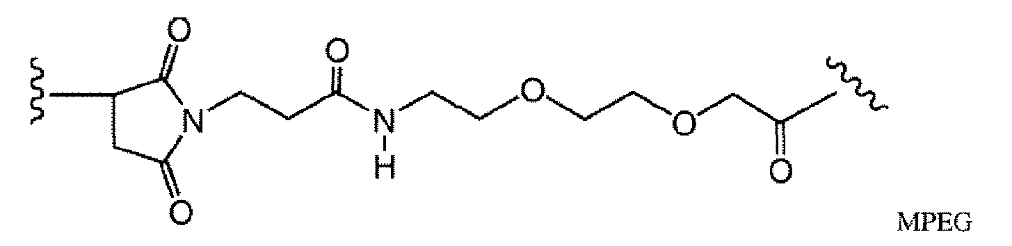


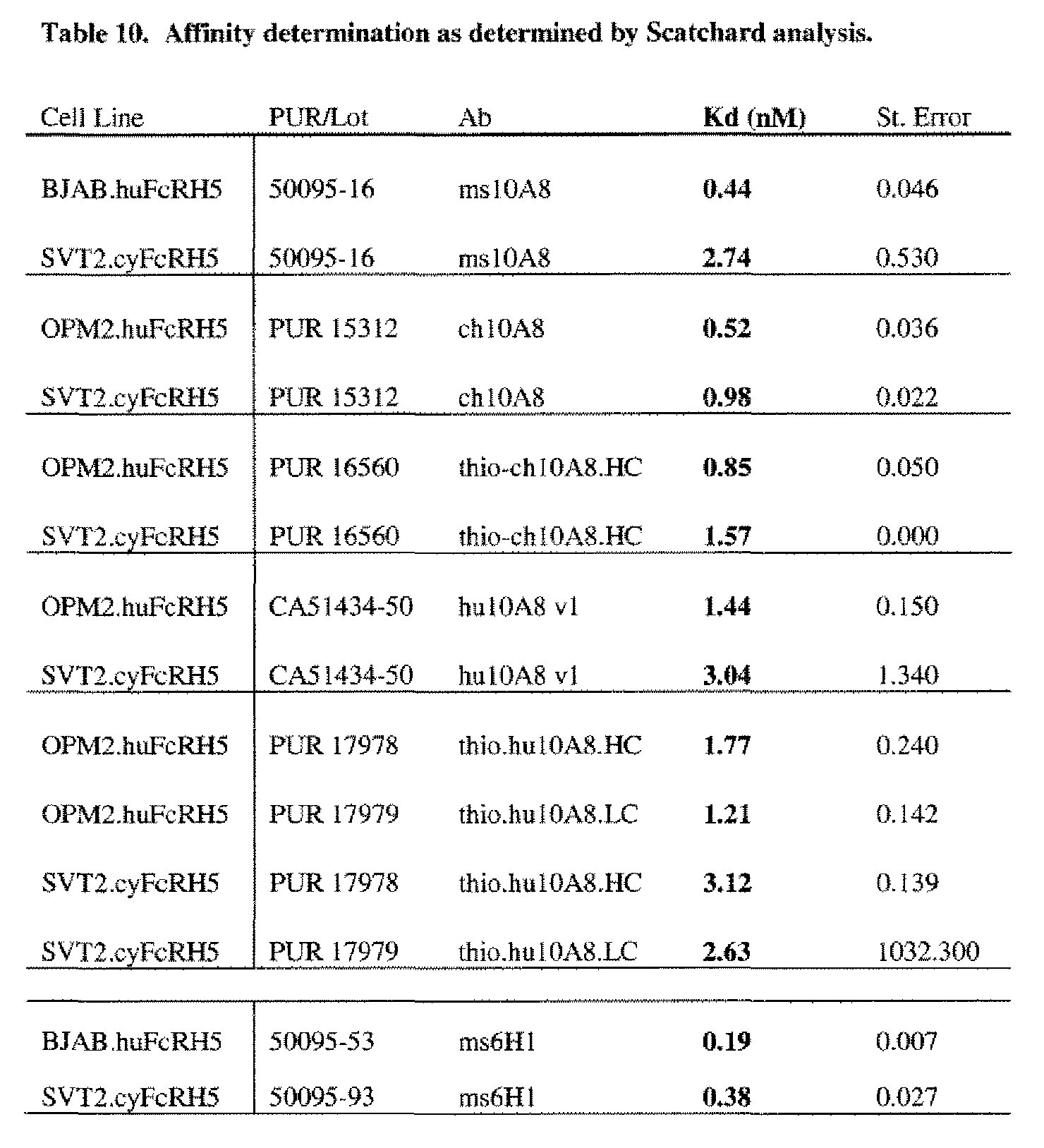


Claims


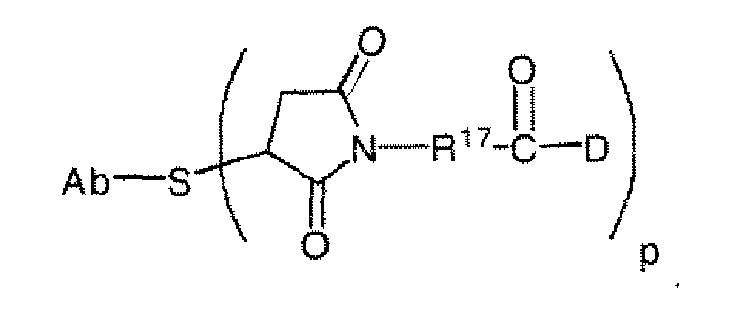


Priority Applications (16)
| Application Number | Priority Date | Filing Date | Title |
|---|---|---|---|
| CA2757382A CA2757382A1 (en) | 2009-04-01 | 2010-03-31 | Anti-fcrh5 antibodies and immunoconjugates |
| NZ595574A NZ595574A (en) | 2009-04-01 | 2010-03-31 | Anti-fcrh5 antibodies and immunoconjugates and methods of use |
| JP2012503679A JP6132548B2 (en) | 2009-04-01 | 2010-03-31 | Anti-FcRH5 antibodies and immunoconjugates and methods of use |
| AU2010236787A AU2010236787A1 (en) | 2009-04-01 | 2010-03-31 | Anti-FcRH5 antibodies and immunoconjugates and methods of use |
| EP10722862A EP2414399A1 (en) | 2009-04-01 | 2010-03-31 | Anti-fcrh5 antibodies and immunoconjugates and methods of use |
| SG2011071313A SG174992A1 (en) | 2009-04-01 | 2010-03-31 | Anti-fcrh5 antibodies and immunoconjugates and methods of use |
| CN201080024087.6A CN102471380B (en) | 2009-04-01 | 2010-03-31 | Anti-fcrh5 antibodies and immunoconjugates and methods of use |
| MA34324A MA33256B1 (en) | 2009-04-01 | 2010-03-31 | Antibody ,,, fcrh5 and methods of use |
| KR1020117025864A KR20120057565A (en) | 2009-04-01 | 2010-03-31 | Anti-fcrh5 antibodies and immunoconjugates and methods of use |
| MX2011010264A MX2011010264A (en) | 2009-04-01 | 2010-03-31 | Anti-fcrh5 antibodies and immunoconjugates and methods of use. |
| UAA201112795A UA104460C2 (en) | 2009-04-01 | 2010-03-31 | Anti-fcrh5 antibodies and immunoconjugates and methods of use |
| RU2011144141/10A RU2587621C2 (en) | 2009-04-01 | 2010-03-31 | ANTI-FcRH5 ANTIBODIES, IMMUNOCONJUGATES THEREOF AND METHODS FOR USE THEREOF |
| BRPI1012676A BRPI1012676A2 (en) | 2009-04-01 | 2010-03-31 | anti-fcrh5 and immunoconjugate antibodies and methods of use |
| ZA2011/06907A ZA201106907B (en) | 2009-04-01 | 2011-09-21 | Anti-fcrh5 antibodies and immunoconjugates and methods of use |
| IL215402A IL215402A0 (en) | 2009-04-01 | 2011-09-27 | Anti-fcrh5 antibodies and immunoconjugates and methods of use |
| HK12111896.8A HK1171236A1 (en) | 2009-04-01 | 2012-11-21 | Anti-fcrh5 antibodies and immunoconjugates and methods of use fcrh5 |
Applications Claiming Priority (6)
| Application Number | Priority Date | Filing Date | Title |
|---|---|---|---|
| US21169509P | 2009-04-01 | 2009-04-01 | |
| US61/211,695 | 2009-04-01 | ||
| US16621709P | 2009-04-02 | 2009-04-02 | |
| US61/166,217 | 2009-04-02 | ||
| US26697209P | 2009-12-04 | 2009-12-04 | |
| US61/266,972 | 2009-12-04 |
Publications (1)
| Publication Number | Publication Date |
|---|---|
| WO2010120561A1 true WO2010120561A1 (en) | 2010-10-21 |
Family
ID=42358044
Family Applications (1)
| Application Number | Title | Priority Date | Filing Date |
|---|---|---|---|
| PCT/US2010/029516 WO2010120561A1 (en) | 2009-04-01 | 2010-03-31 | Anti-fcrh5 antibodies and immunoconjugates and methods of use |
Country Status (24)
| Country | Link |
|---|---|
| US (3) | US8466260B2 (en) |
| EP (1) | EP2414399A1 (en) |
| JP (3) | JP6132548B2 (en) |
| KR (1) | KR20120057565A (en) |
| CN (1) | CN102471380B (en) |
| AR (1) | AR076029A1 (en) |
| AU (1) | AU2010236787A1 (en) |
| BR (1) | BRPI1012676A2 (en) |
| CA (1) | CA2757382A1 (en) |
| CL (1) | CL2011002417A1 (en) |
| CO (1) | CO6430468A2 (en) |
| CR (1) | CR20110558A (en) |
| EC (1) | ECSP11011436A (en) |
| HK (1) | HK1171236A1 (en) |
| IL (1) | IL215402A0 (en) |
| MA (1) | MA33256B1 (en) |
| MX (1) | MX2011010264A (en) |
| NZ (1) | NZ595574A (en) |
| PE (1) | PE20120878A1 (en) |
| RU (1) | RU2587621C2 (en) |
| SG (1) | SG174992A1 (en) |
| TW (1) | TW201038286A (en) |
| WO (1) | WO2010120561A1 (en) |
| ZA (1) | ZA201106907B (en) |
Cited By (29)
| Publication number | Priority date | Publication date | Assignee | Title |
|---|---|---|---|---|
| US8362213B2 (en) | 2009-04-01 | 2013-01-29 | Genentech, Inc. | Anti-FcRH5 antibodies and immunoconjugates and methods of use |
| US8466260B2 (en) | 2009-04-01 | 2013-06-18 | Genentech, Inc. | Anti-FcRH5 antibodies and immunoconjugates and methods of use |
| US8728730B2 (en) | 2009-09-03 | 2014-05-20 | Genentech, Inc. | Methods for treating, diagnosing, and monitoring rheumatoid arthritis |
| WO2014210064A1 (en) * | 2013-06-24 | 2014-12-31 | Genentech, Inc. | Anti-fcrh5 antibodies |
| JP2018093879A (en) * | 2010-11-30 | 2018-06-21 | 中外製薬株式会社 | Cytotoxicity-inducing therapeutic agents |
| US10323094B2 (en) | 2015-06-16 | 2019-06-18 | Genentech, Inc. | Humanized and affinity matured antibodies to FcRH5 and methods of use |
| WO2019237035A1 (en) | 2018-06-08 | 2019-12-12 | Intellia Therapeutics, Inc. | Compositions and methods for immunooncology |
| WO2020012337A1 (en) | 2018-07-10 | 2020-01-16 | Novartis Ag | 3-(5-amino-1-oxoisoindolin-2-yl)piperidine-2,6-dione derivatives and their use in the treatment of i karos family zinc finger 2 (ikzf2)-dependent diseases |
| WO2020128972A1 (en) | 2018-12-20 | 2020-06-25 | Novartis Ag | Dosing regimen and pharmaceutical combination comprising 3-(1-oxoisoindolin-2-yl)piperidine-2,6-dione derivatives |
| WO2020165833A1 (en) | 2019-02-15 | 2020-08-20 | Novartis Ag | 3-(1-oxo-5-(piperidin-4-yl)isoindolin-2-yl)piperidine-2,6-dione derivatives and uses thereof |
| WO2020165834A1 (en) | 2019-02-15 | 2020-08-20 | Novartis Ag | Substituted 3-(1-oxoisoindolin-2-yl)piperidine-2,6-dione derivatives and uses thereof |
| US10934344B2 (en) | 2006-03-31 | 2021-03-02 | Chugai Seiyaku Kabushiki Kaisha | Methods of modifying antibodies for purification of bispecific antibodies |
| US10973922B2 (en) | 2013-05-02 | 2021-04-13 | Glykos Finland Oy | Glycoprotein-toxic payload conjugates |
| US11001643B2 (en) | 2014-09-26 | 2021-05-11 | Chugai Seiyaku Kabushiki Kaisha | Cytotoxicity-inducing therapeutic agent |
| WO2021123996A1 (en) | 2019-12-20 | 2021-06-24 | Novartis Ag | Uses of anti-tgf-beta antibodies and checkpoint inhibitors for the treatment of proliferative diseases |
| US11059891B2 (en) | 2014-12-05 | 2021-07-13 | Memorial Sloan-Kettering Cancer Center | Chimeric antigen receptors targeting Fc receptor-like 5 and uses thereof |
| US11072666B2 (en) | 2016-03-14 | 2021-07-27 | Chugai Seiyaku Kabushiki Kaisha | Cell injury inducing therapeutic drug for use in cancer therapy |
| US11186650B2 (en) | 2013-12-17 | 2021-11-30 | Genentech, Inc. | Anti-CD3 antibodies and methods of use |
| WO2021260528A1 (en) | 2020-06-23 | 2021-12-30 | Novartis Ag | Dosing regimen comprising 3-(1-oxoisoindolin-2-yl)piperidine-2,6-dione derivatives |
| WO2022029573A1 (en) | 2020-08-03 | 2022-02-10 | Novartis Ag | Heteroaryl substituted 3-(1-oxoisoindolin-2-yl)piperidine-2,6-dione derivatives and uses thereof |
| WO2022215011A1 (en) | 2021-04-07 | 2022-10-13 | Novartis Ag | USES OF ANTI-TGFβ ANTIBODIES AND OTHER THERAPEUTIC AGENTS FOR THE TREATMENT OF PROLIFERATIVE DISEASES |
| US11485790B2 (en) | 2014-04-07 | 2022-11-01 | Chugai Seiyaku Kabushiki Kaisha | Immunoactivating antigen-binding molecule |
| WO2022229853A1 (en) | 2021-04-27 | 2022-11-03 | Novartis Ag | Viral vector production system |
| US11505605B2 (en) | 2014-05-13 | 2022-11-22 | Chugai Seiyaku Kabushiki Kaisha | T cell-redirected antigen-binding molecule for cells having immunosuppression function |
| US11649262B2 (en) | 2015-12-28 | 2023-05-16 | Chugai Seiyaku Kabushiki Kaisha | Method for promoting efficiency of purification of Fc region-containing polypeptide |
| US11649293B2 (en) | 2015-11-18 | 2023-05-16 | Chugai Seiyaku Kabushiki Kaisha | Method for enhancing humoral immune response |
| US11660340B2 (en) | 2015-11-18 | 2023-05-30 | Chugai Seiyaku Kabushiki Kaisha | Combination therapy using T cell redirection antigen binding molecule against cell having immunosuppressing function |
| WO2023214325A1 (en) | 2022-05-05 | 2023-11-09 | Novartis Ag | Pyrazolopyrimidine derivatives and uses thereof as tet2 inhibitors |
| US11851476B2 (en) | 2011-10-31 | 2023-12-26 | Chugai Seiyaku Kabushiki Kaisha | Antigen-binding molecule having regulated conjugation between heavy-chain and light-chain |
Families Citing this family (50)
| Publication number | Priority date | Publication date | Assignee | Title |
|---|---|---|---|---|
| WO2012109280A2 (en) * | 2011-02-07 | 2012-08-16 | Neotope Biosciences Limited | Apoe immunotherapy |
| WO2013052582A2 (en) * | 2011-10-05 | 2013-04-11 | Kalobios Pharmaceuticals, Inc. | Generation of antibodies to an epitope of interest that contains a phosphomimetic amino acid |
| US20140079636A1 (en) * | 2012-04-16 | 2014-03-20 | Dinesh U. Chimmanamada | Targeted therapeutics |
| CN103254304A (en) * | 2012-05-30 | 2013-08-21 | 深圳大学 | Immune stimulant couplet, preparation method thereof and antineoplastic application of the immune stimulant couplet |
| CN103626831A (en) * | 2012-08-22 | 2014-03-12 | 中国科学院过程工程研究所 | Method used for selective modification of protein carbon terminal carboxyl groups with polyethylene glycol |
| DK2911700T5 (en) * | 2012-10-24 | 2017-05-22 | Polytherics Ltd | The drug-protein conjugates |
| US9498532B2 (en) * | 2013-03-13 | 2016-11-22 | Novartis Ag | Antibody drug conjugates |
| US9415118B2 (en) | 2013-03-13 | 2016-08-16 | Novartis Ag | Antibody drug conjugates |
| EP2968593A1 (en) * | 2013-03-15 | 2016-01-20 | Regeneron Pharmaceuticals, Inc. | Biologically active molecules, conjugates thereof, and therapeutic uses |
| WO2015138615A2 (en) * | 2014-03-12 | 2015-09-17 | Irm Llc | Specific sites for modifying antibodies to make immunoconjugates |
| SG10202005268WA (en) * | 2015-12-04 | 2020-07-29 | Memorial Sloan Kettering Cancer Center | Antibodies targeting fc receptor-like 5 and methods of use |
| JPWO2017188346A1 (en) * | 2016-04-27 | 2019-02-28 | 国立大学法人 東京大学 | Material for capturing and collecting circulating cells in the blood using microfiber and method using the material |
| US10492904B2 (en) | 2016-05-27 | 2019-12-03 | Terumo Cardiovascular Systems Corporation | Equipment for preparing valve leaflet from membrane |
| BR112020008638A2 (en) | 2017-11-01 | 2020-10-20 | Juno Therapeutics Inc | chimeric antigen receptors specific for b cell maturation antigens (bcma) |
| WO2019089969A2 (en) | 2017-11-01 | 2019-05-09 | Juno Therapeutics, Inc. | Antibodies and chimeric antigen receptors specific for b-cell maturation antigen |
| US11564946B2 (en) | 2017-11-01 | 2023-01-31 | Juno Therapeutics, Inc. | Methods associated with tumor burden for assessing response to a cell therapy |
| US20210132042A1 (en) | 2017-11-01 | 2021-05-06 | Juno Therapeutics, Inc. | Methods of assessing or monitoring a response to a cell therapy |
| WO2019090004A1 (en) | 2017-11-01 | 2019-05-09 | Juno Therapeutics, Inc. | Process for producing a t cell composition |
| BR112020008340A2 (en) | 2017-11-01 | 2020-11-17 | Juno Therapeutics Inc | process for generating therapeutic compositions of modified cells |
| MX2020005908A (en) | 2017-12-08 | 2020-10-07 | Juno Therapeutics Inc | Process for producing a composition of engineered t cells. |
| WO2019113556A1 (en) | 2017-12-08 | 2019-06-13 | Juno Therapeutics, Inc. | Serum-free media formulation for culturing cells and methods of use thereof |
| CN112203680A (en) | 2017-12-08 | 2021-01-08 | 朱诺治疗学股份有限公司 | Phenotypic markers for cell therapy and related methods |
| MX2020006822A (en) * | 2018-01-04 | 2020-09-03 | Shanghai Lumosa Therapeutics Co Ltd | Single-domain antibody-cytosine deaminase fusion proteins. |
| JP2021530246A (en) | 2018-07-03 | 2021-11-11 | マレンゴ・セラピューティクス,インコーポレーテッド | Anti-TCR antibody molecule and its use |
| US20210230671A1 (en) | 2018-08-09 | 2021-07-29 | Juno Therapeutics, Inc. | Methods for assessing integrated nucleic acids |
| CN110850067B (en) * | 2018-08-21 | 2023-08-15 | 上海恒润达生生物科技股份有限公司 | Chimeric antigen receptor affinity detection method |
| TW202021981A (en) | 2018-11-01 | 2020-06-16 | 美商奇諾治療有限公司 | Chimeric antigen receptors specific for g protein-coupled receptor class c group 5 member d (gprc5d) |
| EP3873943A2 (en) | 2018-11-01 | 2021-09-08 | Juno Therapeutics, Inc. | Methods for treatment using chimeric antigen receptors specific for b-cell maturation antigen |
| CN109883932B (en) * | 2019-02-18 | 2021-08-10 | 武汉伊莱瑞特生物科技股份有限公司 | Flow antibody, preparation method and titration method thereof |
| JP2022531577A (en) | 2019-05-01 | 2022-07-07 | ジュノー セラピューティクス インコーポレイテッド | Cells expressing chimeric receptors from the modified CD247 locus, related polynucleotides, and methods. |
| MX2021013219A (en) | 2019-05-01 | 2022-02-17 | Juno Therapeutics Inc | Cells expressing a recombinant receptor from a modified tgfbr2 locus, related polynucleotides and methods. |
| CN110987571A (en) * | 2019-12-31 | 2020-04-10 | 广州金域医学检验中心有限公司 | M protein depolymerization method of monoclonal polymer |
| CA3164090A1 (en) * | 2020-01-10 | 2021-07-15 | Jonathan Kurtis | Antibodies to pfgarp kill plasmodium falciparum malaria parasites and protect against infection and severe disease |
| AU2021251265A1 (en) | 2020-04-10 | 2022-11-03 | Juno Therapeutics, Inc. | Methods and uses related to cell therapy engineered with a chimeric antigen receptor targeting B-cell maturation antigen |
| JP2023528215A (en) | 2020-05-13 | 2023-07-04 | ジュノー セラピューティクス インコーポレイテッド | Methods of identifying features associated with clinical response and uses thereof |
| KR20230042283A (en) | 2020-06-26 | 2023-03-28 | 주노 테라퓨틱스 게엠베하 | Engineered T cells conditionally expressing recombinant receptors, related polynucleotides and methods |
| CN112285361B (en) * | 2020-09-27 | 2023-12-05 | 中国人民解放军空军军医大学 | Agent for eliminating interference of anti-CD 38 monoclonal antibody medicine against human globulin detection |
| WO2022098787A1 (en) | 2020-11-04 | 2022-05-12 | Juno Therapeutics, Inc. | Cells expressing a chimeric receptor from a modified invariant cd3 immunoglobulin superfamily chain locus and related polynucleotides and methods |
| EP4301755A1 (en) | 2021-03-03 | 2024-01-10 | Juno Therapeutics, Inc. | Combination of a t cell therapy and a dgk inhibitor |
| EP4319809A1 (en) * | 2021-04-06 | 2024-02-14 | Memorial Sloan Kettering Cancer Center | Combination therapy with dexamethasone and tumor-specific t cell engaging multi-specific antibodies for treating cancer |
| WO2022216993A2 (en) | 2021-04-08 | 2022-10-13 | Marengo Therapeutics, Inc. | Multifuntional molecules binding to tcr and uses thereof |
| EP4334341A2 (en) | 2021-05-06 | 2024-03-13 | Juno Therapeutics GmbH | Methods for stimulating and transducing t cells |
| WO2023147515A1 (en) | 2022-01-28 | 2023-08-03 | Juno Therapeutics, Inc. | Methods of manufacturing cellular compositions |
| WO2023159182A1 (en) | 2022-02-18 | 2023-08-24 | Rakuten Medical, Inc. | Anti-programmed death-ligand 1 (pd-l1) antibody molecules, encoding polynucleotides, and methods of use |
| WO2023213969A1 (en) | 2022-05-05 | 2023-11-09 | Juno Therapeutics Gmbh | Viral-binding protein and related reagents, articles, and methods of use |
| WO2023220655A1 (en) | 2022-05-11 | 2023-11-16 | Celgene Corporation | Methods to overcome drug resistance by re-sensitizing cancer cells to treatment with a prior therapy via treatment with a t cell therapy |
| WO2023230548A1 (en) | 2022-05-25 | 2023-11-30 | Celgene Corporation | Method for predicting response to a t cell therapy |
| WO2023230581A1 (en) | 2022-05-25 | 2023-11-30 | Celgene Corporation | Methods of manufacturing t cell therapies |
| WO2024054944A1 (en) | 2022-09-08 | 2024-03-14 | Juno Therapeutics, Inc. | Combination of a t cell therapy and continuous or intermittent dgk inhibitor dosing |
| CN116789849B (en) * | 2023-04-12 | 2024-03-08 | 南京紫珑生物科技有限公司 | Chimeric antigen receptor and application thereof |
Citations (215)
| Publication number | Priority date | Publication date | Assignee | Title |
|---|---|---|---|---|
| US633410A (en) | 1898-09-22 | 1899-09-19 | George A Ames | Ice-cutter. |
| US3720760A (en) | 1968-09-06 | 1973-03-13 | Pharmacia Ab | Method for determining the presence of reagin-immunoglobulins(reagin-ig)directed against certain allergens,in aqueous samples |
| US3773919A (en) | 1969-10-23 | 1973-11-20 | Du Pont | Polylactide-drug mixtures |
| US3809611A (en) | 1972-01-13 | 1974-05-07 | Kyowa Hakko Kogyo Kk | Process for producing citric acid |
| US4137230A (en) | 1977-11-14 | 1979-01-30 | Takeda Chemical Industries, Ltd. | Method for the production of maytansinoids |
| US4151042A (en) | 1977-03-31 | 1979-04-24 | Takeda Chemical Industries, Ltd. | Method for producing maytansinol and its derivatives |
| EP0003089A1 (en) | 1978-01-06 | 1979-07-25 | Bernard David | Drier for silkscreen printed sheets |
| US4179337A (en) | 1973-07-20 | 1979-12-18 | Davis Frank F | Non-immunogenic polypeptides |
| US4248870A (en) | 1978-10-27 | 1981-02-03 | Takeda Chemical Industries, Ltd. | Maytansinoids and use |
| US4256746A (en) | 1978-11-14 | 1981-03-17 | Takeda Chemical Industries | Dechloromaytansinoids, their pharmaceutical compositions and method of use |
| US4260608A (en) | 1978-11-14 | 1981-04-07 | Takeda Chemical Industries, Ltd. | Maytansinoids, pharmaceutical compositions thereof and methods of use thereof |
| WO1981001145A1 (en) | 1979-10-18 | 1981-04-30 | Univ Illinois | Hydrolytic enzyme-activatible pro-drugs |
| US4265814A (en) | 1978-03-24 | 1981-05-05 | Takeda Chemical Industries | Matansinol 3-n-hexadecanoate |
| US4275149A (en) | 1978-11-24 | 1981-06-23 | Syva Company | Macromolecular environment control in specific receptor assays |
| EP0036776A2 (en) | 1980-03-24 | 1981-09-30 | Genentech, Inc. | A method of creating an expression plasmid |
| US4294757A (en) | 1979-01-31 | 1981-10-13 | Takeda Chemical Industries, Ltd | 20-O-Acylmaytansinoids |
| US4301144A (en) | 1979-07-11 | 1981-11-17 | Ajinomoto Company, Incorporated | Blood substitute containing modified hemoglobin |
| US4307016A (en) | 1978-03-24 | 1981-12-22 | Takeda Chemical Industries, Ltd. | Demethyl maytansinoids |
| US4308269A (en) | 1979-06-11 | 1981-12-29 | Takeda Chemical Industries, Ltd. | Maytansinoids, pharmaceutical compositions thereof and method of use thereof |
| US4308268A (en) | 1979-06-11 | 1981-12-29 | Takeda Chemical Industries, Ltd. | Maytansinoids, pharmaceutical compositions thereof and method of use thereof |
| US4309428A (en) | 1979-07-30 | 1982-01-05 | Takeda Chemical Industries, Ltd. | Maytansinoids |
| US4313946A (en) | 1981-01-27 | 1982-02-02 | The United States Of America As Represented By The Secretary Of Agriculture | Chemotherapeutically active maytansinoids from Trewia nudiflora |
| US4315929A (en) | 1981-01-27 | 1982-02-16 | The United States Of America As Represented By The Secretary Of Agriculture | Method of controlling the European corn borer with trewiasine |
| US4317821A (en) | 1979-06-08 | 1982-03-02 | Takeda Chemical Industries, Ltd. | Maytansinoids, their use and pharmaceutical compositions thereof |
| US4318980A (en) | 1978-04-10 | 1982-03-09 | Miles Laboratories, Inc. | Heterogenous specific binding assay employing a cycling reactant as label |
| US4322348A (en) | 1979-06-05 | 1982-03-30 | Takeda Chemical Industries, Ltd. | Maytansinoids |
| US4331598A (en) | 1979-09-19 | 1982-05-25 | Takeda Chemical Industries, Ltd. | Maytansinoids |
| USRE30985E (en) | 1978-01-01 | 1982-06-29 | Serum-free cell culture media | |
| US4362663A (en) | 1979-09-21 | 1982-12-07 | Takeda Chemical Industries, Ltd. | Maytansinoid compound |
| US4364866A (en) | 1979-09-21 | 1982-12-21 | Takeda Chemical Industries, Ltd. | Maytansinoids |
| US4371533A (en) | 1980-10-08 | 1983-02-01 | Takeda Chemical Industries, Ltd. | 4,5-Deoxymaytansinoids, their use and pharmaceutical compositions thereof |
| EP0073657A1 (en) | 1981-08-31 | 1983-03-09 | Genentech, Inc. | Preparation of hepatitis B surface antigen in yeast |
| US4399216A (en) | 1980-02-25 | 1983-08-16 | The Trustees Of Columbia University | Processes for inserting DNA into eucaryotic cells and for producing proteinaceous materials |
| US4419446A (en) | 1980-12-31 | 1983-12-06 | The United States Of America As Represented By The Department Of Health And Human Services | Recombinant DNA process utilizing a papilloma virus DNA as a vector |
| US4424219A (en) | 1981-05-20 | 1984-01-03 | Takeda Chemical Industries, Ltd. | 9-Thiomaytansinoids and their pharmaceutical compositions and use |
| US4450254A (en) | 1980-11-03 | 1984-05-22 | Standard Oil Company | Impact improvement of high nitrile resins |
| EP0117060A2 (en) | 1983-01-19 | 1984-08-29 | Genentech, Inc. | Methods of screening and amplification in eukaryotic host cells, and nucleotide sequences and expression vectors for use therein |
| EP0117058A2 (en) | 1983-01-19 | 1984-08-29 | Genentech, Inc. | Methods for producing mature protein in vertebrate host cells |
| US4485045A (en) | 1981-07-06 | 1984-11-27 | Research Corporation | Synthetic phosphatidyl cholines useful in forming liposomes |
| US4496689A (en) | 1983-12-27 | 1985-01-29 | Miles Laboratories, Inc. | Covalently attached complex of alpha-1-proteinase inhibitor with a water soluble polymer |
| EP0139383A1 (en) | 1983-08-16 | 1985-05-02 | Zymogenetics, Inc. | Method for expressing foreign genes in schizosaccharomyces pombe and the use in therapeutic formulations of the products, DNA constructs and transformant strains of schizosaccharomyces pombe usable in such method and their preparation |
| US4544545A (en) | 1983-06-20 | 1985-10-01 | Trustees University Of Massachusetts | Liposomes containing modified cholesterol for organ targeting |
| US4560655A (en) | 1982-12-16 | 1985-12-24 | Immunex Corporation | Serum-free cell culture medium and process for making same |
| EP0183070A2 (en) | 1984-10-30 | 1986-06-04 | Phillips Petroleum Company | Transformation of yeasts of the genus pichia |
| US4601978A (en) | 1982-11-24 | 1986-07-22 | The Regents Of The University Of California | Mammalian metallothionein promoter system |
| WO1987000195A1 (en) | 1985-06-28 | 1987-01-15 | Celltech Limited | Animal cell culture |
| US4640835A (en) | 1981-10-30 | 1987-02-03 | Nippon Chemiphar Company, Ltd. | Plasminogen activator derivatives |
| US4657866A (en) | 1982-12-21 | 1987-04-14 | Sudhir Kumar | Serum-free, synthetic, completely chemically defined tissue culture media |
| US4670417A (en) | 1985-06-19 | 1987-06-02 | Ajinomoto Co., Inc. | Hemoglobin combined with a poly(alkylene oxide) |
| US4675187A (en) | 1983-05-16 | 1987-06-23 | Bristol-Myers Company | BBM-1675, a new antibiotic complex |
| US4676980A (en) | 1985-09-23 | 1987-06-30 | The United States Of America As Represented By The Secretary Of The Department Of Health And Human Services | Target specific cross-linked heteroantibodies |
| WO1987005330A1 (en) | 1986-03-07 | 1987-09-11 | Michel Louis Eugene Bergh | Method for enhancing glycoprotein stability |
| EP0244234A2 (en) | 1986-04-30 | 1987-11-04 | Alko Group Ltd. | Transformation of trichoderma |
| US4737456A (en) | 1985-05-09 | 1988-04-12 | Syntex (U.S.A.) Inc. | Reducing interference in ligand-receptor binding assays |
| US4767704A (en) | 1983-10-07 | 1988-08-30 | Columbia University In The City Of New York | Protein-free culture medium |
| WO1988007378A1 (en) | 1987-03-09 | 1988-10-06 | Cancer Research Campaign Technology Ltd. | Improvements relating to drug delivery systems |
| US4791192A (en) | 1986-06-26 | 1988-12-13 | Takeda Chemical Industries, Ltd. | Chemically modified protein with polyethyleneglycol |
| US4816567A (en) | 1983-04-08 | 1989-03-28 | Genentech, Inc. | Recombinant immunoglobin preparations |
| DD266710A3 (en) | 1983-06-06 | 1989-04-12 | Ve Forschungszentrum Biotechnologie | Process for the biotechnical production of alkaline phosphatase |
| WO1989005859A1 (en) | 1987-12-21 | 1989-06-29 | The Upjohn Company | Agrobacterium mediated transformation of germinating plant seeds |
| GB2211504A (en) | 1987-10-23 | 1989-07-05 | Nat Res Dev | Fowlpox virus promoters |
| US4892538A (en) | 1987-11-17 | 1990-01-09 | Brown University Research Foundation | In vivo delivery of neurotransmitters by implanted, encapsulated cells |
| EP0362179A2 (en) | 1988-08-25 | 1990-04-04 | Smithkline Beecham Corporation | Recombinant saccharomyces |
| WO1990003430A1 (en) | 1988-09-23 | 1990-04-05 | Cetus Corporation | Cell culture medium for enhanced cell growth, culture longevity and product expression |
| US4927762A (en) | 1986-04-01 | 1990-05-22 | Cell Enterprises, Inc. | Cell culture medium with antioxidant |
| US4933294A (en) | 1984-01-30 | 1990-06-12 | Icrf Patents Limited | Method of detecting truncated epidermal growth factor receptors |
| US4943529A (en) | 1982-05-19 | 1990-07-24 | Gist-Brocades Nv | Kluyveromyces as a host strain |
| US4946783A (en) | 1987-01-30 | 1990-08-07 | President And Fellows Of Harvard College | Periplasmic protease mutants of Escherichia coli |
| US4965199A (en) | 1984-04-20 | 1990-10-23 | Genentech, Inc. | Preparation of functional human factor VIII in mammalian cells using methotrexate based selection |
| EP0394538A1 (en) | 1989-04-28 | 1990-10-31 | Rhein Biotech Gesellschaft Für Neue Biotechnologische Prozesse Und Produkte Mbh | A yeast cell of the genus schwanniomyces |
| WO1990013646A1 (en) | 1989-04-28 | 1990-11-15 | Transgene S.A. | Application of novel dna fragments as a coding sequence for a signal peptide for the secretion of mature proteins by recombinant yeast, expression cassettes, transformed yeasts and corresponding process for the preparation of proteins |
| US4975278A (en) | 1988-02-26 | 1990-12-04 | Bristol-Myers Company | Antibody-enzyme conjugates in combination with prodrugs for the delivery of cytotoxic agents to tumor cells |
| EP0402226A1 (en) | 1989-06-06 | 1990-12-12 | Institut National De La Recherche Agronomique | Transformation vectors for yeast yarrowia |
| EP0404097A2 (en) | 1989-06-22 | 1990-12-27 | BEHRINGWERKE Aktiengesellschaft | Bispecific and oligospecific, mono- and oligovalent receptors, production and applications thereof |
| WO1991000357A1 (en) | 1989-06-30 | 1991-01-10 | Cayla | New strain with filamentous fungi mutants, process for the production of recombinant proteins using said strain, and strains and proteins produced by said process |
| WO1991000360A1 (en) | 1989-06-29 | 1991-01-10 | Medarex, Inc. | Bispecific reagents for aids therapy |
| WO1991005264A1 (en) | 1989-09-29 | 1991-04-18 | Oncogenetics Partners | Detection and quantification of neu related proteins in the biological fluids of humans |
| US5010182A (en) | 1987-07-28 | 1991-04-23 | Chiron Corporation | DNA constructs containing a Kluyveromyces alpha factor leader sequence for directing secretion of heterologous polypeptides |
| US5013556A (en) | 1989-10-20 | 1991-05-07 | Liposome Technology, Inc. | Liposomes with enhanced circulation time |
| US5053394A (en) | 1988-09-21 | 1991-10-01 | American Cyanamid Company | Targeted forms of methyltrithio antitumor agents |
| US5078998A (en) | 1985-08-02 | 1992-01-07 | Bevan Michael J | Hybrid ligand directed to activation of cytotoxic effector T lymphocytes and target associated antigen |
| US5091178A (en) | 1986-02-21 | 1992-02-25 | Oncogen | Tumor therapy with biologically active anti-tumor antibodies |
| WO1992009690A2 (en) | 1990-12-03 | 1992-06-11 | Genentech, Inc. | Enrichment method for variant proteins with altered binding properties |
| US5122469A (en) | 1990-10-03 | 1992-06-16 | Genentech, Inc. | Method for culturing Chinese hamster ovary cells to improve production of recombinant proteins |
| WO1992020373A1 (en) | 1991-05-14 | 1992-11-26 | Repligen Corporation | Heteroconjugate antibodies for treatment of hiv infection |
| US5208020A (en) | 1989-10-25 | 1993-05-04 | Immunogen Inc. | Cytotoxic agents comprising maytansinoids and their therapeutic use |
| WO1993008829A1 (en) | 1991-11-04 | 1993-05-13 | The Regents Of The University Of California | Compositions that mediate killing of hiv-infected cells |
| WO1993011161A1 (en) | 1991-11-25 | 1993-06-10 | Enzon, Inc. | Multivalent antigen-binding proteins |
| US5229275A (en) | 1990-04-26 | 1993-07-20 | Akzo N.V. | In-vitro method for producing antigen-specific human monoclonal antibodies |
| WO1993016185A2 (en) | 1992-02-06 | 1993-08-19 | Creative Biomolecules, Inc. | Biosynthetic binding protein for cancer marker |
| WO1993021232A1 (en) | 1992-04-10 | 1993-10-28 | Research Development Foundation | IMMUNOTOXINS DIRECTED AGAINST c-erbB-2 (HER-2/neu) RELATED SURFACE ANTIGENS |
| US5264365A (en) | 1990-11-09 | 1993-11-23 | Board Of Regents, The University Of Texas System | Protease-deficient bacterial strains for production of proteolytically sensitive polypeptides |
| WO1993025673A1 (en) | 1992-06-04 | 1993-12-23 | The Regents Of The University Of California | In vivo gene therapy with intron-free sequence of interest |
| US5283187A (en) | 1987-11-17 | 1994-02-01 | Brown University Research Foundation | Cell culture-containing tubular capsule produced by co-extrusion |
| WO1994004690A1 (en) | 1992-08-17 | 1994-03-03 | Genentech, Inc. | Bispecific immunoadhesins |
| WO1994011026A2 (en) | 1992-11-13 | 1994-05-26 | Idec Pharmaceuticals Corporation | Therapeutic application of chimeric and radiolabeled antibodies to human b lymphocyte restricted differentiation antigen for treatment of b cell lymphoma |
| US5316757A (en) | 1984-10-18 | 1994-05-31 | Board Of Regents, The University Of Texas System | Synthesis of polyazamacrocycles with more than one type of side-chain chelating groups |
| EP0600517A1 (en) | 1992-12-04 | 1994-06-08 | Bristol-Myers Squibb Company | 6,7-Modified paclitaxels |
| US5342606A (en) | 1984-10-18 | 1994-08-30 | Board Of Regents, The University Of Texas System | Polyazamacrocyclic compounds for complexation of metal ions |
| EP0616812A1 (en) | 1993-03-24 | 1994-09-28 | Berlex Biosciences | Combination with anti-hormonal compounds and binding molecules for the treatment of cancer |
| US5362852A (en) | 1991-09-27 | 1994-11-08 | Pfizer Inc. | Modified peptide derivatives conjugated at 2-hydroxyethylamine moieties |
| US5364934A (en) | 1991-02-01 | 1994-11-15 | Genentech, Inc. | Plasma carboxypeptidase |
| US5385893A (en) | 1993-05-06 | 1995-01-31 | The Dow Chemical Company | Tricyclopolyazamacrocyclophosphonic acids, complexes and derivatives thereof, for use as contrast agents |
| US5401638A (en) | 1986-06-04 | 1995-03-28 | Oncogene Science, Inc. | Detection and quantification of neu related proteins in the biological fluids of humans |
| US5428130A (en) | 1989-02-23 | 1995-06-27 | Genentech, Inc. | Hybrid immunoglobulins |
| US5428139A (en) | 1991-12-10 | 1995-06-27 | The Dow Chemical Company | Bicyclopolyazamacrocyclophosphonic acid complexes for use as radiopharmaceuticals |
| US5427908A (en) | 1990-05-01 | 1995-06-27 | Affymax Technologies N.V. | Recombinant library screening methods |
| US5432018A (en) | 1990-06-20 | 1995-07-11 | Affymax Technologies N.V. | Peptide library and screening systems |
| US5462725A (en) | 1993-05-06 | 1995-10-31 | The Dow Chemical Company | 2-pyridylmethylenepolyazamacrocyclophosphonic acids, complexes and derivatives thereof, for use as contrast agents |
| US5480990A (en) | 1991-12-10 | 1996-01-02 | The Dow Chemical Company | Bicyclopolyazamacrocyclocarboxylic acid complexes for use as contrast agents |
| WO1996000732A1 (en) | 1994-06-29 | 1996-01-11 | Schering Corporation | Phosphinic acid derivatives |
| US5498530A (en) | 1991-10-16 | 1996-03-12 | Affymax Technologies, N.V. | Peptide library and screening method |
| WO1996007321A1 (en) | 1994-09-06 | 1996-03-14 | The Uab Research Foundation | Methods for modulating protein function in cells using intracellular antibody homologues |
| WO1996007754A1 (en) | 1994-09-02 | 1996-03-14 | The Scripps Research Institute | Methods for producing antibody libraries using universal or randomized immunoglobulin light chains |
| US5500362A (en) | 1987-01-08 | 1996-03-19 | Xoma Corporation | Chimeric antibody with specificity to human B cell surface antigen |
| US5508192A (en) | 1990-11-09 | 1996-04-16 | Board Of Regents, The University Of Texas System | Bacterial host strains for producing proteolytically sensitive polypeptides |
| WO1996016673A1 (en) | 1994-12-02 | 1996-06-06 | Chiron Corporation | Method of promoting an immune response with a bispecific antibody |
| US5545807A (en) | 1988-10-12 | 1996-08-13 | The Babraham Institute | Production of antibodies from transgenic animals |
| US5545806A (en) | 1990-08-29 | 1996-08-13 | Genpharm International, Inc. | Ransgenic non-human animals for producing heterologous antibodies |
| US5548237A (en) | 1995-03-10 | 1996-08-20 | International Business Machines Corporation | Process tolerant delay circuit |
| EP0425235B1 (en) | 1989-10-25 | 1996-09-25 | Immunogen Inc | Cytotoxic agents comprising maytansinoids and their therapeutic use |
| US5565332A (en) | 1991-09-23 | 1996-10-15 | Medical Research Council | Production of chimeric antibodies - a combinatorial approach |
| US5567610A (en) | 1986-09-04 | 1996-10-22 | Bioinvent International Ab | Method of producing human monoclonal antibodies and kit therefor |
| US5569825A (en) | 1990-08-29 | 1996-10-29 | Genpharm International | Transgenic non-human animals capable of producing heterologous antibodies of various isotypes |
| US5571894A (en) | 1991-02-05 | 1996-11-05 | Ciba-Geigy Corporation | Recombinant antibodies specific for a growth factor receptor |
| US5573905A (en) | 1992-03-30 | 1996-11-12 | The Scripps Research Institute | Encoded combinatorial chemical libraries |
| US5583024A (en) | 1985-12-02 | 1996-12-10 | The Regents Of The University Of California | Recombinant expression of Coleoptera luciferase |
| US5587458A (en) | 1991-10-07 | 1996-12-24 | Aronex Pharmaceuticals, Inc. | Anti-erbB-2 antibodies, combinations thereof, and therapeutic and diagnostic uses thereof |
| US5591669A (en) | 1988-12-05 | 1997-01-07 | Genpharm International, Inc. | Transgenic mice depleted in a mature lymphocytic cell-type |
| WO1997017852A1 (en) | 1995-11-15 | 1997-05-22 | Hoechst Schering Agrevo Gmbh | Synergetic herbicidal mixtures |
| US5635483A (en) | 1992-12-03 | 1997-06-03 | Arizona Board Of Regents Acting On Behalf Of Arizona State University | Tumor inhibiting tetrapeptide bearing modified phenethyl amides |
| US5639635A (en) | 1994-11-03 | 1997-06-17 | Genentech, Inc. | Process for bacterial production of polypeptides |
| US5641870A (en) | 1995-04-20 | 1997-06-24 | Genentech, Inc. | Low pH hydrophobic interaction chromatography for antibody purification |
| US5648237A (en) | 1991-09-19 | 1997-07-15 | Genentech, Inc. | Expression of functional antibody fragments |
| US5658727A (en) | 1991-04-10 | 1997-08-19 | The Scripps Research Institute | Heterodimeric receptor libraries using phagemids |
| US5663149A (en) | 1994-12-13 | 1997-09-02 | Arizona Board Of Regents Acting On Behalf Of Arizona State University | Human cancer inhibitory pentapeptide heterocyclic and halophenyl amides |
| WO1997038731A1 (en) | 1996-04-18 | 1997-10-23 | The Regents Of The University Of California | Immunoliposomes that optimize internalization into target cells |
| US5700670A (en) | 1995-04-13 | 1997-12-23 | Mitsubishi Chemical Corporation | Method for producing optically active ester of γ-substituted-β-hydroxybutyric acid |
| WO1998002463A1 (en) | 1996-07-11 | 1998-01-22 | Medarex, Inc. | THERAPEUTIC MULTISPECIFIC COMPOUNDS COMPRISED OF ANTI-FCα RECEPTOR ANTIBODIES |
| US5712374A (en) | 1995-06-07 | 1998-01-27 | American Cyanamid Company | Method for the preparation of substantiallly monomeric calicheamicin derivative/carrier conjugates |
| US5714586A (en) | 1995-06-07 | 1998-02-03 | American Cyanamid Company | Methods for the preparation of monomeric calicheamicin derivative/carrier conjugates |
| US5731168A (en) | 1995-03-01 | 1998-03-24 | Genentech, Inc. | Method for making heteromultimeric polypeptides |
| US5733743A (en) | 1992-03-24 | 1998-03-31 | Cambridge Antibody Technology Limited | Methods for producing members of specific binding pairs |
| US5739116A (en) | 1994-06-03 | 1998-04-14 | American Cyanamid Company | Enediyne derivatives useful for the synthesis of conjugates of methyltrithio antitumor agents |
| US5739277A (en) | 1995-04-14 | 1998-04-14 | Genentech Inc. | Altered polypeptides with increased half-life |
| US5739294A (en) | 1991-12-10 | 1998-04-14 | The Dow Chemical Company | Bicyclopol yazamacrocyclophosphonic acid complexes for use as contrast agents |
| US5767237A (en) | 1993-10-01 | 1998-06-16 | Teikoku Hormone Mfg. Co., Ltd. | Peptide derivatives |
| US5770710A (en) | 1987-10-30 | 1998-06-23 | American Cyanamid Company | Antitumor and antibacterial substituted disulfide derivatives prepared from compounds possessing a methlytrithio group |
| US5770701A (en) | 1987-10-30 | 1998-06-23 | American Cyanamid Company | Process for preparing targeted forms of methyltrithio antitumor agents |
| US5780588A (en) | 1993-01-26 | 1998-07-14 | Arizona Board Of Regents | Elucidation and synthesis of selected pentapeptides |
| US5789199A (en) | 1994-11-03 | 1998-08-04 | Genentech, Inc. | Process for bacterial production of polypeptides |
| US5807715A (en) | 1984-08-27 | 1998-09-15 | The Board Of Trustees Of The Leland Stanford Junior University | Methods and transformed mammalian lymphocyte cells for producing functional antigen-binding protein including chimeric immunoglobulin |
| US5821337A (en) | 1991-06-14 | 1998-10-13 | Genentech, Inc. | Immunoglobulin variants |
| WO1998045479A1 (en) | 1997-04-04 | 1998-10-15 | Albany Medical College | Method for assessing prostate cancer |
| US5834456A (en) | 1996-02-23 | 1998-11-10 | The Dow Chemical Company | Polyazamacrocyclofluoromonoalkylphosphonic acids, and their complexes, for use as contrast agents |
| US5837234A (en) | 1995-06-07 | 1998-11-17 | Cytotherapeutics, Inc. | Bioartificial organ containing cells encapsulated in a permselective polyether suflfone membrane |
| US5837242A (en) | 1992-12-04 | 1998-11-17 | Medical Research Council | Multivalent and multispecific binding proteins, their manufacture and use |
| US5840523A (en) | 1995-03-01 | 1998-11-24 | Genetech, Inc. | Methods and compositions for secretion of heterologous polypeptides |
| US5869046A (en) | 1995-04-14 | 1999-02-09 | Genentech, Inc. | Altered polypeptides with increased half-life |
| WO1999051642A1 (en) | 1998-04-02 | 1999-10-14 | Genentech, Inc. | Antibody variants and fragments thereof |
| US5969108A (en) | 1990-07-10 | 1999-10-19 | Medical Research Council | Methods for producing members of specific binding pairs |
| US6027888A (en) | 1996-04-05 | 2000-02-22 | Board Of Regents, The University Of Texas System | Methods for producing soluble, biologically-active disulfide-bond containing eukaryotic proteins in bacterial cells |
| US6075181A (en) | 1990-01-12 | 2000-06-13 | Abgenix, Inc. | Human antibodies derived from immunized xenomice |
| US6083715A (en) | 1997-06-09 | 2000-07-04 | Board Of Regents, The University Of Texas System | Methods for producing heterologous disulfide bond-containing polypeptides in bacterial cells |
| US6150584A (en) | 1990-01-12 | 2000-11-21 | Abgenix, Inc. | Human antibodies derived from immunized xenomice |
| WO2001000245A2 (en) | 1999-06-25 | 2001-01-04 | Genentech, Inc. | HUMANIZED ANTI-ErbB2 ANTIBODIES AND TREATMENT WITH ANTI-ErbB2 ANTIBODIES |
| US6172197B1 (en) | 1991-07-10 | 2001-01-09 | Medical Research Council | Methods for producing members of specific binding pairs |
| US6184105B1 (en) | 1997-05-22 | 2001-02-06 | Advanced Micro Devices | Method for post transistor isolation |
| US6194551B1 (en) | 1998-04-02 | 2001-02-27 | Genentech, Inc. | Polypeptide variants |
| US6214345B1 (en) | 1993-05-14 | 2001-04-10 | Bristol-Myers Squibb Co. | Lysosomal enzyme-cleavable antitumor drug conjugates |
| WO2001038490A2 (en) | 1999-11-29 | 2001-05-31 | The Trustees Of Columbia University In The City Of New York | ISOLATION OF FIVE NOVEL GENES CODING FOR NEW Fc RECEPTORS-TYPE MELANOMA INVOLVED IN THE PATHOGENESIS OF LYMPHOMA/MELANOMA |
| US6248564B1 (en) | 1997-08-29 | 2001-06-19 | Harvard University | Mutant MHC class I molecules |
| WO2001045746A2 (en) | 1999-12-24 | 2001-06-28 | Genentech, Inc. | Methods and compositions for prolonging elimination half-times of bioactive compounds |
| US6372907B1 (en) | 1999-11-03 | 2002-04-16 | Apptera Corporation | Water-soluble rhodamine dye peptide conjugates |
| WO2002088172A2 (en) | 2001-04-30 | 2002-11-07 | Seattle Genetics, Inc. | Pentapeptide compounds and uses related thereto |
| US6528624B1 (en) | 1998-04-02 | 2003-03-04 | Genentech, Inc. | Polypeptide variants |
| WO2003026577A2 (en) | 2001-09-24 | 2003-04-03 | Seattle Genetics, Inc. | P-amidobenzylethers in drug delivery agents |
| US20030096743A1 (en) | 2001-09-24 | 2003-05-22 | Seattle Genetics, Inc. | p-Amidobenzylethers in drug delivery agents |
| WO2003043583A2 (en) | 2001-11-20 | 2003-05-30 | Seattle Genetics, Inc. | Treatment of immunological disorders using anti-cd30 antibodies |
| US6602677B1 (en) | 1997-09-19 | 2003-08-05 | Promega Corporation | Thermostable luciferases and methods of production |
| WO2004001993A1 (en) | 2002-06-20 | 2003-12-31 | Snaptrack Incorporated | Reducing cross-interference in a combined gps receiver and communication system |
| WO2004002072A1 (en) | 2002-06-21 | 2003-12-31 | Thomson Licensing S.A. | Linearly expandable broadcast router apparatus |
| US20040001827A1 (en) | 2002-06-28 | 2004-01-01 | Dennis Mark S. | Serum albumin binding peptides for tumor targeting |
| US20040018194A1 (en) | 2000-11-28 | 2004-01-29 | Francisco Joseph A. | Recombinant anti-CD30 antibodies and uses thereof |
| EP1391213A1 (en) | 2002-08-21 | 2004-02-25 | Boehringer Ingelheim International GmbH | Compositions and methods for treating cancer using maytansinoid CD44 antibody immunoconjugates and chemotherapeutic agents |
| WO2004032828A2 (en) | 2002-07-31 | 2004-04-22 | Seattle Genetics, Inc. | Anti-cd20 antibody-drug conjugates for the treatment of cancer and immune disorders |
| WO2004043493A1 (en) | 2002-11-14 | 2004-05-27 | Syntarga B.V. | Prodrugs built as multiple self-elimination-release spacers |
| US6790952B2 (en) | 2001-02-13 | 2004-09-14 | Cincinnati Children's Hospital Research Foundation | Quantitative epstein barr virus PCR rapid assay |
| US20040229310A1 (en) | 2003-01-23 | 2004-11-18 | Genentech, Inc. | Methods for producing humanized antibodies and improving yield of antibodies or antigen binding fragments in cell culture |
| US20050016993A1 (en) | 2003-04-17 | 2005-01-27 | Koskey James Donald | Heated pet mat |
| US20050048572A1 (en) | 2002-10-31 | 2005-03-03 | Genentech, Inc. | Methods and compositions for increasing antibody production |
| WO2005037992A2 (en) | 2003-10-10 | 2005-04-28 | Immunogen, Inc. | Method of targeting specific cell populations using cell-binding agent maytansinoid conjugates linked via a non-cleavable linker, said conjugates, and methods of making said conjugates |
| US6913748B2 (en) | 2002-08-16 | 2005-07-05 | Immunogen, Inc. | Cross-linkers with high reactivity and solubility and their use in the preparation of conjugates for targeted delivery of small molecule drugs |
| WO2005063299A2 (en) | 2003-12-24 | 2005-07-14 | Genentech, Inc. | Compositions and methods for the treatment of tumor of hematopoietic origin |
| US20050170475A1 (en) | 2003-05-08 | 2005-08-04 | Immunogen | Methods for the production of ansamitocins |
| US20050238649A1 (en) | 2003-11-06 | 2005-10-27 | Seattle Genetics, Inc. | Monomethylvaline compounds capable of conjugation to ligands |
| US20050256030A1 (en) | 2004-02-23 | 2005-11-17 | Bainian Feng | Heterocyclic self-immolative linkers and conjugates |
| WO2005117986A2 (en) * | 2004-06-01 | 2005-12-15 | Genentech, Inc. | Antibody drug conjugates and methods |
| WO2006034488A2 (en) | 2004-09-23 | 2006-03-30 | Genentech, Inc. | Cysteine engineered antibodies and conjugates |
| WO2006039238A2 (en) * | 2004-09-30 | 2006-04-13 | The Goverment Of The United States Of America As Represented By The Secretary Of The Department Of Health And Human Services | Irta2 antibodies and methods of use |
| USRE39151E1 (en) | 2000-08-18 | 2006-06-27 | Immunogen, Inc. | Process for the preparation and purification of thiol-containing maytansinoids |
| WO2006076691A2 (en) * | 2005-01-12 | 2006-07-20 | Medarex, Inc. | Irta-2 antibodies and their uses |
| US20060167245A1 (en) | 2005-01-21 | 2006-07-27 | Immunogen, Inc. | Method for the preparation of maytansinoid esters |
| US20060251662A1 (en) | 2001-10-19 | 2006-11-09 | Genentech, Inc. | Compositions and methods for the treatment of tumor of hematopoietic origin |
| WO2007008603A1 (en) | 2005-07-07 | 2007-01-18 | Seattle Genetics, Inc. | Monomethylvaline compounds having phenylalanine side-chain modifications at the c-terminus |
| WO2007008848A2 (en) | 2005-07-07 | 2007-01-18 | Seattle Genetics, Inc. | Monomethylvaline compounds having phenylalanine carboxy modifications at the c-terminus |
| WO2007011968A2 (en) | 2005-07-18 | 2007-01-25 | Seattle Genetics, Inc. | Beta-glucuronide-linker drug conjugates |
| US20070037972A1 (en) | 2005-08-09 | 2007-02-15 | Millennium Pharmaceuticals, Inc. | Method of acylating maytansinol with chiral amino acids |
| US7183076B2 (en) | 1997-05-02 | 2007-02-27 | Genentech, Inc. | Method for making multispecific antibodies having heteromultimeric and common components |
| US7276497B2 (en) | 2003-05-20 | 2007-10-02 | Immunogen Inc. | Cytotoxic agents comprising new maytansinoids |
| US7368565B2 (en) | 2001-05-31 | 2008-05-06 | Immunogen Inc. | Methods for preparation of cytotoxic conjugates of maytansinoids and cell binding agents |
| US20080166742A1 (en) | 2005-01-27 | 2008-07-10 | The Binding Site Limited | Method of Detecting or Monitoring a Malignant Plasma Cell Disease |
| WO2008109533A2 (en) * | 2007-03-02 | 2008-09-12 | Medarex, Inc. | Human antibodies that bind multiple irta family proteins, and uses thereof |
| US20080317745A1 (en) | 2006-09-15 | 2008-12-25 | Boruchov Adam M | Methods of diagnosing, treating, or preventing plasma cell disorders |
| US7491529B2 (en) | 1997-06-16 | 2009-02-17 | Genentech, Inc. | Secreted and transmembrane polypeptides and nucleic acids encoding the same |
Family Cites Families (11)
| Publication number | Priority date | Publication date | Assignee | Title |
|---|---|---|---|---|
| US7105149B1 (en) * | 1999-11-29 | 2006-09-12 | The Trustees Of Columbia University In The City Of New York | Isolation of five novel genes coding for new Fc receptors-type melanoma involved in the pathogenesis of lymphoma/myeloma |
| US20040005561A1 (en) | 2000-03-01 | 2004-01-08 | Corixa Corporation | Compositions and methods for the detection, diagnosis and therapy of hematological malignancies |
| PE20020574A1 (en) * | 2000-12-06 | 2002-07-02 | Wyeth Corp | HUMANIZED ANTIBODIES THAT RECOGNIZE THE AMYLOID PEPTIDE BETA |
| CU23007A1 (en) * | 2001-04-06 | 2004-12-17 | Ct De Inmunologia Molecular Ct De Inmunologia Mole | IMMUNOTHERAPEUTIC COMBINATIONS FOR THE TREATMENT IMMUNOTHERAPEUTIC COMBINATIONS FOR THE TREATMENT OF TUMORS THAT OVER-EXPRESS GANGLIOSIDES TO OF TUMORS THAT OVER-EXPRESS GANGLOSIDES |
| CA2460120A1 (en) | 2001-09-18 | 2003-03-27 | Genentech, Inc. | Compositions and methods for the diagnosis and treatment of tumor |
| US7888478B2 (en) * | 2002-09-11 | 2011-02-15 | Genentech, Inc. | Compositions and methods for the treatment of tumor of hematopoietic origin |
| US20050226869A1 (en) * | 2001-10-19 | 2005-10-13 | Genentech, Inc. | Compositions and methods for the treatment of tumor of hematopoietic origin |
| US20070134243A1 (en) * | 2004-12-01 | 2007-06-14 | Gazzard Lewis J | Antibody drug conjugates and methods |
| US7947839B2 (en) * | 2004-12-01 | 2011-05-24 | Genentech, Inc. | Heterocyclic-substituted bis-1,8 naphthalimide compounds, antibody drug conjugates, and methods of use |
| AR076030A1 (en) | 2009-04-01 | 2011-05-11 | Genentech Inc | ANTI-FCRH5 ANTIBODIES (FC-HOMOLOGICAL RECEIVER) AND IMMUNOCATE AND METHODS OF USE |
| US8466260B2 (en) | 2009-04-01 | 2013-06-18 | Genentech, Inc. | Anti-FcRH5 antibodies and immunoconjugates and methods of use |
-
2010
- 2010-03-31 US US12/751,819 patent/US8466260B2/en not_active Expired - Fee Related
- 2010-03-31 JP JP2012503679A patent/JP6132548B2/en not_active Expired - Fee Related
- 2010-03-31 CA CA2757382A patent/CA2757382A1/en not_active Abandoned
- 2010-03-31 TW TW099109948A patent/TW201038286A/en unknown
- 2010-03-31 NZ NZ595574A patent/NZ595574A/en not_active IP Right Cessation
- 2010-03-31 KR KR1020117025864A patent/KR20120057565A/en not_active Application Discontinuation
- 2010-03-31 CN CN201080024087.6A patent/CN102471380B/en not_active Expired - Fee Related
- 2010-03-31 RU RU2011144141/10A patent/RU2587621C2/en not_active IP Right Cessation
- 2010-03-31 BR BRPI1012676A patent/BRPI1012676A2/en not_active IP Right Cessation
- 2010-03-31 PE PE2011001752A patent/PE20120878A1/en not_active Application Discontinuation
- 2010-03-31 EP EP10722862A patent/EP2414399A1/en not_active Ceased
- 2010-03-31 MA MA34324A patent/MA33256B1/en unknown
- 2010-03-31 MX MX2011010264A patent/MX2011010264A/en active IP Right Grant
- 2010-03-31 WO PCT/US2010/029516 patent/WO2010120561A1/en active Application Filing
- 2010-03-31 AR ARP100101079A patent/AR076029A1/en not_active Application Discontinuation
- 2010-03-31 AU AU2010236787A patent/AU2010236787A1/en not_active Abandoned
- 2010-03-31 SG SG2011071313A patent/SG174992A1/en unknown
-
2011
- 2011-09-21 ZA ZA2011/06907A patent/ZA201106907B/en unknown
- 2011-09-27 IL IL215402A patent/IL215402A0/en unknown
- 2011-09-30 CL CL2011002417A patent/CL2011002417A1/en unknown
- 2011-10-20 CR CR20110558A patent/CR20110558A/en unknown
- 2011-10-31 CO CO11147461A patent/CO6430468A2/en active IP Right Grant
- 2011-11-01 EC EC2011011436A patent/ECSP11011436A/en unknown
-
2012
- 2012-11-20 US US13/682,581 patent/US8617559B2/en not_active Expired - Fee Related
- 2012-11-21 HK HK12111896.8A patent/HK1171236A1/en not_active IP Right Cessation
-
2013
- 2013-11-12 US US14/078,241 patent/US9360484B2/en not_active Expired - Fee Related
-
2015
- 2015-10-22 JP JP2015208374A patent/JP2016063812A/en active Pending
-
2017
- 2017-03-24 JP JP2017058515A patent/JP2017160206A/en active Pending
Patent Citations (233)
| Publication number | Priority date | Publication date | Assignee | Title |
|---|---|---|---|---|
| US633410A (en) | 1898-09-22 | 1899-09-19 | George A Ames | Ice-cutter. |
| US3720760B1 (en) | 1968-09-06 | 1984-02-07 | Pharmacia Ab | |
| US3720760A (en) | 1968-09-06 | 1973-03-13 | Pharmacia Ab | Method for determining the presence of reagin-immunoglobulins(reagin-ig)directed against certain allergens,in aqueous samples |
| US3773919A (en) | 1969-10-23 | 1973-11-20 | Du Pont | Polylactide-drug mixtures |
| US3809611A (en) | 1972-01-13 | 1974-05-07 | Kyowa Hakko Kogyo Kk | Process for producing citric acid |
| US4179337A (en) | 1973-07-20 | 1979-12-18 | Davis Frank F | Non-immunogenic polypeptides |
| US4151042A (en) | 1977-03-31 | 1979-04-24 | Takeda Chemical Industries, Ltd. | Method for producing maytansinol and its derivatives |
| US4137230A (en) | 1977-11-14 | 1979-01-30 | Takeda Chemical Industries, Ltd. | Method for the production of maytansinoids |
| USRE30985E (en) | 1978-01-01 | 1982-06-29 | Serum-free cell culture media | |
| EP0003089A1 (en) | 1978-01-06 | 1979-07-25 | Bernard David | Drier for silkscreen printed sheets |
| US4307016A (en) | 1978-03-24 | 1981-12-22 | Takeda Chemical Industries, Ltd. | Demethyl maytansinoids |
| US4361650A (en) | 1978-03-24 | 1982-11-30 | Takeda Chemical Industries, Ltd. | Fermentation process of preparing demethyl maytansinoids |
| US4265814A (en) | 1978-03-24 | 1981-05-05 | Takeda Chemical Industries | Matansinol 3-n-hexadecanoate |
| US4318980A (en) | 1978-04-10 | 1982-03-09 | Miles Laboratories, Inc. | Heterogenous specific binding assay employing a cycling reactant as label |
| US4248870A (en) | 1978-10-27 | 1981-02-03 | Takeda Chemical Industries, Ltd. | Maytansinoids and use |
| US4256746A (en) | 1978-11-14 | 1981-03-17 | Takeda Chemical Industries | Dechloromaytansinoids, their pharmaceutical compositions and method of use |
| US4260608A (en) | 1978-11-14 | 1981-04-07 | Takeda Chemical Industries, Ltd. | Maytansinoids, pharmaceutical compositions thereof and methods of use thereof |
| US4275149A (en) | 1978-11-24 | 1981-06-23 | Syva Company | Macromolecular environment control in specific receptor assays |
| US4294757A (en) | 1979-01-31 | 1981-10-13 | Takeda Chemical Industries, Ltd | 20-O-Acylmaytansinoids |
| US4322348A (en) | 1979-06-05 | 1982-03-30 | Takeda Chemical Industries, Ltd. | Maytansinoids |
| US4317821A (en) | 1979-06-08 | 1982-03-02 | Takeda Chemical Industries, Ltd. | Maytansinoids, their use and pharmaceutical compositions thereof |
| US4308268A (en) | 1979-06-11 | 1981-12-29 | Takeda Chemical Industries, Ltd. | Maytansinoids, pharmaceutical compositions thereof and method of use thereof |
| US4308269A (en) | 1979-06-11 | 1981-12-29 | Takeda Chemical Industries, Ltd. | Maytansinoids, pharmaceutical compositions thereof and method of use thereof |
| US4301144A (en) | 1979-07-11 | 1981-11-17 | Ajinomoto Company, Incorporated | Blood substitute containing modified hemoglobin |
| US4309428A (en) | 1979-07-30 | 1982-01-05 | Takeda Chemical Industries, Ltd. | Maytansinoids |
| US4331598A (en) | 1979-09-19 | 1982-05-25 | Takeda Chemical Industries, Ltd. | Maytansinoids |
| US4362663A (en) | 1979-09-21 | 1982-12-07 | Takeda Chemical Industries, Ltd. | Maytansinoid compound |
| US4364866A (en) | 1979-09-21 | 1982-12-21 | Takeda Chemical Industries, Ltd. | Maytansinoids |
| WO1981001145A1 (en) | 1979-10-18 | 1981-04-30 | Univ Illinois | Hydrolytic enzyme-activatible pro-drugs |
| US4399216A (en) | 1980-02-25 | 1983-08-16 | The Trustees Of Columbia University | Processes for inserting DNA into eucaryotic cells and for producing proteinaceous materials |
| EP0036776A2 (en) | 1980-03-24 | 1981-09-30 | Genentech, Inc. | A method of creating an expression plasmid |
| US4371533A (en) | 1980-10-08 | 1983-02-01 | Takeda Chemical Industries, Ltd. | 4,5-Deoxymaytansinoids, their use and pharmaceutical compositions thereof |
| US4450254A (en) | 1980-11-03 | 1984-05-22 | Standard Oil Company | Impact improvement of high nitrile resins |
| US4419446A (en) | 1980-12-31 | 1983-12-06 | The United States Of America As Represented By The Department Of Health And Human Services | Recombinant DNA process utilizing a papilloma virus DNA as a vector |
| US4315929A (en) | 1981-01-27 | 1982-02-16 | The United States Of America As Represented By The Secretary Of Agriculture | Method of controlling the European corn borer with trewiasine |
| US4313946A (en) | 1981-01-27 | 1982-02-02 | The United States Of America As Represented By The Secretary Of Agriculture | Chemotherapeutically active maytansinoids from Trewia nudiflora |
| US4424219A (en) | 1981-05-20 | 1984-01-03 | Takeda Chemical Industries, Ltd. | 9-Thiomaytansinoids and their pharmaceutical compositions and use |
| US4485045A (en) | 1981-07-06 | 1984-11-27 | Research Corporation | Synthetic phosphatidyl cholines useful in forming liposomes |
| EP0073657A1 (en) | 1981-08-31 | 1983-03-09 | Genentech, Inc. | Preparation of hepatitis B surface antigen in yeast |
| US4640835A (en) | 1981-10-30 | 1987-02-03 | Nippon Chemiphar Company, Ltd. | Plasminogen activator derivatives |
| US4943529A (en) | 1982-05-19 | 1990-07-24 | Gist-Brocades Nv | Kluyveromyces as a host strain |
| US4601978A (en) | 1982-11-24 | 1986-07-22 | The Regents Of The University Of California | Mammalian metallothionein promoter system |
| US4560655A (en) | 1982-12-16 | 1985-12-24 | Immunex Corporation | Serum-free cell culture medium and process for making same |
| US4657866A (en) | 1982-12-21 | 1987-04-14 | Sudhir Kumar | Serum-free, synthetic, completely chemically defined tissue culture media |
| EP0117058A2 (en) | 1983-01-19 | 1984-08-29 | Genentech, Inc. | Methods for producing mature protein in vertebrate host cells |
| EP0117060A2 (en) | 1983-01-19 | 1984-08-29 | Genentech, Inc. | Methods of screening and amplification in eukaryotic host cells, and nucleotide sequences and expression vectors for use therein |
| US4816567A (en) | 1983-04-08 | 1989-03-28 | Genentech, Inc. | Recombinant immunoglobin preparations |
| US4675187A (en) | 1983-05-16 | 1987-06-23 | Bristol-Myers Company | BBM-1675, a new antibiotic complex |
| DD266710A3 (en) | 1983-06-06 | 1989-04-12 | Ve Forschungszentrum Biotechnologie | Process for the biotechnical production of alkaline phosphatase |
| US4544545A (en) | 1983-06-20 | 1985-10-01 | Trustees University Of Massachusetts | Liposomes containing modified cholesterol for organ targeting |
| EP0139383A1 (en) | 1983-08-16 | 1985-05-02 | Zymogenetics, Inc. | Method for expressing foreign genes in schizosaccharomyces pombe and the use in therapeutic formulations of the products, DNA constructs and transformant strains of schizosaccharomyces pombe usable in such method and their preparation |
| US4767704A (en) | 1983-10-07 | 1988-08-30 | Columbia University In The City Of New York | Protein-free culture medium |
| US4496689A (en) | 1983-12-27 | 1985-01-29 | Miles Laboratories, Inc. | Covalently attached complex of alpha-1-proteinase inhibitor with a water soluble polymer |
| US4933294A (en) | 1984-01-30 | 1990-06-12 | Icrf Patents Limited | Method of detecting truncated epidermal growth factor receptors |
| US4965199A (en) | 1984-04-20 | 1990-10-23 | Genentech, Inc. | Preparation of functional human factor VIII in mammalian cells using methotrexate based selection |
| US5807715A (en) | 1984-08-27 | 1998-09-15 | The Board Of Trustees Of The Leland Stanford Junior University | Methods and transformed mammalian lymphocyte cells for producing functional antigen-binding protein including chimeric immunoglobulin |
| US5342606A (en) | 1984-10-18 | 1994-08-30 | Board Of Regents, The University Of Texas System | Polyazamacrocyclic compounds for complexation of metal ions |
| US5316757A (en) | 1984-10-18 | 1994-05-31 | Board Of Regents, The University Of Texas System | Synthesis of polyazamacrocycles with more than one type of side-chain chelating groups |
| US5428155A (en) | 1984-10-18 | 1995-06-27 | Board Of Regents, The University Of Texas System | Synthesis of polyazamacrocycles with more than one type of side-chain chelating groups |
| EP0183070A2 (en) | 1984-10-30 | 1986-06-04 | Phillips Petroleum Company | Transformation of yeasts of the genus pichia |
| US4737456A (en) | 1985-05-09 | 1988-04-12 | Syntex (U.S.A.) Inc. | Reducing interference in ligand-receptor binding assays |
| US4670417A (en) | 1985-06-19 | 1987-06-02 | Ajinomoto Co., Inc. | Hemoglobin combined with a poly(alkylene oxide) |
| WO1987000195A1 (en) | 1985-06-28 | 1987-01-15 | Celltech Limited | Animal cell culture |
| US5078998A (en) | 1985-08-02 | 1992-01-07 | Bevan Michael J | Hybrid ligand directed to activation of cytotoxic effector T lymphocytes and target associated antigen |
| US4676980A (en) | 1985-09-23 | 1987-06-30 | The United States Of America As Represented By The Secretary Of The Department Of Health And Human Services | Target specific cross-linked heteroantibodies |
| US5583024A (en) | 1985-12-02 | 1996-12-10 | The Regents Of The University Of California | Recombinant expression of Coleoptera luciferase |
| US5674713A (en) | 1985-12-02 | 1997-10-07 | The Regents Of The University Of California | DNA sequences encoding coleoptera luciferase activity |
| US5091178A (en) | 1986-02-21 | 1992-02-25 | Oncogen | Tumor therapy with biologically active anti-tumor antibodies |
| WO1987005330A1 (en) | 1986-03-07 | 1987-09-11 | Michel Louis Eugene Bergh | Method for enhancing glycoprotein stability |
| US4927762A (en) | 1986-04-01 | 1990-05-22 | Cell Enterprises, Inc. | Cell culture medium with antioxidant |
| EP0244234A2 (en) | 1986-04-30 | 1987-11-04 | Alko Group Ltd. | Transformation of trichoderma |
| US5401638A (en) | 1986-06-04 | 1995-03-28 | Oncogene Science, Inc. | Detection and quantification of neu related proteins in the biological fluids of humans |
| US4791192A (en) | 1986-06-26 | 1988-12-13 | Takeda Chemical Industries, Ltd. | Chemically modified protein with polyethyleneglycol |
| US5567610A (en) | 1986-09-04 | 1996-10-22 | Bioinvent International Ab | Method of producing human monoclonal antibodies and kit therefor |
| US5500362A (en) | 1987-01-08 | 1996-03-19 | Xoma Corporation | Chimeric antibody with specificity to human B cell surface antigen |
| US4946783A (en) | 1987-01-30 | 1990-08-07 | President And Fellows Of Harvard College | Periplasmic protease mutants of Escherichia coli |
| WO1988007378A1 (en) | 1987-03-09 | 1988-10-06 | Cancer Research Campaign Technology Ltd. | Improvements relating to drug delivery systems |
| US5010182A (en) | 1987-07-28 | 1991-04-23 | Chiron Corporation | DNA constructs containing a Kluyveromyces alpha factor leader sequence for directing secretion of heterologous polypeptides |
| GB2211504A (en) | 1987-10-23 | 1989-07-05 | Nat Res Dev | Fowlpox virus promoters |
| US5770710A (en) | 1987-10-30 | 1998-06-23 | American Cyanamid Company | Antitumor and antibacterial substituted disulfide derivatives prepared from compounds possessing a methlytrithio group |
| US5770701A (en) | 1987-10-30 | 1998-06-23 | American Cyanamid Company | Process for preparing targeted forms of methyltrithio antitumor agents |
| US4892538A (en) | 1987-11-17 | 1990-01-09 | Brown University Research Foundation | In vivo delivery of neurotransmitters by implanted, encapsulated cells |
| US5283187A (en) | 1987-11-17 | 1994-02-01 | Brown University Research Foundation | Cell culture-containing tubular capsule produced by co-extrusion |
| WO1989005859A1 (en) | 1987-12-21 | 1989-06-29 | The Upjohn Company | Agrobacterium mediated transformation of germinating plant seeds |
| US4975278A (en) | 1988-02-26 | 1990-12-04 | Bristol-Myers Company | Antibody-enzyme conjugates in combination with prodrugs for the delivery of cytotoxic agents to tumor cells |
| EP0362179A2 (en) | 1988-08-25 | 1990-04-04 | Smithkline Beecham Corporation | Recombinant saccharomyces |
| US5053394A (en) | 1988-09-21 | 1991-10-01 | American Cyanamid Company | Targeted forms of methyltrithio antitumor agents |
| WO1990003430A1 (en) | 1988-09-23 | 1990-04-05 | Cetus Corporation | Cell culture medium for enhanced cell growth, culture longevity and product expression |
| US5545807A (en) | 1988-10-12 | 1996-08-13 | The Babraham Institute | Production of antibodies from transgenic animals |
| US5591669A (en) | 1988-12-05 | 1997-01-07 | Genpharm International, Inc. | Transgenic mice depleted in a mature lymphocytic cell-type |
| US5428130A (en) | 1989-02-23 | 1995-06-27 | Genentech, Inc. | Hybrid immunoglobulins |
| EP0394538A1 (en) | 1989-04-28 | 1990-10-31 | Rhein Biotech Gesellschaft Für Neue Biotechnologische Prozesse Und Produkte Mbh | A yeast cell of the genus schwanniomyces |
| WO1990013646A1 (en) | 1989-04-28 | 1990-11-15 | Transgene S.A. | Application of novel dna fragments as a coding sequence for a signal peptide for the secretion of mature proteins by recombinant yeast, expression cassettes, transformed yeasts and corresponding process for the preparation of proteins |
| EP0402226A1 (en) | 1989-06-06 | 1990-12-12 | Institut National De La Recherche Agronomique | Transformation vectors for yeast yarrowia |
| EP0404097A2 (en) | 1989-06-22 | 1990-12-27 | BEHRINGWERKE Aktiengesellschaft | Bispecific and oligospecific, mono- and oligovalent receptors, production and applications thereof |
| WO1991000360A1 (en) | 1989-06-29 | 1991-01-10 | Medarex, Inc. | Bispecific reagents for aids therapy |
| WO1991000357A1 (en) | 1989-06-30 | 1991-01-10 | Cayla | New strain with filamentous fungi mutants, process for the production of recombinant proteins using said strain, and strains and proteins produced by said process |
| WO1991005264A1 (en) | 1989-09-29 | 1991-04-18 | Oncogenetics Partners | Detection and quantification of neu related proteins in the biological fluids of humans |
| US5013556A (en) | 1989-10-20 | 1991-05-07 | Liposome Technology, Inc. | Liposomes with enhanced circulation time |
| US5208020A (en) | 1989-10-25 | 1993-05-04 | Immunogen Inc. | Cytotoxic agents comprising maytansinoids and their therapeutic use |
| EP0425235B1 (en) | 1989-10-25 | 1996-09-25 | Immunogen Inc | Cytotoxic agents comprising maytansinoids and their therapeutic use |
| US5416064A (en) | 1989-10-25 | 1995-05-16 | Immunogen, Inc. | Cytotoxic agents comprising maytansinoids and their therapeutic use |
| US6150584A (en) | 1990-01-12 | 2000-11-21 | Abgenix, Inc. | Human antibodies derived from immunized xenomice |
| US6075181A (en) | 1990-01-12 | 2000-06-13 | Abgenix, Inc. | Human antibodies derived from immunized xenomice |
| US5229275A (en) | 1990-04-26 | 1993-07-20 | Akzo N.V. | In-vitro method for producing antigen-specific human monoclonal antibodies |
| US5427908A (en) | 1990-05-01 | 1995-06-27 | Affymax Technologies N.V. | Recombinant library screening methods |
| US5580717A (en) | 1990-05-01 | 1996-12-03 | Affymax Technologies N.V. | Recombinant library screening methods |
| US5723286A (en) | 1990-06-20 | 1998-03-03 | Affymax Technologies N.V. | Peptide library and screening systems |
| US5432018A (en) | 1990-06-20 | 1995-07-11 | Affymax Technologies N.V. | Peptide library and screening systems |
| US5969108A (en) | 1990-07-10 | 1999-10-19 | Medical Research Council | Methods for producing members of specific binding pairs |
| US5545806A (en) | 1990-08-29 | 1996-08-13 | Genpharm International, Inc. | Ransgenic non-human animals for producing heterologous antibodies |
| US5569825A (en) | 1990-08-29 | 1996-10-29 | Genpharm International | Transgenic non-human animals capable of producing heterologous antibodies of various isotypes |
| US5122469A (en) | 1990-10-03 | 1992-06-16 | Genentech, Inc. | Method for culturing Chinese hamster ovary cells to improve production of recombinant proteins |
| US5508192A (en) | 1990-11-09 | 1996-04-16 | Board Of Regents, The University Of Texas System | Bacterial host strains for producing proteolytically sensitive polypeptides |
| US5264365A (en) | 1990-11-09 | 1993-11-23 | Board Of Regents, The University Of Texas System | Protease-deficient bacterial strains for production of proteolytically sensitive polypeptides |
| WO1992009690A2 (en) | 1990-12-03 | 1992-06-11 | Genentech, Inc. | Enrichment method for variant proteins with altered binding properties |
| US5750373A (en) | 1990-12-03 | 1998-05-12 | Genentech, Inc. | Enrichment method for variant proteins having altered binding properties, M13 phagemids, and growth hormone variants |
| US5364934A (en) | 1991-02-01 | 1994-11-15 | Genentech, Inc. | Plasma carboxypeptidase |
| US5571894A (en) | 1991-02-05 | 1996-11-05 | Ciba-Geigy Corporation | Recombinant antibodies specific for a growth factor receptor |
| US5658727A (en) | 1991-04-10 | 1997-08-19 | The Scripps Research Institute | Heterodimeric receptor libraries using phagemids |
| WO1992020373A1 (en) | 1991-05-14 | 1992-11-26 | Repligen Corporation | Heteroconjugate antibodies for treatment of hiv infection |
| US6407213B1 (en) | 1991-06-14 | 2002-06-18 | Genentech, Inc. | Method for making humanized antibodies |
| US5821337A (en) | 1991-06-14 | 1998-10-13 | Genentech, Inc. | Immunoglobulin variants |
| US6172197B1 (en) | 1991-07-10 | 2001-01-09 | Medical Research Council | Methods for producing members of specific binding pairs |
| US5648237A (en) | 1991-09-19 | 1997-07-15 | Genentech, Inc. | Expression of functional antibody fragments |
| US5565332A (en) | 1991-09-23 | 1996-10-15 | Medical Research Council | Production of chimeric antibodies - a combinatorial approach |
| US5362852A (en) | 1991-09-27 | 1994-11-08 | Pfizer Inc. | Modified peptide derivatives conjugated at 2-hydroxyethylamine moieties |
| US5587458A (en) | 1991-10-07 | 1996-12-24 | Aronex Pharmaceuticals, Inc. | Anti-erbB-2 antibodies, combinations thereof, and therapeutic and diagnostic uses thereof |
| US5498530A (en) | 1991-10-16 | 1996-03-12 | Affymax Technologies, N.V. | Peptide library and screening method |
| WO1993008829A1 (en) | 1991-11-04 | 1993-05-13 | The Regents Of The University Of California | Compositions that mediate killing of hiv-infected cells |
| WO1993011161A1 (en) | 1991-11-25 | 1993-06-10 | Enzon, Inc. | Multivalent antigen-binding proteins |
| US5750660A (en) | 1991-12-10 | 1998-05-12 | The Dow Chemical Company | Bicyclopolyazamacrocyclophosphonic acid half esters |
| US5428139A (en) | 1991-12-10 | 1995-06-27 | The Dow Chemical Company | Bicyclopolyazamacrocyclophosphonic acid complexes for use as radiopharmaceuticals |
| US5739294A (en) | 1991-12-10 | 1998-04-14 | The Dow Chemical Company | Bicyclopol yazamacrocyclophosphonic acid complexes for use as contrast agents |
| US5480990A (en) | 1991-12-10 | 1996-01-02 | The Dow Chemical Company | Bicyclopolyazamacrocyclocarboxylic acid complexes for use as contrast agents |
| WO1993016185A2 (en) | 1992-02-06 | 1993-08-19 | Creative Biomolecules, Inc. | Biosynthetic binding protein for cancer marker |
| US5733743A (en) | 1992-03-24 | 1998-03-31 | Cambridge Antibody Technology Limited | Methods for producing members of specific binding pairs |
| US5573905A (en) | 1992-03-30 | 1996-11-12 | The Scripps Research Institute | Encoded combinatorial chemical libraries |
| WO1993021232A1 (en) | 1992-04-10 | 1993-10-28 | Research Development Foundation | IMMUNOTOXINS DIRECTED AGAINST c-erbB-2 (HER-2/neu) RELATED SURFACE ANTIGENS |
| WO1993025673A1 (en) | 1992-06-04 | 1993-12-23 | The Regents Of The University Of California | In vivo gene therapy with intron-free sequence of interest |
| WO1994004690A1 (en) | 1992-08-17 | 1994-03-03 | Genentech, Inc. | Bispecific immunoadhesins |
| WO1994011026A2 (en) | 1992-11-13 | 1994-05-26 | Idec Pharmaceuticals Corporation | Therapeutic application of chimeric and radiolabeled antibodies to human b lymphocyte restricted differentiation antigen for treatment of b cell lymphoma |
| US5635483A (en) | 1992-12-03 | 1997-06-03 | Arizona Board Of Regents Acting On Behalf Of Arizona State University | Tumor inhibiting tetrapeptide bearing modified phenethyl amides |
| US5837242A (en) | 1992-12-04 | 1998-11-17 | Medical Research Council | Multivalent and multispecific binding proteins, their manufacture and use |
| EP0600517A1 (en) | 1992-12-04 | 1994-06-08 | Bristol-Myers Squibb Company | 6,7-Modified paclitaxels |
| US5780588A (en) | 1993-01-26 | 1998-07-14 | Arizona Board Of Regents | Elucidation and synthesis of selected pentapeptides |
| EP0616812A1 (en) | 1993-03-24 | 1994-09-28 | Berlex Biosciences | Combination with anti-hormonal compounds and binding molecules for the treatment of cancer |
| US5385893A (en) | 1993-05-06 | 1995-01-31 | The Dow Chemical Company | Tricyclopolyazamacrocyclophosphonic acids, complexes and derivatives thereof, for use as contrast agents |
| US5462725A (en) | 1993-05-06 | 1995-10-31 | The Dow Chemical Company | 2-pyridylmethylenepolyazamacrocyclophosphonic acids, complexes and derivatives thereof, for use as contrast agents |
| US6214345B1 (en) | 1993-05-14 | 2001-04-10 | Bristol-Myers Squibb Co. | Lysosomal enzyme-cleavable antitumor drug conjugates |
| US6124431A (en) | 1993-10-01 | 2000-09-26 | Teikoku Hormone Mfg. Co., Ltd. | Peptide derivatives |
| US5767237A (en) | 1993-10-01 | 1998-06-16 | Teikoku Hormone Mfg. Co., Ltd. | Peptide derivatives |
| US5739116A (en) | 1994-06-03 | 1998-04-14 | American Cyanamid Company | Enediyne derivatives useful for the synthesis of conjugates of methyltrithio antitumor agents |
| US5773001A (en) | 1994-06-03 | 1998-06-30 | American Cyanamid Company | Conjugates of methyltrithio antitumor agents and intermediates for their synthesis |
| US5877296A (en) | 1994-06-03 | 1999-03-02 | American Cyanamid Company | Process for preparing conjugates of methyltrithio antitumor agents |
| US5767285A (en) | 1994-06-03 | 1998-06-16 | American Cyanamid Company | Linkers useful for the synthesis of conjugates of methyltrithio antitumor agents |
| WO1996000732A1 (en) | 1994-06-29 | 1996-01-11 | Schering Corporation | Phosphinic acid derivatives |
| WO1996007754A1 (en) | 1994-09-02 | 1996-03-14 | The Scripps Research Institute | Methods for producing antibody libraries using universal or randomized immunoglobulin light chains |
| WO1996007321A1 (en) | 1994-09-06 | 1996-03-14 | The Uab Research Foundation | Methods for modulating protein function in cells using intracellular antibody homologues |
| US5789199A (en) | 1994-11-03 | 1998-08-04 | Genentech, Inc. | Process for bacterial production of polypeptides |
| US5639635A (en) | 1994-11-03 | 1997-06-17 | Genentech, Inc. | Process for bacterial production of polypeptides |
| WO1996016673A1 (en) | 1994-12-02 | 1996-06-06 | Chiron Corporation | Method of promoting an immune response with a bispecific antibody |
| US5663149A (en) | 1994-12-13 | 1997-09-02 | Arizona Board Of Regents Acting On Behalf Of Arizona State University | Human cancer inhibitory pentapeptide heterocyclic and halophenyl amides |
| US5840523A (en) | 1995-03-01 | 1998-11-24 | Genetech, Inc. | Methods and compositions for secretion of heterologous polypeptides |
| US5731168A (en) | 1995-03-01 | 1998-03-24 | Genentech, Inc. | Method for making heteromultimeric polypeptides |
| US5548237A (en) | 1995-03-10 | 1996-08-20 | International Business Machines Corporation | Process tolerant delay circuit |
| US5700670A (en) | 1995-04-13 | 1997-12-23 | Mitsubishi Chemical Corporation | Method for producing optically active ester of γ-substituted-β-hydroxybutyric acid |
| US5869046A (en) | 1995-04-14 | 1999-02-09 | Genentech, Inc. | Altered polypeptides with increased half-life |
| US5739277A (en) | 1995-04-14 | 1998-04-14 | Genentech Inc. | Altered polypeptides with increased half-life |
| US5641870A (en) | 1995-04-20 | 1997-06-24 | Genentech, Inc. | Low pH hydrophobic interaction chromatography for antibody purification |
| US5837234A (en) | 1995-06-07 | 1998-11-17 | Cytotherapeutics, Inc. | Bioartificial organ containing cells encapsulated in a permselective polyether suflfone membrane |
| US5712374A (en) | 1995-06-07 | 1998-01-27 | American Cyanamid Company | Method for the preparation of substantiallly monomeric calicheamicin derivative/carrier conjugates |
| US5714586A (en) | 1995-06-07 | 1998-02-03 | American Cyanamid Company | Methods for the preparation of monomeric calicheamicin derivative/carrier conjugates |
| WO1997017852A1 (en) | 1995-11-15 | 1997-05-22 | Hoechst Schering Agrevo Gmbh | Synergetic herbicidal mixtures |
| US5834456A (en) | 1996-02-23 | 1998-11-10 | The Dow Chemical Company | Polyazamacrocyclofluoromonoalkylphosphonic acids, and their complexes, for use as contrast agents |
| US6027888A (en) | 1996-04-05 | 2000-02-22 | Board Of Regents, The University Of Texas System | Methods for producing soluble, biologically-active disulfide-bond containing eukaryotic proteins in bacterial cells |
| WO1997038731A1 (en) | 1996-04-18 | 1997-10-23 | The Regents Of The University Of California | Immunoliposomes that optimize internalization into target cells |
| WO1998002463A1 (en) | 1996-07-11 | 1998-01-22 | Medarex, Inc. | THERAPEUTIC MULTISPECIFIC COMPOUNDS COMPRISED OF ANTI-FCα RECEPTOR ANTIBODIES |
| WO1998045479A1 (en) | 1997-04-04 | 1998-10-15 | Albany Medical College | Method for assessing prostate cancer |
| US7183076B2 (en) | 1997-05-02 | 2007-02-27 | Genentech, Inc. | Method for making multispecific antibodies having heteromultimeric and common components |
| US6184105B1 (en) | 1997-05-22 | 2001-02-06 | Advanced Micro Devices | Method for post transistor isolation |
| US6083715A (en) | 1997-06-09 | 2000-07-04 | Board Of Regents, The University Of Texas System | Methods for producing heterologous disulfide bond-containing polypeptides in bacterial cells |
| US7491529B2 (en) | 1997-06-16 | 2009-02-17 | Genentech, Inc. | Secreted and transmembrane polypeptides and nucleic acids encoding the same |
| US6248564B1 (en) | 1997-08-29 | 2001-06-19 | Harvard University | Mutant MHC class I molecules |
| US6602677B1 (en) | 1997-09-19 | 2003-08-05 | Promega Corporation | Thermostable luciferases and methods of production |
| US6194551B1 (en) | 1998-04-02 | 2001-02-27 | Genentech, Inc. | Polypeptide variants |
| US6528624B1 (en) | 1998-04-02 | 2003-03-04 | Genentech, Inc. | Polypeptide variants |
| WO1999051642A1 (en) | 1998-04-02 | 1999-10-14 | Genentech, Inc. | Antibody variants and fragments thereof |
| WO2001000245A2 (en) | 1999-06-25 | 2001-01-04 | Genentech, Inc. | HUMANIZED ANTI-ErbB2 ANTIBODIES AND TREATMENT WITH ANTI-ErbB2 ANTIBODIES |
| US6372907B1 (en) | 1999-11-03 | 2002-04-16 | Apptera Corporation | Water-soluble rhodamine dye peptide conjugates |
| WO2001038490A2 (en) | 1999-11-29 | 2001-05-31 | The Trustees Of Columbia University In The City Of New York | ISOLATION OF FIVE NOVEL GENES CODING FOR NEW Fc RECEPTORS-TYPE MELANOMA INVOLVED IN THE PATHOGENESIS OF LYMPHOMA/MELANOMA |
| WO2001045746A2 (en) | 1999-12-24 | 2001-06-28 | Genentech, Inc. | Methods and compositions for prolonging elimination half-times of bioactive compounds |
| USRE39151E1 (en) | 2000-08-18 | 2006-06-27 | Immunogen, Inc. | Process for the preparation and purification of thiol-containing maytansinoids |
| US20040018194A1 (en) | 2000-11-28 | 2004-01-29 | Francisco Joseph A. | Recombinant anti-CD30 antibodies and uses thereof |
| US6790952B2 (en) | 2001-02-13 | 2004-09-14 | Cincinnati Children's Hospital Research Foundation | Quantitative epstein barr virus PCR rapid assay |
| WO2002088172A2 (en) | 2001-04-30 | 2002-11-07 | Seattle Genetics, Inc. | Pentapeptide compounds and uses related thereto |
| US7368565B2 (en) | 2001-05-31 | 2008-05-06 | Immunogen Inc. | Methods for preparation of cytotoxic conjugates of maytansinoids and cell binding agents |
| US20030130189A1 (en) | 2001-09-24 | 2003-07-10 | Senter Peter D. | P-amidobenzylethers in drug delivery agents |
| US20030096743A1 (en) | 2001-09-24 | 2003-05-22 | Seattle Genetics, Inc. | p-Amidobenzylethers in drug delivery agents |
| WO2003026577A2 (en) | 2001-09-24 | 2003-04-03 | Seattle Genetics, Inc. | P-amidobenzylethers in drug delivery agents |
| US20060251662A1 (en) | 2001-10-19 | 2006-11-09 | Genentech, Inc. | Compositions and methods for the treatment of tumor of hematopoietic origin |
| WO2003043583A2 (en) | 2001-11-20 | 2003-05-30 | Seattle Genetics, Inc. | Treatment of immunological disorders using anti-cd30 antibodies |
| WO2004001993A1 (en) | 2002-06-20 | 2003-12-31 | Snaptrack Incorporated | Reducing cross-interference in a combined gps receiver and communication system |
| WO2004002072A1 (en) | 2002-06-21 | 2003-12-31 | Thomson Licensing S.A. | Linearly expandable broadcast router apparatus |
| US20040001827A1 (en) | 2002-06-28 | 2004-01-01 | Dennis Mark S. | Serum albumin binding peptides for tumor targeting |
| WO2004032828A2 (en) | 2002-07-31 | 2004-04-22 | Seattle Genetics, Inc. | Anti-cd20 antibody-drug conjugates for the treatment of cancer and immune disorders |
| US6913748B2 (en) | 2002-08-16 | 2005-07-05 | Immunogen, Inc. | Cross-linkers with high reactivity and solubility and their use in the preparation of conjugates for targeted delivery of small molecule drugs |
| EP1391213A1 (en) | 2002-08-21 | 2004-02-25 | Boehringer Ingelheim International GmbH | Compositions and methods for treating cancer using maytansinoid CD44 antibody immunoconjugates and chemotherapeutic agents |
| US20050048572A1 (en) | 2002-10-31 | 2005-03-03 | Genentech, Inc. | Methods and compositions for increasing antibody production |
| WO2004043493A1 (en) | 2002-11-14 | 2004-05-27 | Syntarga B.V. | Prodrugs built as multiple self-elimination-release spacers |
| US20040229310A1 (en) | 2003-01-23 | 2004-11-18 | Genentech, Inc. | Methods for producing humanized antibodies and improving yield of antibodies or antigen binding fragments in cell culture |
| US20050016993A1 (en) | 2003-04-17 | 2005-01-27 | Koskey James Donald | Heated pet mat |
| US20050170475A1 (en) | 2003-05-08 | 2005-08-04 | Immunogen | Methods for the production of ansamitocins |
| US7276497B2 (en) | 2003-05-20 | 2007-10-02 | Immunogen Inc. | Cytotoxic agents comprising new maytansinoids |
| WO2005037992A2 (en) | 2003-10-10 | 2005-04-28 | Immunogen, Inc. | Method of targeting specific cell populations using cell-binding agent maytansinoid conjugates linked via a non-cleavable linker, said conjugates, and methods of making said conjugates |
| US20050238649A1 (en) | 2003-11-06 | 2005-10-27 | Seattle Genetics, Inc. | Monomethylvaline compounds capable of conjugation to ligands |
| WO2005063299A2 (en) | 2003-12-24 | 2005-07-14 | Genentech, Inc. | Compositions and methods for the treatment of tumor of hematopoietic origin |
| US20050256030A1 (en) | 2004-02-23 | 2005-11-17 | Bainian Feng | Heterocyclic self-immolative linkers and conjugates |
| US20050276812A1 (en) | 2004-06-01 | 2005-12-15 | Genentech, Inc. | Antibody-drug conjugates and methods |
| WO2005117986A2 (en) * | 2004-06-01 | 2005-12-15 | Genentech, Inc. | Antibody drug conjugates and methods |
| WO2006034488A2 (en) | 2004-09-23 | 2006-03-30 | Genentech, Inc. | Cysteine engineered antibodies and conjugates |
| US20070092940A1 (en) | 2004-09-23 | 2007-04-26 | Genentech, Inc. | Cysteine engineered antibodies and conjugates |
| US20080292632A1 (en) | 2004-09-30 | 2008-11-27 | Ira Pastan | Irta2 Antibodies and Methods of Use |
| WO2006039238A2 (en) * | 2004-09-30 | 2006-04-13 | The Goverment Of The United States Of America As Represented By The Secretary Of The Department Of Health And Human Services | Irta2 antibodies and methods of use |
| WO2006076691A2 (en) * | 2005-01-12 | 2006-07-20 | Medarex, Inc. | Irta-2 antibodies and their uses |
| US20060167245A1 (en) | 2005-01-21 | 2006-07-27 | Immunogen, Inc. | Method for the preparation of maytansinoid esters |
| US20080166742A1 (en) | 2005-01-27 | 2008-07-10 | The Binding Site Limited | Method of Detecting or Monitoring a Malignant Plasma Cell Disease |
| WO2007008848A2 (en) | 2005-07-07 | 2007-01-18 | Seattle Genetics, Inc. | Monomethylvaline compounds having phenylalanine carboxy modifications at the c-terminus |
| WO2007008603A1 (en) | 2005-07-07 | 2007-01-18 | Seattle Genetics, Inc. | Monomethylvaline compounds having phenylalanine side-chain modifications at the c-terminus |
| WO2007011968A2 (en) | 2005-07-18 | 2007-01-25 | Seattle Genetics, Inc. | Beta-glucuronide-linker drug conjugates |
| US20070037972A1 (en) | 2005-08-09 | 2007-02-15 | Millennium Pharmaceuticals, Inc. | Method of acylating maytansinol with chiral amino acids |
| US20080317745A1 (en) | 2006-09-15 | 2008-12-25 | Boruchov Adam M | Methods of diagnosing, treating, or preventing plasma cell disorders |
| WO2008109533A2 (en) * | 2007-03-02 | 2008-09-12 | Medarex, Inc. | Human antibodies that bind multiple irta family proteins, and uses thereof |
Non-Patent Citations (382)
| Title |
|---|
| 615TH MEETING BELFAST, 1986 |
| 7HANG ET AL., CANCER RES, vol. 65, 2005, pages 3877 - 3882 |
| AGNEW, CHEM INRL. ED. ENGL., vol. 33, 1994, pages 183 - 186 |
| ALBERT ET AL., BIOORG. MED. CHEM. LETT., vol. 8, 1998, pages 1207 - 1210 |
| ALLEY, S.C. ET AL.: "Controlling the location of drug attachment in antibody-drug conjugates.", AMERICAN ASSOCIATION FOR CANCER RESEARCH, 2004 ANNUAL MEETING, MARCH 27-31, 2004, PROCEEDINGS OF THE AACR, vol. 45, 27 March 2004 (2004-03-27) |
| AMIR ET AL., ANGEW. CHEM. INT. ED., vol. 42, 2003, pages 4494 - 4499 |
| AMSBERRY ET AL., J. ORG. CHEM, vol. 55, 1990, pages 5867 |
| AMSBERRY ET AL., J. ORG. CHEM., vol. 55, 1990, pages 5867 |
| ANDERSON ET AL., SCIENCE, vol. 256, 1992, pages 808 - 813 |
| APLIN; WRISTON, CRC CRIT. REV. BIOCHEM., 1981, pages 259 - 306 |
| ARIE ET AL., MOL. MICROBIOL., vol. 39, 2001, pages 199 - 210 |
| ATWEL ET AL., J. MOL. BIOL., vol. 270, 1997, pages 26 - 35 |
| ATWELL ET AL., J. MOL. BIOL., vol. 270, 1997, pages 26 - 35 |
| AXWORTHY ET AL., PROC. NATL. ACAD. SCI. USA, vol. 97, no. 4, 2000, pages 1802 - 1807 |
| B.LEE; F.M.RICHARDS, J.MOL.BIOL., vol. 55, 1971, pages 379 - 400 |
| BALDWIN ET AL., LANCET, 15 March 1986 (1986-03-15), pages 603 - 05 |
| BALLANCE ET AL., BIOCHEM. BIOPHVS. RES. COMMUN., vol. 112, 1983, pages 284 - 289 |
| BARBAS ET AL., PROC NAT. ACAD. SCI, USA, vol. 91, 1994, pages 3809 - 3813 |
| BARBAS ET AL., PROC. NATL. ACAD. SCI USA, vol. 88, 1991, pages 7978 - 7982 |
| BARBAS ET AL., PROC. NATL. ACAD. SCI. USA, vol. 89, 1992, pages 4457 - 4461 |
| BARBAS, PROC. NATL. ACAD. SCI. USA, vol. 88, 1991, pages 7978 - 7982 |
| BARNES ET AL., ANAL. BIOCHEM., vol. 102, 1980, pages 255 |
| BASS ET AL., PROTEINS, vol. 8, 1990, pages 309 - 314 |
| BASS, S., PROTEINS, vol. 8, 1990, pages 309 |
| BEACH; NURSE, NATURE, vol. 290, 1981, pages 140 |
| BERNHARD ET AL., BIOCONJUGATE CHEM., vol. 5, 1994, pages 126 - 132 |
| BETTER ET AL., J. BIOL. CHEM., vol. 13, 1994, pages 9644 - 9650 |
| BHASKAR ET AL., CANCER RES., vol. 63, 2003, pages 6387 - 6394 |
| BLEND ET AL., CANCER BIOTHERAPY & RADIOPHARMACEUTICALS, vol. 18, 2003, pages 355 - 363 |
| BOERNER ET AL., J. IMMUNOL., vol. 147, no. 1, 1991, pages 86 - 95 |
| BORING ET AL., CA CANCEL J. CLIN., vol. 43, no. 7, 1993 |
| BOTHMANN; PLUCKTHUN, J. BIOL. CHEM., vol. 275, 2000, pages 17100 - 17105 |
| BRENNAN ET AL., SCIENCE, vol. 229, 1985, pages 81 |
| BRIGGS ET AL.: "Synthesis of Functionalised Fluorescent Dyes and Their Coupling to Amines and Amino Acids", J. CHEM. SOC., vol. 1, 1997, pages 1051 - 1058, XP002298160, DOI: doi:10.1039/a605012c |
| BRUGGEMANN ET AL., YEAR IN IMMUNO., vol. 7, 1993, pages 33 |
| C. ANTHONY, THE BIOCHEMISTRY OF METHVLOTROPHS, vol. 269, 1982 |
| CAMERA ET AL., J. NUCL. MED., vol. 21, 1994, pages 640 - 646 |
| CAMERA ET AL., NUD. MED. BIOL., vol. 20, 1993, pages 955 - 62 |
| CAPEL ET AL., IMMUNOMETHODS, vol. 4, 1994, pages 25 - 34 |
| CARLSSON ET AL., BIOCHEM. J., vol. 173, 1978, pages 723 - 737 |
| CARON ET AL., J. EXP MED., vol. 176, 1992, pages 1191 - 1195 |
| CARTER ET AL., BIO/TECHNOLOGY, vol. 10, 1992, pages 163 - 167 |
| CARTER ET AL., BIO/TEHCNOLOGY, vol. 10, 1992, pages 163 - 167 |
| CARTER ET AL., NUCL. ACIDS RES., vol. 13, 1986, pages 331 |
| CARTER ET AL., NUCLEIC ACIDS RES., vol. 13, 1985, pages 4431 - 4443 |
| CARTER ET AL., PROC. NATL. ACAD. SCI. USA, vol. 89, 1992, pages 4285 |
| CARTER, BIO/TECHNOLOGY, vol. 10, 1992, pages 163 - 167 |
| CARTER, PROC. NATL. ACAD. SCI. USA, vol. 89, 1992, pages 4285 |
| CASE ET AL., PROC. NATL. ACAD. SCI. USA, vol. 76, 1979, pages 5259 - 5263 |
| CHAMOW ET AL., J. IMMUNOL., vol. 153, 1994, pages 4268 |
| CHANG, NATURE, vol. 275, 1978, pages 615 |
| CHARI ET AL., CANCER RES., vol. 52, 1992, pages 127 - 131 |
| CHARI ET AL., CANCER RESEARCH, vol. 52, 1992, pages 127 - 131 |
| CHEN ET AL., BIOCONJUGATE CHEM., vol. 15, 2004, pages 41 - 49 |
| CHEN ET AL., J. MOL BIOL, vol. 293, 1999, pages 865 - 881 |
| CHEN, J BIO CHEM, vol. 274, 1999, pages 19601 - 19605 |
| CHEN, Y. ET AL., J. MOL BIOL, vol. 293, 1999, pages 865 - 881 |
| CHEVRAY; NATHANS, PROC. NATL. ACAD. SCI. USA, vol. 89, 1991, pages 5789 - 5793 |
| CHIEN ET AL., PROC. NATL. ACAD. SCI. USA, vol. 88, 1991, pages 9578 - 9582 |
| CHMURA ET AL., PROC. NAT. ACAD. SCI. USA, vol. 98, no. 15, 2001, pages 8480 - 8484 |
| CHOTHIA ET AL.: "196", J. MOL. BIOL., 1987, pages 901 |
| CHOTHIA, J. MOL. BIOL., vol. 150, 1976, pages 1 |
| CHOTHIA; LESK, J. MOL. BIOL., vol. 196, 1987, pages 901 - 917 |
| CLACKSON ET AL., NATURE, vol. 352, 1991, pages 624 - 628 |
| CLACKSON, NATURE, vol. 352, 1991, pages 624 - 628 |
| CLYNES ET AL., PNAS (USA), vol. 95, 1998, pages 652 - 656 |
| CLYNES ET AL., USA, vol. 95, 1998, pages 652 - 656 |
| CREA ET AL., PROC. NAT'L. ACAD. SCI. USA, vol. 75, 1978, pages 5765 |
| CREE ET AL., ANTICANCER DRUGS, vol. 6, 1995, pages 398 - 404 |
| CROUCH ET AL., J. IMMUNOL, METHO., vol. 160, 1993, pages 81 - 88 |
| CROUCH ET AL., J. IMMUNOT. METH., vol. 160, 1993, pages 81 - 88 |
| CUNNINGHAM; WELLS, SCIENCE, vol. 244, 1989, pages 1081 - 1085 |
| DAVIS ET AL., PROC. NATL. ACAD. SCI. USA, vol. 98, 2001, pages 9772 - 7 |
| DE GROOT ET AL., ANGEW. CHEM. INT. ED., vol. 42, 2003, pages 4490 - 4494 |
| DE LEON-RODRIGUEZ ET AL., CHEM.EUR. J., vol. 10, 2004, pages 1149 - 1155 |
| DEBOER ET AL., PROC. NATL. ACAD. SCI. USA, vol. 80, 1983, pages 21 - 25 |
| DENARDO ET AL., CLINICAL CANCER RESEARCH, vol. 4, 1998, pages 2483 - 90 |
| DENNIS ET AL., J BIOL CHEM., vol. 277, 2002, pages 35035 - 35043 |
| DENNIS ET AL.: "Albumin Binding As A General Strategy For Improving The Pharmacokinetics Of Proteins", J BIOL CHEM., vol. 277, 2002, pages 35035 - 35043 |
| DEUTSCHER, METHODS IN ENZYMOLOGY, 1990, pages 182 |
| DORONINA ET AL., BIOCONJUG. CHEM, vol. 17, 2006, pages 114 - 123 |
| DORONINA ET AL., BIOCONJUGATE CHEM., vol. 17, 2006, pages 114 - 124 |
| DORONINA ET AL., BIOCONJUGATE GHEM., vol. 17, 2006, pages 114 - 124 |
| DORONINA ET AL., NAT. BIOTECH., vol. 21, 2003, pages 778 - 784 |
| DORONINA ET AL., NAT. BIOTECHNOL., vol. 21, 2003, pages 778 - 784 |
| DORONINA ET AL., NATURE BIOTECHNOLOGY, vol. 21, no. 7, 2003, pages 778 - 784 |
| DORONINA, BIOCONJUGATE CHEM., vol. 17, 2006, pages 114 - 124 |
| DORONINA, NAT. BIOTECHNOL, vol. 21, no. 7, 2003, pages 778 - 784 |
| DUBOWCHIK, TETRAHEDRON LETTERS, vol. 38, 1997, pages 5257 - 60 |
| EDGE ET AL., ANAL. BIOCHEM., vol. 118, 1981, pages 131 |
| EIGENBROT, J MOL BIOL., vol. 229, 1993, pages 969 - 995 |
| EMBLETON ET AL., NUCL. ACIDS RES., vol. 20, 1992, pages 3831 - 3837 |
| EPSTEIN ET AL., PROC. NATL. ACAD. SCI. USA, vol. 82, 1985, pages 3688 |
| ERICKSON ET AL., CANCER RES., vol. 66, 2006, pages 4426 - 4433 |
| ERICKSON ET AL., CANCER RES., vol. 66, no. 8, 2006, pages 4426 - 4433 |
| EVAN ET AL., MOLECULAR AND CELLULAR BIOLOGY, vol. 5, 1985, pages 3610 - 3616 |
| FIELD ET AL., MOL. CELL. BIOL., vol. 8, 1988, pages 2159 - 2165 |
| FIELDS; SONG, NATURE (LONDON), vol. 340, 1989, pages 245 - 246 |
| FLEER ET AL., BIO/TECHNOLOGY, vol. 9, 1991, pages 968 - 975 |
| FRAKER ET AL., BIOCHEM. BIOPHYS. RES. COMMUN., vol. 80, 1978, pages 49 - 57 |
| FRANCISCO ET AL., BLOOD, vol. 102, no. 4, 2003, pages 1458 - 1465 |
| FRISCH ET AL., BIOCONJUGATE CHEM., vol. 7, 1996, pages 180 - 186 |
| GABIZON ET AL., J. NATIONAL CANCER INST., vol. 81, no. 19, 1989, pages 1484 |
| GAZZANO-SANTORO ET AL., J. IMMUNOL. METHODS, vol. 202, 1996, pages 163 |
| GAZZANO-SANTORO, J. IMMUNOL. METHODS, vol. 202, 1996, pages 163 |
| GEOGHEGAN; STROH, BIOCONJUGATE CHEM., vol. 3, 1992, pages 138 - 146 |
| GETHING ET AL., NATURE, vol. 293, 1981, pages 620 - 625 |
| GETZ ET AL., ANAL. BIOCHEM., vol. 273, 1999, pages 73 - 80 |
| GOEDDEL ET AL., NATURE, vol. 281, 1979, pages 544 |
| GOEDDEL, NUCLEIC ACIDS RES., vol. 8, 1980, pages 4057 |
| GORMAN ET AL., DNA PROT ENG TECH, vol. 2, 1990, pages 3 - 10 |
| GRAHAM ET AL., J. GEN VIROL., vol. 36, 1977, pages 59 |
| GRAHAM ET AL., J. GEN. VIROL., vol. 36, 1977, pages 59 - 74 |
| GRAHAM; VAN DER EB, VIROLOGY, vol. 52, 1978, pages 456 - 457 |
| GRAM ET AL., PROC. NATL. ACAD. SCI USA, vol. 89, 1992, pages 3576 - 3580 |
| GREENWOOD ET AL., THERAPEUTIC IMMUNOLOGY, vol. 1, 1994, pages 247 - 255 |
| GRIFFITH ET AL., EMB0 J., vol. 12, 1993, pages 725 - 734 |
| GRIFFITH ET AL., EMBO J., vol. 12, 1993, pages 725 - 734 |
| GRIFFITHS ET AL., EMBO J, vol. 12, 1993, pages 725 - 734 |
| GRUBER ET AL., J. IMMUNOL., vol. 152, 1994, pages 5368 |
| GUSS ET AL., EMBO J., vol. 5, 1986, pages 15671575 |
| GUYER ET AL., J. IMMUNOL., vol. 117, 1976, pages 587 |
| HAAS ET AL., J. LAB. CLIN. MED., vol. 126, 1995, pages 330 - 41 |
| HAKIMI ET AL., J. IMMUNOL., vol. 147, 1991, pages 1352 |
| HAKIMUDDIN ET AL., ARCH- BIOCHEM. BIOPHYS., vol. 259, 1987, pages 52 |
| HAM ET AL., METH. ENZ., vol. 58, 1979, pages 44 |
| HAMANN, EXPERI OPIN. THER. PATENTS, vol. 15, 2005, pages 1087 - 1103 |
| HAMBLETT ET AL., CLIN. CANCER RES., vol. 10, 2004, pages 7063 - 7070 |
| HAMBLETT, K.J.: "Effect of drug loading on the pharmacology, pharmacokinetics, and toxicity of an anti-CD30 antibody-drug conjugate", AMERICAN ASSOCIATION FOR CANCER RESEARCH, 2004 ANNUAL MEETING, MARCH 27-31, 2004, PROCEEDINGS OF THE AACR, vol. 45, 27 March 2004 (2004-03-27) |
| HARA ET AL., MICROBIAL DRUG RESISTANCE, vol. 2, 1996, pages 63 - 72 |
| HARRIS, BIOCHEM. SOC. TRANSACTIONS., vol. 23, 1995, pages 1035 - 1038 |
| HAWKINS ET AL., J. MOL. BIOL., vol. 226, 1992, pages 889 - 896 |
| HAY ET AL., BIOORG. MED. CHEM LETT., vol. 9, 1999, pages 2237 |
| HAY ET AL., BIOORG. MED. CHEM. LETT., vol. 9, 1999, pages 2237 |
| HESS ET AL., J. ADV. ENZYME REG., vol. 7, 1968, pages 149 |
| HINMAN ET AL., CANCER RES., vol. 53, 1993, pages 3336 - 3342 |
| HINMAN ET AL., CANCER RESEARCH, vol. 53, 1993, pages 3336 - 3342 |
| HITZCMAN ET AL., J. BIOL. CHEM., vol. 255, 1980, pages 2073 |
| HNATOWICH ET AL., J. IMMUNOL. METHODS, vol. 65, 1983, pages 147 - 157 |
| HO ET AL., GENE (AMST.), vol. 77, 1989, pages 51 - 59 |
| HOGREFE ET AL., GENE, vol. 1.28, 1993, pages 119 - 126 |
| HOLLAND, BIOCHEMISTRY, vol. 17, 1978, pages 4900 |
| HOLLINGER ET AL., PROC. NATL. ACAD. SCI. USA, vol. 90, 1993, pages 6444 - 6448 |
| HOLLINGER ET AL., PROC. NATL.ACAD. SCI. USA, vol. 90, 1993, pages 6444 - 6448 |
| HOOGENBOOM ET AL., NUEL. ACIDS RES., vol. 19, 1991, pages 4133 - 4137 |
| HOOGENBOOM; WINTER, J. MOL. BIOL., vol. 227, 1991, pages 381 |
| HOOGENBOOM; WINTER, J. MOL. BIOL., vol. 227, 1992, pages 381 - 388 |
| HOPP ET AL., BIOTECHNOLOGY, vol. 6, 1988, pages 1204 - 1210 |
| HSIAO ET AL., PROC. NATL. ACAD. SCI. (USA), vol. 76, 1979, pages 3829 |
| HUDSON ET AL., NAT. MED., vol. 9, 2003, pages 129 - 134 |
| HURLE; GROSS, CURR. OP. BIOTECH., vol. 5, 1994, pages 428 - 433 |
| HWANG, PROC. NATL ACAD. SCI. USA, vol. 77, 1980, pages 4030 |
| IDUSOGIE ET AL., J. IMMUNOL., vol. 164, 2000, pages 4178 - 4184 |
| ISE T ET AL: "Elevation of soluble CD307 (IRTA2/FcRH5) protein in the blood and expression on malignant cells of patients with multiple myeloma, chronic lymphocytic leukemia, and mantle cell lymphoma", LEUKEMIA, MACMILLAN PRESS LTD, US LNKD- DOI:10.1038/SJ.LEU.2404445, vol. 21, no. 1, 1 January 2007 (2007-01-01), pages 169 - 174, XP002587809, ISSN: 0887-6924, [retrieved on 20061019] * |
| ISE TOMOKO ET AL: "Immunoglobulin superfamily receptor translocation associated 2 protein on lymphoma cell lines and hairy cell leukemia cells detected by novel monoclonal antibodies", CLINICAL CANCER RESEARCH, THE AMERICAN ASSOCIATION FOR CANCER RESEARCH, US, vol. 11, no. 1, 1 January 2005 (2005-01-01), pages 87 - 96, XP002528392, ISSN: 1078-0432 * |
| ITO ET AL., GENE, vol. 102, 1991, pages 67 - 70 |
| IZARD ET AL., BIOCONJUGATE CHEM., vol. 3, 1992, pages 346 - 350 |
| J. AM. CHEM. SOC., vol. 82, 1960, pages 5566 |
| JACKSON ET AL., J. IMMUNOL., vol. 154, no. 7, 1995, pages 3310 - 9 |
| JADAYCL, D.M. ET AL., LEUKEMIA, vol. 11, no. 1, 1997, pages 64 - 72 |
| JAFFERS ET AL., TRANSPLANTATION, vol. 41, 1986, pages 572 |
| JAKOBOVITS ET AL., NATURE, vol. 362, 1993, pages 255 - 258 |
| JAKOBOVITS, PROC. NATL. ACAD. SCI. USA., vol. 90, 1993, pages 2551 |
| JOHNSON, KEVIN S.; CHISWELL, DAVID J., CURRENT OPINION IN STRUCTURAL BIOLOGY, vol. 3, 1993, pages 564 - 571 |
| JONES ET AL., BIOTECHNOL., vol. 9, 1991, pages 88 - 89 |
| JONES ET AL., NATURE, vol. 321, 1986, pages 522 - 525 |
| JONES, GENETICS, vol. 85, 1977, pages 12 |
| JUNGHANS ET AL., CANCER RES., vol. 50, 1990, pages 1495 |
| JUNUTULA, J.R. ET AL., J LMMUNOL METHODS, vol. 332, 2008, pages 41 - 52 |
| JUNUTULA. J.R., J IMMUNOL METHODS, vol. 332, 2008, pages 41 - 52 |
| KANNO ET AL., J. OF BIOTECHNOLOGY, vol. 76, 2000, pages 207 - 214 |
| KELLY; HYNES, EMBO J., vol. 4, 1985, pages 475 - 479 |
| KEOWN ET AL., METHODS IN ENZYMOLOGY, vol. 185, 1990, pages 527 - 537 |
| KHAXZAELI ET AL., J. NAT!. CANCER INST., vol. 80, 1988, pages 937 |
| KIM ET AL., J. IMMUNOL., vol. 24, 1994, pages 249 |
| KING, TETRAHEDRON LETTERS, vol. 43, 2002, pages 1987 - 1990 |
| KINGSBURY ET AL., J. MED. CHEM., vol. 27, 1984, pages 1447 |
| KINGSBURY. ET AL., J. MED. CHEM., vol. 27, 1984, pages 1447 |
| KINGSMAN ET AL., GENE, vol. 7, 1979, pages 141 |
| KLUIN-NEILEMANS, H.C. ET AL., LEUKEMIA, vol. 5, 1991, pages 221 - 224 |
| KLUIN-NEILEMANS, H.C. ET AL., LEUKEMIA, vol. 8, 1994, pages 1385 - 1391 |
| KLUSSMAN ET AL., BIOCONJUGATE CHEMISTRY, vol. 15, no. 4, 2004, pages 765 - 773 |
| KOBAYASHI ET AL., 1. NUCL. MED., vol. 39, 1998, pages 829 - 36 |
| KOBAYASHI ET AL., BIOCONJUGATE CHEM., vol. 10, 1999, pages 103 - 111 |
| KOHLER ET AL., NATURE, vol. 256, 1975, pages 495 |
| KOHLER, NATURE, vol. 256, 1975, pages 495 |
| KOSTELNY ET AL., J. IMMUNOL., vol. 148, no. 5, 1992, pages 1547 - 1553 |
| KOZBOR, J. IMMUNOL., vol. 133, 1984, pages 3001 |
| KUKIS ET AL., J. NUCL. MED., vol. 39, 1998, pages 2105 - 21 10 |
| KUNKEL ET AL., METHODS ENZYMOL., vol. 154, 1987, pages 367 - 382 |
| KUNKEL ET AL., PROC. NATL. ACAD. SCI. USA, vol. 82, 1987, pages 488 |
| KUNKEL, METHODS ENZYMOL., vol. 154, 1987, pages 367 - 382 |
| LAMBERT, J., CURR. OPINION IN PHARMACOLOGY, vol. 5, 2005, pages 543 - 549 |
| LEE ET AL., CANCER RES., vol. 61, 2001, pages 4474 - 4482 |
| LEE ET AL., J. MOL. BIOL., vol. 340, 2004, pages 1073 - 1093 |
| LEUNG ET AL., TECHNIQUE, vol. 1, 1989, pages 11 - 15 |
| LEWIS ET AL., BIOCONJ. CHEM., vol. 9, no. 72, 1998, pages 86 |
| LEWIS ET AL., BIOCONJUGATE CHEM., vol. 12, 2001, pages 320 - 324 |
| LI ET AL., BIOCONJUGATE CHEM., vol. 1.3, 2002, pages 110 - 115 |
| LI ET AL., PROC. NATL. ACAD. SCI. USA, vol. 103, 2006, pages 3557 - 3562 |
| LINDMARK ET AL., J. IMMUNOL. METH., vol. 62, 1983, pages 1 - 13 |
| LIU ET AL., J. BIOL. CHEM., vol. 273, 1998, pages 20252 - 20260 |
| LIU ET AL., PROC. NATL. ACAD. SCI. USA, vol. 93, 1996, pages 8618 - 8623 |
| LIU, PROC. NATL. ACAD. SCI USA, vol. 93, 1996, pages 8618 - 8623 |
| LJONES ET AL., NATURE, vol. 321, 1986, pages 522 - 525 |
| LODE ET AL., CANCER RES., vol. 58, 1998, pages 2928 |
| LODE ET AL., CANCER RESEARCH, vol. 58, 1998, pages 2925 - 2928 |
| LOUVENCOURT ET AL., J. BACTERIOL., vol. 154, no. 2, 1983, pages 737 - 742 |
| LOWMAN ET AL., J. BIOL. CHEM., vol. 266, no. 17, 1991, pages 10982 - 10988 |
| LOWMAN; WELLS, METHODS: A COMPANION TO METHODS IN ENZYMOLOGY, vol. 3, 1991, pages 205 |
| LUTZ-FREYERMUTH ET AL., PROC. NATL. ACAD. SCI. USA, vol. 87, 1990, pages 6393 - 6397 |
| M. IN DAERON, ANNU. REV. IMMUNOL., vol. 15, 1997, pages 203 - 234 |
| MANDLER ET AL., BIOCONJUGATE CHEM., vol. 13, 2002, pages 786 - 791 |
| MANDLER ET AL., BIOORGANIC & MED. CHEM. LETTERS, vol. 10, 2000, pages 1025 - 1028 |
| MANDLER ET AL., J. OF THE NAT. CANCER INST., vol. 92, no. 19, 2000, pages 1573 - 1581 |
| MANSOUR ET AL., NATURE, vol. 336, 1988, pages 348 - 352 |
| MANTEI ET AL., NATURE, vol. 281, 1979, pages 40 - 46 |
| MAO ET AL., CANCER RESEARCH, vol. 64, no. 3, 2004, pages 781 - 788 |
| MARASCO ET AL., PROC. NATL. ACAD. SCI. USA., vol. 90, 1993, pages 7889 - 7893 |
| MARDIROSSIAN, NUCL. MED. BIOL., vol. 20, 1993, pages 65 - 74 |
| MARKS ET AL., BIO/TECHNOLOGY, vol. 10, 1992, pages 779 - 783 |
| MARKS ET AL., BIOL/TECHNOLOGY, vol. 10, 1992, pages 779 - 783 |
| MARKS ET AL., BIOTECHNOL., vol. 10, 1992, pages 779 - 783 |
| MARKS ET AL., J. MOL. BIOL., vol. 222, 1991, pages 581 |
| MARKS ET AL., J. MOL. BIOL., vol. 222, 1991, pages 581 - 597 |
| MARTIN ET AL., J. BIOL. CHEM, vol. 257, 1982, pages 286 - 288 |
| MARTIN ET AL., J. BIOL. CHEM., vol. 257, 1982, pages 286 - 288 |
| MARTIN ET AL., SCIENCE, vol. 255, 1992, pages 192 - 194 |
| MASSEY, NATURE, vol. 328, 1987, pages 457 - 458 |
| MATHER ET AL., ANNALS N.Y. ACAD. SCI., vol. 383, 1982, pages 44 - 68 |
| MATHER., BIOL. REPROD., vol. 23, 1980, pages 243 - 251 |
| MATSUDA ET AL., NATURE GENET., vol. 3, 1993, pages 88 - 94 |
| MCCAFFERRY ET AL., NATURE, vol. 348, 1990, pages 552 - 553 |
| MCCAFFERTY ET AL., NATURE, vol. 348, 1990, pages 552 - 553 |
| MCCAFFERTY ET AL., NATURE, vol. 348, 1990, pages 552 - 554 |
| MCDONAGH ET AL., PROT. ENGR. DESIGN & SELECTION, vol. 19, no. 7, 2006, pages 299 - 307 |
| MEANS, BIOCONJUGATE CHEM., vol. 1, 1990, pages 2 |
| MEARES ET AL., ANAL. BOICHEM., vol. 142, 1984, pages 68 - 78 |
| MEARES ET AL., J. CANCER, no. 10, 1990, pages 21 - 26 |
| MENDOZA ET AL., CANCER RES., vol. 62, 2002, pages 5485 - 5488 |
| MERCHANT ET AL., NAT. BIOTECHN., vol. 16, 1998, pages 677 - 681 |
| MERRIFIELD, J. AM. CHEM. SOC., vol. 85, 1963, pages 2149 - 2154 |
| MERRIFIELD., J. AM. CHEM. SOC., vol. 85, 1963, pages 2149 - 2154 |
| MIEDERER ET AL., J. NUCL. MED., vol. 45, 2004, pages 129 - 137 |
| MIER ET AL., BIOCONJUGATE CHEM., vol. 16, 2005, pages 240 - 237 |
| MILLER ET AL., BLOOD, vol. 99, 2002, pages 2662 - 2669 |
| MILLER, BLOOD, vol. 62, 1983, pages 988 |
| MILLSTEIN ET AL., NATURE, vol. 305, 1983, pages 537 - 539 |
| MIRAGLIA: "Homogeneous cell- and bead-based assays for high throughput screening using fluorometric microvolume assay technology", J. OF BIOMOLECULAR SCREENING, vol. 4, 1999, pages L93 - 204 |
| MIRZADEH ET AL., BIOCONJUGATE CHEM., vol. 1, 1990, pages 59 - 65 |
| MITCHELL ET AL., J. NUCL. MED., vol. 44, 2003, pages 1105 - 1112 |
| MOORE ET AL., CYTOTECHNOLOGY, vol. 17, 1995, pages 1 - 11 |
| MORIMOTO ET AL., JOURNAL OF BIOCHEMICAL AND BIOPHYSICAL METHODS, vol. 24, 1992, pages 107 - 117 |
| MORRISON ET AL., PROC. NAIL. ACAD. SCI. USA, vol. 81, 1984, pages 6851 - 6855 |
| MORRISON ET AL., PROC. NATL. ACAD. SCI. USA., vol. 81, 1984, pages 685 - 6855 |
| MORRISON, PROC. NATL ACAD. SCI. USA, vol. 81, 1984, pages 6851 |
| MOSMANN, J. IMMUNOL. METH., vol. 65, 1983, pages 55 - 63 |
| MUNSON, ANAL. BIOCHEM., vol. 107, 1980, pages 220 |
| NAKAYAMA ET AL., BIOCHM. BIOPHYS. RES. COMMUN., vol. 285, 2001, pages 830 - 7 |
| NEUBERGER ET AL., NATURE, vol. 312, 1984, pages 604 - 608 |
| NICULESCU-DUVAZ; SPRINGER, ADV. DRUG DEL. REV., vol. 26, 1997, pages 151 - 172 |
| NIELSEN, PROT. ENG., vol. 10, 1997, pages 1 - 6 |
| NIKULA ET AL., J. NUCL. MED., vol. 40, 1999, pages 166 - 76 |
| NIKULA ET AL., NUCL. MED. BIOL., vol. 22, 1995, pages 387 - 90 |
| ORLANDI ET AL., PROC. NATL. ACAD. SCI. (USA), vol. 86, 1989, pages 3833 - 3837 |
| ORUM ET AL., NUCLEIC ACIDS RES., vol. 21, 1993, pages 4491 - 4498 |
| PABORSKY ET AL., PROTEIN ENGINEERING, vol. 3, no. 6, 1990, pages 547 - 553 |
| PAYNE, G., CANCER CELL, vol. 3, 2003, pages 207 - 212 |
| PETTIT ET AL., ANTI-CANCER DNIG DESIGN, vol. 13, 1998, pages 243 - 277 |
| PETTIT ET AL., ANTIMICROB. AGENTS CHEMOTHER., vol. 42, 1998, pages 2961 - 2965 |
| PETTIT ET AL., J. AM. CHEM. SOC., vol. 111, 1989, pages 5463 - 5465 |
| PETTIT ET AL., J. CHEM. SOC. PERKIN TRANS., vol. 1, no. 5, 1996, pages 859 - 863 |
| PETTIT, G.R. ET AL., SYNTHESIS, 1996, pages 719 - 725 |
| PLUCKTHUN, IMMUNOL. REVS, vol. T30, 1992, pages 151 |
| PLÜCKTHUN, IMMUNOL. REVS., vol. 134, 1992, pages 151 - 188 |
| PLÜCKTHUN, IMMUNOL. REVS., vol. 30, 1992, pages 151 - 188 |
| POISON ET AL., TNT IMMUNOL., vol. 18, no. 9, September 2006 (2006-09-01), pages 1363 - 73 |
| POLSON ANDREW G ET AL: "Expression pattern of the human FcRH/IRTA receptors in normal tissue and in B-chronic lymphocytic leukemia", INTERNATIONAL IMMUNOLOGY, OXFORD UNIVERSITY PRESS, GB LNKD- DOI:10.1093/INTIMM/DXL069, vol. 18, no. 9, 1 September 2006 (2006-09-01), pages 1363 - 1373, XP002487226, ISSN: 0953-8178, [retrieved on 20060718] * |
| PRESTA ET AL., CANCER RES., vol. 57, 1997, pages 4593 - 4599 |
| PRESTA ET AL., J. IMMUNOL., vol. 151, 1993, pages 2623 |
| PRESTA, CURR. OP. STRUCT. BIOL., vol. 2, 1992, pages 593 - 596 |
| PROBA; PLUCKTHUN, GENE, vol. 159, 1995, pages 203 |
| PTESTA, CURR. OP. STRUCT. BIOI., vol. 2, 1992, pages 593 - 596 |
| RAMM; PLUCKTHUN, J. BIOL. CHEM., vol. 275, 2000, pages 17106 - 17113 |
| RAVCTCH; KINET, ANNU. REV. IMMUNOL., vol. 9, 1991, pages 457 - 92 |
| RAVETCH; KINET, ANNU. REV. IMMUNOL, vol. 9, 1991, pages 457 - 92 |
| RAVETCH; KINET, ANNU. REV. IMMUNOL., vol. 9, 1991, pages 457 - 492 |
| REICHMANN ET AL., NATURE, vol. 332, 1988, pages 323 |
| REYES ET AL., NATURE, vol. 297, 1982, pages 598 - 60 I |
| RIDGWAY ET AL., PROT. ENG., vol. 9, 1996, pages 617 - 621 |
| RIECHMANN ET AL., NATURE, vol. 332, 1988, pages 323 - 327 |
| RIECHMANN ET AL., NATURE, vol. 332, 1988, pages 323 - 329 |
| RODRIGUES ET AL., CANCER RES., vol. 55, 1995, pages 63 - 70 |
| RODRIGUES ET AL., CHEMISTRY BIOLOGY, vol. 2, 1995, pages 223 |
| RODRIGUES, INT. J. CANCER, no. 7, 1992, pages 45 |
| ROSELLI ET AL., CANCER BIOTHERAPY & RADIOPHARMACEUTICALS, vol. 14, 1999, pages 209 - 20 |
| ROWLAND ET AL., CANCER IMMUNOL. IMMUNOTHER., vol. 21, 1986, pages 183 - 87 |
| RUEGG ET AL., CANCER RES., vol. 50, 1990, pages 4221 - 4226 |
| SASTRY ET AL., PROC. NATL. ACAD. SCI. (USA), vol. 86, 1989, pages 5728 - 5732 |
| SCHIER ET AL., GENE, vol. 169, 1995, pages 147 - 155 |
| SCNTER ET AL., PROCEEDINGS OF THE AMERICAN ASSOCIATION FOR CANCER RESEARCH, vol. 45, no. 623, 28 March 2004 (2004-03-28) |
| SEARS ET AL., J. BIOL. RESPONSE MOD., vol. 3, 1984, pages 138 |
| SENTER ET AL., PROCEEDINGS OF THE AMERICAN ASSOCIATION FOR CANCER RESEARCH, vol. 45, 28 March 2004 (2004-03-28) |
| SHALABY ET AL., J. EXP. MED., vol. 175, 1992, pages 217 |
| SHALABY ET AL., J. EXP. MED., vol. 175, 1992, pages 217 - 225 |
| SHAMIS ET AL., J. AMER. CHEM. SOC., vol. 126, 2004, pages 1726 - 1731 |
| SHAW ET AL., GENE, vol. 23, 1983, pages 315 |
| SHAWLER, J. LMMUNOL., vol. 135, 1985, pages 1530 |
| SHIELDS ET AL., J BIOL CHEM, vol. 276, 2000, pages 6591 - 6604 |
| SHIELDS ET AL., J. BIOL. CHEM., vol. 9, no. 2, 2001, pages 6591 - 6604 |
| SHOPES, B., J. IMMUNOL., vol. 148, 1992, pages 2918 - 2922 |
| SIAS ET AL., J. IMMUNOL. METHODS, vol. 1.32, 1990, pages 73 - 80 |
| SIEBENLIST ET AL., CELL, vol. 20, 1980, pages 269 |
| SIMMONS ET AL., J. IMMUNOL. METHODS, vol. 263, 2002, pages 133 - 147 |
| SIMS ET AL., J. IMMUNOL., vol. 151, 1993, pages 2296 |
| SINGH ET AL., ANAL. BIOCHEM., vol. 304, 2002, pages 147 - 15 |
| SINGH ET AL., ANAL. BIOCHEM., vol. 304, 2002, pages 147 - 156 |
| SKERRA ET AL., CURR. OPINION IN IMMUNOL, vol. 5, 1993, pages 256 |
| SKERRA ET AL., CURR. OPINION IN IMMUNOL., vol. 5, 1993, pages 256 - 262 |
| SKINNER ET AL., J. BIOL. CHEM., vol. 266, 1991, pages 15163 - 15166 |
| SREEKRISHNA ET AL., J. BASIC MICROBIOL., vol. 28, 1988, pages 265 - 278 |
| STEVENSON ET AL., ANTI-CANCER DRUG DESIGN, vol. 3, 1989, pages 219 - 230 |
| STINCHCOMB ET AL., MATURE, vol. 282, 1979, pages 39 |
| STORM ET AL., J. AMER. CHEM. SOC., vol. 94, 1972, pages 5815 |
| SUN ET AL., BIOORGANIC & MEDICINAL CHEMISTRY LETTERS, vol. 12, 2002, pages 2213 - 2215 |
| SUN ET AL., BIOORGANIC & MEDICINAL CHEMISTRY, vol. 11, 2003, pages 1761 - 1768 |
| SUN, BIOORGANIC & MEDICINAL CHEMISTRY, vol. 11, 2003, pages 1761 - 1768 |
| SURESH ET AL., METHODS IN ENZYMOLOGY, vol. 121, 1986, pages 210 |
| SWARTZMAN: "A homogeneous and multiplexed immunoassay for high-throughput screening using fluorometric microvolume assay technology", ANAL. BIOCHEM., vol. 271, 1999, pages 143 - 51, XP055271863, DOI: doi:10.1006/abio.1999.4128 |
| SYRIGOS; EPENETOS, ANTICANCER RESEARCH, vol. 19, 1999, pages 605 - 614 |
| SZALAI ET AL., J. AMER. CHEM. SOC., vol. 125, 2003, pages 15688 - 15689 |
| THOMAS, PROC. NATL. ACAD. SCI. USA, vol. 77, 1980, pages 5201 - 5205 |
| THORPE ET AL.: "Antibody Carriers Of Cytotoxic Agents In Cancer Therapy: A Review", MONOCLONAL ANTIBODIES '84: BIOLOGICAL AND CLINICAL APPLICATIONS, 1985, pages 475 - 506 |
| THOTAKURA ET AL., METH. ENZYMOL, vol. 138, 1987, pages 350 |
| TILBURN ET AL., GENE, vol. 26, 1983, pages 205 - 221 |
| TOKI ET AL., J. ORG. CHEM., vol. 67, 2002, pages 1866 - 1872 |
| TOMLINSON ET AL., J. MOL. BIOL., vol. 227, 1992, pages 776 - 798 |
| TRAUNECKER ET AL., EMBO J., vol. 10, 1991, pages 3655 - 3659 |
| TSCHEMPER ET AL., GENE, vol. 10, 1980, pages 157 |
| TU ET AL., PROC. NATL. ACAD. SCI USA, vol. 96, 1999, pages 4862 - 4867 |
| TUN ET AL., J. IMMUNOL, vol. 147, 1991, pages 60 |
| URLAUB ET AL., PROC. NATL. ACAD. SCI. USA, vol. 77, 1980, pages 4216 |
| URLAUB ET AL., PROC. NATT. ACAD. SCI. USA, vol. 77, 1980, pages 4216 |
| VALLETTE ET AL., NUC. ACIDS RES., vol. 17, 1989, pages 723 - 733 |
| VAN DEN BERG, BIO/TECHNOLOGY, vol. 8, 1990, pages 135 |
| VAN DIJK; VAN DE WINKEL, CURR. OPIN. PHARMACOL., vol. 5, 2001, pages 368 - 74 |
| VAN SOLINGEN ET AL., J. BACT., vol. 130, 1977, pages 946 |
| VASWANI; HAMILTON, ANN. ALLERGY, ASTHMA AND IMMUNOL., vol. 1, 1998, pages 105 - 115 |
| VEREL ET AL., J. NUCL. MED., vol. 44, 2003, pages 1663 - 1670 |
| VERHOEYEN ET AL., SCIENCE, vol. 239, 1988, pages 1534 - 1536 |
| VERMES: "A novel assay for apoptosis. Flow cytometric detection of phosphatidylserine expression on early apoptotic cells using fluorescein labelled Annexin V", J. IMMUNOL. METHODS, vol. 184, 1995, pages 39 - 51, XP004021001, DOI: doi:10.1016/0022-1759(95)00072-I |
| VIERA ET AL., METH. ENZYMOL., vol. 153, 1987, pages 3 |
| VITETTA ET AL., SCIENCE, vol. 238, 1987, pages 1098 |
| VITE-TTA ET AL., SCIENCE, vol. 238, 1987, pages 1098 |
| VON HEINJE ET AL., NUCL. ACIDS. RES., vol. 14, 1986, pages 4683 - 4690 |
| WALKER, M.A., J. ORG. CHEM., vol. 60, 1995, pages 5352 - 5355 |
| WARD ET AL., NATURE, vol. 341, 1989, pages 544 - 546 |
| WATERHOUSE ET AL., NUC. ACIDS. RES., vol. 21, 1993, pages 2265 - 2266 |
| WATERHOUSE ET AL., NUCL. ACIDS RES., vol. 21, 1993, pages 2265 - 2266 |
| WELLS ET AL., GENE, vol. 34, 1985, pages 315 |
| WELLS ET AL., GENE, vol. 34, 1985, pages 315 - 323 |
| WELLS ET AL., PHILOS. TRANS. R. SOC. LONDON SERA, vol. 317, 1986, pages 415 |
| WILLIAMS; WINTER, EUR. J. LMMUNOL., vol. 23, 1993, pages 1456 - 1461 |
| WILMAN: "Prodrugs in Cancer Chemotherapy", BIOCHEMICAL SOCIETY TRANSACTIONS, vol. 14, pages 375 - 382 |
| WINTER ET AL., ANN- REV. IMMUNOL., vol. 12, 1994, pages 433 - 455 |
| WINTER ET AL., ANN. REV. IMMUNOL., vol. 12, 1994, pages 433 - 455 |
| WOLFF ET AL., CANCER RESEARCH, vol. 53, 1993, pages 2560 - 2565 |
| WOYKE ET AL., ANTIMICROB. AGENTS AND CHEMOTHER, vol. 45, no. 12, 2001, pages 3580 - 3584 |
| WU ET AL., NATURE BIOTECHNOLOGY, vol. 23, no. 9, 2005, pages 1 137 - 1146 |
| WU ET AL., NATURE BIOTECHNOLOGY, vol. 23, no. 9, 2005, pages 1137 - 1146 |
| YANIV, NATURE, vol. 297, 1982, pages 17 - 18 |
| YANSURA ET AL., METHODS: A COMPANION TO METHODS IN ENZYMOL, vol. 4, 1992, pages 151 - 158 |
| YELTON ET AL., J. IMMUNOL, vol. 155, 1995, pages 1994 - 2004 |
| YELTON ET AL., PROC. NATL. ACAD. SCI. USA, vol. 81, 1984, pages 1470 - 1474 |
| YU, PNAS, vol. 99, 2002, pages 7968 - 7973 |
| ZAPATA ET AL., PROTEIN ENG., vol. 8, no. 1.0, 1995, pages 1057 - 1062 |
| ZHANG ET AL., ANAL. BIOCHEM., vol. 311, 2002, pages 1 - 9 |
| ZHENG: "Caspase-3 controls both cytoplasmic and nuclear events associated with Fas- mediated apoptosis in vivo", PROC. NAT). ACAD. SCI. USA, vol. 95, 1998, pages 618 - 23 |
| ZOLLER ET AL., METHODS ENZYMOL., vol. 100, 1983, pages 468 - 500 |
| ZOLLER ET AL., NUCL. ACIDS RES., vol. 10, 1987, pages 6487 |
| ZOLLER, M.J.; SMITH, M., NUCL. ACIDS RES., vol. 10, 1982, pages 6487 - 6500 |
| ZOLLER, NUCLEIC ACIDS RES., vol. 10, 1987, pages 6487 - 6504 |
Cited By (46)
| Publication number | Priority date | Publication date | Assignee | Title |
|---|---|---|---|---|
| US10934344B2 (en) | 2006-03-31 | 2021-03-02 | Chugai Seiyaku Kabushiki Kaisha | Methods of modifying antibodies for purification of bispecific antibodies |
| US8362213B2 (en) | 2009-04-01 | 2013-01-29 | Genentech, Inc. | Anti-FcRH5 antibodies and immunoconjugates and methods of use |
| US8466260B2 (en) | 2009-04-01 | 2013-06-18 | Genentech, Inc. | Anti-FcRH5 antibodies and immunoconjugates and methods of use |
| US8617559B2 (en) | 2009-04-01 | 2013-12-31 | Genetech, Inc. | Anti-FcRH5 antibodies and immunoconjugates and methods of use |
| US9017951B2 (en) | 2009-04-01 | 2015-04-28 | Genenech, Inc. | Anti-FcRH5 antibodies and immunoconjugates and methods of use |
| US9360484B2 (en) | 2009-04-01 | 2016-06-07 | Genentech, Inc. | Anti-FcRH5 antibodies and immunoconjugates and methods of use |
| US8728730B2 (en) | 2009-09-03 | 2014-05-20 | Genentech, Inc. | Methods for treating, diagnosing, and monitoring rheumatoid arthritis |
| US9822400B2 (en) | 2009-09-03 | 2017-11-21 | Genentech, Inc. | Methods for treating, diagnosing, and monitoring rheumatoid arthritis |
| JP2020202879A (en) * | 2010-11-30 | 2020-12-24 | 中外製薬株式会社 | Cytotoxicity-inducing therapeutic agents |
| JP2020078335A (en) * | 2010-11-30 | 2020-05-28 | 中外製薬株式会社 | Cytotoxicity-inducing therapeutic agents |
| EP2647707B1 (en) | 2010-11-30 | 2018-09-12 | Chugai Seiyaku Kabushiki Kaisha | Cytotoxicity-inducing therapeutic agent |
| KR20190028811A (en) * | 2010-11-30 | 2019-03-19 | 추가이 세이야쿠 가부시키가이샤 | Cytotoxicity-inducing therapeutic agent |
| US11066483B2 (en) | 2010-11-30 | 2021-07-20 | Chugai Seiyaku Kabushiki Kaisha | Cytotoxicity-inducing therapeutic agent |
| JP2018093879A (en) * | 2010-11-30 | 2018-06-21 | 中外製薬株式会社 | Cytotoxicity-inducing therapeutic agents |
| KR102244173B1 (en) * | 2010-11-30 | 2021-04-26 | 추가이 세이야쿠 가부시키가이샤 | Cytotoxicity-inducing therapeutic agent |
| US11851476B2 (en) | 2011-10-31 | 2023-12-26 | Chugai Seiyaku Kabushiki Kaisha | Antigen-binding molecule having regulated conjugation between heavy-chain and light-chain |
| US10973922B2 (en) | 2013-05-02 | 2021-04-13 | Glykos Finland Oy | Glycoprotein-toxic payload conjugates |
| AU2014302617B2 (en) * | 2013-06-24 | 2019-12-19 | Genentech, Inc. | Anti-FcRH5 antibodies |
| US11352431B2 (en) | 2013-06-24 | 2022-06-07 | Genentech, Inc. | Anti-FCRH5 antibodies |
| EP3736291A1 (en) * | 2013-06-24 | 2020-11-11 | Genentech, Inc. | Anti-fcrh5 antibodies |
| EP3336106A1 (en) * | 2013-06-24 | 2018-06-20 | Genentech, Inc. | Anti-fcrh5 antibodies |
| WO2014210064A1 (en) * | 2013-06-24 | 2014-12-31 | Genentech, Inc. | Anti-fcrh5 antibodies |
| US10435471B2 (en) | 2013-06-24 | 2019-10-08 | Genentech, Inc. | Anti-FcRH5 antibodies |
| US11186650B2 (en) | 2013-12-17 | 2021-11-30 | Genentech, Inc. | Anti-CD3 antibodies and methods of use |
| US11485790B2 (en) | 2014-04-07 | 2022-11-01 | Chugai Seiyaku Kabushiki Kaisha | Immunoactivating antigen-binding molecule |
| US11505605B2 (en) | 2014-05-13 | 2022-11-22 | Chugai Seiyaku Kabushiki Kaisha | T cell-redirected antigen-binding molecule for cells having immunosuppression function |
| US11001643B2 (en) | 2014-09-26 | 2021-05-11 | Chugai Seiyaku Kabushiki Kaisha | Cytotoxicity-inducing therapeutic agent |
| US11059891B2 (en) | 2014-12-05 | 2021-07-13 | Memorial Sloan-Kettering Cancer Center | Chimeric antigen receptors targeting Fc receptor-like 5 and uses thereof |
| IL256079B1 (en) * | 2015-06-16 | 2023-09-01 | Genentech Inc | Humanized and affinity matured antibodies to fcrh5 and methods of use |
| US11192950B2 (en) | 2015-06-16 | 2021-12-07 | Genentech, Inc. | Humanized and affinity matured antibodies to FcRH5 and methods of use |
| US10323094B2 (en) | 2015-06-16 | 2019-06-18 | Genentech, Inc. | Humanized and affinity matured antibodies to FcRH5 and methods of use |
| US11649293B2 (en) | 2015-11-18 | 2023-05-16 | Chugai Seiyaku Kabushiki Kaisha | Method for enhancing humoral immune response |
| US11660340B2 (en) | 2015-11-18 | 2023-05-30 | Chugai Seiyaku Kabushiki Kaisha | Combination therapy using T cell redirection antigen binding molecule against cell having immunosuppressing function |
| US11649262B2 (en) | 2015-12-28 | 2023-05-16 | Chugai Seiyaku Kabushiki Kaisha | Method for promoting efficiency of purification of Fc region-containing polypeptide |
| US11072666B2 (en) | 2016-03-14 | 2021-07-27 | Chugai Seiyaku Kabushiki Kaisha | Cell injury inducing therapeutic drug for use in cancer therapy |
| WO2019237035A1 (en) | 2018-06-08 | 2019-12-12 | Intellia Therapeutics, Inc. | Compositions and methods for immunooncology |
| WO2020012337A1 (en) | 2018-07-10 | 2020-01-16 | Novartis Ag | 3-(5-amino-1-oxoisoindolin-2-yl)piperidine-2,6-dione derivatives and their use in the treatment of i karos family zinc finger 2 (ikzf2)-dependent diseases |
| WO2020128972A1 (en) | 2018-12-20 | 2020-06-25 | Novartis Ag | Dosing regimen and pharmaceutical combination comprising 3-(1-oxoisoindolin-2-yl)piperidine-2,6-dione derivatives |
| WO2020165834A1 (en) | 2019-02-15 | 2020-08-20 | Novartis Ag | Substituted 3-(1-oxoisoindolin-2-yl)piperidine-2,6-dione derivatives and uses thereof |
| WO2020165833A1 (en) | 2019-02-15 | 2020-08-20 | Novartis Ag | 3-(1-oxo-5-(piperidin-4-yl)isoindolin-2-yl)piperidine-2,6-dione derivatives and uses thereof |
| WO2021123996A1 (en) | 2019-12-20 | 2021-06-24 | Novartis Ag | Uses of anti-tgf-beta antibodies and checkpoint inhibitors for the treatment of proliferative diseases |
| WO2021260528A1 (en) | 2020-06-23 | 2021-12-30 | Novartis Ag | Dosing regimen comprising 3-(1-oxoisoindolin-2-yl)piperidine-2,6-dione derivatives |
| WO2022029573A1 (en) | 2020-08-03 | 2022-02-10 | Novartis Ag | Heteroaryl substituted 3-(1-oxoisoindolin-2-yl)piperidine-2,6-dione derivatives and uses thereof |
| WO2022215011A1 (en) | 2021-04-07 | 2022-10-13 | Novartis Ag | USES OF ANTI-TGFβ ANTIBODIES AND OTHER THERAPEUTIC AGENTS FOR THE TREATMENT OF PROLIFERATIVE DISEASES |
| WO2022229853A1 (en) | 2021-04-27 | 2022-11-03 | Novartis Ag | Viral vector production system |
| WO2023214325A1 (en) | 2022-05-05 | 2023-11-09 | Novartis Ag | Pyrazolopyrimidine derivatives and uses thereof as tet2 inhibitors |
Also Published As
| Publication number | Publication date |
|---|---|
| CL2011002417A1 (en) | 2012-03-30 |
| JP6132548B2 (en) | 2017-05-24 |
| JP2016063812A (en) | 2016-04-28 |
| IL215402A0 (en) | 2011-12-29 |
| US20100260748A1 (en) | 2010-10-14 |
| US20130089555A1 (en) | 2013-04-11 |
| EP2414399A1 (en) | 2012-02-08 |
| CR20110558A (en) | 2011-12-07 |
| US20140106374A1 (en) | 2014-04-17 |
| RU2011144141A (en) | 2013-05-10 |
| RU2587621C2 (en) | 2016-06-20 |
| CA2757382A1 (en) | 2010-10-21 |
| US9360484B2 (en) | 2016-06-07 |
| US8617559B2 (en) | 2013-12-31 |
| BRPI1012676A2 (en) | 2016-04-05 |
| SG174992A1 (en) | 2011-11-28 |
| KR20120057565A (en) | 2012-06-05 |
| US8466260B2 (en) | 2013-06-18 |
| JP2012522512A (en) | 2012-09-27 |
| PE20120878A1 (en) | 2012-08-06 |
| TW201038286A (en) | 2010-11-01 |
| JP2017160206A (en) | 2017-09-14 |
| MA33256B1 (en) | 2012-05-02 |
| AU2010236787A1 (en) | 2011-11-10 |
| CN102471380B (en) | 2015-01-14 |
| ZA201106907B (en) | 2013-01-30 |
| MX2011010264A (en) | 2011-10-11 |
| CO6430468A2 (en) | 2012-04-30 |
| AR076029A1 (en) | 2011-05-11 |
| ECSP11011436A (en) | 2011-12-30 |
| CN102471380A (en) | 2012-05-23 |
| HK1171236A1 (en) | 2013-03-22 |
| NZ595574A (en) | 2013-11-29 |
Similar Documents
| Publication | Publication Date | Title |
|---|---|---|
| US11866496B2 (en) | Humanized anti-CD79B antibodies and immunoconjugates and methods of use | |
| US20220251197A1 (en) | Anti-cd79b antibodies and immunoconjugates and methods of use | |
| US10544218B2 (en) | Anti-CD79B antibodies and immunoconjugates and methods of use | |
| US9360484B2 (en) | Anti-FcRH5 antibodies and immunoconjugates and methods of use | |
| US9017951B2 (en) | Anti-FcRH5 antibodies and immunoconjugates and methods of use | |
| AU2019202880A1 (en) | Anti-CD79b antibodies and immunoconjugates and methods of use |
Legal Events
| Date | Code | Title | Description |
|---|---|---|---|
| WWE | Wipo information: entry into national phase |
Ref document number: 201080024087.6 Country of ref document: CN |
|
| 121 | Ep: the epo has been informed by wipo that ep was designated in this application |
Ref document number: 10722862 Country of ref document: EP Kind code of ref document: A1 |
|
| WWE | Wipo information: entry into national phase |
Ref document number: 2012503679 Country of ref document: JP |
|
| WWE | Wipo information: entry into national phase |
Ref document number: 7048/CHENP/2011 Country of ref document: IN Ref document number: MX/A/2011/010264 Country of ref document: MX |
|
| WWE | Wipo information: entry into national phase |
Ref document number: 2757382 Country of ref document: CA Ref document number: 001752-2011 Country of ref document: PE Ref document number: 12011501960 Country of ref document: PH |
|
| NENP | Non-entry into the national phase |
Ref country code: DE |
|
| WWE | Wipo information: entry into national phase |
Ref document number: 595574 Country of ref document: NZ |
|
| WWE | Wipo information: entry into national phase |
Ref document number: CR2011-000558 Country of ref document: CR |
|
| REEP | Request for entry into the european phase |
Ref document number: 2010722862 Country of ref document: EP |
|
| WWE | Wipo information: entry into national phase |
Ref document number: 2010722862 Country of ref document: EP |
|
| ENP | Entry into the national phase |
Ref document number: 20117025864 Country of ref document: KR Kind code of ref document: A |
|
| WWE | Wipo information: entry into national phase |
Ref document number: 11147461 Country of ref document: CO |
|
| ENP | Entry into the national phase |
Ref document number: 2011144141 Country of ref document: RU Kind code of ref document: A |
|
| ENP | Entry into the national phase |
Ref document number: 2010236787 Country of ref document: AU Date of ref document: 20100331 Kind code of ref document: A |
|
| REG | Reference to national code |
Ref country code: BR Ref legal event code: B01A Ref document number: PI1012676 Country of ref document: BR |
|
| ENP | Entry into the national phase |
Ref document number: PI1012676 Country of ref document: BR Kind code of ref document: A2 Effective date: 20110928 |