WO2010070094A1 - Myostatin binding proteins - Google Patents
Myostatin binding proteins Download PDFInfo
- Publication number
- WO2010070094A1 WO2010070094A1 PCT/EP2009/067515 EP2009067515W WO2010070094A1 WO 2010070094 A1 WO2010070094 A1 WO 2010070094A1 EP 2009067515 W EP2009067515 W EP 2009067515W WO 2010070094 A1 WO2010070094 A1 WO 2010070094A1
- Authority
- WO
- WIPO (PCT)
- Prior art keywords
- seq
- antigen binding
- myostatin
- binding protein
- variant
- Prior art date
Links
Classifications
-
- C—CHEMISTRY; METALLURGY
- C07—ORGANIC CHEMISTRY
- C07K—PEPTIDES
- C07K16/00—Immunoglobulins [IGs], e.g. monoclonal or polyclonal antibodies
- C07K16/18—Immunoglobulins [IGs], e.g. monoclonal or polyclonal antibodies against material from animals or humans
- C07K16/22—Immunoglobulins [IGs], e.g. monoclonal or polyclonal antibodies against material from animals or humans against growth factors ; against growth regulators
-
- A—HUMAN NECESSITIES
- A61—MEDICAL OR VETERINARY SCIENCE; HYGIENE
- A61K—PREPARATIONS FOR MEDICAL, DENTAL OR TOILETRY PURPOSES
- A61K39/00—Medicinal preparations containing antigens or antibodies
- A61K39/395—Antibodies; Immunoglobulins; Immune serum, e.g. antilymphocytic serum
-
- A—HUMAN NECESSITIES
- A61—MEDICAL OR VETERINARY SCIENCE; HYGIENE
- A61P—SPECIFIC THERAPEUTIC ACTIVITY OF CHEMICAL COMPOUNDS OR MEDICINAL PREPARATIONS
- A61P1/00—Drugs for disorders of the alimentary tract or the digestive system
- A61P1/16—Drugs for disorders of the alimentary tract or the digestive system for liver or gallbladder disorders, e.g. hepatoprotective agents, cholagogues, litholytics
-
- A—HUMAN NECESSITIES
- A61—MEDICAL OR VETERINARY SCIENCE; HYGIENE
- A61P—SPECIFIC THERAPEUTIC ACTIVITY OF CHEMICAL COMPOUNDS OR MEDICINAL PREPARATIONS
- A61P11/00—Drugs for disorders of the respiratory system
-
- A—HUMAN NECESSITIES
- A61—MEDICAL OR VETERINARY SCIENCE; HYGIENE
- A61P—SPECIFIC THERAPEUTIC ACTIVITY OF CHEMICAL COMPOUNDS OR MEDICINAL PREPARATIONS
- A61P13/00—Drugs for disorders of the urinary system
- A61P13/12—Drugs for disorders of the urinary system of the kidneys
-
- A—HUMAN NECESSITIES
- A61—MEDICAL OR VETERINARY SCIENCE; HYGIENE
- A61P—SPECIFIC THERAPEUTIC ACTIVITY OF CHEMICAL COMPOUNDS OR MEDICINAL PREPARATIONS
- A61P17/00—Drugs for dermatological disorders
- A61P17/02—Drugs for dermatological disorders for treating wounds, ulcers, burns, scars, keloids, or the like
-
- A—HUMAN NECESSITIES
- A61—MEDICAL OR VETERINARY SCIENCE; HYGIENE
- A61P—SPECIFIC THERAPEUTIC ACTIVITY OF CHEMICAL COMPOUNDS OR MEDICINAL PREPARATIONS
- A61P19/00—Drugs for skeletal disorders
- A61P19/02—Drugs for skeletal disorders for joint disorders, e.g. arthritis, arthrosis
-
- A—HUMAN NECESSITIES
- A61—MEDICAL OR VETERINARY SCIENCE; HYGIENE
- A61P—SPECIFIC THERAPEUTIC ACTIVITY OF CHEMICAL COMPOUNDS OR MEDICINAL PREPARATIONS
- A61P19/00—Drugs for skeletal disorders
- A61P19/08—Drugs for skeletal disorders for bone diseases, e.g. rachitism, Paget's disease
-
- A—HUMAN NECESSITIES
- A61—MEDICAL OR VETERINARY SCIENCE; HYGIENE
- A61P—SPECIFIC THERAPEUTIC ACTIVITY OF CHEMICAL COMPOUNDS OR MEDICINAL PREPARATIONS
- A61P19/00—Drugs for skeletal disorders
- A61P19/08—Drugs for skeletal disorders for bone diseases, e.g. rachitism, Paget's disease
- A61P19/10—Drugs for skeletal disorders for bone diseases, e.g. rachitism, Paget's disease for osteoporosis
-
- A—HUMAN NECESSITIES
- A61—MEDICAL OR VETERINARY SCIENCE; HYGIENE
- A61P—SPECIFIC THERAPEUTIC ACTIVITY OF CHEMICAL COMPOUNDS OR MEDICINAL PREPARATIONS
- A61P21/00—Drugs for disorders of the muscular or neuromuscular system
-
- A—HUMAN NECESSITIES
- A61—MEDICAL OR VETERINARY SCIENCE; HYGIENE
- A61P—SPECIFIC THERAPEUTIC ACTIVITY OF CHEMICAL COMPOUNDS OR MEDICINAL PREPARATIONS
- A61P25/00—Drugs for disorders of the nervous system
-
- A—HUMAN NECESSITIES
- A61—MEDICAL OR VETERINARY SCIENCE; HYGIENE
- A61P—SPECIFIC THERAPEUTIC ACTIVITY OF CHEMICAL COMPOUNDS OR MEDICINAL PREPARATIONS
- A61P25/00—Drugs for disorders of the nervous system
- A61P25/14—Drugs for disorders of the nervous system for treating abnormal movements, e.g. chorea, dyskinesia
- A61P25/16—Anti-Parkinson drugs
-
- A—HUMAN NECESSITIES
- A61—MEDICAL OR VETERINARY SCIENCE; HYGIENE
- A61P—SPECIFIC THERAPEUTIC ACTIVITY OF CHEMICAL COMPOUNDS OR MEDICINAL PREPARATIONS
- A61P25/00—Drugs for disorders of the nervous system
- A61P25/28—Drugs for disorders of the nervous system for treating neurodegenerative disorders of the central nervous system, e.g. nootropic agents, cognition enhancers, drugs for treating Alzheimer's disease or other forms of dementia
-
- A—HUMAN NECESSITIES
- A61—MEDICAL OR VETERINARY SCIENCE; HYGIENE
- A61P—SPECIFIC THERAPEUTIC ACTIVITY OF CHEMICAL COMPOUNDS OR MEDICINAL PREPARATIONS
- A61P29/00—Non-central analgesic, antipyretic or antiinflammatory agents, e.g. antirheumatic agents; Non-steroidal antiinflammatory drugs [NSAID]
-
- A—HUMAN NECESSITIES
- A61—MEDICAL OR VETERINARY SCIENCE; HYGIENE
- A61P—SPECIFIC THERAPEUTIC ACTIVITY OF CHEMICAL COMPOUNDS OR MEDICINAL PREPARATIONS
- A61P3/00—Drugs for disorders of the metabolism
- A61P3/04—Anorexiants; Antiobesity agents
-
- A—HUMAN NECESSITIES
- A61—MEDICAL OR VETERINARY SCIENCE; HYGIENE
- A61P—SPECIFIC THERAPEUTIC ACTIVITY OF CHEMICAL COMPOUNDS OR MEDICINAL PREPARATIONS
- A61P3/00—Drugs for disorders of the metabolism
- A61P3/08—Drugs for disorders of the metabolism for glucose homeostasis
- A61P3/10—Drugs for disorders of the metabolism for glucose homeostasis for hyperglycaemia, e.g. antidiabetics
-
- A—HUMAN NECESSITIES
- A61—MEDICAL OR VETERINARY SCIENCE; HYGIENE
- A61P—SPECIFIC THERAPEUTIC ACTIVITY OF CHEMICAL COMPOUNDS OR MEDICINAL PREPARATIONS
- A61P31/00—Antiinfectives, i.e. antibiotics, antiseptics, chemotherapeutics
- A61P31/12—Antivirals
- A61P31/14—Antivirals for RNA viruses
- A61P31/18—Antivirals for RNA viruses for HIV
-
- A—HUMAN NECESSITIES
- A61—MEDICAL OR VETERINARY SCIENCE; HYGIENE
- A61P—SPECIFIC THERAPEUTIC ACTIVITY OF CHEMICAL COMPOUNDS OR MEDICINAL PREPARATIONS
- A61P35/00—Antineoplastic agents
-
- A—HUMAN NECESSITIES
- A61—MEDICAL OR VETERINARY SCIENCE; HYGIENE
- A61P—SPECIFIC THERAPEUTIC ACTIVITY OF CHEMICAL COMPOUNDS OR MEDICINAL PREPARATIONS
- A61P43/00—Drugs for specific purposes, not provided for in groups A61P1/00-A61P41/00
-
- A—HUMAN NECESSITIES
- A61—MEDICAL OR VETERINARY SCIENCE; HYGIENE
- A61P—SPECIFIC THERAPEUTIC ACTIVITY OF CHEMICAL COMPOUNDS OR MEDICINAL PREPARATIONS
- A61P7/00—Drugs for disorders of the blood or the extracellular fluid
-
- A—HUMAN NECESSITIES
- A61—MEDICAL OR VETERINARY SCIENCE; HYGIENE
- A61P—SPECIFIC THERAPEUTIC ACTIVITY OF CHEMICAL COMPOUNDS OR MEDICINAL PREPARATIONS
- A61P9/00—Drugs for disorders of the cardiovascular system
-
- A—HUMAN NECESSITIES
- A61—MEDICAL OR VETERINARY SCIENCE; HYGIENE
- A61P—SPECIFIC THERAPEUTIC ACTIVITY OF CHEMICAL COMPOUNDS OR MEDICINAL PREPARATIONS
- A61P9/00—Drugs for disorders of the cardiovascular system
- A61P9/04—Inotropic agents, i.e. stimulants of cardiac contraction; Drugs for heart failure
-
- C—CHEMISTRY; METALLURGY
- C12—BIOCHEMISTRY; BEER; SPIRITS; WINE; VINEGAR; MICROBIOLOGY; ENZYMOLOGY; MUTATION OR GENETIC ENGINEERING
- C12N—MICROORGANISMS OR ENZYMES; COMPOSITIONS THEREOF; PROPAGATING, PRESERVING, OR MAINTAINING MICROORGANISMS; MUTATION OR GENETIC ENGINEERING; CULTURE MEDIA
- C12N15/00—Mutation or genetic engineering; DNA or RNA concerning genetic engineering, vectors, e.g. plasmids, or their isolation, preparation or purification; Use of hosts therefor
- C12N15/09—Recombinant DNA-technology
- C12N15/11—DNA or RNA fragments; Modified forms thereof; Non-coding nucleic acids having a biological activity
-
- A—HUMAN NECESSITIES
- A61—MEDICAL OR VETERINARY SCIENCE; HYGIENE
- A61K—PREPARATIONS FOR MEDICAL, DENTAL OR TOILETRY PURPOSES
- A61K39/00—Medicinal preparations containing antigens or antibodies
- A61K2039/505—Medicinal preparations containing antigens or antibodies comprising antibodies
-
- C—CHEMISTRY; METALLURGY
- C07—ORGANIC CHEMISTRY
- C07K—PEPTIDES
- C07K2317/00—Immunoglobulins specific features
- C07K2317/20—Immunoglobulins specific features characterized by taxonomic origin
- C07K2317/24—Immunoglobulins specific features characterized by taxonomic origin containing regions, domains or residues from different species, e.g. chimeric, humanized or veneered
-
- C—CHEMISTRY; METALLURGY
- C07—ORGANIC CHEMISTRY
- C07K—PEPTIDES
- C07K2317/00—Immunoglobulins specific features
- C07K2317/50—Immunoglobulins specific features characterized by immunoglobulin fragments
- C07K2317/56—Immunoglobulins specific features characterized by immunoglobulin fragments variable (Fv) region, i.e. VH and/or VL
- C07K2317/565—Complementarity determining region [CDR]
-
- C—CHEMISTRY; METALLURGY
- C07—ORGANIC CHEMISTRY
- C07K—PEPTIDES
- C07K2317/00—Immunoglobulins specific features
- C07K2317/70—Immunoglobulins specific features characterized by effect upon binding to a cell or to an antigen
- C07K2317/71—Decreased effector function due to an Fc-modification
-
- C—CHEMISTRY; METALLURGY
- C07—ORGANIC CHEMISTRY
- C07K—PEPTIDES
- C07K2317/00—Immunoglobulins specific features
- C07K2317/70—Immunoglobulins specific features characterized by effect upon binding to a cell or to an antigen
- C07K2317/76—Antagonist effect on antigen, e.g. neutralization or inhibition of binding
-
- C—CHEMISTRY; METALLURGY
- C07—ORGANIC CHEMISTRY
- C07K—PEPTIDES
- C07K2317/00—Immunoglobulins specific features
- C07K2317/90—Immunoglobulins specific features characterized by (pharmaco)kinetic aspects or by stability of the immunoglobulin
- C07K2317/92—Affinity (KD), association rate (Ka), dissociation rate (Kd) or EC50 value
Definitions
- the present invention relates to antigen binding proteins, such as antibodies, which bind to myostatin, polynucleotides encoding such antigen binding proteins, pharmaceutical compositions comprising said antigen binding proteins and methods of manufacture.
- the present invention also concerns the use of such antigen binding proteins in the treatment or prophylaxis of diseases associated with any one or a combination of decreased muscle mass, muscle strength and muscle function.
- Myostatin also known as Growth and Differentiation Factor (GDF-8), is a member of the Transforming Growth Factor-beta (TGF- ⁇ ) superfamily and is a negative regulator of muscle mass.
- TGF- ⁇ Transforming Growth Factor-beta
- Myostatin is highly conserved throughout evolution and the sequences of human, chicken, mouse and rat are 100% identical in the mature C-terminal domain.
- Myostatin is synthesised as a precursor protein that contains a signal sequence, a pro-peptide domain and a C-terminal domain.
- Secreted, circulating forms of myostatin include the active mature C-terminal domain and an inactive form comprising the mature C-terminal domain in a latent complex associated with the pro-peptide domain and/or other inhibitory proteins.
- the present invention provides an antigen binding protein which specifically binds to myostatin.
- the antigen binding protein can be used to treat or prevent a disease associated with any one or a combination of decreased muscle mass, muscle strength, and muscle function.
- the present invention provides an antigen binding protein which specifically binds to myostatin and comprises CDRH3 of SEQ ID NO: 3 or a variant CDRH3.
- the present invention also provides an antigen binding protein which specifically binds to myostatin and comprises the corresponding CDRH3 of the variable domain sequence of SEQ ID NO: 7, or a variant CDRH3 thereof.
- the present invention also provides an antigen binding protein which specifically binds to myostatin and comprises a binding unit H3 comprising Kabat residues 95-101 of SEQ ID NO: 7, or a variant H3.
- the present invention also provides an antigen binding protein which specifically binds to myostatin and comprises:
- the present invention also provides an antigen binding protein which specifically binds to myostatin and comprises:
- a heavy chain variable region selected from any one of SEQ ID NO: 12, 13 or 14; and/or a light chain variable region selected from any one of SEQ ID NO: 15, 16, 17, 18 or 24; or a variant heavy chain variable region or light chain variable region with 75% or greater sequence identity; or (ii) a heavy chain selected from any one of SEQ ID NO: 28, 29, 30, 98 or
- a light chain selected from any one of SEQ ID NO: 31, 32, 33, 34 or 40; or a variant heavy chain or light chain with 75% or greater sequence identity.
- the invention also provides a nucleic acid molecule which encodes an antigen binding protein as defined herein.
- the invention also provides an expression vector comprising a nucleic acid molecule as defined herein.
- the invention also provides a recombinant host cell comprising an expression vector as defined herein.
- the invention also provides a method for the production of an antigen binding protein as defined herein which method comprises the step of culturing a host cell as defined above and recovering the antigen binding protein.
- the invention also provides a pharmaceutical composition comprising an antigen binding protein thereof as defined herein and a pharmaceutically acceptable carrier.
- the invention also provides a method of treating a subject afflicted with a disease which reduces muscle mass, muscle strength and/or muscle function, which method comprises the step of administering an antigen binding protein as defined herein.
- the invention provides a method of treating a subject afflicted with sarcopenia, cachexia, muscle-wasting, disuse muscle atrophy, HIV, AIDS, cancer, surgery, burns, trauma or injury to muscle bone or nerve, obesity, diabetes (including type II diabetes mellitus), arthritis, chronic renal failure (CRF), end stage renal disease (ESRD), congestive heart failure (CHF), chronic obstructive pulmonary disease (COPD), elective joint repair, multiple sclerosis (MS), stroke, muscular dystrophy, motor neuron neuropathy, amyotrophic lateral sclerosis (ALS), Parkinson's disease, osteoporosis, osteoarthritis, fatty acid liver disease, liver cirrhosis, Addison's disease, Cushing's syndrome, acute respiratory distress syndrome, steroid induced muscle wasting, myositis or scoliosis, which method comprises the step of administering an antigen binding protein as described herein.
- the invention provides a method of increasing muscle mass, increasing muscle strength, and/or improving muscle function in a subject which method comprises the step of administering an antigen binding protein as defined herein.
- the invention provides an antigen binding protein as described herein for use in the treatment of a subject afflicted with a disease which reduces any one or a combination of muscle mass, muscle strength and muscle function.
- the invention provides an antigen binding protein as described herein for use in the treatment of sarcopenia, cachexia, muscle-wasting, disuse muscle atrophy, HIV, AIDS, cancer, surgery, burns, trauma or injury to muscle bone or nerve, obesity, diabetes (including type II diabetes mellitus), arthritis, chronic renal failure (CRF), end stage renal disease (ESRD), congestive heart failure (CHF), chronic obstructive pulmonary disease (COPD), elective joint repair, multiple sclerosis (MS), stroke, muscular dystrophy, motor neuron neuropathy, amyotrophic lateral sclerosis (ALS), Parkinson's disease, osteoporosis, osteoarthritis, fatty acid liver disease, liver cirrhosis, Addison's disease, Cushing's muscle wasting, myositis or scoliosis.
- diabetes including type II diabetes mellitus
- CRF chronic renal failure
- ESRD end stage renal disease
- CHF congestive heart failure
- COPD chronic obstructive pulmonary
- the invention provides an antigen binding protein as described herein for use in a method of increasing muscle mass, increasing muscle strength, and/or improving syndrome, acute respiratory distress syndrome, steroid induced muscle function in a subject.
- the invention provides the use of an antigen binding protein as described herein in the manufacture of a medicament for use in the treatment of a subject afflicted with a disease which reduces any one or a combination of muscle mass, muscle strength and muscle function.
- the invention provides the use of an antigen binding protein as described herein in the manufacture of a medicament for use in the treatment of sarcopenia, cachexia, muscle-wasting, disuse muscle atrophy, HIV, AIDS, cancer, surgery, burns, trauma or injury to muscle bone or nerve, obesity, diabetes (including type II diabetes mellitus), arthritis, chronic renal failure (CRF), end stage renal disease (ESRD), congestive heart failure (CHF), chronic obstructive pulmonary disease (COPD), elective joint repair, multiple sclerosis (MS), stroke, muscular dystrophy, motor neuron neuropathy, amyotrophic lateral sclerosis (ALS), Parkinson's disease, osteoporosis, osteoarthritis, fatty acid liver disease, liver cirrhosis, Addison's disease, Cushing's muscle wasting, myositis or scoliosis.
- diabetes including type II diabetes mellitus
- CRF chronic renal failure
- ESRD end stage renal disease
- CHF congestive heart failure
- the invention provides the use of an antigen binding protein as described herein in the manufacture of a medicament for use in a method of increasing muscle mass, increasing muscle strength, and/or improving syndrome, acute respiratory distress syndrome, steroid induced muscle function in a subject.
- Figure 1 shows the LC/MS analysis for purified mature myostatin: predicted Molecular Weight (MW) 12406.25 Da, observed MW 24793.98 Da, which indicates a dimerised molecule with nine pairs of disulphide bonds, matching the predicted myostatin monomer with nine cysteine residues.
- Figure 2 shows a 4-12% NuPAGE Bis-Tris gel with MOPS buffer. Lane 1: mature myostatin reduced with DTT. Lane 2: mature myostatin non-reduced without DTT. Lane 3: Mark 12 protein standard.
- Figure 3A shows dose response curves demonstrating myostatin (R&D Systems and in-house myostatin species) induced activation of cell signalling, resulting in luciferase expression after 6 hours in a dose dependent manner in A204 cells.
- Figure 3 B shows dose response curves demonstrating in-house myostatin induced activation of cell signalling, resulting in luciferase expression in a dose dependent manner in A204 cells, on different test occasions as represented by data obtained on different days.
- Figure 4 shows 10B3 binding to mature myostatin, latent complex and mature myostatin released from latent complex by ELISA.
- Figure 5 shows inhibition of myostatin binding to ActRIIb by 10B3 and 10B3 chimera.
- Figure 6 shows the 10B3 and 10B3 chimera inhibition of myostatin-induced activation of cell signalling, resulting in decreased luciferase expression in A204 cells.
- Figure 7 shows the in vivo effects of 10B3 on body weight (A) and lean mass (B) in mice.
- Figure 8 shows the in vivo effects of 10B3 on muscle mass in gastrocnemius (A), quadriceps (B), and extensor digitorum longus (EDL) (C) in mice.
- Figure 9 shows the ex vivo effects of 10B3 on muscle contractility in EDL, showing tetanic force (A) and tetanic force corrected by muscle mass (B).
- Figure 1OA shows the binding of humanised anti-myostatin antibody variants (in CHOKl supernatants) and 10B3C to myostatin by ELISA.
- Figure 1OB is derived from Figure 1OA and displays antibodies containing the H2 and/or L2 chains and 10B3 chimera.
- Figure 11 shows the binding of purified HOLO, H1L2 and H2L2 humanised anti-myostatin antibody variants and 10B3C to myostatin by ELISA.
- Figure 12 shows 10B3, 10B3C, HOLO and H2L2 inhibition of myostatin- induced activation of cell signalling, resulting in luciferase expression in A204 cells.
- Figure 13 shows the binding of purified H2L2-N54D, H2L2-N54Q, H2L2-
- Figure 14 shows the binding of purified H2L2-N54Q, H2L2-C91S, H2L2- N54Q-C91S humanised anti-myostatin antibody variants, H2L2, HOLO and 10B3C (HCLC) to myostatin by ELISA.
- Figure 15 shows the H2L2-N54Q, H2L2-C91S, H2L2-N54Q-C91S humanised anti-myostatin antibody variants, H0L0, H2L2 and 10B3C inhibition of myostatin- induced activation of cell signalling, resulting in luciferase expression in A204 cells.
- Figure 16 shows binding of the H2L2 humanised anti-myostatin antibody to myostatin following treatment of the antibody with or without ammonium bicarbonate which can induce deamidation of the antibody.
- Figure 17 shows binding of the H2L2-N54Q humanised anti-myostatin antibody variant to myostatin following treatment of the antibody with or without ammonium bicarbonate which can induce deamidation of the antibody.
- Figure 18 shows binding of the H2L2-C91S humanised anti-myostatin antibody variant to myostatin following treatment of the antibody with or without ammonium bicarbonate which can induce deamidation of the antibody.
- Figure 19 shows binding of the H2L2-N54Q-C91S humanised anti-myostatin antibody variant to myostatin following treatment of the antibody with or without ammonium bicarbonate which can induce deamidation of the antibody.
- Figure 20 shows binding of the HOLO humanised anti-myostatin antibody to myostatin following treatment of the antibody with or without ammonium bicarbonate which can induce deamidation of the antibody.
- Figure 21 shows the binding activity in the myostatin capture ELISA of the eleven affinity purified CDRH3 variants; and H2L2-C91S, H0L0, HcLc (10B3 chimera) and a negative control monoclonal antibody which were used as control antibodies.
- Figure 22 shows the binding activity in the myostatin binding ELISA of the five affinity purified CDRH2 variants; and H2L2-C91 S F 100G Y, H2L2-C9 IS, HcLc (10B3 chimera) and a negative control monoclonal antibody which were used as control antibodies.
- Figure 23 shows the effect of 10B3 and control antibody treatment on body weight in C-26 tumour bearing mice from day 0 to day 25.
- Figure 24 shows the effect of 10B3 and control antibody treatment on total body fat (A), epididymal fat pad (B), and lean mass (C), in C-26 tumour bearing mice.
- Figure 25 shows the effect of 10B3 and control antibody treatment on lower limb muscle strength, which was measured by the contraction force upon the electrical stimulation of sciatic nerve on the mid thigh in C-26 tumour bearing mice.
- Figure 26 shows the effect of 10B3 and control antibody treatment in sham operated and tenotomy surgery on mouse tibialis anterior (TA) muscle.
- the present invention provides an antigen binding protein which specifically binds to myostatin, for example homodimeric mature myostatin.
- the antigen binding protein may bind to and neutralise myostatin, for example human myostatin.
- the antigen binding protein may be an antibody, for example a monoclonal antibody.
- Myostatin and GDF-8 both refer to any one of: the full-length unprocessed precursor form of myostatin: mature myostatin which results from post-iranslational cleavage of the C -terminal domain, in latent and non-latent (active) forms.
- the term myostatin also refers to any fragments and variants of myostatin thai retain one or more biological activities associated with myostatin.
- the full-length unprocessed precursor form of myostatin comprises propeptide and the C-terminal domain which forms the mature protein, with or without a signal sequence.
- Myostatin pro-peptide plus C-terminal domain is also known as polyprotcin.
- the myostatin precursor may be present as a monomer or liomodimer.
- Mature myostatin is the protein that is cleaved from the C -terminus of the myostatin precursor protein, also known as the C-terminal domain.
- Mature myostatin may be present as a monomer, liomodimer, or in a myostatin latent complex. Depending on conditions, mature myostatin may establish equilibrium between a combination of these different forms.
- the mature C-tcrmina! domain sequences of human, chicken, mouse and rat myostatin are 100% identical (see for example SEQ ID NO: 104).
- the antigen binding protein of the invention binds to hornodirneric, mature myostatin shown in SEQ ID NO: 104,
- Myostatin pro-peptide is the polypeptide Ui at is cleaved from the N -terminal domain of the myostatin precursor protein following cleavage of the signal sequence.
- Propeptide is aSso known as latency-associated peptide (L. A Pj.
- Myostatin pro-peptide is capable of non-covalently binding to the pro-peptide binding domain on mature myostatin.
- An example of the human propeptide myostatin sequence is provided in SEQ WJ NO: 108.
- Myostatin latent complex is a complex of proteins formed between mature myostatic and myostatin propeptide or other myostatin-bmdmg proteins, For example, two myostatin pro-peptide molecules can associate with two molecules of mature myostatin to form an inactive tetrameric latent complex.
- the myostatin latent complex may include other myosiadn-bindmg proteins in place of or in addition to one or both of the myostatin propeptides.
- Examples of other myostatin-binding proteins include fol ⁇ statin, foilistatin-related gene (FLiIG) and Growth and Differentiation Factor- Associated Serum Protein 1 (GASP-I).
- the myostatin antigen binding protein may bind to any one or any combination of precursor, mature, monomeric, dimeric, latent and active forms of myostatin.
- the antigen binding protein may bind mature myostatin in its monomeric and/or dimeric forms.
- the antigen binding protein may or may not bind myostatin when it is in a complex with pro-peptide and/or follistatin.
- the antigen binding protein may or may not bind myostatin when it is in a complex with foilistatin-related gene (FLRG) and/or Growth and Differentiation Factor-Associated Serum Protein 1 (GASP-I).
- FLRG foilistatin-related gene
- GASP-I Growth and Differentiation Factor-Associated Serum Protein 1
- the antigen binding protein binds to mature dimeric myostatin.
- antigen binding protein refers to antibodies, antibody fragments and other protein constructs, such as domains, which are capable of binding to myostatin.
- antibody is used herein in the broadest sense to refer to molecules with an immunoglobulin-like domain and includes monoclonal, recombinant, polyclonal, chimeric, humanised, bispecific and heteroconjugate antibodies; a single variable domain, a domain antibody, antigen binding fragments, immunologically effective fragments, single chain Fv, diabodies, TandabsTM, etc (for a summary of alternative "antibody” formats see Holliger and Hudson, Nature Biotechnology, 2005, VoI 23, No. 9, 1126-1136).
- single variable domain refers to an antigen binding protein variable domain (for example, V H , V HH , V L ) that specifically binds an antigen or epitope independently of a different variable region or domain.
- a “domain antibody” or “dAb” may be considered the same as a “single variable domain” which is capable of binding to an antigen.
- a single variable domain may be a human antibody variable domain, but also includes single antibody variable domains from other species such as rodent (for example, as disclosed in WO 00/29004), nurse shark and Camelid V HH dAbs.
- Camelid V HH are immunoglobulin single variable domain polypeptides that are derived from species including camel, llama, alpaca, dromedary, and guanaco, which produce heavy chain antibodies naturally devoid of light chains.
- Such V HH domains may be humanised according to standard techniques available in the art, and such domains are considered to be "domain antibodies”.
- V H includes camelid V HH domains.
- domain refers to a folded protein structure which has tertiary structure independent of the rest of the protein. Generally, domains are responsible for discrete functional properties of proteins, and in many cases may be added, removed or transferred to other proteins without loss of function of the remainder of the protein and/or of the domain.
- single variable domain is a folded polypeptide domain comprising sequences characteristic of antibody variable domains.
- variable domains and modified variable domains, for example, in which one or more loops have been replaced by sequences which are not characteristic of antibody variable domains, or antibody variable domains which have been truncated or comprise N- or C-terminal extensions, as well as folded fragments of variable domains which retain at least the binding activity and specificity of the full-length domain.
- a domain can bind an antigen or epitope independently of a different variable region or domain.
- An antigen binding fragment may be provided by means of arrangement of one or more CDRs on non-antibody protein scaffolds such as a domain.
- a non- antibody protein scaffold or domain is one that has been subjected to protein engineering in order to obtain binding to a ligand other than its natural ligand, for example a domain which is a derivative of a scaffold selected from: CTLA-4 (Evibody); lipocalin; Protein A derived molecules such as Z-domain of Protein A (Affibody, SpA), A-domain (Avimer/Maxibody); heat shock proteins such as GroEl and GroES; transferrin (trans-body); ankyrin repeat protein (DARPin); peptide aptamer; C-type lectin domain (Tetranectin); human ⁇ -crystallin and human ubiquitin (affilins); PDZ domains; scorpion toxinkunitz type domains of human protease inhibitors; and fibronectin (adnectin); which has been subjected to protein
- CTLA-4 Cytotoxic T Lymphocyte-associated Antigen 4
- CTLA-4 is a CD28-family receptor expressed on mainly CD4+ T-cells. Its extracellular domain has a variable domain-like Ig fold. Loops corresponding to CDRs of antibodies can be substituted with heterologous sequence to confer different binding properties.
- CTLA-4 molecules engineered to have different binding specificities are also known as Evibodies. For further details see Journal of Immunological Methods 248 (1-2), 31-45 (2001).
- Lipocalins are a family of extracellular proteins which transport small hydrophobic molecules such as steroids, bilins, retinoids and lipids. They have a rigid ⁇ -sheet secondary structure with a number of loops at the open end of the canonical structure which can be engineered to bind to different target antigens. Anticalins are between 160-180 amino acids in size, and are derived from lipocalins. For further details see Biochim Biophys Acta 1482: 337-350 (2000), US7250297B1 and US20070224633.
- An affibody is a scaffold derived from Protein A of Staphylococcus aureus which can be engineered to bind to an antigen.
- the domain consists of a three-helical bundle of approximately 58 amino acids. Libraries have been generated by randomisation of surface residues. For further details see Protein Eng. Des. SeI. 17, 455-462 (2004) and EP1641818Al. Avimers are multidomain proteins derived from the A-domain scaffold family.
- the native domains of approximately 35 amino acids adopt a defined disulphide bonded structure. Diversity is generated by shuffling of the natural variation exhibited by the family of A-domains. For further details see Nature Biotechnology 23(12), 1556 - 1561 (2005) and Expert Opinion on Investigational Drugs 16(6), 909-917 (June 2007).
- a transferrin is a monomeric serum transport glycoprotein. Transferrins can be engineered to bind different target antigens by insertion of peptide sequences, such as one or more CDRs, in a permissive surface loop. Examples of engineered transferrin scaffolds include the Trans-body. For further details see J. Biol. Chem 274, 24066- 24073 (1999).
- Designed Ankyrin Repeat Proteins are derived from Ankyrin which is a family of proteins that mediate attachment of integral membrane proteins to the cytoskeleton.
- a single ankyrin repeat is a 33 residue motif consisting of two ⁇ - helices and a ⁇ -turn. They can be engineered to bind different target antigens by: randomising residues in the first ⁇ -helix and a ⁇ -turn of each repeat; or insertion of peptide sequences, such as one or more CDRs. Their binding interface can be increased by increasing the number of modules (a method of affinity maturation). For further details see J. MoI. Biol. 332, 489-503 (2003), PNAS 100(4), 1700-1705 (2003) and J. MoI. Biol. 369, 1015-1028 (2007) and US20040132028Al.
- Fibronectin is a scaffold which can be engineered to bind to antigen.
- Adnectins consists of a backbone of the natural amino acid sequence of the 10th domain of the 15 repeating units of human fibronectin type III (FN3). Three loops at one end of the ⁇ -sandwich can be engineered to enable an Adnectin to specifically recognize a therapeutic target of interest. For further details see Protein Eng. Des. SeI. 18, 435-444 (2005), US20080139791, WO2005056764 and US6818418Bl.
- Peptide aptamers are combinatorial recognition molecules that consist of a constant scaffold protein, typically thioredoxin (TrxA) which contains a constrained variable peptide loop inserted at the active site.
- TrxA thioredoxin
- Microbodies are derived from naturally occurring microproteins of 25-50 amino acids in length which contain 3-4 cysteine bridges; examples of microproteins include KalataBl and conotoxin and knottins.
- the microproteins have a loop which can be engineered to include up to 25 amino acids without affecting the overall fold of the microprotein.
- engineered knottin domains see WO2008098796.
- binding domains include proteins which have been used as a scaffold to engineer different target antigen binding properties include human ⁇ -crystallin and human ubiquitin (affilins), kunitz type domains of human protease inhibitors, PDZ- domains of the Ras-binding protein AF-6, scorpion toxins (charybdotoxin), C-type lectin domain (tetranectins) are reviewed in Chapter 7 - Non- Antibody Scaffolds from
- Binding domains of the present invention could be derived from any of these alternative protein domains and any combination of the CDRs of the present invention grafted onto the domain.
- An antigen binding fragment or an immunologically effective fragment may comprise partial heavy or light chain variable sequences. Fragments are at least 5, 6, 8 or 10 amino acids in length. Alternatively the fragments are at least 15, at least 20, at least 50, at least 75, or at least 100 amino acids in length.
- the term "specifically binds" as used throughout the present specification in relation to antigen binding proteins means that the antigen binding protein binds to myostatin with no or insignificant binding to other (for example, unrelated) proteins. The term however does not exclude the fact that the antigen binding proteins may also be cross-reactive with closely related molecules (for example, Growth and Differentiation Factor- 11).
- the antigen binding proteins described herein may bind to myostatin with at least 2, 5, 10, 50, 100, or 1000 fold greater affinity than they bind to closely related molecules, such as GDF-11.
- the binding affinity or equilibrium dissociation constant (K D ) of the antigen binding protein-myostatin interaction may be 100 nM or less, 10 nM or less, 2 nM or less or 1 nM or less.
- the K D may be between 5 and 10 nM; or between 1 and 2 nM.
- the K D may be between 1 pM and 500 pM; or between 500 pM and 1 nM.
- the binding affinity may be measured by BIAcoreTM, for example by antigen capture with myostatin coupled onto a CM5 chip by primary amine coupling and antibody capture onto this surface.
- the BIAcoreTM method described in Example 2.3 may be used to measure binding affinity.
- the binding affinity can be measured by
- FORTEbio for example by antigen capture with myostatin coupled onto a CM5 needle by primary amine coupling and antibody capture onto this surface.
- FORTEbio method described in Example 5.1 may be used to measure binding affinity. However, due to the nature of the binding of the antigen binding protein of the invention to myostatin, binding affinity may be used for ranking purposes.
- the kd may be Ix 10 "3 s "1 or less, Ix 10 "4 s “1 or less, or 1x10 "5 s “1 or less.
- the kd may be between Ix 10 "5 s “1 and 1x10 "4 s “1 ; or between 1x10 "4 s “1 and 1x10 "3 s "1 .
- a slow k d may result in a slow dissociation of the antigen binding protein-ligand complex and improved neutralisation of the ligand.
- neutralises as used throughout the present specification means that the biological activity of myostatin is reduced in the presence of an antigen binding protein as described herein in comparison to the activity of myostatin in the absence of the antigen binding protein, in vitro or in vivo.
- Neutralisation may be due to one or more of blocking myostatin binding to its receptor, preventing myostatin from activating its receptor, down regulating myostatin or its receptor, or affecting effector functionality.
- Neutralisation may be due to blocking myostatin binding to its receptor and therefore preventing myostatin from activating its receptor.
- Myostatin activity includes one or more of the growth, regulatory and morphogenetic activities associated with active myostatin, for example modulating muscle mass, muscle strength and muscle function. Further activities associated with active myostatin may include modulation of muscle fibre number, muscle fibre size, muscle regeneration, muscle fibrosis, the proliferation rate of myoblasts, myogenic differentiation; activation of satellite cells, proliferation of satellite cells, self renewal of satellite cells; synthesis or catabolism of proteins involved in muscle growth and function.
- the muscle may be skeletal muscle.
- a neutralising antigen binding protein may neutralise the activity of myostatin by at least 20%, 30% 40%, 50%, 55%, 60%, 65%, 70%, 75%, 80%, 82%, 84%, 86%, 88%, 90%, 92%, 94%, 95%, 96%, 97%, 98%, 99% or 100% relative to myostatin activity in the absence of the antigen binding protein.
- IC50 is the concentration that reduces a biological response by 50% of its maximum.
- Neutralisation may be determined or measured using one or more assays known to the skilled person or as described herein.
- antigen binding protein binding to myostatin can be assessed in a sandwich ELISA, by BIAcoreTM, FMAT, FORTEbioTM, or similar in vitro assays such as surface Plasmon resonance.
- An ELISA-based receptor binding assay can be used to determine the neutralising activity of the antigen binding protein by measuring myostatin binding to soluble ActRIIb receptor immobilised on a plate in the presence of the antigen binding protein (for more detail see Example 2.5).
- the receptor neutralisation assay is a sensitive method which is available for differentiating molecules with IC50s lower than InM on the basis of potency. It is, however, itself sensitive to the precise concentration of binding-competent biotinylated myostatin.
- IC50 values in the range of from 0.1 nM to 5 nM may be obtained, for example, from 0.1 nM to 3 nM, or from 0.1 nM to 2 nM, or from 0.1 nM to 1 nM.
- a cell-based receptor binding assay can be used to determine the neutralising activity of the antigen binding protein by measuring inhibition of receptor binding, downstream signalling and gene activation.
- neutralising antigen binding proteins can be identified by their ability to inhibit myostatin-induced luciferase activity in Rhabdomyosarcoma cells (A204) transfected with a construct encoding a luciferase gene under the control of a PAI-I specific promoter, also known as the myostatin responsive reporter gene assay (for more detail see Example 1.2).
- In vivo neutralisation may be determined using a number of different assays in animals which demonstrate changes in any one or a combination of muscle mass, muscle strength, and muscle function.
- body weight, muscle mass (such as lean muscle mass), muscle contractility (for example tetanic force), grip strength, an animal's ability to suspend itself, and swim test can be used in isolation or in any combination to assess the neutralising activity of the myostatin antigen binding protein.
- the muscle mass of the following muscles may be determined: gastrocnemius, quadriceps, triceps, extensor digitorum longus (EDL), tibialis anterior (TA) and soleus.
- the term "derived” is intended to define not only the source in the sense of it being the physical origin for the material but also to define material which is structurally identical to the material but which does not originate from the reference source.
- “residues found in the donor antibody” need not necessarily have been purified from the donor antibody.
- the molecule such as an antigen binding protein, is removed from the environment in which it may be found in nature.
- the molecule may be purified away from substances with which it would normally exist in nature.
- the antigen binding protein can be purified to at least 95%, 96%, 97%, 98% or 99%, or greater with respect to a culture media containing the antigen binding protein.
- a “chimeric antibody” refers to a type of engineered antibody which contains a naturally-occurring variable region (light chain and heavy chains) derived from a donor antibody in association with light and heavy chain constant regions derived from an acceptor antibody.
- a “humanised antibody” refers to a type of engineered antibody having one or more of its CDRs derived from a non-human donor immunoglobulin, the remaining immunoglobulin-derived parts of the molecule being derived from one or more human immunoglobulin(s).
- framework support residues may be altered to preserve binding affinity (see, e.g., Queen et al. Proc. Natl Acad Sci USA, 86:10029- 10032 (1989), Hodgson et al.
- a suitable human acceptor antibody may be one selected from a conventional database, e.g., the KABAT® database, Los Alamos database, and Swiss Protein database, by homology to the nucleotide and amino acid sequences of the donor antibody.
- a human antibody characterized by a homology to the framework regions of the donor antibody (on an amino acid basis) may be suitable to provide a heavy chain constant region and/or a heavy chain variable framework region for insertion of the donor CDRs.
- a suitable acceptor antibody capable of donating light chain constant or variable framework regions may be selected in a similar manner. It should be noted that the acceptor antibody heavy and light chains are not required to originate from the same acceptor antibody.
- donor antibody refers to an antibody which contributes the amino acid sequences of its variable regions, one or more CDRs, or other functional fragments or analogs thereof to a first immunoglobulin partner.
- the donor therefore provides the altered immunoglobulin coding region and resulting expressed altered antibody with the antigenic specificity and neutralising activity characteristic of the donor antibody.
- acceptor antibody refers to an antibody which is heterologous to the donor antibody, which contributes all (or any portion) of the amino acid sequences encoding its heavy and/or light chain framework regions and/or its heavy and/or light chain constant regions to the first immunoglobulin partner.
- a human antibody may be the acceptor antibody.
- V H and V L are used herein to refer to the heavy chain variable region and light chain variable region respectively of an antigen binding protein.
- CDRs are defined as the complementarity determining region amino acid sequences of an antigen binding protein. These are the hypervariable regions of immunoglobulin heavy and light chains. There are three heavy chain and three light chain CDRs (or CDR regions) in the variable portion of an immunoglobulin. Thus, “CDRs” as used herein refers to all three heavy chain CDRs, all three light chain CDRs, all heavy and light chain CDRs, or at least two CDRs.
- CDR sequences There are also alternative numbering conventions for CDR sequences, for example those set out in Chothia et al. (1989) Nature 342: 877-883.
- the structure and protein folding of the antibody may mean that other residues are considered part of the CDR sequence and would be understood to be so by a skilled person. Therefore, the term "corresponding CDR" is used herein to refer to a CDR sequence using any numbering convention, for example those set out in Table 1.
- AbM and contact methods can be determined to provide the "minimum binding unit".
- the minimum binding unit may be a sub-portion of a CDR.
- Table 1 below represents one definition using each numbering convention for each CDR or binding unit. The Kabat numbering scheme is used in Table 1 to number the variable domain amino acid sequence. It should be noted that some of the CDR definitions may vary depending on the individual publication used. Table 1
- the term "antigen binding site” refers to a site on an antigen binding protein which is capable of specifically binding to an antigen. This may be a single domain (for example, an epitope-binding domain), or single-chain Fv (ScFv) domains or it may be paired V H /V L domains as can be found on a standard antibody.
- epitope refers to that portion of the antigen that makes contact with a particular binding domain of the antigen binding protein.
- An epitope may be linear, comprising an essentially linear amino acid sequence from the antigen.
- an epitope may be conformational or discontinuous.
- a conformational epitope comprises amino acid residues which require an element of structural constraint.
- a discontinuous epitope comprises amino acid residues that are separated by other sequences, i.e. not in a continuous sequence in the antigen's primary sequence.
- the residues of a discontinuous epitope are near enough to each other to be bound by an antigen binding protein.
- nucleotide and amino acid sequences For nucleotide and amino acid sequences, the term “identical” or “sequence identity” indicates the degree of identity between two nucleic acid or two amino acid sequences, and if required when optimally aligned and compared with appropriate insertions or deletions.
- the comparison of sequences and determination of percent identity between two sequences can be accomplished using a mathematical algorithm, as described below.
- the percent identity between two nucleotide sequences can be determined using the GAP program in the GCG software package, using a NWSgapdna.CMP matrix and a gap weight of 40, 50, 60, 70, or 80 and a length weight of 1, 2, 3, 4, 5, or 6.
- the percent identity between two nucleotide or amino acid sequences can also be determined using the algorithm of E. Meyers and W. Miller (Comput. Appl. Biosci., 4:11-17 (1988)) which has been incorporated into the ALIGN program (version 2.0), using a PAM 120 weight residue table, a gap length penalty of 12 and a gap penalty of 4.
- the percent identity between two amino acid sequences can be determined using the Needleman and Wunsch (J. MoI. Biol.
- a polynucleotide sequence may be identical to a reference polynucleotide sequence as described herein (see for example SEQ ID NO: 41-55), that is be 100% identical, or it may include up to a certain integer number of nucleotide alterations as compared to the reference sequence, such as at least 50, 60, 70, 75, 80, 85, 90, 95, 98, or 99% identical.
- Such alterations are selected from at least one nucleotide deletion, substitution, including transition and transversion, or insertion, and wherein said alterations may occur at the 5' or 3' terminal positions of the reference nucleotide sequence or anywhere between those terminal positions, interspersed either individually among the nucleotides in the reference sequence or in one or more contiguous groups within the reference sequence.
- the number of nucleotide alterations is determined by multiplying the total number of nucleotides in the reference polynucleotide sequence as described herein (see for example SEQ ID NO: 41-55), by the numerical percent of the respective percent identity (divided by 100) and subtracting that product from said total number of nucleotides in the reference polynucleotide sequence as described herein (see for example SEQ ID NO: 41-55), or: n n ⁇ x n - (x n • y), wherein n n is the number of nucleotide alterations, x n is the total number of nucleotides in the reference polynucleotide sequence as described herein (see for example SEQ ID NO: 41-55), and y is 0.50 for 50%, 0.60 for 60%, 0.70 for 70%, 0.75 for 75%, 0.80 for 80%, 0.85 for 85%, 0.90 for 90%, 0.95 for 95%, 0.98 for 98%, 0.99 for 99% or 1
- a polypeptide sequence may be identical to a polypeptide reference sequence as described herein (see for example SEQ ID NO: 7-40, 98 or 99) that is be 100% identical, or it may include up to a certain integer number of amino acid alterations as compared to the reference sequence such that the % identity is less than 100%, such as at least 50, 60, 70, 75, 80, 85, 90, 95, 98, or 99% identical.
- Such alterations are selected from the group consisting of at least one amino acid deletion, substitution, including conservative and non-conservative substitution, or insertion, and wherein said alterations may occur at the amino- or carboxy-terminal positions of the reference polypeptide sequence or anywhere between those terminal positions, interspersed either individually among the amino acids in the reference sequence or in one or more contiguous groups within the reference sequence.
- the number of amino acid alterations for a given % identity is determined by multiplying the total number of amino acids in the polypeptide sequence encoded by the polypeptide reference sequence as described herein (see for example SEQ ID NO: 7-40, 98 or 99) by the numerical percent of the respective percent identity (divided by 100) and then subtracting that product from said total number of amino acids in the polypeptide reference sequence as described herein (see for example SEQ ID NO: 7-40 or 82-108, 98 or 99), or: n a ⁇ x a - (x a « y), wherein n a is the number of amino acid alterations, x a is the total number of amino acids in the reference polypeptide sequence as described herein (see for example SEQ ID NO: 7-40, 98 or 99), and y is, 0.50 for 50%, 0.60 for 60%, 0.70 for 70%, 0.75 for 75%, 0.80 for 80%, 0.85 for 85%, 0.90 for 90%, 0.95 for 95%, 0.98 for 98%,
- % identity may be determined across the full length of the sequence, or any fragments thereof; and with or without any insertions or deletions.
- peptide polypeptide
- protein each refers to a molecule comprising two or more amino acid residues.
- a peptide may be monomeric or polymeric.
- the present invention provides an antigen binding protein which binds to myostatin and comprises CDRH3 of SEQ ID NO: 3; or a variant CDRH3 thereof (for example any one of SEQ ID NOs: 82-92, or 110).
- the antigen binding protein may also neutralise myostatin activity.
- the present invention also provides an antigen binding protein which binds to myostatin and comprises CDRH2 of SEQ ID NO: 2; or a variant CDRH2 thereof (for example any one of SEQ ID NOs: 93-97).
- the antigen binding protein may also neutralise myostatin activity.
- the antigen binding protein may further comprise in addition to the CDRH3 or CDRH2 sequences described above, one or more CDRs, or all CDRs, in any combination, selected from: CDRHl (SEQ ID NO: 1), CDRH2 (SEQ ID NO: 2),
- CDRLl (SEQ ID NO: 4), CDRL2 (SEQ ID NO: 5), and CDRL3 (SEQ ID NO: 6 or 109); or a variant thereof (for example any one of CDRH2 variants SEQ ID NOs: 93- 97).
- the antigen binding protein may comprise CDRH3 (SEQ ID NO:
- the antigen binding protein may comprise CDRH3 (SEQ ID NO: 1), or variants thereof (for example any one of CDRH3 variants 82-92, or 110).
- the antigen binding protein may comprise CDRH3 (SEQ ID NO: 1)
- CDRH3 variants SEQ ID NOs: 82-92, or 110; or any one of CDRH2 variants SEQ ID NOs: 82-92, or 110; or any one of CDRH2 variants SEQ ID NOs: 82-92, or 110; or any one of CDRH2 variants SEQ ID NOs: 82-92, or 110; or any one of CDRH2 variants SEQ ID NOs: 82-92, or 110; or any one of CDRH2 variants SEQ ID NOs: 82-92, or 110; or any one of CDRH2 variants SEQ ID NOs: 82-92, or 110; or any one of CDRH2 variants SEQ ID NOs: 82-92, or 110; or any one of CDRH2 variants SEQ ID NOs: 82-92, or 110; or any one of CDRH2 variants SEQ ID NOs: 82-92, or 110; or any one of CDRH2 variants SEQ ID NOs: 82
- the antigen binding protein may comprise CDRHl (SEQ ID NO: 1) and
- CDRH2 (SEQ ID NO: 2), and CDRH3 (SEQ ID NO: 3), or variants thereof (for example any one of CDRH3 variants SEQ ID NOs: 82-92, or 110; or any one of
- the antigen binding protein may comprise CDRLl (SEQ ID NO: 4) and
- CDRL2 (SEQ ID NO: 5), or variants thereof.
- the antigen binding protein may comprise CDRL2 (SEQ ID NO: 5) and CDRL3 (SEQ ID NO: 6 or 109), or variants thereof.
- the antigen binding protein may comprise CDRLl (SEQ ID NO: 4), CDRL2
- the antigen binding protein may comprise CDRH3 (SEQ ID NO: 3) and CDRL3 (SEQ ID NO: 6 or 109), or variants thereof (for example any one of CDRH3 variants SEQ ID NOs: 82-92, or 110).
- the antigen binding protein may comprise CDRH3 (SEQ ID NO: 3), CDRH2 (SEQ ID NO: 2) and CDRL3 (SEQ ID NO: 6 or 109), or variants thereof (for example any one of CDRH3 variants SEQ ID NOs: 82- 92, or 110; or any one of CDRH2 variants SEQ ID NOs: 93-97).
- the antigen binding protein may comprise CDRH3 (SEQ ID NO: 3), CDRH2 (SEQ ID NO: 2), CDRL2 (SEQ ID NO: 5) and CDRL3 (SEQ ID NO: 6 or 109), or variants thereof (for example any one of CDRH3 variants SEQ ID NOs: 82-92, or 110; or any one of CDRH2 variants SEQ ID NOs: 93-97).
- the antigen binding protein may comprise CDRHl (SEQ ID NO: 1), CDRH2 (SEQ ID NO: 2), CDRH3 (SEQ ID NO: 3), CDRLl (SEQ ID NO: 4), CDRL2 (SEQ ID NO: 5) and CDRL3 (SEQ ID NO: 6).
- variant CDRs may be present, such as any one of CDRH3 variants SEQ ID NOs: 82-92, or 110; or any one of CDRH2 variants SEQ ID NOs: 93-97; or CDRH3 variant SEQ ID NO: 109.
- the antigen binding protein may comprise CDRHl (SEQ ID NO: 1), CDRH2 (SEQ ID NO: 95), CDRH3 (SEQ ID NO: 90), CDRLl (SEQ ID NO: 4), CDRL2 (SEQ ID NO: 5) and CDRL3 (SEQ ID NO: 109).
- the present invention also provides an antigen binding protein which binds to myostatin and comprises the corresponding CDRH3 of the variable domain sequence of SEQ ID NO: 7, or a variant CDRH3 thereof.
- the antigen binding protein may also neutralise myostatin activity.
- the antigen binding protein may be a chimeric or a humanised antibody.
- the antigen binding protein may further comprise one or more, or all of the corresponding CDRs selected from the variable domain sequence of SEQ ID NO: 7 or SEQ ID NO: 8, or a variant CDR thereof.
- the antigen binding protein may comprise corresponding CDRH3 and corresponding CDRHl, or variants thereof.
- the antigen binding protein may comprise corresponding CDRH3 and corresponding CDRH2, or variants thereof.
- the antigen binding protein may comprise corresponding CDRHl, corresponding CDRH2, and corresponding CDRH3; or variants thereof.
- the antigen binding protein may comprise corresponding CDRLl and corresponding CDRL2, or variants thereof.
- the antigen binding protein may comprise corresponding CDRL2 and corresponding CDRL3, or variants thereof.
- the antigen binding protein may comprise corresponding CDRLl, corresponding CDRL2 and corresponding CDRL3, or variants thereof.
- the antigen binding protein may comprise corresponding CDRH3 and corresponding CDRL3, or variants thereof.
- the antigen binding protein may comprise corresponding CDRH3, corresponding CDRH2 and corresponding CDRL3, or variants thereof.
- the antigen binding protein may comprise corresponding CDRH3, corresponding CDRH2, corresponding CDRL2 and corresponding CDRL3, or variants thereof.
- the antigen binding protein may comprise corresponding CDRHl, corresponding CDRH2, corresponding CDRH3, corresponding CDRLl, corresponding CDRL2 and corresponding CDRL3, or variants thereof.
- the corresponding CDRs can be defined by reference to Kabat (1987),
- the present invention also provides an antigen binding protein which binds to myostatin, and comprises a binding unit H3 comprising Kabat residues 95-101 of SEQ ID NO: 7, or a variant H3.
- the antigen binding protein may also neutralise myostatin.
- the antigen binding protein may further comprise one or more or all binding units selected from: Hl comprising Kabat residues 31-32 of SEQ ID NO: 7, H2 comprising Kabat residues 52-56 of SEQ ID NO: 7, Ll comprising Kabat residues 30- 34 of SEQ ID NO: 8, L2 comprising Kabat residues 50-55 of SEQ ID NO: 8 and L3 comprising Kabat residues 89-96 of SEQ ID NO: 8; or a variant binding unit.
- the antigen binding protein may comprise a binding unit H3 and a binding unit Hl, or variants thereof.
- the antigen binding protein may comprise a binding unit H3 and a binding unit H2, or variants thereof.
- the antigen binding protein may comprise a binding unit Hl, a binding unit H2, and a binding unit H3; or variants thereof.
- the antigen binding protein may comprise a binding unit Ll and a binding unit L2, or variants thereof.
- the antigen binding protein may comprise a binding unit L2 and a binding unit L3, or variants thereof.
- the antigen binding protein may comprise a binding unit Ll, a binding unit L2, and a binding unit L3; or variants thereof.
- the antigen binding protein may comprise a binding unit H3 and a binding unit L3, or variants thereof.
- the antigen binding protein may comprise a binding unit
- the antigen binding protein may comprise a binding unit H3, a binding unit H2, a binding unit L2, and a binding unit L3; or variants thereof.
- the antigen binding protein may comprise a binding unit Hl, a binding unit H2, a binding unit H3, a binding unit Ll, a binding unit L2, and a binding unit L3; or variants thereof.
- a CDR variant or variant binding unit includes an amino acid sequence modified by at least one amino acid, wherein said modification can be chemical or a partial alteration of the amino acid sequence (for example by no more than 10 amino acids), which modification permits the variant to retain the biological characteristics of the unmodified sequence.
- the variant is a functional variant which binds to myostatin.
- a partial alteration of the CDR amino acid sequence may be by deletion or substitution of one to several amino acids, or by addition or insertion of one to several amino acids, or by a combination thereof (for example by no more than 10 amino acids).
- the CDR variant or binding unit variant may contain 1, 2, 3, 4, 5 or 6 amino acid substitutions, additions or deletions, in any combination, in the amino acid sequence.
- the CDR variant or binding unit variant may contain 1, 2 or 3 amino acid substitutions, insertions or deletions, in any combination, in the amino acid sequence.
- the substitutions in amino acid residues may be conservative substitutions, for example, substituting one hydrophobic amino acid for an alternative hydrophobic amino acid.
- leucine may be substituted with valine, or isoleucine.
- the CDRs Ll, L2, L3, Hl and H2 tend to structurally exhibit one of a finite number of main chain conformations.
- the particular canonical structure class of a CDR is defined by both the length of the CDR and by the loop packing, determined by residues located at key positions in both the CDRs and the framework regions (structurally determining residues or SDRs).
- Martin and Thornton (1996; J MoI Biol 263:800-815) have generated an automatic method to define the "key residue" canonical templates.
- Cluster analysis is used to define the canonical classes for sets of CDRs, and canonical templates are then identified by analysing buried hydrophobics, hydrogen-bonding residues, and conserved glycines and prolines.
- the CDRs of antibody sequences can be assigned to canonical classes by comparing the sequences to the key residue templates and scoring each template using identity or similarity matrices.
- Examples of CDR canonicals, where the amino acid before the Kabat number is the original amino acid sequence of SEQ ID NO: 14 or 24 and the amino acid sequence at the end of the Kabat number is the substituted amino acid, include:
- CDRHl canonicals Y32I, Y32H, Y32F, Y32T, Y32N, Y32C, Y32E, Y32D, F33Y, F33A, F33W, F33G, F33T, F33L, F33V, M34I, M34V, M34W, H35E, H35N, H35Q, H35S, H35Y, H35T;
- CDRH2 canonicals N50R, N50E, N50W, N50Y, N50G, N50Q, N50V, N50L, N50K, N50A, 15 IL, 15 IV, 15 IT, 15 IS, 15 IN, Y52D, Y52L, Y52N, Y52S, Y53A, Y53G, Y53S, Y53K, Y53T, Y53N, N54S, N54T, N54K, N54D, N54G, V56Y, V56R, V56E, V56D, V56G, V56S, V56A, N58K, N58T, N58S, N58D, N58R, N58G, N58F, N58Y;
- CDRH3 canonicals V102Y, V102H, V102I, V102S, V102D, V102G; CDRLl canonicals: D28N, D28S, D28E, D28T, I29V, N30D, N30L, N30Y, N30V, N30I, N30S, N30F, N30H, N30G, N30T, S3 IN, S3 IT, S3 IK, S3 IG, Y32F, Y32N, Y32A, Y32H, Y32S, Y32R, L33M, L33V, L33I, L33F, S34A, S34G, S34N, S34H, S34V, S34F;
- CDRL3 canonicals L89Q, L89S, L89G, L89F, Q90N, Q90H, S91N, S91F, S91G, S91R, S91D, S91H, S91T, S91Y, S91V, D92N, D92Y, D92W, D92T, D92S, D92R, D92Q, D92H, D92A, E93N, E93G, E93H, E93T, E93S, E93R, E93A, F94D, F94Y, F94T, F94V, F94L, F94H, F94N, F94I, F94W, F94P, F94S, L96P, L96Y, L96R, L96I, L96W, L96F.
- CDR variants or variant binding units include (using the Kabat numbering scheme, where the amino acid before the Kabat number is the original amino acid sequence of SEQ ID NO: 14 or 24 and the amino acid sequence at the end of the Kabat number is the substituted amino acid):
- H2 G55D, G55L, G55S, G55T, G55V;
- H3 Y96L, G99D, G99S, GlOOA K, PlOOB F, PlOOB I, WlOOE F, FlOOG N, FlOOG S, FlOOG Y, V102N, V102S;
- an antigen binding protein of the invention which binds to myostatin may comprise CDRH3 of SEQ ID NO: 90.
- the antigen binding protein may further comprise CDRH2 of any one of SEQ ID NO: 93-97.
- the CDRH2 may be SEQ ID NO: 95.
- the antigen binding protein may also comprise
- the antigen binding protein may further comprise any one or a combination or all of CDRHl (SEQ ID NO: 1), CDRLl (SEQ ID NO: 4), and CDRL2 (SEQ ID NO: 5).
- the antigen binding protein may also neutralise myostatin activity.
- the antigen binding protein comprising the CDRs, corresponding CDRs, variant CDRs, binding units or variant binding units described, may display a potency for binding to myostatin, as demonstrated by EC50, of within 10 fold, or within 5 fold of the potency demonstrated by 10B3 or 10B3 chimera (heavy chain: SEQ ID NO: 7 or 25, light chain: SEQ ID NO: 8). Potency for binding to myostatin, as demonstrated by EC50, may be carried out by an ELISA assay.
- the antigen binding protein may or may not have a substitution at amino acid
- the antigen binding protein variant may or may not have a substitution at amino acid position 91 of the light chain from cysteine (C) to serine (S).
- the antigen binding protein has a serine (S) residue at position 91 of the light chain and an asparagine (N) at position 54 of the heavy chain.
- the antigen binding protein variable heavy chain may have a serine (S) or Threonine (T) amino acid residue at position 28; and/or a threonine (T) or glutamine (Q) amino acid residue at position 105.
- the antigen binding protein variable light chain may have an arginine (R) or glycine (G) amino acid residue at position 16; and/or a tyrosine (Y) or phenylalanine (F) amino acid residue at position 71; and/or an alanine (A) or glutamine (Q) amino acid residue at position 100.
- the antigen binding protein may comprise serine (S) at position 28, glutamine (Q) at position 105 of the variable heavy chain; and/or glycine (G) at position 16, tyrosine (Y) at position 71, and glutamine (Q) at position 100 of the variable light chain.
- S serine
- Q glutamine
- G glycine
- Y tyrosine
- Q glutamine
- the canonical framework residues of an antigen binding protein of the invention may include (using Kabat numbering): Heavy chain: V, I or G at position 2; L or V at position 4; L, I, M or V at position 20; C at position 22; T, A, V, G or S at position 24; G at position 26; I, F, L or S at position 29; W at position 36; W or Y at position 47; I, M, V or L at position 48; I, L, F, M or V at position 69; A, L, V, Y or F at position 78; L or M at position 80; Y or F at position 90; C at position 92; and/or R, K, G, S, H or N at position 94; and/or
- Light chain I, L or V at position 2; V, Q, L or E at position 3; M or L at position 4; C at position 23; W at position 35; Y, L or F at position 36; S, L, R or V at position 46; Y, H, F or K at position 49; Y or F at position 71; C at position 88; and/or F at position 98.
- any one, any combination, or all of the framework positions described above may be present in the antigen binding protein of the invention.
- the heavy chain variable framework may comprise V at position 2, L at position 4, V at position 20, C at position 22, A at position 24, G at position 26, F at position 29, W at position 36, W at position 47, M at position 48, M at position 69, A at position 78, M at position 80, Y at position 90, C at position 92, and R at position 94.
- the light chain variable framework may comprise I at position 2, Q at position 3, M at position 4, C at position 23, W at position 35, F at position 36, S at position 46, Y at position 49, Y at position 71, C at position 88 and F at position 98.
- One or more of the CDRs, corresponding CDRs, variant CDRs or binding units described herein may be present in the context of a human framework, for example as a humanised or chimeric variable domain.
- the humanised heavy chain variable domain may comprise the CDRs listed in SEQ ID NO: 1-3; variant CDRs listed in SEQ ID NO: 82-97 and 110, and SEQ ID NO 109; corresponding CDRs; binding units; or variants thereof, within an acceptor antibody framework having 75% or greater, 80% or greater, 85% or greater, 90% or greater, 95% or greater, 98% or greater, 99% or greater or 100% identity in the framework regions to the human acceptor variable domain sequence in SEQ ID NO: 10.
- the humanised light chain variable domain may comprise the CDRs listed in SEQ ID NO: 4-6; variant CDRs listed in SEQ ID NO: 82-97 and 110, and SEQ ID NO 109; corresponding CDRs; binding units; or variants thereof, within an acceptor antibody framework having 75% or greater, 80% or greater, 85% or greater, 90% or greater, 95% or greater, 98% or greater, 99% or greater or 100% identity in the framework regions to the human acceptor variable domain sequence in SEQ ID NO: 11.
- the position of CDRH3 has been denoted by X.
- the 10 X residues in SEQ ID NO: 10 and SEQ ID NO: 11 are a placeholder for the location of the CDR, and not a measure of the number of amino acid sequences in each CDR.
- the invention also provides an antigen binding protein which binds to myostatin and comprises a heavy chain variable region selected from SEQ ID NO: 7 or 25.
- the antigen binding protein may comprise a light chain variable region selected from SEQ ID NO: 8 or 21.
- the invention also provides an antigen binding protein which binds to myostatin and comprises any one of the following heavy chain and light chain variable region combinations: 10B3 (SEQ ID NO: 7 and SEQ ID NO: 8), 10B3C
- the antigen binding protein may also neutralise myostatin.
- the invention also provides an antigen binding protein which binds to myostatin and comprises a heavy chain variable region selected from any one of SEQ ID NO: 12, 13, 14, 22 and 23.
- the antigen binding protein may comprise a light chain variable region selected from any one of SEQ ID NO: 15, 16, 17, 18 or 24. Any of the heavy chain variable regions may be combined with any of the light chain variable regions.
- the antigen binding protein may also neutralise myostatin.
- the antigen binding protein may comprise any one of the following heavy chain and light chain variable region combinations: HOLO (SEQ ID NO: 12 and SEQ ID NO: 15), HOLl (SEQ ID NO: 12 and SEQ ID NO: 16), H0L2 (SEQ ID NO: 12 and SEQ ID NO: 17), H0L3 (SEQ ID NO: 12 and SEQ ID NO: 18), HlLO (SEQ ID NO: 13 and SEQ ID NO: 15), HlLl (SEQ ID NO: 13 and SEQ ID NO: 16), H1L2 (SEQ ID NO: 13 and SEQ ID NO: 17), H1L3 (SEQ ID NO: 13 and SEQ ID NO: 18), H2L0 (SEQ ID NO: 14 and SEQ ID NO: 15), H2L1 (SEQ ID NO: 14 and SEQ ID NO: 16), H2L2 (SEQ ID NO: 14 and SEQ ID NO: 17), H2L3 (SEQ ID NO: 14 and SEQ ID NO: 18), H2L2-C91S (
- the antibody heavy chain variable region may have 75% or greater, 80% or greater, 85% or greater, 90% or greater, 95% or greater, 98% or greater, 99% or greater or 100% identity to any one of SEQ ID NO: 7, 25, 12, 13, 14, 19, 20, 22 or 23.
- the antibody light chain variable region may have 75% or greater, 80% or greater, 85% or greater, 90% or greater, 95% or greater, 98% or greater, 99% or greater, or 100% identity to any one of SEQ ID NO: 8, 15, 16, 17, 18, 21 or 24.
- 20, 22, 23, 8, 15, 16, 17, 18, 21 or 24 may be determined across the full length of the sequence.
- the antibody heavy chain variable region may be a variant of any one of SEQ ID NO: 7, 25, 12, 13, 14, 19, 20, 22 or 23 which contains 30, 25, 20, 15, 10, 9, 8, 7, 6, 5, 4, 3, 2 or 1 amino acid substitutions, insertions or deletions.
- the antibody light chain variable region may be a variant of any one of SEQ ID NO: 8, 15, 16, 17, 18, 21 or 24 which contains 30, 25, 20, 15, 10, 9, 8, 7, 6, 5, 4, 3, 2 or 1 amino acid substitutions, insertions or deletions.
- canonical CDRs and canonical framework residue substitutions described above may also be present in the variant heavy or light chain variable regions as variant sequences that are at least 75% identical or which contain up to 30 amino acid substitutions.
- the substitution may comprise any one of the following: Y96L, G99D, G99S, GlOOA K, PlOOB F, PlOOB I, WlOOE F, FlOOG N, FlOOG S, FlOOG Y, V102N, and V102S; in any one of the antibody heavy chain variable regions described above.
- the antibody heavy chain variable region may also comprise any one of the following substitutions: G55D, G55L, G55S, G55T or G55V, in any one of the antibody heavy chain variable regions described above.
- the antibody heavy chain variable region may have the sequence of SEQ ID
- the antibody heavy chain variable region may have the sequence of SEQ ID NO: 14 with the following substitution: FlOOG Y; or FlOOG Y and G55S.
- the antibody heavy chain variable region may be paired with the light chain variable region of the sequence of SEQ ID NO: 24. Any of the heavy chain variable regions may be combined with a suitable human constant region. Any of the light chain variable regions may be combined with a suitable constant region.
- the invention also provides an antigen binding protein which binds to myostatin and comprises any one of the following heavy chain and light chain combinations: 10B3C (SEQ ID NO: 26 and SEQ ID NO: 27), or 10B3C-C91S (SEQ ID NO: 26 and SEQ ID NO: 27), or 10B3C-C91S (SEQ ID NO: 26 and SEQ ID NO: 27), or 10B3C-C91S (SEQ ID NO: 26 and SEQ ID NO: 27), or 10B3C-C91S (SEQ ID NO: 26 and SEQ ID NO: 27), or 10B3C-C91S (SEQ
- the antigen binding protein may also neutralise myostatin.
- the invention also provides an antigen binding protein which binds to myostatin and comprises a heavy chain selected from any one of SEQ ID NO: 28, 29, 30, 35, 36, 38, 39, 98 or 99.
- the antigen binding protein may comprise a light chain selected from any one of SEQ ID NO: 31, 32, 33, 34 or 40. Any of the heavy chains may be combined with any of the light chains.
- the antigen binding protein may also neutralise myostatin.
- the antigen binding protein may comprise any one of the following heavy chain and light chain combinations: HOLO (SEQ ID NO: 28 and SEQ ID NO: 31), HOLl (SEQ ID NO: 28 and SEQ ID NO: 32), H0L2 (SEQ ID NO: 28 and SEQ ID NO: 33), H0L3 (SEQ ID NO: 28 and SEQ ID NO: 34), HlLO (SEQ ID NO: 29 and SEQ ID NO: 31), HlLl (SEQ ID NO: 29 and SEQ ID NO: 32), H1L2 (SEQ ID NO: 29 and SEQ ID NO: 33), H1L3 (SEQ ID NO: 29 and SEQ ID NO: 34), H2L0 (SEQ ID NO: 30 and SEQ ID NO: 31), H2L1 (SEQ ID NO: 30 and SEQ ID NO: 32), H2L2 (SEQ ID NO: 30 and SEQ ID NO: 33), H2L3 (SEQ ID NO: 30 and SEQ ID NO: 34), H2L2-C91S (SEQ
- the antibody heavy chain may have 75% or greater, 80% or greater, 85% or greater, 90% or greater, 95% or greater, 98% or greater, 99% or greater or 100% identity to any one of SEQ ID NO: 26, 28, 29, 30, 35, 36, 38, 39, 98 or 99.
- the antibody light chain may have 75% or greater, 80% or greater, 85% or greater, 90% or greater, 95% or greater, 98% or greater, 99% or greater, or 100% identity to any one of SEQ ID NO: 27, 31, 32, 33, 34, 37 or 40.
- the percentage identity of the variants of SEQ ID NO: 26, 28, 29, 30, 35, 36, 38, 39, 98, 99, 27, 31, 32, 33, 34, 37 or 40 may be determined across the length of the sequence.
- the antibody heavy chain may be a variant of any one of SEQ ID NO: 26, 28, 29, 30, 35, 36, 38, 39, 98 or 99 which contains 30, 25, 20, 15, 10, 9, 8, 7, 6, 5, 4, 3, 2 or 1 amino acid substitutions, insertions or deletions.
- the antibody light chain may be a variant of any one of SEQ ID NO: 27, 31, 32, 33, 34, 37 or 40 which contains 30, 25, 20, 15, 10, 9, 8, 7, 6, 5, 4, 3, 2 or 1 amino acid substitutions, insertions or deletions.
- canonical CDRs and canonical framework residue substitutions described above may also be present in the variant heavy or light chains as variant sequences that are at least 75% identical or which contain up to 30 amino acid substitutions.
- the substitution may comprise any one of the following: Y96L, G99D, G99S, GlOOA K, PlOOB F, PlOOB I, WlOOE F, FlOOG S, FlOOG N, FlOOG Y, V102N, and V102S; in any one of the antibody heavy chains described above.
- the antibody heavy chain may also comprise any one of the following substitutions: G55D, G55L, G55S, G55T or G55V, in any one of the antibody heavy chains described above.
- the antibody heavy chain may have the sequence of SEQ ID NO: 30 with the substitution FlOOG Y.
- substitution FlOOG Y any one of the following substitutions G55D, G55L, G55S, G55T or G55V may also be present.
- the antibody heavy chain may have the sequence of SEQ ID NO: 30 with the following substitution: FlOOG Y; or FlOOG Y and G55S.
- the antibody heavy chain may be paired with the light chain of the sequence of SEQ ID NO: 40.
- Antigen binding proteins as described above may display a potency for binding to myostatin, as demonstrated by EC50, of within 10 fold, or within 5 fold of the potency demonstrated by 10B3 or 10B3 chimera (heavy chain: SEQ ID NO: 7 or 25, light chain: SEQ ID NO: 8). Potency for binding to myostatin, as demonstrated by EC50, may be carried out by an ELISA assay.
- the antigen binding proteins of the invention may be Fc disabled.
- Fc disablement comprises the substitutions of alanine residues at positions 235 and 237 (EU index numbering) of the heavy chain constant region.
- the antigen binding protein may be Fc disabled and comprise the sequence of SEQ ID NO: 98 (humanised heavy chain: H2 F100G Y Fc disabled); or SEQ ID NO: 99 (humanised heavy chain: H2 G55S - FlOOG Y Fc disabled).
- the antigen binding protein may be Fc enabled and not comprise the alanine substitutions at positions 235 and 237.
- the antigen binding protein may bind to myostatin and compete for binding to myostatin with a reference antibody comprising a heavy chain variable region sequence of SEQ ID NO: 7 or 25, and a light chain variable region sequence of SEQ
- the peptide fragment of myostatin may consist of SEQ ID NO: 81
- the peptide fragment of myostatin may be any fragment consisting of up to 14 amino acids of the myostatin sequence.
- the peptide fragment of myostatin may be linear.
- the peptide fragment of myostatin may be any fragment of the myostatin sequence, including the full length sequence, wherein the peptide fragment is linear. This may be assessed using the method described in Example 2.4 using an SRU BIND reader and biotinylated peptides captured onto a streptavidin coated biosensor plate.
- the antigen binding protein may bind to myostatin and compete for binding to myostatin with a reference antibody comprising a heavy chain variable region sequence of SEQ ID NO: 7 or 25, and a light chain variable region sequence of SEQ ID NO: 8; wherein the antigen binding protein does not bind to an artificial peptide sequence consisting of SEQ ID NO: 74 (artificial myostatin linear peptide 37 - SGSGCCTPTKMSPINMLY).
- the artificial peptide sequence may consist of any one of the sequences described in Table 7.
- the artificial peptide sequence may be linear. This may be assessed using the method described in Example 2.4 using an SRU BIND reader and biotinylated peptides captured onto a streptavidin coated biosensor plate.
- the reference antibody may comprise the following heavy chain and light chain combination: 10B3C (SEQ ID NO: 26 and SEQ ID NO: 27).
- the heavy chain sequence SEQ ID NO: 26 comprises the variable domain sequence SEQ ID NO: 25; and the light chain sequence SEQ ID NO: 27 comprises the variable domain sequence
- Competition between the antigen binding protein and the reference antibody may be determined by competition ELISA.
- Competition for neutralisation of myostatin may be determined by any one or a combination of: competition for binding to myostatin, for example as determined by ELISA, FMAT or BIAcore; competition for inhibition of myostatin binding to the ActRIIb receptor; and competition for inhibition of cell signalling resulting in luciferase expression in an A204 assay.
- a competing antigen binding protein may bind to the same epitope, an overlapping epitope, or an epitope in close proximity of the epitope to which the reference antibody binds.
- the antigen binding protein may not bind significantly to the myostatin peptide fragment or artificial peptide sequence.
- the antigen binding protein may not bind to the myostatin peptide fragment or artificial peptide sequence at a ratio range of 1 : 1 to 1 : 10, of antigen binding protein to peptide, respectively.
- Binding or lack of binding between the antigen binding protein and the myostatin peptide fragment or artificial peptide sequence may be determined by
- binding or lack of binding of the antigen binding protein to the linear full length myostatin sequence may be determined by reducing SDS PAGE.
- the antigen binding proteins described herein may not bind to a peptide fragment of myostatin.
- the peptide fragment of myostatin may consist of SEQ ID NO: 81 (CCTPTKMSPINMLY).
- the peptide fragment of myostatin may be any fragment consisting of up to 14 amino acids of the myostatin sequence.
- the peptide fragment of myostatin may be linear.
- the peptide fragment of myostatin may be any fragment of the myostatin sequence, including the full length sequence, wherein the sequence is linear. This may be assessed using the method described in Example 2.4 using an SRU BIND reader and biotinylated peptides captured onto a streptavidin coated biosensor plate.
- the antigen binding proteins described herein may not bind to an artificial peptide sequence consisting of SEQ ID NO: 74 (artificial myostatin linear peptide 37 - SGSGCCTPTKMSPINMLY).
- the artificial peptide sequence may consist of any one of the sequences described in Table 7.
- the artificial peptide sequence may be linear. This may be assessed using the method described in Example 2.4 using an SRU BIND reader and biotinylated peptides captured onto a streptavidin coated biosensor plate.
- the antigen binding protein may not bind significantly to the myostatin peptide fragment or artificial peptide sequence.
- the antigen binding protein may not bind to the myostatin peptide fragment or artificial peptide sequence at a ratio range of 1 : 1 to 1:10, respectively.
- Binding or lack of binding between the antigen binding protein and the myostatin peptide fragment or artificial peptide sequence may be determined by
- binding or lack of binding of the antigen binding protein to the linear full length myostatin sequence may be determined by reducing (i.e. denaturing) SDS PAGE.
- the method described in Example 2.4 using an SRU BIND reader and biotinylated peptides captured onto a streptavidin coated biosensor plate may be employed.
- the data in Example 2.4 suggest that 10B3 may bind a conformational sequence which may prove beneficial in the binding and neutralisation of native myostatin in vivo for therapeutic treatment.
- the epitope of myostatin to which the antigen binding proteins described herein bind may be a conformational or discontinuous epitope.
- the antigen binding proteins described herein may not bind to a linear epitope on myostatin, for example the antigen binding protein may not bind to a reduced or denatured sample of myostatin.
- the conformational or discontinuous epitope may be identical to, similar to, or overlap with the myostatin receptor binding site.
- the epitope may be accessible when myostatin is in its mature form and as part of a dimer with another myostatin molecule (homodimer).
- the epitope may also be accessible when myostatin is in its mature form and as part of a tetramer with other myostatin binding molecules as described.
- the epitope may be distributed across two myostatin polypeptides.
- This type of discontinuous epitope may comprise sequences from each myostatin molecule.
- the sequences may, in the context of the dimer's tertiary and quaternary structure, be near enough to each other to form an epitope and be bound by an antigen binding protein.
- Conformational and/or discontinuous epitopes may be identified by known methods for example CLIPSTM (Pepscan Systems).
- the antigen binding protein may have a half life of at least 6 hours, at least 1 day, at least 2 days, at least 3 days, at least 4 days, at least 5 days, at least 7 days, or at least 9 days in vivo in humans, or in a murine animal model.
- the myostatin polypeptide to which the antigen binding protein binds may be a recombinant polypeptide.
- Myostatin may be in solution or may be attached to a solid surface.
- myostatin may be attached to beads such as magnetic beads.
- Myostatin may be biotinylated.
- the biotin molecule conjugated to myostatin may be used to immobilize myostatin on a solid surface by coupling biotinstreptavidin on the solid surface.
- the antigen binding protein may be derived from rat, mouse, primate (e.g. cynomolgus, Old World monkey or Great Ape) or human.
- the antigen binding protein may be a humanised or chimeric antibody.
- the antigen binding protein may comprise a constant region, which may be of any isotype or subclass.
- the constant region may be of the IgG isotype, for example IgGl, IgG2, IgG3, IgG4 or variants thereof.
- the antigen binding protein constant region may be IgGl .
- Mutational changes to the Fc effector portion of the antibody can be used to change the affinity of the interaction between the FcRn and antibody to modulate antibody turnover.
- the half life of the antibody can be extended in vivo. This would be beneficial to patient populations as maximal dose amounts and maximal dosing frequencies could be achieved as a result of maintaining in vivo IC50 for longer periods of time.
- the Fc effector function of the antibody may be removed, in its entirety or in part, since myostatin is a soluble target. This removal may result in an increased safety profile.
- the antigen binding protein comprising a constant region may have reduced ADCC and/or complement activation or effector functionality.
- the constant domain may comprise a naturally disabled constant region of IgG2 or IgG4 isotype or a mutated IgGl constant domain. Examples of suitable modifications are described in
- EP0307434 One way to achieve Fc disablement comprises the substitutions of alanine residues at positions 235 and 237 (EU index numbering) of the heavy chain constant region.
- the antigen binding protein may comprise one or more modifications selected from a mutated constant domain such that the antibody has enhanced effector functions/ ADCC and/or complement activation. Examples of suitable modifications are described in Shields et al. J. Biol. Chem (2001) 276:6591-6604, Lazar et al. PNAS (2006) 103:4005-4010 and US6737056, WO2004063351 and WO2004029207.
- the antigen binding protein may comprise a constant domain with an altered glycosylation profile such that the antigen binding protein has enhanced effector functions/ ADCC and/or complement activation. Examples of suitable methodologies to produce an antigen binding protein with an altered glycosylation profile are described in WO2003/011878, WO2006/014679 and EP1229125.
- the present invention also provides a nucleic acid molecule which encodes an antigen binding protein as described herein.
- the nucleic acid molecule may comprise a sequence encoding (i) one or more CDRHs, the heavy chain variable sequence, or the full length heavy chain sequence; and (ii) one or more CDRLs, the light chain variable sequence, or the full length light chain sequence, with (i) and (ii) on the same nucleic acid molecule.
- the nucleic acid molecule which encodes an antigen binding protein described herein may comprise sequences encoding (a) one or more CDRHs, the heavy chain variable sequence, or the full length heavy chain sequence; or (b) one or more CDRLs, the light chain variable sequence, or the full length light chain sequence, with (a) and (b) on separate nucleic acid molecules.
- the nucleic acid molecule which encodes the heavy chain may comprise SEQ ID NO: 41.
- the nucleic acid molecule which encodes the light chain may comprise SEQ ID NO: 42 or SEQ ID NO: 52.
- the nucleic acid molecule which encodes the heavy chain may comprise any one of SEQ ID NO: 43, 44 or 45.
- the nucleic acid molecule which encodes the light chain may comprise any one of SEQ ID NO: 46, 47, 48, 49 or 55.
- the nucleic acid molecule(s) which encodes the antigen binding protein may comprise any one of the following heavy chain and light chain combinations: HOLO (SEQ ID NO: 43 and SEQ ID NO: 46), HOLl (SEQ ID NO: 43 and SEQ ID NO: 47), H0L2 (SEQ ID NO: 43 and SEQ ID NO: 48), H0L3 (SEQ ID NO: 43 and SEQ ID NO: 49), HlLO (SEQ ID NO: 44 and SEQ ID NO: 46), HlLl (SEQ ID NO: 44 and SEQ ID NO: 47), H1L2 (SEQ ID NO: 44 and SEQ ID NO: 48), H1L3 (SEQ ID NO: 44 and SEQ ID NO: 49), H2L0 (SEQ
- the nucleic acid molecules described above may also encode a heavy chain with any one of the following substitutions: Y96L, G99D, G99S, GlOOA K, PlOOB F, PlOOB I, WlOOE F, FlOOG N, FlOOG Y, FlOOG S, V102N, and V102S.
- the nucleic acid molecules may also encode heavy chains comprising any one of the following substitutions: G55D, G55L, G55S, G55T or G55V.
- the nucleic acid molecules described above may also encode a light chain with the following substitution: C91S.
- the nucleic acid molecule may have the sequence of SEQ ID NO: 45 with a substitution that encodes FlOOG Y.
- any one of the following substitutions G55D, G55L, G55S, G55T or G55V may also be present.
- the nucleic acid molecule may have the sequence of SEQ ID NO: 45 with a substitution that encodes FlOOG Y.
- any one of the following substitutions G55D, G55L, G55S, G55T or G55V may also be present.
- the nucleic acid molecule may have the sequence of SEQ ID NO: 45 with a substitution that encodes FlOOG Y.
- any one of the following substitutions G55D, G55L, G55S, G55T or G55V may also be present.
- the nucleic acid molecule may have the sequence of SEQ ID NO: 45 with a substitution that encodes FlOOG Y.
- the nucleic acid molecule that encodes the heavy chain may be paired with a nucleic acid molecule of the sequence of SEQ ID NO: 55 that encodes the light chain.
- the present invention also provides an expression vector comprising a nucleic acid molecule as described herein. Also provided is a recombinant host cell comprising an expression vector as described herein.
- the antigen binding protein described herein may be produced in a suitable host cell.
- a method for the production of the antigen binding protein as described herein may comprise the step of culturing a host cell as described herein and recovering the antigen binding protein.
- a recombinant transformed, transfected, or transduced host cell may comprise at least one expression cassette, whereby said expression cassette comprises a polynucleotide encoding a heavy chain of the antigen binding protein described herein and further comprises a polynucleotide encoding a light chain of the antigen binding protein described herein.
- a recombinant transformed, transfected or transduced host cell may comprise at least one expression cassette, whereby a first expression cassette comprises a polynucleotide encoding a heavy chain of the antigen binding protein described herein and further comprise a second cassette comprising a polynucleotide encoding a light chain of the antigen binding protein described herein.
- a stably transformed host cell may comprise a vector comprising one or more expression cassettes encoding a heavy chain and/or a light chain of the antigen binding protein described herein.
- such host cells may comprise a first vector encoding the light chain and a second vector encoding the heavy chain.
- the host cell may be eukaryotic, for example mammalian. Examples of such cell lines include CHO or NSO.
- the host cell may be a non-human host cell.
- the host cell may be a non-embryonic host cell.
- the host cell may be cultured in a culture media, for example serum- free culture media.
- the antigen binding protein may be secreted by the host cell into the culture media.
- the antigen binding protein can be purified to at least 95% or greater (e.g. 98% or greater) with respect to said culture media containing the antigen binding protein.
- a pharmaceutical composition comprising the antigen binding protein and a pharmaceutically acceptable carrier may be provided.
- a kit-of-parts comprising the pharmaceutical composition together with instructions for use may be provided.
- the kit may comprise the reagents in predetermined amounts with instructions for use.
- the light chains of antibodies from most vertebrate species can be assigned to one of two types called Kappa and Lambda based on the amino acid sequence of the constant region.
- human antibodies can be assigned to five different classes, IgA, IgD, IgE, IgG and IgM.
- IgG and IgA can be further subdivided into subclasses, IgGl, IgG2, IgG3 and IgG4; and IgAl and IgA2.
- Species variants exist with mouse and rat having at least IgG2a, IgG2b. The more conserved portions of the variable region are called Framework regions (FR).
- variable domains of intact heavy and light chains each comprise four FR connected by three CDRs.
- the CDRs in each chain are held together in close proximity by the FR regions and with the CDRs from the other chain contribute to the formation of the antigen binding site of antibodies.
- the constant regions are not directly involved in the binding of the antibody to the antigen but exhibit various effector functions such as participation in antibody dependent cell-mediated cytotoxicity (ADCC), phagocytosis via binding to Fc ⁇ receptor, half-life/clearance rate via neonatal Fc receptor (FcRn) and complement dependent cytotoxicity via the CIq component of the complement cascade.
- ADCC antibody dependent cell-mediated cytotoxicity
- FcRn neonatal Fc receptor
- complement dependent cytotoxicity via the CIq component of the complement cascade.
- the human IgG2 constant region has been reported to essentially lack the ability to activate complement by the classical pathway or to mediate antibody- dependent cellular cytotoxicity.
- the IgG4 constant region has been reported to lack the ability to activate complement by the classical pathway and mediates antibody- dependent cellular cytotoxicity only weakly. Antibodies essentially lacking these effector functions may be termed 'non-lytic' antibodies.
- Human antibodies may be produced by a number of methods known to those of skill in the art. Human antibodies can be made by the hybridoma method using human myeloma or mouse-human heteromyeloma cells lines see Kozbor (1984) J. Immunol 133, 3001, and Brodeu ⁇ Monoclonal Antibody Production Techniques and
- mice Several strains of transgenic mice are now available wherein their mouse immunoglobulin loci has been replaced with human immunoglobulin gene segments (see Tomizuka (2000) PNAS 97: 722-727; Fishwild (1996) Nature Biotechnol. 14: 845-851; Mendez (1997) Nature Genetics, 15: 146-156).
- Phage display technology can be used to produce human antigen binding proteins (and fragments thereof), see McCafferty (1990) Nature 348: 552-553 and Griffiths et al. (1994) EMBO 13: 3245-3260.
- affinity maturation (Marks Bio/technol (1992) 10: 779-783) may be used to improve binding affinity wherein the affinity of the primary human antibody is improved by sequentially replacing the H and L chain variable regions with naturally occurring variants and selecting on the basis of improved binding affinities. Variants of this technique such as "epitope imprinting" are now also available, see for example WO 93/06213; Waterhouse (1993) Nucl. Acids Res. 21: 2265-2266.
- Chimeric antibodies are typically produced using recombinant DNA methods.
- DNA encoding the antibodies e.g. cDNA
- DNA encoding the antibodies is isolated and sequenced using conventional procedures (e.g. by using oligonucleotide probes that are capable of binding specifically to genes encoding the H and L chains of the antibody.
- Hybridoma cells serve as a typical source of such DNA.
- the DNA is placed into expression vectors which are then transfected into host cells such as E. coli, COS cells, CHO cells or myeloma cells that do not otherwise produce immunoglobulin protein to obtain synthesis of the antibody.
- the DNA may be modified by substituting the coding sequence for human L and H chains for the corresponding non-human (e.g. murine) H and L constant regions, see for example Morrison (1984) PNAS 81: 6851.
- a large decrease in immunogenicity can be achieved by grafting only the CDRs of a non-human (e.g. murine) antibodies ("donor” antibodies) onto human framework ("acceptor framework") and constant regions to generate humanised antibodies (see Jones et al. (1986) Nature 321: 522-525; and Verhoeyen et al. (1988) Science 239: 1534-1536).
- donor antibodies non-human antibodies
- acceptor framework human framework
- CDR grafting per se may not result in the complete retention of antigen-binding properties and it is frequently found that some framework residues (sometimes referred to as "back mutations") of the donor antibody need to be preserved in the humanised molecule if significant antigen- binding affinity is to be recovered (see Queen et al.
- human variable regions showing the greatest sequence homology to the non-human donor antibody are chosen from a database in order to provide the human framework (FR).
- the selection of human FRs can be made either from human consensus or individual human antibodies. Where necessary, key residues from the donor antibody can be substituted into the human acceptor framework to preserve CDR conformations. Computer modelling of the antibody maybe used to help identify such structurally important residues, see WO 99/48523.
- humanisation maybe achieved by a process of "veneering".
- a statistical analysis of unique human and murine immunoglobulin heavy and light chain variable regions revealed that the precise patterns of exposed residues are different in human and murine antibodies, and most individual surface positions have a strong preference for a small number of different residues (see Padlan et al. (1991) MoI. Immunol. 28: 489-498; and Pedersen et al. (1994) J. MoI. Biol. 235: 959-973). Therefore it is possible to reduce the immunogenicity of a non-human Fv by replacing exposed residues in its framework regions that differ from those usually found in human antibodies.
- a bispecific antigen binding protein is an antigen binding protein having binding specificities for at least two different epitopes. Methods of making such antigen binding proteins are known in the art. Traditionally, the recombinant production of bispecific antigen binding proteins is based on the co-expression of two immunoglobulin H chain-L chain pairs, where the two H chains have different binding specificities, see Millstein et al. (1983) Nature 305: 537-539; WO 93/08829; and Traunecker et al. (1991) EMBO 10: 3655-3659. Because of the random assortment of H and L chains, a potential mixture of ten different antibody structures are produced of which only one has the desired binding specificity.
- variable domains with the desired binding specificities to heavy chain constant region comprising at least part of the hinge region, CH2 and CH3 regions.
- the CHl region containing the site necessary for light chain binding may be present in at least one of the fusions.
- DNA encoding these fusions, and if desired the L chain are inserted into separate expression vectors and are then co- transfected into a suitable host organism. It is possible though to insert the coding sequences for two or all three chains into one expression vector.
- the bispecific antibody is composed of a H chain with a first binding specificity in one arm and a H-L chain pair, providing a second binding specificity in the other arm, see WO 94/04690. Also see Suresh et al. (1986) Methods in Enzymology 121: 210.
- Fragments lacking the constant region lack the ability to activate complement by the classical pathway or to mediate antibody-dependent cellular cytotoxicity.
- fragments are produced by the proteolytic digestion of intact antibodies by e.g. papain digestion (see for example, WO 94/29348) but may be produced directly from recombinantly transformed host cells.
- papain digestion see for example, WO 94/29348
- ScFv fragments can be produced by methods well known to those skilled in the art, see Whitlow et al. (1991) Methods Companion Methods Enzymol, 2: 97-105 and Huston et al. (1993) Int. Rev. Immunol 10: 195-217.
- ScFv may be produced in bacterial cells such as E. coli or in eukaryotic cells.
- One disadvantage of ScFv is the monovalency of the product, which precludes an increased avidity due to polyvalent binding, and their short half-life.
- ScFv' bivalent (ScFv') 2 produced from ScFv containing an additional C-terminal cysteine by chemical coupling
- ScFv can be forced to form multimers by shortening the peptide linker to 3 to 12 residues to form "diabodies", see Holliger et al. (1993) PNAS 90: 6444-6448.
- ScFv-Sc-Fv tandems may also be produced by linking two ScFv units by a third peptide linker, see Kurucz et al. (1995) J. Immol. 154: 4576- 4582.
- Bispecific diabodies can be produced through the noncovalent association of two single chain fusion products consisting of V H domain from one antibody connected by a short linker to the V L domain of another antibody, see Kipriyanov et al. (1998) Int. J. Can 77: 763-772.
- bispecific diabodies can be enhanced by the introduction of disulphide bridges or "knob in hole” mutations as described supra or by the formation of single chain diabodies (ScDb) wherein two hybrid ScFv fragments are connected through a peptide linker see Kontermann et al. (1999) J. Immunol. Methods 226:179-188.
- Tetravalent bispecific molecules are available by e.g. fusing a ScFv fragment to the CH3 domain of an IgG molecule or to a Fab fragment through the hinge region, see Coloma et al. (1997) Nature Biotechnol. 15: 159-163.
- tetravalent bispecific molecules have been created by the fusion of bispecific single chain diabodies (see Alt et al. (1999) FEBS Lett 454: 90- 94.
- Smaller tetravalent bispecific molecules can also be formed by the dimerization of either ScFv-ScFv tandems with a linker containing a helix-loop-helix motif (DiBi miniantibodies, see Muller et al. (1998) FEBS Lett 432: 45-49) or a single chain molecule comprising four antibody variable domains (V H and V L ) in an orientation preventing intramolecular pairing (tandem diabody, see Kipriyanov et al. (1999) J. MoI.
- Bispecific F(ab') 2 fragments can be created by chemical coupling of Fab' fragments or by heterodimerization through leucine zippers (see Shalaby et al. (1992) J. Exp. Med. 175: 217-225; and Kostelny et al. (1992), J. Immunol. 148: 1547-1553). Also available are isolated V H and V L domains (Domantis pic), see US 6,248,516; US 6,291,158; and US 6,172,197.
- Heteroconjugate antibodies are composed of two covalently joined antibodies formed using any convenient cross-linking methods. See, for example, US 4,676,980.
- the antigen binding proteins of the present invention may comprise other modifications to enhance or change their effector functions.
- the interaction between the Fc region of an antibody and various Fc receptors (Fc ⁇ R) is believed to mediate the effector functions of the antibody which include antibody-dependent cellular cytotoxicity (ADCC), fixation of complement, phagocytosis and half-life/clearance of the antibody.
- ADCC antibody-dependent cellular cytotoxicity
- Various modifications to the Fc region of antibodies may be carried out depending on the desired property. For example, specific mutations in the Fc region to render an otherwise lytic antibody, non- lytic is detailed in EP 0629 240 and EP 0307 434 or one may incorporate a salvage receptor binding epitope into the antibody to increase serum half life see US 5,739,277 ' .
- Human Fc ⁇ receptors include Fc ⁇ R (I), Fc ⁇ RIIa, Fc ⁇ RIIb, Fc ⁇ RIIIa and neonatal FcRn. Shields et al. (2001) J. Biol. Chem 276: 6591-6604 demonstrated that a common set of IgGl residues is involved in binding all Fc ⁇ Rs, while Fc ⁇ RII and Fc ⁇ RIII utilize distinct sites outside of this common set.
- One group of IgGl residues reduced binding to all Fc ⁇ Rs when altered to alanine: Pro-238, Asp-265, Asp-270, Asn-297 and Pro-239.
- AU are in the IgG CH2 domain and clustered near the hinge joining CHl and CH2.
- Fc ⁇ RI utilizes only the common set of IgGl residues for binding
- Fc ⁇ RII and Fc ⁇ RIII interact with distinct residues in addition to the common set.
- Alteration of some residues reduced binding only to Fc ⁇ RII (e.g. Arg-292) or Fc ⁇ RIII (e.g. Glu-293).
- Some variants showed improved binding to Fc ⁇ RII or Fc ⁇ RIII but did not affect binding to the other receptor (e.g. Ser-267Ala improved binding to Fc ⁇ RII but binding to Fc ⁇ RIII was unaffected).
- Other variants exhibited improved binding to Fc ⁇ RII or Fc ⁇ RIII with reduction in binding to the other receptor (e.g.
- glycosylation variants of the antibodies include glycosylation variants of the antibodies. Glycosylation of antibodies at conserved positions in their constant regions is known to have a profound effect on antibody function, particularly effector functioning such as those described above, see for example, Boyd et al. (1996) MoI. Immunol. 32: 1311-1318. Glycosylation variants of the antibodies or antigen binding fragments thereof wherein one or more carbohydrate moiety is added, substituted, deleted or modified are contemplated. Introduction of an asparagine-X-serine or asparagine-X- threonine motif creates a potential site for enzymatic attachment of carbohydrate moieties and may therefore be used to manipulate the glycosylation of an antibody. In Raju et al.
- the antibodies for example of the IgG isotype, e.g. IgGl
- the antibodies may comprise a defined number (e.g. 7 or less, for example 5 or less, such as two or a single) of glycoform(s).
- the antibodies may be coupled to a non-proteinaeous polymer such as polyethylene glycol (PEG), polypropylene glycol or polyoxyalkylene.
- PEG polyethylene glycol
- PEG polypropylene glycol
- polyoxyalkylene polyethylene glycol
- Conjugation of proteins to PEG is an established technique for increasing half-life of proteins, as well as reducing antigenicity and immunogenicity of proteins.
- the use of PEGylation with different molecular weights and styles (linear or branched) has been investigated with intact antibodies as well as Fab' fragments, see Koumenis et al. (2000) Int. J. Pharmaceut. 198: 83-95.
- Antigen binding proteins may be produced in transgenic organisms such as goats (see Pollock et al. (1999) J. Immunol. Methods 231: 147-157), chickens (see Morrow (2000) Genet. Eng. News 20: 1-55, mice (see Pollock et al.) or plants (see Doran (2000) Curr. Opinion Biotechnol. 11: 199-204; Ma (1998) Nat. Med. 4: 601- 606; Baez et al. (2000) BioPharm 13: 50-54; Stoger et al. (2000) Plant MoI. Biol. 42: 583-590). Antigen binding proteins may also be produced by chemical synthesis.
- antigen binding proteins are typically produced using recombinant cell culturing technology well known to those skilled in the art.
- a polynucleotide encoding the antigen binding protein is isolated and inserted into a replicable vector such as a plasmid for further cloning (amplification) or expression.
- a replicable vector such as a plasmid for further cloning (amplification) or expression.
- One expression system is a glutamate synthetase system (such as sold by Lonza Biologies), particularly where the host cell is CHO or NSO.
- Polynucleotide encoding the antigen binding protein is readily isolated and sequenced using conventional procedures (e.g. oligonucleotide probes).
- Vectors that may be used include plasmid, virus, phage, transposons, minichromosomes of which plasmids are typically used. Generally such vectors further include a signal sequence, origin of replication, one or more marker genes, an enhancer element, a promoter and transcription termination sequences operably linked to the antigen binding protein polynucleotide so as to facilitate expression. Polynucleotide encoding the light and heavy chains may be inserted into separate vectors and introduced (for example by transformation, transfection, electroporation or transduction) into the same host cell concurrently or sequentially or, if desired both the heavy chain and light chain can be inserted into the same vector prior to said introduction.
- Codon optimisation may be used with the intent that the total level of protein produced by the host cell is greater when transfected with the codon-optimised gene in comparison with the level when transfected with the wild-type sequence.
- Several methods have been published (Nakamura et al. (1996) Nucleic Acids Research 24: 214-215; W098/34640; W097/11086). Due to the redundancy of the genetic code, alternative polynucleotides to those disclosed herein (particularly those codon optimised for expression in a given host cell) may also encode the antigen binding proteins described herein.
- the codon usage of the antigen binding protein of this invention thereof can be modified to accommodate codon bias of the host cell such to augment transcript and/or product yield (eg Hoekema et al MoI Cell Biol 1987 7(8): 2914-24).
- the choice of codons may be based upon suitable compatibility with the host cell used for expression.
- Antigen binding proteins may be produced as a fusion protein with a heterologous signal sequence having a specific cleavage site at the N-terminus of the mature protein.
- the signal sequence should be recognised and processed by the host cell.
- the signal sequence may be for example an alkaline phosphatase, penicillinase, or heat stable enterotoxin II leaders.
- yeast secretion the signal sequences may be for example a yeast invertase leader, ⁇ factor leader or acid phosphatase leaders see e.g. WO90/13646.
- viral secretory leaders such as herpes simplex gD signal and a native immunoglobulin signal sequence may be suitable.
- the signal sequence is ligated in reading frame to DNA encoding the antigen binding protein.
- a signal sequence such as that shown in SEQ ID NO: 9 may be used. Origin of replication
- Origin of replications are well known in the art with pBR322 suitable for most gram-negative bacteria, 2 ⁇ plasmid for most yeast and various viral origins such as SV40, polyoma, adenovirus, VSV or BPV for most mammalian cells.
- origin of replication component is not needed for mammalian expression vectors but the SV40 may be used since it contains the early promoter.
- Selection marker Typical selection genes encode proteins that (a) confer resistance to antibiotics or other toxins e.g. ampicillin, neomycin, methotrexate or tetracycline or (b) complement auxiotrophic deficiencies or supply nutrients not available in the complex media or (c) combinations of both.
- the selection scheme may involve arresting growth of the host cell. Cells, which have been successfully transformed with the genes encoding the antigen binding protein, survive due to e.g. drug resistance conferred by the co-delivered selection marker.
- One example is the DHFR selection marker wherein transformants are cultured in the presence of methotrexate. Cells can be cultured in the presence of increasing amounts of methotrexate to amplify the copy number of the exogenous gene of interest.
- CHO cells are a particularly useful cell line for the DHFR selection.
- a further example is the glutamate synthetase expression system (Lonza Biologies).
- An example of a selection gene for use in yeast is the trpl gene, see Stinchcomb et al. (1979) Nature 282: 38.
- Suitable promoters for expressing antigen binding proteins are operably linked to DNA/polynucleotide encoding the antigen binding protein.
- Promoters for prokaryotic hosts include phoA promoter, beta-lactamase and lactose promoter systems, alkaline phosphatase, tryptophan and hybrid promoters such as Tac.
- Promoters suitable for expression in yeast cells include 3-phosphoglycerate kinase or other glycolytic enzymes e.g. enolase, glyceralderhyde 3 phosphate dehydrogenase, hexokinase, pyruvate decarboxylase, phosphofructokinase, glucose 6 phosphate isomerase, 3-phosphoglycerate mutase and glucokinase.
- Inducible yeast promoters include alcohol dehydrogenase 2, isocytochrome C, acid phosphatase, metallothionein and enzymes responsible for nitrogen metabolism or maltose/galactose utilization.
- Promoters for expression in mammalian cell systems include viral promoters such as polyoma, fowlpox and adenoviruses (e.g. adenovirus 2), bovine papilloma virus, avian sarcoma virus, cytomegalovirus (in particular the immediate early gene promoter), retrovirus, hepatitis B virus, actin, rous sarcoma virus (RSV) promoter and the early or late Simian virus 40.
- viral promoters such as polyoma, fowlpox and adenoviruses (e.g. adenovirus 2), bovine papilloma virus, avian sarcoma virus, cytomegalovirus (in particular the immediate early gene promoter), retrovirus, hepatitis B virus, actin, rous sarcoma virus (RSV) promoter and the early or late Simian virus 40.
- adenoviruses e.g. a
- a first plasmid may comprise a RSV and/or SV40 and/or CMV promoter, DNA encoding light chain variable region (V L ), KC region together with neomycin and ampicillin resistance selection markers and a second plasmid comprising a RSV or SV40 promoter, DNA encoding the heavy chain variable region (V H ), DNA encoding the ⁇ l constant region, DHFR and ampicillin resistance markers.
- an enhancer element operably linked to the promoter element in a vector may be used.
- Mammalian enhancer sequences include enhancer elements from globin, elastase, albumin, fetoprotein and insulin.
- an enhancer element from a eukaroytic cell virus such as SV40 enhancer (at bp 100-270), cytomegalovirus early promoter enhancer, polyma enhancer, baculoviral enhancer or murine IgG2a locus (see WO04/009823).
- the enhancer may be located on the vector at a site upstream to the promoter. Alternatively, the enhancer may be located elsewhere, for example within the untranslated region or downstream of the polyadenylation signal. The choice and positioning of enhancer may be based upon suitable compatibility with the host cell used for expression. Polvadenylation/termination
- polyadenylation signals are operably linked to DNA/polynucleotide encoding the antigen binding protein. Such signals are typically placed 3' of the open reading frame.
- non-limiting examples include signals derived from growth hormones, elongation factor- 1 alpha and viral (eg SV40) genes or retroviral long terminal repeats.
- polydenylation/termination signals include those derived from the phosphoglycerate kinase (PGK) and the alcohol dehydrogenase 1 (ADH) genes.
- PGK phosphoglycerate kinase
- ADH alcohol dehydrogenase 1
- prokaryotic system polyadenylation signals are typically not required and it is instead usual to employ shorter and more defined terminator sequences. The choice of polyadenylation/ termination sequences may be based upon suitable compatibility with the host cell used for expression. Other methods/elements for enhanced yields
- Suitable host cells for cloning or expressing vectors encoding antigen binding proteins are prokaroytic, yeast or higher eukaryotic cells.
- Suitable prokaryotic cells include eubacteria e.g. enterobacteriaceae such as Escherichia e.g. E. coli (for example ATCC 31,446; 31,537; 27,325), Enterobacter, Erwinia, Klebsiella Proteus, Salmonella e.g. Salmonella typhimurium, Serratia e.g. Serratia marcescans and Shigella as well as Bacilli such as B. subtilis and B. licheniformis (see DD 266 710), Pseudomonas such as P.
- enterobacteriaceae such as Escherichia e.g. E. coli (for example ATCC 31,446; 31,537; 27,325)
- Enterobacter Erwinia
- Klebsiella Proteus Salmonella
- yeast host cells Saccharomyces cerevisiae, Schizosaccharomyces pombe, Kluyveromyces (e.g. ATCC 16,045; 12,424; 24178; 56,500), yarrowia (EP402, 226), Pichia pastoris (EP 183 070, see also Peng et al. (2004) J. Biotechnol. 108: 185-192), Candida, Trichoderma reesia (EP 244 234), Penicillin, Tolypocladium and Aspergillus hosts such as A. nidulans and A. niger are also contemplated.
- Higher eukaryotic host cells include mammalian cells such as COS-I (ATCC No.CRL 1650) COS-7 (ATCC CRL 1651), human embryonic kidney line 293, baby hamster kidney cells (BHK) (ATCC CRL.1632), BHK570 (ATCC NO: CRL 10314), 293 (ATCC NO.CRL 1573), Chinese hamster ovary cells CHO (e.g. CHO-Kl, ATCC NO: CCL 61, DHFR-CHO cell line such as DG44 (see Urlaub et al. (1986) Somatic Cell MoI.
- COS-I ATCC No.CRL 1650
- COS-7 ATCC CRL 1651
- BHK baby hamster kidney cells
- BHK ATCC CRL.1632
- BHK570 ATCC NO: CRL 10314
- 293 ATCC NO.CRL 1573
- Chinese hamster ovary cells CHO e.g. CHO-Kl
- Genet.12 555-556
- those CHO cell lines adapted for suspension culture mouse Sertoli cells, monkey kidney cells, African green monkey kidney cells (ATCC CRL- 1587), HELA cells, canine kidney cells (ATCC CCL 34), human lung cells (ATCC CCL 75), Hep G2 and myeloma or lymphoma cells e.g. NSO (see US 5,807,715), Sp2/0, YO.
- Such host cells may also be further engineered or adapted to modify quality, function and/or yield of the antigen binding protein.
- Non-limiting examples include expression of specific modifying (e.g. glycosylation) enzymes and protein folding chaperones.
- Host cells transformed with vectors encoding antigen binding proteins may be cultured by any method known to those skilled in the art.
- Host cells may be cultured in spinner flasks, roller bottles or hollow fibre systems but for large scale production that stirred tank reactors are used particularly for suspension cultures.
- the stirred tankers may be adapted for aeration using e.g. spargers, baffles or low shear impellers. For bubble columns and airlift reactors direct aeration with air or oxygen bubbles maybe used.
- the host cells are cultured in a serum free culture media, the media is supplemented with a cell protective agent such as pluronic F-68 to help prevent cell damage as a result of the aeration process.
- a cell protective agent such as pluronic F-68 to help prevent cell damage as a result of the aeration process.
- microcarriers may be used as growth substrates for anchorage dependent cell lines or the cells maybe adapted to suspension culture (which is typical).
- the culturing of host cells, particularly invertebrate host cells may utilise a variety of operational modes such as fed-batch, repeated batch processing (see Drapeau et al. (1994) Cytotechnology 15: 103-109), extended batch process or perfusion culture.
- recombinantly transformed mammalian host cells may be cultured in serum-containing media such as fetal calf serum (FCS), for example such host cells are cultured in synthetic serum -free media such as disclosed in Keen et al.
- FCS fetal calf serum
- Antigen binding proteins secreted into the media may be recovered and purified using a variety of techniques to provide a degree of purification suitable for the intended use.
- the use of antigen binding proteins for the treatment of human patients typically mandates at least 95% purity, more typically 98% or 99% or greater purity (compared to the crude culture medium).
- Cell debris from the culture media is typically removed using centrifugation followed by a clarification step of the supernatant using e.g. microfiltration, ultrafiltration and/or depth filtration.
- HA hydroxyapatite
- affinity chromatography optionally involving an affinity tagging system such as polyhistidine
- HIC hydrophobic interaction chromatography
- the antibodies following various clarification steps, can be captured using Protein A or G affinity chromatography. Further chromatography steps can follow such as ion exchange and/or HA chromatography, anion or cation exchange, size exclusion chromatography and ammonium sulphate precipitation.
- virus removal steps may also be employed (e.g. nanofiltration using e.g. a DV-20 filter).
- a purified (for example a monoclonal) preparation comprising at least 75mg/ml or greater, or 100mg/ml or greater, of the antigen binding protein is provided.
- Such preparations are substantially free of aggregated forms of antigen binding proteins.
- Bacterial systems may be used for the expression of antigen binding fragments. Such fragments can be localised intracellularly, within the periplasm or secreted extracellularly. Insoluble proteins can be extracted and refolded to form active proteins according to methods known to those skilled in the art, see Sanchez et al. (1999) J. Biotechnol. 72: 13-20; and Cupit et al. (1999) Lett Appl Microbiol 29: 273-277.
- Deamidation is a chemical reaction in which an amide functional group is removed. In biochemistry, the reaction is important in the degradation of proteins because it damages the amide-containing side chains of the amino acids asparagine and glutamine. Deamidation reactions are believed to be one of the factors that can limit the useful lifetime of a protein, they are also one of the most common post- translational modifications occurring during the manufacture of therapeutic proteins. For example, a reduction or loss of in vitro or in vivo biological activity has been reported for recombinant human DNAse and recombinant soluble CD4, whereas other recombinant proteins appear to be unaffected. The ability of the antigen binding proteins described herein to bind to myostatin seems to be unaffected under stress conditions that induce deamidation. Thus, the biological activity of the antigen binding proteins described herein and their useful lifetime is unlikely to be affected by deamidation. Pharmaceutical Compositions
- Purified preparations of an antigen binding protein as described herein may be incorporated into pharmaceutical compositions for use in the treatment of the human diseases described herein.
- the pharmaceutical composition can be used in the treatment of diseases where myostatin contributes to the disease or where neutralising the activity of myostatin will be beneficial.
- the pharmaceutical composition comprising a therapeutically effective amount of the antigen binding protein described herein can be used in the treatment of diseases responsive to neutralisation of myostatin.
- the pharmaceutical preparation may comprise an antigen binding protein in combination with a pharmaceutically acceptable carrier.
- the antigen binding protein may be administered alone, or as part of a pharmaceutical composition.
- compositions comprise a pharmaceutically acceptable carrier as known and called for by acceptable pharmaceutical practice, see e.g. Remingtons
- Such carriers include sterilised carriers such as saline, Ringers solution or dextrose solution, optionally buffered with suitable buffers to a pH within a range of 5 to 8.
- compositions may be administered by injection or continuous infusion (e.g. intravenous, intraperitoneal, intradermal, subcutaneous, intramuscular or intraportal). Such compositions are suitably free of visible particulate matter.
- compositions may comprise between lmg to 1Og of antigen binding protein, for example between 5mg and Ig of antigen binding protein.
- the composition may comprise between 5mg and 500mg, for example between 5mg and 50mg.
- compositions may comprise between lmg to 1Og of antigen binding protein in unit dosage form, optionally together with instructions for use.
- Pharmaceutical compositions may be lyophilised (freeze dried) for reconstitution prior to administration according to methods well known or apparent to those skilled in the art.
- a chelator of copper such as citrate (e.g. sodium citrate) or EDTA or histidine, may be added to the pharmaceutical composition to reduce the degree of copper-mediated degradation of antibodies of this isotype, see EP0612251.
- compositions may also comprise a solubiliser such as arginine base, a detergent/anti-aggregation agent such as polysorbate 80, and an inert gas such as nitrogen to replace vial headspace oxygen.
- a solubiliser such as arginine base
- a detergent/anti-aggregation agent such as polysorbate 80
- an inert gas such as nitrogen to replace vial headspace oxygen.
- Effective doses and treatment regimes for administering the antigen binding protein are generally determined empirically and may be dependent on factors such as the age, weight and health status of the patient and disease or disorder to be treated. Such factors are within the purview of the attending physician. Guidance in selecting appropriate doses may be found in e.g. Smith et al (1977) Antibodies in human diagnosis and therapy, Raven Press, New York.
- the antigen binding protein of the invention may be administered at a therapeutically effective amount.
- the dosage of antigen binding protein administered to a subject is generally between 1 ⁇ g/kg to 150 mg/kg, between 0.1 mg/kg and 100 mg/kg, between 0.5 mg/kg and 50 mg/kg, between 1 and 25 mg/kg or between 1 and 10 mg/kg of the subject's body weight.
- the dose may be 10 mg/kg, 30 mg/kg, or 60 mg/kg.
- the antigen binding protein may be administered parenterally, for example subcutaneously, intravenously or intramuscularly.
- the effective daily dose of a therapeutic composition may be administered as two, three, four, five, six or more sub-doses administered separately at appropriate intervals, optionally, in unit dosage forms.
- the dose may be administered subcutaneously, once every 14 or 28 days in the form of multiple sub- doses on each day of administration.
- the administration of a dose may be by intravenous infusion, typically over a period of from 15 minutes to 24 hours, such as of from 2 to 12 hours, or from 2 to 6 hours. This may result in reduced toxic side effects.
- the administration of a dose may be repeated one or more times as necessary, for example, three times daily, once every day, once every 2 days, once a week, once a fortnight, once a month, once every 3 months, once every 6 months, or once every 12 months.
- the antigen binding proteins may be administered by maintenance therapy, for example once a week for a period of 6 months or more.
- the antigen binding proteins may be administered by intermittent therapy, for example for a period of 3 to 6 months and then no dose for 3 to 6 months, followed by administration of antigen binding proteins again for 3 to 6 months, and so on in a cycle.
- the dosage may be determined or adjusted by measuring the amount of circulating anti-myostatin antigen binding proteins after administration in a biological sample by using anti-idiotypic antibodies which target the anti-myostatin antigen binding proteins.
- Other means of determining or adjusting dosage may be utilized, including but not limited to biologic markers ('biomarkers') of pharmacology, measures of muscle mass and/or function, safety, tolerability, and therapeutic response.
- the antigen binding protein can be administered in an amount and for a duration effective to down-regulate myostatin activity in the subject.
- the antigen binding protein may be administered to the subject in such a way as to target therapy to a particular site.
- the antigen binding protein may be injected locally into muscle, for example skeletal muscle.
- the antigen binding protein may be used in combination with one or more other therapeutically active agents, for example Mortazapine (Remeron, Zispin:
- Oxandrolone (Oxandrin: Sagent), testosterone, recombinant growth hormone (for example Somatropin (Serostim: Serono), Nutropin (Genentech),
- Monaslim, Slimona used in the treatment of the diseases described herein. Such combinations may be used in the treatment of diseases where myostatin contributes to the disease or where neutralising the activity of myostatin will be beneficial.
- the individual components may be administered either together or separately, sequentially or simultaneously, in separate or combined pharmaceutical formulations, by any appropriate route. If administered separately or sequentially, the antigen binding protein and the therapeutically active agent(s) may be administered in any order.
- the combinations referred to above may be presented for use in the form of a single pharmaceutical formulation comprising a combination as defined above optionally together with a pharmaceutically acceptable carrier or excipient.
- the components When combined in the same formulation it will be appreciated that the components must be stable and compatible with each other and the other components of the formulation and may be formulated for administration. When formulated separately they may be provided in any convenient formulation, for example in such a manner as known for antigen binding proteins in the art.
- each component may differ from that when the antigen binding protein is used alone. Appropriate doses will be readily appreciated by those skilled in the art.
- the antigen binding protein and the therapeutically active agent(s) may act synergistically. In other words, administering the antigen binding protein and the therapeutically active agent(s) in combination may have a greater effect on the disease, disorder, or condition described herein than the sum of the effect of each alone.
- the pharmaceutical composition may comprise a kit of parts of the antigen binding protein together with other medicaments, optionally with instructions for use.
- the kit may comprise the reagents in predetermined amounts with instructions for use.
- the terms "individual”, “subject” and “patient” are used herein interchangeably.
- the subject is typically a human.
- the subject may also be a mammal, such as a mouse, rat or primate (e.g. a marmoset or monkey).
- the subject can be a non-human animal.
- the antigen binding proteins may also have veterinary use.
- the subject to be treated may be a farm animal for example, a cow or bull, sheep, pig, ox, goat or horse or may be a domestic animal such as a dog or cat.
- the animal may be any age, or a mature adult animal.
- the subject is a laboratory animal such as a mouse, rat or primate, the animal can be treated to induce a disease or condition associated with muscle wasting, myopathy, or muscle loss.
- Treatment may be therapeutic, prophylactic or preventative.
- the subject may be one who is in need thereof.
- Those in need of treatment may include individuals already suffering from a particular medical disease in addition to those who may develop the disease in the future.
- the antigen binding protein described herein can be used for prophylactic or preventative treatment.
- the antigen binding protein described herein is administered to an individual in order to prevent or delay the onset of one or more aspects or symptoms of the disease.
- the subject can be asymptomatic.
- the subject may have a genetic predisposition to the disease.
- a prophylactically effective amount of the antigen binding protein is administered to such an individual.
- a prophylactically effective amount is an amount which prevents or delays the onset of one or more aspects or symptoms of a disease described herein.
- the antigen binding protein described herein may also be used in methods of therapy.
- Tbc term "therapy” on corn passes allegation, reduction, or prev ention of at least one aspect or symptom of a disease
- the antigen binding protein described herein may be used to ameliorate or reduce one or more aspects or symptoms of a disease described herein.
- the antigen binding protein described herein is used in an effective amount for therapeutic, prophylactic or preventative treatment.
- a therapeutically effective amount of the antigen binding protein described herein is an amount effective to ameliorate or reduce one or more aspects or symptoms of the disease.
- the antigen binding protein described herein may also be used to treat, prevent, or cure the disease described herein.
- the antigen binding protein described herein may have a generally beneficial effect on the subject's health, for example it ⁇ ,an increase the subject ' s expected longevity
- the antigen binding protein described herein need not affect a complete cure, or eradicate every symptom or manifestation of the disease to constitute a viable therapeutic treatment.
- drugs employed as therapeutic agents may reduce the severity of a given disease state, but need not abolish every manifestation of the disease to be regarded as useful therapeutic agents.
- a prophylactically administered treatment need not be completely effective in preventing the onset of a disease in order to constitute a viable prophylactic agent.
- the disorder, disease, or condition include sarcopenia, cachexia, muscle- wasting, disuse muscle atrophy, HIV, AIDS, cancer, surgery, burns, trauma or injury to muscle bone or nerve, obesity, diabetes (including type II diabetes mellitus), arthritis, chronic renal failure (CRF), end stage renal disease (ESRD), congestive heart failure (CHF), chronic obstructive pulmonary disease (COPD), elective joint repair, multiple sclerosis (MS), stroke, muscular dystrophy, motor neuron neuropathy, amyotrophic lateral sclerosis (ALS), Parkinson's disease, osteoporosis, osteoarthritis, fatty acid liver disease, liver cirrhosis, Addison's disease, Cushing's syndrome, acute respiratory distress syndrome, steroid induced muscle wasting, myositis and scoliosis.
- sarcopenia cachexia, muscle- wasting, disuse muscle atrophy, HIV, AIDS, cancer, surgery, burns, trauma or injury to muscle bone or nerve, obesity,
- Age-related muscle wasting (also called myopathy), or sarcopenia, is the progressive loss of muscle mass and muscle strength that occurs with age. This condition is thought to be a consequence of decreased muscle synthesis and repair in addition to increased muscle breakdown. In age-related muscle wasting the bundles of muscle fibers can shrink because individual fibers are lost. Furthermore, due to disuse muscle atrophy in such subjects, muscle fibers also get smaller. Treatments may reverse this muscle atrophy. Thus, the antigen binding proteins described herein may be used to treat sarcopenia.
- Age-related muscle wasting begins at middle age and accelerates throughout the remainder of life.
- the most commonly used definition for the condition is appendicular skeletal mass/height (kg/m ) less than two standard deviations below the mean value for young adults. This disorder can lead to decreased mobility, functional disability and loss of independence.
- Disuse muscle atrophy can be associated with a number of different conditions, diseases or disorders, for example immobilization, post-operative surgery, dialysis, critical care (e.g. burns, ICU), trauma or injury to muscle or bone.
- Disuse atrophy can result from numerous causes or incidents which lead to prolonged periods of muscle disuse.
- Muscle atrophy involves the decrease in size and/or number and/or function of muscle fibers.
- Cachexia is a condition which is associated with any one or a combination of loss of weight, loss of muscle mass, muscle atrophy, fatigue, weakness and loss of appetite in an individual not actively trying to lose weight.
- Cachexia can be associated with various other disorders, including any one of the diseases mentioned herein.
- cachexia may be associated with cancer, infection (for example by HIV or
- cardiac cachexia may be treated using the antigen binding proteins described herein, for example in patients who have experienced myocardial infarction or patients with congestive heart failure.
- a patient with cancer cachexia may be treated by the antigen binding proteins described herein.
- COPD chronic obstructive pulmonary disease
- COPD includes patients with emphysema and bronchitis.
- Patients with emphysema are generally very thin or frail, and their disease is generally considered to be irreversible. Therefore, the antigen binding proteins described herein can be used to treat patients with emphysema since it is more difficult to improve the patient's underlying lung function.
- Patients with bronchitis are generally more robust, although they may also lack muscle, and their disease is thought to have some degree of reversibility. Therefore, the antigen binding proteins described herein can be used to treat patients with bronchitis, optionally in combination with treatment of the patient's underlying lung function. Treatment with the antigen binding proteins described herein can have a direct effect on improving the function of muscles involved in respiration in patients with emphysema or bronchitis.
- Cancer patients often display muscle wasting which can lead to hospitalization, infection, dehydration, hip fracture, and ultimately death. For example, a 10% loss of muscle mass can be associated with a dramatically inferior prognosis of the cancer patient.
- Treatment with the antigen binding proteins described herein may improve the performance status of the cancer patient, for example to allow full chemotherapy or a more aggressive use of chemotherapy, and to improve patient quality of life.
- the antigen binding proteins described herein may be used to treat cancer cachexia.
- Cancer includes, for example, prostate, pancreatic, lung, head and neck, colorectal cancer and lymphoma.
- prostate cancer the subject may have metastatic prostate cancer and/or may be undergoing androgen deprivation therapy (ADT).
- Subjects with cancer may have locally advanced or metastatic cancer, for example early stage metastatic cancer.
- ADT androgen deprivation therapy
- a patient undergoing ADT following prostate cancer may be treated by the antigen binding proteins of the invention.
- Patients with chronic renal failure (CRF) or end stage renal disease (ESRD) may be treated with the antigen binding proteins described herein.
- patients may be treated pre-dialysis to delay the start of dialysis.
- patients who have been on dialysis for 1 year or more, 2 years or more, or 3 years or more may be treated with the antigen binding proteins described herein.
- Use of the antigen binding proteins described herein may prevent or treat muscle wasting in the short term, or long-term via chronic use of the antigen binding proteins.
- hip fractures examples include hip fractures and acute knee injuries.
- Patients with hip fractures often have muscle atrophy prior to fracture and muscle wasting is a key contributor to hip fracture in many patients. Following hip fracture, muscle and strength is lost due to disuse, and often hip fracture patients do not return to pre-fracture levels of ambulation or function.
- hip fracture patients are also afflicted with conditions such as COPD, ESRD and cancer, which can contribute to significant muscle wasting and predispose them to hip fracture. Therefore, patients may be treated with the antigen binding proteins described herein if they are at risk of hip fracture. There is considerable therapeutic urgency associated with hip fracture patients since these patients must be operated on immediately.
- post operative treatment with the antigen binding proteins described herein can help aid the recovery of hip fracture patients by diminishing the loss of muscle mass and strength, and/or improving the recovery of muscle mass and strength.
- a subject at risk of hip fracture or a subject with a hip fracture may be treated by the antigen binding protein of the invention.
- Antigen binding proteins described herein can help to treat elective surgery patients to build muscle in the patient prior to surgery.
- Muscular dystrophy refers to a group of genetic, hereditary muscle diseases that cause progressive muscle weakness. Muscular dystrophies are characterized by progressive skeletal muscle weakness, defects in muscle proteins, and the death of muscle cells and tissue. Examples of muscular dystrophies include Duchenne (DMD), Becker, limb-girdle (LGMD), congenital, facioscapulohumeral (FSHD), myotonic, oculopharyngeal, distal, and Emery-Dreifuss.
- the antigen binding proteins described herein can be used to treat Duchenne, Becker or limb-girdle muscular dystrophies.
- diffuse muscle atrophy rather than local atrophy may be treated by the antigen binding proteins described herein.
- myotonic dystrophy may be treated by the antigen binding proteins described herein because of more focalized muscle atrophy/dysfunction and the role of skeletal/bone and cardiac issues in the disease.
- BMI body mass index
- Obesity can be associated with various diseases, including cardiovascular diseases, diabetes mellitus type 2, obstructive sleep apnea, cancer, and osteoarthritis. As a result, obesity has been found to reduce life expectancy. Typical treatments for obesity include dieting, physical exercise and surgery. Obesity may be treated by the antigen binding proteins described herein which increase muscle mass and as a result can increase basal metabolic rates.
- Typical aspects or symptoms of decreases in muscle mass, muscle strength, and muscle function include any one or any combination of general weakness, fatigue, reduction in physical activities, vulnerability to falls, functional disability, loss of autonomy, depression due to decreasing mobility, loss of appetite, malnutrition, and abnormal weight loss.
- the disease may be associated with high levels of myostatin.
- the antigen binding proteins described herein can be used to modulate the level of myostatin and/or the activity of myostatin.
- endpoints can be used to demonstrate changes in muscle mass, muscle strength, and muscle function.
- endpoints include the Short Physical Performance Battery, Leg Press, a directed quality of life survey, activities of daily living (ADLs), functional independence measure (FIM), functional tests and scales
- Muscle strength can be assessed using bilateral limb muscles, neck muscles or abdominal muscles.
- Short Physical Performance Battery is a multi-component measure of lower extremity function that is assessed by measures of standing balance, walking speed, and ability to rise from a chair, rated on a scale of 0-4.
- Walk test is an assessment of lower extremity function that times how long it takes a patient to walk a certain distance.
- Leg Press measures leg strength using weights and assessment of force. Multiple scales and systems are used in the art to qualitatively assess a patient's quality of life.
- Dual-energy X-ray absorptiometry is a measure of estimated skeletal muscle mass. A number of assays in animals can also be used to demonstrate changes in muscle mass and muscle strength, and muscle function.
- the grip strength test measures an animal's strength when pulled against a grip strength meter.
- the inclined plane test measures an animal's ability to suspend itself.
- the swim test measures functional ability through a representative activity, for example swimming, and is similar to the walk test in humans.
- the Hindlimb Exertion Force Test (HEFT) measures the maximum force exerted following applied tail stimulus.
- Other physical performance tests in animals include walking speed and wheel running. These tests/models can be used alone or in any combination.
- a High Fat Diet (HFD) induced insulin resistance mouse model may be used as a model for obesity.
- Glucocorticoids are commonly used in the treatment of a vast array of chronic inflammatory illnesses, such as systemic lupus erythematosus, sarcoidosis, rheumatoid arthritis, and bronchial asthma.
- administration of high doses of glucocorticoids causes muscle atrophy in human and animals.
- hypercortisolism plays a major role in muscle atrophy in Cushing's disease.
- Dexamethasone (dex)-induced muscle atrophy is associated with a dose-dependent marked induction of muscle myostatin mRNA and protein expression (Ma K, et al. 2003 Am J Physiol Endocrinol Metab 285:E363-E371). Increased myostatin expression has been also reported in several models of muscle atrophy such as immobilization and burn injuries, in which glucocorticoids play a major role (Lalani R, et al. 2000 J Endocrinol 167:417-428; Kawada S, et al. 2001 J Muscle Res Cell Motil 22:627-633; and Lang CH, et al. 2001 FASEB J 15:NIL323-NIL338).
- a mouse model of glucocorticoid-induced muscle wasting may be used to study the antigen binding proteins of the invention.
- tenotomy refers to surgical transection of a tendon due to congenital and/or acquired deformations in the myotendinous unit, although loss of tendon continuity may also occur during trauma or degenerative musculoskeletal diseases.
- Tenotomy results in an immediate loss of resting tension, sarcomere shortening, and subsequent decreases in muscle mass and force generation capacity (Jamali et al. 2000 Muscle Nerve 23: 851-862). Therefore, a mouse tenotomy model which induces skeletal muscle atrophy may be used to study the antigen binding proteins of the invention.
- the antigen binding proteins described can be used for acute, chronic, and/or prophylactic therapy.
- Acute therapy can quickly build strength and bring the patient to an adequate level of functional ability that could then be maintained by exercise or chronic therapy.
- Chronic therapy could be used to maintain or slowly build muscle strength over time.
- Prophylactic therapy could be used to prevent the declines in muscle mass and strength that typically occur over time in the patient populations described. Improvement of muscle function is not always necessary to define successful treatment since early intervention in less severe muscle wasting requires only maintenance of muscle function.
- the antigen binding proteins described may also have cosmetic uses for increasing muscle strength, mass and function.
- the antigen binding proteins described may also have uses during space flight and training exercises for astronauts.
- the antigen binding proteins described may have a direct biological effect on muscle, such as skeletal muscle.
- the antigen binding proteins described may have an indirect biological effect on muscle, such as skeletal muscle.
- the antigen binding proteins may have an effect on one or more of muscle histology, muscle mass, muscle fibre number, muscle fibre size, muscle regeneration and muscle fibrosis.
- muscle mass may be increased.
- lean mass of a subject may be increased.
- the mass of any one or a combination of the following muscles: quadriceps, triceps, soleus, tibialis anterior (TA), and extensor digitorum longus (EDL); may be increased.
- the antigen binding proteins described may increase muscle fibre number and/or muscle fibre size.
- the antigen binding proteins described may enhance muscle regeneration and/or reduce muscle fibrosis.
- the antigen binding proteins described may increase the proliferation rate of myoblasts and/or activate myogenic differentiation.
- the antigen binding proteins may increase the proliferation and/or differentiation of muscle precursor cells.
- the antigen binding proteins described may have one or a combination of the following effects on satellite cells: activate, increase proliferation and promote self renewal.
- the antigen binding proteins described may modulate myostatin levels.
- the antigen binding proteins described may increase body weight of the subject.
- the antigen binding proteins described may increase muscle contractility and/or improve muscle function.
- the antigen binding proteins may increase bone density.
- the antigen binding proteins described herein may modulate the synthesis and/or catabolism of proteins involved in muscle growth, function and contractility.
- muscle-related proteins such as myosin, dystrophin, myogenin may be upregulated by use of the antigen binding proteins described herein.
- muscle-related proteins such as myosin, dystrophin, myogenin
- myogenin may be downregulated by use of the antigen binding proteins described herein.
- the antigen binding proteins described herein may be used to detect myostatin in a biological sample in vitro or in vivo for diagnostic purposes.
- the anti-myostatin antigen binding proteins can be used to detect myostatin in cultured cells, in a tissue or in serum.
- the tissue may have been first removed (for example a biopsy) from a human or animal body.
- Conventional immunoassays may be employed, including ELISA, Western blot, immunohistochemistry, or immunoprecipitation.
- myostatin By correlating the presence or level of myostatin with a disease, one of skill in the art can diagnose the associated disease. Furthermore, detection of increased levels of myostatin in a subject may be indicative of a patient population that would be responsive to treatment with the antigen binding proteins described herein. Detection of a reduction in myostatin levels may be indicative of the biological effect of increased muscle strength, mass and function in subjects treated with the antigen binding proteins described herein.
- the antigen binding proteins may be provided in a diagnostic kit comprising one or more antigen binding proteins, a detectable label, and instructions for use of the kit.
- the kit may comprise the reagents in predetermined amounts with instructions for use.
- Nucleic acid molecules encoding the antigen binding proteins described herein may be administered to a subject in need thereof.
- the nucleic acid molecule may express the CDRs in an appropriate scaffold or domain, the variable domain, or the full length antibody.
- the nucleic acid molecule may be comprised in a vector which allows for expression in a human or animal cell.
- the nucleic acid molecule or vector may be formulated for administration with a pharmaceutically acceptable excipient and/or one or more therapeutically active agents as discussed above.
- the HexaHisGBITev/ (D76A) mouse myostatin polyprotein sequence (SEQ ID NO: 101) was expressed in a CHO secretion system.
- the GBl tag (SEQ ID NO: 102) is described in WO2006/127682 and was found to enable the expression of myostatin at higher levels and enabled the proper folding of myostatin compared with constructs which used an Fc tag.
- the mouse polyprotein sequence (SEQ ID NO: 103) was used to generate the mature myostatin sequence (SEQ ID NO: 104) because the sequences of human and mouse mature myostatin are 100% identical. To reduce any potential degradation of myostatin, the mouse polyprotein sequence was engineered with a D76A mutation in the region "DVQRADSSD".
- Furin cleaves polyprotein between the pro-peptide and mature myostatin (between "TPKRSRR" and "DFGLDCD”) to generate pro-peptide and mature myostatin.
- the myostatin responsive reporter gene assay (Thies et al., (2001) Growth Factors 18(4) 251-259) was used to assess in vitro activity of myostatin in Rhabdomyosarcoma cells (A204).
- A204 cells LGC Promochem HTB-82 were grown in DMEM high glucose without phenol red (Invitrogen), 5% charcoal stripped FCS (Hyclone) and IX Glutamax (Invitrogen).
- Cells were then trypsinised to generate a suspension and transfected with a pLG3 plasmid containing a luciferase gene under the control of 12x CAGA boxes of the PAI-I promoter using Gemini transfection reagent (in-house reagent, described in patent WO2006/053782). Cells were seeded at 40,000 cells per well of a 96 well Fluoronunc Plate (VWR) and allowed to settle and grow overnight.
- VWR Fluoronunc Plate
- A204 cells (LGC Promochem HTB-82) were grown in McCoys media (Invitrogen) and 10% heat inactivated FBS (Invitrogen). Cells were then detached with a 1:1 mixture of versene (Invitrogen) and TrypLE (Invitrogen) and resuspended in DMEM high glucose without phenol red, 5% charcoal-stripped serum (Hyclone) and 2mM glutamax (Invitrogen) (Assay Media).
- 14x10 6 cells were transfected by mixing 18.2 ⁇ g of pLG3 plasmid - containing a luciferase gene under the control of 12x CAGA boxes of the PAI-I promoter - with 182 ⁇ l of ImM Gemini transfection reagent (in-house reagent, described in patent WO2006/053782) in suspension. Cells were transferred into a T 175 culture flask and incubated overnight.
- SJL/J mice (Jackson Laboratories) were immunised by intraperitoneal injection each with mature myostatin (prepared as described above in Example 1.1). Before immunisation, the myostatin was conjugated to C. parvum and mice immunised with the conjugate (2.5 ⁇ g myostatin conjugated to 10 ⁇ g C.parvum) and a further 7.5 ⁇ g of soluble myostatin.
- mice Spleen cells from the mice were removed and B lymphocytes fused with mouse myeloma cells derived from P3X63BCL2-13 cells (generated in-house, see Kilpatrick et al, 1997 Hybridoma 16(4) pages 381-389) in the presence of PEG1500 (Boehringer) to generate hybridomas.
- Individual hybridoma cell lines were cloned by limiting dilution (using the method described in E Harlow and D Lane). Wells containing single colonies were identified microscopically and supernatants tested for activity.
- hybridoma supernatants were screened for binding activity against recombinant myostatin in an FMAT sandwich assay format.
- a secondary screen of these positives was completed using a BIAcoreTM method to detect binding to recombinant myostatin (R&D Systems, 788-G8-010/CF) and in-house expressed purified myostatin (see 1.1 above).
- Monoclonal antibody 10B3 was identified as a potent antibody that bound to recombinant myostatin.
- RNA was extracted from the 10B3 hybridoma cells and the cDNA of the heavy and light variable domains was produced by reverse transcription using primers specific for the leader sequence and the antibody constant regions according to the pre-determined isotype (IgG2a/ ⁇ ).
- the cDNA of the variable heavy and light domains was then cloned into a plasmid for sequencing.
- the 10B3 V H region amino acid sequence is shown in SEQ ID NO: 7.
- the 10B3 V L region amino acid sequence is shown in SEQ ID NO: 8.
- the Kabat CDR sequences for 10B3 are shown in Table 3 and Table 4.
- a chimeric antibody was constructed by taking variable regions from the 10B3 murine monoclonal antibody (V n : SEQ ID NO: 7; V L : SEQ ID NO: 8) and grafting these on to human IgGl/k wild type constant regions.
- V n SEQ ID NO: 7
- V L SEQ ID NO: 8
- a signal sequence as shown in SEQ ID NO: 9 was used in the construction of these constructs.
- the cloned murine variable regions were amplified by PCR to introduce restriction sites required for cloning into mammalian expression vectors (RId EfI and RIn EfI).
- Hind III and Spe I sites were designed to frame the V H domain and allow cloning into a vector (RId EfI) containing the human ⁇ l wild type constant region.
- Hind III and BsiW I sites were designed to frame the V L domain and allow cloning into a vector (RIn EfI) containing the human K constant region.
- V H SEQ ID NO: 25
- V L SEQ ID NO: 8
- the resulting chimeric antibody was termed 10B3 chimera (10B3C or HCLC).
- the 10B3 chimeric antibody has a heavy chain amino acid sequence as set out in SEQ ID NO: 26.
- the 10B3 chimeric antibody has a light amino acid sequence as set out in SEQ ID NO: 27.
- 10B3 and 10B3 chimera (10B3C) bound myostatin (R&D Systems, 788-G8- 010/CF) in a sandwich ELISA. Plates were coated with myostatin at lOng/well and blocked with Block solution (PBS, 0.1% TWEEN and 1% BSA). Following washing (PBS, 0.1% TWEEN), antibody was incubated at 37°C for 2 hours over a dilution series and plates washed again prior to incubation at 37°C for 1 hour with anti-mouse HRP or anti-human HRP (Dako, P0161 & Sigma, A-8400, respectively).
- Block solution PBS, 0.1% TWEEN and 1% BSA
- the affinity of 10B3 mouse parental and 10B3C for recombinant myostatin was assessed by BIAcoreTM (surface plasmon resonance) analysis. Analysis was carried out by the use of a capture surface: anti-mouse IgG was coupled to a Cl chip by primary amine coupling for 10B3 mouse parental; and a protein A surface was created on a Cl chip by primary amine coupling for 10B3 chimera.
- recombinant myostatin was passed over the surface at 64nM, 16nM, 4nM, InM, 0.25nM and 0.0625nM, with a buffer injection (i.e. OnM) used for double referencing.
- OnM buffer injection
- the data was analysed using both the 1 : 1 model and the Bivalent model inherent to the TlOO machines analysis software (see Table 6). Both capture surfaces could be regenerated using 10OmM phosphoric acid, the work was carried out using HBS-EP as the running buffer and using 25°C as the analysis temperature.
- ELISA based assays were undertaken to determine whether binding was specific for pure mature myostatin or if binding could still occur with other myostatin antigens including latent complex, and mature myostatin released from latent complex following BMP-I cleavage between Arg 75 and Asp 76 of the myostatin pro-peptide (Wolfman et al (2003) PNAS 100: pages 15842-15846). Purification of human myostatin pro-peptide was carried out using a
- HexaHisGBITev/Human Myostatin pro-peptide sequence (SEQ ID NO: 106). This sequence was expressed in the CHO secretion system, and expressed protein was captured by Ni-NTA (GE Healthcare, NJ) from the CHO medium. The HexaHisGBltag was cleaved by Tev protease (expressed in-house, sequence shown in SEQ ID NO: 107). Tev protease cleaves between the tag and the pro-peptide (between "ENLYFQ" and "ENSEQK”) of SEQ ID NO: 106 to yield the sequence of SEQ ID NO: 108.
- the cleaved tag and non-cleaved hexaHisGBITev/Human Myostatin polyprotein were captured on Ni-NTA in the presence of 6M Guanidine HCL, with the tag cleaved human myostatin polyprotein in the unbound flowthrough.
- the flowthrough was applied on Superdex 200 column (GE Healthcare, NJ) in IxPBS buffer and the aggregated, dimer and monomer forms were separated on the column.
- the human myostatin pro-peptide (SEQ ID NO: 108) dimer form was used in latent complex formation.
- Myostatin latent complex was prepared by mixture of the purified human myostatin pro-peptide (SEQ ID NO: 108) and mature myostatin (SEQ ID NO: 104) in 6M Guanidine HCl at 3:l(w/w) ratio for 2 hours at room temperature, followed by dialysis into IxPBS overnight at 4°C, and loaded onto Superdex 200 (GE Healthcare, NJ) in IxPBS buffer. The fractions in the peak which contained both myostatin pro- peptide and mature myostatin were pooled. The latent complex was confirmed by both LC/MS and SDS-PAGE (data not shown).
- BMP-I digestion 150 ⁇ l of human myostatin latent complex (1.5mg/ml) was incubated with 225 ⁇ l of BMP-I (0.217mg/ml), 75 ⁇ l of 25mM HEPES (pH 7.5) and 150 ⁇ l of: 2OmM CaC12, 4 ⁇ M ZnCl 2 and 0.04% Brij 35. The reaction was incubated at 30 0 C overnight. BMP-I protein was expressed in-house (sequence shown in SEQ ID NO: 111) using a CHO secretion system.
- the myostatin antigens were coated onto wells of an EIA/RIA plate (Costar) at lOOng/well at 4 0 C overnight in PBS, prior to blocking (PBS, 3% BSA) for 30 minutes at room temperature. Plates were washed (PBS, 1% BSA and 0.1% Tween20), prior to the addition of a dilution series of 10B3 in wash buffer and incubation for 2 hours at room temperature. Plates were again washed prior to addition of peroxidase-conjugated Affinipure F(ab')2 fragment donkey anti-mouse IgG (Jackson Laboratories cat 715-036-151) diluted 1:10,000 in wash buffer and incubated for 1 hour at room temperature.
- FIG. 4 shows that 10B3 is able to bind mature dimeric myostatin, latent complex (tetramer), and myostatin released from the latent complex following BMP-I cleavage. It was also found that 10B3 does not bind to the propeptide dimer (data not shown).
- Recombinant soluble ActRIIb (R&D Systems 339-RBB) was coated in wells of an ELISA plate at l ⁇ g/ml in carbonate buffer overnight at 4°C. Plates were blocked (see Block solution above at 2.3) and washed following standard ELISA protocols.
- 2nM biotinylated myostatin (in-house, as described in 1.1, biotinylated material) was pre-incubated with an antibody dilution series consisting of 10B3, 10B3C, and a negative control (IgGl isotype control) for 2 hours at 37°C.
- the myostatin responsive reporter gene assay described above at 1.2, was used to assess the in vitro effect of anti-myostatin antibodies on the activity of myostatin.
- the assay was modified so that myostatin at a concentration of 2.8nM (equivalent to ED70 in cell activation assays) was pre-incubated with varying concentrations of 10B3 or 10B3C antibody (0.1-2OnM) at 37°C prior to addition to transfected A204 cells. Luciferase readouts were performed, from which the inhibition curves shown in Figure 6 were generated.
- Table 9 shows the IC50 values determined for the antibodies following 3 repeats of the assay and ANOVA analysis. The data clearly demonstrate a dose dependant inhibition of myostatin activation of the A204 muscle cell line, whereas the control antibody showed no inhibition of myostatin activity.
- a suitable human acceptor framework for the 10B3 V H was identified (IGHV 1 18 and the JH3 human J segment sequence): SEQ ID NO: 10.
- a suitable human acceptor framework for the 10B3 V L was identified (IGKV 1 16 and the JK2 human J segment sequence): SEQ ID NO: 11.
- CDRHl and CDRH2 of the acceptor framework are present, and CDRH3 is represented by XXXXXXXXX.
- CDRH3 is represented by XXXXXXXXXXX.
- CDRLl and CDRL2 of the acceptor framework are present, and CDRL3 is represented by XXXXXXXXXX. (The 10 X residues are a placeholder for the location of the CDR, and is not a measure of the number of amino acid sequences in each CDR).
- CDR grafting it is typical to require one or more framework residues from the donor antibody to be included in place of their orthologues in the acceptor frameworks in order to obtain satisfactory binding.
- the following murine framework residues in 10B3 were identified as being potentially important in the design of a CDR-grafted (humanised) version of the antibody (position is according to the Kabat et al numbering convention):
- HO Three humanised V H constructs with different back-mutations were designed to obtain a humanised antibody with satisfactory activity. These are numbered HO to H2.
- HO SEQ ID NO: 12
- Hl SEQ ID NO: 13
- H2 SEQ ID NO: 14
- H2 SEQ ID NO: 14
- LO (SEQ ID NO: 15) consists of a CDR graft of the 10B3 V L CDRs into the specified acceptor sequence, using the Kabat definition of CDRs.
- Ll (SEQ ID NO: 16) is identical to LO, but with a back-mutation where the amino acid at position 16 is arginine in place of glycine.
- L2 (SEQ ID NO: 17) is identical to LO, but with a back- mutation where the amino acid at position 71 is tyrosine in place of phenylalanine.
- L3 (SEQ ID NO: 18) is identical to LO, but with a back-mutation where the amino acid at position 100 is alanine in place of glutamine.
- Humanised V H and V L constructs were prepared by de novo build up of overlapping oligonucleotides including restriction sites for cloning into RId EfI and
- Humanised V H constructs (HO, Hl and H2) and humanised V L constructs (LO, Ll, L2 and L3) were prepared in RId EfI and RIn EfI mammalian expression vectors. Plasmid heavy chain-light chain combinations (HOLO, HOLl, H0L2, H0L3, HlLO, HlLl, H1L2, H1L3, H2L0, H2L1, H2L2, H2L3) were transiently co- transfected into CHOKl cells and expressed at small scale to give twelve different humanised antibodies.
- Plasmid heavy chain-light chain combinations (HOLO, HOLl, H0L2, H0L3, HlLO, HlLl, H1L2, H1L3, H2L0, H2L1, H2L2, H2L3 were transiently co- transfected into CHOKl cells and expressed at small scale to give twelve different humanised antibodies.
- the plasmids for each antibody were transfected into CHOKl cells in duplicate and in two separate experiments.
- 10B3 chimera was expressed as a positive control.
- Antibodies produced in the CHOKl cell supernatant were analysed for activity in the myostatin binding ELISA (see 4.2).
- the ELISA data for just one experiment are illustrated in the graph in Figure 1OA. All twelve humanised mAbs show binding to recombinant myostatin in this ELISA. Across both experiments, the mAbs containing the H2 or L2 chains tended to have better binding activity for myostatin which was similar to that observed for 10B3 chimera.
- Figure 1OB is derived from Figure 1OA and displays antibodies containing the H2 and/or L2 chains and 10B3 chimera.
- HOLO, H1L2 and H2L2 were selected for larger scale expression, purification and further analysis. Purified HOLO, H1L2 and H2L2 bound recombinant myostatin by direct
- 10B3 chimeric antibody was included in the ELISA as a positive control (this material was made in CHOEIa). H2L2 binding activity for myostatin was equivalent to 10B3 chimera and better than that observed for HOLO.
- the myostatin binding ELISA was carried out approximately according to this protocol.
- a 96-well ELISA plate was coated at 4 0 C overnight with lOng/well recombinant myostatin. This plate was then washed 3-times in wash buffer (PBS, 0.1% Tween-20). The wells were blocked for 1 hour at room temperature with block solution (PBS, 0.1% Tween-20 + 1% bovine serum albumin [BSA]), before washing 3-times in wash buffer.
- Antibodies were then titrated out to a suitable concentration range (approximately 100 to 0.001 ⁇ g/ml), added to the plate and incubated for 1 hour at room temperature. The plate was then washed 3-times in wash buffer.
- An anti- mouse IgG HRP-conjugated antibody (P0260 by Dako, this reagent was used according to the manufacturer's instructions) was used to detect binding of mouse antibodies, such as 10B3.
- An anti-human kappa light chain HRP-conjugated antibody (A7164 by Sigma Aldridge, this reagent was used according to the manufacturer's instructions) was used to detect binding of humanized or chimeric antibodies, such as 10B3 chimera or H0L0.
- the plate was then washed 3-times in wash buffer and developed with an OPD substrate (from Sigma, used according to the manufacturer's instructions) and read at 490nm on a plate reader.
- H0L0, H1L2 and H2L2 bound recombinant myostatin by BIAcoreTM.
- Recombinant myostatin was immobilised at three different densities (low, medium and high, to give R-max values of approximately 35, 120 and 350 RU's respectively) onto a BIAcoreTM chip.
- Antibodies were passed over at 256, 64, 16, 4 and InM. OnM antibody was used for double referencing and data was fitted to the 1 : 1 model.
- immobilising myostatin onto the chip surface may cause a conformation change in the protein, or it may obscure the antibody binding epitope on the protein, and will lead to a heterogeneous surface (possibly generating multiple binding events).
- Low density immobilisation of myostatin should give 1:1 binding (predominantly), medium and high density immobilisation of myostatin are likely to be affected by bivalent (avidity) binding events.
- the correct antibody concentration is essential for the determination of accurate values in this assay.
- Table 10 BIAcoreTM analysis of 10B3 chimera. HOLO. H1L2 and H2L2 binding to a low density myostatin surface
- Table 11 BIAcoreTM analysis of 10B3 chimera. HOLO. H1L2 and H2L2 binding to a medium density myostatin surface.
- Table 12 BIAcoreTM analysis of 10B3 chimera. HOLO. H1L2 and H2L2 binding to a high density myostatin surface.
- the humanised antibodies inhibit myostatin-induced activation of A204 cells, however, compared to the chimeric 10B3 some loss in activity has been observed, possibly due to the effects of the human framework region. Losses in activity are minimal however and are certainly within 2 fold in the assay.
- the light chain of 10B3 chimera and the humanised antibodies have a cysteine (C) residue at Kabat position 91 in CDRL3.
- C cysteine
- Unpaired cysteines can be chemically reactive leading to modifications during antibody process development, resulting in possible heterogeneity of product and potential variations in affinity.
- this residue might be able to promote misfolding or aggregation due to mis-pairing with other cysteines in the variable regions which are essential for making the Immunoglobulin fold.
- the heavy and light chain constructs necessary to express these antibodies were prepared by site directed mutagenesis of the relevant H2 heavy chain and L2 light chain expression vectors. Plasmid heavy chain-light chain combinations (H2L2- N54D; H2L2-N54Q; H2L2-N54D-C91S; H2L2-N54Q-C91S; H2L2-C91S) were transiently co-transfected into CHO cells and expressed at small scale to give five different humanised antibodies. In addition, 10B3 chimera (HCLC) and H2L2 were expressed as positive controls.
- Plasmid heavy chain-light chain combinations H2L2- N54D; H2L2-N54Q; H2L2-N54D-C91S; H2L2-N54Q-C91S; H2L2-C91S
- 10B3 chimera (HCLC) and H2L2 were expressed as positive controls.
- the plasmids for each antibody were transfected into CHOKl cells in duplicate and in two separate experiments. Antibodies produced in the CHOKl cell supernatant were analysed for activity in the myostatin binding ELISA.
- the ELISA method was carried out as described in 4.2 and the ELISA data for just one experiment are illustrated in the graph in Figure 13.
- H2L2 mAbs containing the N54Q and / or the C91S substitution showed binding to recombinant myostatin in this ELISA, and this binding was approximately equivalent to 10B3 chimera (HCLC) or H2L2 respectively.
- 10B3 chimera and H2L2 mAbs containing the N54D substitution alone (or in combination with the C91S substitution) did not bind to recombinant myostatin in this ELISA.
- H2L2-N54Q, H2L2-C91S, and H2L2-N54Q C91S were selected for larger scale expression (in both CHOKl and CHOEIa expression systems), purification and further analysis. These antibodies were analysed for activity in the myostatin binding ELISA.
- the ELISA method was carried out as described in section 4.2 and the ELISA data for just one experiment (from a total of three) are illustrated in the graph in Figure 14.
- H2L2 C91S appeared to have similar binding activity to myostatin as 10B3 chimera, HOLO and H2L2.
- H2L2 N54Q and H2L2 N54Q C91 S appeared to have reduced binding activity for myostatin.
- the affinity of 10B3 mouse parental and H2L2-C91S developability variant for recombinant myostatin was also assessed by FORTEbioTM (bio-layer inferometry) analysis.
- FORTEbioTM analyses were carried out by antigen capture.
- Myostatin in- house, see above at 1.1
- Antibodies were then captured onto this surface at 2OnM concentrations.
- the data was analysed using the evaluation software inherent in the machine and the data analysed using 1:1 fit (see Table 16). Due to the limited number of myostatin molecules bound to the sensor surface and the low antibody concentration, avidity effects are reduced, enabling a more accurate measure of affinity compared to the Biacore analyses.
- H2L2, H2L2-C91S, H2L2-N54Q and H2L2-N54Q-C91S antibodies were subjected to stress conditions that induce deamidation, by incubation with 1% ammonium bicarbonate at pH9.0 at 37°C for 48 hours.
- H0L0, H2L2, H2L2-C91S, H2L2-N54Q and H2L2-N54Q-C91S were analysed for functional activity in a myostatin binding ELISA (as described in 4.2).
- the ELISA data for just one experiment are illustrated in Figures 16 to 20. These data clearly indicate that the treatment procedure did not affect the ability of any of the antibodies to bind to myostatin.
- the initial kinetic analyses for the CDRH3 screen were carried out on the ProteOn XPR36 (Biorad Laboratories). For residues R95 to Pl 00 B, analysis was carried out using a Protein A/G capture surface (Pierce 21186) was used and for residues AlOO C to V102, an anti-human IgG surface was used (Biacore/GE Healthcare BR-1008-39). Both capture surfaces were prepared similarly using primary amine coupling to immobilise the capture molecule on a GLM chip (Biorad Laboratories 176-5012).
- CDRH3 variants were captured directly on either the Protein A/G or anti-human IgG surface (depending on the residue mutated) from tissue culture supernatants from transient transfections expressing the particular variant of interest.
- in-house recombinant human myostatin (see 1.1 above) was used as an analyte at 256nM, 32nM, 4nM, 0.5nM and 0.0625nM, with a buffer injection alone (i.e. OnM) used to double reference the binding curves.
- the capture surfaces were regenerated: for the Protein A/G capture surface, 10OmM phosphoric acid was used to regenerate the capture surface; and for the anti-human IgG surface, 3M MgCl 2 was used to regenerate the capture surface; the regeneration removed the previously captured antibody ready for another cycle of capture and binding analysis.
- the data was then fitted to the 1 : 1 model (with mass transport) inherent to the ProteOn analysis software. The run was carried out using HBS-EP (Biacore/GE-Healthcare BR- 1006-69) and the analysis temperature was 25°C.
- Reference to the antibodies by code means the antibody generated by co-transfection and expression of a first and second plasmid encoding the light and heavy chains, for example a plasmid containing the pTT5_ H2 Y96L sequence and a plasmid containing the pTT5_ L2-C91S sequence in a suitable cell line.
- Heavy and light chains of the eleven CDRH3 variants set out in Table 18 were expressed in HEK 293 6E cells (as described in 6.2), affinity purified using immobilised Protein A columns (GE Healthcare), and quantified by reading absorbance at 280nm.
- the initial rate of dissociation was calculated using the software inherent to the Biacore 3000 machine for the interaction of all the antibodies against each density of myostatin surface. Regeneration was by using 10OmM phosphoric acid, and the assay was run using HBS-EP buffer at 25°C.
- Myostatin (recombinant in-house, see 1.1 above) was immobilised on Series S CM5 chip (Biacore/GE Healthcare BR- 1006-68) at low, medium and high density which resulted in surfaces that gave a maximal binding signal of approximately 15 RUs, 37 RUs and 500 RUs respectively.
- the CDRH3 variants were passed over all three surfaces at 256nM, 64nM, 16nM, 4nM, InM with a buffer injection (i.e. OnM) used for double referencing, regeneration was using 10OmM phosphoric acid.
- the data was fitted to the Bivalent model inherent to the TlOO Biacore machine and was run using HBS-EP at 25°C.
- the actual affinities derived may not reflect the affinity that may be seen in vivo. However, this data is useful for ranking purposes.
- the top 5 constructs based on overall affinity (equilibrium constant KD) but excluding the chimera 10B3, were FlOOG Y, PlOOB I, PlOOB F, FlOOG N and WlOOE F. However all other constructs affinities were within two fold of FlOOG Y.
- the eleven affinity purified CDRH3 variants were also analyzed for binding activity in the myostatin capture ELISA.
- a 96-well ELISA plate was coated at 4°C overnight with 2.5 ⁇ g/ml polyclonal Antibody to Myostatin (R&D Systems AF788). This plate was then washed 3-times in wash buffer (PBS, 0.1% Tween-20) and blocked for 1 hour at room temperature with block solution (PBS, 0.1% Tween-20 + 1% bovine serum albumin [BSA]). Then, myostatin was added at 1 ⁇ g/ml in block buffer during 1 hour followed by 3-times in wash buffer. Antibodies were then titrated out to a suitable concentration range (approximately 10 to 0.01 ⁇ g/ml), added to the plate and incubated for 1 hour at room temperature.
- a suitable concentration range approximately 10 to 0.01 ⁇ g/ml
- the plate was then washed 3-times in wash buffer.
- An anti-human kappa light chain HRP-conjugated antibody (Sigma A7164, used according to the manufacturer's instructions) was used to detect binding of humanized or chimeric antibodies, such as 10B3 chimera (HcLc) or HOLO.
- the plate was then washed 3- times in wash buffer and developed with an OPD substrate (according to the manufacturer's instructions) and read at 490nm on a plate reader.
- H2L2-C91S, HOLO, HcLc (10B3 chimera) and a negative control monoclonal antibody were used as control antibodies. All eleven CDRH3 variant antibodies bound to recombinant myostatin in this ELISA.
- H2L2-C91S PlOOB I, H2L2-C91S V102N, H2L2-C91S GlOOA K, H2L2-C91S PlOOB F and H2L2-C91S FlOOG Y tended to have better binding activity for myostatin than base H2L2-C91S and H0L0.
- the CDRH3 variants were further investigated in three different myostatin competition ELISAs.
- the purified antibodies were analyzed for the ability to compete with the 10B3 murine mAb.
- the protocol set out in 6.7 was used with the addition of 10B3 (final concentration of 0.3 ⁇ g/ml) to each well and mixed with the antibodies titrated out to a suitable concentration range (approximately 10 to 0.01 ⁇ g/ml).
- An anti-mouse HRP- conjugated antibody (DAKO P0260, used according to the manufacturer's instructions) was used to detect binding of the 10B3 antibody.
- the ranking obtained from the ELISA data is shown in Table 19.
- the five selected CDRH3 variants of 6.8 were tested in the myostatin responsive reporter gene assay (see 1.2 above), to assess in vitro efficacy.
- Myostatin at a concentration of 5nM was pre-incubated with varying concentrations of antibody at 37°C prior to addition to transfected A204 cells. The cells were incubated at 37°C for a further 6 hours before relative luciferase expression was determined by luminescence.
- the resulting IC50s are shown in Table 20.
- the gene encoding the sequence for the variable heavy region of the CDRH3 variant H2 FlOOG Y was transferred from the existing construct to an expression vector containing the hlgGl Fc disabled constant region.
- Full length DNA expression constructs encoding the heavy chain (SEQ ID NO: 98 H2 _F100G_Y_ Fc Disabled) and the light chain (SEQ ID NO: 40 L2-C91S) were produced using pTT vectors. Details of the heavy chain are in Table 21.
- the pTT plasmids encoding the heavy and light chains respectively were transiently co-trans fection in HEK 293 6E cells as described above at 6.2.
- H2L2-C91S FlOOG Y was expressed as a positive control.
- Antibodies produced in the HEK293 cell supernatant were analyzed for binding to recombinant myostatin by BIAcore.
- the screen of the CDRH2 variants indicated that all bind to recombinant myostatin.
- FIG. 22 shows the results for H2L2-C91 S FlOOG Y, H2L2 C91S, HcLc (10B3C) and a negative control mAb; and all five CDRH2 variant antibodies.
- the CDRH2 variants had better or similar binding activity for myostatin as H2L2-C91S FlOOG Y.
- CDRH2 variant BIAcore Analysis The CDRH2 variants were also tested to determine any changes in myostatin binding affinity by BIAcore.
- Protein A was immobilised on a Cl Biacore biosensor chip, purified antibodies were captured at a low density so that maximal binding of myostatin resulted in less than 30 resonance units.
- Myostatin was passed over the captured antibody surface at a concentration of 256nM only; a buffer injection (i.e. OnM) was used to double reference the binding data.
- Regeneration of the Protein A surface was using 10OmM phosphoric acid.
- Data was fitted to the Bivalent model and to the Two State Model, both inherent to the TlOO Biacore analysis software. However since myostatin is a dimer more weight was given to the Bivalent model data.
- the run was carried out using HBS-EP and at a temperature of 25°C.
- the models used may not reflect the true binding in vivo and the models themselves may not accurately reflect the interaction, so the calculated values were for ranking only.
- the data suggests that compared to H2L2-C91S FlOOG Y, the CDRH2 variants do not impact too significantly on affinity, with the worst construct by the Bivalent model (H2L2 C91S G55L FlOOG Y) showing a 6.8 fold worsening of overall affinity.
- H2L2 C91S G55S FlOOG Y the developability enhanced molecule with the highest apparent potency in the A204 assay was Fc-disabled (by making the following substitutions, using EU numbering system: Leu 235 Ala; and GIy 237 Ala) as exemplified in SEQ ID NO: 99.
- the receptor binding assay (Example 2.5) was used to demonstrate that this new molecule H2L2 C91S G55S FlOOG Y-Fc disabled had slightly improved potency relative to H2L2 C91S G55S FlOOG Y (see Table 25).
- mice C57BL mice were treated with PBS, mIgG2a or 10B3. Dexamethasone was used as the steroid to induce muscle loss
- Dexamethasone treatment caused body weight loss in animals pre-treated with the control antibody.
- the dexamethasone-induced weight loss was attenuated by pre- treatment with 10B3.
- Animals pre-treated with the control antibody showed muscle atrophy in extensor digitorum longus (EDL), tibialis anterior (TA), and gastrocnemius.
- dexamethasone treatment in animals pre-treated with 10B3 did not cause atrophy in TA, EDL, and gastrocnemius.
- Animals pre-treated with the control antibody showed an increase in body fat accumulation. However, there was no increase in % body fat after dexamethasone treatment in animals pre-treated with 10B3.
- Example 9 suggest that 10B3 or the humanised antibody thereof may be used for treatment of glucocorticoids-induced muscle wasting.
- prophylactic treatment of muscle wasting in patients on glucocorticoid therapy may be advantageous.
- Colon-26 (C-26) tumour cells were subcutaneously implanted into 20 mice at 1x10 6 cells/mouse. Several hours later, animals began to receive antibody injections. Mice were injected i.p. with either mouse IgG2a control antibody or 10B3 at the dose of 30 mg/kg on day 0, 3, 7, 14, 21. Body weight and fat mass were monitored throughout the experiment.
- FIG. 23 shows the effect of antibody treatment on body weight in C-26 tumour bearing mice from day 0 to day 25. Tumour bearing mice start to lose body weight dramatically at 21 days after tumour implantation. Treatment with 10B3 effectively mitigated weight loss in tumour bearing mice. The average body weight of the tumour bearing mice treated with 10B3 was 8% higher than that of tumour bearing mice treated with mIgGa2a control antibody. There was no significant difference in tumour size (2.2 g for IgG2a vs 1.9 g for 10B3) between 10B3 treated and mIgG2a control treated groups.
- Figure 24 shows the effect of antibody treatment on total body fat (A), epididymal fat pad (B) and lean mass (C) in C-26 tumour bearing mice.
- Tumour bearing mice had significantly less total body fat (Figure 24A).
- Epididymal fat pad almost completely disappeared in both 10B3 and mIgG2a control treated tumour bearing mice ( Figure 24B), suggesting that 10B3 does not protect tumour bearing animals against body fat loss.
- 10B3 treatment causes significant (p ⁇ 0.01) increase in lean mass in both normal animals as well as tumour bearing mice.
- Tumour bearing mice treated with control IgG2a had significantly lower lean mass after tumour removal.
- tumour bearing mice treated with 10B3 had significantly (p ⁇ 0.01) greater lean mass than IgG2a treated tumour bearing mice.
- Table 26 shows the effect of antibody treatment on muscle mass.
- tumour bearing mice had significant loss of TA, EDL, quadriceps, soleus and gastrocnemius muscle (Table 26).
- 10B3 treatment increased muscle mass in normal animals. Most importantly, 10B3 treatment attenuated muscle loss in tumor bearing mice. In tumour bearing mice treated with 10B3, the weights of TA, EDL, quadriceps, soleus and gastrocnemius muscles were 17.8%, 11.3%, 16.9%, 13.4% and 14.6% greater than those of tumour bearing mice treated with IgG2a control, respectively.
- Table 26 10B3 treatment attenuated muscle loss in tumor bearing mice. Data are mean muscle mass (mg) +/- SEM. The means with the superscripts * and # indicates significantly (p ⁇ 0.05) different from IgG2a group and C-26+IgG2a group, respectively according to Student T tests.
- Figure 25 shows the effect of antibody treatment on lower limb muscle strength, which was assessed by measuring the contraction force upon the electrical stimulation of sciatic nerve in the mid thigh. After 25 days of tumour implant, lower limb muscle contraction force was significantly (p ⁇ 0.001) reduced by 20% in the control antibody groups. 10B3 treatment increased maximum contraction force by 10.2% and 17.5% in healthy animals and tumour bearing mice, respectively, as compared to the control groups (p ⁇ 0.05). There was no significant difference in maximum force measurement between 10B3 treated tumour bearing mice and healthy controls. Thus, 10B3 treatment improved muscle function in both healthy and tumour bearing mice.
- TA tibialis anterior
- mice were euthanized to assess changes in TA muscle mass.
- Three-week treatment of 10B3 significantly increased TA muscle mass following both sham and tenotomy surgeries in mice ( Figure 26).
- the effect of 10B3 was more pronounced in the presence of tenotomy (+21%) compared to the intact sham condition (+14%).
- SEQ ID NO: 7 (mouse monoclonal 10B3 V n ) EVQLQQSGPELVKPGASVKISCKASGYSFTGYFMHWVKQSHGNILDWIGNIY PYNGVSNYNQRFKAKATLTVDKSSSTAYMELRSLTSEDSAVYYCARRYYYG TGPADWYFDVWGTGTTVTVSS
- SEQ ID NO: 8 (mouse monoclonal 10B3 and 10B3 chimera V L ) DIKMTQSPSSMYASLRERVTITCKASQDINSYLSWFQQKPGKSPKTLIYRANR LVDGVPSRFSGSGSGQDYSLTISSLEYEDMGIYYCLQCDEFPLTFGAGTKLEL
- SEQ ID NO: 10 human acceptor framework for V H
- SEQ ID NO: 17 (humanised V L : L2) DIQMTQSPSSLSASVGDRVTITCKASQDINSYLSWFQQKPGKAPKSLIYRANR LVDGVPSKFSGSGSGTD YTLTISSLQPEDF ATYYCLQCDEFPLTFGQGTKLEIK
- SEQ ID NO: 27 (10B3 chimera light chain)
- DIKMTQSPSSMYASLRERVTITCKASQDINSYLSWFQQKPGKSPKTLIYRANR LVDGVPSRFSGSGSGQDYSLTISSLEYEDMGIYYCLQCDEFPLTFGAGTKLEL KRTVAAPSVFIFPPSDEQLKSGTASVVCLLNNFYPREAKVQWKVDNALQSGN SQESVTEQDSKDSTYSLSSTLTLSKADYEKHKVYACEVTHQGLSSPVTKSFNR GEC
- SEQ ID NO: 28 (humanised heavy chain: HO)
- SEQ ID NO: 30 (humanised heavy chain: H2)
- SEQ ID NO: 31 (humanised light chain: LO)
- SEQ ID NO: 33 (humanised light chain: L2)
- DIQMTQSPSSLSASVGDRVTITCKASQDINSYLSWFQQKPGKAPKSLIYRANR LVDGVPSKFSGSGSGTD YTLTISSLQPEDF ATYYCLQCDEFPLTFGQGTKLEIK RTVAAPSVFIFPPSDEQLKSGTASVVCLLNNFYPREAKVQWKVDNALQSGNS QESVTEQDSKDSTYSLSSTLTLSKADYEKHKVYACEVTHQGLSSPVTKSFNRG EC
- SEQ ID NO: 34 (humanised light chain: L3) DIQMTQSPSSLSASVGDRVTITCKASQDINSYLSWFQQKPGKAPKSLIYRANR LVDGVPSKFSGSGSGTDFTLTISSLQPEDF ATYYCLQCDEFPLTFGAGTKLEIK
- SEQ ID NO: 36 (10B3 chimera N54Q heavy chain)
- SEQ ID NO: 39 (humanised heavy chain: H2 N54Q)
- SEQ ID NO: 41 (10B3 chimera heavy chain, DNA sequence) ATGGGATGGAGCTGTATCATCCTCTTCTTGGTAGCAACAGCTACAGGTGTC CACTCCGAGGTTCAGCTGCAGCAGTCTGGACCTGAACTGGTGAAGCCTGG GGCTTCAGTGAAGATATCCTGCAAGGCTTCTGGTTACTCATTCACTGGCTA CTTCATGCACTGGGTGAAGCAGAGCCATGGCAATATCCTCGATTGGATTG GAAATATTTATCCTTACAATGGTGTTTCTAACTACAACCAGAGATTCAAGG CCAAGGCCACATTGACTGTAGACAAGTCCTCTAGTACAGCCTACATGGAG CTCCGCAGCCTTACATCTGAGGACTCTGCAGTCTATTACTGTGCAAGACGC TATTACTACGGTACCGGACCGGCTGATTGGTACTTCGATGTCTGGGGCACT GGGACACTAGTGACCGTGTCCAGCGCCAGCCACCAAGGGCCAGCGTGTT CCCCCTGGCCCCCAGCGTGTT CCCCCCCCCAG
- SEQ ID NO: 43 (humanised heavy chain: HO, DNA sequence) ATGGGCTGGTCCTGCATCATCCTGTTTCTGGTGGCCACCGCCACCGGCGTG CACAGCCAGGTGCAGCTGGTGCAGAGCGGCGCAGAGGTGAAGAAGCCCG GCGCCAGCGTGAAAGTGAGCTGCAAGGCCAGCGGCTACACCTTCACCGGC TACTTCATGCACTGGGTGAGGCAGGCTCCCGGCCAGGGCCTGGAGTGGAT GGGCAACATCTACCCCTACAACGGCGTCAGCAACTACAACCAGAGGTTCA AGGCCAGGGTGACCATGACCACCACCGACACCTCTACCAGCACCGCCTACATG GAACTGAGGAGCCTGAGGAGCGACGACACCGCCGTGTACTACTGCGCCAG GAGGTACTATTACGGCACCGGACCCGCCGATTGGTACTTCGACGTGTGGG GACAGGGGACACTAGTGACCGTGTCCAGCGCCAGCCAGCCAG CGTGTTCCCCCAGCCAG GAGGTACTATTACGGCACCGG
- SEQ ID NO: 44 humanised heavy chain: Hl, DNA sequence
- SEQ ID NO: 45 (humanised heavy chain: H2, DNA sequence) ATGGGCTGGTCCTGCATCATCCTGTTTCTGGTGGCCACCGCCACCGGCGTG CACAGCCAGGTGCAGCTGGTGCAGAGCGGCGCAGAGGTGAAGAAGCCCG GCGCCAGCGTGAAAGTGAGCTGCAAGGCCAGCGGCTACTCCTTCACCGGC TACTTCATGCACTGGGTGAGGCAGGCTCCCGGCCAGGGCCTGGAGTGGAT GGGCAACATCTACCCCTACAACGGCGTCAGCAACTACAACCAGAGGTTCA AGGCCAGGGTGACCATGACCACCACCGACACCTCTACCAGCACCGCCTACATG GAACTGAGGAGCCTGAGGAGCGACGACACCGCCGTGTACTACTGCGCCAG GAGGTACTATTACGGCACCGGACCCGCCGATTGGTACTTCGACGTGTGGG GACAGGGGACACTAGTGACCGTGTCCAGCGCCAGCCAGCCAG CGTGTTCCCCCCCAGCAGCAAGAGCACCAG
- SEQ ID NO: 47 (humanised light chain: Ll, DNA sequence) ATGGGCTGGTCCTGCATCATCCTGTTTCTGGTGGCCACCGCCACCGGCGTG CACAGCGACATTCAGATGACCCAGAGCCCCAGCTCTCTGAGCGCCAGCGT GCGCGATAGGGTGACCATCACCTGCAAGGCCAGCCAGGACATCAACAGCT ACCTGAGCTGGTTCCAGCAGAAGCCCGGCAAGGCTCCCAAGAGCCTGATC TACAGGGCCAACAGGCTCGTGGACGGCGTGCCTAGCAAGTTTAGCGGCAG CGGAAGCGGCACAGACTTCACCCTGACCATCAGCTCCCTGCAGCCCGAGG ACTTCGCCACCTACTACTGCCTGCAGTGCGACGAGTTCCCCCTGACCTTCG GCCAGGGCACCAAACTGGAGATCAAGCGTACGGTGGCCGCCCCCAGCGTG TTCATCTTCCCCCAGCGATGAGCAGCTGAAGAGCGGCACCGCCAGCGT GGTGTGTCTGCTGAACAACTTCT
- SEQ ID NO: 50 (10B3 chimera N54D heavy chain, DNA sequence) ATGGGATGGAGCTGTATCATCCTCTTCTTGGTAGCAACAGCTACAGGTGTC CACTCCGAGGTTCAGCTGCAGCAGTCTGGACCTGAACTGGTGAAGCCTGG GGCTTCAGTGAAGATATCCTGCAAGGCTTCTGGTTACTCATTCACTGGCTA CTTCATGCACTGGGTGAAGCAGAGCCATGGCAATATCCTCGATTGGATTG GAAATATTTATCCTTACGATGGTGTTTCTAACTACAACCAGAGATTCAAGG CCAAGGCCACATTGACTGTAGACAAGTCCTCTAGTACAGCCTACATGGAG CTCCGCAGCCTTACATCTGAGGACTCTGCAGTCTATTACTGTGCAAGACGC TATTACTACGGTACCGGACCGGCTGATTGGTACTTCGATGTCTGGGGCACT GGGACACTAGTGACCGTGTCCAGCGCCAGCCAGCGTGTT CCCTGGCCCCCAGCGTGTT CCCTGGCCCCCAGCGTG
- SEQ ID NO: 53 (humanised heavy chain: H2 N54D, DNA sequence) ATGGGCTGGTCCTGCATCATCCTGTTTCTGGTGGCCACCGCCACCGGCGTG CACAGCCAGGTGCAGCTGGTGCAGAGCGGCGCAGAGGTGAAGAAGCCCG GCGCCAGCGTGAAAGTGAGCTGCAAGGCCAGCGGCTACTCCTTCACCGGC TACTTCATGCACTGGGTGAGGCAGGCTCCCGGCCAGGGCCTGGAGTGGAT GGGCAACATCTACCCCTACGACGGCGTCAGCAACTACAACCAGAGGTTCA AGGCCAGGGTGACCATGACCACCACCGACACCTCTACCAGCACCGCCTACATG GAACTGAGGAGCCTGAGGAGCGACGACACCGCCGTGTACTACTGCGCCAG GAGGTACTATTACGGCACCGGACCCGCCGATTGGTACTTCGACGTGTGGG GACAGGGGACACTAGTGACCGTGTCCAGCGCCAGCCAGCCAG CGTGTTCCCCCAGCAGCAA
- SEQ ID NO: 54 (humanised heavy chain: H2 N54Q, DNA sequence) ATGGGCTGGTCCTGCATCATCCTGTTTCTGGTGGCCACCGCCACCGGCGTG CACAGCCAGGTGCAGCTGGTGCAGAGCGGCGCAGAGGTGAAGAAGCCCG GCGCCAGCGTGAAAGTGAGCTGCAAGGCCAGCGGCTACTCCTTCACCGGC TACTTCATGCACTGGGTGAGGCAGGCTCCCGGCCAGGGCCTGGAGTGGAT GGGCAACATCTACCCCTACCAGGGCGTCAGCAACTACAACCAGAGGTTCA AGGCCAGGGTGACCATGACCACCACCGACACCTCTACCAGCACCGCCTACATG GAACTGAGGAGCCTGAGGAGCGACGACACCGCCGTGTACTACTGCGCCAG GAGGTACTATTACGGCACCGGACCCGCCGATTGGTACTTCGACGTGTGGG GACAGGGGACACTAGTGACCGTGTCCAGCGCCAGCCACCAAGGGCCCCCCAG CGTGTTCCCCCAGCAGCAA
- SEQ ID NO: 55 (humanised light chain: L2 C91S, DNA sequence) ATGGGCTGGTCCTGCATCATCCTGTTTCTGGTGGCCACCGCCACCGGCGTG CACAGCGACATTCAGATGACCCAGAGCCCCAGCTCTCTGAGCGCCAGCGT GGGCGATAGGGTGACCATCACCTGCAAGGCCAGCCAGGACATCAACAGCT ACCTGAGCTGGTTCCAGCAGAAGCCCGGCAAGGCTCCCAAGAGCCTGATC TACAGGGCCAACAGGCTCGTGGACGGCGTGCCTAGCAAGTTTAGCGGCAG CGGAAGCGGCACAGACTACACCCTGACCATCAGCTCCCTGCAGCCCGAGG ACTTCGCCACCTACTACTGCCTGCAGAGCGACGAGTTCCCCCTGACCTTCG GCCAGGGCACCAAACTGGAGATCAAGCGTACGGTGGCCGCCCAGCGTG TTCATCTTCCCCCAGCGATGAGCAGCTGAAGCGGCACCGCCAGCGT GGTGTGTCTGCTGAACAACAG
- SEQ ID NO: 58 artificial myostatin linear peptide 5
- SEQ ID NO: 62 (artificial myostatin linear peptide 13) SGSGEAFGWDWIIAPKRY SEQ ID NO: 63 (artificial myostatin linear peptide 15) SGSGWDWIIAPKRYKANY
- SEQ ID NO: 64 artificial myostatin linear peptide 17
- SEQ ID NO: 65 (artificial myostatin linear peptide 19) SGSGRYKANYCSGECEFV
- SEQ ID NO: 66 (artificial myostatin linear peptide 21) SGSGNYCSGECEFVFLQK
- SEQ ID NO: 68 artificial myostatin linear peptide 25
- SEQ ID NO: 70 artificial myostatin linear peptide 29
- SEQ ID NO: 71 artificial myostatin linear peptide 31
- SEQ ID NO: 75 (artificial myostatin linear peptide 39) SGSGTKMSPINMLYFNGK
- SEQ ID NO: 76 (artificial myostatin linear peptide 41) SGSGPINMLYFNGKEQII
- SEQ ID NO: 78 artificial myostatin linear peptide 45
- SEQ ID NO: 79 (artificial myostatin linear peptide 47) SGSGIIYGKIPAMVVDRC SEQ ID NO: 80 (artificial myostatin linear peptide 49) SGSGGKIPAMVVDRCGCS SEQ ID NO: 81 (artificial myostatin linear peptide) CCTPTKMSPINMLY
- SEQ ID NO: 98 humanised heavy chain: H2 F100G Y Fc disabled
- SEQ ID NO: 99 (humanised heavy chain: H2 G55S - FlOOG Y Fc disabled) QVQLVQSGAEVKKPGASVKVSCKASGYSFTGYFMHWVRQAPGQGLEWMG NIYPYNSVSNYNQRFKARVTMTTDTSTSTAYMELRSLRSDDTAVYYCARRY YYGTGPADWYYDVWGQGTLVTVSSASTKGPSVFPLAPSSKSTSGGTAALGC LVKDYFPEPVTVSWNSGALTSGVHTFPAVLQSSGLYSLSSVVTVPSSSLGTQT YICNVNHKPSNTKVDKKVEPKSCDKTHTCPPCPAPELAGAPSVFLFPPKPKDT LMISRTPEVTCVVVDVSHEDPEVKFNWYVDGVEVHN AKTKPREEQYNSTYR VVSVLTVLHQDWLNGKEYKCKVSNKALPAPIEKTISKAKGQPREPQVYTLPP SRDELTKNQV
- SEQ ID NO: 100 human acceptor framework for V L
- SEQ ID NO: 101 HexaHisGBITev/ (D76A) mouse myostatin polyprotein
- SEQ ID NO: 103 mouse myostatin polyprotein (D76A)
- SEQ ID NO: 107 (Tev protease expression construct) MHGHHHHHHGESLFKGPRDYNPISSTICHLTNESDGHTTSLYGIGFGPFIITNK HLFRRNNGTLLVQSLHGVFKVKNTTTLQQHLIDGRDMIIIRMPKDFPPFPQKL KFREPQREERICLVTTNFQTKSMSSMVSDTSCTFPSSDGIFWKHWIQTKDGQC GSPLVSTRDGFIVGIHSASNFTNTNNYFTSVPKNFMELLTNQEAQQWVSGWR LNADSVLWGGHKVFMVKPEEPFQPVKEATQLMNE SEQ ID NO: 108 (human myostatin pro-peptide)
Abstract
Description


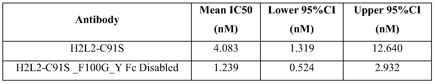


Claims
Priority Applications (14)
| Application Number | Priority Date | Filing Date | Title |
|---|---|---|---|
| EA201190018A EA201190018A1 (en) | 2008-12-19 | 2009-12-18 | MYOSTATINCONNECTING PROTEINS |
| US13/140,841 US20110256132A1 (en) | 2008-12-19 | 2009-12-18 | Myostatin binding proteins |
| CN2009801573934A CN102325793A (en) | 2008-12-19 | 2009-12-18 | Myostatin binding proteins |
| AU2009329533A AU2009329533A1 (en) | 2008-12-19 | 2009-12-18 | Myostatin binding proteins |
| JP2011541469A JP2012512641A (en) | 2008-12-19 | 2009-12-18 | Myostatin binding protein |
| NZ593297A NZ593297A (en) | 2008-12-19 | 2009-12-18 | Myostatin binding proteins |
| BRPI0922405A BRPI0922405A2 (en) | 2008-12-19 | 2009-12-18 | antigen binding protein, nucleic acid molecule, expression vector, recombinant host cell, method for producing an antigen binding protein, pharmaceutical composition, and methods for treating a patient, and for increasing muscle mass, increasing muscle strength, and / or improve muscle function in a patient |
| CA2747062A CA2747062A1 (en) | 2008-12-19 | 2009-12-18 | Myostatin binding proteins |
| MA34031A MA32980B1 (en) | 2008-12-19 | 2009-12-18 | Proteins binding to myostatin |
| SG2011041886A SG172039A1 (en) | 2008-12-19 | 2009-12-18 | Myostatin binding proteins |
| EP09804126A EP2358753A1 (en) | 2008-12-19 | 2009-12-18 | Myostatin binding proteins |
| MX2011006611A MX2011006611A (en) | 2008-12-19 | 2009-12-18 | Myostatin binding proteins. |
| IL213243A IL213243A0 (en) | 2008-12-19 | 2011-05-31 | Myostatin binding proteins |
| ZA2011/04397A ZA201104397B (en) | 2008-12-19 | 2011-06-13 | Myostatin binding proteins |
Applications Claiming Priority (2)
| Application Number | Priority Date | Filing Date | Title |
|---|---|---|---|
| US13898008P | 2008-12-19 | 2008-12-19 | |
| US61/138,980 | 2008-12-19 |
Publications (1)
| Publication Number | Publication Date |
|---|---|
| WO2010070094A1 true WO2010070094A1 (en) | 2010-06-24 |
Family
ID=41786068
Family Applications (1)
| Application Number | Title | Priority Date | Filing Date |
|---|---|---|---|
| PCT/EP2009/067515 WO2010070094A1 (en) | 2008-12-19 | 2009-12-18 | Myostatin binding proteins |
Country Status (24)
| Country | Link |
|---|---|
| US (1) | US20110256132A1 (en) |
| EP (1) | EP2358753A1 (en) |
| JP (1) | JP2012512641A (en) |
| KR (1) | KR20110103431A (en) |
| CN (1) | CN102325793A (en) |
| AR (1) | AR074777A1 (en) |
| AU (1) | AU2009329533A1 (en) |
| BR (1) | BRPI0922405A2 (en) |
| CA (1) | CA2747062A1 (en) |
| CL (1) | CL2011001503A1 (en) |
| CO (1) | CO6400149A2 (en) |
| CR (1) | CR20110358A (en) |
| DO (1) | DOP2011000189A (en) |
| EA (1) | EA201190018A1 (en) |
| IL (1) | IL213243A0 (en) |
| MA (1) | MA32980B1 (en) |
| MX (1) | MX2011006611A (en) |
| NZ (1) | NZ593297A (en) |
| PE (1) | PE20120429A1 (en) |
| SG (1) | SG172039A1 (en) |
| TW (1) | TW201029662A (en) |
| UY (1) | UY32341A (en) |
| WO (1) | WO2010070094A1 (en) |
| ZA (1) | ZA201104397B (en) |
Cited By (30)
| Publication number | Priority date | Publication date | Assignee | Title |
|---|---|---|---|---|
| WO2011150008A1 (en) | 2010-05-26 | 2011-12-01 | Regeneron Pharmaceuticals, Inc. | Antibodies to human gdf8 |
| WO2013074557A1 (en) | 2011-11-14 | 2013-05-23 | Regeneron Pharmaceuticals, Inc. | Compositions and methods for increasing muscle mass and muscle strength by specifically antagonizing gdf8 and/or activin a |
| WO2014043344A1 (en) * | 2012-09-13 | 2014-03-20 | Bristol-Myers Squibb Company | Fibronectin based scaffold domain proteins that bind to myostatin |
| JP2014523737A (en) * | 2011-06-02 | 2014-09-18 | ダイアックス コーポレーション | Fc receptor binding protein |
| WO2016168613A1 (en) * | 2015-04-15 | 2016-10-20 | Regeneron Pharmaceuticals, Inc. | Methods of increasing strength and functionality with gdf8 inhibitors |
| WO2017104783A1 (en) * | 2015-12-18 | 2017-06-22 | Chugai Seiyaku Kabushiki Kaisha | Anti-myostatin antibodies, polypeptides containing variant fc regions, and methods of use |
| US9718881B2 (en) | 2013-07-30 | 2017-08-01 | Regeneron Pharmaceuticals, Inc. | Anti-Activin A antibodies and uses thereof |
| US9828429B2 (en) | 2007-09-26 | 2017-11-28 | Chugai Seiyaku Kabushiki Kaisha | Method of modifying isoelectric point of antibody via amino acid substitution in CDR |
| WO2017217525A1 (en) * | 2016-06-17 | 2017-12-21 | Chugai Seiyaku Kabushiki Kaisha | Anti-myostatin antibodies and methods of use |
| US9868948B2 (en) | 2008-04-11 | 2018-01-16 | Chugai Seiyaku Kabushiki Kaisha | Antigen-binding molecule capable of binding to two or more antigen molecules repeatedly |
| US9969800B2 (en) | 2015-02-05 | 2018-05-15 | Chugai Seiyaku Kabushiki Kaisha | IL-8 antibodies |
| KR101860280B1 (en) | 2014-12-19 | 2018-05-21 | 추가이 세이야쿠 가부시키가이샤 | Anti-myostatin antibodies, polypeptides containing variant fc regions, and methods of use |
| WO2018129395A1 (en) * | 2017-01-06 | 2018-07-12 | Scholar Rock, Inc. | Methods for treating metabolic diseases by inhibiting myostatin activation |
| WO2019169283A1 (en) | 2018-03-01 | 2019-09-06 | Regeneron Pharmaceuticals, Inc. | Methods for altering body composition |
| WO2020131910A1 (en) | 2018-12-18 | 2020-06-25 | Regeneron Pharmaceuticals, Inc. | Compositions and methods for enhancing body weight and lean muscle mass using antagonists against leptin receptor, gdf8 and activin a |
| US10751413B2 (en) | 2015-09-15 | 2020-08-25 | Scholar Rock, Inc. | Anti-pro/latent-Myostatin antibodies and uses thereof |
| US10882904B2 (en) | 2016-01-08 | 2021-01-05 | Scholar Rock, Inc. | Methods for inhibiting myostatin activation by administering anti-pro/latent myostatin antibodies |
| US10919953B2 (en) | 2012-08-24 | 2021-02-16 | Chugai Seiyaku Kabushiki Kaisha | FcgammaRIIB-specific Fc region variant |
| US10946036B2 (en) | 2016-06-13 | 2021-03-16 | Scholar Rock, Inc. | Use of myostatin inhibitors and combination therapies |
| US11046784B2 (en) | 2006-03-31 | 2021-06-29 | Chugai Seiyaku Kabushiki Kaisha | Methods for controlling blood pharmacokinetics of antibodies |
| US11053308B2 (en) | 2016-08-05 | 2021-07-06 | Chugai Seiyaku Kabushiki Kaisha | Method for treating IL-8-related diseases |
| US11135291B2 (en) | 2014-11-06 | 2021-10-05 | Scholar Rock, Inc. | Methods for making and using anti-myostatin antibodies |
| US11155611B2 (en) | 2017-01-06 | 2021-10-26 | Scholar Rock, Inc. | Compositions and methods for making and using anti-myostatin antibodies |
| US11236168B2 (en) | 2012-08-24 | 2022-02-01 | Chugai Seiyaku Kabushiki Kaisha | Mouse FcγammaRII-specific Fc antibody |
| US11267868B2 (en) | 2013-04-02 | 2022-03-08 | Chugai Seiyaku Kabushiki Kaisha | Fc region variant |
| US11359009B2 (en) | 2015-12-25 | 2022-06-14 | Chugai Seiyaku Kabushiki Kaisha | Anti-myostatin antibodies and methods of use |
| US11369595B2 (en) | 2016-09-05 | 2022-06-28 | Metabrain Research | Use of tryptophan metabolites for treating muscle atrophy |
| RU2778945C2 (en) * | 2016-06-17 | 2022-08-29 | Чугаи Сейяку Кабусики Кайся | Antibodies to myostatin and their application methods |
| US11891434B2 (en) | 2010-11-30 | 2024-02-06 | Chugai Seiyaku Kabushiki Kaisha | Antigen-binding molecule capable of binding to plurality of antigen molecules repeatedly |
| WO2024064842A1 (en) | 2022-09-21 | 2024-03-28 | Regeneron Pharmaceuticals, Inc. | Methods of treating obesity, diabetes, and liver dysfunction |
Families Citing this family (2)
| Publication number | Priority date | Publication date | Assignee | Title |
|---|---|---|---|---|
| HUE052232T2 (en) | 2013-05-06 | 2021-04-28 | Scholar Rock Inc | Compositions and methods for growth factor modulation |
| WO2023069605A1 (en) * | 2021-10-20 | 2023-04-27 | Arizona Board Of Regents On Behalf Of Arizona State University | Systems, methods, and apparatuses for implementing a cloud-based health, nutritional, and body composition analysis platform |
Citations (53)
| Publication number | Priority date | Publication date | Assignee | Title |
|---|---|---|---|---|
| EP0054951A1 (en) | 1980-12-24 | 1982-06-30 | Chugai Seiyaku Kabushiki Kaisha | Dibenz(b,f)(1,4)oxazepine derivatives, process for preparing the same, and pharmaceutical compositions comprising the same |
| EP0183070A2 (en) | 1984-10-30 | 1986-06-04 | Phillips Petroleum Company | Transformation of yeasts of the genus pichia |
| US4676980A (en) | 1985-09-23 | 1987-06-30 | The United States Of America As Represented By The Secretary Of The Department Of Health And Human Services | Target specific cross-linked heteroantibodies |
| EP0239400A2 (en) | 1986-03-27 | 1987-09-30 | Medical Research Council | Recombinant antibodies and methods for their production |
| EP0244234A2 (en) | 1986-04-30 | 1987-11-04 | Alko Group Ltd. | Transformation of trichoderma |
| EP0307434A1 (en) | 1987-03-18 | 1989-03-22 | Medical Res Council | Altered antibodies. |
| DD266710A3 (en) | 1983-06-06 | 1989-04-12 | Ve Forschungszentrum Biotechnologie | Process for the biotechnical production of alkaline phosphatase |
| WO1990013646A1 (en) | 1989-04-28 | 1990-11-15 | Transgene S.A. | Application of novel dna fragments as a coding sequence for a signal peptide for the secretion of mature proteins by recombinant yeast, expression cassettes, transformed yeasts and corresponding process for the preparation of proteins |
| EP0402226A1 (en) | 1989-06-06 | 1990-12-12 | Institut National De La Recherche Agronomique | Transformation vectors for yeast yarrowia |
| WO1993006213A1 (en) | 1991-09-23 | 1993-04-01 | Medical Research Council | Production of chimeric antibodies - a combinatorial approach |
| WO1993008829A1 (en) | 1991-11-04 | 1993-05-13 | The Regents Of The University Of California | Compositions that mediate killing of hiv-infected cells |
| WO1994004690A1 (en) | 1992-08-17 | 1994-03-03 | Genentech, Inc. | Bispecific immunoadhesins |
| EP0612251A1 (en) | 1991-10-28 | 1994-08-31 | The Wellcome Foundation Limited | Stabilised antibodies |
| WO1994021681A1 (en) | 1993-03-19 | 1994-09-29 | Johns Hopkins University School Of Medicine | Growth differentiation factor-8 |
| EP0629240A1 (en) | 1992-02-19 | 1994-12-21 | Scotgen Limited | Altered antibodies, products and processes relating thereto |
| WO1994029348A2 (en) | 1993-06-03 | 1994-12-22 | Therapeutic Antibodies Inc. | Production of antibody fragments |
| US5429746A (en) | 1994-02-22 | 1995-07-04 | Smith Kline Beecham Corporation | Antibody purification |
| WO1997011086A1 (en) | 1995-09-22 | 1997-03-27 | The General Hospital Corporation | High level expression of proteins |
| US5739277A (en) | 1995-04-14 | 1998-04-14 | Genentech Inc. | Altered polypeptides with increased half-life |
| WO1998034640A2 (en) | 1997-02-07 | 1998-08-13 | Merck & Co., Inc. | Synthetic hiv gag genes |
| US5807715A (en) | 1984-08-27 | 1998-09-15 | The Board Of Trustees Of The Leland Stanford Junior University | Methods and transformed mammalian lymphocyte cells for producing functional antigen-binding protein including chimeric immunoglobulin |
| WO1999048523A2 (en) | 1998-03-26 | 1999-09-30 | Glaxo Group Limited | Antagonists of the inflammatory mediator oncostatin m (osm) |
| WO2000029004A1 (en) | 1998-11-18 | 2000-05-25 | Peptor Ltd. | Small functional units of antibody heavy chain variable regions |
| US6172197B1 (en) | 1991-07-10 | 2001-01-09 | Medical Research Council | Methods for producing members of specific binding pairs |
| US6248516B1 (en) | 1988-11-11 | 2001-06-19 | Medical Research Council | Single domain ligands, receptors comprising said ligands methods for their production, and use of said ligands and receptors |
| US6291158B1 (en) | 1989-05-16 | 2001-09-18 | Scripps Research Institute | Method for tapping the immunological repertoire |
| EP1229125A1 (en) | 1999-10-19 | 2002-08-07 | Kyowa Hakko Kogyo Co., Ltd. | Process for producing polypeptide |
| WO2003011878A2 (en) | 2001-08-03 | 2003-02-13 | Glycart Biotechnology Ag | Antibody glycosylation variants having increased antibody-dependent cellular cytotoxicity |
| WO2003027248A2 (en) | 2001-09-26 | 2003-04-03 | Wyeth | Antibody inhibitors of gdf-8 and uses thereof |
| WO2004006955A1 (en) | 2001-07-12 | 2004-01-22 | Jefferson Foote | Super humanized antibodies |
| WO2004009823A1 (en) | 2002-07-18 | 2004-01-29 | Lonza Biologics Plc. | Method of expressing recombinant protein in cho cells |
| WO2004014953A2 (en) | 2002-08-06 | 2004-02-19 | Glaxo Group Limited | Anti-myelin associated glycoprotein (mag) antibodies |
| WO2004029207A2 (en) | 2002-09-27 | 2004-04-08 | Xencor Inc. | Optimized fc variants and methods for their generation |
| WO2004037861A2 (en) | 2002-10-22 | 2004-05-06 | Wyeth | Neutralizing antibodies against gdf-8 and uses therefor |
| US6737056B1 (en) | 1999-01-15 | 2004-05-18 | Genentech, Inc. | Polypeptide variants with altered effector function |
| US20040132028A1 (en) | 2000-09-08 | 2004-07-08 | Stumpp Michael Tobias | Collection of repeat proteins comprising repeat modules |
| WO2004063351A2 (en) | 2003-01-09 | 2004-07-29 | Macrogenics, Inc. | IDENTIFICATION AND ENGINEERING OF ANTIBODIES WITH VARIANT Fc REGIONS AND METHODS OF USING SAME |
| US6818418B1 (en) | 1998-12-10 | 2004-11-16 | Compound Therapeutics, Inc. | Protein scaffolds for antibody mimics and other binding proteins |
| WO2005056764A2 (en) | 2003-12-05 | 2005-06-23 | Compound Therapeutics, Inc. | Inhibitors of type 2 vascular endothelial growth factor receptors |
| WO2005094446A2 (en) | 2004-03-23 | 2005-10-13 | Eli Lilly And Company | Anti-myostatin antibodies |
| WO2006014679A1 (en) | 2004-07-21 | 2006-02-09 | Glycofi, Inc. | Immunoglobulins comprising predominantly a glcnac2man3glcnac2 glycoform |
| EP1641818A1 (en) | 2003-07-04 | 2006-04-05 | Affibody AB | Polypeptides having binding affinity for her2 |
| WO2006053782A1 (en) | 2004-11-19 | 2006-05-26 | Glaxo Group Limited | Amide and peptide derivatives of dialkylenetriamines and their use as transfection agents |
| WO2006116269A2 (en) | 2005-04-25 | 2006-11-02 | Pfizer Inc. | Antibodies to myostatin |
| WO2006127682A2 (en) | 2005-05-20 | 2006-11-30 | Smithkline Beecham Corporation | Novel methods |
| US20070087000A1 (en) * | 2005-08-19 | 2007-04-19 | Walsh Frank S | Antagonist antibodies against GDF-8 and uses in treatment of ALS and other GDF-8-associated disorders |
| WO2007044411A2 (en) | 2005-10-06 | 2007-04-19 | Eli Lilly And Company | Anti-myostatin antibodies |
| WO2007047112A2 (en) | 2005-10-12 | 2007-04-26 | Eli Lilly And Company | Anti-myostatin antibodies |
| US7250297B1 (en) | 1997-09-26 | 2007-07-31 | Pieris Ag | Anticalins |
| US20070224633A1 (en) | 2003-08-25 | 2007-09-27 | Pieris Ag | Muteins of Tear Lipocalin |
| WO2008030706A2 (en) | 2006-09-05 | 2008-03-13 | Eli Lilly And Company | Anti-myostatin antibodies |
| US20080139791A1 (en) | 1998-12-10 | 2008-06-12 | Adnexus Therapeutics, Inc. | Pharmaceutically acceptable Fn3 Polypeptides for human treatments |
| WO2008098796A1 (en) | 2007-02-16 | 2008-08-21 | Nascacell Technologies Ag | Polypeptide comprising a knottin protein moiety |
Family Cites Families (1)
| Publication number | Priority date | Publication date | Assignee | Title |
|---|---|---|---|---|
| GB0702888D0 (en) * | 2007-02-14 | 2007-03-28 | Glaxo Group Ltd | Novel Antibodies |
-
2009
- 2009-12-17 TW TW098143432A patent/TW201029662A/en unknown
- 2009-12-17 UY UY0001032341A patent/UY32341A/en unknown
- 2009-12-17 AR ARP090104946A patent/AR074777A1/en not_active Application Discontinuation
- 2009-12-18 US US13/140,841 patent/US20110256132A1/en not_active Abandoned
- 2009-12-18 EP EP09804126A patent/EP2358753A1/en not_active Withdrawn
- 2009-12-18 SG SG2011041886A patent/SG172039A1/en unknown
- 2009-12-18 JP JP2011541469A patent/JP2012512641A/en not_active Ceased
- 2009-12-18 EA EA201190018A patent/EA201190018A1/en unknown
- 2009-12-18 BR BRPI0922405A patent/BRPI0922405A2/en not_active IP Right Cessation
- 2009-12-18 MA MA34031A patent/MA32980B1/en unknown
- 2009-12-18 KR KR1020117016877A patent/KR20110103431A/en not_active Application Discontinuation
- 2009-12-18 CN CN2009801573934A patent/CN102325793A/en active Pending
- 2009-12-18 CA CA2747062A patent/CA2747062A1/en not_active Abandoned
- 2009-12-18 AU AU2009329533A patent/AU2009329533A1/en not_active Abandoned
- 2009-12-18 NZ NZ593297A patent/NZ593297A/en not_active IP Right Cessation
- 2009-12-18 WO PCT/EP2009/067515 patent/WO2010070094A1/en active Application Filing
- 2009-12-18 MX MX2011006611A patent/MX2011006611A/en not_active Application Discontinuation
- 2009-12-18 PE PE2011001257A patent/PE20120429A1/en not_active Application Discontinuation
-
2011
- 2011-05-31 IL IL213243A patent/IL213243A0/en unknown
- 2011-06-13 ZA ZA2011/04397A patent/ZA201104397B/en unknown
- 2011-06-15 DO DO2011000189A patent/DOP2011000189A/en unknown
- 2011-06-17 CL CL2011001503A patent/CL2011001503A1/en unknown
- 2011-06-23 CR CR20110358A patent/CR20110358A/en unknown
- 2011-06-28 CO CO11080753A patent/CO6400149A2/en not_active Application Discontinuation
Patent Citations (54)
| Publication number | Priority date | Publication date | Assignee | Title |
|---|---|---|---|---|
| EP0054951A1 (en) | 1980-12-24 | 1982-06-30 | Chugai Seiyaku Kabushiki Kaisha | Dibenz(b,f)(1,4)oxazepine derivatives, process for preparing the same, and pharmaceutical compositions comprising the same |
| DD266710A3 (en) | 1983-06-06 | 1989-04-12 | Ve Forschungszentrum Biotechnologie | Process for the biotechnical production of alkaline phosphatase |
| US5807715A (en) | 1984-08-27 | 1998-09-15 | The Board Of Trustees Of The Leland Stanford Junior University | Methods and transformed mammalian lymphocyte cells for producing functional antigen-binding protein including chimeric immunoglobulin |
| EP0183070A2 (en) | 1984-10-30 | 1986-06-04 | Phillips Petroleum Company | Transformation of yeasts of the genus pichia |
| US4676980A (en) | 1985-09-23 | 1987-06-30 | The United States Of America As Represented By The Secretary Of The Department Of Health And Human Services | Target specific cross-linked heteroantibodies |
| EP0239400A2 (en) | 1986-03-27 | 1987-09-30 | Medical Research Council | Recombinant antibodies and methods for their production |
| EP0244234A2 (en) | 1986-04-30 | 1987-11-04 | Alko Group Ltd. | Transformation of trichoderma |
| EP0307434A1 (en) | 1987-03-18 | 1989-03-22 | Medical Res Council | Altered antibodies. |
| US6248516B1 (en) | 1988-11-11 | 2001-06-19 | Medical Research Council | Single domain ligands, receptors comprising said ligands methods for their production, and use of said ligands and receptors |
| WO1990013646A1 (en) | 1989-04-28 | 1990-11-15 | Transgene S.A. | Application of novel dna fragments as a coding sequence for a signal peptide for the secretion of mature proteins by recombinant yeast, expression cassettes, transformed yeasts and corresponding process for the preparation of proteins |
| US6291158B1 (en) | 1989-05-16 | 2001-09-18 | Scripps Research Institute | Method for tapping the immunological repertoire |
| EP0402226A1 (en) | 1989-06-06 | 1990-12-12 | Institut National De La Recherche Agronomique | Transformation vectors for yeast yarrowia |
| US6172197B1 (en) | 1991-07-10 | 2001-01-09 | Medical Research Council | Methods for producing members of specific binding pairs |
| WO1993006213A1 (en) | 1991-09-23 | 1993-04-01 | Medical Research Council | Production of chimeric antibodies - a combinatorial approach |
| EP0612251A1 (en) | 1991-10-28 | 1994-08-31 | The Wellcome Foundation Limited | Stabilised antibodies |
| WO1993008829A1 (en) | 1991-11-04 | 1993-05-13 | The Regents Of The University Of California | Compositions that mediate killing of hiv-infected cells |
| EP0629240A1 (en) | 1992-02-19 | 1994-12-21 | Scotgen Limited | Altered antibodies, products and processes relating thereto |
| WO1994004690A1 (en) | 1992-08-17 | 1994-03-03 | Genentech, Inc. | Bispecific immunoadhesins |
| WO1994021681A1 (en) | 1993-03-19 | 1994-09-29 | Johns Hopkins University School Of Medicine | Growth differentiation factor-8 |
| WO1994029348A2 (en) | 1993-06-03 | 1994-12-22 | Therapeutic Antibodies Inc. | Production of antibody fragments |
| US5429746A (en) | 1994-02-22 | 1995-07-04 | Smith Kline Beecham Corporation | Antibody purification |
| US5739277A (en) | 1995-04-14 | 1998-04-14 | Genentech Inc. | Altered polypeptides with increased half-life |
| WO1997011086A1 (en) | 1995-09-22 | 1997-03-27 | The General Hospital Corporation | High level expression of proteins |
| WO1998034640A2 (en) | 1997-02-07 | 1998-08-13 | Merck & Co., Inc. | Synthetic hiv gag genes |
| US7250297B1 (en) | 1997-09-26 | 2007-07-31 | Pieris Ag | Anticalins |
| WO1999048523A2 (en) | 1998-03-26 | 1999-09-30 | Glaxo Group Limited | Antagonists of the inflammatory mediator oncostatin m (osm) |
| WO2000029004A1 (en) | 1998-11-18 | 2000-05-25 | Peptor Ltd. | Small functional units of antibody heavy chain variable regions |
| US20080139791A1 (en) | 1998-12-10 | 2008-06-12 | Adnexus Therapeutics, Inc. | Pharmaceutically acceptable Fn3 Polypeptides for human treatments |
| US6818418B1 (en) | 1998-12-10 | 2004-11-16 | Compound Therapeutics, Inc. | Protein scaffolds for antibody mimics and other binding proteins |
| US6737056B1 (en) | 1999-01-15 | 2004-05-18 | Genentech, Inc. | Polypeptide variants with altered effector function |
| EP1229125A1 (en) | 1999-10-19 | 2002-08-07 | Kyowa Hakko Kogyo Co., Ltd. | Process for producing polypeptide |
| US20040132028A1 (en) | 2000-09-08 | 2004-07-08 | Stumpp Michael Tobias | Collection of repeat proteins comprising repeat modules |
| WO2004006955A1 (en) | 2001-07-12 | 2004-01-22 | Jefferson Foote | Super humanized antibodies |
| WO2003011878A2 (en) | 2001-08-03 | 2003-02-13 | Glycart Biotechnology Ag | Antibody glycosylation variants having increased antibody-dependent cellular cytotoxicity |
| WO2003027248A2 (en) | 2001-09-26 | 2003-04-03 | Wyeth | Antibody inhibitors of gdf-8 and uses thereof |
| US20030138422A1 (en) * | 2001-09-26 | 2003-07-24 | Jane Aghajanian | Antibody inhibitors of GDF-8 and uses thereof |
| WO2004009823A1 (en) | 2002-07-18 | 2004-01-29 | Lonza Biologics Plc. | Method of expressing recombinant protein in cho cells |
| WO2004014953A2 (en) | 2002-08-06 | 2004-02-19 | Glaxo Group Limited | Anti-myelin associated glycoprotein (mag) antibodies |
| WO2004029207A2 (en) | 2002-09-27 | 2004-04-08 | Xencor Inc. | Optimized fc variants and methods for their generation |
| WO2004037861A2 (en) | 2002-10-22 | 2004-05-06 | Wyeth | Neutralizing antibodies against gdf-8 and uses therefor |
| WO2004063351A2 (en) | 2003-01-09 | 2004-07-29 | Macrogenics, Inc. | IDENTIFICATION AND ENGINEERING OF ANTIBODIES WITH VARIANT Fc REGIONS AND METHODS OF USING SAME |
| EP1641818A1 (en) | 2003-07-04 | 2006-04-05 | Affibody AB | Polypeptides having binding affinity for her2 |
| US20070224633A1 (en) | 2003-08-25 | 2007-09-27 | Pieris Ag | Muteins of Tear Lipocalin |
| WO2005056764A2 (en) | 2003-12-05 | 2005-06-23 | Compound Therapeutics, Inc. | Inhibitors of type 2 vascular endothelial growth factor receptors |
| WO2005094446A2 (en) | 2004-03-23 | 2005-10-13 | Eli Lilly And Company | Anti-myostatin antibodies |
| WO2006014679A1 (en) | 2004-07-21 | 2006-02-09 | Glycofi, Inc. | Immunoglobulins comprising predominantly a glcnac2man3glcnac2 glycoform |
| WO2006053782A1 (en) | 2004-11-19 | 2006-05-26 | Glaxo Group Limited | Amide and peptide derivatives of dialkylenetriamines and their use as transfection agents |
| WO2006116269A2 (en) | 2005-04-25 | 2006-11-02 | Pfizer Inc. | Antibodies to myostatin |
| WO2006127682A2 (en) | 2005-05-20 | 2006-11-30 | Smithkline Beecham Corporation | Novel methods |
| US20070087000A1 (en) * | 2005-08-19 | 2007-04-19 | Walsh Frank S | Antagonist antibodies against GDF-8 and uses in treatment of ALS and other GDF-8-associated disorders |
| WO2007044411A2 (en) | 2005-10-06 | 2007-04-19 | Eli Lilly And Company | Anti-myostatin antibodies |
| WO2007047112A2 (en) | 2005-10-12 | 2007-04-26 | Eli Lilly And Company | Anti-myostatin antibodies |
| WO2008030706A2 (en) | 2006-09-05 | 2008-03-13 | Eli Lilly And Company | Anti-myostatin antibodies |
| WO2008098796A1 (en) | 2007-02-16 | 2008-08-21 | Nascacell Technologies Ag | Polypeptide comprising a knottin protein moiety |
Non-Patent Citations (118)
| Title |
|---|
| "Remingtons Pharmaceutical Sciences, 16th edition", 1980, MACK PUBLISHING CO. |
| ADAMS ET AL., CAN. RES, vol. 53, 1993, pages 4026 - 4034 |
| ALT ET AL., FEBS LETT, vol. 454, 1999, pages 90 - 94 |
| BAEZ ET AL., BIOPHARM, vol. 13, 2000, pages 50 - 54 |
| BIOCHIM BIOPHYS ACTA, vol. 1482, 2000, pages 337 - 350 |
| BIRD ET AL., SCIENCE, vol. 242, 1988, pages 423 - 426 |
| BOGDANOVICH SASHA ET AL: "Functional improvement of dystrophic muscle by myostatin blockade.", NATURE 28 NOV 2002, vol. 420, no. 6914, 28 November 2002 (2002-11-28), pages 418 - 421, XP009120477, ISSN: 0028-0836 * |
| BOGDANOVICH SASHA ET AL: "Myostatin blockade improves function but not histopathology in a murine model of limb-girdle muscular dystrophy 2C.", MUSCLE & NERVE MAR 2008, vol. 37, no. 3, March 2008 (2008-03-01), pages 308 - 316, XP007912264, ISSN: 0148-639X * |
| BOYD ET AL., MOL. IMMUNOL., vol. 32, 1996, pages 1311 - 1318 |
| BRADLEY L ET AL: "Myostatin as a therapeutic target for musculoskeletal disease", CMLS CELLULAR AND MOLECULAR LIFE SCIENCES, BIRKHÄUSER-VERLAG, BA, vol. 65, no. 14, 21 April 2008 (2008-04-21), pages 2119 - 2124, XP019619981, ISSN: 1420-9071 * |
| BRODEUR: "Monoclonal Antibody Production Techniques and Applications", 1987, MARCEL DEKKER INC, pages: 51 - 63 |
| CHOTHIA ET AL., NATURE, vol. 342, 1989, pages 877 - 883 |
| CO ET AL., NATURE, vol. 351, 1991, pages 501 - 502 |
| COLOMA ET AL., NATURE BIOTECHNOL, vol. 15, 1997, pages 159 - 163 |
| CUPIT ET AL., LETT APPL MICROBIOL, vol. 29, 1999, pages 273 - 277 |
| DAVIS ET AL., CHEM. REV., vol. 102, 2002, pages 579 |
| DEWYE WD.: "Clinics in Oncology", vol. 5, 1986, SAUNDERS, pages: 251 - 261 |
| DORAN, CURR. OPINION BIOTECHNOL., vol. 11, 2000, pages 199 - 204 |
| DRAPEAU ET AL., CYTOTECHNOLOGY, vol. 15, 1994, pages 103 - 109 |
| E. MEYERS; W. MILLER, COMPUT. APPL. BIOSCI., vol. 4, 1988, pages 11 - 17 |
| EXPERT OPIN. BIOL. THER., vol. 5, 2005, pages 783 - 797 |
| EXPERT OPINION ON INVESTIGATIONAL DRUGS, vol. 16, no. 6, June 2007 (2007-06-01), pages 909 - 917 |
| FISHWILD, NATURE BIOTECHNOL., vol. 14, 1996, pages 845 - 851 |
| GHETIE ET AL., ANNU. REV. IMMUNOL., vol. 18, 2000, pages 739 - 766 |
| GLOCKSHUBER ET AL., BIOCHEMISTRY, vol. 29, 1990, pages 1362 - 1367 |
| GREEN, J. IMMUNOL. METHODS, vol. 231, 1999, pages 11 - 23 |
| GRIFFITHS ET AL., EMBO, vol. 13, 1994, pages 3245 - 3260 |
| HANG ET AL., ACC. CHEM. RES, vol. 34, 2001, pages 727 |
| HODGSON ET AL., BIO/TECHNOLOGY, vol. 9, 1991, pages 421 |
| HOEKEMA ET AL., MOL CELL BIOL, vol. 7, no. 8, 1987, pages 2914 - 24 |
| HOLLIGER ET AL., PNAS, vol. 90, 1993, pages 6444 - 6448 |
| HOLLIGER; HUDSON, NATURE BIOTECHNOLOGY, vol. 23, no. 9, 2005, pages 1126 - 1136 |
| HOLZBAUR ET AL: "Myostatin inhibition slows muscle atrophy in rodent models of amyotrophic lateral sclerosis", NEUROBIOLOGY OF DISEASE, BLACKWELL SCIENTIFIC PUBLICATIONS, OXFORD, GB, vol. 23, no. 3, 1 September 2006 (2006-09-01), pages 697 - 707, XP005599201, ISSN: 0969-9961 * |
| HU ET AL., CANCER RES., vol. 56, 1996, pages 3055 - 3061 |
| HUSTON ET AL., INT. REV. IMMUNOL, vol. 10, 1993, pages 195 - 217 |
| HUSTON ET AL., PNAS, vol. 85, no. 16, 1988, pages 5879 - 5883 |
| J. BIOL. CHEM, vol. 274, 1999, pages 24066 - 24073 |
| J. MOL. BIOL., vol. 332, 2003, pages 489 - 503 |
| J. MOL. BIOL., vol. 369, 2007, pages 1015 - 1028 |
| JAMALI ET AL., MUSCLE NERVE, vol. 23, 2000, pages 851 - 862 |
| JONES ET AL., NATURE, vol. 321, 1986, pages 522 - 525 |
| JOURNAL OF IMMUNOLOGICAL METHODS, vol. 248, no. 1-2, 2001, pages 31 - 45 |
| JUNGHANS, IMMUNOL. RES, vol. 16, 1997, pages 29 - 57 |
| KABAT ET AL.: "Sequences of Proteins of Immunological Interest", 1987, NATIONAL INSTITUTES OF HEALTH |
| KAWADA S ET AL., J MUSCLE RES CELL MOTIL, vol. 22, 2001, pages 627 - 633 |
| KEEN ET AL., CYTOTECHNOLOGY, vol. 17, 1995, pages 153 - 163 |
| KILPATRICK ET AL., HYBRIDOMA, vol. 16, no. 4, 1997, pages 381 - 389 |
| KIPRIYANOV ET AL., CELL. BIOPHYS, vol. 26, 1995, pages 187 - 204 |
| KIPRIYANOV ET AL., INT. J. CAN, vol. 77, 1998, pages 763 - 772 |
| KIPRIYANOV ET AL., J. MOL. BIOL., vol. 293, 1999, pages 41 - 56 |
| KONTERMANN ET AL., J. IMMUNOL. METHODS, vol. 226, 1999, pages 179 - 188 |
| KORTT ET AL., PROTEIN ENG, vol. 10, 1997, pages 423 - 433 |
| KOSTELNY ET AL., J. IMMUNOL., vol. 148, 1992, pages 1547 - 1553 |
| KOTTER DP ET AL., J PARENT ENTERAL NUTR, vol. 14, 1990, pages 454 - 358 |
| KOUMENIS ET AL., INT. J. PHARMACEUT., vol. 198, 2000, pages 83 - 95 |
| KOZBOR, J. IMMUNOL, vol. 133, 1984, pages 3001 |
| KURUCZ ET AL., J. IMMOL., vol. 154, 1995, pages 4576 - 4582 |
| LALANI R ET AL., J ENDOCRINOL, vol. 167, 2000, pages 417 - 428 |
| LANG CH ET AL., FASEB J, vol. 15, 2001, pages NIL323 - NIL338 |
| LAZAR ET AL., PNAS, vol. 103, 2006, pages 4005 - 4010 |
| LE GALL ET AL., FEBS LETT, vol. 453, 1999, pages 164 - 168 |
| MA K ET AL., AM J PHYSIOL ENDOCRINOL METAB, vol. 285, 2003, pages E363 - E371 |
| MA, NAT. MED., vol. 4, 1998, pages 601 - 606 |
| MARK ET AL.: "Handbook of Experimental Pharmacology Vol. 113: The pharmacology of Monoclonal Antibodies", vol. 113, 1994, SPRINGER-VERLAG, pages: 105 - 134 |
| MARKS, BIO/TECHNOL, vol. 10, 1992, pages 779 - 783 |
| MARTIN; THORNTON, J MOL BIOL, vol. 263, 1996, pages 800 - 815 |
| MCCAFFERTY, NATURE, vol. 348, 1990, pages 552 - 553 |
| MCCARTNEY ET AL., PROTEIN ENG., vol. 8, 1995, pages 301 - 314 |
| MENDEZ, NATURE GENETICS, vol. 15, 1997, pages 146 - 156 |
| MILLSTEIN ET AL., NATURE, vol. 305, 1983, pages 537 - 539 |
| MORRISON, PNAS, vol. 81, 1984, pages 6851 |
| MORROW, GENET. ENG. NEWS, vol. 20, 2000, pages 1 - 55 |
| MULLER ET AL., FEBS LETT, vol. 432, 1998, pages 45 - 49 |
| NADEAU AMELIE ET AL: "Are big muscles necessarily good muscles?", ANNALS OF NEUROLOGY MAY 2008, vol. 63, no. 5, May 2008 (2008-05-01), pages 543 - 545, XP007912263, ISSN: 1531-8249 * |
| NAKAMURA ET AL., NUCLEIC ACIDS RESEARCH, vol. 24, 1996, pages 214 - 215 |
| NATURE BIOTECHNOLOGY, vol. 23, no. 12, 2005, pages 1556 - 1561 |
| NEEDLEMAN; WUNSCH, J. MOL. BIOL., vol. 48, 1970, pages 444 - 453 |
| PACK ET AL., BIOCHEMISTRY, vol. 31, 1992, pages 1579 - 1584 |
| PADLAN ET AL., MOL. IMMUNOL., vol. 28, 1991, pages 489 - 498 |
| PEDERSEN ET AL., J. MOL. BIOL., vol. 235, 1994, pages 959 - 973 |
| PENG ET AL., J. BIOTECHNOL., vol. 108, 2004, pages 185 - 192 |
| PNAS, vol. 100, no. 4, 2003, pages 1700 - 1705 |
| POLLOCK ET AL., J. IMMUNOL. METHODS, vol. 231, 1999, pages 147 - 157 |
| PROTEIN ENG. DES. SEL., vol. 17, 2004, pages 455 - 462 |
| PROTEIN ENG. DES. SEL., vol. 18, 2005, pages 435 - 444 |
| PROTEIN SCIENCE, vol. 15, 2006, pages 14 - 27 |
| QUEEN ET AL., PNAS, vol. 86, 1989, pages 10,029 - 10,033 |
| QUEEN ET AL., PROC. NATL ACAD SCI USA, vol. 86, 1989, pages 10029 - 10032 |
| RAJU ET AL., BIOCHEMISTRY, vol. 40, 2001, pages 8868 - 8876 |
| SANCHEZ ET AL., J. BIOTECHNOL., vol. 72, 1999, pages 13 - 20 |
| SCHARFENBERG ET AL.: "Animal Cell Technology: Developments towards the 21st century", 1995, KLUWER ACADEMIC PUBLISHERS, pages: 619 - 623 |
| SEARS ET AL., SCIENCE, vol. 291, 2001, pages 2344 |
| See also references of EP2358753A1 |
| SHALABY ET AL., J. EXP. MED., vol. 175, 1992, pages 217 - 225 |
| SHIELDS ET AL., J. BIOL. CHEM, vol. 276, 2001, pages 6591 - 6604 |
| SMITH ET AL.: "Antibodies in human diagnosis and therapy", 1977, RAVEN PRESS |
| STEFAN DUBEL: "Handbook of Therapeutic Antibodies", 2007 |
| STINCHCOMB ET AL., NATURE, vol. 282, 1979, pages 38 |
| STOGER ET AL., PLANT MOL. BIOL., vol. 42, 2000, pages 583 - 590 |
| SURESH ET AL., METHODS IN ENZYMOLOGY, vol. 121, 1986, pages 210 |
| THIES ET AL., GROWTH FACTORS, vol. 18, no. 4, 2001, pages 251 - 259 |
| TOMIZUKA, PNAS, vol. 97, 2000, pages 722 - 727 |
| TRAUNECKER ET AL., EMBO, vol. 10, 1991, pages 3655 - 3659 |
| TSUCHIDA KUNIHIRO: "Targeting myostatin for therapies against muscle-wasting disorders", CURRENT OPINION IN DRUG DISCOVERY AND DEVELOPMENT, CURRENT DRUGS, LONDON, GB, vol. 11, no. 4, 1 July 2008 (2008-07-01), pages 487 - 494, XP008120304, ISSN: 1367-6733 * |
| URLAUB ET AL., SOMATIC CELL MOL. GENET., vol. 12, 1986, pages 555 - 556 |
| VERHOEYEN ET AL., SCIENCE, vol. 239, 1988, pages 1534 - 1536 |
| WACKER ET AL., SCIENCE, vol. 298, 2002, pages 1790 |
| WAGNER KATHRYN R ET AL: "A phase I/IItrial of MYO-029 in adult subjects with muscular dystrophy", ANNALS OF NEUROLOGY, JOHN WILEY AND SONS, BOSTON, US, vol. 63, no. 5, 1 May 2008 (2008-05-01), pages 561 - 571, XP002514515, ISSN: 0364-5134 * |
| WAGNER, ANN NEUROL., vol. 63, no. 5, 2008, pages 561 - 71 |
| WALSH F S ET AL: "MYOSTATIN: A MODULATOR OF SKELETAL-MUSCLE STEM CELLS", BIOCHEMICAL SOCIETY TRANSACTIONS, PORTLAND PRESS LTD, GB, vol. 33, no. 6, 1 December 2005 (2005-12-01), pages 1513 - 1517, XP009077202, ISSN: 0300-5127 * |
| WATERHOUSE, NUCL. ACIDS RES., vol. 21, 1993, pages 2265 - 2266 |
| WHITLOW ET AL., METHODS COMPANION METHODS ENZYMOL, vol. 2, 1991, pages 97 - 105 |
| WHITTEMORE LISA-ANNE ET AL: "Inhibition of myostatin in adult mice increases skeletal muscle mass and strength.", BIOCHEMICAL AND BIOPHYSICAL RESEARCH COMMUNICATIONS 24 JAN 2003, vol. 300, no. 4, 24 January 2003 (2003-01-24), pages 965 - 971, XP002574357, ISSN: 0006-291X * |
| WIGMORE SJ ET AL., BR J CANCER, vol. 75, 1997, pages 106 - 109 |
| WINTER, ANNU. REV. IMMUNOL, vol. 12, 1994, pages 433 - 455 |
| WOLFMAN ET AL., PNAS, vol. 100, 2003, pages 15842 - 15846 |
| ZHANG ET AL., SCIENCE, vol. 303, 2004, pages 371 |
| ZHU ET AL., PROTEIN SCI., vol. 6, 1997, pages 781 - 788 |
Cited By (94)
| Publication number | Priority date | Publication date | Assignee | Title |
|---|---|---|---|---|
| US11046784B2 (en) | 2006-03-31 | 2021-06-29 | Chugai Seiyaku Kabushiki Kaisha | Methods for controlling blood pharmacokinetics of antibodies |
| US11248053B2 (en) | 2007-09-26 | 2022-02-15 | Chugai Seiyaku Kabushiki Kaisha | Method of modifying isoelectric point of antibody via amino acid substitution in CDR |
| US9828429B2 (en) | 2007-09-26 | 2017-11-28 | Chugai Seiyaku Kabushiki Kaisha | Method of modifying isoelectric point of antibody via amino acid substitution in CDR |
| US11359194B2 (en) | 2008-04-11 | 2022-06-14 | Chugai Seiyaku Kabushiki Kaisha | Antigen-binding molecule capable of binding two or more antigen molecules repeatedly |
| US9890377B2 (en) | 2008-04-11 | 2018-02-13 | Chugai Seiyaku Kabushiki Kaisha | Antigen-binding molecule capable of binding to two or more antigen molecules repeatedly |
| US10472623B2 (en) | 2008-04-11 | 2019-11-12 | Chugai Seiyaku Kabushiki Kaisha | Antigen-binding molecule capable of binding two or more antigen molecules repeatedly |
| US11371039B2 (en) | 2008-04-11 | 2022-06-28 | Chugai Seiyaku Kabushiki Kaisha | Antigen-binding molecule capable of binding to two or more antigen molecules repeatedly |
| US9868948B2 (en) | 2008-04-11 | 2018-01-16 | Chugai Seiyaku Kabushiki Kaisha | Antigen-binding molecule capable of binding to two or more antigen molecules repeatedly |
| US9260515B2 (en) | 2010-05-26 | 2016-02-16 | Regeneron Pharmaceuticals, Inc. | Antibodies to human GDF8 |
| WO2011150008A1 (en) | 2010-05-26 | 2011-12-01 | Regeneron Pharmaceuticals, Inc. | Antibodies to human gdf8 |
| US8840894B2 (en) | 2010-05-26 | 2014-09-23 | Regeneron Pharmaceuticals, Inc. | Antibodies to human GDF8 |
| US9890212B2 (en) | 2010-05-26 | 2018-02-13 | Regeneron Pharmaceuticals, Inc. | Antibodies to human GDF8 |
| US11891434B2 (en) | 2010-11-30 | 2024-02-06 | Chugai Seiyaku Kabushiki Kaisha | Antigen-binding molecule capable of binding to plurality of antigen molecules repeatedly |
| JP2018121657A (en) * | 2011-06-02 | 2018-08-09 | ダイアックス コーポレーション | Fc receptor binding protein |
| US11014988B2 (en) | 2011-06-02 | 2021-05-25 | Dyax Corp. | Nucleic acids encoding anti-neonatal Fc receptor (FcRn) antibodies |
| JP2014523737A (en) * | 2011-06-02 | 2014-09-18 | ダイアックス コーポレーション | Fc receptor binding protein |
| US11739152B2 (en) | 2011-06-02 | 2023-08-29 | Takeda Pharmaceutical Company Limited | Antibodies which bind Fc receptors (FcRn) |
| JP2017169559A (en) * | 2011-06-02 | 2017-09-28 | ダイアックス コーポレーション | Fc receptor binding protein |
| US10479834B2 (en) | 2011-06-02 | 2019-11-19 | Dyax Corp. | Fc receptor antibodies and methods of use thereof |
| US9862768B2 (en) | 2011-06-02 | 2018-01-09 | Dyax Corp. | Methods of producing antibodies to neonatal Fc receptor (FcRn) |
| US10400036B2 (en) | 2011-11-14 | 2019-09-03 | Regeneron Pharmaceuticals, Inc. | Compositions and methods for increasing muscle mass and muscle strength by specifically antagonizing GDF8 and or activin A |
| WO2013074557A1 (en) | 2011-11-14 | 2013-05-23 | Regeneron Pharmaceuticals, Inc. | Compositions and methods for increasing muscle mass and muscle strength by specifically antagonizing gdf8 and/or activin a |
| US11655291B2 (en) | 2011-11-14 | 2023-05-23 | Regeneron Pharmaceuticals, Inc. | Compositions and methods for increasing muscle mass and muscle strength by specifically antagonizing GDF8 and or activin A |
| US8871209B2 (en) | 2011-11-14 | 2014-10-28 | Regeneron Pharmaceuticals, Inc. | Compositions and methods for increasing muscle mass and muscle strength by specifically antagonizing GDF8 and or Activin A |
| US11236168B2 (en) | 2012-08-24 | 2022-02-01 | Chugai Seiyaku Kabushiki Kaisha | Mouse FcγammaRII-specific Fc antibody |
| US10919953B2 (en) | 2012-08-24 | 2021-02-16 | Chugai Seiyaku Kabushiki Kaisha | FcgammaRIIB-specific Fc region variant |
| IL270110B (en) * | 2012-09-13 | 2022-08-01 | Bristol Myers Squibb Co | Fibronectin based scaffold domain proteins that bind to myostatin |
| US9662373B2 (en) | 2012-09-13 | 2017-05-30 | Bristol-Myers Squibb Company | Fibronectin based scaffold domain proteins that bind to myostatin |
| KR20150054938A (en) * | 2012-09-13 | 2015-05-20 | 브리스톨-마이어스 스큅 컴퍼니 | Fibronectin based scaffold domain proteins that bind to myostatin |
| EP3835310A1 (en) * | 2012-09-13 | 2021-06-16 | Bristol-Myers Squibb Company | Fibronectin based scaffold domain proteins that bind to myostatin |
| US9493546B2 (en) | 2012-09-13 | 2016-11-15 | Bristol-Myers Squibb Company | Fibronectin based scaffold domain proteins that bind to myostatin |
| US8993265B2 (en) | 2012-09-13 | 2015-03-31 | Bristol-Myers Squibb Company | Fibronectin based scaffold domain proteins that bind to myostatin |
| TWI633117B (en) * | 2012-09-13 | 2018-08-21 | 必治妥美雅史谷比公司 | Fibronectin based scaffold domain proteins that bind to myostatin |
| US11813315B2 (en) | 2012-09-13 | 2023-11-14 | Bristol-Myers Squibb Company | Fibronectin based scaffold domain proteins that bind to myostatin |
| KR102424590B1 (en) | 2012-09-13 | 2022-07-27 | 브리스톨-마이어스 스큅 컴퍼니 | Fibronectin based scaffold domain proteins that bind to myostatin |
| US10245302B2 (en) | 2012-09-13 | 2019-04-02 | Bristol-Myers Squibb Company | Fibronectin based scaffold domain proteins that bind to myostatin |
| US8933199B2 (en) | 2012-09-13 | 2015-01-13 | Bristol-Myers Squibb Company | Fibronectin based scaffold domain proteins that bind to myostatin |
| AU2013315482B2 (en) * | 2012-09-13 | 2018-03-15 | Bristol-Myers Squibb Company | Fibronectin based scaffold domain proteins that bind to myostatin |
| US10406212B2 (en) | 2012-09-13 | 2019-09-10 | Bristol-Myers Squibb Company | Fibronectin based scaffold domain proteins that bind to myostatin |
| EA033536B1 (en) * | 2012-09-13 | 2019-10-31 | Bristol Myers Squibb Co | Fibronectin based scaffold domain proteins that bind to myostatin |
| EP3564258A1 (en) * | 2012-09-13 | 2019-11-06 | Bristol-Myers Squibb Company | Fibronectin based scaffold domain proteins that bind to myostatin |
| US8853154B2 (en) | 2012-09-13 | 2014-10-07 | Bristol-Myers Squibb Company | Fibronectin based scaffold domain proteins that bind to myostatin |
| WO2014043344A1 (en) * | 2012-09-13 | 2014-03-20 | Bristol-Myers Squibb Company | Fibronectin based scaffold domain proteins that bind to myostatin |
| KR20200138435A (en) * | 2012-09-13 | 2020-12-09 | 브리스톨-마이어스 스큅 컴퍼니 | Fibronectin based scaffold domain proteins that bind to myostatin |
| KR102187714B1 (en) | 2012-09-13 | 2020-12-07 | 브리스톨-마이어스 스큅 컴퍼니 | Fibronectin based scaffold domain proteins that bind to myostatin |
| AU2018201273B2 (en) * | 2012-09-13 | 2020-04-09 | Bristol-Myers Squibb Company | Fibronectin based scaffold domain proteins that bind to myostatin |
| TWI694085B (en) * | 2012-09-13 | 2020-05-21 | 美商必治妥美雅史谷比公司 | Fibronectin based scaffold domain proteins that bind to myostatin |
| US11267868B2 (en) | 2013-04-02 | 2022-03-08 | Chugai Seiyaku Kabushiki Kaisha | Fc region variant |
| US9718881B2 (en) | 2013-07-30 | 2017-08-01 | Regeneron Pharmaceuticals, Inc. | Anti-Activin A antibodies and uses thereof |
| US10526403B2 (en) | 2013-07-30 | 2020-01-07 | Regeneron Pharmaceuticals, Inc. | Anti-activin A antibodies and uses thereof |
| US11135291B2 (en) | 2014-11-06 | 2021-10-05 | Scholar Rock, Inc. | Methods for making and using anti-myostatin antibodies |
| US11925683B2 (en) | 2014-11-06 | 2024-03-12 | Scholar Rock, Inc. | Compositions for making and using anti-Myostatin antibodies |
| US10738111B2 (en) | 2014-12-19 | 2020-08-11 | Chugai Seiyaku Kabushiki Kaisha | Anti-myostatin antibodies, polypeptides containing variant Fc regions, and methods of use |
| KR20220034918A (en) | 2014-12-19 | 2022-03-18 | 추가이 세이야쿠 가부시키가이샤 | Anti-myostatin antibodies, polypeptides containing variant fc regions, and methods of use |
| US11454633B2 (en) | 2014-12-19 | 2022-09-27 | Chugai Seiyaku Kabushiki Kaisha | Anti-myostatin antibodies, polypeptides containing variant Fc regions, and methods of use |
| US10000560B2 (en) | 2014-12-19 | 2018-06-19 | Chugai Seiyaku Kabushiki Kaisha | Anti-myostatin antibodies, polypeptides containing variant Fc regions, and methods of use |
| KR101860280B1 (en) | 2014-12-19 | 2018-05-21 | 추가이 세이야쿠 가부시키가이샤 | Anti-myostatin antibodies, polypeptides containing variant fc regions, and methods of use |
| KR102650420B1 (en) | 2014-12-19 | 2024-03-21 | 추가이 세이야쿠 가부시키가이샤 | Anti-myostatin antibodies, polypeptides containing variant fc regions, and methods of use |
| US10519229B2 (en) | 2015-02-05 | 2019-12-31 | Chugai Seiyaku Kabushiki Kaisha | Nucleic acids encoding IL-8 antibodies |
| US9969800B2 (en) | 2015-02-05 | 2018-05-15 | Chugai Seiyaku Kabushiki Kaisha | IL-8 antibodies |
| US11180548B2 (en) | 2015-02-05 | 2021-11-23 | Chugai Seiyaku Kabushiki Kaisha | Methods of neutralizing IL-8 biological activity |
| AU2016249015B2 (en) * | 2015-04-15 | 2022-03-24 | Regeneron Pharmaceuticals, Inc. | Methods of increasing strength and functionality with GDF8 inhibitors |
| US10934349B2 (en) | 2015-04-15 | 2021-03-02 | Regeneron Pharmaceuticals, Inc. | Methods for increasing lean body mass with resistance training and a GDF8 inhibitor that is an anti-GDF8 antibody |
| WO2016168613A1 (en) * | 2015-04-15 | 2016-10-20 | Regeneron Pharmaceuticals, Inc. | Methods of increasing strength and functionality with gdf8 inhibitors |
| US11439704B2 (en) | 2015-09-15 | 2022-09-13 | Scholar Rock, Inc. | Methods for using anti-myostatin antibodies |
| US10751413B2 (en) | 2015-09-15 | 2020-08-25 | Scholar Rock, Inc. | Anti-pro/latent-Myostatin antibodies and uses thereof |
| AU2016372934B2 (en) * | 2015-12-18 | 2023-10-05 | Chugai Seiyaku Kabushiki Kaisha | Anti-myostatin antibodies, polypeptides containing variant Fc regions, and methods of use |
| CN108473562A (en) * | 2015-12-18 | 2018-08-31 | 中外制药株式会社 | Anti- myostatin antibodies, the polypeptide and application method for including the areas variant FC |
| WO2017104783A1 (en) * | 2015-12-18 | 2017-06-22 | Chugai Seiyaku Kabushiki Kaisha | Anti-myostatin antibodies, polypeptides containing variant fc regions, and methods of use |
| KR101820637B1 (en) | 2015-12-18 | 2018-01-19 | 추가이 세이야쿠 가부시키가이샤 | Anti-myostatin antibodies, polypeptides containing variant fc regions, and methods of use |
| RU2789884C2 (en) * | 2015-12-18 | 2023-02-14 | Чугаи Сейяку Кабусики Кайся | Antibodies to myostatin, polypeptides containing fc-region variants, and their application methods |
| CN108473562B (en) * | 2015-12-18 | 2022-06-17 | 中外制药株式会社 | Anti-myostatin antibodies, polypeptides comprising variant FC regions, and methods of use |
| US11359009B2 (en) | 2015-12-25 | 2022-06-14 | Chugai Seiyaku Kabushiki Kaisha | Anti-myostatin antibodies and methods of use |
| US10882904B2 (en) | 2016-01-08 | 2021-01-05 | Scholar Rock, Inc. | Methods for inhibiting myostatin activation by administering anti-pro/latent myostatin antibodies |
| US10946036B2 (en) | 2016-06-13 | 2021-03-16 | Scholar Rock, Inc. | Use of myostatin inhibitors and combination therapies |
| RU2778945C2 (en) * | 2016-06-17 | 2022-08-29 | Чугаи Сейяку Кабусики Кайся | Antibodies to myostatin and their application methods |
| KR20220038530A (en) | 2016-06-17 | 2022-03-28 | 추가이 세이야쿠 가부시키가이샤 | Anti-myostatin antibodies and methods of use |
| KR20190009272A (en) | 2016-06-17 | 2019-01-28 | 추가이 세이야쿠 가부시키가이샤 | Anti-myostatin antibodies and methods of use |
| KR20210118979A (en) | 2016-06-17 | 2021-10-01 | 추가이 세이야쿠 가부시키가이샤 | Anti-myostatin antibodies and methods of use |
| WO2017217525A1 (en) * | 2016-06-17 | 2017-12-21 | Chugai Seiyaku Kabushiki Kaisha | Anti-myostatin antibodies and methods of use |
| TWI611811B (en) * | 2016-06-17 | 2018-01-21 | 中外製藥股份有限公司 | Anti-myostatin antibodies and methods of use |
| KR101822352B1 (en) | 2016-06-17 | 2018-01-25 | 추가이 세이야쿠 가부시키가이샤 | Anti-myostatin antibodies and methods of use |
| KR20220143961A (en) | 2016-06-17 | 2022-10-25 | 추가이 세이야쿠 가부시키가이샤 | Anti-myostatin antibodies and methods of use |
| TWI760396B (en) * | 2016-06-17 | 2022-04-11 | 日商中外製藥股份有限公司 | Anti-myostatin antibodies and methods of use |
| US11053308B2 (en) | 2016-08-05 | 2021-07-06 | Chugai Seiyaku Kabushiki Kaisha | Method for treating IL-8-related diseases |
| US11780912B2 (en) | 2016-08-05 | 2023-10-10 | Chugai Seiyaku Kabushiki Kaisha | Composition for prophylaxis or treatment of IL-8 related diseases |
| US11369595B2 (en) | 2016-09-05 | 2022-06-28 | Metabrain Research | Use of tryptophan metabolites for treating muscle atrophy |
| WO2018129395A1 (en) * | 2017-01-06 | 2018-07-12 | Scholar Rock, Inc. | Methods for treating metabolic diseases by inhibiting myostatin activation |
| US11155611B2 (en) | 2017-01-06 | 2021-10-26 | Scholar Rock, Inc. | Compositions and methods for making and using anti-myostatin antibodies |
| US11248044B2 (en) | 2018-03-01 | 2022-02-15 | Regeneron Pharmaceuticals, Inc. | Methods for altering body composition by administering a GDF8 inhibitor and an Activin A inhibitor |
| WO2019169283A1 (en) | 2018-03-01 | 2019-09-06 | Regeneron Pharmaceuticals, Inc. | Methods for altering body composition |
| WO2020131910A1 (en) | 2018-12-18 | 2020-06-25 | Regeneron Pharmaceuticals, Inc. | Compositions and methods for enhancing body weight and lean muscle mass using antagonists against leptin receptor, gdf8 and activin a |
| TWI836364B (en) | 2021-04-28 | 2024-03-21 | 沛爾生技醫藥股份有限公司 | Lentivirus packaging system and the method of using the same for improving lentivirus production in a host cell |
| WO2024064842A1 (en) | 2022-09-21 | 2024-03-28 | Regeneron Pharmaceuticals, Inc. | Methods of treating obesity, diabetes, and liver dysfunction |
Also Published As
| Publication number | Publication date |
|---|---|
| CL2011001503A1 (en) | 2012-02-03 |
| JP2012512641A (en) | 2012-06-07 |
| AR074777A1 (en) | 2011-02-09 |
| AU2009329533A1 (en) | 2011-06-30 |
| EP2358753A1 (en) | 2011-08-24 |
| CN102325793A (en) | 2012-01-18 |
| CR20110358A (en) | 2011-10-04 |
| PE20120429A1 (en) | 2012-05-08 |
| BRPI0922405A2 (en) | 2018-10-23 |
| UY32341A (en) | 2010-07-30 |
| MX2011006611A (en) | 2011-06-30 |
| DOP2011000189A (en) | 2011-07-31 |
| NZ593297A (en) | 2012-10-26 |
| IL213243A0 (en) | 2011-07-31 |
| CO6400149A2 (en) | 2012-03-15 |
| MA32980B1 (en) | 2012-01-02 |
| US20110256132A1 (en) | 2011-10-20 |
| EA201190018A1 (en) | 2013-02-28 |
| SG172039A1 (en) | 2011-07-28 |
| KR20110103431A (en) | 2011-09-20 |
| CA2747062A1 (en) | 2010-06-24 |
| TW201029662A (en) | 2010-08-16 |
| ZA201104397B (en) | 2012-11-28 |
Similar Documents
| Publication | Publication Date | Title |
|---|---|---|
| US20110256132A1 (en) | Myostatin binding proteins | |
| US20130142788A1 (en) | Humanised antigen binding proteins to myostatin6 | |
| US8940303B2 (en) | CD127 binding proteins | |
| WO2009026117A2 (en) | Novel compounds | |
| US20100047243A1 (en) | Novel antibodies against igf-ir | |
| JP6236478B2 (en) | Antigen-binding protein specific for serum amyloid P component | |
| WO2011080050A2 (en) | Binding molecules | |
| US9434716B2 (en) | Antigen binding proteins | |
| JP2023535840A (en) | antigen binding protein |
Legal Events
| Date | Code | Title | Description |
|---|---|---|---|
| WWE | Wipo information: entry into national phase |
Ref document number: 200980157393.4 Country of ref document: CN |
|
| 121 | Ep: the epo has been informed by wipo that ep was designated in this application |
Ref document number: 09804126 Country of ref document: EP Kind code of ref document: A1 |
|
| DPE1 | Request for preliminary examination filed after expiration of 19th month from priority date (pct application filed from 20040101) | ||
| WWE | Wipo information: entry into national phase |
Ref document number: 213243 Country of ref document: IL |
|
| WWE | Wipo information: entry into national phase |
Ref document number: 201190018 Country of ref document: EA |
|
| WWE | Wipo information: entry into national phase |
Ref document number: 593297 Country of ref document: NZ |
|
| WWE | Wipo information: entry into national phase |
Ref document number: 2747062 Country of ref document: CA |
|
| WWE | Wipo information: entry into national phase |
Ref document number: 2496/KOLNP/2011 Country of ref document: IN |
|
| WWE | Wipo information: entry into national phase |
Ref document number: 2011541469 Country of ref document: JP Ref document number: 12011501256 Country of ref document: PH Ref document number: MX/A/2011/006611 Country of ref document: MX |
|
| NENP | Non-entry into the national phase |
Ref country code: DE |
|
| WWE | Wipo information: entry into national phase |
Ref document number: 001257-2011 Country of ref document: PE Ref document number: 13140841 Country of ref document: US |
|
| WWE | Wipo information: entry into national phase |
Ref document number: 2009804126 Country of ref document: EP |
|
| WWE | Wipo information: entry into national phase |
Ref document number: CR2011-000358 Country of ref document: CR |
|
| WWE | Wipo information: entry into national phase |
Ref document number: 11080753 Country of ref document: CO |
|
| ENP | Entry into the national phase |
Ref document number: 2009329533 Country of ref document: AU Date of ref document: 20091218 Kind code of ref document: A |
|
| ENP | Entry into the national phase |
Ref document number: 20117016877 Country of ref document: KR Kind code of ref document: A |
|
| WWE | Wipo information: entry into national phase |
Ref document number: a201106653 Country of ref document: UA |
|
| ENP | Entry into the national phase |
Ref document number: PI0922405 Country of ref document: BR Kind code of ref document: A2 Effective date: 20110617 |