WO2000044808A1 - Biomaterials formed by nucleophilic addition reaction to conjugated unsaturated groups - Google Patents
Biomaterials formed by nucleophilic addition reaction to conjugated unsaturated groups Download PDFInfo
- Publication number
- WO2000044808A1 WO2000044808A1 PCT/US2000/002608 US0002608W WO0044808A1 WO 2000044808 A1 WO2000044808 A1 WO 2000044808A1 US 0002608 W US0002608 W US 0002608W WO 0044808 A1 WO0044808 A1 WO 0044808A1
- Authority
- WO
- WIPO (PCT)
- Prior art keywords
- biomaterial
- poly
- conjugated unsaturated
- group
- peg
- Prior art date
Links
- 239000012620 biological material Substances 0.000 title claims abstract description 171
- 238000005935 nucleophilic addition reaction Methods 0.000 title claims abstract description 15
- 108090000765 processed proteins & peptides Proteins 0.000 claims description 194
- 239000002243 precursor Substances 0.000 claims description 162
- 108090000623 proteins and genes Proteins 0.000 claims description 124
- 150000003573 thiols Chemical class 0.000 claims description 118
- 102000004169 proteins and genes Human genes 0.000 claims description 115
- 238000006243 chemical reaction Methods 0.000 claims description 101
- 229920001223 polyethylene glycol Polymers 0.000 claims description 95
- 239000012038 nucleophile Substances 0.000 claims description 94
- 210000004027 cell Anatomy 0.000 claims description 91
- 238000000034 method Methods 0.000 claims description 91
- -1 poly(ethylene glycol) Polymers 0.000 claims description 85
- 210000001519 tissue Anatomy 0.000 claims description 75
- 102000004196 processed proteins & peptides Human genes 0.000 claims description 72
- 150000001412 amines Chemical class 0.000 claims description 55
- 239000003102 growth factor Substances 0.000 claims description 53
- 239000000017 hydrogel Substances 0.000 claims description 52
- 229920000642 polymer Polymers 0.000 claims description 48
- NIXOWILDQLNWCW-UHFFFAOYSA-M Acrylate Chemical compound [O-]C(=O)C=C NIXOWILDQLNWCW-UHFFFAOYSA-M 0.000 claims description 45
- HTTJABKRGRZYRN-UHFFFAOYSA-N Heparin Chemical compound OC1C(NC(=O)C)C(O)OC(COS(O)(=O)=O)C1OC1C(OS(O)(=O)=O)C(O)C(OC2C(C(OS(O)(=O)=O)C(OC3C(C(O)C(O)C(O3)C(O)=O)OS(O)(=O)=O)C(CO)O2)NS(O)(=O)=O)C(C(O)=O)O1 HTTJABKRGRZYRN-UHFFFAOYSA-N 0.000 claims description 37
- 229920000669 heparin Polymers 0.000 claims description 36
- 229960002897 heparin Drugs 0.000 claims description 36
- AZQWKYJCGOJGHM-UHFFFAOYSA-N 1,4-benzoquinone Chemical compound O=C1C=CC(=O)C=C1 AZQWKYJCGOJGHM-UHFFFAOYSA-N 0.000 claims description 29
- 238000006116 polymerization reaction Methods 0.000 claims description 28
- 238000002156 mixing Methods 0.000 claims description 25
- 108091005804 Peptidases Proteins 0.000 claims description 22
- 239000000126 substance Substances 0.000 claims description 20
- 239000004365 Protease Substances 0.000 claims description 18
- 230000001225 therapeutic effect Effects 0.000 claims description 16
- 150000007523 nucleic acids Chemical class 0.000 claims description 15
- 108020004707 nucleic acids Proteins 0.000 claims description 14
- 102000039446 nucleic acids Human genes 0.000 claims description 14
- 108010088751 Albumins Proteins 0.000 claims description 13
- 102000009027 Albumins Human genes 0.000 claims description 13
- 239000002245 particle Substances 0.000 claims description 13
- 229920001577 copolymer Polymers 0.000 claims description 11
- 241001465754 Metazoa Species 0.000 claims description 10
- 229920001400 block copolymer Polymers 0.000 claims description 10
- AFOSIXZFDONLBT-UHFFFAOYSA-N divinyl sulfone Chemical compound C=CS(=O)(=O)C=C AFOSIXZFDONLBT-UHFFFAOYSA-N 0.000 claims description 10
- 210000003491 skin Anatomy 0.000 claims description 10
- WHNWPMSKXPGLAX-UHFFFAOYSA-N N-Vinyl-2-pyrrolidone Chemical compound C=CN1CCCC1=O WHNWPMSKXPGLAX-UHFFFAOYSA-N 0.000 claims description 9
- PEEHTFAAVSWFBL-UHFFFAOYSA-N Maleimide Chemical compound O=C1NC(=O)C=C1 PEEHTFAAVSWFBL-UHFFFAOYSA-N 0.000 claims description 8
- 102000007056 Recombinant Fusion Proteins Human genes 0.000 claims description 8
- 108010008281 Recombinant Fusion Proteins Proteins 0.000 claims description 8
- WDRZVZVXHZNSFG-UHFFFAOYSA-N 1-ethenylpyridin-1-ium Chemical compound C=C[N+]1=CC=CC=C1 WDRZVZVXHZNSFG-UHFFFAOYSA-N 0.000 claims description 7
- HRPVXLWXLXDGHG-UHFFFAOYSA-N Acrylamide Chemical compound NC(=O)C=C HRPVXLWXLXDGHG-UHFFFAOYSA-N 0.000 claims description 7
- 210000004204 blood vessel Anatomy 0.000 claims description 7
- 210000000988 bone and bone Anatomy 0.000 claims description 7
- 230000000379 polymerizing effect Effects 0.000 claims description 7
- 229920002125 Sokalan® Polymers 0.000 claims description 6
- 150000004676 glycans Chemical class 0.000 claims description 6
- 210000000056 organ Anatomy 0.000 claims description 6
- 210000004789 organ system Anatomy 0.000 claims description 6
- 229920001713 poly(ethylene-co-vinyl alcohol) Polymers 0.000 claims description 6
- 229920001282 polysaccharide Polymers 0.000 claims description 6
- 239000005017 polysaccharide Substances 0.000 claims description 6
- 229940002612 prodrug Drugs 0.000 claims description 6
- 239000000651 prodrug Substances 0.000 claims description 6
- 229920003171 Poly (ethylene oxide) Polymers 0.000 claims description 5
- 239000011976 maleic acid Substances 0.000 claims description 5
- 229920002401 polyacrylamide Polymers 0.000 claims description 5
- 229920001444 polymaleic acid Polymers 0.000 claims description 5
- 229920002451 polyvinyl alcohol Polymers 0.000 claims description 5
- KFDVPJUYSDEJTH-UHFFFAOYSA-O 4-ethenylpyridine;hydron Chemical compound C=CC1=CC=[NH+]C=C1 KFDVPJUYSDEJTH-UHFFFAOYSA-O 0.000 claims description 4
- 208000007536 Thrombosis Diseases 0.000 claims description 4
- 230000000692 anti-sense effect Effects 0.000 claims description 4
- 230000004888 barrier function Effects 0.000 claims description 4
- 210000000845 cartilage Anatomy 0.000 claims description 4
- 239000012530 fluid Substances 0.000 claims description 4
- 210000004072 lung Anatomy 0.000 claims description 4
- 210000005036 nerve Anatomy 0.000 claims description 4
- 230000001172 regenerating effect Effects 0.000 claims description 4
- 241001269524 Dura Species 0.000 claims description 3
- 210000000936 intestine Anatomy 0.000 claims description 3
- 208000037803 restenosis Diseases 0.000 claims description 3
- 238000007789 sealing Methods 0.000 claims description 3
- 230000003612 virological effect Effects 0.000 claims description 2
- 102100037486 Reverse transcriptase/ribonuclease H Human genes 0.000 claims 2
- 238000011282 treatment Methods 0.000 abstract description 2
- 239000000499 gel Substances 0.000 description 211
- 235000018102 proteins Nutrition 0.000 description 109
- 239000000463 material Substances 0.000 description 106
- 239000000243 solution Substances 0.000 description 103
- 239000002202 Polyethylene glycol Substances 0.000 description 86
- 238000007792 addition Methods 0.000 description 65
- 239000003814 drug Substances 0.000 description 52
- XLYOFNOQVPJJNP-UHFFFAOYSA-N water Substances O XLYOFNOQVPJJNP-UHFFFAOYSA-N 0.000 description 49
- WEVYAHXRMPXWCK-UHFFFAOYSA-N Acetonitrile Chemical compound CC#N WEVYAHXRMPXWCK-UHFFFAOYSA-N 0.000 description 47
- 229940079593 drug Drugs 0.000 description 47
- 230000015572 biosynthetic process Effects 0.000 description 45
- 238000005755 formation reaction Methods 0.000 description 43
- 235000018417 cysteine Nutrition 0.000 description 42
- 229910001868 water Inorganic materials 0.000 description 42
- LOKCTEFSRHRXRJ-UHFFFAOYSA-I dipotassium trisodium dihydrogen phosphate hydrogen phosphate dichloride Chemical compound P(=O)(O)(O)[O-].[K+].P(=O)(O)([O-])[O-].[Na+].[Na+].[Cl-].[K+].[Cl-].[Na+] LOKCTEFSRHRXRJ-UHFFFAOYSA-I 0.000 description 41
- 239000000203 mixture Substances 0.000 description 41
- 239000002953 phosphate buffered saline Substances 0.000 description 41
- XUJNEKJLAYXESH-REOHCLBHSA-N L-Cysteine Chemical compound SC[C@H](N)C(O)=O XUJNEKJLAYXESH-REOHCLBHSA-N 0.000 description 39
- XUJNEKJLAYXESH-UHFFFAOYSA-N cysteine Natural products SCC(N)C(O)=O XUJNEKJLAYXESH-UHFFFAOYSA-N 0.000 description 39
- 229920000671 polyethylene glycol diacrylate Polymers 0.000 description 34
- YMWUJEATGCHHMB-UHFFFAOYSA-N Dichloromethane Chemical compound ClCCl YMWUJEATGCHHMB-UHFFFAOYSA-N 0.000 description 33
- 108010088842 Fibrinolysin Proteins 0.000 description 30
- 238000004132 cross linking Methods 0.000 description 29
- 238000007259 addition reaction Methods 0.000 description 28
- 150000001413 amino acids Chemical class 0.000 description 27
- 229940012957 plasmin Drugs 0.000 description 26
- 239000000872 buffer Substances 0.000 description 25
- 239000007787 solid Substances 0.000 description 25
- 235000001014 amino acid Nutrition 0.000 description 24
- 229940024606 amino acid Drugs 0.000 description 24
- 239000000758 substrate Substances 0.000 description 24
- RTZKZFJDLAIYFH-UHFFFAOYSA-N Diethyl ether Chemical compound CCOCC RTZKZFJDLAIYFH-UHFFFAOYSA-N 0.000 description 23
- 150000001252 acrylic acid derivatives Chemical class 0.000 description 21
- 102000035195 Peptidases Human genes 0.000 description 20
- 230000009257 reactivity Effects 0.000 description 19
- CSCPPACGZOOCGX-UHFFFAOYSA-N Acetone Chemical compound CC(C)=O CSCPPACGZOOCGX-UHFFFAOYSA-N 0.000 description 18
- 102000004190 Enzymes Human genes 0.000 description 18
- 108090000790 Enzymes Proteins 0.000 description 18
- 230000015556 catabolic process Effects 0.000 description 18
- 229940088598 enzyme Drugs 0.000 description 18
- VLKZOEOYAKHREP-UHFFFAOYSA-N n-Hexane Chemical compound CCCCCC VLKZOEOYAKHREP-UHFFFAOYSA-N 0.000 description 18
- 238000002360 preparation method Methods 0.000 description 18
- 238000006731 degradation reaction Methods 0.000 description 17
- 230000007062 hydrolysis Effects 0.000 description 16
- 238000006460 hydrolysis reaction Methods 0.000 description 16
- 230000002209 hydrophobic effect Effects 0.000 description 16
- 239000000047 product Substances 0.000 description 16
- 125000004386 diacrylate group Chemical group 0.000 description 15
- 238000001879 gelation Methods 0.000 description 15
- JVTAAEKCZFNVCJ-UHFFFAOYSA-N lactic acid Chemical compound CC(O)C(O)=O JVTAAEKCZFNVCJ-UHFFFAOYSA-N 0.000 description 15
- GSEJCLTVZPLZKY-UHFFFAOYSA-N Triethanolamine Chemical compound OCCN(CCO)CCO GSEJCLTVZPLZKY-UHFFFAOYSA-N 0.000 description 14
- 238000002835 absorbance Methods 0.000 description 14
- 230000000694 effects Effects 0.000 description 14
- 239000000843 powder Substances 0.000 description 14
- LFQSCWFLJHTTHZ-UHFFFAOYSA-N Ethanol Chemical compound CCO LFQSCWFLJHTTHZ-UHFFFAOYSA-N 0.000 description 13
- 102100030856 Myoglobin Human genes 0.000 description 13
- 108010062374 Myoglobin Proteins 0.000 description 13
- 125000000524 functional group Chemical group 0.000 description 13
- 239000000178 monomer Substances 0.000 description 13
- HEMHJVSKTPXQMS-UHFFFAOYSA-M Sodium hydroxide Chemical compound [OH-].[Na+] HEMHJVSKTPXQMS-UHFFFAOYSA-M 0.000 description 12
- 238000013459 approach Methods 0.000 description 12
- 150000001875 compounds Chemical class 0.000 description 12
- 239000007943 implant Substances 0.000 description 12
- 229920001184 polypeptide Polymers 0.000 description 12
- 239000002904 solvent Substances 0.000 description 12
- JKMHFZQWWAIEOD-UHFFFAOYSA-N 2-[4-(2-hydroxyethyl)piperazin-1-yl]ethanesulfonic acid Chemical compound OCC[NH+]1CCN(CCS([O-])(=O)=O)CC1 JKMHFZQWWAIEOD-UHFFFAOYSA-N 0.000 description 11
- 102000029816 Collagenase Human genes 0.000 description 11
- 108060005980 Collagenase Proteins 0.000 description 11
- 239000007995 HEPES buffer Substances 0.000 description 11
- 239000007975 buffered saline Substances 0.000 description 11
- 229960002424 collagenase Drugs 0.000 description 11
- 230000001965 increasing effect Effects 0.000 description 11
- SECXISVLQFMRJM-UHFFFAOYSA-N N-Methylpyrrolidone Chemical compound CN1CCCC1=O SECXISVLQFMRJM-UHFFFAOYSA-N 0.000 description 10
- JOBBTVPTPXRUBP-UHFFFAOYSA-N [3-(3-sulfanylpropanoyloxy)-2,2-bis(3-sulfanylpropanoyloxymethyl)propyl] 3-sulfanylpropanoate Chemical compound SCCC(=O)OCC(COC(=O)CCS)(COC(=O)CCS)COC(=O)CCS JOBBTVPTPXRUBP-UHFFFAOYSA-N 0.000 description 10
- 230000001413 cellular effect Effects 0.000 description 10
- 125000000151 cysteine group Chemical group N[C@@H](CS)C(=O)* 0.000 description 10
- 150000002148 esters Chemical class 0.000 description 10
- 239000004310 lactic acid Substances 0.000 description 10
- 235000014655 lactic acid Nutrition 0.000 description 10
- 230000000269 nucleophilic effect Effects 0.000 description 10
- 239000004094 surface-active agent Substances 0.000 description 10
- 108020004414 DNA Proteins 0.000 description 9
- KDXKERNSBIXSRK-YFKPBYRVSA-N L-lysine Chemical compound NCCCC[C@H](N)C(O)=O KDXKERNSBIXSRK-YFKPBYRVSA-N 0.000 description 9
- YXFVVABEGXRONW-UHFFFAOYSA-N Toluene Chemical compound CC1=CC=CC=C1 YXFVVABEGXRONW-UHFFFAOYSA-N 0.000 description 9
- 238000003260 vortexing Methods 0.000 description 9
- KIUMMUBSPKGMOY-UHFFFAOYSA-N 3,3'-Dithiobis(6-nitrobenzoic acid) Chemical compound C1=C([N+]([O-])=O)C(C(=O)O)=CC(SSC=2C=C(C(=CC=2)[N+]([O-])=O)C(O)=O)=C1 KIUMMUBSPKGMOY-UHFFFAOYSA-N 0.000 description 8
- KDXKERNSBIXSRK-UHFFFAOYSA-N Lysine Natural products NCCCCC(N)C(O)=O KDXKERNSBIXSRK-UHFFFAOYSA-N 0.000 description 8
- 125000003277 amino group Chemical group 0.000 description 8
- 230000000975 bioactive effect Effects 0.000 description 8
- 238000013461 design Methods 0.000 description 8
- 238000001727 in vivo Methods 0.000 description 8
- 238000011065 in-situ storage Methods 0.000 description 8
- 235000018977 lysine Nutrition 0.000 description 8
- 150000003254 radicals Chemical class 0.000 description 8
- 238000003756 stirring Methods 0.000 description 8
- 125000003396 thiol group Chemical group [H]S* 0.000 description 8
- IMQFZQVZKBIPCQ-UHFFFAOYSA-N 2,2-bis(3-sulfanylpropanoyloxymethyl)butyl 3-sulfanylpropanoate Chemical compound SCCC(=O)OCC(CC)(COC(=O)CCS)COC(=O)CCS IMQFZQVZKBIPCQ-UHFFFAOYSA-N 0.000 description 7
- 239000004472 Lysine Substances 0.000 description 7
- VYPSYNLAJGMNEJ-UHFFFAOYSA-N Silicium dioxide Chemical compound O=[Si]=O VYPSYNLAJGMNEJ-UHFFFAOYSA-N 0.000 description 7
- FAPWRFPIFSIZLT-UHFFFAOYSA-M Sodium chloride Chemical compound [Na+].[Cl-] FAPWRFPIFSIZLT-UHFFFAOYSA-M 0.000 description 7
- 125000002915 carbonyl group Chemical group [*:2]C([*:1])=O 0.000 description 7
- 125000003178 carboxy group Chemical group [H]OC(*)=O 0.000 description 7
- 230000012292 cell migration Effects 0.000 description 7
- 125000002887 hydroxy group Chemical group [H]O* 0.000 description 7
- 239000010954 inorganic particle Substances 0.000 description 7
- 239000007788 liquid Substances 0.000 description 7
- 239000002609 medium Substances 0.000 description 7
- 239000003960 organic solvent Substances 0.000 description 7
- 239000012071 phase Substances 0.000 description 7
- 230000007704 transition Effects 0.000 description 7
- 229960004418 trolamine Drugs 0.000 description 7
- ZORQXIQZAOLNGE-UHFFFAOYSA-N 1,1-difluorocyclohexane Chemical compound FC1(F)CCCCC1 ZORQXIQZAOLNGE-UHFFFAOYSA-N 0.000 description 6
- XEKOWRVHYACXOJ-UHFFFAOYSA-N Ethyl acetate Chemical compound CCOC(C)=O XEKOWRVHYACXOJ-UHFFFAOYSA-N 0.000 description 6
- 229920002594 Polyethylene Glycol 8000 Polymers 0.000 description 6
- ZMANZCXQSJIPKH-UHFFFAOYSA-N Triethylamine Chemical compound CCN(CC)CC ZMANZCXQSJIPKH-UHFFFAOYSA-N 0.000 description 6
- 239000002253 acid Substances 0.000 description 6
- 230000003592 biomimetic effect Effects 0.000 description 6
- 230000008859 change Effects 0.000 description 6
- 238000012377 drug delivery Methods 0.000 description 6
- 210000002950 fibroblast Anatomy 0.000 description 6
- 229910021485 fumed silica Inorganic materials 0.000 description 6
- 238000010348 incorporation Methods 0.000 description 6
- 230000007246 mechanism Effects 0.000 description 6
- 239000008363 phosphate buffer Substances 0.000 description 6
- 239000000376 reactant Substances 0.000 description 6
- 239000001593 sorbitan monooleate Substances 0.000 description 6
- 235000011069 sorbitan monooleate Nutrition 0.000 description 6
- 229940035049 sorbitan monooleate Drugs 0.000 description 6
- 241000894007 species Species 0.000 description 6
- 230000008961 swelling Effects 0.000 description 6
- OVXIMRGEBNSORH-UHFFFAOYSA-N 2-[[2-[2-[[2-[[1-[1-[5-amino-2-[[2-amino-3-(1h-indol-3-yl)propanoyl]amino]-5-oxopentanoyl]pyrrolidine-2-carbonyl]pyrrolidine-2-carbonyl]amino]-5-(diaminomethylideneamino)pentanoyl]amino]propanoylamino]-5-(diaminomethylideneamino)pentanoyl]amino]-3-methylp Chemical class CCC(C)C(C(O)=O)NC(=O)C(CCCN=C(N)N)NC(=O)C(C)NC(=O)C(CCCN=C(N)N)NC(=O)C1CCCN1C(=O)C1N(C(=O)C(CCC(N)=O)NC(=O)C(N)CC=2C3=CC=CC=C3NC=2)CCC1 OVXIMRGEBNSORH-UHFFFAOYSA-N 0.000 description 5
- LYCAIKOWRPUZTN-UHFFFAOYSA-N Ethylene glycol Chemical compound OCCO LYCAIKOWRPUZTN-UHFFFAOYSA-N 0.000 description 5
- AEMRFAOFKBGASW-UHFFFAOYSA-N Glycolic acid Chemical compound OCC(O)=O AEMRFAOFKBGASW-UHFFFAOYSA-N 0.000 description 5
- OUYCCCASQSFEME-QMMMGPOBSA-N L-tyrosine Chemical compound OC(=O)[C@@H](N)CC1=CC=C(O)C=C1 OUYCCCASQSFEME-QMMMGPOBSA-N 0.000 description 5
- 102000004887 Transforming Growth Factor beta Human genes 0.000 description 5
- 108090001012 Transforming Growth Factor beta Proteins 0.000 description 5
- 108010073929 Vascular Endothelial Growth Factor A Proteins 0.000 description 5
- 102000005789 Vascular Endothelial Growth Factors Human genes 0.000 description 5
- 108010019530 Vascular Endothelial Growth Factors Proteins 0.000 description 5
- 239000000654 additive Substances 0.000 description 5
- 150000004054 benzoquinones Chemical class 0.000 description 5
- 239000011203 carbon fibre reinforced carbon Substances 0.000 description 5
- 230000008614 cellular interaction Effects 0.000 description 5
- 238000007906 compression Methods 0.000 description 5
- 230000006835 compression Effects 0.000 description 5
- 230000007423 decrease Effects 0.000 description 5
- 230000003247 decreasing effect Effects 0.000 description 5
- 150000002009 diols Chemical class 0.000 description 5
- 230000008034 disappearance Effects 0.000 description 5
- 150000004662 dithiols Chemical class 0.000 description 5
- 125000006575 electron-withdrawing group Chemical group 0.000 description 5
- 210000002889 endothelial cell Anatomy 0.000 description 5
- 230000002255 enzymatic effect Effects 0.000 description 5
- 238000002474 experimental method Methods 0.000 description 5
- 230000004927 fusion Effects 0.000 description 5
- 102000037865 fusion proteins Human genes 0.000 description 5
- 108020001507 fusion proteins Proteins 0.000 description 5
- 229910052739 hydrogen Inorganic materials 0.000 description 5
- 239000012705 liquid precursor Substances 0.000 description 5
- 239000004005 microsphere Substances 0.000 description 5
- 230000004048 modification Effects 0.000 description 5
- 238000012986 modification Methods 0.000 description 5
- 239000011148 porous material Substances 0.000 description 5
- 230000008569 process Effects 0.000 description 5
- 102000005962 receptors Human genes 0.000 description 5
- 239000000565 sealant Substances 0.000 description 5
- 238000001542 size-exclusion chromatography Methods 0.000 description 5
- 231100000419 toxicity Toxicity 0.000 description 5
- 230000001988 toxicity Effects 0.000 description 5
- OUYCCCASQSFEME-UHFFFAOYSA-N tyrosine Natural products OC(=O)C(N)CC1=CC=C(O)C=C1 OUYCCCASQSFEME-UHFFFAOYSA-N 0.000 description 5
- 235000002374 tyrosine Nutrition 0.000 description 5
- IYMAXBFPHPZYIK-BQBZGAKWSA-N Arg-Gly-Asp Chemical group NC(N)=NCCC[C@H](N)C(=O)NCC(=O)N[C@@H](CC(O)=O)C(O)=O IYMAXBFPHPZYIK-BQBZGAKWSA-N 0.000 description 4
- UHOVQNZJYSORNB-UHFFFAOYSA-N Benzene Chemical compound C1=CC=CC=C1 UHOVQNZJYSORNB-UHFFFAOYSA-N 0.000 description 4
- 108091003079 Bovine Serum Albumin Proteins 0.000 description 4
- XDTMQSROBMDMFD-UHFFFAOYSA-N Cyclohexane Chemical compound C1CCCCC1 XDTMQSROBMDMFD-UHFFFAOYSA-N 0.000 description 4
- BWGNESOTFCXPMA-UHFFFAOYSA-N Dihydrogen disulfide Chemical compound SS BWGNESOTFCXPMA-UHFFFAOYSA-N 0.000 description 4
- KCXVZYZYPLLWCC-UHFFFAOYSA-N EDTA Chemical compound OC(=O)CN(CC(O)=O)CCN(CC(O)=O)CC(O)=O KCXVZYZYPLLWCC-UHFFFAOYSA-N 0.000 description 4
- 108010037362 Extracellular Matrix Proteins Proteins 0.000 description 4
- 102000010834 Extracellular Matrix Proteins Human genes 0.000 description 4
- PEDCQBHIVMGVHV-UHFFFAOYSA-N Glycerine Chemical compound OCC(O)CO PEDCQBHIVMGVHV-UHFFFAOYSA-N 0.000 description 4
- 102100031000 Hepatoma-derived growth factor Human genes 0.000 description 4
- 101001083798 Homo sapiens Hepatoma-derived growth factor Proteins 0.000 description 4
- 239000004793 Polystyrene Substances 0.000 description 4
- PXIPVTKHYLBLMZ-UHFFFAOYSA-N Sodium azide Chemical compound [Na+].[N-]=[N+]=[N-] PXIPVTKHYLBLMZ-UHFFFAOYSA-N 0.000 description 4
- HVVWZTWDBSEWIH-UHFFFAOYSA-N [2-(hydroxymethyl)-3-prop-2-enoyloxy-2-(prop-2-enoyloxymethyl)propyl] prop-2-enoate Chemical compound C=CC(=O)OCC(CO)(COC(=O)C=C)COC(=O)C=C HVVWZTWDBSEWIH-UHFFFAOYSA-N 0.000 description 4
- 230000004913 activation Effects 0.000 description 4
- 150000001408 amides Chemical group 0.000 description 4
- 125000003118 aryl group Chemical group 0.000 description 4
- TZCXTZWJZNENPQ-UHFFFAOYSA-L barium sulfate Inorganic materials [Ba+2].[O-]S([O-])(=O)=O TZCXTZWJZNENPQ-UHFFFAOYSA-L 0.000 description 4
- 230000004071 biological effect Effects 0.000 description 4
- 239000003795 chemical substances by application Substances 0.000 description 4
- 230000021615 conjugation Effects 0.000 description 4
- 239000011243 crosslinked material Substances 0.000 description 4
- 239000008367 deionised water Substances 0.000 description 4
- 229910021641 deionized water Inorganic materials 0.000 description 4
- 239000000839 emulsion Substances 0.000 description 4
- 239000012091 fetal bovine serum Substances 0.000 description 4
- 230000006870 function Effects 0.000 description 4
- 230000035876 healing Effects 0.000 description 4
- 125000005842 heteroatom Chemical group 0.000 description 4
- 239000001257 hydrogen Substances 0.000 description 4
- 229920001477 hydrophilic polymer Polymers 0.000 description 4
- 230000001976 improved effect Effects 0.000 description 4
- 239000003999 initiator Substances 0.000 description 4
- 208000014674 injury Diseases 0.000 description 4
- 230000005012 migration Effects 0.000 description 4
- 238000013508 migration Methods 0.000 description 4
- 229920000747 poly(lactic acid) Polymers 0.000 description 4
- 239000012704 polymeric precursor Substances 0.000 description 4
- 229920002223 polystyrene Polymers 0.000 description 4
- 230000017854 proteolysis Effects 0.000 description 4
- 239000011541 reaction mixture Substances 0.000 description 4
- 238000007634 remodeling Methods 0.000 description 4
- 150000003384 small molecules Chemical class 0.000 description 4
- 239000011780 sodium chloride Substances 0.000 description 4
- 239000011343 solid material Substances 0.000 description 4
- 229920001059 synthetic polymer Polymers 0.000 description 4
- 231100000331 toxic Toxicity 0.000 description 4
- 230000002588 toxic effect Effects 0.000 description 4
- 125000003088 (fluoren-9-ylmethoxy)carbonyl group Chemical group 0.000 description 3
- FOWAVPREFGTTQS-UPHRSURJSA-N (z)-4-hydrazinyl-4-oxobut-2-enoic acid Chemical class NNC(=O)\C=C/C(O)=O FOWAVPREFGTTQS-UPHRSURJSA-N 0.000 description 3
- KUDUQBURMYMBIJ-UHFFFAOYSA-N 2-prop-2-enoyloxyethyl prop-2-enoate Chemical compound C=CC(=O)OCCOC(=O)C=C KUDUQBURMYMBIJ-UHFFFAOYSA-N 0.000 description 3
- 102100023995 Beta-nerve growth factor Human genes 0.000 description 3
- 108010049931 Bone Morphogenetic Protein 2 Proteins 0.000 description 3
- 102100024506 Bone morphogenetic protein 2 Human genes 0.000 description 3
- 108010035532 Collagen Proteins 0.000 description 3
- 102000008186 Collagen Human genes 0.000 description 3
- 102100024785 Fibroblast growth factor 2 Human genes 0.000 description 3
- 108090000379 Fibroblast growth factor 2 Proteins 0.000 description 3
- 101000844802 Lacticaseibacillus rhamnosus Teichoic acid D-alanyltransferase Proteins 0.000 description 3
- 102000007547 Laminin Human genes 0.000 description 3
- 108010085895 Laminin Proteins 0.000 description 3
- 102000002274 Matrix Metalloproteinases Human genes 0.000 description 3
- 108010000684 Matrix Metalloproteinases Proteins 0.000 description 3
- 229930192627 Naphthoquinone Natural products 0.000 description 3
- 108010025020 Nerve Growth Factor Proteins 0.000 description 3
- 108020004511 Recombinant DNA Proteins 0.000 description 3
- DTQVDTLACAAQTR-UHFFFAOYSA-N Trifluoroacetic acid Chemical compound OC(=O)C(F)(F)F DTQVDTLACAAQTR-UHFFFAOYSA-N 0.000 description 3
- 108090000631 Trypsin Proteins 0.000 description 3
- 102000004142 Trypsin Human genes 0.000 description 3
- 230000003213 activating effect Effects 0.000 description 3
- 239000002585 base Substances 0.000 description 3
- 230000008901 benefit Effects 0.000 description 3
- UVXXFOQQHBSTIC-UHFFFAOYSA-N benzimidazole-4,7-quinone Chemical compound O=C1C=CC(=O)C2=C1N=CN2 UVXXFOQQHBSTIC-UHFFFAOYSA-N 0.000 description 3
- 239000013060 biological fluid Substances 0.000 description 3
- CREMABGTGYGIQB-UHFFFAOYSA-N carbon carbon Chemical compound C.C CREMABGTGYGIQB-UHFFFAOYSA-N 0.000 description 3
- 230000021164 cell adhesion Effects 0.000 description 3
- 125000003636 chemical group Chemical group 0.000 description 3
- 238000004587 chromatography analysis Methods 0.000 description 3
- KRKNYBCHXYNGOX-UHFFFAOYSA-N citric acid Chemical compound OC(=O)CC(O)(C(O)=O)CC(O)=O KRKNYBCHXYNGOX-UHFFFAOYSA-N 0.000 description 3
- 229920001436 collagen Polymers 0.000 description 3
- 150000001945 cysteines Chemical class 0.000 description 3
- 230000001627 detrimental effect Effects 0.000 description 3
- 238000009792 diffusion process Methods 0.000 description 3
- 239000003995 emulsifying agent Substances 0.000 description 3
- 238000005538 encapsulation Methods 0.000 description 3
- 238000005516 engineering process Methods 0.000 description 3
- 230000007515 enzymatic degradation Effects 0.000 description 3
- 238000001914 filtration Methods 0.000 description 3
- ZFKJVJIDPQDDFY-UHFFFAOYSA-N fluorescamine Chemical compound C12=CC=CC=C2C(=O)OC1(C1=O)OC=C1C1=CC=CC=C1 ZFKJVJIDPQDDFY-UHFFFAOYSA-N 0.000 description 3
- 210000003953 foreskin Anatomy 0.000 description 3
- 239000007789 gas Substances 0.000 description 3
- RAXXELZNTBOGNW-UHFFFAOYSA-N imidazole Natural products C1=CNC=N1 RAXXELZNTBOGNW-UHFFFAOYSA-N 0.000 description 3
- 238000002513 implantation Methods 0.000 description 3
- 238000002347 injection Methods 0.000 description 3
- 239000007924 injection Substances 0.000 description 3
- 150000002576 ketones Chemical class 0.000 description 3
- 230000014759 maintenance of location Effects 0.000 description 3
- 238000004519 manufacturing process Methods 0.000 description 3
- 238000001840 matrix-assisted laser desorption--ionisation time-of-flight mass spectrometry Methods 0.000 description 3
- 125000002496 methyl group Chemical group [H]C([H])([H])* 0.000 description 3
- 150000002791 naphthoquinones Chemical class 0.000 description 3
- 229940053128 nerve growth factor Drugs 0.000 description 3
- 238000010899 nucleation Methods 0.000 description 3
- 230000011164 ossification Effects 0.000 description 3
- 238000010647 peptide synthesis reaction Methods 0.000 description 3
- 150000004053 quinones Chemical class 0.000 description 3
- 230000008929 regeneration Effects 0.000 description 3
- 238000011069 regeneration method Methods 0.000 description 3
- 229920005989 resin Polymers 0.000 description 3
- 239000011347 resin Substances 0.000 description 3
- 230000004044 response Effects 0.000 description 3
- 230000007017 scission Effects 0.000 description 3
- 230000035945 sensitivity Effects 0.000 description 3
- 230000007480 spreading Effects 0.000 description 3
- 238000003892 spreading Methods 0.000 description 3
- 238000003786 synthesis reaction Methods 0.000 description 3
- 238000012360 testing method Methods 0.000 description 3
- 230000008733 trauma Effects 0.000 description 3
- QZQIWEZRSIPYCU-UHFFFAOYSA-N trithiole Chemical compound S1SC=CS1 QZQIWEZRSIPYCU-UHFFFAOYSA-N 0.000 description 3
- 239000012588 trypsin Substances 0.000 description 3
- 230000029663 wound healing Effects 0.000 description 3
- YMMPZCZOLKBHAP-IHPCNDPISA-N (2s)-2-[[(2s)-1-[2-[[2-[[(2s)-1-[2-(phenylmethoxycarbonylamino)acetyl]pyrrolidine-2-carbonyl]amino]acetyl]amino]acetyl]pyrrolidine-2-carbonyl]amino]propanoic acid Chemical compound OC(=O)[C@H](C)NC(=O)[C@@H]1CCCN1C(=O)CNC(=O)CNC(=O)[C@H]1N(C(=O)CNC(=O)OCC=2C=CC=CC=2)CCC1 YMMPZCZOLKBHAP-IHPCNDPISA-N 0.000 description 2
- MWOGMBZGFFZBMK-LJZWMIMPSA-N (2s)-2-[[(2s)-2-[[2-[[(2s,3s)-2-[[(2s)-2-amino-3-(4-hydroxyphenyl)propanoyl]amino]-3-methylpentanoyl]amino]acetyl]amino]-3-hydroxypropanoyl]amino]-5-(diaminomethylideneamino)pentanoic acid Chemical group NC(N)=NCCC[C@@H](C(O)=O)NC(=O)[C@H](CO)NC(=O)CNC(=O)[C@H]([C@@H](C)CC)NC(=O)[C@@H](N)CC1=CC=C(O)C=C1 MWOGMBZGFFZBMK-LJZWMIMPSA-N 0.000 description 2
- BGRWYRAHAFMIBJ-UHFFFAOYSA-N 1,3-di(propan-2-yl)urea Chemical compound CC(C)NC(=O)NC(C)C BGRWYRAHAFMIBJ-UHFFFAOYSA-N 0.000 description 2
- DLFVBJFMPXGRIB-UHFFFAOYSA-N Acetamide Chemical compound CC(N)=O DLFVBJFMPXGRIB-UHFFFAOYSA-N 0.000 description 2
- 239000004475 Arginine Substances 0.000 description 2
- XKRFYHLGVUSROY-UHFFFAOYSA-N Argon Chemical compound [Ar] XKRFYHLGVUSROY-UHFFFAOYSA-N 0.000 description 2
- 241000894006 Bacteria Species 0.000 description 2
- 206010011985 Decubitus ulcer Diseases 0.000 description 2
- 208000008960 Diabetic foot Diseases 0.000 description 2
- AOJJSUZBOXZQNB-TZSSRYMLSA-N Doxorubicin Chemical compound O([C@H]1C[C@@](O)(CC=2C(O)=C3C(=O)C=4C=CC=C(C=4C(=O)C3=C(O)C=21)OC)C(=O)CO)[C@H]1C[C@H](N)[C@H](O)[C@H](C)O1 AOJJSUZBOXZQNB-TZSSRYMLSA-N 0.000 description 2
- 239000006144 Dulbecco’s modified Eagle's medium Substances 0.000 description 2
- 241000588724 Escherichia coli Species 0.000 description 2
- VGGSQFUCUMXWEO-UHFFFAOYSA-N Ethene Chemical group C=C VGGSQFUCUMXWEO-UHFFFAOYSA-N 0.000 description 2
- 239000005977 Ethylene Substances 0.000 description 2
- 108090000386 Fibroblast Growth Factor 1 Proteins 0.000 description 2
- 102100031706 Fibroblast growth factor 1 Human genes 0.000 description 2
- 102100037362 Fibronectin Human genes 0.000 description 2
- 108010067306 Fibronectins Proteins 0.000 description 2
- DHMQDGOQFOQNFH-UHFFFAOYSA-N Glycine Chemical compound NCC(O)=O DHMQDGOQFOQNFH-UHFFFAOYSA-N 0.000 description 2
- WZUVPPKBWHMQCE-UHFFFAOYSA-N Haematoxylin Chemical compound C12=CC(O)=C(O)C=C2CC2(O)C1C1=CC=C(O)C(O)=C1OC2 WZUVPPKBWHMQCE-UHFFFAOYSA-N 0.000 description 2
- ODKSFYDXXFIFQN-BYPYZUCNSA-N L-arginine Chemical compound OC(=O)[C@@H](N)CCCN=C(N)N ODKSFYDXXFIFQN-BYPYZUCNSA-N 0.000 description 2
- ODKSFYDXXFIFQN-BYPYZUCNSA-P L-argininium(2+) Chemical compound NC(=[NH2+])NCCC[C@H]([NH3+])C(O)=O ODKSFYDXXFIFQN-BYPYZUCNSA-P 0.000 description 2
- LEVWYRKDKASIDU-IMJSIDKUSA-N L-cystine Chemical compound [O-]C(=O)[C@@H]([NH3+])CSSC[C@H]([NH3+])C([O-])=O LEVWYRKDKASIDU-IMJSIDKUSA-N 0.000 description 2
- 239000002841 Lewis acid Substances 0.000 description 2
- 206010028980 Neoplasm Diseases 0.000 description 2
- 108091028043 Nucleic acid sequence Proteins 0.000 description 2
- 108010067372 Pancreatic elastase Proteins 0.000 description 2
- 102000016387 Pancreatic elastase Human genes 0.000 description 2
- ISWSIDIOOBJBQZ-UHFFFAOYSA-N Phenol Chemical compound OC1=CC=CC=C1 ISWSIDIOOBJBQZ-UHFFFAOYSA-N 0.000 description 2
- 229920002565 Polyethylene Glycol 400 Polymers 0.000 description 2
- 208000004210 Pressure Ulcer Diseases 0.000 description 2
- 241001415846 Procellariidae Species 0.000 description 2
- 108010067787 Proteoglycans Proteins 0.000 description 2
- 102000016611 Proteoglycans Human genes 0.000 description 2
- 101000781681 Protobothrops flavoviridis Disintegrin triflavin Proteins 0.000 description 2
- KYQCOXFCLRTKLS-UHFFFAOYSA-N Pyrazine Chemical compound C1=CN=CC=N1 KYQCOXFCLRTKLS-UHFFFAOYSA-N 0.000 description 2
- 241000700159 Rattus Species 0.000 description 2
- 101000611641 Rattus norvegicus Protein phosphatase 1 regulatory subunit 15A Proteins 0.000 description 2
- PMZURENOXWZQFD-UHFFFAOYSA-L Sodium Sulfate Chemical compound [Na+].[Na+].[O-]S([O-])(=O)=O PMZURENOXWZQFD-UHFFFAOYSA-L 0.000 description 2
- 208000027418 Wounds and injury Diseases 0.000 description 2
- 150000007513 acids Chemical class 0.000 description 2
- 150000003926 acrylamides Chemical class 0.000 description 2
- 239000000853 adhesive Substances 0.000 description 2
- 230000001070 adhesive effect Effects 0.000 description 2
- 150000001298 alcohols Chemical class 0.000 description 2
- 238000004458 analytical method Methods 0.000 description 2
- 239000003242 anti bacterial agent Substances 0.000 description 2
- 229940088710 antibiotic agent Drugs 0.000 description 2
- 239000003429 antifungal agent Substances 0.000 description 2
- 239000007864 aqueous solution Substances 0.000 description 2
- 239000003125 aqueous solvent Substances 0.000 description 2
- ODKSFYDXXFIFQN-UHFFFAOYSA-N arginine Natural products OC(=O)C(N)CCCNC(N)=N ODKSFYDXXFIFQN-UHFFFAOYSA-N 0.000 description 2
- 229960003121 arginine Drugs 0.000 description 2
- 238000003556 assay Methods 0.000 description 2
- 238000010533 azeotropic distillation Methods 0.000 description 2
- 230000009286 beneficial effect Effects 0.000 description 2
- 230000002902 bimodal effect Effects 0.000 description 2
- 230000003115 biocidal effect Effects 0.000 description 2
- 230000008033 biological extinction Effects 0.000 description 2
- 230000036760 body temperature Effects 0.000 description 2
- 238000006664 bond formation reaction Methods 0.000 description 2
- 230000010478 bone regeneration Effects 0.000 description 2
- 229910052799 carbon Inorganic materials 0.000 description 2
- 125000004432 carbon atom Chemical group C* 0.000 description 2
- 239000003054 catalyst Substances 0.000 description 2
- 239000006143 cell culture medium Substances 0.000 description 2
- 230000010261 cell growth Effects 0.000 description 2
- 239000003153 chemical reaction reagent Substances 0.000 description 2
- 238000003776 cleavage reaction Methods 0.000 description 2
- 238000010367 cloning Methods 0.000 description 2
- 239000000515 collagen sponge Substances 0.000 description 2
- 239000002299 complementary DNA Substances 0.000 description 2
- 238000011109 contamination Methods 0.000 description 2
- 239000007822 coupling agent Substances 0.000 description 2
- 229920003020 cross-linked polyethylene Polymers 0.000 description 2
- 239000004703 cross-linked polyethylene Substances 0.000 description 2
- 230000007123 defense Effects 0.000 description 2
- 238000001514 detection method Methods 0.000 description 2
- XBDQKXXYIPTUBI-UHFFFAOYSA-N dimethylselenoniopropionate Natural products CCC(O)=O XBDQKXXYIPTUBI-UHFFFAOYSA-N 0.000 description 2
- 238000004090 dissolution Methods 0.000 description 2
- 229940093499 ethyl acetate Drugs 0.000 description 2
- 235000019439 ethyl acetate Nutrition 0.000 description 2
- 239000013604 expression vector Substances 0.000 description 2
- 210000002744 extracellular matrix Anatomy 0.000 description 2
- 238000000605 extraction Methods 0.000 description 2
- 238000011049 filling Methods 0.000 description 2
- 238000009472 formulation Methods 0.000 description 2
- RWSXRVCMGQZWBV-WDSKDSINSA-N glutathione Chemical compound OC(=O)[C@@H](N)CCC(=O)N[C@@H](CS)C(=O)NCC(O)=O RWSXRVCMGQZWBV-WDSKDSINSA-N 0.000 description 2
- 229920000578 graft copolymer Polymers 0.000 description 2
- 230000002439 hemostatic effect Effects 0.000 description 2
- 238000004128 high performance liquid chromatography Methods 0.000 description 2
- WGCNASOHLSPBMP-UHFFFAOYSA-N hydroxyacetaldehyde Natural products OCC=O WGCNASOHLSPBMP-UHFFFAOYSA-N 0.000 description 2
- 230000006872 improvement Effects 0.000 description 2
- 230000001939 inductive effect Effects 0.000 description 2
- 230000008595 infiltration Effects 0.000 description 2
- 238000001764 infiltration Methods 0.000 description 2
- NOESYZHRGYRDHS-UHFFFAOYSA-N insulin Chemical compound N1C(=O)C(NC(=O)C(CCC(N)=O)NC(=O)C(CCC(O)=O)NC(=O)C(C(C)C)NC(=O)C(NC(=O)CN)C(C)CC)CSSCC(C(NC(CO)C(=O)NC(CC(C)C)C(=O)NC(CC=2C=CC(O)=CC=2)C(=O)NC(CCC(N)=O)C(=O)NC(CC(C)C)C(=O)NC(CCC(O)=O)C(=O)NC(CC(N)=O)C(=O)NC(CC=2C=CC(O)=CC=2)C(=O)NC(CSSCC(NC(=O)C(C(C)C)NC(=O)C(CC(C)C)NC(=O)C(CC=2C=CC(O)=CC=2)NC(=O)C(CC(C)C)NC(=O)C(C)NC(=O)C(CCC(O)=O)NC(=O)C(C(C)C)NC(=O)C(CC(C)C)NC(=O)C(CC=2NC=NC=2)NC(=O)C(CO)NC(=O)CNC2=O)C(=O)NCC(=O)NC(CCC(O)=O)C(=O)NC(CCCNC(N)=N)C(=O)NCC(=O)NC(CC=3C=CC=CC=3)C(=O)NC(CC=3C=CC=CC=3)C(=O)NC(CC=3C=CC(O)=CC=3)C(=O)NC(C(C)O)C(=O)N3C(CCC3)C(=O)NC(CCCCN)C(=O)NC(C)C(O)=O)C(=O)NC(CC(N)=O)C(O)=O)=O)NC(=O)C(C(C)CC)NC(=O)C(CO)NC(=O)C(C(C)O)NC(=O)C1CSSCC2NC(=O)C(CC(C)C)NC(=O)C(NC(=O)C(CCC(N)=O)NC(=O)C(CC(N)=O)NC(=O)C(NC(=O)C(N)CC=1C=CC=CC=1)C(C)C)CC1=CN=CN1 NOESYZHRGYRDHS-UHFFFAOYSA-N 0.000 description 2
- 230000003993 interaction Effects 0.000 description 2
- 150000002500 ions Chemical class 0.000 description 2
- RBTARNINKXHZNM-UHFFFAOYSA-K iron trichloride Chemical compound Cl[Fe](Cl)Cl RBTARNINKXHZNM-UHFFFAOYSA-K 0.000 description 2
- 150000007517 lewis acids Chemical class 0.000 description 2
- 230000000670 limiting effect Effects 0.000 description 2
- 239000007791 liquid phase Substances 0.000 description 2
- 229920002521 macromolecule Polymers 0.000 description 2
- 210000002540 macrophage Anatomy 0.000 description 2
- 239000012046 mixed solvent Substances 0.000 description 2
- 230000007935 neutral effect Effects 0.000 description 2
- LQNUZADURLCDLV-UHFFFAOYSA-N nitrobenzene Chemical compound [O-][N+](=O)C1=CC=CC=C1 LQNUZADURLCDLV-UHFFFAOYSA-N 0.000 description 2
- 239000012299 nitrogen atmosphere Substances 0.000 description 2
- 231100000252 nontoxic Toxicity 0.000 description 2
- 230000003000 nontoxic effect Effects 0.000 description 2
- 238000010534 nucleophilic substitution reaction Methods 0.000 description 2
- 229910052760 oxygen Inorganic materials 0.000 description 2
- 230000036961 partial effect Effects 0.000 description 2
- 125000001151 peptidyl group Chemical group 0.000 description 2
- 239000013612 plasmid Substances 0.000 description 2
- 239000004626 polylactic acid Substances 0.000 description 2
- 230000002980 postoperative effect Effects 0.000 description 2
- 238000001556 precipitation Methods 0.000 description 2
- 150000003141 primary amines Chemical class 0.000 description 2
- 238000000746 purification Methods 0.000 description 2
- 229920005604 random copolymer Polymers 0.000 description 2
- 230000009467 reduction Effects 0.000 description 2
- 230000002829 reductive effect Effects 0.000 description 2
- 238000000518 rheometry Methods 0.000 description 2
- CXMXRPHRNRROMY-UHFFFAOYSA-N sebacic acid Chemical compound OC(=O)CCCCCCCCC(O)=O CXMXRPHRNRROMY-UHFFFAOYSA-N 0.000 description 2
- 210000002966 serum Anatomy 0.000 description 2
- 229910052938 sodium sulfate Inorganic materials 0.000 description 2
- 235000011152 sodium sulphate Nutrition 0.000 description 2
- 239000007921 spray Substances 0.000 description 2
- 125000001424 substituent group Chemical group 0.000 description 2
- 239000000725 suspension Substances 0.000 description 2
- 230000008685 targeting Effects 0.000 description 2
- 231100000027 toxicology Toxicity 0.000 description 2
- 238000002054 transplantation Methods 0.000 description 2
- ZIBGPFATKBEMQZ-UHFFFAOYSA-N triethylene glycol Chemical compound OCCOCCOCCO ZIBGPFATKBEMQZ-UHFFFAOYSA-N 0.000 description 2
- YFHICDDUDORKJB-UHFFFAOYSA-N trimethylene carbonate Chemical compound O=C1OCCCO1 YFHICDDUDORKJB-UHFFFAOYSA-N 0.000 description 2
- CSRZQMIRAZTJOY-UHFFFAOYSA-N trimethylsilyl iodide Chemical compound C[Si](C)(C)I CSRZQMIRAZTJOY-UHFFFAOYSA-N 0.000 description 2
- LENZDBCJOHFCAS-UHFFFAOYSA-N tris Chemical compound OCC(N)(CO)CO LENZDBCJOHFCAS-UHFFFAOYSA-N 0.000 description 2
- 210000003606 umbilical vein Anatomy 0.000 description 2
- 230000002792 vascular Effects 0.000 description 2
- 201000002282 venous insufficiency Diseases 0.000 description 2
- 238000010792 warming Methods 0.000 description 2
- PAPBSGBWRJIAAV-UHFFFAOYSA-N ε-Caprolactone Chemical compound O=C1CCCCCO1 PAPBSGBWRJIAAV-UHFFFAOYSA-N 0.000 description 2
- VYMPLPIFKRHAAC-UHFFFAOYSA-N 1,2-ethanedithiol Chemical compound SCCS VYMPLPIFKRHAAC-UHFFFAOYSA-N 0.000 description 1
- ZFFMLCVRJBZUDZ-UHFFFAOYSA-N 2,3-dimethylbutane Chemical group CC(C)C(C)C ZFFMLCVRJBZUDZ-UHFFFAOYSA-N 0.000 description 1
- NAZDVUBIEPVUKE-UHFFFAOYSA-N 2,5-dimethoxyaniline Chemical compound COC1=CC=C(OC)C(N)=C1 NAZDVUBIEPVUKE-UHFFFAOYSA-N 0.000 description 1
- 150000003923 2,5-pyrrolediones Chemical group 0.000 description 1
- VZSRBBMJRBPUNF-UHFFFAOYSA-N 2-(2,3-dihydro-1H-inden-2-ylamino)-N-[3-oxo-3-(2,4,6,7-tetrahydrotriazolo[4,5-c]pyridin-5-yl)propyl]pyrimidine-5-carboxamide Chemical compound C1C(CC2=CC=CC=C12)NC1=NC=C(C=N1)C(=O)NCCC(N1CC2=C(CC1)NN=N2)=O VZSRBBMJRBPUNF-UHFFFAOYSA-N 0.000 description 1
- GVNVAWHJIKLAGL-UHFFFAOYSA-N 2-(cyclohexen-1-yl)cyclohexan-1-one Chemical compound O=C1CCCCC1C1=CCCCC1 GVNVAWHJIKLAGL-UHFFFAOYSA-N 0.000 description 1
- JKNCOURZONDCGV-UHFFFAOYSA-N 2-(dimethylamino)ethyl 2-methylprop-2-enoate Chemical compound CN(C)CCOC(=O)C(C)=C JKNCOURZONDCGV-UHFFFAOYSA-N 0.000 description 1
- ZUYKJZQOPXDNOK-UHFFFAOYSA-N 2-(ethylamino)-2-thiophen-2-ylcyclohexan-1-one;hydrochloride Chemical class Cl.C=1C=CSC=1C1(NCC)CCCCC1=O ZUYKJZQOPXDNOK-UHFFFAOYSA-N 0.000 description 1
- CZMRCDWAGMRECN-UHFFFAOYSA-N 2-{[3,4-dihydroxy-2,5-bis(hydroxymethyl)oxolan-2-yl]oxy}-6-(hydroxymethyl)oxane-3,4,5-triol Chemical compound OCC1OC(CO)(OC2OC(CO)C(O)C(O)C2O)C(O)C1O CZMRCDWAGMRECN-UHFFFAOYSA-N 0.000 description 1
- DKIDEFUBRARXTE-UHFFFAOYSA-N 3-mercaptopropanoic acid Chemical compound OC(=O)CCS DKIDEFUBRARXTE-UHFFFAOYSA-N 0.000 description 1
- DKIDEFUBRARXTE-UHFFFAOYSA-M 3-mercaptopropionate Chemical compound [O-]C(=O)CCS DKIDEFUBRARXTE-UHFFFAOYSA-M 0.000 description 1
- YYPNJNDODFVZLE-UHFFFAOYSA-N 3-methylbut-2-enoic acid Chemical compound CC(C)=CC(O)=O YYPNJNDODFVZLE-UHFFFAOYSA-N 0.000 description 1
- FWMNVWWHGCHHJJ-SKKKGAJSSA-N 4-amino-1-[(2r)-6-amino-2-[[(2r)-2-[[(2r)-2-[[(2r)-2-amino-3-phenylpropanoyl]amino]-3-phenylpropanoyl]amino]-4-methylpentanoyl]amino]hexanoyl]piperidine-4-carboxylic acid Chemical compound C([C@H](C(=O)N[C@H](CC(C)C)C(=O)N[C@H](CCCCN)C(=O)N1CCC(N)(CC1)C(O)=O)NC(=O)[C@H](N)CC=1C=CC=CC=1)C1=CC=CC=C1 FWMNVWWHGCHHJJ-SKKKGAJSSA-N 0.000 description 1
- 125000004042 4-aminobutyl group Chemical group [H]C([*])([H])C([H])([H])C([H])([H])C([H])([H])N([H])[H] 0.000 description 1
- KFDVPJUYSDEJTH-UHFFFAOYSA-N 4-ethenylpyridine Chemical compound C=CC1=CC=NC=C1 KFDVPJUYSDEJTH-UHFFFAOYSA-N 0.000 description 1
- SQDAZGGFXASXDW-UHFFFAOYSA-N 5-bromo-2-(trifluoromethoxy)pyridine Chemical compound FC(F)(F)OC1=CC=C(Br)C=N1 SQDAZGGFXASXDW-UHFFFAOYSA-N 0.000 description 1
- STQGQHZAVUOBTE-UHFFFAOYSA-N 7-Cyan-hept-2t-en-4,6-diinsaeure Natural products C1=2C(O)=C3C(=O)C=4C(OC)=CC=CC=4C(=O)C3=C(O)C=2CC(O)(C(C)=O)CC1OC1CC(N)C(O)C(C)O1 STQGQHZAVUOBTE-UHFFFAOYSA-N 0.000 description 1
- 229920002126 Acrylic acid copolymer Polymers 0.000 description 1
- 108010059616 Activins Proteins 0.000 description 1
- 108020000948 Antisense Oligonucleotides Proteins 0.000 description 1
- 102000004411 Antithrombin III Human genes 0.000 description 1
- 108090000935 Antithrombin III Proteins 0.000 description 1
- DCXYFEDJOCDNAF-UHFFFAOYSA-N Asparagine Natural products OC(=O)C(N)CC(N)=O DCXYFEDJOCDNAF-UHFFFAOYSA-N 0.000 description 1
- 108010049870 Bone Morphogenetic Protein 7 Proteins 0.000 description 1
- 102100022544 Bone morphogenetic protein 7 Human genes 0.000 description 1
- BTBUEUYNUDRHOZ-UHFFFAOYSA-N Borate Chemical compound [O-]B([O-])[O-] BTBUEUYNUDRHOZ-UHFFFAOYSA-N 0.000 description 1
- 241001264766 Callistemon Species 0.000 description 1
- 108010001857 Cell Surface Receptors Proteins 0.000 description 1
- 229920001287 Chondroitin sulfate Polymers 0.000 description 1
- 208000000094 Chronic Pain Diseases 0.000 description 1
- 101150065749 Churc1 gene Proteins 0.000 description 1
- 241000157855 Cinchona Species 0.000 description 1
- 235000001258 Cinchona calisaya Nutrition 0.000 description 1
- 108091033380 Coding strand Proteins 0.000 description 1
- 239000004971 Cross linker Substances 0.000 description 1
- FBPFZTCFMRRESA-KVTDHHQDSA-N D-Mannitol Chemical compound OC[C@@H](O)[C@@H](O)[C@H](O)[C@H](O)CO FBPFZTCFMRRESA-KVTDHHQDSA-N 0.000 description 1
- 125000002038 D-arginyl group Chemical group N[C@@H](C(=O)*)CCCNC(=N)N 0.000 description 1
- KDXKERNSBIXSRK-RXMQYKEDSA-N D-lysine group Chemical group N[C@H](CCCCN)C(=O)O KDXKERNSBIXSRK-RXMQYKEDSA-N 0.000 description 1
- 101800003838 Epidermal growth factor Proteins 0.000 description 1
- 102400001368 Epidermal growth factor Human genes 0.000 description 1
- 239000004386 Erythritol Substances 0.000 description 1
- UNXHWFMMPAWVPI-UHFFFAOYSA-N Erythritol Natural products OCC(O)C(O)CO UNXHWFMMPAWVPI-UHFFFAOYSA-N 0.000 description 1
- IMROMDMJAWUWLK-UHFFFAOYSA-N Ethenol Chemical compound OC=C IMROMDMJAWUWLK-UHFFFAOYSA-N 0.000 description 1
- IAYPIBMASNFSPL-UHFFFAOYSA-N Ethylene oxide Chemical compound C1CO1 IAYPIBMASNFSPL-UHFFFAOYSA-N 0.000 description 1
- 241000206602 Eukaryota Species 0.000 description 1
- 108010073385 Fibrin Proteins 0.000 description 1
- 102000009123 Fibrin Human genes 0.000 description 1
- BWGVNKXGVNDBDI-UHFFFAOYSA-N Fibrin monomer Chemical compound CNC(=O)CNC(=O)CN BWGVNKXGVNDBDI-UHFFFAOYSA-N 0.000 description 1
- 108090000385 Fibroblast growth factor 7 Proteins 0.000 description 1
- 102000003972 Fibroblast growth factor 7 Human genes 0.000 description 1
- 108010010803 Gelatin Proteins 0.000 description 1
- WQZGKKKJIJFFOK-GASJEMHNSA-N Glucose Natural products OC[C@H]1OC(O)[C@H](O)[C@@H](O)[C@@H]1O WQZGKKKJIJFFOK-GASJEMHNSA-N 0.000 description 1
- 108010024636 Glutathione Proteins 0.000 description 1
- 239000004471 Glycine Substances 0.000 description 1
- 108090000288 Glycoproteins Proteins 0.000 description 1
- 102000003886 Glycoproteins Human genes 0.000 description 1
- 229920002971 Heparan sulfate Polymers 0.000 description 1
- 241000282412 Homo Species 0.000 description 1
- 102100026818 Inhibin beta E chain Human genes 0.000 description 1
- 102100023915 Insulin Human genes 0.000 description 1
- 108090001061 Insulin Proteins 0.000 description 1
- 229910021578 Iron(III) chloride Inorganic materials 0.000 description 1
- KFZMGEQAYNKOFK-UHFFFAOYSA-N Isopropanol Chemical compound CC(C)O KFZMGEQAYNKOFK-UHFFFAOYSA-N 0.000 description 1
- DCXYFEDJOCDNAF-REOHCLBHSA-N L-asparagine Chemical compound OC(=O)[C@@H](N)CC(N)=O DCXYFEDJOCDNAF-REOHCLBHSA-N 0.000 description 1
- CKLJMWTZIZZHCS-REOHCLBHSA-N L-aspartic acid Chemical compound OC(=O)[C@@H](N)CC(O)=O CKLJMWTZIZZHCS-REOHCLBHSA-N 0.000 description 1
- 229930195725 Mannitol Natural products 0.000 description 1
- 102000000380 Matrix Metalloproteinase 1 Human genes 0.000 description 1
- 108010016113 Matrix Metalloproteinase 1 Proteins 0.000 description 1
- CERQOIWHTDAKMF-UHFFFAOYSA-M Methacrylate Chemical compound CC(=C)C([O-])=O CERQOIWHTDAKMF-UHFFFAOYSA-M 0.000 description 1
- 102000001068 Neural Cell Adhesion Molecules Human genes 0.000 description 1
- 108010069196 Neural Cell Adhesion Molecules Proteins 0.000 description 1
- ZYYUIIWMMMJIGK-UHFFFAOYSA-N O=C(C=C1)C=CC1=NOP Chemical compound O=C(C=C1)C=CC1=NOP ZYYUIIWMMMJIGK-UHFFFAOYSA-N 0.000 description 1
- NRBVOBOCCOCGJH-PKNBQFBNSA-N O=C(C=C1)c2ccccc2/C1=N/OP Chemical compound O=C(C=C1)c2ccccc2/C1=N/OP NRBVOBOCCOCGJH-PKNBQFBNSA-N 0.000 description 1
- VQJRGZJRMALWMF-UHFFFAOYSA-N O=C(C=C1NP)C(C=CCC2)=C2C1=O Chemical compound O=C(C=C1NP)C(C=CCC2)=C2C1=O VQJRGZJRMALWMF-UHFFFAOYSA-N 0.000 description 1
- 108010038807 Oligopeptides Proteins 0.000 description 1
- 102000015636 Oligopeptides Human genes 0.000 description 1
- 241000283973 Oryctolagus cuniculus Species 0.000 description 1
- 102000009890 Osteonectin Human genes 0.000 description 1
- 108010077077 Osteonectin Proteins 0.000 description 1
- 208000012868 Overgrowth Diseases 0.000 description 1
- 229910019142 PO4 Inorganic materials 0.000 description 1
- 208000002193 Pain Diseases 0.000 description 1
- PCNDJXKNXGMECE-UHFFFAOYSA-N Phenazine Natural products C1=CC=CC2=NC3=CC=CC=C3N=C21 PCNDJXKNXGMECE-UHFFFAOYSA-N 0.000 description 1
- 102000004211 Platelet factor 4 Human genes 0.000 description 1
- 108090000778 Platelet factor 4 Proteins 0.000 description 1
- 108010038512 Platelet-Derived Growth Factor Proteins 0.000 description 1
- 102000010780 Platelet-Derived Growth Factor Human genes 0.000 description 1
- 239000004698 Polyethylene Substances 0.000 description 1
- 206010060932 Postoperative adhesion Diseases 0.000 description 1
- ZLMJMSJWJFRBEC-UHFFFAOYSA-N Potassium Chemical compound [K] ZLMJMSJWJFRBEC-UHFFFAOYSA-N 0.000 description 1
- ONIBWKKTOPOVIA-UHFFFAOYSA-N Proline Natural products OC(=O)C1CCCN1 ONIBWKKTOPOVIA-UHFFFAOYSA-N 0.000 description 1
- 102100038239 Protein Churchill Human genes 0.000 description 1
- CZPWVGJYEJSRLH-UHFFFAOYSA-N Pyrimidine Chemical compound C1=CN=CN=C1 CZPWVGJYEJSRLH-UHFFFAOYSA-N 0.000 description 1
- 108010071390 Serum Albumin Proteins 0.000 description 1
- 102000007562 Serum Albumin Human genes 0.000 description 1
- 206010059516 Skin toxicity Diseases 0.000 description 1
- KDYFGRWQOYBRFD-UHFFFAOYSA-N Succinic acid Natural products OC(=O)CCC(O)=O KDYFGRWQOYBRFD-UHFFFAOYSA-N 0.000 description 1
- NINIDFKCEFEMDL-UHFFFAOYSA-N Sulfur Chemical compound [S] NINIDFKCEFEMDL-UHFFFAOYSA-N 0.000 description 1
- 239000005864 Sulphur Substances 0.000 description 1
- 108700005078 Synthetic Genes Proteins 0.000 description 1
- 108090000190 Thrombin Proteins 0.000 description 1
- XSTXAVWGXDQKEL-UHFFFAOYSA-N Trichloroethylene Chemical compound ClC=C(Cl)Cl XSTXAVWGXDQKEL-UHFFFAOYSA-N 0.000 description 1
- ZJCCRDAZUWHFQH-UHFFFAOYSA-N Trimethylolpropane Chemical compound CCC(CO)(CO)CO ZJCCRDAZUWHFQH-UHFFFAOYSA-N 0.000 description 1
- 239000007983 Tris buffer Substances 0.000 description 1
- 208000025865 Ulcer Diseases 0.000 description 1
- 241000700605 Viruses Species 0.000 description 1
- 102100035140 Vitronectin Human genes 0.000 description 1
- 108010031318 Vitronectin Proteins 0.000 description 1
- 206010052428 Wound Diseases 0.000 description 1
- 230000001133 acceleration Effects 0.000 description 1
- 231100000230 acceptable toxicity Toxicity 0.000 description 1
- QTBSBXVTEAMEQO-UHFFFAOYSA-N acetic acid Substances CC(O)=O QTBSBXVTEAMEQO-UHFFFAOYSA-N 0.000 description 1
- 239000003929 acidic solution Substances 0.000 description 1
- HFBMWMNUJJDEQZ-UHFFFAOYSA-N acryloyl chloride Chemical compound ClC(=O)C=C HFBMWMNUJJDEQZ-UHFFFAOYSA-N 0.000 description 1
- 239000000488 activin Substances 0.000 description 1
- 230000000996 additive effect Effects 0.000 description 1
- 230000002411 adverse Effects 0.000 description 1
- 238000000246 agarose gel electrophoresis Methods 0.000 description 1
- 125000001931 aliphatic group Chemical group 0.000 description 1
- 239000003513 alkali Substances 0.000 description 1
- 229910052783 alkali metal Inorganic materials 0.000 description 1
- 229910052784 alkaline earth metal Inorganic materials 0.000 description 1
- 150000001336 alkenes Chemical class 0.000 description 1
- 125000000539 amino acid group Chemical group 0.000 description 1
- 230000033115 angiogenesis Effects 0.000 description 1
- 238000002399 angioplasty Methods 0.000 description 1
- 150000008064 anhydrides Chemical class 0.000 description 1
- 239000002246 antineoplastic agent Substances 0.000 description 1
- 229940041181 antineoplastic drug Drugs 0.000 description 1
- 239000000074 antisense oligonucleotide Substances 0.000 description 1
- 238000012230 antisense oligonucleotides Methods 0.000 description 1
- 229960005348 antithrombin iii Drugs 0.000 description 1
- 239000012062 aqueous buffer Substances 0.000 description 1
- 239000008346 aqueous phase Substances 0.000 description 1
- 108010072041 arginyl-glycyl-aspartic acid Proteins 0.000 description 1
- 229910052786 argon Inorganic materials 0.000 description 1
- 210000001367 artery Anatomy 0.000 description 1
- 235000009582 asparagine Nutrition 0.000 description 1
- 229960001230 asparagine Drugs 0.000 description 1
- 235000003704 aspartic acid Nutrition 0.000 description 1
- 230000003416 augmentation Effects 0.000 description 1
- JXLHNMVSKXFWAO-UHFFFAOYSA-N azane;7-fluoro-2,1,3-benzoxadiazole-4-sulfonic acid Chemical group N.OS(=O)(=O)C1=CC=C(F)C2=NON=C12 JXLHNMVSKXFWAO-UHFFFAOYSA-N 0.000 description 1
- 230000001580 bacterial effect Effects 0.000 description 1
- WQZGKKKJIJFFOK-VFUOTHLCSA-N beta-D-glucose Chemical compound OC[C@H]1O[C@@H](O)[C@H](O)[C@@H](O)[C@@H]1O WQZGKKKJIJFFOK-VFUOTHLCSA-N 0.000 description 1
- OQFSQFPPLPISGP-UHFFFAOYSA-N beta-carboxyaspartic acid Natural products OC(=O)C(N)C(C(O)=O)C(O)=O OQFSQFPPLPISGP-UHFFFAOYSA-N 0.000 description 1
- 230000033558 biomineral tissue development Effects 0.000 description 1
- 229960000074 biopharmaceutical Drugs 0.000 description 1
- 229920001222 biopolymer Polymers 0.000 description 1
- 210000001772 blood platelet Anatomy 0.000 description 1
- 210000001124 body fluid Anatomy 0.000 description 1
- 239000010839 body fluid Substances 0.000 description 1
- 238000010504 bond cleavage reaction Methods 0.000 description 1
- 239000002639 bone cement Substances 0.000 description 1
- 239000007853 buffer solution Substances 0.000 description 1
- KDYFGRWQOYBRFD-NUQCWPJISA-N butanedioic acid Chemical compound O[14C](=O)CC[14C](O)=O KDYFGRWQOYBRFD-NUQCWPJISA-N 0.000 description 1
- 229910002092 carbon dioxide Inorganic materials 0.000 description 1
- UBAZGMLMVVQSCD-UHFFFAOYSA-N carbon dioxide;molecular oxygen Chemical compound O=O.O=C=O UBAZGMLMVVQSCD-UHFFFAOYSA-N 0.000 description 1
- 150000001735 carboxylic acids Chemical class 0.000 description 1
- 230000003197 catalytic effect Effects 0.000 description 1
- 125000002091 cationic group Chemical group 0.000 description 1
- 150000001768 cations Chemical class 0.000 description 1
- 230000003915 cell function Effects 0.000 description 1
- 230000004709 cell invasion Effects 0.000 description 1
- 230000004700 cellular uptake Effects 0.000 description 1
- 229920001429 chelating resin Polymers 0.000 description 1
- 238000010382 chemical cross-linking Methods 0.000 description 1
- 238000007385 chemical modification Methods 0.000 description 1
- 239000007795 chemical reaction product Substances 0.000 description 1
- 229940059329 chondroitin sulfate Drugs 0.000 description 1
- 238000004440 column chromatography Methods 0.000 description 1
- 238000004891 communication Methods 0.000 description 1
- 230000000295 complement effect Effects 0.000 description 1
- 239000000470 constituent Substances 0.000 description 1
- 239000000356 contaminant Substances 0.000 description 1
- 238000013270 controlled release Methods 0.000 description 1
- 239000000599 controlled substance Substances 0.000 description 1
- 238000001816 cooling Methods 0.000 description 1
- 239000006184 cosolvent Substances 0.000 description 1
- 230000008878 coupling Effects 0.000 description 1
- 238000010168 coupling process Methods 0.000 description 1
- 238000005859 coupling reaction Methods 0.000 description 1
- 238000010614 crotonylation reaction Methods 0.000 description 1
- 238000012136 culture method Methods 0.000 description 1
- 230000001186 cumulative effect Effects 0.000 description 1
- 125000004122 cyclic group Chemical group 0.000 description 1
- WMNNFUMNTPIROQ-UHFFFAOYSA-N cyclopent-3-ene-1,2-dione Chemical class O=C1CC=CC1=O WMNNFUMNTPIROQ-UHFFFAOYSA-N 0.000 description 1
- 125000002697 cystyl group Chemical group 0.000 description 1
- 230000006378 damage Effects 0.000 description 1
- STQGQHZAVUOBTE-VGBVRHCVSA-N daunorubicin Chemical compound O([C@H]1C[C@@](O)(CC=2C(O)=C3C(=O)C=4C=CC=C(C=4C(=O)C3=C(O)C=21)OC)C(C)=O)[C@H]1C[C@H](N)[C@H](O)[C@H](C)O1 STQGQHZAVUOBTE-VGBVRHCVSA-N 0.000 description 1
- 229960000975 daunorubicin Drugs 0.000 description 1
- 230000034994 death Effects 0.000 description 1
- 230000007850 degeneration Effects 0.000 description 1
- 230000003413 degradative effect Effects 0.000 description 1
- 230000003111 delayed effect Effects 0.000 description 1
- 238000004925 denaturation Methods 0.000 description 1
- 230000036425 denaturation Effects 0.000 description 1
- 230000030609 dephosphorylation Effects 0.000 description 1
- 238000006209 dephosphorylation reaction Methods 0.000 description 1
- 238000011161 development Methods 0.000 description 1
- 230000018109 developmental process Effects 0.000 description 1
- JQZRVMZHTADUSY-UHFFFAOYSA-L di(octanoyloxy)tin Chemical compound [Sn+2].CCCCCCCC([O-])=O.CCCCCCCC([O-])=O JQZRVMZHTADUSY-UHFFFAOYSA-L 0.000 description 1
- GUJOJGAPFQRJSV-UHFFFAOYSA-N dialuminum;dioxosilane;oxygen(2-);hydrate Chemical compound O.[O-2].[O-2].[O-2].[Al+3].[Al+3].O=[Si]=O.O=[Si]=O.O=[Si]=O.O=[Si]=O GUJOJGAPFQRJSV-UHFFFAOYSA-N 0.000 description 1
- 239000002037 dichloromethane fraction Substances 0.000 description 1
- DCOPUUMXTXDBNB-UHFFFAOYSA-N diclofenac Chemical compound OC(=O)CC1=CC=CC=C1NC1=C(Cl)C=CC=C1Cl DCOPUUMXTXDBNB-UHFFFAOYSA-N 0.000 description 1
- 229960001259 diclofenac Drugs 0.000 description 1
- 230000004069 differentiation Effects 0.000 description 1
- 230000029087 digestion Effects 0.000 description 1
- 239000000539 dimer Substances 0.000 description 1
- 239000006185 dispersion Substances 0.000 description 1
- 238000009826 distribution Methods 0.000 description 1
- 229960004679 doxorubicin Drugs 0.000 description 1
- 239000003937 drug carrier Substances 0.000 description 1
- 238000001035 drying Methods 0.000 description 1
- 239000012039 electrophile Substances 0.000 description 1
- 239000003480 eluent Substances 0.000 description 1
- 238000010828 elution Methods 0.000 description 1
- 230000002708 enhancing effect Effects 0.000 description 1
- 230000007613 environmental effect Effects 0.000 description 1
- 238000006911 enzymatic reaction Methods 0.000 description 1
- YQGOJNYOYNNSMM-UHFFFAOYSA-N eosin Chemical compound [Na+].OC(=O)C1=CC=CC=C1C1=C2C=C(Br)C(=O)C(Br)=C2OC2=C(Br)C(O)=C(Br)C=C21 YQGOJNYOYNNSMM-UHFFFAOYSA-N 0.000 description 1
- SEACYXSIPDVVMV-UHFFFAOYSA-L eosin Y Chemical compound [Na+].[Na+].[O-]C(=O)C1=CC=CC=C1C1=C2C=C(Br)C(=O)C(Br)=C2OC2=C(Br)C([O-])=C(Br)C=C21 SEACYXSIPDVVMV-UHFFFAOYSA-L 0.000 description 1
- 229940116977 epidermal growth factor Drugs 0.000 description 1
- UNXHWFMMPAWVPI-ZXZARUISSA-N erythritol Chemical compound OC[C@H](O)[C@H](O)CO UNXHWFMMPAWVPI-ZXZARUISSA-N 0.000 description 1
- 229940009714 erythritol Drugs 0.000 description 1
- 235000019414 erythritol Nutrition 0.000 description 1
- 230000032050 esterification Effects 0.000 description 1
- 238000005886 esterification reaction Methods 0.000 description 1
- PQJJJMRNHATNKG-UHFFFAOYSA-N ethyl bromoacetate Chemical compound CCOC(=O)CBr PQJJJMRNHATNKG-UHFFFAOYSA-N 0.000 description 1
- 230000002349 favourable effect Effects 0.000 description 1
- 229950003499 fibrin Drugs 0.000 description 1
- 239000010419 fine particle Substances 0.000 description 1
- 239000012634 fragment Substances 0.000 description 1
- 238000004108 freeze drying Methods 0.000 description 1
- 229920001002 functional polymer Polymers 0.000 description 1
- 238000007306 functionalization reaction Methods 0.000 description 1
- 230000002538 fungal effect Effects 0.000 description 1
- 239000008273 gelatin Substances 0.000 description 1
- 229920000159 gelatin Polymers 0.000 description 1
- 235000019322 gelatine Nutrition 0.000 description 1
- 235000011852 gelatine desserts Nutrition 0.000 description 1
- 239000008103 glucose Substances 0.000 description 1
- 229960001031 glucose Drugs 0.000 description 1
- 239000003292 glue Substances 0.000 description 1
- 229960003180 glutathione Drugs 0.000 description 1
- 238000000227 grinding Methods 0.000 description 1
- 229910052736 halogen Inorganic materials 0.000 description 1
- 125000005843 halogen group Chemical group 0.000 description 1
- 102000022382 heparin binding proteins Human genes 0.000 description 1
- 108091012216 heparin binding proteins Proteins 0.000 description 1
- HNDVDQJCIGZPNO-UHFFFAOYSA-N histidine Natural products OC(=O)C(N)CC1=CN=CN1 HNDVDQJCIGZPNO-UHFFFAOYSA-N 0.000 description 1
- 239000005556 hormone Substances 0.000 description 1
- 229940088597 hormone Drugs 0.000 description 1
- 210000005260 human cell Anatomy 0.000 description 1
- 230000003301 hydrolyzing effect Effects 0.000 description 1
- 229920001600 hydrophobic polymer Polymers 0.000 description 1
- 150000003949 imides Chemical class 0.000 description 1
- 230000003100 immobilizing effect Effects 0.000 description 1
- 230000006058 immune tolerance Effects 0.000 description 1
- 239000012535 impurity Substances 0.000 description 1
- 238000000338 in vitro Methods 0.000 description 1
- 238000011534 incubation Methods 0.000 description 1
- 230000006698 induction Effects 0.000 description 1
- 208000000509 infertility Diseases 0.000 description 1
- 230000036512 infertility Effects 0.000 description 1
- 231100000535 infertility Toxicity 0.000 description 1
- 239000002198 insoluble material Substances 0.000 description 1
- 229940125396 insulin Drugs 0.000 description 1
- 230000002452 interceptive effect Effects 0.000 description 1
- 208000003243 intestinal obstruction Diseases 0.000 description 1
- JMMWKPVZQRWMSS-UHFFFAOYSA-N isopropanol acetate Natural products CC(C)OC(C)=O JMMWKPVZQRWMSS-UHFFFAOYSA-N 0.000 description 1
- 125000000654 isopropylidene group Chemical group C(C)(C)=* 0.000 description 1
- 229940124280 l-arginine Drugs 0.000 description 1
- JJTUDXZGHPGLLC-UHFFFAOYSA-N lactide Chemical compound CC1OC(=O)C(C)OC1=O JJTUDXZGHPGLLC-UHFFFAOYSA-N 0.000 description 1
- 125000005647 linker group Chemical group 0.000 description 1
- 239000002502 liposome Substances 0.000 description 1
- 239000006193 liquid solution Substances 0.000 description 1
- 231100000053 low toxicity Toxicity 0.000 description 1
- 125000003588 lysine group Chemical group [H]N([H])C([H])([H])C([H])([H])C([H])([H])C([H])([H])C([H])(N([H])[H])C(*)=O 0.000 description 1
- 150000002669 lysines Chemical class 0.000 description 1
- 239000000594 mannitol Substances 0.000 description 1
- 235000010355 mannitol Nutrition 0.000 description 1
- 238000005259 measurement Methods 0.000 description 1
- 230000001404 mediated effect Effects 0.000 description 1
- 230000005541 medical transmission Effects 0.000 description 1
- 239000012528 membrane Substances 0.000 description 1
- 102000006240 membrane receptors Human genes 0.000 description 1
- 150000002734 metacrylic acid derivatives Chemical class 0.000 description 1
- 229920003145 methacrylic acid copolymer Polymers 0.000 description 1
- 239000000693 micelle Substances 0.000 description 1
- 239000011859 microparticle Substances 0.000 description 1
- 238000003801 milling Methods 0.000 description 1
- 230000003278 mimic effect Effects 0.000 description 1
- 238000012544 monitoring process Methods 0.000 description 1
- 210000003205 muscle Anatomy 0.000 description 1
- CHDKQNHKDMEASZ-UHFFFAOYSA-N n-prop-2-enoylprop-2-enamide Chemical compound C=CC(=O)NC(=O)C=C CHDKQNHKDMEASZ-UHFFFAOYSA-N 0.000 description 1
- 229930014626 natural product Natural products 0.000 description 1
- 230000017074 necrotic cell death Effects 0.000 description 1
- 229940099990 ogen Drugs 0.000 description 1
- JRZJOMJEPLMPRA-UHFFFAOYSA-N olefin Natural products CCCCCCCC=C JRZJOMJEPLMPRA-UHFFFAOYSA-N 0.000 description 1
- 230000003287 optical effect Effects 0.000 description 1
- 239000012074 organic phase Substances 0.000 description 1
- 230000003647 oxidation Effects 0.000 description 1
- 238000007254 oxidation reaction Methods 0.000 description 1
- 150000002923 oximes Chemical class 0.000 description 1
- 150000002924 oxiranes Chemical group 0.000 description 1
- NFHFRUOZVGFOOS-UHFFFAOYSA-N palladium;triphenylphosphane Chemical compound [Pd].C1=CC=CC=C1P(C=1C=CC=CC=1)C1=CC=CC=C1.C1=CC=CC=C1P(C=1C=CC=CC=1)C1=CC=CC=C1.C1=CC=CC=C1P(C=1C=CC=CC=1)C1=CC=CC=C1.C1=CC=CC=C1P(C=1C=CC=CC=1)C1=CC=CC=C1 NFHFRUOZVGFOOS-UHFFFAOYSA-N 0.000 description 1
- 238000005192 partition Methods 0.000 description 1
- 230000037361 pathway Effects 0.000 description 1
- 230000006320 pegylation Effects 0.000 description 1
- 230000035515 penetration Effects 0.000 description 1
- WXZMFSXDPGVJKK-UHFFFAOYSA-N pentaerythritol Chemical compound OCC(CO)(CO)CO WXZMFSXDPGVJKK-UHFFFAOYSA-N 0.000 description 1
- 230000035699 permeability Effects 0.000 description 1
- 235000019271 petrolatum Nutrition 0.000 description 1
- 238000005191 phase separation Methods 0.000 description 1
- NBIIXXVUZAFLBC-UHFFFAOYSA-K phosphate Chemical compound [O-]P([O-])([O-])=O NBIIXXVUZAFLBC-UHFFFAOYSA-K 0.000 description 1
- 239000010452 phosphate Substances 0.000 description 1
- 125000005541 phosphonamide group Chemical group 0.000 description 1
- UEZVMMHDMIWARA-UHFFFAOYSA-M phosphonate Chemical compound [O-]P(=O)=O UEZVMMHDMIWARA-UHFFFAOYSA-M 0.000 description 1
- 229910052698 phosphorus Inorganic materials 0.000 description 1
- 239000003504 photosensitizing agent Substances 0.000 description 1
- 230000004962 physiological condition Effects 0.000 description 1
- 239000002504 physiological saline solution Substances 0.000 description 1
- 229920003213 poly(N-isopropyl acrylamide) Polymers 0.000 description 1
- 229920001504 poly(N-isopropylacrylamide-co-acrylic acid) Polymers 0.000 description 1
- 229920001308 poly(aminoacid) Polymers 0.000 description 1
- 238000002264 polyacrylamide gel electrophoresis Methods 0.000 description 1
- 229920000447 polyanionic polymer Polymers 0.000 description 1
- 229920001610 polycaprolactone Polymers 0.000 description 1
- 229920000573 polyethylene Polymers 0.000 description 1
- 229920001451 polypropylene glycol Polymers 0.000 description 1
- 229910052700 potassium Inorganic materials 0.000 description 1
- 239000011591 potassium Substances 0.000 description 1
- 239000002244 precipitate Substances 0.000 description 1
- 238000002203 pretreatment Methods 0.000 description 1
- 230000002265 prevention Effects 0.000 description 1
- 230000003449 preventive effect Effects 0.000 description 1
- 125000002924 primary amino group Chemical group [H]N([H])* 0.000 description 1
- 230000002035 prolonged effect Effects 0.000 description 1
- 235000019260 propionic acid Nutrition 0.000 description 1
- QQONPFPTGQHPMA-UHFFFAOYSA-N propylene Natural products CC=C QQONPFPTGQHPMA-UHFFFAOYSA-N 0.000 description 1
- 125000004805 propylene group Chemical group [H]C([H])([H])C([H])([*:1])C([H])([H])[*:2] 0.000 description 1
- 239000002089 prostaglandin antagonist Substances 0.000 description 1
- 230000009822 protein phosphorylation Effects 0.000 description 1
- 230000003161 proteinsynthetic effect Effects 0.000 description 1
- 229940024999 proteolytic enzymes for treatment of wounds and ulcers Drugs 0.000 description 1
- PBMFSQRYOILNGV-UHFFFAOYSA-N pyridazine Chemical compound C1=CC=NN=C1 PBMFSQRYOILNGV-UHFFFAOYSA-N 0.000 description 1
- HNJBEVLQSNELDL-UHFFFAOYSA-N pyrrolidin-2-one Chemical compound O=C1CCCN1 HNJBEVLQSNELDL-UHFFFAOYSA-N 0.000 description 1
- IUVKMZGDUIUOCP-BTNSXGMBSA-N quinbolone Chemical group O([C@H]1CC[C@H]2[C@H]3[C@@H]([C@]4(C=CC(=O)C=C4CC3)C)CC[C@@]21C)C1=CCCC1 IUVKMZGDUIUOCP-BTNSXGMBSA-N 0.000 description 1
- 125000003410 quininyl group Chemical group 0.000 description 1
- 125000002943 quinolinyl group Chemical class N1=C(C=CC2=CC=CC=C12)* 0.000 description 1
- 125000004151 quinonyl group Chemical group 0.000 description 1
- 238000007348 radical reaction Methods 0.000 description 1
- 230000035484 reaction time Effects 0.000 description 1
- 239000012925 reference material Substances 0.000 description 1
- 230000008439 repair process Effects 0.000 description 1
- 230000003362 replicative effect Effects 0.000 description 1
- 238000011160 research Methods 0.000 description 1
- 108091008146 restriction endonucleases Proteins 0.000 description 1
- 238000012552 review Methods 0.000 description 1
- 150000003839 salts Chemical class 0.000 description 1
- 238000007086 side reaction Methods 0.000 description 1
- 239000000377 silicon dioxide Substances 0.000 description 1
- 238000002741 site-directed mutagenesis Methods 0.000 description 1
- 230000036560 skin regeneration Effects 0.000 description 1
- 231100000438 skin toxicity Toxicity 0.000 description 1
- 239000001488 sodium phosphate Substances 0.000 description 1
- 229910000162 sodium phosphate Inorganic materials 0.000 description 1
- 230000002269 spontaneous effect Effects 0.000 description 1
- 238000001694 spray drying Methods 0.000 description 1
- 230000006641 stabilisation Effects 0.000 description 1
- 238000011105 stabilization Methods 0.000 description 1
- 230000003068 static effect Effects 0.000 description 1
- 230000004936 stimulating effect Effects 0.000 description 1
- 238000007920 subcutaneous administration Methods 0.000 description 1
- 238000006467 substitution reaction Methods 0.000 description 1
- 235000000346 sugar Nutrition 0.000 description 1
- 150000008163 sugars Chemical class 0.000 description 1
- 229940124530 sulfonamide Drugs 0.000 description 1
- 150000003456 sulfonamides Chemical class 0.000 description 1
- BDHFUVZGWQCTTF-UHFFFAOYSA-M sulfonate Chemical compound [O-]S(=O)=O BDHFUVZGWQCTTF-UHFFFAOYSA-M 0.000 description 1
- 150000003457 sulfones Chemical class 0.000 description 1
- 150000003462 sulfoxides Chemical class 0.000 description 1
- 239000013589 supplement Substances 0.000 description 1
- 238000001356 surgical procedure Methods 0.000 description 1
- 230000002195 synergetic effect Effects 0.000 description 1
- 230000002194 synthesizing effect Effects 0.000 description 1
- 229920006250 telechelic polymer Polymers 0.000 description 1
- ZRKFYGHZFMAOKI-QMGMOQQFSA-N tgfbeta Chemical compound C([C@H](NC(=O)[C@H](C(C)C)NC(=O)CNC(=O)[C@H](CCC(O)=O)NC(=O)[C@H](CCCNC(N)=N)NC(=O)[C@H](CC(N)=O)NC(=O)[C@H](CC(C)C)NC(=O)[C@H]([C@@H](C)O)NC(=O)[C@H](CCC(O)=O)NC(=O)[C@H]([C@@H](C)O)NC(=O)[C@H](CC(C)C)NC(=O)CNC(=O)[C@H](C)NC(=O)[C@H](CO)NC(=O)[C@H](CCC(N)=O)NC(=O)[C@@H](NC(=O)[C@H](C)NC(=O)[C@H](C)NC(=O)[C@@H](NC(=O)[C@H](CC(C)C)NC(=O)[C@@H](N)CCSC)C(C)C)[C@@H](C)CC)C(=O)N[C@@H]([C@@H](C)O)C(=O)N[C@@H](C(C)C)C(=O)N[C@@H](CC=1C=CC=CC=1)C(=O)N[C@@H](C)C(=O)N1[C@@H](CCC1)C(=O)N[C@@H]([C@@H](C)O)C(=O)N[C@@H](CC(N)=O)C(=O)N[C@@H](CCC(O)=O)C(=O)N[C@@H](C)C(=O)N[C@@H](CC=1C=CC=CC=1)C(=O)N[C@@H](CCCNC(N)=N)C(=O)N[C@@H](C)C(=O)N[C@@H](CC(C)C)C(=O)N1[C@@H](CCC1)C(=O)N1[C@@H](CCC1)C(=O)N[C@@H](CCCNC(N)=N)C(=O)N[C@@H](CCC(O)=O)C(=O)N[C@@H](CCCNC(N)=N)C(=O)N[C@@H](CO)C(=O)N[C@@H](CCCNC(N)=N)C(=O)N[C@@H](CC(C)C)C(=O)N[C@@H](CC(C)C)C(O)=O)C1=CC=C(O)C=C1 ZRKFYGHZFMAOKI-QMGMOQQFSA-N 0.000 description 1
- 229960004072 thrombin Drugs 0.000 description 1
- 230000000451 tissue damage Effects 0.000 description 1
- 231100000827 tissue damage Toxicity 0.000 description 1
- 230000017423 tissue regeneration Effects 0.000 description 1
- 238000012876 topography Methods 0.000 description 1
- 238000013518 transcription Methods 0.000 description 1
- 230000035897 transcription Effects 0.000 description 1
- 238000012546 transfer Methods 0.000 description 1
- 238000013519 translation Methods 0.000 description 1
- 230000014616 translation Effects 0.000 description 1
- ZGYICYBLPGRURT-UHFFFAOYSA-N tri(propan-2-yl)silicon Chemical compound CC(C)[Si](C(C)C)C(C)C ZGYICYBLPGRURT-UHFFFAOYSA-N 0.000 description 1
- ITMCEJHCFYSIIV-UHFFFAOYSA-M triflate Chemical compound [O-]S(=O)(=O)C(F)(F)F ITMCEJHCFYSIIV-UHFFFAOYSA-M 0.000 description 1
- 239000003656 tris buffered saline Substances 0.000 description 1
- RYFMWSXOAZQYPI-UHFFFAOYSA-K trisodium phosphate Chemical compound [Na+].[Na+].[Na+].[O-]P([O-])([O-])=O RYFMWSXOAZQYPI-UHFFFAOYSA-K 0.000 description 1
- 231100000397 ulcer Toxicity 0.000 description 1
- VBEQCZHXXJYVRD-GACYYNSASA-N uroanthelone Chemical compound C([C@@H](C(=O)N[C@H](C(=O)N[C@@H](CS)C(=O)N[C@@H](CC(N)=O)C(=O)N[C@@H](CS)C(=O)N[C@H](C(=O)N[C@@H]([C@@H](C)CC)C(=O)NCC(=O)N[C@@H](CC=1C=CC(O)=CC=1)C(=O)N[C@@H](CO)C(=O)NCC(=O)N[C@@H](CC(O)=O)C(=O)N[C@@H](CCCNC(N)=N)C(=O)N[C@@H](CS)C(=O)N[C@@H](CCC(N)=O)C(=O)N[C@@H]([C@@H](C)O)C(=O)N[C@@H](CCCNC(N)=N)C(=O)N[C@@H](CC(O)=O)C(=O)N[C@@H](CC(C)C)C(=O)N[C@@H](CCCNC(N)=N)C(=O)N[C@@H](CC=1C2=CC=CC=C2NC=1)C(=O)N[C@@H](CC=1C2=CC=CC=C2NC=1)C(=O)N[C@@H](CCC(O)=O)C(=O)N[C@@H](CC(C)C)C(=O)N[C@@H](CCCNC(N)=N)C(O)=O)C(C)C)[C@@H](C)O)NC(=O)[C@H](CO)NC(=O)[C@H](CC(O)=O)NC(=O)[C@H](CC(C)C)NC(=O)[C@H](CO)NC(=O)[C@H](CCC(O)=O)NC(=O)[C@@H](NC(=O)[C@H](CC=1NC=NC=1)NC(=O)[C@H](CCSC)NC(=O)[C@H](CS)NC(=O)[C@@H](NC(=O)CNC(=O)CNC(=O)[C@H](CC(N)=O)NC(=O)[C@H](CC(C)C)NC(=O)[C@H](CS)NC(=O)[C@H](CC=1C=CC(O)=CC=1)NC(=O)CNC(=O)[C@H](CC(O)=O)NC(=O)[C@H](CC=1C=CC(O)=CC=1)NC(=O)[C@H](CO)NC(=O)[C@H](CO)NC(=O)[C@H]1N(CCC1)C(=O)[C@H](CS)NC(=O)CNC(=O)[C@H]1N(CCC1)C(=O)[C@H](CC=1C=CC(O)=CC=1)NC(=O)[C@H](CO)NC(=O)[C@@H](N)CC(N)=O)C(C)C)[C@@H](C)CC)C1=CC=C(O)C=C1 VBEQCZHXXJYVRD-GACYYNSASA-N 0.000 description 1
- 210000003556 vascular endothelial cell Anatomy 0.000 description 1
- 239000013598 vector Substances 0.000 description 1
- 229920002554 vinyl polymer Polymers 0.000 description 1
- 108010047303 von Willebrand Factor Proteins 0.000 description 1
- 102100036537 von Willebrand factor Human genes 0.000 description 1
- 229960001134 von willebrand factor Drugs 0.000 description 1
- 229920003169 water-soluble polymer Polymers 0.000 description 1
- 229910052724 xenon Inorganic materials 0.000 description 1
- FHNFHKCVQCLJFQ-UHFFFAOYSA-N xenon atom Chemical compound [Xe] FHNFHKCVQCLJFQ-UHFFFAOYSA-N 0.000 description 1
Classifications
-
- A—HUMAN NECESSITIES
- A61—MEDICAL OR VETERINARY SCIENCE; HYGIENE
- A61K—PREPARATIONS FOR MEDICAL, DENTAL OR TOILETRY PURPOSES
- A61K9/00—Medicinal preparations characterised by special physical form
- A61K9/0012—Galenical forms characterised by the site of application
- A61K9/0019—Injectable compositions; Intramuscular, intravenous, arterial, subcutaneous administration; Compositions to be administered through the skin in an invasive manner
- A61K9/0024—Solid, semi-solid or solidifying implants, which are implanted or injected in body tissue
-
- A—HUMAN NECESSITIES
- A61—MEDICAL OR VETERINARY SCIENCE; HYGIENE
- A61K—PREPARATIONS FOR MEDICAL, DENTAL OR TOILETRY PURPOSES
- A61K47/00—Medicinal preparations characterised by the non-active ingredients used, e.g. carriers or inert additives; Targeting or modifying agents chemically bound to the active ingredient
- A61K47/50—Medicinal preparations characterised by the non-active ingredients used, e.g. carriers or inert additives; Targeting or modifying agents chemically bound to the active ingredient the non-active ingredient being chemically bound to the active ingredient, e.g. polymer-drug conjugates
- A61K47/69—Medicinal preparations characterised by the non-active ingredients used, e.g. carriers or inert additives; Targeting or modifying agents chemically bound to the active ingredient the non-active ingredient being chemically bound to the active ingredient, e.g. polymer-drug conjugates the conjugate being characterised by physical or galenical forms, e.g. emulsion, particle, inclusion complex, stent or kit
- A61K47/6903—Medicinal preparations characterised by the non-active ingredients used, e.g. carriers or inert additives; Targeting or modifying agents chemically bound to the active ingredient the non-active ingredient being chemically bound to the active ingredient, e.g. polymer-drug conjugates the conjugate being characterised by physical or galenical forms, e.g. emulsion, particle, inclusion complex, stent or kit the form being semi-solid, e.g. an ointment, a gel, a hydrogel or a solidifying gel
-
- A—HUMAN NECESSITIES
- A61—MEDICAL OR VETERINARY SCIENCE; HYGIENE
- A61K—PREPARATIONS FOR MEDICAL, DENTAL OR TOILETRY PURPOSES
- A61K9/00—Medicinal preparations characterised by special physical form
- A61K9/06—Ointments; Bases therefor; Other semi-solid forms, e.g. creams, sticks, gels
-
- A—HUMAN NECESSITIES
- A61—MEDICAL OR VETERINARY SCIENCE; HYGIENE
- A61L—METHODS OR APPARATUS FOR STERILISING MATERIALS OR OBJECTS IN GENERAL; DISINFECTION, STERILISATION OR DEODORISATION OF AIR; CHEMICAL ASPECTS OF BANDAGES, DRESSINGS, ABSORBENT PADS OR SURGICAL ARTICLES; MATERIALS FOR BANDAGES, DRESSINGS, ABSORBENT PADS OR SURGICAL ARTICLES
- A61L24/00—Surgical adhesives or cements; Adhesives for colostomy devices
- A61L24/001—Use of materials characterised by their function or physical properties
- A61L24/0031—Hydrogels or hydrocolloids
-
- A—HUMAN NECESSITIES
- A61—MEDICAL OR VETERINARY SCIENCE; HYGIENE
- A61L—METHODS OR APPARATUS FOR STERILISING MATERIALS OR OBJECTS IN GENERAL; DISINFECTION, STERILISATION OR DEODORISATION OF AIR; CHEMICAL ASPECTS OF BANDAGES, DRESSINGS, ABSORBENT PADS OR SURGICAL ARTICLES; MATERIALS FOR BANDAGES, DRESSINGS, ABSORBENT PADS OR SURGICAL ARTICLES
- A61L24/00—Surgical adhesives or cements; Adhesives for colostomy devices
- A61L24/04—Surgical adhesives or cements; Adhesives for colostomy devices containing macromolecular materials
- A61L24/043—Mixtures of macromolecular materials
-
- A—HUMAN NECESSITIES
- A61—MEDICAL OR VETERINARY SCIENCE; HYGIENE
- A61L—METHODS OR APPARATUS FOR STERILISING MATERIALS OR OBJECTS IN GENERAL; DISINFECTION, STERILISATION OR DEODORISATION OF AIR; CHEMICAL ASPECTS OF BANDAGES, DRESSINGS, ABSORBENT PADS OR SURGICAL ARTICLES; MATERIALS FOR BANDAGES, DRESSINGS, ABSORBENT PADS OR SURGICAL ARTICLES
- A61L26/00—Chemical aspects of, or use of materials for, wound dressings or bandages in liquid, gel or powder form
- A61L26/0009—Chemical aspects of, or use of materials for, wound dressings or bandages in liquid, gel or powder form containing macromolecular materials
- A61L26/0052—Mixtures of macromolecular compounds
-
- A—HUMAN NECESSITIES
- A61—MEDICAL OR VETERINARY SCIENCE; HYGIENE
- A61L—METHODS OR APPARATUS FOR STERILISING MATERIALS OR OBJECTS IN GENERAL; DISINFECTION, STERILISATION OR DEODORISATION OF AIR; CHEMICAL ASPECTS OF BANDAGES, DRESSINGS, ABSORBENT PADS OR SURGICAL ARTICLES; MATERIALS FOR BANDAGES, DRESSINGS, ABSORBENT PADS OR SURGICAL ARTICLES
- A61L26/00—Chemical aspects of, or use of materials for, wound dressings or bandages in liquid, gel or powder form
- A61L26/0061—Use of materials characterised by their function or physical properties
- A61L26/008—Hydrogels or hydrocolloids
-
- A—HUMAN NECESSITIES
- A61—MEDICAL OR VETERINARY SCIENCE; HYGIENE
- A61L—METHODS OR APPARATUS FOR STERILISING MATERIALS OR OBJECTS IN GENERAL; DISINFECTION, STERILISATION OR DEODORISATION OF AIR; CHEMICAL ASPECTS OF BANDAGES, DRESSINGS, ABSORBENT PADS OR SURGICAL ARTICLES; MATERIALS FOR BANDAGES, DRESSINGS, ABSORBENT PADS OR SURGICAL ARTICLES
- A61L27/00—Materials for grafts or prostheses or for coating grafts or prostheses
- A61L27/14—Macromolecular materials
- A61L27/26—Mixtures of macromolecular compounds
-
- A—HUMAN NECESSITIES
- A61—MEDICAL OR VETERINARY SCIENCE; HYGIENE
- A61L—METHODS OR APPARATUS FOR STERILISING MATERIALS OR OBJECTS IN GENERAL; DISINFECTION, STERILISATION OR DEODORISATION OF AIR; CHEMICAL ASPECTS OF BANDAGES, DRESSINGS, ABSORBENT PADS OR SURGICAL ARTICLES; MATERIALS FOR BANDAGES, DRESSINGS, ABSORBENT PADS OR SURGICAL ARTICLES
- A61L27/00—Materials for grafts or prostheses or for coating grafts or prostheses
- A61L27/50—Materials characterised by their function or physical properties, e.g. injectable or lubricating compositions, shape-memory materials, surface modified materials
- A61L27/52—Hydrogels or hydrocolloids
-
- A—HUMAN NECESSITIES
- A61—MEDICAL OR VETERINARY SCIENCE; HYGIENE
- A61L—METHODS OR APPARATUS FOR STERILISING MATERIALS OR OBJECTS IN GENERAL; DISINFECTION, STERILISATION OR DEODORISATION OF AIR; CHEMICAL ASPECTS OF BANDAGES, DRESSINGS, ABSORBENT PADS OR SURGICAL ARTICLES; MATERIALS FOR BANDAGES, DRESSINGS, ABSORBENT PADS OR SURGICAL ARTICLES
- A61L31/00—Materials for other surgical articles, e.g. stents, stent-grafts, shunts, surgical drapes, guide wires, materials for adhesion prevention, occluding devices, surgical gloves, tissue fixation devices
- A61L31/04—Macromolecular materials
- A61L31/041—Mixtures of macromolecular compounds
-
- A—HUMAN NECESSITIES
- A61—MEDICAL OR VETERINARY SCIENCE; HYGIENE
- A61L—METHODS OR APPARATUS FOR STERILISING MATERIALS OR OBJECTS IN GENERAL; DISINFECTION, STERILISATION OR DEODORISATION OF AIR; CHEMICAL ASPECTS OF BANDAGES, DRESSINGS, ABSORBENT PADS OR SURGICAL ARTICLES; MATERIALS FOR BANDAGES, DRESSINGS, ABSORBENT PADS OR SURGICAL ARTICLES
- A61L31/00—Materials for other surgical articles, e.g. stents, stent-grafts, shunts, surgical drapes, guide wires, materials for adhesion prevention, occluding devices, surgical gloves, tissue fixation devices
- A61L31/14—Materials characterised by their function or physical properties, e.g. injectable or lubricating compositions, shape-memory materials, surface modified materials
- A61L31/145—Hydrogels or hydrocolloids
-
- A—HUMAN NECESSITIES
- A61—MEDICAL OR VETERINARY SCIENCE; HYGIENE
- A61P—SPECIFIC THERAPEUTIC ACTIVITY OF CHEMICAL COMPOUNDS OR MEDICINAL PREPARATIONS
- A61P41/00—Drugs used in surgical methods, e.g. surgery adjuvants for preventing adhesion or for vitreum substitution
-
- A—HUMAN NECESSITIES
- A61—MEDICAL OR VETERINARY SCIENCE; HYGIENE
- A61P—SPECIFIC THERAPEUTIC ACTIVITY OF CHEMICAL COMPOUNDS OR MEDICINAL PREPARATIONS
- A61P43/00—Drugs for specific purposes, not provided for in groups A61P1/00-A61P41/00
-
- A—HUMAN NECESSITIES
- A61—MEDICAL OR VETERINARY SCIENCE; HYGIENE
- A61P—SPECIFIC THERAPEUTIC ACTIVITY OF CHEMICAL COMPOUNDS OR MEDICINAL PREPARATIONS
- A61P9/00—Drugs for disorders of the cardiovascular system
- A61P9/10—Drugs for disorders of the cardiovascular system for treating ischaemic or atherosclerotic diseases, e.g. antianginal drugs, coronary vasodilators, drugs for myocardial infarction, retinopathy, cerebrovascula insufficiency, renal arteriosclerosis
-
- C—CHEMISTRY; METALLURGY
- C08—ORGANIC MACROMOLECULAR COMPOUNDS; THEIR PREPARATION OR CHEMICAL WORKING-UP; COMPOSITIONS BASED THEREON
- C08G—MACROMOLECULAR COMPOUNDS OBTAINED OTHERWISE THAN BY REACTIONS ONLY INVOLVING UNSATURATED CARBON-TO-CARBON BONDS
- C08G81/00—Macromolecular compounds obtained by interreacting polymers in the absence of monomers, e.g. block polymers
-
- Y—GENERAL TAGGING OF NEW TECHNOLOGICAL DEVELOPMENTS; GENERAL TAGGING OF CROSS-SECTIONAL TECHNOLOGIES SPANNING OVER SEVERAL SECTIONS OF THE IPC; TECHNICAL SUBJECTS COVERED BY FORMER USPC CROSS-REFERENCE ART COLLECTIONS [XRACs] AND DIGESTS
- Y10—TECHNICAL SUBJECTS COVERED BY FORMER USPC
- Y10S—TECHNICAL SUBJECTS COVERED BY FORMER USPC CROSS-REFERENCE ART COLLECTIONS [XRACs] AND DIGESTS
- Y10S524/00—Synthetic resins or natural rubbers -- part of the class 520 series
- Y10S524/916—Hydrogel compositions
Definitions
- the present invention relates to biomaterials formed by nucleophilic addition reactions to conjugated unsaturated groups, and the uses of such biomaterials.
- Synthetic biomaterials including polymeric hydrogels can be used in a variety of applications, including pharmaceutical and surgical applications. They can be used, for example, to deliver therapeutic molecules to a subject, as adhesives or sealants, as tissue engineering and wound healing scaffolds, and as cell transplant devices. While much progress has been made in the field of polymeric biomaterials, further developments must be made in order for such biomaterials to be used optimally in the body. For, example the formation of biomaterials in the presence of sensitive biological materials is difficult to achieve because the components ofthe biomaterials do not exhibit a high degree of self selectivity.
- New polymeric biomaterials including polymeric hydrogels, have been developed for medical treatments. They are unique in their use of addition reactions between a strong nucleophile and a conjugated unsaturation, for polymerizing or cross-linking two or more components in a manner that can be accomplished in the presence of sensitive biological materials. This would include formation of biomaterials in the presence of WOOO/44808 PCT/USOO/02608
- drugs including proteins and DNA
- formation of biomaterials in the presence of cells and cell aggregates and also formation of biomaterials in vivo either within the body or upon the surface ofthe body. It is possible to form these biomaterials in the presence of sensitive biological materials because ofthe high self-selectivity ofthe addition reactions between strong nucleophiles and conjugated unsaturations, that are employed.
- the strong nucleophile of particular interest in the method described herein is the thiol.
- two or more liquid components can be mixed together and react to form either an elastic solid, a viscoelastic solid (like a typical solid gel, for example, a gel like gelatin), a viscoelastic liquid (like a typical gel that can be induced to flow, for example, a gel like petroleum jelly), a viscoelastic liquid that is formed of gel microparticles (such as a CarbopolTM gel) or even a viscous liquid of a considerably higher viscosity than either of the two precursor components that are mixed together.
- the chemical conversion from the precursors to the final material is so selective that it can be carried out in the presence ofthe sensitive biological material, including the case when the biological material is the body itself.
- a novel family of potentially highly biomimetic synthetic polymers has been developed. These polymers can: (i) be converted from liquid precursors to polymeric linear or cross-linked biomaterials either in the laboratory or in situ at a site of implantation; (ii) be hydrogels or more substantially non-swelling materials; (iii) present bioactive molecules that serve as adhesion sites, to provide traction for cell invasion; (iv) present bioactive molecules that serve as protease substrate sites, to make the material degrade in response to enzymes, such as collagenase or plasmin, which are produced by cells during cell migration; (v) present growth factor binding sites, to make the material interact with growth factors in a biomimetic manner, by binding them and then releasing them on cellular demand; and (vi) provide for the delivery of protein drugs by hydrolysis or enzymatic degradation of groups contained within the backbone ofthe polymers that form the gel.
- the invention features a method for making a biomaterial, involving combining two or more precursor components ofthe biomaterial under conditions that allow polymerization ofthe two components, where polymerization occurs through self selective reaction between a strong nucleophile and a conjugated unsaturated bond or a conjugated unsaturated group, by nucleophilic addition.
- the functionality of each component is at least two, and the biomaterial does not comprise unprocessed albumin.
- the conjugated unsaturated bond or group is not a maleimide or a vinyl sulfone.
- the components are selected from the group consisting of oligomers, polymers, biosynthetic proteins or peptides, naturally occurring peptides or proteins, processed naturally occurring peptides or proteins, and polysaccharides.
- the polymer may be poly(ethylene glycol), poly(ethylene oxide), poly( vinyl alcohol), poly(ethylene-co- vinyl alcohol), poly(acrylic acid), poly(ethylene-co-acrylic acid), poly(ethyloxazoline), poly( vinyl pyrrolidone), poly(ethylene-co- vinyl pyrrolidone), poly(maleic acid), poly(ethylene-co-maleic acid), poly(acrylamide), or ⁇ oly(ethylene oxide)-co-poly(propylene oxide) block copolymers.
- the peptide may comprise an adhesion site, growth factor binding site, or protease binding site.
- the components are functionalized to comprise a strong nucleophile or a conjugated unsaturated group or a conjugated unsaturated bond.
- the strong nucleophile is a thiol or a group containing a thiol.
- the conjugated unsaturated group is an acrylate, an acrylamide, a quinone, or a vinylpyridinium, for example, 2- or 4-vinylpyridinium.
- one component has a functionality of at least three.
- the method further comprises combining the precursor components with a molecule that comprises an adhesion site, a growth factor binding site, or a heparin binding site and also comprises either a strong nucleophile or a conjugated unsaturated bond or a conjugated unsaturated group.
- a strong nucleophile is a thiol or the conjugated unsaturated bond or conjugated unsaturated group is an acrylate, an acrylamide, a quinone, or a vinyl pyridinium.
- the biomaterial is a hydrogel.
- the biomaterial may also be degradable.
- the biomaterial may be made in the presence of sensitive biological molecules, or in the presence of cells or tissues.
- the biomaterial may also be made within or upon the body of an animal.
- the method further comprises combining the precursor components with an accelerator prior to polymerization.
- the method may also further comprise mixing the precursor components with a component that comprises at least one conjugated unsaturated bond or conjugated unsaturated group and at least one amine reactive group.
- An additional component may also be applied to the cell or tissue surface site of polymerization, the additional component comprising at least one conjugated unsaturated bond or conjugated unsaturated group and at least one amine reactive group.
- the invention features a biomaterial formed by combining two or more precursor components of a biomaterial under conditions that allow polymerization ofthe two components, where polymerization occurs through self selective reaction between a strong nucleophile and a conjugated unsaturated bond or a conjugated unsaturated group, by nucleophilic addition.
- the functionality of each component is at least two, the biomaterial does not comprise unprocessed albumin, and the conjugated unsaturated bond or conjugated unsaturated group is not a maleimide or a WOOO/44808 PCT/USOO/02608
- the components are selected from the group consisting of oligomers, polymers, biosynthetic proteins or peptides, naturally occurring peptides or proteins, processed naturally occurring peptides or proteins, and polysaccharides.
- the polymer may be poly(ethylene glycol), poly(ethylene oxide), poly(vinyl alcohol), poly(ethylene-co-vinyl alcohol), poly(acrylic acid), poly(ethylene-co-acrylic acid), poly(ethyloxazoline), poly(vinyl pyrrolidone), poly(ethylene-co-vmyl pyrrolidone), poly(maleic acid), poly(ethylene-co-maleic acid), poly(acrylamide), or poly(ethylene oxide)-co-poly(propylene oxide) block copolymers.
- the peptide may comprise an adhesion site, growth factor binding site, or protease binding site.
- the components are functionalized to comprise a strong nucleophile or a conjugated unsaturated group or a conjugated unsaturated bond.
- the strong nucleophile is a thiol or a group containing a thiol.
- the conjugated unsaturated group is an acrylate, an acrylamide, a quinone, or a vinylpyridinium, for example, 2- or 4-vinylpyridinium.
- one component has a functionality of at least three.
- the method further comprises combining the precursor components with a molecule that comprises an adhesion site, a growth factor binding site, or a heparin binding site and also comprises either a strong nucleophile or a conjugated unsaturated bond or a conjugated unsaturated group.
- the strong nucleophile is a thiol or the conjugated unsaturated bond or conjugated unsaturated group is an acrylate, an acrylamide, a quinone, or a vinyl pyridinium.
- the biomaterial is a hydrogel.
- the biomaterial may also be degradable.
- the biomaterial may be made in the presence of sensitive biological molecules, or in the presence of cells or tissues.
- the biomaterial may also be made within or upon the body of an animal.
- the method further comprises combining the precursor components with an accelerator prior to polymerization.
- the method may also further comprise mixing the precursor components with a component that comprises at least one conjugated unsaturated bond or conjugated unsaturated group and at least one amine reactive group.
- An additional component may also be applied to the cell or tissue surface site of polymerization, the additional component comprising at least one conjugated unsaturated bond or conjugated unsaturated group and at least one amine reactive group.
- the invention features a method for delivering a therapeutic substance to a cell, tissue, organ, organ system, or body of an animal said method involving contacting the cell, tissue, organ, organ system or body with the biomaterial of the second aspect ofthe invention, wherein the biomaterial contains a therapeutic substance, whereby the therapeutic substance is delivered to the cell, tissue, organ, organ system, or body of an animal.
- the therapeutic substance is selected from the group consisting of proteins, naturally occurring or synthetic organic molecules, nucleic acid molecules, for example DNA or RNA, and a viral particle.
- the therapeutic substance is a prodrug.
- the nucleic acid molecule is an antisense nucleic acid molecule.
- the invention features a method of regenerating a tissue, involving introducing a scaffold to a site, under conditions which permit cell ingrowth.
- the scaffold may comprising the biomaterial ofthe second aspect ofthe invention.
- the scaffold has been pre- seeded with cells.
- the tissue may be selected from the group consisting of bone, skin, nerve, blood vessel, and cartilage.
- the invention features a method of preventing adhesions, thrombosis, or restenosis, involving contacting a site with the biomaterial precursor components ofthe second aspect ofthe invention; and polymerizing the components at the site.
- the invention features a method of sealing a fluid or gas flow, said method comprising the steps of contacting a site within the body of an animal with the biomaterial precursor components ofthe second aspect ofthe invention, which may further comprise a component that includes at least one conjugated unsaturated bond or conjugated unsaturated group and a least one amine reactive group; and polymerizing the components at the site.
- the site is a lung, blood vessel, skin, dura barrier, or intestine.
- the invention features a method of encapsulating a cell or tissue, involving combining the precursor components of a biomaterial with a cell or tissue; and polymerizing the components, where polymerization occurs through self selected reaction between a strong nucleophile and a conjugated unsaturated bond or a conjugate unsaturated group, and where the cell or tissue is encapsulated by the polymerized biomaterial.
- the invention features a method for making a biomaterial, involving combining two or more precursor components ofthe biomaterial under conditions that allow polymerization ofthe two components, where the polymerization occurs through self selective reaction between an amine and a conjugated unsaturated bond or a conjugated unsaturated group, by nucleophilic addition, wherein the functionality of each component is at least two, and wherein the biomaterial does not comprise unprocessed albumin, and the unsaturated bond or group is not a maleimide or a vinyl sulfone.
- the invention features a biomaterial, formed by combining two or more precursor components ofthe biomaterial under conditions that allow polymerization ofthe two components, where the polymerization occurs through self selective reaction between an amine and a conjugated unsaturated bond or a conjugated unsaturated group, by nucleophilic addition, wherein the functionality of each component is at least two, and wherein the biomaterial does not comprise unprocessed albumin, and the unsaturated bond or group is not a maleimide or a vinyl sulfone.
- biomaterials material which is intended for contact with the body, either upon the surface of it or implanted within it.
- the biomaterial is formed by a conjugate addition reaction between a strong nucleophile and a conjugated unsaturation.
- polymerization and “cross-linking” are used to indicate a linking of multiple precursor component molecules to result in a substantial increase in molecular weight.
- Cross-linking further indicates branching, typically to yield a polymer network.
- a first precursor component ofthe reaction reacts much faster with a second precursor component of the reaction than with other compounds present in the mixture at the site ofthe reaction, and the second precursor component reacts much faster with the first precursor component than with other compounds present in the mixture at the site of the reaction.
- the mixture may contain other biological materials, for example, drugs, peptides, proteins, DNA, cells, cell aggregates, and tissues.
- a strong nucleophile preferentially binds to a conjugated unsaturation, rather than to other biological compounds
- a conjugated unsaturated group preferentially binds to a strong nucleophile rather than to other biological compounds.
- a thiol is the nucleophile of choice.
- an amine may be used as the strong nucleophile.
- Conditions utilized to complete the self selective reaction ofthe present invention can be altered to increase the degree of self selectivity , as provided herein. For example, if an amine is used as the strong nucleophile in the formation of a biomaterial by selection of an amine with a low pK, and the final precursor solution to be polymerized is formulated such that the pH is near the pK, the reaction ofthe unsaturation with the provided amine is favored and thus self selectivity is achieved.
- strong nucleophile is meant a molecule which is capable of donating an electron pair to an electrophile in a polar-bond forming reaction.
- the strong nucleophile is more nucleophilic than H 2 0 at physiologic pH.
- strong nucleophiles are thiols and amines.
- a thiol is the preferred strong nucleophile to be used in the present invention, as it exhibits high self-selectivity. Very few sterically accessible thiols are present in proteins that are found outside cells. Amines may also be useful and self-selective especially when the biomaterial-forming reaction is conducted in the presence of sensitive biological molecules that do not bear amines, for example, many drugs.
- conjugated unsaturated bond is meant the alternation of carbon-carbon, carbon-heteroatom or heteroatom-heteroatom multiple bonds with single bonds, or the linking of a functional group to a macromolecule, such as a synthetic polymer or a protein. Such bonds can undergo addition reactions.
- conjugated unsaturated group is meant a molecule or a region of a molecule, containing an alternation of carbon-carbon, carbon-heteroatom or heteroatom-heteroatom multiple bonds with single bonds, which has a multiple bond which can undergo addition reactions.
- conjugated unsaturated groups include, but are not limited to acrylates, acrylamides, quinones, and vinylpyridiniums, for example, 2- or 4- vinylpyridinium.
- substantially pure peptide By “substantially pure peptide,” “substantially pure polypeptide”, or “substantially pure protein” is meant a polypeptide that has been separated from the components that naturally accompany it. As used herein the terms peptide, polypeptide, and protein are used interchangeably. Typically, the polypeptide is substantially pure when it is at least 60%, by weight, free from the proteins and naturally-occurring organic molecules with which it is naturally associated. Preferably, the polypeptide is at least 75%, more preferably, at least 90%, and most preferably, at least 99%, by weight, pure.
- a substantially pure polypeptide of interest may be obtained, for example, by extraction from a natural source (e.g., a cell, cell aggregate, or tissue) by expression of a recombinant nucleic acid encoding the desired polypeptide, or by chemically synthesizing the protein. Purity can be assayed by any appropriate method, for example, by column chromatography, polyacrylamide gel electrophoresis, agarose gel electrophoresis, optical density, or HPLC analysis.
- a natural source e.g., a cell, cell aggregate, or tissue
- Purity can be assayed by any appropriate method, for example, by column chromatography, polyacrylamide gel electrophoresis, agarose gel electrophoresis, optical density, or HPLC analysis.
- a protein is substantially free of naturally associated components when it is separated from those contaminants which accompany it in its natural state.
- a protein which is chemically synthesized or produced in a cellular system different from the cell from which it naturally originates will be substantially free from its naturally associated components.
- substantially pure polypeptides include those derived from eukaryotic organisms but synthesized in E. coli or other prokaryotes.
- purified nucleic acid is meant a nucleic acid molecule that is free ofthe genes which, in the naturally- occurring genome ofthe organism from which the nucleic acid ofthe invention is derived, flank the gene.
- the term therefore includes, for example, a recombinant DNA which is incorporated into a vector; into an autonomously replicating plasmid or virus; or into the genomic DNA of a prokaryote or eukaryote; or which exists as a separate molecule (e.g., a cDNA or a genomic or cDNA fragment produced by PCR or restriction endonuclease digestion) independent of other sequences. It also includes recombinant DNA which is part of a hybrid gene encoding additional polypeptide sequence.
- a molecule may be functionalized by the introduction of a molecule which makes the molecule a strong nucleophile or a conjugated unsaturation.
- a molecule for example PEG, is functionalized to become a thiol, amine, acrylate, or quinone.
- Proteins in particular may also be effectively functionalized by partial or complete reduction of disulfide bonds to create free thiols.
- functionality is meant the number of reactive sites on a molecule.
- the functionality of a strong nucleophile and a conjugated unsaturation will each be at least two. Mixing two components, for example, a strong nucleophile and a conjugated unsaturation, with functionalities of two each will result in a linear polymeric biomaterial, and the mixing to two components with functionalities of at least two each, one ofthe components having a functionality of more than two, will result in a cross- linked biomaterial.
- adhesion site is meant a peptide sequence to which a molecule, for example, an adhesion-promoting receptor on the surface of a cell, binds.
- adhesions sites include, but are not limited to, the RGD sequence from fibronectin, and the YIGSR sequence from laminin.
- adhesion sites are incorporated into the biomaterial of the present invention.
- growth factor binding site is meant a peptide sequence to which a growth factor, or a molecule(s) which binds a growth factor binds.
- the growth factor binding site may include a heparin binding site. This site will bind heparin, which will in turn, bind heparin-binding growth factors, for example, bFGF, VEGF, BMP, or TGF ⁇ .
- prote binding site is meant a peptide sequence which is a substrate for an enzyme.
- antisense nucleic acid is meant a nucleic acid sequence, regardless of length, that is complementary to the coding strand gene encoding a protein of interest.
- the antisense nucleic acid is capable of decreasing the biological activity of said protein of interest when present in a cell.
- the decrease is at least 10%, relative to a control, more preferably, 25%, and most preferably, 100%.
- biological activity is meant functional events mediated by a protein of interest. In some embodiments, this includes events assayed by measuring the interactions of a polypeptide with another polypeptide. It also includes assaying the effect which the protein of interest has on cell growth, differentiation, death, migration, adhesion, interactions with other proteins, enzymatic activity, protein phosphorylation or dephosphorylation, transcription, or translation.
- sensitive biological molecule is meant a molecule that is found in a cell, or in a body, or which can be used as a therapeutic for a cell or a body, which may react with other molecules in its presence.
- sensitive biological molecules include, but are not limited to, peptides, proteins, nucleic acids, and drugs.
- biomaterials can be made in the presence of sensitive biological materials, without adversely affecting the sensitive biological materials.
- regenerate is meant to grow back a portion, or all of, a tissue.
- the present invention features methods of regenerating bone following trauma, tumor removal, or spinal fusion, or for regenerating skin to aid in the healing of diabetic foot ulcers, pressure sores, and venous insufficiency.
- Other tissues which may be regenerated include, but are not limited to, nerve, blood vessel, and cartilage tissue.
- cell transplantation is meant transplanting a cell, cell aggregate, or tissue into a subject.
- the biomaterial ofthe present invention can be used to isolate transplanted cells, cell aggregates, or tissues in the subject from the subject's defense system, while allowing the selective transport of molecules required for normal cell function.
- Fig. 1 is a graph ofthe effect of changing the amino acid residues adjacent to cysteine on the rate of conjugate addition on acrylates (PEG-acrylate).
- Fig. 2 is a schematic representation of a conjugate addition reaction, used as a model to study kinetics of a thiol (on cysteine) addition to the acrylate on PEG diacrylate.
- Fig. 3 is a graph showing the effect of pH on the addition reaction between a thiol
- Fig. 4 is a graph ofthe effect of different PEGDA contents on the absorbance per mole of reagent, the average extinction coefficient (i.e., absorbance divided by the sum of the PEGDA and cysteine concentration; this sum is kept constant to 2.5xl0 "3 M).
- Fig. 5 is a graph showing the effect the steric influence of groups near the site of the conjugated unsaturation has on the reaction between a thiol (on cysteine) and an acrylate, crotonoate, or dimethylacrylate of an accordingly functionalized PEG.
- Fig. 6 is a graph showing the effect ofthe incorporation of an RGD peptide sequence into hydrogels ofthe present invention on cell adherence and spreading.
- Fig. 7 is a graph showing the release of myoglobin from hydrogel-embedded collagen (Helistat) sponges. Note that at day 14, plasmin was added to the materials and this lead to the release of more myoglobin from the plasmin-sensitive hydrogels.
- Fig. 8 is a strain-stress curve for a 75% solid gel prepared in an aqueous system.
- the gels were prepared using pentaerythritol tetrakis (3-mercaptopropionate) and PEG diacrylate 570 at 75% solid in phosphate buffered saline at pH 9.0.
- the gels showed approximately 37% deformation and 2 MPa Ultimate strength when submitted to compressive loads.
- Fig. 9 shows stress-strain curves for a 75% solid gel prepared in an aqueous system with various contents of pentaerythritol triacrylate replacing the PEG diacrylate 570.
- the gels were prepared using pentaerythritol tetrakis (3-mercaptopropionate) and
- the gels showed that the stiffness ofthe gel was manipulated by the content of the hydrophobic triacrylate.
- Fig. 10 is a graph showing the effect ofthe addition of inorganic particles or surfactants to the gels on the ultimate strength ofthe gels.
- Gels prepared in the aqueous system at 75% solid (75% solid gels) were compared to those in which BaS0 4 was added at 10%), or when a surfactant, sorbitan monooleate (Emulsion), was added at 1%.
- Gel obtained from precursors pre-reacted were also compared to gels obtained by the pentaerythritol tetrakis (3-mercaptopropionate) and PEG diacrylate 570 precursors (Pre- reacted precursors).
- Fig. 11 is a graph showing the effect ofthe addition of inorganic particles or surfactants to the gels on the stiffness ofthe gels.
- Gels prepared in the aqueous system at 75% solid (75%o solid gels) were compared to those in which BaS0 4 was added at 10%, or when a surfactant, sorbitan monooleate (Emulsion), was added at 1%.
- Gels obtained from precursors pre-reacted were also compared to gels obtained by the pentaerythritol tetrakis (3-mercaptopropionate) and PEG diacrylate 570 precursors (Pre-reacted precursors).
- Fig. 12 is a stress-strain curve for a gel prepared in an aqueous system loaded with fumed silica (14 nm).
- the gels were prepared using pentaerythritol tetrakis (3- mercaptopropionate) and PEG diacrylate 570 in phosphate buffered saline at pH 9.0, reinforced with fumed silica particles (14 nm).
- Fig. 13 shows a stress-strain curve for a 10% solid gel prepared in a N-methyl pyrrolidone/PEG 400 cosolvent.
- Fig. 14 shows elastic and complex moduli (G' and G") for pentaerythritol tetrakis
- Fig. 15 shows elastic and complex moduli (G' and G") at 37°C for pentaerythritol tetrakis (3-mercaptopropionate) and PEG diacrylate 570 activated with phosphate buffered saline at pH 9.0.
- a novel chemical reaction scheme has been developed by which to polymerize or cross-link (the words are used as synonyms herein) two or more precursor components of a biomaterial in situ or in the presence of sensitive biological materials in a very self- selective manner.
- two precursor components are mixed together. These two precursor components are self-selective in their reaction rates (i.e., a first precursor component reacts much faster with a second precursor component than with other components in the sensitive biological material and the second precursor component reacts much quicker with the first precursor component than with other components in the sensitive biological material).
- both of these precursor components have a functionality of at least two, and when one of them has a functionality greater than two, the system will self-selectively react to form a cross-linked biomaterial.
- both ofthe precursor components have a functionality of two, a linear polymeric biomaterial will result. Both situations can be useful.
- the components can be very hydrophilic and the overall material can yet remain as an intact solid, not dispersing throughout the body. If such a non-dispersing system is desired for a linear polymeric biomaterial, it is useful if at least one precursor component be hydrophobic, such that the resulting biomaterial also be insoluble in water or body fluids.
- Other approaches are also possible, for example, when the two precursor components otherwise interact to become insoluble, or. when one or both precursors respond to pH, temperature or other stimuli to become more or less soluble, or when one precursor component is a polycation and the other precursor component is a polyanion, or when one precursor component strongly hydrogen bonds to the other.
- the chemical reaction system ofthe present invention makes use of addition reactions, in which one component possesses a strong nucleophile and the other component possesses a conjugated unsaturation, or a conjugated unsaturation.
- strong nucleophiles are thiols.
- the system makes use of conjugate addition reactions between a thiol and a conjugated unsaturation (e.g., an acrylate or a quinone).
- conjugate addition reactions between a thiol and a conjugated unsaturation e.g., an acrylate or a quinone.
- This reaction system can be made to be self- selective, meaning substantially unreactive with other chemical groups found in most sensitive biological compounds of interest (most drugs, peptides, proteins, DNA, cells, cell aggregates, and tissues). It is particularly useful when one or both of these components is part of a polymer or oligomer, however other possibilities are also indicated herein.
- Proteins contain the amino acid cysteine, the side chain of which terminates in a thiol. In spite of this, there are very few free thiols within the protein: most proteins contain an even number of cysteine residues, and these are then paired and form disulfide cross-links between various regions ofthe protein. Some proteins contain an odd number of cysteine residues and most of these are present as disulfide linked dimers, again resulting in no free thiol residues being present in the native protein. Thus, there are very few free thiols in proteins. Some important electron transferring molecules, such as glutathione, contain a free thiol, but these molecules are generally restricted in their spatial location to the inside of a cell.
- conjugated unsaturated structures presented outside the cell will be substantially unreactive with most proteins at near- physiological conditions.
- Amines are also nucleophiles, although not as good a nucleophile as thiols.
- the pH ofthe reaction environment is important in this consideration.
- unprotonated amines are generally better nucleophiles than protonated amines.
- amines on the side chain of lysine are almost exclusively protonated, and thus not very reactive.
- the alpha amine ofthe N-terminus of peptides and proteins has a much lower pK than the side chain epsilon amine; accordingly, at physiological pH it is more reactive to conjugate additions than are the epsilon amines of the lysine side chain. Notwithstanding, the thiol is substantially more reactive than the unprotonated amine. As stated, the pH is an important in this consideration: the deprotonated thiol is substantially more reactive than the protonated thiol.
- conjugated structures are kept outside of cells, there are very few reactive nucleophiles with which to react to induce toxicity.
- Polyethylene glycol provides a very convenient building block.
- a strong nucleophile such as a thiol
- a conjugated structure such as an acrylate or a quinone.
- a hydrogel material will form.
- peptides provide a very convenient building block. It is straightforward to synthesize peptides that contain two or more cysteine residues, and this component can then readily serve as the nucleophilic precursor component of a biomaterial, especially a hydrogel biomaterial. For example, a peptide with two free cysteine residues will readily form a hydrogel when mixed with a PEG triacrylate at physiological or slightly higher pH (e.g., 8 to 9; the gelation will also proceed well at even higher pH, but at the potential expense of self-selectivity).
- the two liquid precursor components react over a period of a few minutes to form an elastic gel, consisting of a network of PEG chains, bearing the nodes ofthe network, with the peptides as connecting links.
- the peptides can be selected as protease substrates, so as to make the network capable of being infiltrated and degraded by cells, much as they would do in a protein- based network.
- the gelation is self-selective, meaning the peptide reacts mostly with the PEG component and no other components, and the PEG component reacts mostly with the peptide and no other components; if desired, one can design and incorporate biofunctional agents to provide chemical bonding to other species (e.g., a tissue surface).
- gels are operationally simple to form: one mixes two liquid precursors, one containing the peptide and the other containing the functionalized PEG. Because, in this example, physiological saline can serve as the solvent, and because minimal heat is generated by reaction, and because neither the PEG triacrylate nor the peptide can readily diffuse inside cells, the gelation can be carried out in vivo or in vitro, in direct contact with tissue, without untoward toxicity. It is clear that polymers other than PEG may be used, either telechelically modified or modified on their side groups.
- a peptide used as a dithiol for cross-linking purposes can be designed to be a substrate for an enzyme used by cells migration through tissues and remodel tissues (e.g., as a substrate for plasmin, elastase or matrix metalloproteinases (MMPs), such as collagenase).
- MMPs matrix metalloproteinases
- the degradation characteristics ofthe gels can be manipulated by changing the details ofthe peptide that serves as the cross-linking nodes.
- One can thus make a biomaterial that is biomimetic, in that it is capable of being remodeled by the normal remodeling characteristics of cells.
- the adhesion-promoting peptide will become incorporated into one end ofthe PEG multiply functionalized with a conjugated unsaturation; when the remaining multithiol is added to the system, a cross- linked network will form.
- a cross- linked network will form.
- an adhesion peptide containing one cysteine is mixed with a PEG triacrylate (at, e.g., 0.1 mole of peptide per mole of acrylate end group), and then a protease substrate peptide containing two cysteine residues is added to form the three-dimensional network (at, e.g., equimolar less 0.1 mole peptide per mole of acrylate end group), the resulting material will be highly biomimetic: the combination of incorporated adhesion sites and protease sites permits a cell to establish traction in the material as it degrades a pathway for its migration, exactly as the cell would naturally do in the extracellular matrix in vivo.
- the adhesion site is pendantly incorporated into the material.
- One way would be to include two or more thiols (e.g., cysteine) in the adhesion peptide or protein.
- biomaterials ofthe present invention can further enhance the biomimetic nature ofthe biomaterials ofthe present invention, especially when they are formed from water-soluble components so as to be hydrogels, by the incorporation of growth factor binding domains.
- heparin- binding peptides can be employed to bind heparin, which can in turn be employed to bind heparin-binding growth factors, such as bFGF, VEGF, BMP or TGF ⁇ .
- the resulting gel will slowly release the growth factor, holding most of it until an invading cell released the growth factor by degradation ofthe gel. This is one ofthe natural functions ofthe extracellular matrix in vivo, to serve as a depot for growth factors which become released in injury by local cellular activity.
- Another related way to sequester heparin-binding growth factors would be more directly through the use of covalently incorporated heparin mimics, for example, peptides with negatively charged side chains, that directly bind growth factors.
- the biomaterial itself is a network, it can be used to release a growth factor that is simply physically incorporated and is released slowly by degradation or diffusion, or a combination thereof.
- the gelation chemistry is self-selective, the growth factor itself and the other bioactive peptides are not chemically modified so as to destroy their biological activity. This important aspect of self-selectivity obviates the need, for example, to encapsulate the growth factor in polymer particles (to thereby protect it from the gelation chemistry, if the gelation chemistry were to react with side groups that are present free on the growth factor, such as the epsilon amines present on the side chains of lysine in the protein).
- Hydrogels are particularly useful for the delivery of protein therapeutics.
- Hydrogels are biocompatible, and provide a gentle environment for proteins so as to minimize denaturation ofthe proteins.
- Conjugate addition reactions with thiols are utilized for the production of gels in the presence of proteins, because ofthe self- selectivity of these reactions as compared with nucleophilic substitution reactions, free- radical reactions or reactions involving amines for reactivity.
- the proteins are physically entrapped within the gels.
- degradable segments can be incorporated within the polymers that form the hydrogel, and via degradation of segments within the gel, the proteins will be released as the gel degrades.
- a particularly useful embodiment ofthe invention occurs in the case when the conjugate addition reaction itself leads to a structure that is particularly prone to hydrolysis.
- protein drugs or high molecular weight therapeutics such as antisense oligonucleotides or genes are delivered from degradable hydrophobic materials, such as polylactic acid.
- hydrophilic materials such as cross-linked polyethylene glycol functionalized with thiols, with conjugated unsaturations, or both.
- Other examples exist, including photo-cross-linked polyethylene glycol (Pathak et al., Journal ofthe American Chemical Society 114:8311-8312, 1992) and polyethylene glycol cross-linked by nucleophilic substitution reactions (Zhao et al., Polymer Preprints 38:526-527,1997; WO 99/2270; WO 99/34833, and WO 99/14259).
- the cross-linking via conjugate addition chemistries with thiols exhibits excellent self- selectivity, in that reaction between the conjugated group and other groups, such as amines, in proteins, will be quite slow.
- the protein to be incorporated contains a free thiol, this will be reacted with the biomaterial system unless it is otherwise protected or reacted.
- An additional advantage to the use of biomaterials formed by conjugate addition with thiols to encapsulate and release proteins arises due to the chemistry of groups generated by the conjugate addition cross-linking. If the conjugated group is an acrylate, then a relatively unstable ester is present in the system.
- Hydrogels are polymeric materials that are highly swollen with water. For many applications, hydrogels are especially useful. Hydrogels are of interest for myriad biomedical applications. These include but are not limited to barrier applications (adhesion preventives, sealants), drug delivery devices, tissue engineering and wound healing scaffolds, materials for cell encapsulation and transplantation, materials for surgical augmentation of tissues and materials for sealants and adhesives.
- barrier applications adheresion preventives, sealants
- drug delivery devices include but are not limited to barrier applications (adhesion preventives, sealants), drug delivery devices, tissue engineering and wound healing scaffolds, materials for cell encapsulation and transplantation, materials for surgical augmentation of tissues and materials for sealants and adhesives.
- Hydrogels for adhesion prevention are desirable to minimize unwanted post- operative or other post-traumatic adhesions.
- adhesions can be proteinaceous or cellular, or both.
- postoperative abdominopelvic adhesions can lead to chronic pain, bowel obstruction, and infertility.
- unwanted adhesion between blood platelets and the blood vessel wall surface after balloon angioplasty in the vascular system can lead to thrombosis and restenosis.
- Materials cured in situ upon a surgical site may be useful in preventing postoperative adhesions, especially when these materials degrade over a period of several days to weeks.
- Materials cured in situ upon the surface of an injured artery may be useful in preventing thrombosis upon the site of vascular trauma associated with catheter intervention, deployment of a stent, or surgery.
- Hydrogels as glues or sealants are desirable to seal leaks in tissues that isolate (gas or liquid phase) fluid-containing cavities.
- Some examples are blood vessels, the skin, the lung, the dura barrier, and the intestine.
- the materials may be useful internally, for example, in sealing air leaks on the lung, and externally, for example, in closing incisions on the skin.
- Hydrogels can also be useful as localized drug delivery devices.
- a drug may be any biologically active molecule, for example, a natural product, synthetic drug, protein
- the functional properties of such a drug must be preserved by its carrier.
- the drug may be released by diffusive mechanisms or by degradation ofthe gel carrier through a variety of mechanisms (such as hydrolysis or enzymatic degradation) or by other sensing mechanisms (for example, pH induced swelling).
- mechanisms such as hydrolysis or enzymatic degradation
- other sensing mechanisms for example, pH induced swelling
- Hydrogels as scaffolds are desirable for tissue engineering and wound healing applications: nerve regeneration, angiogenesis, and skin, bone and cartilage repair and regeneration.
- Such scaffolds may be introduced to the body pre-seeded with cells or may depend upon cell infiltration from outside the material in the tissues near the implanted biomaterial.
- Such scaffolds may contain (through covalent or non-covalent bonds) cell interactive molecules like adhesion peptides and growth factors.
- Hydrogels also have biomedical applications as cell transplant devices. Such devices serve to isolate cells (e.g., allograft or xenograft) from a host's defense system
- immunoprotect while allowing selective transport of molecules such as oxygen, carbon dioxide, glucose, hormones, and insulin and other growth factors, thus enabling encapsulated cells to retain their normal functions and to provide desired benefits, such as the release of a therapeutic protein that can diffuse through the immunoprotection hydrogel membrane to the recipient.
- Hydrogels can be responsive to their environment. They can be designed to increase network formation, and thus attachment, between gel and tissue because when initially injected the components are water borne and water soluble. Upon transition of the active stimuli (e.g., temperature or pH) one or both ofthe precursors become water insoluble giving lower average water content and result in increased stiffness and improved mechanical properties ofthe resulting gel. In some of these examples cited above, it is desirable to form therapeutic hydrogels at their final destination in the body. Implantable materials which can be injected in the liquid phase to a target site where they can then be transformed into solid materials are therefore of interest. The shape of such an implant can match the tissue topography, and a relatively large implant can be delivered through minimally invasive methods.
- active stimuli e.g., temperature or pH
- good adhesion to the underlying tissue substrate can be achieved, for example, by intimate penetration of the liquid precursors into texture on the tissue surface or by phase interpenetration to form an interpenetrating polymer network between the biomaterial polymer network and the natural tissue extracellular materials, which are also a polymer network.
- additional materials to serve a role as coupling agent to enhance adhesion.
- a heterobifunctional coupling agent with an activated ester (such as an N-succinimidyl activated ester derivative) or an epoxide group on one end and a conjugated structure that reacts slowly with amines on the other end.
- Such an agent would react with proteins on the tissue surface when applied to the tissue surface and would then immobilize conjugated groups for chemical incorporation into the biomaterial network during polymerization or cross-linking.
- This pre-treatment step would thereby introduce upon the surface ofthe tissue chemical groups that could participate in the self-selective cross-linking between the two components of the final precursor solution.
- biomaterials including hydrogels.
- materials made in contact with sensitive biological materials, including cells or tissue, or intended for implantation or other contact with the body are subject to special constraints. In the text below, the situation of formation of a biomaterial hydrogel is considered, because of the special usefulness of biomaterial hydrogels.
- the approaches are generally the same for non-hydrogel materials, and the approaches described below should be understood to be generalizable.
- the network formation process must proceed in relatively mild conditions with regard to solvent system, temperature and exothermicity, and pH.
- Precursors and products should be substantially non-toxic, with toxic being defined as inducing a medically unacceptable tissue reaction in a medically relevant context.
- the approaches described herein using conjugate addition reactions with thiols to form biomaterials simplify the process of gel formation (no light or temperature changes are required) and add greatly to usefulness by being self selective (in general not reacting with proteins that are incorporated as biopharmaceuticals or are present on cell and tissue surfaces).
- peptides into the biomaterial itself, for example, as protease cleavage sites (to provide degradation), cell adhesion sites, or heparin or growth factor binding sites.
- hydrogels there exist numerous applications in medicine where in situ cross-linking is desired but where hydrogels are not desired. These can include applications where a high strength material is desired. High strength hydrogels can be formed, but in general non- hydrogel materials can be stronger. These materials can be obtained either by cross- linking, using the scheme of this invention, in the presence of a low toxicity non-aqueous solvent, such as ethylacetate, a low molecular weight PEG, or from cross-linking neat without any solvent, from liquid precursors.
- a hydrolytically degradable strong material could be formed from a low molecular weight poly(epsilon caprolactone) diacrylate (which is a liquid) as a hydrophobic component.
- Such materials can be either linear polymeric biomaterials or cross-linked polymeric biomaterials. This may also be achieved by using precursors that exhibit sensitivity to pH, temperature or other stimuli which can be manipulated. In this manner, the precursors will undergo a transition from soluble to insoluble after/during application. This will allow easy handling but allow the improvement of mechanical properties by using non-hydrogel (low water content) materials.
- the mechanical properties ofthe structural gels obtained by this method can be modified by the addition of inorganic particles, hydrophilic or hydrophobic additives, or by the use of multimodal molecular weight precursors (precursors with multiple discreet molecular weights).
- the addition of inorganic particles increases the stiffness ofthe cross-linked material and can increase the ultimate strength and the fatigue resistance ofthe material.
- hydrophilic additives can be used to increase the water content and to soften the materials.
- hydrophobic additives can be used to reduce the water content ofthe gel and can be used to harden and/or strengthen the materials. This may also be used to enhance elasticity.
- the density of cross-linking can be affected by the molecular weight ofthe original precursors. Increase ofthe molecular weight can reduce the cross-linking density and be used to modulate the mechanical properties ofthe final biomaterial.
- the symbol P is employed to indicate the part of a molecule that lies between two reactive sites (telechelic sense) or is grafted with reactive sites (grafted WO-00/44808 PCT/USOO/02608
- P will lie between two strong nucleophiles, such as two thiols, or between two conjugated unsaturations (e.g., in the case of a PEG diacrylate or a PEG dithiol, P is a PEG chain). In the case of a PEG triquinone or trithiol, P is a three- armed, branched PEG.
- P is the (lactic acid oligomer)-PEG-(lactic acid oligomer) block copolymer.
- a graft copolymer e.g., polylysine-graft-(PEG acrylate) or polylysine-graft-(PEG quinone) or polylysine-graft-(PEG thiol)
- P in which the geometry ofthe polymer is as a bottle-brush with the tips ofthe bristles containing either the conjugated unsaturations or the strong nucleophile, P is polylysine-graft-(PEG).
- P can also present the reactive groups in the side chains: every polymer bearing alcohols or amines in the side chains is easily acrylated, for example, in order to present multiple conjugated unsaturated groups for the conjugate addition reaction.
- Polymers containing carboxylic acids can be derivatized to expose, for example, quinines groups.
- P need not be polymeric in the usual sense ofthe word.
- P is the ethylene unit.
- a peptide for example, YCXXXXXXCY (SEQ ID NO: 1) or CXXXXXC (SEQ ID NO: 2), where C is the amino acid cysteine and X and Y are other amino acids, such that XXXXX (SEQ ID NO: 3) could be a sequence that functions as a substrate for a protease such as collagenase, P is XXXXXX.
- P is the ethylene.
- P is the molecular part ofthe precursor component that is interposed between the two, or more, reactive groups on the precursor component. It is often convenient when this is polymeric or oligomeric, but neither case is necessary; small molecules are also of interest and use.
- small molecules which may be used include, but are not limited to reduced sugars or analogous compounds, such as mannitol, erythritol, pentaeritrol, trimethylol propane, and glycerol, which can be totally or partially acrylated, or reacted with beta-mercapto propionic acid to give thiols.
- Di- or multicarboxylic acids such as EDTA, citric acid, succinic acid, and sebacic acid, can be converted to quinones.
- Michael-type reaction The 1 ,4 addition reaction of a nucleophile on a conjugate unsaturated system is referred to as a Michael-type reaction.
- the addition mechanism could be purely polar, or proceed through a radical-like intermediate state(s); Lewis acids or appropriately designed hydrogen bonding species can act as catalysts.
- conjugation can refer both to alternation of carbon-carbon, carbon-heteroatom or heteroatom-heteroatom multiple bonds with single bonds, or to the linking of a functional group to a macromolecule, such as a synthetic polymer or a protein. Double bonds spaced by a CH or CH 2 unit are referred to as homoconjugated double bonds.
- Conjugated unsaturated groups such as vinyl sulfones (Pathak, supra) or acrylamides (Mathur, supra), have been used to link PEG or polysaccharides to proteins through Michael-type reactions with amino- or mercapto-groups.
- the innovation ofthe present invention consists in the fact that a biocompatible gelling of biomaterial precursors to form a biomaterial is rapidly provided by the use of a wide variety of conjugated unsaturated compounds reacting with thiols in a self-selective manner.
- the gel formation kinetics and the mechanical and transport properties ofthe product are tailored to the needs ofthe application.
- the possibility to incorporate proteinaceous or peptidyl material is envisaged mainly in order to obtain a proteolytically degradable material or for specific recognition processes within it, but primarily by reaction with intentionally incorporated cysteine residues; pure protein PEGylation is outside ofthe scope ofthe present invention, since it does not result in a biomaterial.
- P can be coupled to reactive conjugated unsaturated groups in structures such as those numbered 1 to 20.
- P is intended as terminated with a CH 2 , CH or C group.
- Reactive double bonds can be conjugated to one or more carbonyl groups in a linear ketone, ester or amide structure (1, 2) or to two in a ring system, as in a maleic or paraquinoid derivative (3, 4, 5, 6, 7, 8, 9, 10).
- the ring can be fused to give a naphthoquinone (6, 7, 10) or a 4,7-benzimidazoledione (8) (Pathak, supra) and the carbonyl groups can be converted to an oxime (9, 10).
- the double bond can be conjugated to a heteroatom-heteroatom double bond, such as a sulfone (11), a sulfoxide (12), a sulfonate or a sulfonamide (13), a phosphonate or phosphonamide (14).
- a heteroatom-heteroatom double bond such as a sulfone (11), a sulfoxide (12), a sulfonate or a sulfonamide (13), a phosphonate or phosphonamide (14).
- an electron-poor aromatic system such as a 4- vinylpirydinium ion (15).
- Triple bonds can be used in conjugation with carbonyl or heteroatom-based multiple bonds (16, 17, 18, 19, 20).
- X halogen, sulphonate
- Structures such as 1 and 2 are based on the conjugation of a carbon-carbon double bond with one or two electron-withdrawing groups. One of them is always a carbonyl, increasing the reactivity passing from an amide, to an ester, and then to a phenone structure.
- the nucleophilic addition is easier upon decreasing the steric hindrance, or increasing the electron-withdrawing power in the alpha-position: CH 3 ⁇ H ⁇ COOW ⁇ CN.
- the higher reactivity obtained by using the last two structures can be modulated by varying the bulkiness of the substituents in the beta-position, where the nucleophilic attack takes place; the reactivity decreases in the order P ⁇ W ⁇ Ph ⁇ H. So, the position of P too can be used to tune the reactivity towards nucleophiles.
- This family includes some compounds for which a great deal is known about their toxicology and use in medicine.
- water-soluble polymers with acrylates and methacrylates on their termini are polymerized (by free radical mechanisms) in vivo, in hydrogel sealants and bone cements, respectively.
- acrylate and methacrylate-containing polymers have been seen in the body before in clinical products, but for use with a dramatically different chemical reaction scheme.
- the structures 3-10 exhibit very high reactivity towards nucleophiles, due both to the eis configuration of the double bond and the presence of two electron-withdrawing groups.
- P can be placed in positions where it does not reduce the reactivity of the unsaturated group, that is in the opposite part ofthe ring (3, 5), on another ring (7, 8) or
- P O-linked through a para-quinone mono-oxime (9, 10).
- P can be also linked to the reactive double bond (6, 8), if the nucleophilic addition rate is to be decreased.
- the activation of double bonds to nucleophilic addition can be obtained also by using hetheroatoms-based electron-withdrawing groups.
- heteroatom-containing analogous of ketones (11, 12), esters and amides (13, 14) provide a similar electronic behavior.
- Structures 13 and 14 can also be used as easily hydrolyzable groups, that can promote a quick gel degradation.
- the reactivity towards nucleophilic addition increases with electronegativity of the group, that is in the order 11>12>13>14, and is enhanced by the linkage with an aromatic ring.
- a strong activation of double bonds can also be obtained, using electron-withdrawing groups based on aromatic rings.
- Any aromatic structure containing a pyridinium-like cation e.g., derivatives of quinoline, imidazole, pyrazine, pyrimidine, pyridazine, and similar sp 2 -nitrogen containing compounds strongly polarizes the double bond and makes possible quick Michael-type additions.
- Carbon-carbon triple bonds, conjugated with carbon- or heteroatom-based electron-withdrawing groups, can easily react with sulphur nucleophiles, to give products from simple and double addition.
- the reactivity is influenced by the substituents, as for the double bond-containing analogous compounds.
- the nucleophiles that are useful are those that are reactive towards conjugated unsaturated groups via addition reactions.
- the reactivity ofthe nucleophile depends on the identity ofthe unsaturated group, as discussed in more detail elsewhere herein, but the identity ofthe unsaturated group is first limited by its reaction with water at physiologic pH.
- the useful nucleophiles will generally be more nucleophilic than H : 0 at physiologic pH.
- Preferred nucleophiles will be ones that are commonly found in biological systems, for reasons of toxicology, but ones that are not commonly found free in biological systems outside of cells.
- the most preferred nucleophile is the thiol.
- Thiols are present in biological systems outside of cells in paired form, as disulfide linkages.
- a thiol will represent the strong nucleophile of choice.
- thiols are the preferred strong nucleophile of this invention, for reasons of pH in the precursor solution and maximal self-selectivity, but there are situations in which amines will also serve as useful strong nucleophiles; the usefulness of particular nucleophiles depends upon the situation envisioned and the amount of self- selectivity desired.
- nucleophilic group is extended herein, so that the term is sometimes used to include not only the functional groups themselves (e.g.. thiol or amine), but also molecules which contain the functional group (e.g., cysteine or cystyl residue, or lysine or lysyl residue).
- the nucleophilic groups may be contained in molecules with great flexibility in overall structure.
- a diflinctional nucleophile could be presented in the form of Nuc-P-Nuc, where P is used in the sense described herein and Nuc refers to the nucleophile.
- P- Nuc a repeating structure
- not all of the P or the Nuc in such a structure need to be identical. It is only necessary that one nucleophilic precursor contain greater than or equal to two such Nuc groups.
- conjugated unsaturated precursor may contain greater than or equal to two such conjugated unsaturated groups. It should be noted and understood, that it is not necessary that both precursor components, for example, both the nucleophilic precursor component and the conjugated unsaturated precursor component, actually be polymeric in the usual sense of the word. It is only the functionality that matters. In practice, it is convenient if at least one component is polymeric in the usual sense ofthe word, but this is not absolutely- necessary.
- useful materials result from the reaction of a PEG triacrylate with dicysteine
- useful materials result from the reaction of a PEG trithiol and a low molecular weight diacrylate.
- useful materials for some applications also result from reaction of a dicysteine and a low molecular diacrylate.
- P can be synthetic hydrophilic polymers, synthetic hydrophobic polymeric liquids, synthetic hydrophobic polymers that are soluble in solvents of acceptable toxicity or biological influence for the envisioned application, biosynthetic proteins or peptides, naturally occurring proteins or processed naturally occurring proteins, or polysaccharides.
- the synthetic polymer P can be poly(ethylene glycol), poly( ethylene oxide), poly(vinyl alcohol), poly(ethylene-co-vinyl alcohol), poly(acrylic acid), poly(ethylene-co-acrylic acid), poly(ethyloxazoline), poly( vinyl pyrrolidone), poly(ethylene-co- vinyl pyrrolidone), poly(maleic acid), poly(ethylene-co-maleic acid), poly( acrylamide), or poly(ethylene oxide)-co-poly(propylene oxide) block copolymers This is not an exhaustive list as other hydrophilic polymers could also be used.
- P can also be copolymers, block copolymers. graft copolymers, or random copolymers.
- Blocks, which are polymerized on the ends of the hydrophilic polymers can be composed of, for example, lactic acid, glycolic acid, epsilon-caprolactone, lactic-co- glycolic acid oligomers, trimethylene carbonate, anhydrides, and amino acids, for example, to confer degradability by hydrolytic or enzymatic means. This list is not exhaustive; other oligomers may also be used for block copolymers.
- Random copolymers can be based on vinyl alcohol, such as polyfN- vinylpyrrolidone-co-vinyl alcohol) or poly(ethylene-co-vinyl alcohol), with different compositions, can be derivatized with conjugated unsaturated groups, such as acrylates, benzoquinones, naphthoquinones and others.
- the vinyl alcohol copolymers can be functionalized with (CH 2 ) n COOH groups by reaction with ⁇ -bromo-carboxylic acids.
- the resulting polymers or acrylic or methacrylic acid copolymers can be used for the attachment of quinone groups.
- Comonomer composition and extent of functionalization do not influence dramatically the reaction rates, unless they determine solubility or phase transition. On the other hand, they determine the final mechanical properties.
- one component P could even be a solid, such as a colloidal particle with either nucleophiles or sites of conjugated unsaturation upon it.
- the protein can be a naturally occurring or recombinant protein.
- the recombinant proteins are any length amino acid material g enerated through recombinant DNA technology. Examples of components these can have include peptide sequences which encode degradation sites for proteases, peptide sequences for other biological signals and non biointeractive sequences.
- Naturally occurring protein can also be P. More specifically, a naturally occurring protein is composed of several Ps which are separated by nucleophiles. For example, serum albumin, a 584 amino acid protein, contains 5.7 % cysteine, 9.9 % lysine and 3.1 % tyrosine. The amino acid sequences which occur between, for example, cysteine, tyrosine and lysine make up distinct Ps.
- albumin in its natural state may be less than useful for the purposes of cross-linking by conjugate addition reactions between conjugated unsaturations and thiols on the protein
- albumin can be readily processed by reduction so as to form a poly(amino acid) with some or all of its cysteine residues free or it can be chemically derivatized to introduce multiple thiol groups.
- P may be a peptide or a polypeptide, where the nucleophile is the amino acid cysteine, resulting in peptides of structures similar to H 2 N-
- One such particularly useful sequence, as a substrate for the enzyme plasmin is described in the examples. A variety of such peptides may be learned from a study of the literature of these enzymes. For example, such a study shows substrate sites for the important protease plasmin (Table 1 ; SEQ ID NOS: 8-24):
- plasmin is an important enzyme in cell migration and tissue / clot
- collagenase is an important enzyme in cell migration and tissue
- enzyme degradation sites within P is that the rate of biomaterial resorption or remodeling may be linked to the rate and progress of healing, for example, as indicated by cell infiltration.
- the rate of biomaterial resorption may be modulated by adjustments to the oligopeptide sequence so as to alter the K m and k,. at of the substrate site.
- a study of the literature on the enzymology of collagenase substrate sites shows that it is possible to adjust the rate of degradation of substrates by the design of the sequence of the substrates (Table 3: SEQ ID NOS: 32-38):
- Radical initiators are referred to as organic or water-soluble molecules undergoing spontaneous, thermally- or photochemically-initiated homolytic scission of carbon-heteroatom or heteroatom-heteroatom bonds, to produce carbon- or heteroatom-based radicals.
- the use of such radical initiators as accelerators, while not preferred, should be understood to be superior to polymerization, in that the concentration of free radicals employed can be much lower.
- the addition rate of poorly reacting nucleophiles to conjugated unsaturated groups can be enhanced by the presence of accelerating substances; these can be radical initiators, photosensitizers; (alone or in combination with radical initiators; Pathak, supra), low molecular weight Lewis acids (Pathak, supra), solid-state catalysts characterized by Lewis acidity or by the presence of quaternary ammonium ions, such as an Amberlyst resin (Pathak, supra) or a montmorillonite clay (Pathak, supra , or hydrogen bonding receptors, based on N,N- disubstituted urea or peptidic structures (Pathak, supra).
- accelerating substances can be radical initiators, photosensitizers; (alone or in combination with radical initiators; Pathak, supra), low molecular weight Lewis acids (Pathak, supra), solid-state catalysts characterized by Lewis acidity or by the presence of quaternary ammonium ions, such as an Amberlyst resin (Pathak,
- the acceleration mechanism is based on the stabilization by hydrogen bonding of the enolate-like transition state, following the attack of the nucleophile on the conjugated olefin; tailor- made antibodies can be used on this purpose (Pathak, supra).
- P residue is quickly mixed with a concentrated (>10%, but simply at a sufficiently high concentration to achieve the desired behavior) solution of a thiol- or suitable amino- containing compound (especially thiols, in applications where the highest degree of self- selectivity may not be required), with a number of nucleophilic species greater than two.
- An accelerating species in catalytic quantities ( ⁇ l-2% w/w) can be introduced during the mixing stage.
- Higher temperatures up to 60°C
- injection temperatures up to approximately 50 °C may be acceptable.
- polymers can be made by reaction of monomers with a functionality of 2.
- Cross-linked networks of polymers can be made if some or all of the monomers have a functionality greater than 2.
- Molecules are described herein having a functionality greater than or equal to 2 (monomers or macromers), which can be reacted together to form a cross-linked network, where functionality is defined in terms of addition reactions.
- polymerization refers to the reaction of monomers or macromers with functionality of 2
- cross-linking refers to the reaction of monomers or macromers some or all of which have a functionality greater than 2.
- monomers here is not limited to small molecules, but can also refer to polymers and biopolymers.
- the monomers described are of two classes, which when reacted together form a linear or cross-linked biomaterial. Both classes of monomers are required to be mixed together for cross-linking to occur (different approaches for mixing are described immediately below).
- One class of monomer contains 2 or more conjugated unsaturated groups (thus, a functionality of 2 or more), preferably conjugated.
- the other class of monomer contains 2 or more nucleophiles (thus, a functionality of 2 or more) , preferably nucleophiles that are stronger nucleophiles than others present as other components ofthe system, for example, thiols when compared with amines that may be present as desirably non-reactive components of the system.
- the final precursor solution When water-soluble precursor monomers are mixed together (referred to as the final precursor solution) , linear or cross-linked gels or networks are formed, and such reactions can proceed in water at physiologic or nearly physiologic salt concentrations and pH. It is not necessary that the monomers be entirely soluble in water, and indeed it is sometimes beneficial that they not be soluble in water. In such cases, gels may not be obtained as the final material, but rather more hydrophobic, less water-swelling materials. These can be particularly useful in the delivery of hydrophobic drugs and in the formation of materials with substantial structural strength. It is only necessary that the two components be either soluble in each other or at least finely dispersible in each other, perhaps in the presence of an emulsifying agent. In this manner, the two components can come close enough to each other to react to form the linear or cross-linked material.
- NMP N-methyl pyrrolidone
- one solution contains the nucleophilic precursor component and one solution contains the conjugated unsaturated precursor component.
- These two components are formulated in solvent and buffer systems such that the pH and concentrations obtained after mixing are appropriate for the chemical reaction to proceed.
- Such mixing could occur in a static mixer at the function of two syringes, for example.
- Other mixing approaches can be imagined. For example, mixing can occur between fine particles of each of the two precursor solutions in an air spray.
- One solution could be prepared from both precursor components, but at a pH, for example, such that the reaction did not proceed or proceeded only slowly.
- pH could be adjusted (e.g., by change of temperature, or mixing with acid or base, or by a chemical reaction to create an acid or base, or diffusion of an acid or base), to result in a final condition in the final precursor solution that was appropriate for the chemical reaction to proceed.
- Another approach can be to prepare the final precursor solution at a temperature such that the reaction did not proceed or proceeded only very slowly, either related to the activation energy ofthe reaction or to a buffer with temperature-sensitive characteristics or both. Upon warming or cooling (most usefully warming) to the final application temperature (e.g., to body temperature after injection), the conditions in the final precursor solution would be appropriate for the chemical reaction to proceed.
- the system of molecules used in the precursor solution must meet certain criteria. These include:
- the rate ofthe Michael-type reaction must occur over a clinically relevant period of time at a clinically relevant temperature and pH. Generally, gelation over a period of less than approximately 15 minutes, at a pH generally more than 7 and less than 9 and at a temperature greater than 25 and less than 40 °C is desirable. 2.
- the reaction must be sufficiently self-selective, with self-selectivity considerations including the following. For the formation of gels in the presence of drugs containing amines or where reaction with cell and tissue components is undesirable, the conjugated unsaturation must react very slowly with amines at the pH of application of the final precursor solution.
- a ratio of reactivity ofthe conjugated unsaturation for the nucleophile of intentional reactivity to the amine, in this case the nucleophile of unintentional or undesirable reactivity, in excess often and more preferably even higher is desired.
- the approach of Michael-type addition between conjugated unsaturations and thiols will not be useful for drugs that contain themselves conjugate unsaturations or thiols. Exceptions include cases when the reactivity ofthe group on the drug is considerably less than the reactivity on the corresponding group in the biomaterial precursor and cases when such reactions are not detrimental , for example, when grafting to the biomaterial network are not detrimental.
- the reactants must be stable in water, when the precursor solutions are prepared in water. Stable is defined as reacting slowly, with slowly defined as sufficiently slow to allow the reaction between the two components to proceed and still result in the formation of the desired biomaterial.
- the addition reaction in the final precursor solution must not be exothermic to the point of causing tissue damage, drug breakdown or other detrimental results to the biological material under consideration.
- the temperature ofthe gelling solution generally should not be raised above 60 °C during gelation, and preferably even cooler maximum reaction temperatures are desirable.
- the components of the precursor solution must not be toxic at concentrations which diffuse out of the final precursor solution as it is applied, with the word toxic being def i ned as inducing a medically unacceptable tissue reaction in a medically relevant context.
- bioactive biofunctional groups can be incorporated into the biomate ⁇ al, for example, to provide sites for binding of adhesion-promoting receptors on the cell surface or sites for ⁇ rowth factor binding.
- Adhesion peptides can be incorporated into the biomate ⁇ al, for example, to provide sites for binding of adhesion-promoting receptors on the cell surface or sites for ⁇ rowth factor binding.
- adhesion-promoting peptides have been identified as being the active domains of adhesion-promoting proteins such as fibronectin, vitronectin, laminin. collagen, von Willebrand factor, osteonectin, and so forth. These peptides can be readily incorporated into the biomaterial, when they are designed with a strong nucleophile in the peptide chain, such as a cysteine. Such an example is demonstrated in the examples.
- a partial list ofthe peptides that would be of interest follows (Table 4; SEQ ID NOS: 39-
- peptides are potentially useful in controlling a variety of cellular reactions, such as cell attachment, migration and overgrowth upon a material surface (especially when the material is not degradable or is slowly degradable), cell migration through a material (especially when the material is more readily degradable by the inco ⁇ oration of protease substrates within one of the two precursor components), and the induction of particular cellular phenotypes (e.g., stimulating a macrophage to release beneficial growth factors but not to form foreign body giant cells).
- the peptides shown in Table 5 (SEQ ID NOS: 50-57) bind to cell surface receptors that are glycoproteins.
- heparin-binding peptides There are other such peptide sequences that bind to cell-surface heparan-sulfate and chondroitin-sulfate containing proteoglycans, called as a family heparin-binding peptides. These can also be inco ⁇ orated to confer cell adhesion via binding to such cell-surface components.
- nucleophile e.g.. cysteine with a free thiol
- PEG diacrylate or the multifunctional PEG conjugate unsaturated structure
- a second sort of biofunctionality that is useful in biomaterials are structures that bind growth factors. These can be used in the controlled delivery and release of growth factors.
- An excellent example can be found for heparin-binding growth factors, which include aFGF, bFGF, VEGF. TGF ⁇ , and BMP. It is straightforward to incorporate peptides that bind heparin (as described above, and further below). Heparin can be added to this mixture, along with the growth factor. Because ofthe self-selectivity of the svstem. chemical reaction with the heparin and the growth factor would not be expected to occur.
- a heparin-binding peptide containing a single free thiol at a cysteine residue were mixed with heparin and a heparin-binding growth factor, and if these components were mixed with, for example, a PEG-triacrylate, and if this were mixed with a protease substrate peptide with two thiols by the inco ⁇ oration of two cysteine residues (one each on both sides of the substrate domain), the following biomimetic biomaterial would result: the biomaterial would be degradable by cell-associated proteases, and the growth factor would be bound into the biomaterial by non-covIER binding to heparin, which is in turn non-covalently bound to the heparin-binding peptide, which is, in turn, covalently bound to the hydrogel biomaterial.
- heparin directly so that it contains a single strong nucleophile and is directly chemically bound into the polymer network.
- Another related way to sequester heparin-binding growth factors would be more directly through the use of covalently inco ⁇ orated heparin mimics (e.g., peptides with negatively charged side chains) that directly bind growth factors.
- Example 1 Preparation of Basic Reagents Acrylation of polyethylene glycol) diol Polyethylene glycol, mol. wt. 8000 (50 g, 6.25 mmol, 12.5 mmol hydroxyl groups,
- PEG-triacrylate (PEG-3A) is a three-armed PEG with glycerol core.
- Molecular weight notations PEG-2500-3 A, PEG-3500-3 A refer to total average molecular weight and not to the molecular weight of a single arm. The acrylation was carried out using exactly the same molar ratios of reactants as described for the PEG diol.
- 27 g PEG-8000 (3.375 mmol, 6.75 mmol hydroxyl groups; Aldrich) was dissolved in benzene and azeotropically distilled until the distillate appeared clear.
- the PEG-benzene solution was allowed to cool to room temperature. Then 100 ml of the solution was anhydrously transferred to a separate round bottom flask.
- Triethylamine (1.1 ml to the 100 ml sample and 1.7 ml to the larger ( 150 ml) sample, 3 equivalents based on hydroxyl groups; Aldrich) was added to each flask. Crotonoyl-Cl on hydroxyl groups; Fluka) was added dropwise to the 150 ml sample. Dimethacryloyl-Cl (0.9 ml, 3 equivalents based on hydroxyl groups; Fluka) was added dropwise to the 100 ml sample. The reactions were run 20 hours in the dark. The solutions were filtered through paper and precipitated in hexane. Both precipitates were dried in vacuo. The degrees of modification were determined by IH NMR to be 85% for the PEG-8000-2C and 89% for the PEG-8000-
- Steps B to E are analogous to the steps A to D in the preparation of bis(benzoquinones) PEG.
- Peptides were synthesized on a Perseptive Biosystems (Framingham, MA. USA) Pioneer peptide synthesizer using the standard Fmoc protection scheme. Peptides were cleaved from the resin using 8.8 ml trifluoroacetic acid (Perseptive Biosystems). 0.5 ml of water, 0.5 ml phenol (Aldrich), and 0.2 ml triisopropylsilane (Aldrich) per gram of resin, for 2 hr at room temperature. The solution was precipitated in ether, and product recovered by filtration, and dried under vacuum. Peptides were purified by CIS chromatography, and fractions containing product were identified by MALDI-TOF mass spectrometry. Peptides were stored under Ar at -20°C. Prior to application, cysteine-containing peptides were handled wet in acidic solutions and/or degassed solutions, or dry under vacuum or under argon as much as possible to prevent oxidation.
- PEG-8000-2A 50 mg PEG-8000-2A was dissolved at 0.1 g ml in 500 microliters of 4:1 50 mM bicarbonate buffer (pH 8.4): acetonitrile.
- 1.1 microliters of trimethylolpropane tris(3- mercaptopropionate) 1.25 equivalents based on acrylates were added and the solution mixed by vortexing.
- the trimethylolpropane tris(3-mercaptopropionate) was not perfectly miscible in the solution but formed a suspension of small droplets in the aqueous phase. The material did not gel in two hours but was let to sit overnight.
- a gel of higher concentration of PEG and with stronger mechanical properties was formed by first dissolving 0.2 g PEG-8000-2A in 750 microliters of unbuffered water and 250 microliters acetone. 4.4 microliters of trimethylolpropane tris(3-mercaptopropionate) (1.25 equivalents based on acrylate groups) were added. While trimethylolpropane tris(3-mercaptopropionate) is soluble in acetone at these concentrations (4.4 microliters trimethylolpropane tris(3-mercaptopropionate)/250 microliters acetone), it still formed an visibly insoluble suspension with the PEG solution upon vortexing. After 2- 4 hours, a highly cross-linked water insoluble material had formed.
- the peptide GCYKNRDCG (SEQ ID NO: 58) was designed to be sensitive to hydrolysis by the enzyme plasmin, to contain more than one thiol (cysteine) for addition reaction with conjugated unsaturated groups, and to be very water soluble.
- the peptide was synthesized according to the methods described above. The peptide was extremely water soluble, up to at least 120 mg/ml.
- Gels were formed from PEG-2500-3A and GCYKNRDCG as well as from PEG- 3500-3 A and GCYKNRDCG. Gels have been formed at three ratios of acrylates to sulfhydryls (1 : 1, 1.1 : 1 , and 1.25: 1). Gels were formed in 10 mM phosphate buffered saline with triethanolamine to adjust the pH to 8.0-9.0 as tested by paper pH strips (gel formation reactions were performed at 50 microliter and smaller scales).
- Gels have been made by: predissolving the peptide and then adding peptide solution to PEG-3A; by predissolving the PEG-3 A and adding its solution to the peptide; and by predissolving both solutions and then mixing them in appropriate ratios.
- the following protocol has been used for gel formation at the 40 microliter scale.
- the amount of PEG-2500-3A weighed into an Eppendorf varies due to the sticky qualities of the material that make is somewhat difficult to maneuver.
- the buffer volume added to the PEG-2500-3A is always adjusted to give the same final concentration on a mass/volume basis.
- MMP-1 sensitive peptides Two collagenase (MMP-1) sensitive peptides were synthesized: GCDDGPQGIWGQDDCG (SEQ ID NO: 59) and GCRDGPQGIWGQDRCG (SEQ ID NO: 60; using standard Fmoc techniques described in Example 1.
- the thiol in cysteine, C
- D residue bearing negative charge
- the thiol was near a residue bearing positive charge (arginine, R) when close to neutral pH.
- Each peptide was reacted separately with acrylate containing polymers of PEG at pH 8. The rate of conjugate addition was followed by the consumption of thiols by using DTNB, Ellman's reagent.
- Gels were made with 0.1 g/ml, 0.15 g/ml, and 0.2 g/ml PEG-2500-3A at a 20 microliter scale.
- the gels contained 1.1 acrylates per sulfhydryl in the peptide (nucleophile) component, GCYKNRDCG.
- PBS buffers were adjusted to account for added acidity of additional peptide in higher concentration gels and to give reactions at pH 8.0-8.5. Gels were made in quadruplicate.
- Material precursors may also be delivered to tissues in the dry state.
- PEG dithiol can be formulated as a powder
- PEG tetraacrylate can be formulated as a powder, one or the other but preferably both components containing buffer components such that when the two powder components are mixed and dissolved in water, saline or physiological fluids a solution forms at a pH such that reaction ofthe two precursor components occurs, e.g. pH 8.
- These powder components may be sprayed upon a tissue surface, either along with an aqueous stream or without one. In the case that the powders are sprayed with an aqueous stream or without one.
- the polymer components dissolve in the aqueous steam, along with the layer of biological fluids on the tissue surface, and then react to form the final biomaterial implant.
- the polymeric precursors and the buffer components dissolve in the biological fluids and form a precursor solution, capable of reaction to form the final biomaterial implant.
- the concentration of the polymeric precursor components may be high, resulting in a strong biomaterial implant and good adhesion to the tissue.
- the powders may be mixed together and then applied as a single powder mixture to the moist tissue surface, or they may be mixed in a spray from two components.
- the powder components may be formed by methods know to those skilled in the a ⁇ of powder technology, such as precipitation, grinding, milling, lyophilization, spray drying, and so forth. Small particles will lead to more effective and rapid dissolution, either in an aqueous stream or in moisture at the tissue surface.
- the two polymeric precursor components should be mixed together at a ratio such that the thiol components and the acrylate components are approximately equi-equivalent on a mole of thiol per mole of acrylate basis.
- the nature ofthe biomaterial implant may be controlled by adding other asents to the precursor powders, such as particles that are slow to dissolve in aqueous solution or gas formers, both of which will lead to the formation of pores within the material implant after curing.
- Example 3 General protocols for immobilizing peptides and testing activit y with cells in culture
- PEG-2500-3A was dissolved in 69.5 microliters (5.0 mg/25 microliters) of PBS* TEA ( 10 mM PBS containing 13 microliters of triethanolamine/ml) containing
- GCGYGRGDSPG (SEQ ID NO: 61) at a concentration of 3.2 mg GCGYGRGDSPG/ml. 7.0 mg GCYKNRDCG was dissolved in 65 microliters of PBS*TEA (2.7 mg/25 microliter). The GCYKNRDCG was filtered through a 0.22 micron filter. After 9 minutes of reaction time, the PEG-2500-3A GCGYGRGDSPG solution was separately filtered through a 0.22 micron filter. As soon as the filtrations were complete, equivolumes (25 microliters) ofthe two solutions were added to wells of a Corning flat- bottomed tissue culture treated polystyrene 96 well plates. As the second of the two precursor solutions was added, the pipet tip was used to stir the mixture for 2-3 seconds. Then the gels were allowed to set at 37° C. Cell-resistance of gels made by conjugate addition lacking incorporated adhesion peptides
- Conjugate addition gels were made with 0.1 g/ml PEG-2500-3A and 1.1 acrylates per sulfhydryl in GCYKNRDCG.
- the gels were swollen for 24 hours in Dulbecco's modified Eagle's medium (some in serum-free conditions and some in 10% fetal bovine serum) containing 1% antibiotic and antimycotic agents.
- Human foreskin fibroblasts (passage 7; passaged with trypsin/EDTA) were seeded onto the gels. From time points two hours to 48 hours, the cells remained round and did not spread. The cells became increasingly clumped together. The cellular behavior was independent of serum in the medium. Control cells seeded on tissue culture treated polystyrene spread normally.
- Conjugate addition gels were made with PEG-2500-3A, GCYKNRDCG, and an RGD-containing peptide (GCGYGRGDSPG) inco ⁇ orated in a pendant fashion.
- the gels were made with 0.1 g PEG-2500-3A/ml and 1.1 acrylates per sulfhydryl in
- GCYKNRDCG The gels were swollen for more than 36 hours in Dulbecco's modified Eagle's medium (some in serum-free conditions and some in 10% fetal bovine serum) containing 1% antibiotic and antimycotic agents.
- Dulbecco's modified Eagle's medium some in serum-free conditions and some in 10% fetal bovine serum
- 1% antibiotic and antimycotic agents When the RGD peptide was inco ⁇ orated on one of every 12 acrylates of the PEG-2500-3A, human foreskin fibroblasts (passage 8; passaged by trypsin/EDTA) adhered to the gels (both those swollen in serum-free conditions and those in serum-containing medium).
- the cells were uniformly distributed over the gel surface, and approximately 50% of the seeded cells were spread (in both medium conditions). Cell interactions with the polyethylene glycol networks
- Human foreskin fibroblasts or human umbilical vein endothelial cells were purchased from Clonetics (San Diego, CA, USA). Fibroblasts were cultured in Dulbecco's Modified Eagles Medium containing 10% fetal bovine serum and 1% antibiotics (all from GIBCO BRL, Grand Island, NY, USA) at 37°C and 5% C0 2 . Endothelial cells were cultured in M199 media with 10% fetal bovine serum and 1% antibiotics (all from GIBCO BRL). Per 50 ml of media were added 100 ⁇ g/ml heparin (Sigma, St. Louis, MO, USA) and 3 mg of Endothelial cell growth supplement (Becton Dickinson Labware, Bedford, MA, USA).
- Cells were removed from culture substrates using Trypsin/EDTA (GIBCO BRL), centrifuged (500 g for 5 min for fibroblasts, 250 g for 5 min for endothelial cells), and resuspended in the normal cell culture media before seeding onto polyethylene glycol gels.
- Trypsin/EDTA Gibco BRL
- centrifuged 500 g for 5 min for fibroblasts, 250 g for 5 min for endothelial cells
- resuspended in the normal cell culture media before seeding onto polyethylene glycol gels.
- Ellman's reagent was used to measure the concentration of thiols in a solution.
- the assay utilized a solution of 40 mg Dinitrobisthiol nitrobenzene (Sigma) in 10 ml of 0.1 M phosphate buffer pH 8 (Ellman's reagent). A solution was tested for the presence of thiols by the addition of the solution to 3 ml of the phosphate buffer.
- Another method to on-line follow the reaction between thiols and PEG diacrylate is monitoring the absorbance ofthe reaction mixture at 233 nm. At this wavelength, the absorbance is in principle due to four substances: the thiol components, the disulfide impurities in the thiol component, the acrylate and the product (the ⁇ -alkylthio propionic ester).
- the experiments were conducted in 10 "2 M PBS buffer at various temperatures between 20°C and 37 °C.
- PEG diacrylate was mixed with a peptide with the amino acid sequence GDGSGYGRGDSPG (SEQ ID NO: 63), which contains only one primary amine at the amine terminus of the peptide.
- the presence of amines was measured using the fluorescamine assay. Fluorescamine (Sigma) was dissolved in dry acetone at 1.5 mg/ml. The peptide (1 mg) was added to 100 ⁇ l of 0.1 M phosphate buffer at pH 8. PEG diacrylate, mol. wt. 8000 (100 mg), was dissolved to 900 ⁇ l in 0.1 M phosphate buffer, pH 8 and mixed with the peptide solution. Samples were taken from the reaction and added to 100 ⁇ l of 1.5 mg fluorescamine in dry acetone, and raised to 1 ml with 50 mM borate buffer at pH 9.
- the fluorescence intensity was measured using a spectrofluorimeter, and concentrations calculated by comparison with a standard curve produced using the amino acid glycine.
- the half life for the reaction of the amine with an acrylate was about 90 min at pH 8 and 37°C. Production of PEG-peptide adducts assessed using size exclusion chromatography
- Aqueous size exclusion chromatography was performed using a Shodex OHpak SB-803 column (Showa Denko, Tokyo, Japan), using UN detection, measuring absorbance from 200-400 nm.
- the eluent was phosphate buffered saline ( 10 mM sodium phosphate, 140 mM ⁇ aCl, pH 7.4).
- PEG diacrylate has maximum absorbance at 205 nm, whereas the peptide used, GCGYGRGDS (SEQ ID NO: 64) has absorbance maxima at 220 and 270 nm, due to the presence of amide bonds, and a tyrosine.
- PEG diacrylate was dissolved in 0.1 M phosphate buffer at pH 8 at a concentration of 25 ⁇ mol in 1 ml. A sample of the solution was separated using size exclusion chromatography, and the polyethylene glycol eluted as a single peak with an absorbance maximum at 205 nm, and no absorbance at 220 or 270 nm. Next, the peptide ( 12.5 ⁇ mol) was added to the PEG diacrylate solution, and reacted at room temperature for 5 min. A sample was then separated using size exclusion chromatography, and a single peak was detected, with absorbance maxima at 205, 220, and 270 nm, with the same retention time as PEG diacrylate.
- PEG-3400 diacrylate and the peptide molecular weights Analysis of reaction kinetics of acrylate, crotonylate and dimethylacrylate- terminated PEGs with cysteine
- the amino acid cysteine was mixed in solution (0. IM phosphate, pH 8.0) with functionalized PEGs (PEG-8000-2A, PEG-8000-2C, and PEG-8000-2DMA) such that thiols and conjugated unsaturated groups were initially in equimolar concentrations (20 micromolar).
- PEG-8000-2A, PEG-8000-2C, and PEG-8000-2DMA functionalized PEGs
- thiols and conjugated unsaturated groups were initially in equimolar concentrations (20 micromolar).
- dimethacryloyl functionalities the rate of thiol consumption was essentially zero over the time scale followed (10 minutes).
- the rate of thiol consumption was increased.
- the concentration of thiols decreased more rapidly, but did not go to completion in the time course followed. See Figure 5.
- Example 5 Demonstration of hydrolysis of the bond formed between a cysteine- containing peptide and acrylated polymers Hydrolysis in solution
- the peptide Ac-GCGYGRGDSP-NH 2 was dissolved in deionized water, and PEG- 8000 diacrylate was dissolved in deionized water buffered with 10 mM HEPES and 115 mM triethanolamine at pH 8. After mixing 1 mol of the peptide per 2 mol of the PEG- 8000 diacrylate, the reaction was followed by CI S chromatography, using a gradient from 95% water with 0.1% trifluoroacetic acid, 5% acetonitrile to 40% water with 0.1% trifluoroacetic acid, 60% acetonitrile.
- the peptide Ac-GCGYGRGDSP-NH 2 eluted at about 20% acteonitrile, whereas PEG or PEG-8000 diacrylate eluted at about 40% acetonitrile. Rapidly, the free peptide peak at 20% acetonitrile disappeared, and the peptide then coeluted with the PEG peak at 40% acetonitrile.
- the solution containing the PEG-peptide adduct was then incubated at 37°C, and C IS chromatographic injections were made at later time points to detect hydrolysis of the peptide from the polymer.
- PEG-3400 triacrylate was dissolved in 50 mM HEPES buffered saline, pH 8 at a concentration of 20% (w/v).
- PEG-3400 dithiol Shearwater Polymers, Huntsville, AL, USA
- the solutions were mixed at a ratio of 1 acrylate : 1 thiol.
- the HEPES buffered saline was replaced daily for the first week, and the presence of a gel remaining in the tube was assessed daily. Solid gels were found to be gone from the tubes after about 3 weeks, with solid gels absent from the tubes between 18 and 24 days after cross-linking. This is compared with gels formed from PEG-8000 diacrylate by free-radical cross-linking (Pathak, supra), which are still present after 4 months at pH 7.4 , 3 " 3 C.
- PEG-2500-3A was dissolved in 69.5 microliters (5.0 mg/25 microliters) of PBS «TEA (10 mM PBS containing 13 microliters of triethanoiamine/ml). 7.0 mg of GCYKNRDCG was dissolved in 65 microliters of PBS*TEA (2.7 mg/25 microliter).
- PBS TEA (10 mM PBS containing 13 microliters of triethanolamine/ml). 7.0 mg of GKKKKGCYKNRDCG (SEQ ID NO: 65) is dissolved in 65 microliters of PBS «TEA (2.7 mg/25 microliter). Equivolumes (25 microliters) of the two solutions are added to wells of a Corning flat-bottomed tissue culture treated polystyrene 96 well plates. As the second of the two precursor solutions is added, the pipet tip is used to stir the mixture for
- Example 6 Demonstration of plasmin hydrol y sis of gels formed bv conjugate addition with a peptide containing two cysteine residues with a plasmin substrate sequence in between, and lack of hydrolysis of a substituted peptide Synthesis of gels by conjugate addition
- GCYKNRDCG Since enzymes and peptides are chiral, the stereochemistry of GCYKNRDCG was altered to make a plasmin-stable nucleophile for gels made by conjugate addition.
- This plasmin stable peptide was: GCY-DLys-N-DArg-DCG (SEQ ID NO: 66). The sequence was otherwise not altered in order to maintain the extremely good water solubility properties of GCYKNRDCG.
- Plasmin micromolar
- concentration ofthe peptide millimolar
- Example 7 Incorporation of peptides into pol y eth y lene glycol gels formed by photopolymerization Gel synthesis
- Polyethylene glycol diacrylate of mol. wt. 8000 (230 mg/ml) was allowed to dissolve in HEPES buffered saline (10 mmol HEPES, Sigma, 8 g'L NaCl, pH 7.4) for 1 hr. Triethanolamine (Aldrich, 15.3 ⁇ l/ml) was added, and the pH of the solution was adjusted to pH 8 with 6 N HCI. Cysteine containing peptides were dissolved in 1 16.5 ⁇ l of HEPES buffered saline, and added to 870 ⁇ l of the PEG solution with vortexing. After
- PEG gels were prepared as described above, using the peptide GCGYGRGDSPG. Most cells have receptors that recognize the sequence GRGDSPG, and cells will interact WO ⁇ )0/44808 PCT/US00/02608 _ with surfaces displaying immobilized RGD containing peptides. To test cellular interactions of cells with PEG gels containing peptides inco ⁇ orated via conjugate addition, gels were formed and human umbilical vein endothelial cells were seeded onto the gels. The change in the shape of the cells on the surface was observed, which indicated that the cells were interacting with the peptides on the surface.
- the change in shape is referred to as spreading, and refers to the change ofthe cell shape from spherical to flattened and polygonal on the surface.
- Cells were seeded onto the gels at a concentration of 400 cells per mm 2 , and the number of spread cells per area were counted at different times (see Figure 6). The experiments were performed using the normal cell culture medium. Cells could only spread on gels that contained the peptide GCGYGRGDSPG, which was inco ⁇ orated into the gels utilizing a conjugate addition reaction.
- the peptide GCCGHHHHHGCCG (SEQ ID NO: 67) was synthesized as described above.
- the peptide (10 mg) was dissolved in 15 ⁇ l of 10 mM phosphate buffered saline, pH 7.4 and 25 ⁇ l ethanol.
- the pH of the solution was adjusted to pH 5.8 using I N NaOH, and then PEG diacrylate, mol. wt. 400 (7.5 ⁇ l, Aldrich), was added.
- Gels were polymerized as spheres by adding the gelling solution from above to 1 ml of cyclohexane containing 94 mg of Hypermer B239 (ICI Surfactants, Wilmington, DE, USA), with vortexing at 37°C for 10 min. Spheres were produced with diameters ranging from 2 ⁇ m to 20 ⁇ m, which demonstrated about a 50% increase in diameter upon changing from pH 7.4 to pH 5.S.
- PEG-3400 triacrylate is dissolved in 50 mM HEPES buffered saline. pH 8 at a concentration of 20% (w/v), with 2% albumin (Sigma, St. Louis. MO, USA).
- PEG-3400 dithiol (Shearwater Polymers, Huntsville, AL, USA) is dissolved in 1 M MES buffered saline, pH 5.6 at a concentration of 20% (w/v). The solutions are mixed at a ratio of 1 acrylate : 1 thiol.
- the liquid solution (50 ⁇ l) is rapidly added to 1 ml of cyclohexane containing 100 mg Hypermer B239 (ICI Surfactants, Wilmington. DE, USA), with rapid stirring. The mixture is heated to 37°C for 30 min.
- the polymerized, protein-containing spheres are then washed with additional cyclohexane to remove surfactant, followed by drying in vacuum to remove cyclohexane.
- the particles are then resuspended in HEPES buffered saline, pH 7.4. Release of protein from the microspheres is measured by changing the resuspending medium daily, and protein in the resuspending medium is assessed using size exclusion chromatography combined with UN detection at 280 nm.
- Protein concentrations in the resuspending medium are determined from a concentration standard curve for albumin at 280 nm.
- Example 10 Targeting PEG-triacrylate microspheres to cells and tissues using peptides incorporated via conjugate addition Microspheres are formed via conjugate addition cross-linking of PEG-triacrylate and the peptide GCYdKNdRDCG (SEQ ID NO: 68) as in Example 7, but additionally the peptide GCGYGRGDSPG is also included in the reaction mixture, at a ratio of 1 GCGYGRGDSPG to 8 GCYdKNdRDCG. The bioactive peptide is tested for the ability to localize microspheres to the surfaces of cells, as compared with microspheres containing no bioactive peptide.
- Example 1 Drug encapsulation and deliver y bv gels made bv con j ugate addition
- drugs such as protein drugs are inco ⁇ orated into the gels for delivery. Such mild conditions do not denature most proteins.
- the drug is inco ⁇ orated in a number of fashions.
- pro t ein or other drug soluble in water, ethanol, acetonitrile or other solvent for both the PEG and the enzyme sensitive peptide and which can be exchanged for aqueous buffer
- pro t ein or other drug is en t rapped in the pore spaces of the gel during gel formation.
- Myoglobin release following entrapment in gel pore spaces The protein myoglobin (17,000 Da) was released from hydrogels made by conjugate addition between thiols and acrylates.
- PEG-3500-3A at 0.2 g/ml in PBS, pH 7.4 was mixed with a solution of the plasmin sensitive peptide GCYKNRDCG such that the concentration of thiols and acrylates was the same and the final concentration of PEG- 3500-3A was 10% (precursor solution).
- myoglobin was added (5.2 ⁇ l of 9.8 mg/ml myoglobin solution per 195 ⁇ l ot precursor solution).
- Myoglobin was chosen as a model protein for growth factors, such as BMP-2, because of its similar size. 200 ⁇ l aliquots of precursor solution with and without myoglobin were made onto hemostatic collagen sponges. To some control sponges 5.2 ⁇ l of the 9.8 mg/ml myoglobin solution were added without gel precursors. To some sponges, PBS was added instead of myoglobin. After gels had solidified within the sponges, each sample was incubated in 4 ml of 10 mM PBS, pH 7.4, containing 0.1% sodium azide to prevent bacterial and fungal contamination. At 6 hr, 12 hr, 24 hr.
- drugs such as heparin binding growth factors are electrostatically sequestered within the gel.
- This method is effective for trapping relatively low molecular weight compounds that otherwise diffuse out of the gel through its pores, especially in the swollen state of the gel.
- the sequestering is accomplished in a variety of methods.
- one includes during gel formation by conjugate addition a heparin-binding peptide that contains one or more cysteines (i.e., the peptide can be pendant or serve as cross-linker), heparin, and the heparin-binding growth factor.
- a heparin-binding peptide that contains one or more cysteines (i.e., the peptide can be pendant or serve as cross-linker), heparin, and the heparin-binding growth factor.
- cysteines i.e., the peptide can be pendant or serve as cross-linker
- the heparin-binding growth factor i.e., the peptide can be pendant or serve as cross-linker
- one makes one's unnatural proteins with molecular biological techniques and engineers in heparin-binding regions where none (or where only low affinity ones) existed before.
- one makes unnatural proteins that contain unpaired cysteines. Then one covalently couples the protein via this cysteine link
- This protein is also covalently inco ⁇ orated into the conjugate addition gels at gel formation, but this protein is released in the presence of the proper protease, which can be the same that degrades the bulk of the gel or a distinct enzyme.
- a heparin mimic containing a thiol, for example, a cys residue, or a conjugated unsaturation and covalently inco ⁇ orates it into the material in the presence of a heparin-binding growth factor such that the growth factor is electrostatically sequestered.
- Heparin-binding proteins including heparin-binding growth factors are non- covalently bound to the material at material formation. If the protein to be bound does not contain a native heparin-binding sequence, a fusion protein is constructed (using molecular biological techniques and starting from the DNA level) to contain the native protein sequence and a synthetic heparin-binding domain.
- nerve growth factor is expressed as a fusion protein in E. coli such that the protein contains a heparin-binding domain at the N-terminus and the NGF sequence at the C-terminus of the protein. This is accomplished by constructing a synthetic gene containing the DNA which codes for the desired fusion protein.
- the protein sequence to expressed is as follows: MGSSHHHHHHSSGL VPRG HMKDVKRL ⁇ RSRKL?V ⁇ ⁇ SSSU? ⁇ HRGE ⁇ S ⁇ ' COS NSVWNGDKTTATDIKGKENMNLGEN ⁇ I ⁇ SNFKQYFFETKCRDP ⁇ PNDSGCRGI DSKHW ⁇ SYCTTTHTFVKALTMDGKQAAWRFIRIDTACNCNLSRKANRZ (SEQ ID NO: 69)
- region in italics is the Histidine tag derived from the expression vector
- the cloning plasmid used for gene assembly is pUC 18.
- This gene is inserted between the EcoRI and Hindlll sites in the polylinker cloning region of pUC 18. After assembly, this gene is inserted into the expression vector. Expression and purification are then performed.
- a heparin binding peptide such as GCGK( ⁇ A)FAKLAARLYRKA (SEQ ID NO: 71 ; see Table 5) is synthesized.
- the peptide is preincubated with the conjugated unsaturated precursor; the fusion protein is preincubated with heparin; then the fusion protein-heparin complex is added to the peptide-unsaturation such that the peptide is covalently coupled to the conjugated unsaturation and simultaneously heparin forms a non-covalent bridge between the peptide and the protein.
- the thiol containing cross- linking precursor is added, and the material is formed with the heparin-bound protein throughout.
- a fusion protein is constructed to contain the protein of interest and at one terminus a short peptidyl sequence degradable by an enzyme, such as plasmin, and a cysteine distal to the site for proteolysis.
- the cysteine allows covalent inco ⁇ oration of the protein in the material at material formation.
- the site for proteolysis allows for release ofthe protein in its native form at a rate determined by cellular activity, for example, activation of proteases such as plasmin or collagenase used in cell migration.
- the release of the protein can be controlled by the same or by a different enzyme than the one that degrades the material itself.
- the protein is engineered starting from the DNA level to contain an unpaired cysteine (e.g., at one of the termini ofthe protein) but no new site for proteolysis.
- VEGF vascular endothelial growth factor
- Example 12 Drug (including non-protein drug) delivery from a material via covalently linked prodrugs which can be released as drugs bv proteolysis
- Another method for the covalent inco ⁇ oration of prodrugs within a material is described.
- Large molecular weight modifications of drugs are used to modulate circulation time, prodrug targeting, immune tolerance and mode of cellular uptake.
- a stable carrier-drug linkage is desired for transport.
- a bond that can be cleaved upon arrival at the desired location is of interest.
- An amide bond where one constituent of the bond is an L- amino acid recognized by a proteolytic enzyme is appropriate.
- Kopecek et al. Pato, J., M. Azori, K. Ulbrich, and J. Kopecek, Makromol. Chem.
- the functional groups of a drug are protected with the exception of amine functional groups.
- the amine groups on the drug are coupled to an amino acid or peptide by formation of an amide bond.
- the amino acid or peptide is chosen to be degradable amino-terminal to the amino acid (Y) or peptide (XXXXY; SEQ ID NO:72). hence at the amide bond that joins the amino acid or peptide to the drug, by a proteolytic enzyme, such as plasmin which cleaves amino-terminal to lysine and arginine.
- Either a thiol e.g., cysteine
- a thiol is included in the coupled peptide or is next coupled to the amino acid or peptide portion of the peptide- drug conjugate.
- the drug and peptide functional groups are deprotected (to give SH- XXXXY-drug).
- the thiol-peptide-drug conjugate is covalently coupled to the material by way of conjugate addition of the thiol on a conjugated unsaturation in the material precursor.
- the drug is released from the material by enzymatic activity, such as plasminolysis, that cleaves the amide bond (Y-drug) linking the drug to the material.
- the functional groups of a drag are protected with the exception of carboxyl functional groups.
- the carboxyl groups are activated and coupled to a peptide by formation of an amide bond at the peptide amino terminus.
- the peptide is designed to contain a thiol (e.g., cysteine) and to be degradable carboxyl-terminal to the peptide, hence at the amide bond that joins the peptide to the drug, by a proteolytic enzyme which cleaves carboxyl-terminal to specific amino acids (Y).
- the drug and peptide functional groups are deprotected.
- the thiol-peptide-drug conjugate (drug-YXXXX-SH i is covalently coupled to the material by way of conjugate addition of the thiol on a conjugated unsaturation in the material precursor.
- the drug is released from the material by enzymatic activity that cleaves the amide bond (drug-Y) linking the drug to the material.
- Ectopic (subcutaneous) bone formation in the rat Materials were made essentially according to Example 3. but under sterile conditions and with PEG-3500-3 A, a molar ratio of acrylates: thiols of 1 : 1 , and a molar ratio of GCGYGRGDSPG: acrylates of 1/12.
- a recombinant human growth factor, BMP-2 which induces bone formation was added to the precursor solution at a concentration of 250 ⁇ g/ml of precursor solution.
- Precursor solution was added to hemostatic collagen sponges (Helistat; S mm diameter, approximately 3.5 mm height). Precursor solution was added until the sponges could not absorb more solution (approximately 160 ⁇ l).
- the gels were allowed to solidify in the sponges. Gels were briefly washed with PBS then kept minimally wet until implantation subcutaneously in rats. The implants were removed after two weeks, fixed, and hematoxylin and eosin stained. The materials were well infiltrated by cells with very little residual material remaining and promoted bone formation (mineralization and marrow formation) and vascularization. This indicates that the materials can deliver active biomolecules (e.g., growth factors) and can be infiltrated by cells in vivo.
- active biomolecules e.g., growth factors
- Bone regeneration Hydrogel materials can be useful in bone regeneration in a variety of healing situations, for example, following trauma, tumor removal, or spinal fusion.
- hydrogel material for example, as described above, is applied in the space within a spinal fusion cage, containing within that material a bone mo ⁇ hogenetic protein, such as BMP-2, TGF- ⁇ , or BMP-7. This bioactive formulation is useful in enhancing the probability of successful spinal fusion within the case.
- Hydrogel material can be useful in skin healing and regeneration, for example, in the closure of diabetic foot ulcers, pressure sores, and venous insufficiency ulcers.
- a hydrogel for example, as described above, can be used to delivery growth factors that enhance the closure of these wounds, such as vascular endothelial cell growth factor, a TGF ⁇ , activin, keratinocyte growth factor, platelet-derived growth factor, epidermal growth factor, or a number of other growth factors. These growth factors may be inco ⁇ orated within the hydrogel either by entrapment or by biospecific affinity (e.g., by affinity for heparin).
- Example 14 Hvdrogel and Non-hydrogel Materials for Bearing Structural Loads Structural materials formed by conjugate addition reactions
- Pentaerythritol tetrakis (3-mercaptopropionate)(QT) (424 mg) and 997 mg of polyethylene glycol diacrylate 570 (PEGDA) were combined neat and mixed well by vortexing. Air bubbles were removed by sonicating. PBS 0.01 M solution prepared at pH 9.0 ( 10 mM PBS adjusted to pH 9.0 with triethanolamine (EtOH 3 N) mixed with an equal volume of 10 mM PBS adjusted to pH 9.0 with IN NaOH) (473 mg) was add to the mixed precursors. The mixture was again vortexed for about 2 minutes to mix well and disperse the precursors in the aqueous solution. Following vortexing, the mixture was again sonicated to remove air bubbles. At room temperature the material gelled in about
- QT and pentaerythritol triacrylate were mixed neat at a ratio of 489 mg to 596 mg (mixture 1).
- QT and PEGDA 570 were mixed at the ratio indicated above (mixture 2).
- QT and PEGDA 570 were combined neat to achieve ratios of thiol to acrylate of 0.8, 0.9, 1.0, 1.1, 1.3 by adding the appropriate amount of QT to 1000 mg of PEGDA 570 and adding PBS 9.0 in a quantity to make 75 wt% solid gels.
- PEGDA 570 for the 0.8 thiol to acrylate ratio, 343 mg of QT were added to 1000 mg of PEGDA. The two components were mixed by vortexing and then 448 mg of PBS 9.0 was added. Again the mixture was vortexed and then allow to gel.
- thiol acrylate ratios from 1 the resulting gels exhibit significant decreases in ultimate strength.
- ratios from 1.0 to 1.3 the gels were less sensitive to changes in the thiol/acrylate ratio.
- Table 8 presents the ultimate strengths obtain at each ofthe thiol/acrylate ratios.
- Precursors, QT and PEGDA 570 were combined as outlined above. Prior to addition of the activating buffer (pH 9.0 PBS), 10 wt% of BaS04 particles, balance fixe (0.8 ⁇ m), were added to the mixed precursors. The activating buffer was added and then the entire mixtures were vortexed and then allowed to gel. The same quantities of the precursors, as noted in the example above, were used. The gels resulting from addition of the BaS04 showed some increase in ultimate strength and substantial increase in stiffness ( Figures 10 and 1 1 ). QT and PEGDA 570 gels were also prepared that were loaded with fumed silica particles (14 nm). 424 mg of QT was combined with 997 mg of PEGDA. Prior to addition ofthe PBS pH 9.0 buffer, the buffer was loaded with 10% fumed silica.
- Gels were prepared by emulsion by adding sorbitan monooleate to the PBS 9.0 buffer at 4 wt% prior to addition of the buffer to the mixed precursors. QT and PEGDA 570. The surfactant/pH 9.0 buffer mixture was then added to the mixed precursors. Otherwise the same quantities and procedures were used as noted above. The resulting gels exhibited a similar increase in ultimate strength compared to the gels with inorganic particles added but without the associated increase in stiffness ( Figures 1 and 11).
- Precursors, QT and PEGDA 570, the buffers, and sorbitan monooleate were all filter sterilized.
- Blanc Fixe particles were sterilized by autoclave.
- Gel pins were prepared using the precursors, Blanc Fixe and sorbitan monooleate. Other pins were prepared using only the two precursors.
- the gel pins prepared with only the two precursors were prepared using the same procedure cited above, except that the activated and vortexed mixture was placed in molds to form pins prior to gelation and the gel pins containing the inorganic particle and the surfactant were prepared by combining the procedures described above.
- the pins were implanted into the right and left dorsal muscles of rabbits. Reference pins of polyethylene were also used.
- the result of this process is a tetrafiinctional acrylate consisting of each thiol of the QT capped with a diacrylate leaving a terminal acrylate free.
- a similar reaction with QT in excess gives a peg capped at each end with three free thiols giving a hexafunctional thiol.
- the combination of these precursors with 1: 1 thiol to acrylate ratio gives similar gels as obtained by the direct application of QT and PEGDA 570 (refer Figures 10 and 11. noting the HT and QA values).
- End functional polymer cross-linked systems have shown that mechanical properties can be manipulated by using multimodal molecular weight distributions.
- Including a low molar content of a high molecular weight precursor in a low molecular weight system has a synergistic effect giving improved mechanical properties than are achievable by either molecular weight alone.
- Networks containing only short chains are brittle and networks containing only the larger component have very low ultimate strength.
- bimodal systems having predominately the small chains with a small molar ratio of the larger component show networks with a high ultimate strength compared to the larger molecular weight system and with improved extensibility compared to the short chain monomodal system (Pathak, supra. Llorente, M. A., et al.. J Polym. Sci., Polym. Phys. Ed., 19:621, 1981.
- Low molar content of a larger molecular weight precursor i.e., PEGDA 20,000 or PEVAL 20,000
- PEGDA 20,000 or PEVAL 20,000 low molar content of a larger molecular weight precursor
- the three precursor system can be combined (i.e.. QT, PEGDA 570 and PEGDA 20,000) in an aqueous system at a pH providing sufficient reaction kinetics.
- the results are tougher gels.
- the hydrophilic/hydrophobic balance of this third (larger molecular weight precursor) can also be exploited to further modulate properties.
- the precursors are temperature sensitive (i.e., soluble below a critical temperature below 37°C and insoluble above the same temperature) but still possess thiol or conjugated unsaturated groups, they can be used to prepare gels with easy manipulation during mixing, but show the increased properties exhibited by gels obtained with the hydrophobic precursors.
- temperature sensitive polymers i.e., telechelic or grafted functional groups on poly(N-isopropylacrylamide), poly(propylene glycol-co-ethylene glycol), or other temperature sensitive polymers may be used.
- the precursors can be dissolved in water at a temperature below the critical temperature.
- the precursors solutions can be combined and allowed to gel. If the gel experiences a temperature increase above the critical temperature, then the gel will undergo a transition to a more hydrophobic state. The transition may or may not be associated with syneresis depending on the design of the temperature sensitive precursors and original concentrations.
- the precursors are pH sensitive (i.e.. soluble above or below a critical pH) but still possess thiol or conjugated unsaturated groups, they can be used to prepare gels with easy manipulation during mixing but show the increased properties exhibited by gels obtained with the hydrophobic precursors.
- pH sensitive i.e.. soluble above or below a critical pH
- poly(N-isopropylacrylamide-co acrylic acid) poly(N- isopropylacrylamide-co dimethylaminoethylmethacrylate) or poly( acrylic acid) or other pH sensitive polymers may be used.
- the solubility of these materials can be altered by pH.
- the precursors solutions can be combined and allowed to gel.
- a pH change in the environment changes the hydrophobicity of the gel by protonating or deprotonating the gel. The transition may or may not be associated with syneresis depending on the design ofthe pH sensitive precursors and original concentrations.
Abstract
Description


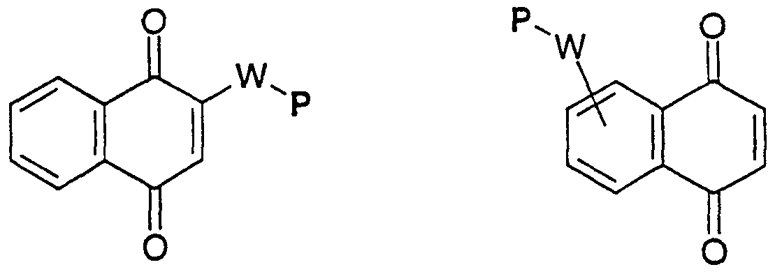


Claims
Priority Applications (7)
| Application Number | Priority Date | Filing Date | Title |
|---|---|---|---|
| AU32204/00A AU773914B2 (en) | 1999-02-01 | 2000-02-01 | Biomaterials formed by nucleophilic addition reaction to conjugated unsaturated groups |
| JP2000596061A JP4954370B2 (en) | 1999-02-01 | 2000-02-01 | Biocompatible materials made by nucleophilic addition reactions to conjugated unsaturated groups |
| EP00910049A EP1181323B1 (en) | 1999-02-01 | 2000-02-01 | Biomaterials formed by nucleophilic addition reaction to conjugated unsaturated groups |
| AT00910049T ATE514729T1 (en) | 1999-02-01 | 2000-02-01 | BIOMATERIALS ADDED BY NUCLEOPHILIC REACTION ON CONJUGATE UNSATURATED GROUPS |
| CA002359318A CA2359318C (en) | 1999-02-01 | 2000-02-01 | Biomaterials formed by nucleophilic addition reaction to conjugated unsaturated groups |
| MXPA01007777A MXPA01007777A (en) | 1999-02-01 | 2000-02-01 | Biomaterials formed by nucleophilic addition reaction to conjugated unsaturated groups. |
| DK00910049.6T DK1181323T3 (en) | 1999-02-01 | 2000-02-01 | Biomaterials formed by nucleophilic addition reaction with conjugated unsaturated groups |
Applications Claiming Priority (2)
| Application Number | Priority Date | Filing Date | Title |
|---|---|---|---|
| US11809399P | 1999-02-01 | 1999-02-01 | |
| US60/118,093 | 1999-02-01 |
Publications (1)
| Publication Number | Publication Date |
|---|---|
| WO2000044808A1 true WO2000044808A1 (en) | 2000-08-03 |
Family
ID=22376466
Family Applications (1)
| Application Number | Title | Priority Date | Filing Date |
|---|---|---|---|
| PCT/US2000/002608 WO2000044808A1 (en) | 1999-02-01 | 2000-02-01 | Biomaterials formed by nucleophilic addition reaction to conjugated unsaturated groups |
Country Status (10)
| Country | Link |
|---|---|
| US (1) | US7744912B1 (en) |
| EP (2) | EP1181323B1 (en) |
| JP (2) | JP4954370B2 (en) |
| AT (1) | ATE514729T1 (en) |
| AU (1) | AU773914B2 (en) |
| CA (1) | CA2359318C (en) |
| DK (1) | DK1181323T3 (en) |
| ES (2) | ES2632564T3 (en) |
| MX (1) | MXPA01007777A (en) |
| WO (1) | WO2000044808A1 (en) |
Cited By (102)
| Publication number | Priority date | Publication date | Assignee | Title |
|---|---|---|---|---|
| EP1187859A1 (en) * | 1999-03-23 | 2002-03-20 | The University Of Melbourne | Polymer gels, methods for their preparation and their use in electrophoretic applications |
| WO2002088331A1 (en) * | 2001-05-01 | 2002-11-07 | Universite Du Quebec A Montreal | Tridimensional biocompatible support structure for bioartificial organs and uses thereof |
| EP1292709A1 (en) * | 2000-06-02 | 2003-03-19 | Eidgenössische Technische Hochschule Zürich | Conjugate addition reactions for the controlled delivery of pharmaceutically active compounds |
| WO2003040235A1 (en) * | 2001-11-07 | 2003-05-15 | Universität Zürich | Synthetic matrix for controlled cell ingrowth and tissue regeneration |
| JP2004531534A (en) * | 2001-04-25 | 2004-10-14 | アイトゲネシッシェ テヒニッシェ ホーホシューレ チューリッヒ | Drug delivery matrix to promote wound healing |
| US6872799B2 (en) | 2002-12-18 | 2005-03-29 | Ethicon, Inc. | Functionalized polymers for medical applications |
| US6872384B1 (en) | 1998-02-23 | 2005-03-29 | Life Medical Sciences, Inc. | Treatment of trauma |
| JP2005102612A (en) * | 2003-09-30 | 2005-04-21 | Toyota Central Res & Dev Lab Inc | Method for implanting cell and method for preparing cellular tissue |
| WO2005042046A1 (en) | 2003-11-03 | 2005-05-12 | Medtronic, Inc. | Hydrogel providing cell-specific ingrowth |
| JP2005517658A (en) * | 2001-12-18 | 2005-06-16 | エイドゲントシッシュ テクニーシェ ホッシュール チューリッヒ | Growth factor-modified protein matrix for tissue manipulation |
| WO2005061701A1 (en) * | 2003-12-24 | 2005-07-07 | Shionogi & Co., Ltd. | Process for producing polymer composite of protein |
| EP1558215A1 (en) * | 2003-10-14 | 2005-08-03 | Wockhardt Limited | Sterile gelling agents |
| US6933328B2 (en) | 2000-04-03 | 2005-08-23 | Universiteit Gent | Composition of crosslinkable prepolymers for biodegradable implants |
| JP2005525851A (en) * | 2002-03-22 | 2005-09-02 | クロス、バイオサージェリー、アクチエンゲゼルシャフト | Composition for reinforcing hard tissue |
| US6943211B1 (en) | 1999-02-23 | 2005-09-13 | Life Medical Sciences, Inc. | Polymer compounds |
| US6958212B1 (en) | 1999-02-01 | 2005-10-25 | Eidgenossische Technische Hochschule Zurich | Conjugate addition reactions for the controlled delivery of pharmaceutically active compounds |
| US6967234B2 (en) | 2002-12-18 | 2005-11-22 | Ethicon, Inc. | Alkyd-lactone copolymers for medical applications |
| WO2006003014A2 (en) * | 2004-07-05 | 2006-01-12 | Complex Biosystems Gmbh | Hydrogel polymeric conjugates of a prodrug |
| EP1625856A1 (en) * | 2004-08-13 | 2006-02-15 | Complex Biosystems GmbH | Hydrogel polymeric conjugates of a prodrug |
| US7005136B2 (en) | 2002-03-29 | 2006-02-28 | Ethicon, Inc. | Bone replacement materials utilizing bioabsorbable liquid polymers |
| US7026374B2 (en) | 2002-06-25 | 2006-04-11 | Aruna Nathan | Injectable microdispersions for medical applications |
| US7030127B2 (en) | 2001-06-29 | 2006-04-18 | Ethicon, Inc. | Composition and medical devices utilizing bioabsorbable polymeric waxes |
| US7034037B2 (en) | 2001-06-29 | 2006-04-25 | Ethicon, Inc. | Compositions and medical devices utilizing bioabsorbable polymeric waxes and rapamycin |
| US7101566B2 (en) | 2002-06-28 | 2006-09-05 | Ethicon, Inc. | Polymer coated microparticles for sustained release |
| US7132475B2 (en) | 2000-10-19 | 2006-11-07 | Ecole Polytechnique Federale De Lausanne | Block copolymers for multifunctional self-assembled systems |
| EP1764404A1 (en) | 2005-09-16 | 2007-03-21 | Straumann Holding AG | Use of methylene blue as polymerization inhibitor |
| US7208145B2 (en) | 2002-12-31 | 2007-04-24 | Nektar Therapeutics Al, Corporation | Polymeric reagents comprising a ketone or a related functional group |
| EP1820522A1 (en) * | 2006-02-20 | 2007-08-22 | Straumann Holding AG | Granulate-matrix |
| US7282584B2 (en) | 2004-06-16 | 2007-10-16 | Straumann Holding Ag | Methylene blue |
| US7291673B2 (en) | 2000-06-02 | 2007-11-06 | Eidgenossiche Technische Hochschule Zurich | Conjugate addition reactions for the controlled delivery of pharmaceutically active compounds |
| WO2007128059A1 (en) * | 2006-05-05 | 2007-11-15 | The University Of Sydney | Bis-pyridinium compounds |
| EP1862170A1 (en) | 2004-12-10 | 2007-12-05 | Straumann Holding AG | Protein formulation |
| US7326426B2 (en) | 2002-03-29 | 2008-02-05 | Ethicon, Inc. | Compositions and medical devices utilizing bioabsorbable liquid polymers |
| WO2008051864A2 (en) * | 2006-10-24 | 2008-05-02 | Neville Alleyne | Method of treating spinal internal disk derangement |
| US7368125B2 (en) | 2002-06-05 | 2008-05-06 | Ethicon, Inc. | Amphiphilic polymers for medical applications |
| US7371719B2 (en) | 2002-02-15 | 2008-05-13 | Northwestern University | Self-assembly of peptide-amphiphile nanofibers under physiological conditions |
| US7390526B2 (en) | 2003-02-11 | 2008-06-24 | Northwestern University | Methods and materials for nanocrystalline surface coatings and attachment of peptide amphiphile nanofibers thereon |
| DE202008005131U1 (en) | 2008-04-14 | 2008-08-28 | Eidgenössische Technische Hochschule Zürich | Synthetic matrix for the controlled ingrowth of cells and tissue regeneration |
| US7423013B2 (en) | 2004-12-10 | 2008-09-09 | Straumann Holding Ag | Protein formulation |
| WO2008136536A1 (en) | 2007-05-01 | 2008-11-13 | National University Corporation Tokyo Medical And Dental University | Hybrid gel comprising chemically cross-linked hyaluronic acid derivative, and pharmaceutical composition using the same |
| US7452679B2 (en) | 2003-12-05 | 2008-11-18 | Northwestern University | Branched peptide amphiphiles, related epitope compounds and self assembled structures thereof |
| EP2014256A1 (en) * | 2007-07-12 | 2009-01-14 | Straumann Holding AG | Composite bone repair material |
| US7491690B2 (en) * | 2001-11-14 | 2009-02-17 | Northwestern University | Self-assembly and mineralization of peptide-amphiphile nanofibers |
| US7534761B1 (en) | 2002-08-21 | 2009-05-19 | North Western University | Charged peptide-amphiphile solutions and self-assembled peptide nanofiber networks formed therefrom |
| US7544661B2 (en) | 2003-12-05 | 2009-06-09 | Northwestern University | Self-assembling peptide amphiphiles and related methods for growth factor delivery |
| US7554021B2 (en) | 2002-11-12 | 2009-06-30 | Northwestern University | Composition and method for self-assembly and mineralization of peptide amphiphiles |
| US7632291B2 (en) | 2003-06-13 | 2009-12-15 | Trivascular2, Inc. | Inflatable implant |
| EP2111872A3 (en) * | 2004-07-05 | 2009-12-23 | Complex Biosystems GmbH | Hydrogel polymeric conjugates of a prodrug |
| US7683025B2 (en) | 2002-11-14 | 2010-03-23 | Northwestern University | Synthesis and self-assembly of ABC triblock bola peptide amphiphiles |
| US20100111917A1 (en) * | 2006-03-21 | 2010-05-06 | Ying Jackie Y | Cell-adhesive polyelectrolyte material for use as membrane and coating |
| US7741427B2 (en) | 2004-06-16 | 2010-06-22 | Straumann Holding Ag | Barrier membrane |
| US7744912B1 (en) | 1999-02-01 | 2010-06-29 | Eidgenossische Technische Hochschule Zurich | Biomaterials formed by nucleophilic addition reaction to conjugated unsaturated groups |
| US7803178B2 (en) | 2004-01-30 | 2010-09-28 | Trivascular, Inc. | Inflatable porous implants and methods for drug delivery |
| US7816316B2 (en) | 2002-11-21 | 2010-10-19 | Chugai Seiyaku Kabushiki Kaisha | Sustained release drug carrier |
| US7851445B2 (en) | 2005-03-04 | 2010-12-14 | Northwestern University | Angiogenic heparin-binding epitopes, peptide amphiphiles, self-assembled compositions and related methods of use |
| WO2011004255A2 (en) | 2009-07-10 | 2011-01-13 | Kuros Biosurgery Ag | Compositions for tissue augmentation |
| US7883694B2 (en) | 1995-12-18 | 2011-02-08 | Angiodevice International Gmbh | Method for preventing the formation of adhesions following surgery or injury |
| EP2329832A1 (en) | 2004-12-10 | 2011-06-08 | Straumann Holding AG | Protein formulation |
| US8034618B2 (en) | 2001-12-18 | 2011-10-11 | Eldgenossische Technische Hochschule Zurich | PTH containing cell growth matrix |
| EP2380920A1 (en) | 2010-04-22 | 2011-10-26 | QGel SA | Hydrogel precursor formulation and production process thereof |
| US8067031B2 (en) | 2004-04-28 | 2011-11-29 | Angiodevice International Gmbh | Compositions and systems for forming crosslinked biomaterials and associated methods of preparation and use |
| US8076295B2 (en) | 2007-04-17 | 2011-12-13 | Nanotope, Inc. | Peptide amphiphiles having improved solubility and methods of using same |
| EP2418233A1 (en) * | 2010-08-14 | 2012-02-15 | Fraunhofer-Gesellschaft zur Förderung der angewandten Forschung e.V. | Polymer-supported culture systems |
| US8127770B2 (en) | 2004-08-30 | 2012-03-06 | Spineovations, Inc. | Method of using an implant for treament of ligaments and tendons |
| US20120156289A1 (en) * | 2010-12-21 | 2012-06-21 | Confluent Surgical, Inc. | Biodegradable Osmotic Pump Implant For Drug Delivery |
| US8226942B2 (en) | 2007-12-28 | 2012-07-24 | Kuros Biosurgery Ag | PDGF fusion proteins incorporated into fibrin foams |
| WO2012123028A1 (en) | 2011-03-16 | 2012-09-20 | Kuros Biosurgery Ag | Pharmaceutical formulation for use in spinal fusion |
| US8318674B2 (en) | 2005-01-06 | 2012-11-27 | Kuros Biosurgery Ag | Local treatment of bone defects |
| US8398638B2 (en) | 2004-08-30 | 2013-03-19 | Spineovations, Inc. | Method of treating spinal internal disk derangement |
| US8450271B2 (en) | 2009-04-13 | 2013-05-28 | Northwestern University | Peptide-based scaffolds for cartilage regeneration and methods for their use |
| US8469961B2 (en) | 2008-03-05 | 2013-06-25 | Neville Alleyne | Methods and compositions for minimally invasive capsular augmentation of canine coxofemoral joints |
| US8575332B2 (en) | 2003-11-14 | 2013-11-05 | Chugai Seiyaku Kabushiki Kaisha | Crosslinked polysaccharide microparticles and method for their preparation |
| US8575101B2 (en) | 2005-01-06 | 2013-11-05 | Kuros Biosurgery Ag | Supplemented matrices for the repair of bone fractures |
| US8617584B2 (en) | 1995-12-18 | 2013-12-31 | Angiodevice International Gmbh | Adhesive tissue repair patch and collagen sheets |
| WO2014070973A1 (en) * | 2012-11-02 | 2014-05-08 | Board Of Supervisors Of Louisiana State University And Agricural And Mechanical College | Thiol acrylate nanocomposite foams |
| US8961947B2 (en) | 2007-04-13 | 2015-02-24 | Kuros Biosurgery Ag | Polymeric tissue sealant |
| US8992595B2 (en) | 2012-04-04 | 2015-03-31 | Trivascular, Inc. | Durable stent graft with tapered struts and stable delivery methods and devices |
| WO2015008152A3 (en) * | 2013-05-31 | 2015-05-28 | M-S Medical Sagl | Methods and compositions for blood vessel tissue repair and engineering |
| US9133276B2 (en) | 2010-09-17 | 2015-09-15 | Sanofi-Aventis Deutschland Gmbh | Prodrugs comprising an exendin linker conjugate |
| US9138462B2 (en) | 2009-07-31 | 2015-09-22 | Sanofi-Aventis Deutschland Gmbh | Prodrugs comprising an insulin linker conjugate |
| US9198568B2 (en) | 2010-03-04 | 2015-12-01 | The General Hospital Corporation | Methods and systems of matching voice deficits with a tunable mucosal implant to restore and enhance individualized human sound and voice production |
| US9216188B2 (en) | 2008-09-04 | 2015-12-22 | The General Hospital Corporation | Hydrogels for vocal cord and soft tissue augmentation and repair |
| WO2016007586A1 (en) | 2014-07-10 | 2016-01-14 | Cambridge Polymer Group, Inc. | Thiolated peg-pva hydrogels |
| US9265723B2 (en) | 2009-07-31 | 2016-02-23 | Sanofi-Aventis Deutschland Gmbh | Long acting insulin composition |
| US9271929B2 (en) | 2008-11-25 | 2016-03-01 | École Polytechnique Fédérale De Lausanne (Epfl) | Block copolymers and uses thereof |
| US9353218B2 (en) | 2004-09-17 | 2016-05-31 | Angiotech Pharmaceuticals, Inc. | Kit for multifunctional compounds forming crosslinked biomaterials |
| US9498363B2 (en) | 2012-04-06 | 2016-11-22 | Trivascular, Inc. | Delivery catheter for endovascular device |
| US9505867B2 (en) | 2005-05-31 | 2016-11-29 | Ecole Polytechmique Fédérale De Lausanne | Triblock copolymers for cytoplasmic delivery of gene-based drugs |
| US9701940B2 (en) | 2005-09-19 | 2017-07-11 | Histogenics Corporation | Cell-support matrix having narrowly defined uniformly vertically and non-randomly organized porosity and pore density and a method for preparation thereof |
| EP3281605A1 (en) | 2003-08-20 | 2018-02-14 | Histogenics Corporation | Acellular matrix implants for treatment of articular cartilage, bone or osteochondral defects and injuries and a method for use thereof |
| US10058837B2 (en) | 2009-08-28 | 2018-08-28 | The Trustees Of Columbia University In The City Of New York | Systems, methods, and devices for production of gas-filled microbubbles |
| US10077420B2 (en) | 2014-12-02 | 2018-09-18 | Histogenics Corporation | Cell and tissue culture container |
| US10159557B2 (en) | 2007-10-04 | 2018-12-25 | Trivascular, Inc. | Modular vascular graft for low profile percutaneous delivery |
| US10279080B2 (en) | 2015-01-16 | 2019-05-07 | Spineovations, Inc. | Method of treating spinal disk |
| EP3620188A1 (en) * | 2005-04-01 | 2020-03-11 | TriVascular, Inc. | A non-degradable, low swelling, water soluble, radiopaque hyrdrogel polymer |
| US10711106B2 (en) | 2013-07-25 | 2020-07-14 | The University Of Chicago | High aspect ratio nanofibril materials |
| WO2020156968A2 (en) | 2019-01-30 | 2020-08-06 | Ventana Medical Systems, Inc. | Calibration slides for digital pathology |
| US10959761B2 (en) | 2015-09-18 | 2021-03-30 | Ortho-Space Ltd. | Intramedullary fixated subacromial spacers |
| WO2021053400A3 (en) * | 2019-09-21 | 2021-05-14 | Aratinga.Bio Tnp | Dmso-free synthesis of oligopeptide-modified poly(beta-amino ester)s and their use in nanoparticle delivery systems |
| US11033398B2 (en) | 2007-03-15 | 2021-06-15 | Ortho-Space Ltd. | Shoulder implant for simulating a bursa |
| US11045981B2 (en) | 2017-01-30 | 2021-06-29 | Ortho-Space Ltd. | Processing machine and methods for processing dip-molded articles |
| US11826228B2 (en) | 2011-10-18 | 2023-11-28 | Stryker European Operations Limited | Prosthetic devices |
Families Citing this family (35)
| Publication number | Priority date | Publication date | Assignee | Title |
|---|---|---|---|---|
| EP1245272A1 (en) * | 2001-03-30 | 2002-10-02 | Ucb S.A. | Substrates, preparation and use |
| WO2003031483A1 (en) * | 2001-10-10 | 2003-04-17 | The Regents Of The University Of Colorado | Degradable thiol-ene polymers |
| US20070264227A1 (en) * | 2001-11-07 | 2007-11-15 | Eidgenossische Technische Hochschule Zurich | Synthetic Matrix for Controlled Cell Ingrowth and Tissue Regeneration |
| WO2003053288A1 (en) | 2001-12-20 | 2003-07-03 | Trivascular, Inc. | Advanced endovascular graft |
| US20050090804A1 (en) * | 2003-10-22 | 2005-04-28 | Trivascular, Inc. | Endoluminal prosthesis endoleak management |
| CA2552649A1 (en) * | 2004-01-07 | 2005-07-28 | Boston Scientific Santa Rosa Corporation | Methods, compositions, and devices for embolizing body lumens |
| EP2422751A3 (en) | 2004-05-05 | 2013-01-02 | Direct Flow Medical, Inc. | Unstented heart valve with formed in place support structure |
| WO2006133294A2 (en) | 2005-06-07 | 2006-12-14 | Direct Flow Medical, Inc. | Stentless aortic valve replacement with high radial strength |
| US7935144B2 (en) | 2006-10-19 | 2011-05-03 | Direct Flow Medical, Inc. | Profile reduction of valve implant |
| US8133213B2 (en) | 2006-10-19 | 2012-03-13 | Direct Flow Medical, Inc. | Catheter guidance through a calcified aortic valve |
| AR064286A1 (en) * | 2006-12-13 | 2009-03-25 | Quiceno Gomez Alexandra Lorena | PRODUCTION OF OPHTHALMIC DEVICES BASED ON POLYMERIZATION BY PHOTOINDUCIDED SCALE GROWTH |
| EP2190379B1 (en) | 2007-08-23 | 2016-06-15 | Direct Flow Medical, Inc. | Translumenally implantable heart valve with formed in place support |
| AU2011206908B2 (en) * | 2010-01-13 | 2014-09-04 | Georg Bischof | Composition for producing a temporary intestinal occlusion |
| US8962823B2 (en) | 2010-01-19 | 2015-02-24 | Organoclick Ab | Heterogeneous thiol-ene click modifications of solid polysaccharide-based materials |
| US9603708B2 (en) | 2010-05-19 | 2017-03-28 | Dfm, Llc | Low crossing profile delivery catheter for cardiovascular prosthetic implant |
| CN102375065A (en) * | 2010-05-27 | 2012-03-14 | 杭州景杰生物科技有限公司 | Identifying Method For Acylation Of Lysine Butane In Protein And Preparation Of Affine Reagent Thereof |
| US20130224880A1 (en) | 2011-05-27 | 2013-08-29 | Ptm Biolabs, Inc. | Reagents and methods for detecting protein crotonylation |
| CN103189015B (en) | 2010-07-09 | 2016-07-06 | 海莱夫简易股份公司 | Transcatheter atrioventricular valves (A V valves) prosthese |
| US20140038826A1 (en) | 2011-01-28 | 2014-02-06 | The Regents Of The University Of Colorado, A Body Corporate | Covalently cross linked hydrogels and methods of making and using same |
| EP2720722A4 (en) | 2011-06-16 | 2014-12-03 | Univ Hong Kong Science & Techn | Multi-vinylsulfone containing molecule |
| US9988433B2 (en) | 2013-03-14 | 2018-06-05 | Mosaic Biosciences, Inc. | Covalent modification of biological macromolecules |
| EP2822533B1 (en) | 2012-02-02 | 2021-01-20 | Mosaic Biosciences, Inc. | Biomaterials for delivery of blood extracts and methods of using same |
| US9993328B2 (en) | 2012-04-03 | 2018-06-12 | Trivascular, Inc. | Advanced kink resistant stent graft |
| US9445897B2 (en) | 2012-05-01 | 2016-09-20 | Direct Flow Medical, Inc. | Prosthetic implant delivery device with introducer catheter |
| US9540428B2 (en) | 2012-07-03 | 2017-01-10 | Ecole Polytechnique Federale De Lausanne (Epfl) | Extracellular matrix heparin-binding domains |
| US9879062B2 (en) | 2012-07-03 | 2018-01-30 | Ecole Polytechnique Federale De Lausanne | Protein-binding peptide isolated from placenta growth factor |
| US9395468B2 (en) | 2012-08-27 | 2016-07-19 | Ocular Dynamics, Llc | Contact lens with a hydrophilic layer |
| US9724186B2 (en) | 2012-10-10 | 2017-08-08 | Trivascular, Inc. | Endovascular graft for aneurysms involving major branch vessels |
| WO2014190317A2 (en) | 2013-05-24 | 2014-11-27 | Cedars-Sinai Medical Center | Left atrial appendage occlusion devices and methods |
| US11123205B2 (en) | 2013-09-24 | 2021-09-21 | Trivascular, Inc. | Tandem modular endograft |
| EP3570093B1 (en) | 2013-11-15 | 2021-09-15 | Tangible Science, Inc. | Contact lens with a hydrophilic layer |
| US10525170B2 (en) | 2014-12-09 | 2020-01-07 | Tangible Science, Llc | Medical device coating with a biocompatible layer |
| WO2016130573A2 (en) | 2015-02-09 | 2016-08-18 | Mosaic Biosciences, Inc. | Degradable thiol-ene polymers and methods of making thereof |
| WO2018021331A1 (en) * | 2016-07-27 | 2018-02-01 | 東レ株式会社 | Resin composition |
| WO2020131871A1 (en) * | 2018-12-19 | 2020-06-25 | Tangible Science, Inc. | Systems and methods of treating a hydrogel-coated medical device |
Citations (3)
| Publication number | Priority date | Publication date | Assignee | Title |
|---|---|---|---|---|
| US5268305A (en) * | 1989-06-15 | 1993-12-07 | Biocircuits Corporation | Multi-optical detection system |
| US5567422A (en) * | 1993-02-02 | 1996-10-22 | Enzon, Inc. | Azlactone activated polyalkylene oxides conjugated to biologically active nucleophiles |
| US5635207A (en) * | 1993-02-22 | 1997-06-03 | Vivorx Pharmaceuticals, Inc. | Methods for the preparation of blood substitutes for in vivo delivery |
Family Cites Families (39)
| Publication number | Priority date | Publication date | Assignee | Title |
|---|---|---|---|---|
| CA992105A (en) | 1970-03-26 | 1976-06-29 | Exxon Research And Engineering Company | Polythioethers formed by anionic ring opening of episulfides |
| JPS6198734A (en) * | 1984-10-19 | 1986-05-17 | Showa Denko Kk | Curable composition |
| US4618400A (en) | 1984-12-28 | 1986-10-21 | Shell Oil Company | Wastepaper deinking process |
| US4711903A (en) * | 1986-01-31 | 1987-12-08 | G. D. Searle & Co. | Phenolic thioethers as inhibitors of 5-lipoxygenase |
| US5575815A (en) | 1988-08-24 | 1996-11-19 | Endoluminal Therapeutics, Inc. | Local polymeric gel therapy |
| US5247041A (en) * | 1989-02-22 | 1993-09-21 | Toray Industries, Inc. | Thiol methacrylate or acrylate resin obtained by polymerizing a thiol methacrylate or acrylate compound |
| US5330911A (en) | 1989-09-28 | 1994-07-19 | Board Of Regents, The University Of Texas System | Surfaces having desirable cell adhesive effects |
| US5852182A (en) | 1990-01-11 | 1998-12-22 | Isis Pharmaceuticals Inc. | Thiol-derivatized oligonucleosides |
| US5410016A (en) | 1990-10-15 | 1995-04-25 | Board Of Regents, The University Of Texas System | Photopolymerizable biodegradable hydrogels as tissue contacting materials and controlled-release carriers |
| US5529914A (en) | 1990-10-15 | 1996-06-25 | The Board Of Regents The Univeristy Of Texas System | Gels for encapsulation of biological materials |
| US5573934A (en) | 1992-04-20 | 1996-11-12 | Board Of Regents, The University Of Texas System | Gels for encapsulation of biological materials |
| WO1993024476A1 (en) | 1992-06-04 | 1993-12-09 | Clover Consolidated, Limited | Water-soluble polymeric carriers for drug delivery |
| US5292514A (en) * | 1992-06-24 | 1994-03-08 | Minnesota Mining And Manufacturing Company | Azlactone-functional substrates, corneal prostheses, and manufacture and use thereof |
| DE69316223T2 (en) | 1992-08-25 | 1998-05-28 | Searle & Co | SULFONYLALKANOYLAMINOHYDROXYETHYLAMINOSULFONAMIDES USED AS RETROVIRAL PROTEASE INHIBITORS |
| WO1994005282A1 (en) | 1992-09-04 | 1994-03-17 | The Scripps Research Institute | Water soluble taxol derivatives |
| US5428014A (en) | 1993-08-13 | 1995-06-27 | Zymogenetics, Inc. | Transglutaminase cross-linkable polypeptides and methods relating thereto |
| US5880131A (en) | 1993-10-20 | 1999-03-09 | Enzon, Inc. | High molecular weight polymer-based prodrugs |
| US5446090A (en) | 1993-11-12 | 1995-08-29 | Shearwater Polymers, Inc. | Isolatable, water soluble, and hydrolytically stable active sulfones of poly(ethylene glycol) and related polymers for modification of surfaces and molecules |
| JPH0848770A (en) * | 1994-06-03 | 1996-02-20 | Toyo Ink Mfg Co Ltd | Liquid resin composition, its production and curing resin composition |
| JPH0892369A (en) * | 1994-07-29 | 1996-04-09 | Toyo Ink Mfg Co Ltd | Curable liquid resin composition and production of cured product therefrom |
| US5932462A (en) | 1995-01-10 | 1999-08-03 | Shearwater Polymers, Inc. | Multiarmed, monofunctional, polymer for coupling to molecules and surfaces |
| US5702717A (en) | 1995-10-25 | 1997-12-30 | Macromed, Inc. | Thermosensitive biodegradable polymers based on poly(ether-ester)block copolymers |
| AU7566396A (en) | 1995-11-16 | 1997-06-05 | Boehringer Mannheim Gmbh | Process for the preparation of peptides by way of streptavidin fusion proteins |
| US5752974A (en) | 1995-12-18 | 1998-05-19 | Collagen Corporation | Injectable or implantable biomaterials for filling or blocking lumens and voids of the body |
| PT1704878E (en) | 1995-12-18 | 2013-07-17 | Angiodevice Internat Gmbh | Crosslinked polymer compositions and methods for their use |
| US5914182A (en) | 1996-06-03 | 1999-06-22 | Gore Hybrid Technologies, Inc. | Materials and methods for the immobilization of bioactive species onto polymeric substrates |
| US20020064546A1 (en) | 1996-09-13 | 2002-05-30 | J. Milton Harris | Degradable poly(ethylene glycol) hydrogels with controlled half-life and precursors therefor |
| US6258351B1 (en) | 1996-11-06 | 2001-07-10 | Shearwater Corporation | Delivery of poly(ethylene glycol)-modified molecules from degradable hydrogels |
| ATE200030T1 (en) | 1997-01-29 | 2001-04-15 | Polymasc Pharmaceuticals Plc | PEGYLATION PROCEDURE |
| DE19705963A1 (en) | 1997-02-17 | 1998-08-20 | Hoechst Ag | Crosslinked vinyl polymers with bile acid adsorber effect |
| DK1053019T3 (en) | 1998-01-07 | 2004-04-13 | Debio Rech Pharma Sa | Degradable heterobifunctional polyethylene glycol acrylates and gels and conjugates derived therefrom |
| US5958874A (en) | 1998-02-18 | 1999-09-28 | The Research Foundation Of State University Of New York | Recombinant fibronectin-based extracellular matrix for wound healing |
| US6514534B1 (en) | 1998-08-14 | 2003-02-04 | Incept Llc | Methods for forming regional tissue adherent barriers and drug delivery systems |
| JP4954370B2 (en) | 1999-02-01 | 2012-06-13 | エイドジェノシスク テクニスク ホクシューレ チューリッヒ | Biocompatible materials made by nucleophilic addition reactions to conjugated unsaturated groups |
| PE20010288A1 (en) | 1999-07-02 | 2001-03-07 | Hoffmann La Roche | ERYTHROPOYETIN DERIVATIVES |
| US7291673B2 (en) * | 2000-06-02 | 2007-11-06 | Eidgenossiche Technische Hochschule Zurich | Conjugate addition reactions for the controlled delivery of pharmaceutically active compounds |
| WO2002055185A2 (en) | 2000-10-19 | 2002-07-18 | Eidgenoess Tech Hochschule | Block copolymers for multifunctional self-assembled systems |
| EP1379133A2 (en) | 2001-03-20 | 2004-01-14 | Eidgenössische Technische Hochschule Zürich | Two-phase processing of thermosensitive polymers for use as biomaterials |
| EP1446453A1 (en) | 2001-11-07 | 2004-08-18 | Universität Zürich | Synthetic matrix for controlled cell ingrowth and tissue regeneration and endometriosis |
-
2000
- 2000-02-01 JP JP2000596061A patent/JP4954370B2/en not_active Expired - Lifetime
- 2000-02-01 WO PCT/US2000/002608 patent/WO2000044808A1/en active IP Right Grant
- 2000-02-01 EP EP00910049A patent/EP1181323B1/en not_active Expired - Lifetime
- 2000-02-01 DK DK00910049.6T patent/DK1181323T3/en active
- 2000-02-01 CA CA002359318A patent/CA2359318C/en not_active Expired - Lifetime
- 2000-02-01 US US09/496,231 patent/US7744912B1/en not_active Expired - Fee Related
- 2000-02-01 EP EP10181654.4A patent/EP2311895B1/en not_active Expired - Lifetime
- 2000-02-01 AT AT00910049T patent/ATE514729T1/en active
- 2000-02-01 ES ES10181654.4T patent/ES2632564T3/en not_active Expired - Lifetime
- 2000-02-01 AU AU32204/00A patent/AU773914B2/en not_active Expired
- 2000-02-01 ES ES00910049T patent/ES2368988T3/en not_active Expired - Lifetime
- 2000-02-01 MX MXPA01007777A patent/MXPA01007777A/en active IP Right Grant
-
2011
- 2011-12-26 JP JP2011282597A patent/JP2012101095A/en active Pending
Patent Citations (4)
| Publication number | Priority date | Publication date | Assignee | Title |
|---|---|---|---|---|
| US5268305A (en) * | 1989-06-15 | 1993-12-07 | Biocircuits Corporation | Multi-optical detection system |
| US5427915A (en) * | 1989-06-15 | 1995-06-27 | Biocircuits Corporation | Multi-optical detection system |
| US5567422A (en) * | 1993-02-02 | 1996-10-22 | Enzon, Inc. | Azlactone activated polyalkylene oxides conjugated to biologically active nucleophiles |
| US5635207A (en) * | 1993-02-22 | 1997-06-03 | Vivorx Pharmaceuticals, Inc. | Methods for the preparation of blood substitutes for in vivo delivery |
Cited By (164)
| Publication number | Priority date | Publication date | Assignee | Title |
|---|---|---|---|---|
| US8197802B2 (en) | 1995-12-18 | 2012-06-12 | Angiodevice International Gmbh | Method for treating or inhibiting the formation of adhesions following surgery or injury |
| US8617584B2 (en) | 1995-12-18 | 2013-12-31 | Angiodevice International Gmbh | Adhesive tissue repair patch and collagen sheets |
| US7883694B2 (en) | 1995-12-18 | 2011-02-08 | Angiodevice International Gmbh | Method for preventing the formation of adhesions following surgery or injury |
| US6872384B1 (en) | 1998-02-23 | 2005-03-29 | Life Medical Sciences, Inc. | Treatment of trauma |
| US7670605B2 (en) | 1999-02-01 | 2010-03-02 | Eidgenossische Technische Hochschule Zurich | Conjugate addition reactions for the controlled delivery of pharmaceutically active compounds |
| US7413739B2 (en) | 1999-02-01 | 2008-08-19 | Eidgenossische Technische Hochschule Zurich | Conjugate addition reactions for the controlled delivery of pharmaceutically active compounds |
| US6958212B1 (en) | 1999-02-01 | 2005-10-25 | Eidgenossische Technische Hochschule Zurich | Conjugate addition reactions for the controlled delivery of pharmaceutically active compounds |
| US7744912B1 (en) | 1999-02-01 | 2010-06-29 | Eidgenossische Technische Hochschule Zurich | Biomaterials formed by nucleophilic addition reaction to conjugated unsaturated groups |
| US6943211B1 (en) | 1999-02-23 | 2005-09-13 | Life Medical Sciences, Inc. | Polymer compounds |
| EP1187859A4 (en) * | 1999-03-23 | 2005-03-23 | Univ Melbourne | Polymer gels, methods for their preparation and their use in electrophoretic applications |
| EP1187859A1 (en) * | 1999-03-23 | 2002-03-20 | The University Of Melbourne | Polymer gels, methods for their preparation and their use in electrophoretic applications |
| US6933328B2 (en) | 2000-04-03 | 2005-08-23 | Universiteit Gent | Composition of crosslinkable prepolymers for biodegradable implants |
| US7291673B2 (en) | 2000-06-02 | 2007-11-06 | Eidgenossiche Technische Hochschule Zurich | Conjugate addition reactions for the controlled delivery of pharmaceutically active compounds |
| EP1292709A4 (en) * | 2000-06-02 | 2004-09-22 | Eidgenoess Tech Hochschule | Conjugate addition reactions for the controlled delivery of pharmaceutically active compounds |
| EP1292709A1 (en) * | 2000-06-02 | 2003-03-19 | Eidgenössische Technische Hochschule Zürich | Conjugate addition reactions for the controlled delivery of pharmaceutically active compounds |
| EP2363424A1 (en) * | 2000-10-19 | 2011-09-07 | Ecole Polytechnique Fédérale de Lausanne (EPFL) | Block copolymers for multifunctional self-assembled systems |
| US7132475B2 (en) | 2000-10-19 | 2006-11-07 | Ecole Polytechnique Federale De Lausanne | Block copolymers for multifunctional self-assembled systems |
| US8309518B2 (en) | 2001-04-25 | 2012-11-13 | Eidgenossische Technische Hochschule Zurich | Drug delivery matrices to enhance wound healing |
| JP2004531534A (en) * | 2001-04-25 | 2004-10-14 | アイトゲネシッシェ テヒニッシェ ホーホシューレ チューリッヒ | Drug delivery matrix to promote wound healing |
| WO2002088331A1 (en) * | 2001-05-01 | 2002-11-07 | Universite Du Quebec A Montreal | Tridimensional biocompatible support structure for bioartificial organs and uses thereof |
| US7030127B2 (en) | 2001-06-29 | 2006-04-18 | Ethicon, Inc. | Composition and medical devices utilizing bioabsorbable polymeric waxes |
| US7034037B2 (en) | 2001-06-29 | 2006-04-25 | Ethicon, Inc. | Compositions and medical devices utilizing bioabsorbable polymeric waxes and rapamycin |
| JP2005508237A (en) * | 2001-11-07 | 2005-03-31 | ユニバーシテート チューリッヒ | Synthetic matrix for controlled cell ingrowth and tissue regeneration |
| KR101054023B1 (en) * | 2001-11-07 | 2011-08-04 | 아이드게노시셰 테크시셰 호흐쉘 취리히 | Synthetic Matrix to Control Intracellular Proliferation and Tissue Regeneration |
| WO2003040235A1 (en) * | 2001-11-07 | 2003-05-15 | Universität Zürich | Synthetic matrix for controlled cell ingrowth and tissue regeneration |
| CN1328319C (en) * | 2001-11-07 | 2007-07-25 | 苏黎世大学 | Synthetic matrix for controlled cell ingrowth and tissue regeneration |
| US7491690B2 (en) * | 2001-11-14 | 2009-02-17 | Northwestern University | Self-assembly and mineralization of peptide-amphiphile nanofibers |
| US7838491B2 (en) | 2001-11-14 | 2010-11-23 | Northwestern University | Self-assembly and mineralization of peptide-amphiphile nanofibers |
| JP2005517658A (en) * | 2001-12-18 | 2005-06-16 | エイドゲントシッシュ テクニーシェ ホッシュール チューリッヒ | Growth factor-modified protein matrix for tissue manipulation |
| US8034618B2 (en) | 2001-12-18 | 2011-10-11 | Eldgenossische Technische Hochschule Zurich | PTH containing cell growth matrix |
| US7371719B2 (en) | 2002-02-15 | 2008-05-13 | Northwestern University | Self-assembly of peptide-amphiphile nanofibers under physiological conditions |
| US8063014B2 (en) | 2002-02-15 | 2011-11-22 | Northwestern University | Self-assembly of peptide-amphiphile nanofibers under physiological conditions |
| US7745708B2 (en) | 2002-02-15 | 2010-06-29 | Northwestern University | Self-assembly of peptide-amphiphile nanofibers under physiological conditions |
| JP2005525851A (en) * | 2002-03-22 | 2005-09-02 | クロス、バイオサージェリー、アクチエンゲゼルシャフト | Composition for reinforcing hard tissue |
| US7575740B2 (en) * | 2002-03-22 | 2009-08-18 | Kuros Biosurgery Ag | Compositions for tissue augmentation |
| US8282912B2 (en) * | 2002-03-22 | 2012-10-09 | Kuros Biosurgery, AG | Compositions for tissue augmentation |
| AU2003226688B2 (en) * | 2002-03-22 | 2009-08-13 | Kuros Biosurgery Ag | Composition for hard tissue augmentation |
| US7005136B2 (en) | 2002-03-29 | 2006-02-28 | Ethicon, Inc. | Bone replacement materials utilizing bioabsorbable liquid polymers |
| US8623413B2 (en) | 2002-03-29 | 2014-01-07 | Ethicon, Inc. | Compositions and medical devices utilizing bioabsorbable liquid polymers |
| US7326426B2 (en) | 2002-03-29 | 2008-02-05 | Ethicon, Inc. | Compositions and medical devices utilizing bioabsorbable liquid polymers |
| US7368125B2 (en) | 2002-06-05 | 2008-05-06 | Ethicon, Inc. | Amphiphilic polymers for medical applications |
| US7026374B2 (en) | 2002-06-25 | 2006-04-11 | Aruna Nathan | Injectable microdispersions for medical applications |
| US7101566B2 (en) | 2002-06-28 | 2006-09-05 | Ethicon, Inc. | Polymer coated microparticles for sustained release |
| US7534761B1 (en) | 2002-08-21 | 2009-05-19 | North Western University | Charged peptide-amphiphile solutions and self-assembled peptide nanofiber networks formed therefrom |
| US7554021B2 (en) | 2002-11-12 | 2009-06-30 | Northwestern University | Composition and method for self-assembly and mineralization of peptide amphiphiles |
| US8124583B2 (en) | 2002-11-12 | 2012-02-28 | Northwestern University | Composition and method for self-assembly and mineralization of peptide-amphiphiles |
| US7683025B2 (en) | 2002-11-14 | 2010-03-23 | Northwestern University | Synthesis and self-assembly of ABC triblock bola peptide amphiphiles |
| US7816316B2 (en) | 2002-11-21 | 2010-10-19 | Chugai Seiyaku Kabushiki Kaisha | Sustained release drug carrier |
| US6967234B2 (en) | 2002-12-18 | 2005-11-22 | Ethicon, Inc. | Alkyd-lactone copolymers for medical applications |
| US6872799B2 (en) | 2002-12-18 | 2005-03-29 | Ethicon, Inc. | Functionalized polymers for medical applications |
| US8865149B2 (en) | 2002-12-31 | 2014-10-21 | Nektar Therapeutics | Polymeric reagents comprising a ketone or a related functional group |
| US7208145B2 (en) | 2002-12-31 | 2007-04-24 | Nektar Therapeutics Al, Corporation | Polymeric reagents comprising a ketone or a related functional group |
| US7390526B2 (en) | 2003-02-11 | 2008-06-24 | Northwestern University | Methods and materials for nanocrystalline surface coatings and attachment of peptide amphiphile nanofibers thereon |
| US10201350B2 (en) | 2003-06-13 | 2019-02-12 | Trivascular, Inc. | Inflatable implant |
| EP2436340A1 (en) | 2003-06-13 | 2012-04-04 | TriVascular, Inc. | Inflatable implant |
| US7632291B2 (en) | 2003-06-13 | 2009-12-15 | Trivascular2, Inc. | Inflatable implant |
| US11000288B2 (en) | 2003-06-13 | 2021-05-11 | Trivascular, Inc. | Inflatable implant |
| EP3281605A1 (en) | 2003-08-20 | 2018-02-14 | Histogenics Corporation | Acellular matrix implants for treatment of articular cartilage, bone or osteochondral defects and injuries and a method for use thereof |
| JP4742491B2 (en) * | 2003-09-30 | 2011-08-10 | 株式会社豊田中央研究所 | Cell implantation method and cell tissue preparation method |
| JP2005102612A (en) * | 2003-09-30 | 2005-04-21 | Toyota Central Res & Dev Lab Inc | Method for implanting cell and method for preparing cellular tissue |
| EP1558215A4 (en) * | 2003-10-14 | 2007-09-12 | Wockhardt Ltd | Sterile gelling agents |
| EP1558215A1 (en) * | 2003-10-14 | 2005-08-03 | Wockhardt Limited | Sterile gelling agents |
| WO2005042046A1 (en) | 2003-11-03 | 2005-05-12 | Medtronic, Inc. | Hydrogel providing cell-specific ingrowth |
| US8575332B2 (en) | 2003-11-14 | 2013-11-05 | Chugai Seiyaku Kabushiki Kaisha | Crosslinked polysaccharide microparticles and method for their preparation |
| US8138140B2 (en) | 2003-12-05 | 2012-03-20 | Northwestern University | Self-assembling peptide amphiphiles and related methods for growth factor delivery |
| US7544661B2 (en) | 2003-12-05 | 2009-06-09 | Northwestern University | Self-assembling peptide amphiphiles and related methods for growth factor delivery |
| US8580923B2 (en) | 2003-12-05 | 2013-11-12 | Northwestern University | Self-assembling peptide amphiphiles and related methods for growth factor delivery |
| US7452679B2 (en) | 2003-12-05 | 2008-11-18 | Northwestern University | Branched peptide amphiphiles, related epitope compounds and self assembled structures thereof |
| WO2005061701A1 (en) * | 2003-12-24 | 2005-07-07 | Shionogi & Co., Ltd. | Process for producing polymer composite of protein |
| US8267989B2 (en) | 2004-01-30 | 2012-09-18 | Trivascular, Inc. | Inflatable porous implants and methods for drug delivery |
| US7803178B2 (en) | 2004-01-30 | 2010-09-28 | Trivascular, Inc. | Inflatable porous implants and methods for drug delivery |
| EP2332493A1 (en) | 2004-01-30 | 2011-06-15 | TriVascular, Inc. | Inflatable porous implants and methods for drug delivery |
| US8481073B2 (en) | 2004-04-28 | 2013-07-09 | Angiodevice International Gmbh | Compositions and systems for forming crosslinked biomaterials and associated methods of preparation and use |
| US8460708B2 (en) | 2004-04-28 | 2013-06-11 | Angiodevice International Gmbh | Compositions and systems for forming crosslinked biomaterials and associated methods of preparation and use |
| US8067031B2 (en) | 2004-04-28 | 2011-11-29 | Angiodevice International Gmbh | Compositions and systems for forming crosslinked biomaterials and associated methods of preparation and use |
| US8044172B2 (en) | 2004-06-16 | 2011-10-25 | Straumann Holding Ag | Barrier membrane |
| US8227460B2 (en) | 2004-06-16 | 2012-07-24 | Straumann Holding Ag | Methylene blue |
| US7741427B2 (en) | 2004-06-16 | 2010-06-22 | Straumann Holding Ag | Barrier membrane |
| US7282584B2 (en) | 2004-06-16 | 2007-10-16 | Straumann Holding Ag | Methylene blue |
| EP2594291A1 (en) * | 2004-07-05 | 2013-05-22 | Ascendis Pharma GmbH | Hydrogel |
| EP2111872A3 (en) * | 2004-07-05 | 2009-12-23 | Complex Biosystems GmbH | Hydrogel polymeric conjugates of a prodrug |
| US10029015B2 (en) | 2004-07-05 | 2018-07-24 | Ascendis Pharma A/S | Hydrogel formulations |
| WO2006003014A2 (en) * | 2004-07-05 | 2006-01-12 | Complex Biosystems Gmbh | Hydrogel polymeric conjugates of a prodrug |
| US7968085B2 (en) | 2004-07-05 | 2011-06-28 | Ascendis Pharma A/S | Hydrogel formulations |
| WO2006003014A3 (en) * | 2004-07-05 | 2007-01-11 | Complex Biosystems Gmbh | Hydrogel polymeric conjugates of a prodrug |
| US9056126B2 (en) | 2004-07-05 | 2015-06-16 | Ascendis Pharma A/S | Hydrogel formulations |
| EP1625856A1 (en) * | 2004-08-13 | 2006-02-15 | Complex Biosystems GmbH | Hydrogel polymeric conjugates of a prodrug |
| US8951255B2 (en) | 2004-08-30 | 2015-02-10 | Spineovations, Inc. | Method of treating spinal internal disk derangement |
| US8398638B2 (en) | 2004-08-30 | 2013-03-19 | Spineovations, Inc. | Method of treating spinal internal disk derangement |
| US8127770B2 (en) | 2004-08-30 | 2012-03-06 | Spineovations, Inc. | Method of using an implant for treament of ligaments and tendons |
| US9351769B2 (en) | 2004-08-30 | 2016-05-31 | Spineovations, Inc. | Method of treating spinal internal disk derangement |
| US9353218B2 (en) | 2004-09-17 | 2016-05-31 | Angiotech Pharmaceuticals, Inc. | Kit for multifunctional compounds forming crosslinked biomaterials |
| US8029769B2 (en) | 2004-12-10 | 2011-10-04 | Straumann Holding Ag | Protein formulation |
| US7423013B2 (en) | 2004-12-10 | 2008-09-09 | Straumann Holding Ag | Protein formulation |
| US7985845B2 (en) | 2004-12-10 | 2011-07-26 | Straumann Holding Ag | Protein formulation |
| EP2329832A1 (en) | 2004-12-10 | 2011-06-08 | Straumann Holding AG | Protein formulation |
| EP1862170A1 (en) | 2004-12-10 | 2007-12-05 | Straumann Holding AG | Protein formulation |
| US8318674B2 (en) | 2005-01-06 | 2012-11-27 | Kuros Biosurgery Ag | Local treatment of bone defects |
| US8575101B2 (en) | 2005-01-06 | 2013-11-05 | Kuros Biosurgery Ag | Supplemented matrices for the repair of bone fractures |
| US7851445B2 (en) | 2005-03-04 | 2010-12-14 | Northwestern University | Angiogenic heparin-binding epitopes, peptide amphiphiles, self-assembled compositions and related methods of use |
| EP3620188A1 (en) * | 2005-04-01 | 2020-03-11 | TriVascular, Inc. | A non-degradable, low swelling, water soluble, radiopaque hyrdrogel polymer |
| US9505867B2 (en) | 2005-05-31 | 2016-11-29 | Ecole Polytechmique Fédérale De Lausanne | Triblock copolymers for cytoplasmic delivery of gene-based drugs |
| EP2053066A1 (en) | 2005-09-16 | 2009-04-29 | Straumann Holding AG | Use of methylene blue as polymerization inhibitor |
| EP1764404A1 (en) | 2005-09-16 | 2007-03-21 | Straumann Holding AG | Use of methylene blue as polymerization inhibitor |
| US9701940B2 (en) | 2005-09-19 | 2017-07-11 | Histogenics Corporation | Cell-support matrix having narrowly defined uniformly vertically and non-randomly organized porosity and pore density and a method for preparation thereof |
| EP1820522A1 (en) * | 2006-02-20 | 2007-08-22 | Straumann Holding AG | Granulate-matrix |
| US20100111917A1 (en) * | 2006-03-21 | 2010-05-06 | Ying Jackie Y | Cell-adhesive polyelectrolyte material for use as membrane and coating |
| WO2007128059A1 (en) * | 2006-05-05 | 2007-11-15 | The University Of Sydney | Bis-pyridinium compounds |
| WO2008051864A2 (en) * | 2006-10-24 | 2008-05-02 | Neville Alleyne | Method of treating spinal internal disk derangement |
| WO2008051864A3 (en) * | 2006-10-24 | 2008-08-07 | Neville Alleyne | Method of treating spinal internal disk derangement |
| US11033398B2 (en) | 2007-03-15 | 2021-06-15 | Ortho-Space Ltd. | Shoulder implant for simulating a bursa |
| US9180222B2 (en) | 2007-04-13 | 2015-11-10 | Kuros Biosurgery Ag | Polymeric tissue sealant |
| US8961947B2 (en) | 2007-04-13 | 2015-02-24 | Kuros Biosurgery Ag | Polymeric tissue sealant |
| US8076295B2 (en) | 2007-04-17 | 2011-12-13 | Nanotope, Inc. | Peptide amphiphiles having improved solubility and methods of using same |
| WO2008136536A1 (en) | 2007-05-01 | 2008-11-13 | National University Corporation Tokyo Medical And Dental University | Hybrid gel comprising chemically cross-linked hyaluronic acid derivative, and pharmaceutical composition using the same |
| US8987230B2 (en) | 2007-05-01 | 2015-03-24 | National University Corporation Tokyo Medical And Dental University | Hybrid gel comprising chemically crosslinked hyaluronic acid derivative and pharmaceutical composition comprising the same |
| EP2564813A3 (en) * | 2007-07-12 | 2013-07-10 | Straumann Holding AG | Composite Bone Repair Material |
| AU2008274585B2 (en) * | 2007-07-12 | 2014-05-15 | Cam Bioceramics B.V. | Composite bone repair material |
| US8574611B2 (en) | 2007-07-12 | 2013-11-05 | Straumann Holding Ag | Composite bone repair material |
| EP2014256A1 (en) * | 2007-07-12 | 2009-01-14 | Straumann Holding AG | Composite bone repair material |
| WO2009007034A1 (en) | 2007-07-12 | 2009-01-15 | Straumann Holding Ag | Composite bone repair material |
| US10159557B2 (en) | 2007-10-04 | 2018-12-25 | Trivascular, Inc. | Modular vascular graft for low profile percutaneous delivery |
| US10682222B2 (en) | 2007-10-04 | 2020-06-16 | Trivascular, Inc. | Modular vascular graft for low profile percutaneous delivery |
| US8226942B2 (en) | 2007-12-28 | 2012-07-24 | Kuros Biosurgery Ag | PDGF fusion proteins incorporated into fibrin foams |
| US8469961B2 (en) | 2008-03-05 | 2013-06-25 | Neville Alleyne | Methods and compositions for minimally invasive capsular augmentation of canine coxofemoral joints |
| DE202008005131U1 (en) | 2008-04-14 | 2008-08-28 | Eidgenössische Technische Hochschule Zürich | Synthetic matrix for the controlled ingrowth of cells and tissue regeneration |
| US9216188B2 (en) | 2008-09-04 | 2015-12-22 | The General Hospital Corporation | Hydrogels for vocal cord and soft tissue augmentation and repair |
| US9682169B2 (en) | 2008-09-04 | 2017-06-20 | Massachusetts Institute Of Technology | Hydrogels for vocal cord and soft tissue augmentation and repair |
| US9271929B2 (en) | 2008-11-25 | 2016-03-01 | École Polytechnique Fédérale De Lausanne (Epfl) | Block copolymers and uses thereof |
| US10335499B2 (en) | 2008-11-25 | 2019-07-02 | École Polytechnique Fédérale De Lausanne (Epfl) | Block copolymers and uses thereof |
| US8450271B2 (en) | 2009-04-13 | 2013-05-28 | Northwestern University | Peptide-based scaffolds for cartilage regeneration and methods for their use |
| WO2011004255A2 (en) | 2009-07-10 | 2011-01-13 | Kuros Biosurgery Ag | Compositions for tissue augmentation |
| US9138462B2 (en) | 2009-07-31 | 2015-09-22 | Sanofi-Aventis Deutschland Gmbh | Prodrugs comprising an insulin linker conjugate |
| US9265723B2 (en) | 2009-07-31 | 2016-02-23 | Sanofi-Aventis Deutschland Gmbh | Long acting insulin composition |
| US9457066B2 (en) | 2009-07-31 | 2016-10-04 | Sanofi-Aventis Deutschland Gmbh | Prodrugs comprising an insulin linker conjugate |
| US10058837B2 (en) | 2009-08-28 | 2018-08-28 | The Trustees Of Columbia University In The City Of New York | Systems, methods, and devices for production of gas-filled microbubbles |
| US9198568B2 (en) | 2010-03-04 | 2015-12-01 | The General Hospital Corporation | Methods and systems of matching voice deficits with a tunable mucosal implant to restore and enhance individualized human sound and voice production |
| EP2380920A1 (en) | 2010-04-22 | 2011-10-26 | QGel SA | Hydrogel precursor formulation and production process thereof |
| US9850461B2 (en) | 2010-04-22 | 2017-12-26 | Qgel Sa | Hydrogel precursor formulation and production process thereof |
| WO2011131642A1 (en) | 2010-04-22 | 2011-10-27 | Qgel Sa | Hydrogel precursor formulation and production process thereof |
| EP2418233A1 (en) * | 2010-08-14 | 2012-02-15 | Fraunhofer-Gesellschaft zur Förderung der angewandten Forschung e.V. | Polymer-supported culture systems |
| US9133276B2 (en) | 2010-09-17 | 2015-09-15 | Sanofi-Aventis Deutschland Gmbh | Prodrugs comprising an exendin linker conjugate |
| US8518440B2 (en) * | 2010-12-21 | 2013-08-27 | Confluent Surgical, Inc. | Biodegradable osmotic pump implant for drug delivery |
| US20120156289A1 (en) * | 2010-12-21 | 2012-06-21 | Confluent Surgical, Inc. | Biodegradable Osmotic Pump Implant For Drug Delivery |
| US8932618B2 (en) | 2010-12-21 | 2015-01-13 | Confluent Surgical, Inc. | Biodegradable osmotic pump implant for drug delivery |
| US10589001B2 (en) | 2011-03-16 | 2020-03-17 | Kuros Biosurgery Ag | Pharmaceutical formulation for use in spinal fusion |
| WO2012123028A1 (en) | 2011-03-16 | 2012-09-20 | Kuros Biosurgery Ag | Pharmaceutical formulation for use in spinal fusion |
| US11826228B2 (en) | 2011-10-18 | 2023-11-28 | Stryker European Operations Limited | Prosthetic devices |
| US8992595B2 (en) | 2012-04-04 | 2015-03-31 | Trivascular, Inc. | Durable stent graft with tapered struts and stable delivery methods and devices |
| US9498363B2 (en) | 2012-04-06 | 2016-11-22 | Trivascular, Inc. | Delivery catheter for endovascular device |
| WO2014070973A1 (en) * | 2012-11-02 | 2014-05-08 | Board Of Supervisors Of Louisiana State University And Agricural And Mechanical College | Thiol acrylate nanocomposite foams |
| WO2015008152A3 (en) * | 2013-05-31 | 2015-05-28 | M-S Medical Sagl | Methods and compositions for blood vessel tissue repair and engineering |
| US10711106B2 (en) | 2013-07-25 | 2020-07-14 | The University Of Chicago | High aspect ratio nanofibril materials |
| WO2016007586A1 (en) | 2014-07-10 | 2016-01-14 | Cambridge Polymer Group, Inc. | Thiolated peg-pva hydrogels |
| EP3167000A4 (en) * | 2014-07-10 | 2018-01-17 | Cambridge Polymer Group, Inc. | Thiolated peg-pva hydrogels |
| US10077420B2 (en) | 2014-12-02 | 2018-09-18 | Histogenics Corporation | Cell and tissue culture container |
| US11555172B2 (en) | 2014-12-02 | 2023-01-17 | Ocugen, Inc. | Cell and tissue culture container |
| US10806825B2 (en) | 2015-01-16 | 2020-10-20 | Spineovations, Inc. | Method of treating spinal disk |
| US10279080B2 (en) | 2015-01-16 | 2019-05-07 | Spineovations, Inc. | Method of treating spinal disk |
| US11607475B2 (en) | 2015-01-16 | 2023-03-21 | Sprezzatura Innovations, Llc | Method of treating spinal disk |
| US10959761B2 (en) | 2015-09-18 | 2021-03-30 | Ortho-Space Ltd. | Intramedullary fixated subacromial spacers |
| US11045981B2 (en) | 2017-01-30 | 2021-06-29 | Ortho-Space Ltd. | Processing machine and methods for processing dip-molded articles |
| WO2020156968A2 (en) | 2019-01-30 | 2020-08-06 | Ventana Medical Systems, Inc. | Calibration slides for digital pathology |
| WO2021053400A3 (en) * | 2019-09-21 | 2021-05-14 | Aratinga.Bio Tnp | Dmso-free synthesis of oligopeptide-modified poly(beta-amino ester)s and their use in nanoparticle delivery systems |
Also Published As
| Publication number | Publication date |
|---|---|
| CA2359318A1 (en) | 2000-08-03 |
| DK1181323T3 (en) | 2011-10-17 |
| ES2632564T3 (en) | 2017-09-14 |
| JP2002535108A (en) | 2002-10-22 |
| EP1181323B1 (en) | 2011-06-29 |
| AU3220400A (en) | 2000-08-18 |
| EP1181323A4 (en) | 2005-04-20 |
| EP1181323A1 (en) | 2002-02-27 |
| JP2012101095A (en) | 2012-05-31 |
| ATE514729T1 (en) | 2011-07-15 |
| EP2311895A1 (en) | 2011-04-20 |
| ES2368988T3 (en) | 2011-11-24 |
| EP2311895B1 (en) | 2017-04-12 |
| AU773914B2 (en) | 2004-06-10 |
| JP4954370B2 (en) | 2012-06-13 |
| MXPA01007777A (en) | 2003-10-06 |
| US7744912B1 (en) | 2010-06-29 |
| CA2359318C (en) | 2009-06-30 |
Similar Documents
| Publication | Publication Date | Title |
|---|---|---|
| US7744912B1 (en) | Biomaterials formed by nucleophilic addition reaction to conjugated unsaturated groups | |
| US6958212B1 (en) | Conjugate addition reactions for the controlled delivery of pharmaceutically active compounds | |
| CA2410526C (en) | Conjugate addition reactions for the controlled delivery of pharmaceutically active compounds | |
| US7291673B2 (en) | Conjugate addition reactions for the controlled delivery of pharmaceutically active compounds | |
| AU2001275226A1 (en) | Conjugate addition reactions for the controlled delivery of pharmaceutically active compounds | |
| Bae et al. | Injectable biodegradable hydrogels: progress and challenges | |
| KR101054023B1 (en) | Synthetic Matrix to Control Intracellular Proliferation and Tissue Regeneration | |
| Dikovsky et al. | The effect of structural alterations of PEG-fibrinogen hydrogel scaffolds on 3-D cellular morphology and cellular migration | |
| Bakaic et al. | Injectable hydrogels based on poly (ethylene glycol) and derivatives as functional biomaterials | |
| JP4560291B2 (en) | Growth factor-modified protein matrix for tissue manipulation | |
| US20070264227A1 (en) | Synthetic Matrix for Controlled Cell Ingrowth and Tissue Regeneration | |
| AU2005318097A1 (en) | Michael-type addition reaction functionalised peg hydrogels with factor XIIIA incorporated biofactors | |
| Kader et al. | Plasmin-cleavable nanoparticles for on-demand release of morphogens in vascularized osteogenesis | |
| WO2017103914A1 (en) | Ready for use organic solvent free compositions comprising protein-polymer conjugates and uses thereof | |
| Metters et al. | Biodegradable hydrogels: tailoring properties and function through chemistry and structure | |
| EP1465989B1 (en) | Growth factor modified protein matrices for tissue engineering | |
| EP1864681A1 (en) | Growth factor modified protein matrices for tissue engineering |
Legal Events
| Date | Code | Title | Description |
|---|---|---|---|
| AK | Designated states |
Kind code of ref document: A1 Designated state(s): AU BR CA CN CZ GE HU ID IL IS JP KR MX NO NZ PL RO RU SG TR UA US YU |
|
| AL | Designated countries for regional patents |
Kind code of ref document: A1 Designated state(s): AT BE CH CY DE DK ES FI FR GB GR IE IT LU MC NL PT SE |
|
| 121 | Ep: the epo has been informed by wipo that ep was designated in this application | ||
| DFPE | Request for preliminary examination filed prior to expiration of 19th month from priority date (pct application filed before 20040101) | ||
| WWE | Wipo information: entry into national phase |
Ref document number: 32204/00 Country of ref document: AU |
|
| ENP | Entry into the national phase |
Ref document number: 2359318 Country of ref document: CA Ref country code: CA Ref document number: 2359318 Kind code of ref document: A Format of ref document f/p: F |
|
| ENP | Entry into the national phase |
Ref country code: JP Ref document number: 2000 596061 Kind code of ref document: A Format of ref document f/p: F |
|
| WWE | Wipo information: entry into national phase |
Ref document number: PA/a/2001/007777 Country of ref document: MX |
|
| WWE | Wipo information: entry into national phase |
Ref document number: 2000910049 Country of ref document: EP |
|
| WWP | Wipo information: published in national office |
Ref document number: 2000910049 Country of ref document: EP |
|
| WWG | Wipo information: grant in national office |
Ref document number: 32204/00 Country of ref document: AU |