US9770232B2 - Heart occlusion devices - Google Patents
Heart occlusion devices Download PDFInfo
- Publication number
- US9770232B2 US9770232B2 US13/571,046 US201213571046A US9770232B2 US 9770232 B2 US9770232 B2 US 9770232B2 US 201213571046 A US201213571046 A US 201213571046A US 9770232 B2 US9770232 B2 US 9770232B2
- Authority
- US
- United States
- Prior art keywords
- wire
- plate
- plane
- geometric form
- waist
- Prior art date
- Legal status (The legal status is an assumption and is not a legal conclusion. Google has not performed a legal analysis and makes no representation as to the accuracy of the status listed.)
- Active, expires
Links
Images


Classifications
-
- A—HUMAN NECESSITIES
- A61—MEDICAL OR VETERINARY SCIENCE; HYGIENE
- A61B—DIAGNOSIS; SURGERY; IDENTIFICATION
- A61B17/00—Surgical instruments, devices or methods, e.g. tourniquets
- A61B17/0057—Implements for plugging an opening in the wall of a hollow or tubular organ, e.g. for sealing a vessel puncture or closing a cardiac septal defect
-
- A—HUMAN NECESSITIES
- A61—MEDICAL OR VETERINARY SCIENCE; HYGIENE
- A61B—DIAGNOSIS; SURGERY; IDENTIFICATION
- A61B17/00—Surgical instruments, devices or methods, e.g. tourniquets
- A61B17/08—Wound clamps or clips, i.e. not or only partly penetrating the tissue ; Devices for bringing together the edges of a wound
-
- A—HUMAN NECESSITIES
- A61—MEDICAL OR VETERINARY SCIENCE; HYGIENE
- A61B—DIAGNOSIS; SURGERY; IDENTIFICATION
- A61B17/00—Surgical instruments, devices or methods, e.g. tourniquets
- A61B17/10—Surgical instruments, devices or methods, e.g. tourniquets for applying or removing wound clamps, e.g. containing only one clamp or staple; Wound clamp magazines
-
- A—HUMAN NECESSITIES
- A61—MEDICAL OR VETERINARY SCIENCE; HYGIENE
- A61B—DIAGNOSIS; SURGERY; IDENTIFICATION
- A61B17/00—Surgical instruments, devices or methods, e.g. tourniquets
- A61B17/12—Surgical instruments, devices or methods, e.g. tourniquets for ligaturing or otherwise compressing tubular parts of the body, e.g. blood vessels, umbilical cord
- A61B17/12022—Occluding by internal devices, e.g. balloons or releasable wires
- A61B17/12027—Type of occlusion
- A61B17/12031—Type of occlusion complete occlusion
-
- A—HUMAN NECESSITIES
- A61—MEDICAL OR VETERINARY SCIENCE; HYGIENE
- A61F—FILTERS IMPLANTABLE INTO BLOOD VESSELS; PROSTHESES; DEVICES PROVIDING PATENCY TO, OR PREVENTING COLLAPSING OF, TUBULAR STRUCTURES OF THE BODY, e.g. STENTS; ORTHOPAEDIC, NURSING OR CONTRACEPTIVE DEVICES; FOMENTATION; TREATMENT OR PROTECTION OF EYES OR EARS; BANDAGES, DRESSINGS OR ABSORBENT PADS; FIRST-AID KITS
- A61F2/00—Filters implantable into blood vessels; Prostheses, i.e. artificial substitutes or replacements for parts of the body; Appliances for connecting them with the body; Devices providing patency to, or preventing collapsing of, tubular structures of the body, e.g. stents
- A61F2/02—Prostheses implantable into the body
- A61F2/24—Heart valves ; Vascular valves, e.g. venous valves; Heart implants, e.g. passive devices for improving the function of the native valve or the heart muscle; Transmyocardial revascularisation [TMR] devices; Valves implantable in the body
-
- A—HUMAN NECESSITIES
- A61—MEDICAL OR VETERINARY SCIENCE; HYGIENE
- A61B—DIAGNOSIS; SURGERY; IDENTIFICATION
- A61B17/00—Surgical instruments, devices or methods, e.g. tourniquets
- A61B17/12—Surgical instruments, devices or methods, e.g. tourniquets for ligaturing or otherwise compressing tubular parts of the body, e.g. blood vessels, umbilical cord
- A61B17/12022—Occluding by internal devices, e.g. balloons or releasable wires
- A61B17/12131—Occluding by internal devices, e.g. balloons or releasable wires characterised by the type of occluding device
- A61B17/1214—Coils or wires
- A61B17/12145—Coils or wires having a pre-set deployed three-dimensional shape
-
- A—HUMAN NECESSITIES
- A61—MEDICAL OR VETERINARY SCIENCE; HYGIENE
- A61B—DIAGNOSIS; SURGERY; IDENTIFICATION
- A61B17/00—Surgical instruments, devices or methods, e.g. tourniquets
- A61B17/0057—Implements for plugging an opening in the wall of a hollow or tubular organ, e.g. for sealing a vessel puncture or closing a cardiac septal defect
- A61B2017/00575—Implements for plugging an opening in the wall of a hollow or tubular organ, e.g. for sealing a vessel puncture or closing a cardiac septal defect for closure at remote site, e.g. closing atrial septum defects
- A61B2017/00597—Implements comprising a membrane
-
- A—HUMAN NECESSITIES
- A61—MEDICAL OR VETERINARY SCIENCE; HYGIENE
- A61B—DIAGNOSIS; SURGERY; IDENTIFICATION
- A61B17/00—Surgical instruments, devices or methods, e.g. tourniquets
- A61B17/0057—Implements for plugging an opening in the wall of a hollow or tubular organ, e.g. for sealing a vessel puncture or closing a cardiac septal defect
- A61B2017/00575—Implements for plugging an opening in the wall of a hollow or tubular organ, e.g. for sealing a vessel puncture or closing a cardiac septal defect for closure at remote site, e.g. closing atrial septum defects
- A61B2017/00606—Implements H-shaped in cross-section, i.e. with occluders on both sides of the opening
-
- A—HUMAN NECESSITIES
- A61—MEDICAL OR VETERINARY SCIENCE; HYGIENE
- A61B—DIAGNOSIS; SURGERY; IDENTIFICATION
- A61B17/00—Surgical instruments, devices or methods, e.g. tourniquets
- A61B2017/00831—Material properties
- A61B2017/00867—Material properties shape memory effect
Definitions
- the present disclosure generally relates to medical devices, and more particularly relates to devices for occluding apertures in tissues and vessels.
- Heart occlusion devices are used in the medical field for correcting congenital heart defects, such as atrial septal defects (“ASD”), patent foramen ovale (“PFO”) defects, ventricular septal defects (“VSD”), and patent ductus arteriosus (“PDA”) defects.
- a PFO illustrated in FIG. 1 at 110 , is a persistent, one-way, usually flap-like opening in the wall between the right atrium 102 and left atrium 104 of the heart 100 .
- the foramen ovale 110 serves a desired purpose when a fetus is gestating in utero.
- the circulatory system of the fetal heart allows the blood to flow through the foramen ovale as a physiologic conduit for right-to-left shunting.
- the increased left atrial blood flow and pressure results in functional closure of the foramen ovale. This functional closure is subsequently followed by anatomical closure of the two over-lapping layers of tissue: septum primum 118 and septum secundum 120 .
- LA left atrial
- RA right atrial
- PFO defect The presence of a PFO defect is generally considered to have no therapeutic consequence in otherwise healthy adults.
- Paradoxical embolism via a PFO defect is considered in the diagnosis for patients who have suffered a stroke or transient ischemic attack (TIA) in the presence of a PFO and without another identified cause of ischemic stroke.
- TIA transient ischemic attack
- TIA transient ischemic attack
- patients at such an increased risk are considered for prophylactic medical therapy to reduce the risk of a recurrent embolic event.
- These patients are commonly treated with oral anticoagulants, which potentially have adverse side effects, such as hemorrhaging, hematoma, and interactions with a variety of other drugs.
- oral anticoagulants which potentially have adverse side effects, such as hemorrhaging, hematoma, and interactions with a variety of other drugs.
- the use of these drugs can alter a person's recovery and necessitate adjustments in a person's daily living pattern.
- surgical may be necessary or desirable to close a PFO defect.
- the surgery would typically include suturing a PFO closed by attaching septum secundum to septum primum. This sutured attachment can be accomplished using either an interrupted or a continuous stitch and is a common way a surgeon shuts a PFO under direct visualization.
- Umbrella devices and a variety of other similar mechanical closure devices developed initially for percutaneous closure of atrial septal defects (ASDs), have been used in some instances to close PFOs. These devices potentially allow patients to avoid or lessen the side effects often associated with anticoagulation therapies and the risks of invasive surgery.
- umbrella devices and the like that are designed for ASDs may not be optimally suited for use as PFO closure devices.
- Certain currently available septal closure devices present possible drawbacks, including technically complex implantation procedures. Additionally, complications are possible due to thrombus, fractures of the components, conduction system disturbances, perforations of heart tissue, and residual leaks. Certain devices have a high septal profile and include large masses of foreign material, which may lead to unfavorable body adaptation of a device. Given that ASD devices are designed to occlude holes, certain of such devices lack anatomic conformability to the flap-like anatomy of PFOs. The flap-like opening of the PFO is complex, and devices with a central post or devices that are self-centering may not close the defect completely, an outcome that is highly desired when closing a PFO defect.
- a device with a waist which can conform to the defect will have much higher chance of completely closing the defect. Even if an occlusive seal is formed, the device may be deployed in the heart on an angle, leaving some components insecurely seated against the septum and, thereby, risking thrombus formation due to hemodynamic disturbances. Finally, some septal closure devices are complex to manufacture, which may result in inconsistent product performance.
- Certain devices for occluding other heart defects e.g., ASD, VSD, PDA, also have potential drawbacks.
- certain currently available devices tend to be either self-centering or non-self-centering and may not properly conform to the intra-cardiac anatomy. Both of these characteristics have distinct advantages and disadvantages.
- the non-self-centering device may not close the defect completely and may need to be over-sized significantly. This type of device may not be available for larger defects.
- the self-centering device if not sized properly, may cause injury to the heart.
- Some devices have sharp edges, which may damage the heart causing potential clinical problems.
- Some devices contain too much nitinol/metal, which may cause an undesired reaction in the patient.
- Some currently marketed devices have numerous model numbers (several available sizes), making it difficult and uneconomical for hospitals and markets to invest in starting a congenital and structural heart interventional program. The present disclosure is designed to address these and other deficiencies of certain existing closure devices.
- Devices are also used for occluding other apertures, including uses such as occluding the lumen of a vessel and occluding apertures in vessel walls.
- a device for occluding an aperture in a tissue or vessel comprises a first flexible wire and a second flexible wire.
- Each of the first and second wires is comprised of a shape memory material.
- Each of the first and second wires is shaped into first and second geometric forms separated by a waist formed from two portions of the first wire and two portions of the second wire.
- the first geometric form of the first wire and the first geometric form of the second wire form a first plate in a first plane.
- the second geometric form of the first wire and the second geometric form of the second wire form a second plate in a second plane that is parallel to and remote from the first plane.
- the first plane has a first quadrant, a second quadrant that is adjacent to the first quadrant, a third quadrant that is below the first quadrant, and a fourth quadrant that is below the second quadrant and adjacent to the third quadrant.
- the second plane has a first quadrant, a second quadrant that is adjacent to the first quadrant, a third quadrant that is below the first quadrant, and a fourth quadrant that is below the second quadrant and adjacent to the third quadrant.
- the first quadrant of the first plane is closer to the first quadrant of the second plane than to the second, third, or fourth quadrants of the second plane.
- the second quadrant of the first plane is closer to the second quadrant of the second plane than to the first, third, or fourth quadrants of the second plane.
- the third quadrant of the first plane is closer to the third quadrant of the second plane than to the first, second, or fourth quadrants of the second plane.
- the fourth quadrant of the first plane is closer to the fourth quadrant of the second plane than to the first, second, or third quadrants of the second plane.
- the first geometric form of the first wire extends through the first and second quadrants of the first plane.
- the second geometric form of the first wire extends through the third and fourth quadrants of the second plane.
- the first geometric form of the second wire extends through the third and fourth quadrants of the first plane.
- the second geometric form of the second wire extends through the first and second quadrants of the second plane.
- a device for occluding an aperture in a tissue or vessel comprises a first flexible wire and a second flexible wire.
- Each of the first and second wires is comprised of a shape memory material.
- Each of the first and second wires is shaped into first and second geometric forms separated by a waist formed from two portions of the first wire and two portions of the second wire.
- the first geometric form of the first wire and the first geometric form of the second wire form a first plate in a first plane.
- the second geometric form of the first wire and the second geometric form of the second wire form a second plate in a second plane that is parallel to and remote from the first plane.
- the first plane has a first half-plane and a second half-plane.
- the second half-plane is adjacent to the first half-plane.
- the second plane has a third half-plane and a fourth half-plane.
- the third half-plane is parallel to the first half-plane.
- the fourth half-plane is parallel to the second half-plane and adjacent to the third half-plane.
- the first geometric form of the first wire is disposed in the first half-plane.
- the second geometric form of the first wire is disposed in the fourth half-plane.
- the first geometric form of the second wire is disposed in the second half-plane.
- the second geometric form of the second wire is disposed in the third half-plane.
- a device for occluding an aperture in a tissue or vessel comprises a first flexible wire and a second flexible wire.
- Each of the first and second wires is comprised of a shape memory material.
- Each of the first and second wires is shaped into first and second geometric forms separated by a waist formed from two portions of the first wire and two portions of the second wire.
- the first geometric form of the first wire and the first geometric form of the second wire form a first plate in a first plane.
- the second geometric form of the first wire and the second geometric form of the second wire form a second plate in a second plane that is parallel to and remote from the first plane.
- the first plane is disposed within a first spatial quartile and a second spatial quartile that is adjacent to the first spatial quartile.
- the second plane is disposed within a third spatial quartile and a fourth spatial quartile.
- the third spatial quartile is parallel to the first spatial quartile.
- the fourth spatial quartile is parallel to the second spatial quartile and adjacent to the third spatial quartile.
- the first geometric form of the first wire is disposed in the first spatial quartile.
- the second geometric form of the first wire is disposed in the fourth spatial quartile.
- the first geometric form of the second wire is disposed in the second spatial quartile.
- the second geometric form of the second wire is disposed in the third spatial quartile.
- a device for occluding an aperture in a tissue or vessel comprises a first flexible wire and a second flexible wire.
- Each of the first and second wires is comprised of a shape memory material.
- Each of the first and second wires is shaped into a first, a second, and a third geometric form.
- the first geometric form of the first wire and the first geometric form of the second wire form a first plate in a first plane.
- the second geometric form of the first wire and the second geometric form of the second wire form a second plate in a second plane that is parallel to and remote from the first plane.
- the third geometric form of the first wire and the third geometric form of the second wire form a third plate in a third plane that is parallel to and remote from both the first and second planes.
- the first and second plates are separated by a first waist formed from two portions of the first wire and two portions of the second wire.
- the second and third plates are separated by a second waist formed from an additional two portions of the first wire and an additional two portions of the second wire.
- a device for occluding an aperture in a tissue or vessel comprises a first flexible wire and a second flexible wire.
- Each of the first and second wires is comprised of a shape memory material.
- Each of the first and second wires is shaped into first and second geometric forms.
- the first geometric form of the first wire and the first geometric form of the second wire form a first plate in a first plane
- the second geometric form of the first wire and the second geometric form of the second wire form a second plate in a second plane that is parallel to and remote from the first plane.
- the first and second plates are separated by a waist formed from two portions of the first wire and two portions of the second wire, the waist comprising a flexible connection between the first and second plates.
- a device for occluding an aperture in a tissue or vessel comprises a first flexible wire and a second flexible wire.
- Each of the first and second wires is comprised of a shape memory material.
- Each of the first and second wires is shaped into first and second geometric forms.
- the first geometric form of the first wire and the first geometric form of the second wire form a first plate in a first plane
- the second geometric form of the first wire and the second geometric form of the second wire form a second plate in a second plane that is parallel to and remote from the first plane.
- the first and second plates are separated by a waist formed from two portions of the first wire and two portions of the second wire, the waist having a stored length.
- a device for occluding an aperture in a tissue or vessel comprises a first flexible wire and a second flexible wire.
- Each of the first and second wires is comprised of a shape memory material.
- Each of the first and second wires is shaped into first and second geometric forms.
- the first geometric form of the first wire and the first geometric form of the second wire form a first plate in a first plane
- the second geometric form of the first wire and the second geometric form of the second wire form a second plate in a second plane that is parallel to and remote from the first plane.
- the first and second plates are separated by a waist formed from two portions of the first wire and two portions of the second wire.
- the two portions of the first wire and the two portions of the second wire form a spring between the first and second plates.
- a device for occluding an aperture in a tissue or vessel comprises a first flexible wire and a second flexible wire.
- Each of the first and second wires is comprised of a shape memory material.
- Each of the first and second wires is shaped into first and second geometric forms.
- the first geometric form of the first wire and the first geometric form of the second wire form a first plate in a first plane
- the second geometric form of the first wire and the second geometric form of the second wire form a second plate in a second plane that is parallel to and remote from the first plane.
- the first and second plates are separated by a waist formed from two portions of the first wire and two portions of the second wire.
- the first plate, the second plate, or both, includes a flexible connection formed therein.
- a device for occluding an aperture in a tissue or vessel comprises a first flexible wire and a second flexible wire.
- Each of the first and second wires is comprised of a shape memory material.
- Each of the first and second wires is shaped into first and second geometric forms.
- the first geometric form of the first wire and the first geometric form of the second wire form a first plate in a first plane
- the second geometric form of the first wire and the second geometric form of the second wire form a second plate in a second plane that is parallel to and remote from the first plane.
- the first and second plates are separated by a waist formed from two portions of the first wire and two portions of the second wire.
- the first plate, the second plate, or both, has a stored length.
- a device for occluding an aperture in a tissue or vessel comprises a first flexible wire and a second flexible wire.
- Each of the first and second wires is comprised of a shape memory material.
- Each of the first and second wires is shaped into first and second geometric forms.
- the first geometric form of the first wire and the first geometric form of the second wire form a first plate in a first plane
- the second geometric form of the first wire and the second geometric form of the second wire form a second plate in a second plane that is parallel to and remote from the first plane.
- the first and second plates are separated by a waist formed from two portions of the first wire and two portions of the second wire.
- the first plate, the second plate, or both, includes a spring formed therein.
- a device for occluding an aperture in a tissue or vessel comprises a first flexible wire and a second flexible wire.
- Each of the first and second wires is comprised of a shape memory material.
- Each of the first and second wires is shaped into first and second geometric forms.
- the first geometric form of the first wire and the first geometric form of the second wire form a first plate in a first plane
- the second geometric form of the first wire and the second geometric form of the second wire form a second plate in a second plane that is parallel to and remote from the first plane.
- the first and second plates are separated by a waist comprising a first waist component from the first wire and a second waist component from the second wire.
- the first and second waist components are not substantially centered about a center axis of the device.
- a device for occluding an aperture in a tissue or vessel comprises a first flexible wire and a second flexible wire.
- Each of the first and second wires is comprised of a shape memory material.
- Each of the first and second wires is shaped into first and second geometric forms.
- the first geometric form of the first wire and the first geometric form of the second wire form a first plate in a first plane
- the second geometric form of the first wire and the second geometric form of the second wire form a second plate in a second plane that is parallel to and remote from the first plane.
- the first and second plates are separated by a waist formed by the first wire and the second wire.
- the first wire crosses a center region of the device at a first point and a second point.
- the second wire crosses the center region at a third point and a fourth point.
- the first, second, third, and fourth points form a substantially square shape therebetween.
- methods for occluding an aperture in a tissue or vessel comprise the steps of providing an occluder device of a type corresponding to one of the various occluder device embodiments described herein.
- the occluder device further comprises a sealed covering over at least one of the first and second plates, wherein the covering provides a seal for the aperture.
- Each of the first and second wires has a first and second end.
- Each of the first and second ends of the first and second wires is connected to a hub.
- the hub further comprises a delivery attachment mechanism for attachment to a removable deployment cable.
- the methods further comprise attaching the occluder device to the removable deployment cable, placing the occluder device within a flexible delivery catheter having an open channel, feeding the catheter into a blood vessel system and advancing the catheter via the blood vessel system to the aperture.
- the catheter is advanced through the aperture, and is withdrawn from the occluder device such that the first plate of the occluder device expands on a first side of the aperture.
- the catheter is further withdrawn from the occluder device such that the second plate of the occluder device expands on a second side of the aperture, such that the waist of the occluder device expands by memory retention within the aperture to self-center the occluder device.
- the catheter is further withdrawn from the blood vessel system, and the deployment cable is removed from the hub.
- FIG. 1 is a schematic representation of a human heart including various septal defects, in accordance with an exemplary embodiment
- FIG. 2 is a perspective view of an occluder device, in accordance with an exemplary embodiment
- FIG. 3 is a top plan view of the occluder device of FIG. 2 , in accordance with an exemplary embodiment
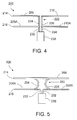
- FIG. 4 is a side plan view of the occluder device of FIG. 2 , in accordance with an exemplary embodiment
- FIG. 5 is a side plan view of the occluder device of FIG. 2 , in accordance with an exemplary embodiment
- FIG. 6 is a perspective view of the occluder device of FIG. 2 , and illustrating a cover for the occluder device, in accordance with an exemplary embodiment
- FIG. 7 is a top plan view of the occluder device of FIG. 2 , depicted along with the cover of FIG. 6 , in accordance with an exemplary embodiment
- FIG. 8 is a perspective view of the occluder device of FIG. 2 , depicted as first emerging from a catheter, in accordance with an exemplary embodiment
- FIG. 9 is a perspective view of the occluder device of FIG. 2 , depicted as half-way merged from the catheter, in accordance with an exemplary embodiment
- FIG. 10 is a perspective view of the occluder device of FIG. 2 , depicted as fully emerged from the catheter and separated from a deployment cable, in accordance with an exemplary embodiment
- FIG. 11 is a perspective view of another exemplary alternative embodiment of an occluder device, depicted with reference to planar quadrants in FIG. 11A ;
- FIG. 12 is a side view of another exemplary alternative embodiment of an occluder device
- FIG. 13 is a side view of a further exemplary alternative embodiment of an occluder device
- FIG. 14 is a perspective view of another exemplary embodiment of an occluder device
- FIG. 15 is a perspective view of yet another exemplary alternative embodiment of an occluder device
- FIG. 16 is a flowchart of an exemplary embodiment of a method for occluding an aperture in tissue or a vessel, and that may be implemented using the occluder devices of FIGS. 2-15 ;
- FIG. 17 depicts an exemplary deployment of an occluder device within a vessel.
- the present disclosure provides a device for occluding an aperture within body tissue or vessel.
- One skilled in the art will recognize that the device and methods of the present disclosure may be used to treat other anatomical conditions in addition to those specifically discussed herein. As such, the disclosure should not be considered limited in applicability to any particular anatomical condition.
- FIG. 1 illustrates a human heart 100 , having a right atrium 102 , a left atrium 104 , a right ventricle 106 , and a left ventricle 108 . Shown are various anatomical anomalies 110 , 112 , and 114 .
- the atrial septum 116 includes septum primum 118 and septum secundum 120 .
- the anatomy of the septum 116 varies widely within the population. In some people, the septum primum 118 extends to and overlaps with the septum secundum 120 .
- the septum primum 118 may be quite thin.
- the human blood circulation comprises a systemic circuit and a pulmonary circuit.
- the two circuits are joined to one another by the ductus arteriosus.
- the ductus connects the aorta (circulation to the body) to the pulmonary artery (pulmonary circuit).
- this ductus closes after birth. If development is defective, it can happen that the ductus does not close, and as a result the two blood circuits are still joined even after birth.
- distal refers to the direction away from a catheter insertion location and “proximal” refers to the direction nearer the insertion location.
- memory or “shape memory” refers to a property of materials to resume and maintain an intended shape despite being distorted for periods of time, such as during storage or during the process of delivery in vivo.
- aperture refers to a gap, hole or opening in a patient's body. Apertures may be in a tissue (including, for example, in an organ), or a vessel.
- tissue including, for example, in an organ
- apertures in heart tissue include, but are not limited to, PFO, ASD, VSD, and PDA, among others.
- Apertures in vessels include apertures in the walls of vessels (e.g., focal aortic defects, pseudoaneurysms, penetrating ulcers or communicative defects between the true and false lumen in aortic dissections) as well as the arteries or veins themselves wherein the aperture refers to the lumen of the vessel.
- an occluder device 200 of the present disclosure is provided. While for the sake of brevity, the term “occluder device 200 ” is used generically throughout, it is to be understood that in embodiments or descriptions where no covering is depicted or described, the embodiment is referring to an “occluder frame”. Similarly, it is to be understood that in embodiments where a covering is depicted or described, the embodiment is referring to an “occluder device”.
- the occluder device 200 is configured to occlude an aperture, including, for example, a defect of a heart, such as one or more of the anomalies 110 , 112 , 114 of the heart 100 depicted in FIG. 1 .
- a defect of a heart such as one or more of the anomalies 110 , 112 , 114 of the heart 100 depicted in FIG. 1 .
- One skilled in the art would also recognize the device's application for use as a vascular occluder or plug as well as an atrial appendage occluder.
- the occluder device 200 comprises two separate uniquely shaped memory wires 201 . While in some embodiments one member of the pair of shaped memory wires has a shape different than the shape of the other member of the pair, in some embodiments each member of the pair of shaped memory wire has a shape identical to the shape of the other member of the pair.
- the memory wires 201 can be formed of biocompatible metals or polymers, such as bioresorbable polymers, shape memory polymers, shape memory metal alloys, biocompatible metals, bioresorbable metals, or combinations thereof. Specific examples include but are not limited to iron, magnesium, stainless steel, nitinol, or combinations of these and/or similar materials.
- a preferred metal for the present disclosure is a nitinol alloy.
- Nitinol an acronym for Nickel Titanium Naval Ordinance Laboratory
- Nitinol is a family of intermetallic materials, which contain a nearly equal mixture of nickel (55 wt. %) and titanium. Other elements can be added to adjust or “tune” the material properties.
- Nitinol exhibits unique behavior, specifically, a well-defined “shape memory” and super elasticity.
- any biocompatible material with a memory capability can be used with the present disclosure.
- the thermal shape memory and/or superelastic properties of shape memory polymers and alloys permit the occluder device 200 to resume and maintain its intended shape in vivo despite being distorted during the delivery process.
- the memory may also assist in pressing an aperture, such as a PFO tunnel, closed.
- the diameter or thickness of the wire depends on the size and type of the device, i.e., the larger the device, the larger the diameter of the wire. In general, wire having a diameter between about 0.2 mm and 0.8 mm can be used.
- the occluders include three or more, four or more, five or more, or six or more separate uniquely shaped memory wires. While in some such embodiments, one or more of the shaped memory wires has a shape different than the shape of the other shaped memory wires, in some embodiments two or more of the memory wires have an identical shape, in other embodiments each of the shaped memory wires has an identical shape.
- first wire 202 there are two second, or proximal, geometric forms 208 of the first wire 202 (namely, 208 (A) and 208 (B)).
- 208 (A) and 208 (B) the number and configuration of the first and/or second geometric forms 206 , 208 of the first wire 202 may vary.
- the second wire 204 forms a first geometric form 210 and a second geometric form 212 .
- the first geometric form 210 of the second wire 204 preferably comprises a distal geometric form
- the second geometric form 212 of the second wire preferably comprises a proximal geometric form.
- there are two second, or proximal, geometric forms 212 of the second wire 204 namely, 212 (A) and 212 (B)
- the number and configuration of the first and/or second geometric focus 210 , 212 of the second wire 204 may vary.
- the first geometric forms 206 of the first wire 202 and the first geometric forms 210 of the second wire 204 form a first plate, such as a disc, or another otherwise relatively flat surface (hereinafter referred to as a “plate”) 214 in a first plane 218 .
- the second geometric forms 208 of the first wire 202 and the second geometric forms 212 of the second wire 204 form a second plate 216 in a second plane 220 that is parallel to and remote from the first plane 218 .
- the first and second plates 214 , 216 each comprise one or more semi-circular discs. However, this may vary in other embodiments, as the first and second plates 214 , 216 may comprise any one or more of a number of other different types of geometric forms.
- each wire 202 , 204 forms a respective distal semi-circle or half disc 206 , 210 in addition to two proximal quarter-circles or quarter-discs 208 (A), 208 (B) or 212 (A), 212 (B).
- the two proximal quarter-circles of each wire together form proximal semi-circles or half-discs 208 (A), 208 (B) or 212 (A), 212 (B).
- the two distal semi-circles of each respective wire 202 , 204 together comprise a distal plate 214 (depicted in FIGS. 2-5 as a distal disc) of the occluder device 200 .
- the four proximal quarter-circles 208 (A), 208 (B), 212 (A), 212 (B), which form a “four-leaf clover” configuration in the embodiment of FIGS. 2-5 comprise a proximal plate 216 (depicted in FIGS. 2-5 as a proximal disc) of the occluder device 200 .
- the proximal semi-circle 208 (A), 208 (B) or 212 (A), 212 (B) of each wire 201 is connected to the distal semi-circle 206 or 210 by a waist 222 formed by waist components 224 , 226 .
- waist components 224 of the first wire 202 there are two waist components 224 of the first wire 202 and two waist components 226 of the second wire 204 .
- the four waist components (two from each wire) 224 , 226 together comprise restricted area or waist 222 of the occluder device 200 .
- the distance between the waist components, both within the same wire and from wire to wire, determines the size of the waist 222 .
- the size of the waist 222 is dependent on the particular application and the size of the occluder device 200 .
- the resiliency and memory of the waist components 224 , 226 and capacity to expand radially serves as a self-centering mechanism of the occluder device 200 in heart apertures.
- the first and second wires 202 , 204 are attached, joined, or otherwise coupled to the delivery attachment mechanism or hub 230 .
- the ends 232 , 234 of wires 202 , 204 are welded, glued, or otherwise affixed to the hub 230 .
- the distal plate 214 and/or proximal plate 216 may include membranous coverings 236 and 238 illustrated in FIGS. 6 and 7 .
- the membranous coverings 236 and 238 ensure more complete coverage of an aperture and promote encapsulation and endothelialization of tissue, thereby further encouraging anatomical closure of the tissue and improving closure rate.
- the coverings 236 and 238 also help stabilize the occluder device 200 .
- the membranous coverings 236 and 238 may be formed of any flexible, biocompatible material capable of promoting tissue growth and/or acting as a sealant.
- suitable membranous coverings include, but are not limited to DACRON®, polyester fabrics, Teflon-based materials, ePTFE, polyurethanes, metallic materials, polyvinyl alcohol (PVA), extracellular matrix (ECM) or other bioengineered materials, synthetic bioabsorbable polymeric materials, other natural materials (e.g. collagen), or combinations of the foregoing materials.
- the membranous coverings 236 and 238 may be formed of a thin, metallic film or foil, e.g. a nitinol film or foil, as described in U.S. Pat. No.
- ePTFE expanded polytetrafluoroethylene
- Loops may also be stitched to the membranous coverings 236 and 238 to securely fasten the coverings to occluder device 200 .
- the coverings may additionally or alternatively be glued, welded or otherwise attached to the occluder device 200 via the wires (not shown in FIG. 6 or FIG. 7 ).
- the microporous structure of the membranous coverings can be tailored to promote tissue ingrowth and/or endothelialization.
- the coverings can be modified by various chemical or physical processes to enhance certain mechanical or physical properties.
- a hydrophilic coating can be applied to the covering to promote its wetability and/or echo translucency.
- physiochemical modifications can be employed whereby the covering includes chemical moieties that promote endothelial cell attachment, migration, and/or proliferation or resist thrombosis.
- a surface modified with covalently attached heparin is one example of a covering modification.
- the coverings prevent blood flow through the aperture, e.g. acute occlusion
- the microporosity of the coverings permits some blood flow through the aperture, e.g. partial occlusion. In some such embodiments, this blood flow is reduced over time by tissue ingrowth and/or endothelialization of the covering.
- the plates 214 , 216 are of equal size and are centered around the hub 230 . In other embodiments, the plates 214 , 216 may be of unequal sizes. In yet other embodiments, the plates 214 , 216 may be of equal size yet offset from each other via a shift in opposite directions from the hub 230 .
- the diameters of the distal plate 214 and proximal plate 216 are generally 5-8 mm larger than the diameter of the connecting waist 222 .
- the diameters of the plates 214 , 216 are generally about 9 mm each. Because of the flexibility in the waist 222 , a 12 mm waist device will be able to be placed in a 6 mm to 12 mm defect. For larger waists 222 or larger devices, the diameter of the plate size will increase proportionately.
- devices include waist sizes having the following diameters: 6 mm, 12 mm, 18 mm, 24 min, 30 mm, 36 mm, and 42 mm.
- the occluder device 200 may be inserted into an aperture to prevent the flow of blood therethrough.
- the occluder device 200 may extend through a PFO 110 or an ASD 112 such that the distal plate 214 is located in the left atrium 104 and the proximal plate 216 is located in the right atrium 102 (as shown in the heart 100 in FIG. 1 ).
- the occluder device 200 for use as a vascular occluder or plug as well as an atrial appendage occluder. The closure of apertures in these and other tissues, as well as other types of apertures, will become apparent as described below.
- the occluder device 200 is attached to a deployment cable 240 which is removably attached to the occluder device 200 at the hub 230 .
- a deployment cable 240 which is removably attached to the occluder device 200 at the hub 230 .
- one method of releasably attaching the deployment cable 240 to the hub 230 is by threaded engagement utilizing a screw end 248 which engages unseen female threads within the hub 230 .
- Other known means of attachment can be used to releasably connect the deployment cable 240 to the hub 230 .
- the occluder device 200 includes hub 230 at both the proximal and distal ends of the device to allow the user to conveniently select the orientation of the device. As described below, the hub 230 also permits the user to reposition the device if so desired.
- the occluder device 200 When the deployment cable 240 is engaged with the hub 230 , as illustrated in FIGS. 8 and 9 , the occluder device 200 is initially housed within a flexible delivery catheter 242 having an open channel 244 .
- FIG. 8 illustrates the occluder device 200 in which the distal plate 214 is expanded, due to the memory expansion of the wires 202 and 204 , and housed within the open channel 244 of the delivery catheter 242 .
- both the distal plate 214 and the proximal plate 216 as well as the coverings 236 and 238 are housed within the open channel 244 of the delivery catheter 242 .
- the catheter 242 is fed into a blood vessel through an already placed sheath and advanced via the blood vessel system to an aperture, including, for example, apertures in tissue (e.g. apertures in the heart including the PFO, the ASD, the VSD, the PDA, or an atrial appendage).
- an aperture including, for example, apertures in tissue (e.g. apertures in the heart including the PFO, the ASD, the VSD, the PDA, or an atrial appendage).
- occluders may also be used to close or block the lumen of a vessel or to close or block an aperture in the wall of a vessel.
- the occluder device 200 will be partially advanced from the catheter 242 as illustrated in FIG. 8 .
- the distal plate 214 which includes the covering 236 , begins to expand on the distal side of the aperture. Due to the memory capabilities of the wires 202 and 204 , the occluder device 200 begins to return to its normal shape such that the distal plate 214 expands on the distal side of the aperture. Once the distal plate 214 is completely out of the catheter opening 244 , as shown in FIG. 9 , the distal plate 214 and the attached covering 236 become fully expanded.
- the catheter 242 is further withdrawn to expose the waist 222 , which then begins to emerge and expand due to the memory shape of the wires 202 and 204 .
- the waist 222 is designed to expand such that each of the wires forming the waist 222 is urged against the aperture causing a custom fit device of the occluder device 200 within the aperture.
- the proximal plate 216 and the covering 238 begin their process of expansion on the proximal side of the aperture. When the proximal plate 216 is fully delivered from the catheter 242 , it will expand and effectively form a seal over the aperture.
- the distal plate 214 and proximal plate 216 are secured in place by the action of the wires in the waist 222 urging against the aperture.
- the deployment cable 240 is removed from the hub 230 and the catheter 242 and the deployment cable 240 are removed from the body.
- the occluder device 200 is left at the region of the aperture. Over several months, tissue and other membranous structures will bind to the occluder device 200 thereby permanently locking the occluder device 200 to the specific area of the aperture.
- the two wires 202 , 204 function to form round plates 214 , 216 on each side of the aperture.
- the plates 214 , 216 maintain the circular shape because of the memory capability of the wires 202 , 204 .
- the coverings 236 , 238 help to stabilize the discs, and preferably act to completely occlude the defect.
- the wires 202 , 204 at the waist components 224 , 226 will be separated enough at the waist 222 to make the occluder device 200 self-centering. Due to the conformity of this design, the occluder device 200 will self-center within commonly (e.g. round, oval) shaped septal defects as the waist 222 can adjust to any type of opening.
- the waist 222 has the capability to expand (if needed) to a larger size with the help of a balloon.
- a center channel 246 extends through the deployment cable 240 , the hub 230 , and the screw end 248 .
- a balloon (not shown) is urged through the center channel 246 after the occluder device has been removed from the catheter 242 and expanded but before the hub 230 has been detached from the deployment cable 240 .
- the balloon is placed within the waist 222 and expanded.
- the waist 222 is dilatable, i.e., expandable, when gentle pressure of the balloon is applied. The dilation will expand the waist components 224 , 226 .
- the balloon is deflated and removed by withdrawal through the center channel 246 .
- the occluder device 200 appears stable, the occluder device 200 is separated from the deployment cable 240 as discussed above. In the majority of cases, balloon dilation will not be required.
- FIGS. 11-15 various exemplary embodiments are provided with respect to the occluder device and/or components thereof.
- FIGS. 11 and 11A depict an embodiment of an occluder device contemplated herein with first wire 202 and staggered geometric forms 206 , 208 of the first wire 202 . Not shown in FIG. 11 are the second wire and staggered geometric forms of the second wire.
- FIG. 11A depicts an exemplary classification of planar quadrants for the first and second planes 218 , 220 of FIG. 2 for reference with respect to the embodiment of FIG. 11 .
- One skilled in the art will recognize that less or more than four quadrants can be utilized.
- the first plane 218 of FIG. 2 has a first quadrant 1401 (A), a second quadrant 1402 (A) that is adjacent to the first quadrant 1401 (A), a third quadrant 1403 (A) that is below the first quadrant 1401 (A), and a fourth quadrant 1404 (A) that is below the second quadrant 1402 (A) and adjacent to the third quadrant 1403 (A).
- the first quadrant 1401 (B) of the first plane 218 is closer to the first quadrant 1401 (B) of the second plane 220 than to the second, third, or fourth quadrants 1402 (B), 1403 (B), 1404 (B) of the second plane 220 .
- the second quadrant 1402 (A) of the first plane 218 is closer to the second quadrant 1402 (B) of the second plane 220 than to the first, third, or fourth quadrants 1401 (B), 1403 (B), 1404 (B) of the second plane 220 .
- the third quadrant 1403 (A) of the first plane 218 is closer to the third quadrant 1403 (B) of the second plane 220 than to the first, second, or fourth quadrants 1401 (B), 1402 (B), 1404 (B) of the second plane 220 .
- the fourth quadrant 1404 (A) of the first plane 218 is closer to the fourth quadrant 1404 (B) of the second plane 220 than to the first, second, or third quadrants 1401 (B), 1402 (B), 1403 (B) of the second plane 220 .
- the first geometric form 206 of the first wire 202 extends through the first and second quadrants 1401 (A), 1402 (A) of the first plane 218 , preferably in a hemispheric shape.
- the second geometric form 208 of the first wire 202 extends through the third and fourth quadrants 1403 (B), 1404 (B) of the second plane 220 , also preferably in a hemispheric shape.
- the two hemispheric shapes are joined by a portion of the first wire 202 that extends in an angled manner from the first plane 218 to the second plane 220 . Not shown in FIG.
- FIG. 11 are depictions of the first geometric form of the second wire extending through the third and fourth quadrants of the first plane, preferably in a hemispheric shape.
- the second geometric form of the second wire 204 extending through the first and second quadrants of the second plane, also preferably in a hemispheric shape.
- the two hemispheric shapes are joined by a portion of the second wire that extends in an angled manner from the first plane to the second plane.
- the first wire 202 and second wire preferably form hemispheric shapes in planes 218 , 220 , the invention is not so limited, and various shapes suitable for in an occlusive device may be used.
- the first plane 218 may also be considered to include a first half-plane 1411 (A) and a second half-plane 1412 (A).
- the first half-plane 1411 (A) may comprise the first and second quadrants 1401 (A), 1402 (A) of the first plane 218 .
- the second half-plane 1412 (A) may comprise the third and fourth quadrants 1403 (A), 1404 (A) of the first plane 218 .
- the second plane 220 may also be considered to include a first half-plane 1411 (B) and a second half-plane 1412 (B).
- the first half-plane 1411 (B) may comprise the first and second quadrants 1401 (B), 1402 (B) of the second plane 220 .
- the second half-plane 1412 (B) may comprise the third and fourth quadrants 1403 (B), 1404 (B) of the second plane 220 .
- the first geometric form 206 of the first wire 202 extends through and is disposed within the first half-plane 1411 (A) of the first plane 218 .
- the second geometric form 208 of the first wire 202 extends through and is disposed within the second half-plane 1412 (B) of the second plane 220 .
- FIG. 11 depictions of the first geometric form of the second wire extending through and disposed within the second half-plane of the first plane.
- a depiction of the second geometric form of the second wire extending through and disposed within the first half-plane of the second plane.
- the first and second planes 218 , 220 may also collectively be considered to include four spatial quartiles 1421 , 1422 , 1423 , and 1424 .
- the first spatial quartile 1421 may comprise the first half-plane 1411 (A) of the first plane 218
- the second spatial quartile 1422 may comprise the second half-plane 1412 (A) of the first plane 218
- the third spatial quartile 1403 may comprise the first half-plane 1411 (B) of the second plane 220
- the fourth spatial quartile 1424 may comprise the second half-plane 1412 (B) of the second plane 220 .
- the first geometric form 206 of the first wire 202 extends through and is disposed within the first spatial quartile 1421
- the second geometric form 208 of the first wire 202 extends through and is disposed within the fourth spatial quartile 1424 .
- the first geometric form of the second wire extending through and disposed within the second spatial quartile
- the second geometric form of the second wire extending through and disposed within the third spatial quartile.
- FIG. 12 depicts an embodiment of an occluder device contemplated herein with an increased number of plates and waists as compared with the embodiment of FIG. 2 .
- Additional plates and waists may be beneficial, for example, in providing additional support and/or stability, and/or reaching apertures that are deeper within the heart or vessels, closing multiple apertures, and/or closing apertures that are surrounded by non-uniform tissue.
- the occluder device may be designed or used to close apertures in arteries or veins (e.g., focal aortic defects, pseudoaneurysms, penetrating ulcers or communicative defects between the true and false lumen in aortic dissections, or arteries or veins themselves).
- the 12 includes a third plate 217 , in addition to the first and second plates 214 , 216 referenced above.
- the first plate 214 is disposed within the first plane 218
- the second plate 216 is disposed in the second plane 220 .
- the first and second planes 218 , 220 are parallel to and remote from one another.
- the third plate 217 is disposed in a third plane 221 that is parallel to and remote from both the first and second planes 218 , 220 .
- one or more of the plates may not be parallel to the other of the plates when deployed while in certain embodiments, none of the plates are parallel to the other plates when deployed. In other embodiments and as depicted in FIG. 16 , the three (or more) plates may be substantially parallel to each other when deployed.
- the embodiment of FIG. 12 also includes a second waist 223 , in addition to the first waist 222 .
- the first waist 222 is formed by first components 224 of the first wire 202 and first components 226 of the second wire 204 .
- the second waist 223 is formed by second, or additional, components 225 of the first wire 202 and second, or additional, components 227 of the second wire 204 .
- the first waist 222 is attached between the first and second plates 214 , 216
- the second waist 223 is attached between the second and third plates 216 , 217 .
- the first, second, and third plates 214 , 216 , 217 are of unequal sizes, and are arranged in order of increasing size.
- the third plate 217 is larger than the second plate 216 (for example in terms of diameter and/or surface area)
- the second plate 216 is larger than the first plate 214 (for example in terms of diameter and surface area).
- the first waist 222 and the second waist 223 may be of approximately equal size as depicted in FIG. 12 , in some embodiments the length and/or diameter of the first and second waists may differ.
- the number, sizes, and/or shapes of the plates 214 , 216 , 217 and/or the waists 222 , 223 may vary in other embodiments.
- the device includes three or more plates 214 , 216 , 217 , wherein each plate is substantially the same size.
- the first and third plates 214 and 217 are of substantially the same size and are larger than the second plate 216 .
- the first and third plates 214 and 217 are of substantially the same size and are smaller than the second plate 216 .
- two of the plates are of substantially the same size and are larger than the third plate.
- two of the plates are of substantially the same size and are smaller than the third plate.
- all of the plates include a covering
- all of the plates include a covering
- one or more of the plates 214 , 216 , 217 of the occluder device 200 of FIG. 12 may be bent and/or inverted. Furthermore, in certain embodiments, a shortest distance between the first and second plates 214 , 216 may differ from the shortest distance between the second and third pates 216 , 217 . In certain embodiments, one or more of the plates 214 , 216 , 217 may have one or more hook, anchor or barb, or combinations thereof on one or both sides thereof to reduce to eliminate unintentional migration of the device. In some embodiments at least one hook, anchor or barb is affixed to the distal most plate of the device.
- certain embodiments include at least one hook, anchor or barb is affixed to the two distal most plates of the device.
- the spatial arrangement of hooks, anchors or barbs may be selected according to the location of the aperture to be occluded
- At least one hook, anchor or barb is affixed to the first and/or second wires. In certain embodiments at least one hook, anchor or barb is positioned on the periphery of the occlusive face. In some embodiments at least one hook, anchor or barb protrudes or projects in a direction from the occlusive face of the device. Some embodiments have at least one hook, anchor or barb protruding or projecting substantially tangent or at an acute angle to the peripheral edge of the occlusive face. Hooks, anchors or barbs can be made of any suitable material. In some embodiments, hooks, anchors or barbs are made of a biocompatible material. In some embodiments hooks, anchors or barbs are constructed of a non-permanent biodegradable or bioabsorbable material. Hooks, anchors and barbs can be attached to the first and/or second wire by any suitable method.
- a device may have relatively fewer hooks, anchors or barbs than a device for non-vascular implementation.
- the device includes no hooks, anchors or barbs.
- FIGS. 13 and 14 depict embodiments of an occluder device contemplated herein having a flexible connection.
- the first wire waist components 224 and the second wire waist components 226 are configured such that the waist 222 comprises a flexible connection 2000 between the first and second plates 214 , 216 .
- the waist 222 preferably includes a stored length as a result of the flexible connection 2000 .
- the term “stored length” means the additional length of the waist 222 to which the waist 222 can be extended from its resting length when the plates 214 , 216 are distanced from each other.
- the flexible connection 2000 comprises a spring that is attached between the first and second plates 214 , 216 .
- the flexible connection and/or spring may be beneficial, for example, in providing flexibility or greater ability of the occluder device to adjust to apertures of different shapes and sizes.
- one or more of the plates of the occluder device may also include one or more strain relief mechanisms, such as springs or flexible connections.
- both of the plates 214 , 216 include flexible connections 2000 similar to those described above in connection with FIG. 13 .
- each flexible connection 2000 comprises a spring that is formed within one or more of the geometric forms 206 - 212 .
- Certain of the plates 214 , 216 and/or geometric forms 206 - 212 may therefore include a stored length similar to that described above in connection with FIG. 13 .
- FIG. 14 depicts each of the geometric forms 206 - 212 as having a flexible connection 2000
- one or more of the geometric forms 206 - 212 may include a flexible connection while one or more other of the geometric forms 206 - 212 may not include a flexible connection 2000
- one of the plates 214 , 216 may include a flexible connection 2000 while the other of the plates 214 , 216 does not.
- the third plate 217 of FIG. 12 may similarly include a flexible connection 2000 , instead of or in addition to one or both of the first and second plates 214 , 216 .
- FIG. 15 depicts an embodiment of an occluder device contemplated herein in which the waist is aligned off-center.
- the various waist components 224 of the first wire 202 (denoted as 224 (A) and 224 (B) in FIG. 15 ) are offset with respect to a center portion or central axis 2100 of the occluder device 200 .
- the various waist components 226 of the second wire 204 (denoted as 226 (A), 226 (B) and 226 (C) in FIG. 15 ) are also offset with respect to the center portion or central axis 2100 of the occluder device 200 .
- the first wire 202 crosses a center region of the occluder device 200 at a first point and a second point
- the second wire 204 crosses the center region at a third point and a fourth point.
- each plate 214 , 216 may be reduced in size as compared with other embodiments.
- each plate 214 , 216 has a surface area that is preferably twice the surface area of the aperture.
- each plate 214 , 216 has a surface area that is only approximately twenty five percent larger than the surface area of the aperture.
- FIG. 16 is a flowchart of an exemplary embodiment of a method 1600 for occluding an aperture defect in a heart.
- the method 1600 can be utilized in connection with the heart 100 of FIG. 1 and the various embodiments of the occluder device 200 of FIGS. 2-15 .
- the method 1600 preferably utilizes one or more embodiments of the occluder devices 200 of FIGS. 2-15 to occlude an aperture defect of a heart, such as one or more of the anomalies 110 , 112 , 114 of the heart 100 depicted in FIG. 1 .
- One skilled in the art would also recognize the method's application for use as a vascular occluder or plug as well as an atrial appendage occluder.
- the method 1600 includes the step of providing an occluder device (step 1602 ).
- the occluder device corresponds to the occluder device 200 depicted in any of the embodiments depicted in FIGS. 2-15 and/or described above.
- the occluder device preferably comprises a first flexible wire (such as wire 202 described above) and a second flexible wire (such as wire 204 described above). Each of the first and second wires is comprised of a shape memory material.
- Each of the first and second wires is shaped into first and second geometric forms (such as forms 206 , 208 , 210 , and 212 described above) around an inner region such that the first geometric form of the first wire and the first geometric form of the second wire form a first plate (such as pate 214 described above) in a first plane, and the second geometric form 208 of the first wire 202 and the second geometric form 212 of the second wire 204 form a second plate (such as plate 216 described above) in a second plane that is parallel to and remote from the first plane.
- the first and second plates are separated by a waist (such as waist 222 described above) formed from two portions of the first wire and two portions of the second wire.
- a sealed covering (such as covering 236 or 238 described above) is preferably disposed over at least one of the first and second plates.
- the covering provides a seal for the aperture defect (such as one or more of the anomalies 110 , 112 , 114 of the heart 100 described above).
- Each of the first and second wires has a first end and a second end.
- Each of the first and second ends of the first and second wires is connected to a hub (such as hub 230 described above).
- the hub further comprises a delivery attachment mechanism (for example, that includes or is used in connection with the catheter 242 described above) for attachment to a removable deployment cable (such as deployment cable 240 described above).
- the method 1600 also includes the step of attaching the occluder device to the removable deployment cable (step 1604 ).
- the occluder device is placed within a flexible delivery catheter (such as the catheter 242 described above) having an open channel (such as the channel 244 described above) (step 1606 ).
- the catheter is fed into a blood vessel system (such as a blood vessel system of the heart 100 described above) and advanced via the blood vessel system to the aperture defect in the heart (step 1608 ).
- the catheter, with the occluder device disposed within, is similarly advanced through the aperture defect (step 1610 ).
- a balloon sub-process 1612 is also utilized in occluding the aperture defect in the heart.
- a balloon is advanced into the heart through the open channel toward the occluder device at the aperture defect (step 1614 ).
- the balloon is also inserted into the waist of the occluder device (step 1616 ).
- the balloon is then inflated (step 1618 ), in order to help position the occluder device proximate the heart defect.
- the balloon is deflated (step 1620 ) and then removed from the waist of the occluder device (step 1622 ).
- a hook sub-process 1624 may be utilized in occluding the aperture defect in the heart.
- one or more books is engaged with the delivery attachment mechanism (such as the catheter) (step 1626 ), preferably via a screw system.
- the hook is manipulated using the delivery attachment mechanism and used to reposition the occluder device (step 1628 ).
- the hook may also be utilized to retrieve the occluder device by exerting force on the delivery attachment mechanism in a direction away from the heart (step 1630 ).
- the catheter next is withdrawn from the occluder device (step 1632 ).
- the catheter is withdrawn from the occluder device in step 1632 in a manner such that the first plate of the occluder device expands on a first side of the aperture defect.
- the catheter is further withdrawn from the occluder device such that the second plate of the occluder device expands on a second side of the aperture defect (step 1634 ).
- the catheter is withdrawn from the occluder device in step 1634 in a manner, such that the waist of the occluder device expands by memory retention within the aperture defect to self-center the occluder device.
- the catheter is then withdrawn from the blood vessel system (step 1636 ), and the deployment cable is removed from the hub of the occluder device (step 1638 ).
- certain steps of the method 1600 may vary in certain embodiments. It will also be appreciated that certain steps of the method 1600 may occur in a different order than is depicted in FIG. 16 .
- the optional hook sub-process 1624 may be performed before the optional balloon sub-process 1612 . It will similarly be appreciated that certain steps of the method 1600 may occur simultaneously with one another. Additional optional steps may also be performed.
- the clinician may wish to visualize the location of the device within the aperture. Such visualization may be performed using imaging techniques well known to those of ordinary skill. If the clinician is not satisfied with the positioning of the device based, for example, on visualization of the device, the clinician may choose to remove and/or reposition the device.
- FIG. 17 depicts an embodiment of an occluder device 200 contemplated herein with three plates 214 , 216 and 217 and deployed in a vessel 250 .
- the first waist 222 is formed by first components 224 of the first wire and first components 226 of the second wire.
- the second waist 223 is formed by second, or additional, components 225 of the first wire and second, or additional, components 227 of the second wire.
- the first plate 214 is disposed within the first plane 218
- the second plate 216 is disposed in the second plane 220 .
- the first and second planes 218 , 220 are parallel to and remote from one another.
- the third plate 217 is disposed in a third plane 221 that is parallel to and remote from both the first and second planes 218 , 220 .
- hubs 230 , 231 are located at distal and proximal ends of the device.
Abstract
Devices for occluding an aperture in tissue or a vessel include a first flexible wire and a second flexible wire. Each of the first and second wires is formed of a shape memory material. Each of the first and second wires is shaped into first and second geometric forms such that the first geometric form of the first wire and the first geometric form of the second wire form a first plate in a first plane, and the second geometric form of the first wire and the second geometric form of the second wire form a second plate in a second plane that is parallel to and remote from the first plane. The first and second plates are separated by a waist formed from two portions of the first wire and two portions of the second wire. Methods for occluding an aperture in tissue or a vessel using such devices are also provided.
Description
This application claims priority under 35 U.S.C. §119(e) to provisional U.S. Patent Application 61/523,175, filed on Aug. 12, 2011, the entire contents of which are hereby incorporated by reference.
The present disclosure generally relates to medical devices, and more particularly relates to devices for occluding apertures in tissues and vessels.
Heart occlusion devices are used in the medical field for correcting congenital heart defects, such as atrial septal defects (“ASD”), patent foramen ovale (“PFO”) defects, ventricular septal defects (“VSD”), and patent ductus arteriosus (“PDA”) defects. A PFO, illustrated in FIG. 1 at 110, is a persistent, one-way, usually flap-like opening in the wall between the right atrium 102 and left atrium 104 of the heart 100. The foramen ovale 110 serves a desired purpose when a fetus is gestating in utero. Because blood is oxygenated through the umbilical cord and not through the developing lungs, the circulatory system of the fetal heart allows the blood to flow through the foramen ovale as a physiologic conduit for right-to-left shunting. After birth, with the establishment of pulmonary circulation, the increased left atrial blood flow and pressure results in functional closure of the foramen ovale. This functional closure is subsequently followed by anatomical closure of the two over-lapping layers of tissue: septum primum 118 and septum secundum 120.
However, a PFO has been shown to persist in a number of adults. Because left atrial (LA) pressure is normally higher than right atrial (RA) pressure, the flap usually stays closed. Under certain conditions, however, right atrial pressure can exceed left atrial pressure, creating the possibility that blood could pass from the right atrium 102 to the left atrium 104, and blood clots could enter the systemic circulation. It is desirable that this circumstance be eliminated.
The presence of a PFO defect is generally considered to have no therapeutic consequence in otherwise healthy adults. Paradoxical embolism via a PFO defect is considered in the diagnosis for patients who have suffered a stroke or transient ischemic attack (TIA) in the presence of a PFO and without another identified cause of ischemic stroke. While there is currently no definitive proof of a cause-effect relationship, many studies have confirmed a strong association between the presence of a PFO defect and the risk for paradoxical embolism or stroke. In addition, there is significant evidence that patients with a PFO defect who have had a cerebral vascular event are at increased risk for future, recurrent cerebrovascular events.
Accordingly, patients at such an increased risk are considered for prophylactic medical therapy to reduce the risk of a recurrent embolic event. These patients are commonly treated with oral anticoagulants, which potentially have adverse side effects, such as hemorrhaging, hematoma, and interactions with a variety of other drugs. The use of these drugs can alter a person's recovery and necessitate adjustments in a person's daily living pattern.
In certain cases, such as when anticoagulation is contraindicated, surgery may be necessary or desirable to close a PFO defect. The surgery would typically include suturing a PFO closed by attaching septum secundum to septum primum. This sutured attachment can be accomplished using either an interrupted or a continuous stitch and is a common way a surgeon shuts a PFO under direct visualization.
Umbrella devices and a variety of other similar mechanical closure devices, developed initially for percutaneous closure of atrial septal defects (ASDs), have been used in some instances to close PFOs. These devices potentially allow patients to avoid or lessen the side effects often associated with anticoagulation therapies and the risks of invasive surgery. However, umbrella devices and the like that are designed for ASDs may not be optimally suited for use as PFO closure devices.
Certain currently available septal closure devices present possible drawbacks, including technically complex implantation procedures. Additionally, complications are possible due to thrombus, fractures of the components, conduction system disturbances, perforations of heart tissue, and residual leaks. Certain devices have a high septal profile and include large masses of foreign material, which may lead to unfavorable body adaptation of a device. Given that ASD devices are designed to occlude holes, certain of such devices lack anatomic conformability to the flap-like anatomy of PFOs. The flap-like opening of the PFO is complex, and devices with a central post or devices that are self-centering may not close the defect completely, an outcome that is highly desired when closing a PFO defect. Hence, a device with a waist which can conform to the defect will have much higher chance of completely closing the defect. Even if an occlusive seal is formed, the device may be deployed in the heart on an angle, leaving some components insecurely seated against the septum and, thereby, risking thrombus formation due to hemodynamic disturbances. Finally, some septal closure devices are complex to manufacture, which may result in inconsistent product performance.
Certain devices for occluding other heart defects, e.g., ASD, VSD, PDA, also have potential drawbacks. For example, certain currently available devices tend to be either self-centering or non-self-centering and may not properly conform to the intra-cardiac anatomy. Both of these characteristics have distinct advantages and disadvantages. The non-self-centering device may not close the defect completely and may need to be over-sized significantly. This type of device may not be available for larger defects. Further, the self-centering device, if not sized properly, may cause injury to the heart. Some devices have sharp edges, which may damage the heart causing potential clinical problems. Some devices contain too much nitinol/metal, which may cause an undesired reaction in the patient. Some currently marketed devices have numerous model numbers (several available sizes), making it difficult and uneconomical for hospitals and markets to invest in starting a congenital and structural heart interventional program. The present disclosure is designed to address these and other deficiencies of certain existing closure devices.
Devices are also used for occluding other apertures, including uses such as occluding the lumen of a vessel and occluding apertures in vessel walls.
Accordingly, it is desirable to provide improved devices for occluding apertures in tissues or vessels. Furthermore, other desirable features and characteristics of the present invention will become apparent from the subsequent detailed description and the appended claims, taken in conjunction with the accompanying drawings and this background
In accordance with an exemplary embodiment, a device for occluding an aperture in a tissue or vessel is provided. The device comprises a first flexible wire and a second flexible wire. Each of the first and second wires is comprised of a shape memory material. Each of the first and second wires is shaped into first and second geometric forms separated by a waist formed from two portions of the first wire and two portions of the second wire. The first geometric form of the first wire and the first geometric form of the second wire form a first plate in a first plane. The second geometric form of the first wire and the second geometric form of the second wire form a second plate in a second plane that is parallel to and remote from the first plane. The first plane has a first quadrant, a second quadrant that is adjacent to the first quadrant, a third quadrant that is below the first quadrant, and a fourth quadrant that is below the second quadrant and adjacent to the third quadrant. The second plane has a first quadrant, a second quadrant that is adjacent to the first quadrant, a third quadrant that is below the first quadrant, and a fourth quadrant that is below the second quadrant and adjacent to the third quadrant. The first quadrant of the first plane is closer to the first quadrant of the second plane than to the second, third, or fourth quadrants of the second plane. The second quadrant of the first plane is closer to the second quadrant of the second plane than to the first, third, or fourth quadrants of the second plane. The third quadrant of the first plane is closer to the third quadrant of the second plane than to the first, second, or fourth quadrants of the second plane. The fourth quadrant of the first plane is closer to the fourth quadrant of the second plane than to the first, second, or third quadrants of the second plane. The first geometric form of the first wire extends through the first and second quadrants of the first plane. The second geometric form of the first wire extends through the third and fourth quadrants of the second plane. The first geometric form of the second wire extends through the third and fourth quadrants of the first plane. The second geometric form of the second wire extends through the first and second quadrants of the second plane.
In accordance with an exemplary embodiment, a device for occluding an aperture in a tissue or vessel is provided. The device comprises a first flexible wire and a second flexible wire. Each of the first and second wires is comprised of a shape memory material. Each of the first and second wires is shaped into first and second geometric forms separated by a waist formed from two portions of the first wire and two portions of the second wire. The first geometric form of the first wire and the first geometric form of the second wire form a first plate in a first plane. The second geometric form of the first wire and the second geometric form of the second wire form a second plate in a second plane that is parallel to and remote from the first plane. The first plane has a first half-plane and a second half-plane. The second half-plane is adjacent to the first half-plane. The second plane has a third half-plane and a fourth half-plane. The third half-plane is parallel to the first half-plane. The fourth half-plane is parallel to the second half-plane and adjacent to the third half-plane. The first geometric form of the first wire is disposed in the first half-plane. The second geometric form of the first wire is disposed in the fourth half-plane. The first geometric form of the second wire is disposed in the second half-plane. The second geometric form of the second wire is disposed in the third half-plane.
In accordance with an exemplary embodiment, a device for occluding an aperture in a tissue or vessel is provided. The device comprises a first flexible wire and a second flexible wire. Each of the first and second wires is comprised of a shape memory material. Each of the first and second wires is shaped into first and second geometric forms separated by a waist formed from two portions of the first wire and two portions of the second wire. The first geometric form of the first wire and the first geometric form of the second wire form a first plate in a first plane. The second geometric form of the first wire and the second geometric form of the second wire form a second plate in a second plane that is parallel to and remote from the first plane. The first plane is disposed within a first spatial quartile and a second spatial quartile that is adjacent to the first spatial quartile. The second plane is disposed within a third spatial quartile and a fourth spatial quartile. The third spatial quartile is parallel to the first spatial quartile. The fourth spatial quartile is parallel to the second spatial quartile and adjacent to the third spatial quartile. The first geometric form of the first wire is disposed in the first spatial quartile. The second geometric form of the first wire is disposed in the fourth spatial quartile. The first geometric form of the second wire is disposed in the second spatial quartile. The second geometric form of the second wire is disposed in the third spatial quartile.
In accordance with an exemplary embodiment, a device for occluding an aperture in a tissue or vessel is provided. The device comprises a first flexible wire and a second flexible wire. Each of the first and second wires is comprised of a shape memory material. Each of the first and second wires is shaped into a first, a second, and a third geometric form. The first geometric form of the first wire and the first geometric form of the second wire form a first plate in a first plane. The second geometric form of the first wire and the second geometric form of the second wire form a second plate in a second plane that is parallel to and remote from the first plane. The third geometric form of the first wire and the third geometric form of the second wire form a third plate in a third plane that is parallel to and remote from both the first and second planes. The first and second plates are separated by a first waist formed from two portions of the first wire and two portions of the second wire. The second and third plates are separated by a second waist formed from an additional two portions of the first wire and an additional two portions of the second wire.
In accordance with another exemplary embodiment, a device for occluding an aperture in a tissue or vessel is provided. The device comprises a first flexible wire and a second flexible wire. Each of the first and second wires is comprised of a shape memory material. Each of the first and second wires is shaped into first and second geometric forms. The first geometric form of the first wire and the first geometric form of the second wire form a first plate in a first plane, and the second geometric form of the first wire and the second geometric form of the second wire form a second plate in a second plane that is parallel to and remote from the first plane. The first and second plates are separated by a waist formed from two portions of the first wire and two portions of the second wire, the waist comprising a flexible connection between the first and second plates.
In accordance with another exemplary embodiment, a device for occluding an aperture in a tissue or vessel is provided. The device comprises a first flexible wire and a second flexible wire. Each of the first and second wires is comprised of a shape memory material. Each of the first and second wires is shaped into first and second geometric forms. The first geometric form of the first wire and the first geometric form of the second wire form a first plate in a first plane, and the second geometric form of the first wire and the second geometric form of the second wire form a second plate in a second plane that is parallel to and remote from the first plane. The first and second plates are separated by a waist formed from two portions of the first wire and two portions of the second wire, the waist having a stored length.
In accordance with another exemplary embodiment, a device for occluding an aperture in a tissue or vessel is provided. The device comprises a first flexible wire and a second flexible wire. Each of the first and second wires is comprised of a shape memory material. Each of the first and second wires is shaped into first and second geometric forms. The first geometric form of the first wire and the first geometric form of the second wire form a first plate in a first plane, and the second geometric form of the first wire and the second geometric form of the second wire form a second plate in a second plane that is parallel to and remote from the first plane. The first and second plates are separated by a waist formed from two portions of the first wire and two portions of the second wire. The two portions of the first wire and the two portions of the second wire form a spring between the first and second plates.
In accordance with another exemplary embodiment, a device for occluding an aperture in a tissue or vessel is provided. The device comprises a first flexible wire and a second flexible wire. Each of the first and second wires is comprised of a shape memory material. Each of the first and second wires is shaped into first and second geometric forms. The first geometric form of the first wire and the first geometric form of the second wire form a first plate in a first plane, and the second geometric form of the first wire and the second geometric form of the second wire form a second plate in a second plane that is parallel to and remote from the first plane. The first and second plates are separated by a waist formed from two portions of the first wire and two portions of the second wire. The first plate, the second plate, or both, includes a flexible connection formed therein.
In accordance with another exemplary embodiment, a device for occluding an aperture in a tissue or vessel is provided. The device comprises a first flexible wire and a second flexible wire. Each of the first and second wires is comprised of a shape memory material. Each of the first and second wires is shaped into first and second geometric forms. The first geometric form of the first wire and the first geometric form of the second wire form a first plate in a first plane, and the second geometric form of the first wire and the second geometric form of the second wire form a second plate in a second plane that is parallel to and remote from the first plane. The first and second plates are separated by a waist formed from two portions of the first wire and two portions of the second wire. The first plate, the second plate, or both, has a stored length.
In accordance with another exemplary embodiment, a device for occluding an aperture in a tissue or vessel is provided. The device comprises a first flexible wire and a second flexible wire. Each of the first and second wires is comprised of a shape memory material. Each of the first and second wires is shaped into first and second geometric forms. The first geometric form of the first wire and the first geometric form of the second wire form a first plate in a first plane, and the second geometric form of the first wire and the second geometric form of the second wire form a second plate in a second plane that is parallel to and remote from the first plane. The first and second plates are separated by a waist formed from two portions of the first wire and two portions of the second wire. The first plate, the second plate, or both, includes a spring formed therein.
In accordance with another exemplary embodiment, a device for occluding an aperture in a tissue or vessel is provided. The device comprises a first flexible wire and a second flexible wire. Each of the first and second wires is comprised of a shape memory material. Each of the first and second wires is shaped into first and second geometric forms. The first geometric form of the first wire and the first geometric form of the second wire form a first plate in a first plane, and the second geometric form of the first wire and the second geometric form of the second wire form a second plate in a second plane that is parallel to and remote from the first plane. The first and second plates are separated by a waist comprising a first waist component from the first wire and a second waist component from the second wire. In some embodiments the first and second waist components are not substantially centered about a center axis of the device.
In accordance with another exemplary embodiment, a device for occluding an aperture in a tissue or vessel is provided. The device comprises a first flexible wire and a second flexible wire. Each of the first and second wires is comprised of a shape memory material. Each of the first and second wires is shaped into first and second geometric forms. The first geometric form of the first wire and the first geometric form of the second wire form a first plate in a first plane, and the second geometric form of the first wire and the second geometric form of the second wire form a second plate in a second plane that is parallel to and remote from the first plane. The first and second plates are separated by a waist formed by the first wire and the second wire. The first wire crosses a center region of the device at a first point and a second point. The second wire crosses the center region at a third point and a fourth point. The first, second, third, and fourth points form a substantially square shape therebetween.
In accordance with yet other exemplary embodiments, methods for occluding an aperture in a tissue or vessel are provided. The methods comprise the steps of providing an occluder device of a type corresponding to one of the various occluder device embodiments described herein. The occluder device further comprises a sealed covering over at least one of the first and second plates, wherein the covering provides a seal for the aperture. Each of the first and second wires has a first and second end. Each of the first and second ends of the first and second wires is connected to a hub. The hub further comprises a delivery attachment mechanism for attachment to a removable deployment cable. The methods further comprise attaching the occluder device to the removable deployment cable, placing the occluder device within a flexible delivery catheter having an open channel, feeding the catheter into a blood vessel system and advancing the catheter via the blood vessel system to the aperture. The catheter is advanced through the aperture, and is withdrawn from the occluder device such that the first plate of the occluder device expands on a first side of the aperture. The catheter is further withdrawn from the occluder device such that the second plate of the occluder device expands on a second side of the aperture, such that the waist of the occluder device expands by memory retention within the aperture to self-center the occluder device. The catheter is further withdrawn from the blood vessel system, and the deployment cable is removed from the hub.
Other advantages, benefits and novel features of the embodiments of the present invention will become apparent from the following detailed description and accompanying drawings. All references, publications and patents, including the figures and drawings included therewith, are incorporated by reference in their entirety.
The present disclosure will hereinafter be described in conjunction with the following drawing figures, wherein like numerals denote like elements.
The following detailed description is merely exemplary in nature and is not intended to limit the disclosure or the application and uses of the disclosure. Furthermore, there is no intention to be bound by any theory presented in the preceding background or the following detailed description.
The present disclosure provides a device for occluding an aperture within body tissue or vessel. One skilled in the art will recognize that the device and methods of the present disclosure may be used to treat other anatomical conditions in addition to those specifically discussed herein. As such, the disclosure should not be considered limited in applicability to any particular anatomical condition.
As described herein, FIG. 1 illustrates a human heart 100, having a right atrium 102, a left atrium 104, a right ventricle 106, and a left ventricle 108. Shown are various anatomical anomalies 110, 112, and 114. The atrial septum 116 includes septum primum 118 and septum secundum 120. The anatomy of the septum 116 varies widely within the population. In some people, the septum primum 118 extends to and overlaps with the septum secundum 120. The septum primum 118 may be quite thin. When a PFO is present, blood could travel through the passage 110 between septum primum 118 and septum secundum 120 (referred to as “the PFO tunnel”). Additionally or alternatively, the presence of an ASD could permit blood to travel through an aperture in the septal tissue, such as that schematically illustrated by aperture 112. A VSD is similar to an ASD, except that an aperture 114 exists in the septum between the left and right ventricle of the heart.
PDA results from defects in the ductus arteriosus. The human blood circulation comprises a systemic circuit and a pulmonary circuit. In the embryonic phase of human development, the two circuits are joined to one another by the ductus arteriosus. The ductus connects the aorta (circulation to the body) to the pulmonary artery (pulmonary circuit). In normal development of an infant, this ductus closes after birth. If development is defective, it can happen that the ductus does not close, and as a result the two blood circuits are still joined even after birth.
As used herein, “distal” refers to the direction away from a catheter insertion location and “proximal” refers to the direction nearer the insertion location. As used herein, “memory” or “shape memory” refers to a property of materials to resume and maintain an intended shape despite being distorted for periods of time, such as during storage or during the process of delivery in vivo.
As used herein, “aperture” refers to a gap, hole or opening in a patient's body. Apertures may be in a tissue (including, for example, in an organ), or a vessel. By way of example, apertures in heart tissue include, but are not limited to, PFO, ASD, VSD, and PDA, among others. Apertures in vessels include apertures in the walls of vessels (e.g., focal aortic defects, pseudoaneurysms, penetrating ulcers or communicative defects between the true and false lumen in aortic dissections) as well as the arteries or veins themselves wherein the aperture refers to the lumen of the vessel.
Referring now to FIGS. 2-15 , an occluder device 200 of the present disclosure is provided. While for the sake of brevity, the term “occluder device 200” is used generically throughout, it is to be understood that in embodiments or descriptions where no covering is depicted or described, the embodiment is referring to an “occluder frame”. Similarly, it is to be understood that in embodiments where a covering is depicted or described, the embodiment is referring to an “occluder device”.
The occluder device 200 is configured to occlude an aperture, including, for example, a defect of a heart, such as one or more of the anomalies 110, 112, 114 of the heart 100 depicted in FIG. 1 . One skilled in the art would also recognize the device's application for use as a vascular occluder or plug as well as an atrial appendage occluder.
As depicted in FIG. 2 the occluder device 200 comprises two separate uniquely shaped memory wires 201. While in some embodiments one member of the pair of shaped memory wires has a shape different than the shape of the other member of the pair, in some embodiments each member of the pair of shaped memory wire has a shape identical to the shape of the other member of the pair. The memory wires 201 can be formed of biocompatible metals or polymers, such as bioresorbable polymers, shape memory polymers, shape memory metal alloys, biocompatible metals, bioresorbable metals, or combinations thereof. Specific examples include but are not limited to iron, magnesium, stainless steel, nitinol, or combinations of these and/or similar materials. A preferred metal for the present disclosure is a nitinol alloy. Nitinol (an acronym for Nickel Titanium Naval Ordinance Laboratory) is a family of intermetallic materials, which contain a nearly equal mixture of nickel (55 wt. %) and titanium. Other elements can be added to adjust or “tune” the material properties. Nitinol exhibits unique behavior, specifically, a well-defined “shape memory” and super elasticity. In general, any biocompatible material with a memory capability can be used with the present disclosure. The thermal shape memory and/or superelastic properties of shape memory polymers and alloys permit the occluder device 200 to resume and maintain its intended shape in vivo despite being distorted during the delivery process. In certain embodiments, the memory may also assist in pressing an aperture, such as a PFO tunnel, closed. The diameter or thickness of the wire depends on the size and type of the device, i.e., the larger the device, the larger the diameter of the wire. In general, wire having a diameter between about 0.2 mm and 0.8 mm can be used.
In some embodiments the occluders include three or more, four or more, five or more, or six or more separate uniquely shaped memory wires. While in some such embodiments, one or more of the shaped memory wires has a shape different than the shape of the other shaped memory wires, in some embodiments two or more of the memory wires have an identical shape, in other embodiments each of the shaped memory wires has an identical shape.
In the embodiments of FIGS. 2-5 , the memory wires comprise a first wire 202 and a second wire 204. In certain embodiments, the memory wires 201 may further comprise one or more additional wires. In the embodiment of FIGS. 2-5 , each wire 202, 204 forms a shape which mirrors that of the other respective wire 204, 202. In other embodiments, the respective shapes of the different wires 201 may be otherwise related to one another, for example via a diagonal or double-mirrored relationship.
The first wire 202 forms one or more first geometric forms 206 and one or more second geometric forms 208. “Geometric forms” as used herein comprises symmetric as well as asymmetric forms. Relative to a delivery attachment mechanism or hub 230, discussed below in greater detail, the first geometric form 206 of the first wire 202 preferably comprises a distal geometric form, and the second geometric forms 208 of the first wire preferably each comprise proximal geometric forms. In the embodiments of FIGS. 2-5 , there is a single first, or distal, geometric form 206 of the first wire 202. Also in the embodiment of FIGS. 2-5 , there are two second, or proximal, geometric forms 208 of the first wire 202 (namely, 208(A) and 208(B)). However, the number and configuration of the first and/or second geometric forms 206, 208 of the first wire 202 may vary.
Similarly the second wire 204 forms a first geometric form 210 and a second geometric form 212. Relative to the hub 230, the first geometric form 210 of the second wire 204 preferably comprises a distal geometric form, and the second geometric form 212 of the second wire preferably comprises a proximal geometric form. In the embodiment of FIGS. 2-5 , there is a single first, or distal, geometric form 210 of the second wire 204. Also in the embodiment of FIGS. 2-5 , there are two second, or proximal, geometric forms 212 of the second wire 204 (namely, 212(A) and 212(B)). However, the number and configuration of the first and/or second geometric focus 210, 212 of the second wire 204 may vary.
The first geometric forms 206 of the first wire 202 and the first geometric forms 210 of the second wire 204 form a first plate, such as a disc, or another otherwise relatively flat surface (hereinafter referred to as a “plate”) 214 in a first plane 218. The second geometric forms 208 of the first wire 202 and the second geometric forms 212 of the second wire 204 form a second plate 216 in a second plane 220 that is parallel to and remote from the first plane 218. In the embodiment of FIGS. 2-5 , the first and second plates 214, 216 each comprise one or more semi-circular discs. However, this may vary in other embodiments, as the first and second plates 214, 216 may comprise any one or more of a number of other different types of geometric forms.
Specifically, in the embodiment of FIGS. 2-5 , each wire 202, 204 forms a respective distal semi-circle or half disc 206, 210 in addition to two proximal quarter-circles or quarter-discs 208(A), 208(B) or 212(A), 212(B). The two proximal quarter-circles of each wire together form proximal semi-circles or half-discs 208(A), 208(B) or 212(A), 212(B). The two distal semi-circles of each respective wire 202, 204 together comprise a distal plate 214 (depicted in FIGS. 2-5 as a distal disc) of the occluder device 200. The four proximal quarter-circles 208(A), 208(B), 212(A), 212(B), which form a “four-leaf clover” configuration in the embodiment of FIGS. 2-5 , comprise a proximal plate 216 (depicted in FIGS. 2-5 as a proximal disc) of the occluder device 200.
In the embodiment of FIGS. 2-5 , the proximal semi-circle 208(A), 208(B) or 212(A), 212(B) of each wire 201 is connected to the distal semi-circle 206 or 210 by a waist 222 formed by waist components 224, 226. As shown in FIG. 2 , there are two waist components 224 of the first wire 202 and two waist components 226 of the second wire 204. The four waist components (two from each wire) 224, 226 together comprise restricted area or waist 222 of the occluder device 200. The distance between the waist components, both within the same wire and from wire to wire, determines the size of the waist 222. The size of the waist 222 is dependent on the particular application and the size of the occluder device 200. The resiliency and memory of the waist components 224, 226 and capacity to expand radially serves as a self-centering mechanism of the occluder device 200 in heart apertures. The first and second wires 202, 204 are attached, joined, or otherwise coupled to the delivery attachment mechanism or hub 230. The ends 232, 234 of wires 202, 204 are welded, glued, or otherwise affixed to the hub 230.
According to certain embodiments contemplated herein, the distal plate 214 and/or proximal plate 216 may include membranous coverings 236 and 238 illustrated in FIGS. 6 and 7 . The membranous coverings 236 and 238 ensure more complete coverage of an aperture and promote encapsulation and endothelialization of tissue, thereby further encouraging anatomical closure of the tissue and improving closure rate. The coverings 236 and 238 also help stabilize the occluder device 200.
In FIGS. 6 and 7 , the plates are depicted as including respective coverings. For example, coverings 236 and 238 are depicted in FIG. 6 and covering 236 is depicted in FIG. 7 . Similarly, the third plate 217 described further below in connection with FIG. 12 may also include a similar membranous covering. However, in certain embodiments one or more of the plates may include a covering, while certain other of the plates may not include a covering. In some embodiments, one or more of the plates are at least partially covered with a membranous covering. In addition, in certain embodiments, the waist (not shown in FIG. 6 or FIG. 7 ) may also include a membranous covering, while in other embodiments the waist may not include a membranous covering. In some embodiments the waist is partially covered with a membranous covering.
The membranous coverings 236 and 238 may be formed of any flexible, biocompatible material capable of promoting tissue growth and/or acting as a sealant. Examples of suitable membranous coverings include, but are not limited to DACRON®, polyester fabrics, Teflon-based materials, ePTFE, polyurethanes, metallic materials, polyvinyl alcohol (PVA), extracellular matrix (ECM) or other bioengineered materials, synthetic bioabsorbable polymeric materials, other natural materials (e.g. collagen), or combinations of the foregoing materials. For example, the membranous coverings 236 and 238 may be formed of a thin, metallic film or foil, e.g. a nitinol film or foil, as described in U.S. Pat. No. 7,335,426 (the entirety of which is incorporated herein by reference). One preferred material is expanded polytetrafluoroethylene (ePTFE) as it combines several important features such as thickness and the ability to stretch. Loops may also be stitched to the membranous coverings 236 and 238 to securely fasten the coverings to occluder device 200. The coverings may additionally or alternatively be glued, welded or otherwise attached to the occluder device 200 via the wires (not shown in FIG. 6 or FIG. 7 ).
As noted above, the microporous structure of the membranous coverings can be tailored to promote tissue ingrowth and/or endothelialization. For example, the coverings can be modified by various chemical or physical processes to enhance certain mechanical or physical properties. A hydrophilic coating can be applied to the covering to promote its wetability and/or echo translucency. Additionally, physiochemical modifications can be employed whereby the covering includes chemical moieties that promote endothelial cell attachment, migration, and/or proliferation or resist thrombosis. A surface modified with covalently attached heparin is one example of a covering modification.
While in some embodiments the coverings prevent blood flow through the aperture, e.g. acute occlusion, in other embodiments the microporosity of the coverings permits some blood flow through the aperture, e.g. partial occlusion. In some such embodiments, this blood flow is reduced over time by tissue ingrowth and/or endothelialization of the covering.
In some embodiments, the plates 214, 216 are of equal size and are centered around the hub 230. In other embodiments, the plates 214, 216 may be of unequal sizes. In yet other embodiments, the plates 214, 216 may be of equal size yet offset from each other via a shift in opposite directions from the hub 230.
The diameters of the distal plate 214 and proximal plate 216 are generally 5-8 mm larger than the diameter of the connecting waist 222. For example, if the diameter of the connecting waist 222 is 4 mm, the diameters of the plates 214, 216 are generally about 9 mm each. Because of the flexibility in the waist 222, a 12 mm waist device will be able to be placed in a 6 mm to 12 mm defect. For larger waists 222 or larger devices, the diameter of the plate size will increase proportionately.
It is within the scope of the present disclosure to envision occluder devices available in multiple different sizes. In some embodiments, devices include waist sizes having the following diameters: 6 mm, 12 mm, 18 mm, 24 min, 30 mm, 36 mm, and 42 mm.
In general, the occluder device 200 may be inserted into an aperture to prevent the flow of blood therethrough. As a non-limiting example, the occluder device 200 may extend through a PFO 110 or an ASD 112 such that the distal plate 214 is located in the left atrium 104 and the proximal plate 216 is located in the right atrium 102 (as shown in the heart 100 in FIG. 1 ). As mentioned above, one skilled in the art would also recognize the application of the occluder device 200 for use as a vascular occluder or plug as well as an atrial appendage occluder. The closure of apertures in these and other tissues, as well as other types of apertures, will become apparent as described below.
Referring now to FIGS. 8-10 , the occluder device 200 is attached to a deployment cable 240 which is removably attached to the occluder device 200 at the hub 230. As illustrated in FIG. 10 , one method of releasably attaching the deployment cable 240 to the hub 230 is by threaded engagement utilizing a screw end 248 which engages unseen female threads within the hub 230. Other known means of attachment can be used to releasably connect the deployment cable 240 to the hub 230.
In some embodiments the occluder device 200 includes hub 230 at both the proximal and distal ends of the device to allow the user to conveniently select the orientation of the device. As described below, the hub 230 also permits the user to reposition the device if so desired.
When the deployment cable 240 is engaged with the hub 230, as illustrated in FIGS. 8 and 9 , the occluder device 200 is initially housed within a flexible delivery catheter 242 having an open channel 244. Reference is made to FIG. 8 which illustrates the occluder device 200 in which the distal plate 214 is expanded, due to the memory expansion of the wires 202 and 204, and housed within the open channel 244 of the delivery catheter 242. During the initial stages of placement of the occluder device 200, both the distal plate 214 and the proximal plate 216 as well as the coverings 236 and 238 are housed within the open channel 244 of the delivery catheter 242. In this manner, the catheter 242 is fed into a blood vessel through an already placed sheath and advanced via the blood vessel system to an aperture, including, for example, apertures in tissue (e.g. apertures in the heart including the PFO, the ASD, the VSD, the PDA, or an atrial appendage). As described above, occluders may also be used to close or block the lumen of a vessel or to close or block an aperture in the wall of a vessel.
Once the delivery catheter 242 traverses the aperture to be occluded, e.g., a hole in the heart, the occluder device 200 will be partially advanced from the catheter 242 as illustrated in FIG. 8 . As the occluder device 200 leaves the catheter 242, the distal plate 214, which includes the covering 236, begins to expand on the distal side of the aperture. Due to the memory capabilities of the wires 202 and 204, the occluder device 200 begins to return to its normal shape such that the distal plate 214 expands on the distal side of the aperture. Once the distal plate 214 is completely out of the catheter opening 244, as shown in FIG. 9 , the distal plate 214 and the attached covering 236 become fully expanded. The catheter 242 is further withdrawn to expose the waist 222, which then begins to emerge and expand due to the memory shape of the wires 202 and 204. Advantageously, the waist 222 is designed to expand such that each of the wires forming the waist 222 is urged against the aperture causing a custom fit device of the occluder device 200 within the aperture. As the catheter 242 is further withdrawn, the proximal plate 216 and the covering 238 begin their process of expansion on the proximal side of the aperture. When the proximal plate 216 is fully delivered from the catheter 242, it will expand and effectively form a seal over the aperture. The distal plate 214 and proximal plate 216 are secured in place by the action of the wires in the waist 222 urging against the aperture. At this stage, as shown in FIG. 10 , the deployment cable 240 is removed from the hub 230 and the catheter 242 and the deployment cable 240 are removed from the body. The occluder device 200 is left at the region of the aperture. Over several months, tissue and other membranous structures will bind to the occluder device 200 thereby permanently locking the occluder device 200 to the specific area of the aperture.
The two wires 202, 204 function to form round plates 214, 216 on each side of the aperture. The plates 214, 216 maintain the circular shape because of the memory capability of the wires 202, 204. In some embodiments the coverings 236, 238 help to stabilize the discs, and preferably act to completely occlude the defect.
In embodiments where self-centering is desired, the wires 202, 204 at the waist components 224, 226 will be separated enough at the waist 222 to make the occluder device 200 self-centering. Due to the conformity of this design, the occluder device 200 will self-center within commonly (e.g. round, oval) shaped septal defects as the waist 222 can adjust to any type of opening.
If a larger-diameter waist 222 is required, the waist 222 has the capability to expand (if needed) to a larger size with the help of a balloon. In this manner, a center channel 246 extends through the deployment cable 240, the hub 230, and the screw end 248. A balloon (not shown) is urged through the center channel 246 after the occluder device has been removed from the catheter 242 and expanded but before the hub 230 has been detached from the deployment cable 240. The balloon is placed within the waist 222 and expanded. The waist 222 is dilatable, i.e., expandable, when gentle pressure of the balloon is applied. The dilation will expand the waist components 224, 226. Once the desired diameter is reached, the balloon is deflated and removed by withdrawal through the center channel 246. Once the occluder device 200 appears stable, the occluder device 200 is separated from the deployment cable 240 as discussed above. In the majority of cases, balloon dilation will not be required.
With reference to FIGS. 11-15 , various exemplary embodiments are provided with respect to the occluder device and/or components thereof. FIGS. 11 and 11A depict an embodiment of an occluder device contemplated herein with first wire 202 and staggered geometric forms 206, 208 of the first wire 202. Not shown in FIG. 11 are the second wire and staggered geometric forms of the second wire. FIG. 11A depicts an exemplary classification of planar quadrants for the first and second planes 218, 220 of FIG. 2 for reference with respect to the embodiment of FIG. 11 . One skilled in the art will recognize that less or more than four quadrants can be utilized.
With reference to FIGS. 11 and 11A , the first plane 218 of FIG. 2 has a first quadrant 1401(A), a second quadrant 1402(A) that is adjacent to the first quadrant 1401(A), a third quadrant 1403(A) that is below the first quadrant 1401(A), and a fourth quadrant 1404(A) that is below the second quadrant 1402(A) and adjacent to the third quadrant 1403(A). The second plane 220 of FIG. 2 has a first quadrant 1401(B), a second quadrant 1402(B) that is adjacent to the first quadrant 1401(B), a third quadrant 1403(B) that is below the first quadrant 1401(B), and a fourth quadrant 1404(B) that is below the second quadrant 1402 (B) and adjacent to the third quadrant 1403(B). The first quadrant 1401(A) of the first plane 218 is closer to the first quadrant 1401(B) of the second plane 220 than to the second, third, or fourth quadrants 1402(B), 1403(B), 1404(B) of the second plane 220. The second quadrant 1402(A) of the first plane 218 is closer to the second quadrant 1402(B) of the second plane 220 than to the first, third, or fourth quadrants 1401(B), 1403(B), 1404(B) of the second plane 220. The third quadrant 1403(A) of the first plane 218 is closer to the third quadrant 1403(B) of the second plane 220 than to the first, second, or fourth quadrants 1401(B), 1402(B), 1404(B) of the second plane 220. The fourth quadrant 1404(A) of the first plane 218 is closer to the fourth quadrant 1404(B) of the second plane 220 than to the first, second, or third quadrants 1401(B), 1402(B), 1403(B) of the second plane 220.
In the depicted embodiment of FIG. 11 , the first geometric form 206 of the first wire 202 extends through the first and second quadrants 1401(A), 1402(A) of the first plane 218, preferably in a hemispheric shape. The second geometric form 208 of the first wire 202 extends through the third and fourth quadrants 1403(B), 1404(B) of the second plane 220, also preferably in a hemispheric shape. The two hemispheric shapes are joined by a portion of the first wire 202 that extends in an angled manner from the first plane 218 to the second plane 220. Not shown in FIG. 11 are depictions of the first geometric form of the second wire extending through the third and fourth quadrants of the first plane, preferably in a hemispheric shape. Not shown in FIG. 11 is the second geometric form of the second wire 204 extending through the first and second quadrants of the second plane, also preferably in a hemispheric shape. The two hemispheric shapes are joined by a portion of the second wire that extends in an angled manner from the first plane to the second plane. Although the first wire 202 and second wire preferably form hemispheric shapes in planes 218, 220, the invention is not so limited, and various shapes suitable for in an occlusive device may be used.
The first plane 218 may also be considered to include a first half-plane 1411(A) and a second half-plane 1412(A). For example, the first half-plane 1411(A) may comprise the first and second quadrants 1401(A), 1402(A) of the first plane 218. Similarly, the second half-plane 1412(A) may comprise the third and fourth quadrants 1403(A), 1404(A) of the first plane 218.
Likewise, the second plane 220 may also be considered to include a first half-plane 1411(B) and a second half-plane 1412(B). For example, the first half-plane 1411(B) may comprise the first and second quadrants 1401(B), 1402(B) of the second plane 220. Similarly, the second half-plane 1412(B) may comprise the third and fourth quadrants 1403(B), 1404(B) of the second plane 220.
Accordingly, also in the depicted embodiment, the first geometric form 206 of the first wire 202 extends through and is disposed within the first half-plane 1411(A) of the first plane 218. The second geometric form 208 of the first wire 202 extends through and is disposed within the second half-plane 1412(B) of the second plane 220. Not shown in FIG. 11 are depictions of the first geometric form of the second wire extending through and disposed within the second half-plane of the first plane. Also not shown in FIG. 11 is a depiction of the second geometric form of the second wire extending through and disposed within the first half-plane of the second plane.
The first and second planes 218, 220 may also collectively be considered to include four spatial quartiles 1421, 1422, 1423, and 1424. For example, the first spatial quartile 1421 may comprise the first half-plane 1411(A) of the first plane 218, the second spatial quartile 1422 may comprise the second half-plane 1412(A) of the first plane 218, the third spatial quartile 1403 may comprise the first half-plane 1411(B) of the second plane 220, and the fourth spatial quartile 1424 may comprise the second half-plane 1412(B) of the second plane 220.
Accordingly, also in the depicted embodiment, the first geometric form 206 of the first wire 202 extends through and is disposed within the first spatial quartile 1421, the second geometric form 208 of the first wire 202 extends through and is disposed within the fourth spatial quartile 1424. Not shown is the first geometric form of the second wire extending through and disposed within the second spatial quartile, and the second geometric form of the second wire extending through and disposed within the third spatial quartile.
In some embodiments, one or more of the plates may not be parallel to the other of the plates when deployed while in certain embodiments, none of the plates are parallel to the other plates when deployed. In other embodiments and as depicted in FIG. 16 , the three (or more) plates may be substantially parallel to each other when deployed.
The embodiment of FIG. 12 also includes a second waist 223, in addition to the first waist 222. As mentioned above, the first waist 222 is formed by first components 224 of the first wire 202 and first components 226 of the second wire 204. In addition, as shown in FIG. 12 , the second waist 223 is formed by second, or additional, components 225 of the first wire 202 and second, or additional, components 227 of the second wire 204.
Also in the embodiment of FIG. 12 , the first waist 222 is attached between the first and second plates 214, 216, and the second waist 223 is attached between the second and third plates 216, 217. The first, second, and third plates 214, 216, 217 are of unequal sizes, and are arranged in order of increasing size. In some embodiments the third plate 217 is larger than the second plate 216 (for example in terms of diameter and/or surface area), and the second plate 216 is larger than the first plate 214 (for example in terms of diameter and surface area). While the first waist 222 and the second waist 223 may be of approximately equal size as depicted in FIG. 12 , in some embodiments the length and/or diameter of the first and second waists may differ. In accord, the number, sizes, and/or shapes of the plates 214, 216, 217 and/or the waists 222, 223 may vary in other embodiments.
In some embodiments, the device includes three or more plates 214, 216, 217, wherein each plate is substantially the same size. In other embodiments, the first and third plates 214 and 217 are of substantially the same size and are larger than the second plate 216. In other embodiments, the first and third plates 214 and 217 are of substantially the same size and are smaller than the second plate 216. In some embodiments, two of the plates are of substantially the same size and are larger than the third plate. In some embodiments, two of the plates are of substantially the same size and are smaller than the third plate.
While in certain embodiments not all of the plates include a covering, in some embodiments, all of the plates include a covering.
In certain embodiments, one or more of the plates 214, 216, 217 of the occluder device 200 of FIG. 12 may be bent and/or inverted. Furthermore, in certain embodiments, a shortest distance between the first and second plates 214, 216 may differ from the shortest distance between the second and third pates 216, 217. In certain embodiments, one or more of the plates 214, 216, 217 may have one or more hook, anchor or barb, or combinations thereof on one or both sides thereof to reduce to eliminate unintentional migration of the device. In some embodiments at least one hook, anchor or barb is affixed to the distal most plate of the device. In some of the embodiments wherein the device has more than two plates, certain embodiments include at least one hook, anchor or barb is affixed to the two distal most plates of the device. The spatial arrangement of hooks, anchors or barbs may be selected according to the location of the aperture to be occluded
In some embodiments at least one hook, anchor or barb is affixed to the first and/or second wires. In certain embodiments at least one hook, anchor or barb is positioned on the periphery of the occlusive face. In some embodiments at least one hook, anchor or barb protrudes or projects in a direction from the occlusive face of the device. Some embodiments have at least one hook, anchor or barb protruding or projecting substantially tangent or at an acute angle to the peripheral edge of the occlusive face. Hooks, anchors or barbs can be made of any suitable material. In some embodiments, hooks, anchors or barbs are made of a biocompatible material. In some embodiments hooks, anchors or barbs are constructed of a non-permanent biodegradable or bioabsorbable material. Hooks, anchors and barbs can be attached to the first and/or second wire by any suitable method.
In some vascular implementations, for example, a device may have relatively fewer hooks, anchors or barbs than a device for non-vascular implementation. In some embodiments, the device includes no hooks, anchors or barbs.
With reference to FIG. 14 , one or more of the plates of the occluder device may also include one or more strain relief mechanisms, such as springs or flexible connections. As depicted in FIG. 14 , both of the plates 214, 216 include flexible connections 2000 similar to those described above in connection with FIG. 13 . In certain embodiments, each flexible connection 2000 comprises a spring that is formed within one or more of the geometric forms 206-212. Certain of the plates 214, 216 and/or geometric forms 206-212 may therefore include a stored length similar to that described above in connection with FIG. 13 .
While FIG. 14 depicts each of the geometric forms 206-212 as having a flexible connection 2000, in certain embodiments one or more of the geometric forms 206-212 may include a flexible connection while one or more other of the geometric forms 206-212 may not include a flexible connection 2000. Similarly, in certain embodiments, one of the plates 214, 216 may include a flexible connection 2000 while the other of the plates 214, 216 does not. In yet other embodiments, the third plate 217 of FIG. 12 may similarly include a flexible connection 2000, instead of or in addition to one or both of the first and second plates 214, 216.
The embodiment of FIG. 15 provides for increased stability and self-centering of the waist 222. In addition, because of the increased stability and self-centering, in the embodiment of FIG. 15 , the plates 214, 216 may be reduced in size as compared with other embodiments. For example, in certain versions of the embodiment of FIG. 2 described earlier, each plate 214, 216 has a surface area that is preferably twice the surface area of the aperture. In contrast, in certain versions of the embodiment of FIG. 15 , each plate 214, 216 has a surface area that is only approximately twenty five percent larger than the surface area of the aperture.
As depicted in FIG. 16 , the method 1600 includes the step of providing an occluder device (step 1602). In various embodiments, the occluder device corresponds to the occluder device 200 depicted in any of the embodiments depicted in FIGS. 2-15 and/or described above. The occluder device preferably comprises a first flexible wire (such as wire 202 described above) and a second flexible wire (such as wire 204 described above). Each of the first and second wires is comprised of a shape memory material. Each of the first and second wires is shaped into first and second geometric forms (such as forms 206, 208, 210, and 212 described above) around an inner region such that the first geometric form of the first wire and the first geometric form of the second wire form a first plate (such as pate 214 described above) in a first plane, and the second geometric form 208 of the first wire 202 and the second geometric form 212 of the second wire 204 form a second plate (such as plate 216 described above) in a second plane that is parallel to and remote from the first plane. The first and second plates are separated by a waist (such as waist 222 described above) formed from two portions of the first wire and two portions of the second wire. A sealed covering (such as covering 236 or 238 described above) is preferably disposed over at least one of the first and second plates. The covering provides a seal for the aperture defect (such as one or more of the anomalies 110, 112, 114 of the heart 100 described above). Each of the first and second wires has a first end and a second end. Each of the first and second ends of the first and second wires is connected to a hub (such as hub 230 described above). The hub further comprises a delivery attachment mechanism (for example, that includes or is used in connection with the catheter 242 described above) for attachment to a removable deployment cable (such as deployment cable 240 described above).
The method 1600 also includes the step of attaching the occluder device to the removable deployment cable (step 1604). The occluder device is placed within a flexible delivery catheter (such as the catheter 242 described above) having an open channel (such as the channel 244 described above) (step 1606). The catheter is fed into a blood vessel system (such as a blood vessel system of the heart 100 described above) and advanced via the blood vessel system to the aperture defect in the heart (step 1608). The catheter, with the occluder device disposed within, is similarly advanced through the aperture defect (step 1610).
In some embodiments, a balloon sub-process 1612 is also utilized in occluding the aperture defect in the heart. In some embodiments, depicted in FIG. 16 , a balloon is advanced into the heart through the open channel toward the occluder device at the aperture defect (step 1614). The balloon is also inserted into the waist of the occluder device (step 1616). The balloon is then inflated (step 1618), in order to help position the occluder device proximate the heart defect. Once the occluder device is properly positioned, the balloon is deflated (step 1620) and then removed from the waist of the occluder device (step 1622).
In some embodiments, a hook sub-process 1624 may be utilized in occluding the aperture defect in the heart. In some embodiments, as set forth, for example, in FIG. 16 , one or more books is engaged with the delivery attachment mechanism (such as the catheter) (step 1626), preferably via a screw system. The hook is manipulated using the delivery attachment mechanism and used to reposition the occluder device (step 1628). In some embodiments, the hook may also be utilized to retrieve the occluder device by exerting force on the delivery attachment mechanism in a direction away from the heart (step 1630).
The catheter next is withdrawn from the occluder device (step 1632). Preferably, the catheter is withdrawn from the occluder device in step 1632 in a manner such that the first plate of the occluder device expands on a first side of the aperture defect. In addition, the catheter is further withdrawn from the occluder device such that the second plate of the occluder device expands on a second side of the aperture defect (step 1634). Preferably, the catheter is withdrawn from the occluder device in step 1634 in a manner, such that the waist of the occluder device expands by memory retention within the aperture defect to self-center the occluder device. The catheter is then withdrawn from the blood vessel system (step 1636), and the deployment cable is removed from the hub of the occluder device (step 1638).
It will be appreciated that certain steps of the method 1600 may vary in certain embodiments. It will also be appreciated that certain steps of the method 1600 may occur in a different order than is depicted in FIG. 16 . For example, the optional hook sub-process 1624 may be performed before the optional balloon sub-process 1612. It will similarly be appreciated that certain steps of the method 1600 may occur simultaneously with one another. Additional optional steps may also be performed. For example, in some embodiments the clinician may wish to visualize the location of the device within the aperture. Such visualization may be performed using imaging techniques well known to those of ordinary skill. If the clinician is not satisfied with the positioning of the device based, for example, on visualization of the device, the clinician may choose to remove and/or reposition the device.
Other embodiments may comprise any combinations of the embodiments described herein and/or described in the drawings. It is understood that the disclosure is not confined to the particular construction and arrangement of parts herein illustrated and described, but embraces such modified forms thereof as come within the scope of the following claims. Additionally, it will be appreciated that various embodiments may be freely combined together, and/or that various features of different embodiments may be freely combined together.
While several exemplary embodiments have been presented in the foregoing detailed description, it should be appreciated that a vast number of variations exist. It should also be appreciated that the exemplary embodiment or exemplary embodiments are only examples, and are not intended to limit the scope, applicability, or configuration of the invention in any way. Rather, the foregoing detailed description will provide those skilled in the art with a convenient road map for implementing an exemplary embodiment of the invention, it being understood that various changes may be made in the function and arrangement of elements described in an exemplary embodiment without departing from the scope of the invention as set forth in the appended claims and their legal equivalents.
Claims (19)
1. A device for occluding an aperture in tissue or a vessel, the device comprising:
a hub aligned with a central axis of the device and configured to removably attach to a deployment tool for deploying the device at the aperture;
a first flexible wire attached to the hub and a second flexible wire attached to the hub, wherein each of the first and second wires is comprised of a shape memory material, wherein each of the first and second wires is shaped into a first geometric form, a second geometric form, and a third geometric form, such that, in a deployed configuration, the first geometric form of the first wire and the first geometric form of the second wire form a first plate in a first plane, the second geometric form of the first wire and the second geometric form of the second wire form a second plate in a second plane that is parallel to and remote from the first plane, and the third geometric form of the first wire and the third geometric form of the second wire form a third plate in a third plane that is parallel to and remote from both the first and second planes, and the first and second plates are separated by a first waist formed from two portions of the first wire and two portions of the second wire, and the second and third plates are separated by a second waist formed from an additional two portions of the first wire and an additional two portions of the second wire, and the two portions of the first wire and the two portions of the second wire each include a first curvature and a first length, the additional two portions of the first wire and the additional two portions of the second wire each include a second curvature and a second length, the first length differs from the second length, and the first curvature differs from the second curvature, and
wherein the additional two portions of the first wire and the additional two portions of the second wire of the first waist and the second waist are substantially centered about a longitudinal axis of the device in the deployed configuration.
2. The device of claim 1 , wherein at least two of the first plate, the second plate, and the third plate are of unequal sizes.
3. The device of claim 2 , wherein the first plate, the second plate, and the third plate are each of unequal sizes.
4. The device of claim 3 , wherein the second plate is larger than the first plate.
5. The device of claim 3 , wherein the third plate is larger than the second plate.
6. The device of claim 1 , wherein the first plate, the second plate, and the third plate are of equal sizes.
7. The device of claim 1 , wherein the first plate has a first surface area; and
the second plate has a second surface area that is larger than the first surface area.
8. The device of claim 7 , wherein the third plate has a third surface area that is larger than the second surface area.
9. The device of claim 1 , wherein the first plate has a first diameter; and
the second plate has a second diameter that is larger than the first diameter.
10. The device of claim 9 , wherein the third plate has a third diameter that is larger than the second diameter.
11. The device of claim 1 , wherein the first, second, and third and second geometric forms of the first and second wires are generally semi-circular.
12. The device of claim 1 , wherein:
the first plate comprises a first disc;
the second plate comprises a second disc; and
the third plate comprises a third disc.
13. The device of claim 1 , further comprising:
a covering arranged on the first plate.
14. The device of claim 1 , further comprising: a covering arranged on the second plate.
15. The device of claim 1 , further comprising a covering arranged on the first plate and the second plate.
16. The device of claim 15 , wherein the covering comprises polytetrafluoroethylene.
17. The device of claim 1 , wherein the aperture is a defect selected from the group consisting of a PFO, an ASD, a VSD, and a PDA.
18. The device of claim 1 , wherein the aperture is in an artery, vein or atrial appendage.
19. The device of claim 1 , further comprising at least one delivery attachment hub, wherein the hub is attached to one or more ends of the first and second wires.
Priority Applications (12)
| Application Number | Priority Date | Filing Date | Title |
|---|---|---|---|
| US13/571,046 US9770232B2 (en) | 2011-08-12 | 2012-08-09 | Heart occlusion devices |
| CA2843489A CA2843489C (en) | 2011-08-12 | 2012-08-10 | Heart occlusion devices |
| KR1020147005210A KR101932308B1 (en) | 2011-08-12 | 2012-08-10 | Heart occlusion devices |
| PCT/US2012/050358 WO2013025511A2 (en) | 2011-08-12 | 2012-08-10 | Heart occlusion devices |
| JP2014525173A JP6042889B2 (en) | 2011-08-12 | 2012-08-10 | Cardiac occlusion device |
| AU2012295293A AU2012295293A1 (en) | 2011-08-12 | 2012-08-10 | Heart occlusion devices |
| BR112014003291A BR112014003291A2 (en) | 2011-08-12 | 2012-08-10 | heart occlusion devices |
| CN201280049877.9A CN104159524B (en) | 2011-08-12 | 2012-08-10 | Heart occlusion devices |
| RU2014109379/14A RU2014109379A (en) | 2011-08-12 | 2012-08-10 | DEVICE FOR HEART Occlusion |
| EP12748353.5A EP2741679B1 (en) | 2011-08-12 | 2012-08-10 | Heart occlusion devices |
| HK14112327.3A HK1198893A1 (en) | 2011-08-12 | 2014-12-08 | Heart occlusion devices |
| AU2016201859A AU2016201859B2 (en) | 2011-08-12 | 2016-03-24 | Heart occlusion devices |
Applications Claiming Priority (2)
| Application Number | Priority Date | Filing Date | Title |
|---|---|---|---|
| US201161523175P | 2011-08-12 | 2011-08-12 | |
| US13/571,046 US9770232B2 (en) | 2011-08-12 | 2012-08-09 | Heart occlusion devices |
Publications (2)
| Publication Number | Publication Date |
|---|---|
| US20130041404A1 US20130041404A1 (en) | 2013-02-14 |
| US9770232B2 true US9770232B2 (en) | 2017-09-26 |
Family
ID=47678009
Family Applications (1)
| Application Number | Title | Priority Date | Filing Date |
|---|---|---|---|
| US13/571,046 Active 2033-06-25 US9770232B2 (en) | 2011-08-12 | 2012-08-09 | Heart occlusion devices |
Country Status (11)
| Country | Link |
|---|---|
| US (1) | US9770232B2 (en) |
| EP (1) | EP2741679B1 (en) |
| JP (1) | JP6042889B2 (en) |
| KR (1) | KR101932308B1 (en) |
| CN (1) | CN104159524B (en) |
| AU (2) | AU2012295293A1 (en) |
| BR (1) | BR112014003291A2 (en) |
| CA (1) | CA2843489C (en) |
| HK (1) | HK1198893A1 (en) |
| RU (1) | RU2014109379A (en) |
| WO (1) | WO2013025511A2 (en) |
Cited By (10)
| Publication number | Priority date | Publication date | Assignee | Title |
|---|---|---|---|---|
| US20180021133A1 (en) * | 2016-07-20 | 2018-01-25 | Abbott Cardiovascular Systems Inc. | System for tricuspid valve repair |
| US10376267B2 (en) | 2017-02-24 | 2019-08-13 | Inceptus Medical, Llc | Vascular occlusion devices and methods |
| US10420565B2 (en) | 2016-11-29 | 2019-09-24 | Abbott Cardiovascular Systems Inc. | Cinch and post for tricuspid valve repair |
| US10548614B2 (en) | 2016-11-29 | 2020-02-04 | Evalve, Inc. | Tricuspid valve repair system |
| US10577733B2 (en) | 2016-10-14 | 2020-03-03 | Inceptus Medical, Llc | Braiding machine and methods of use |
| US10918392B2 (en) | 2018-01-26 | 2021-02-16 | Syntheon 2.0, LLC | Left atrial appendage clipping device and methods for clipping the LAA |
| US10925615B2 (en) | 2019-05-03 | 2021-02-23 | Syntheon 2.0, LLC | Recapturable left atrial appendage clipping device and methods for recapturing a left atrial appendage clip |
| US10952852B2 (en) | 2017-02-24 | 2021-03-23 | Abbott Cardiovascular Systems Inc. | Double basket assembly for valve repair |
| US11534303B2 (en) | 2020-04-09 | 2022-12-27 | Evalve, Inc. | Devices and systems for accessing and repairing a heart valve |
| US11885051B2 (en) | 2017-10-14 | 2024-01-30 | Inceptus Medical, Llc | Braiding machine and methods of use |
Families Citing this family (19)
| Publication number | Priority date | Publication date | Assignee | Title |
|---|---|---|---|---|
| US9861346B2 (en) | 2003-07-14 | 2018-01-09 | W. L. Gore & Associates, Inc. | Patent foramen ovale (PFO) closure device with linearly elongating petals |
| US9005242B2 (en) | 2007-04-05 | 2015-04-14 | W.L. Gore & Associates, Inc. | Septal closure device with centering mechanism |
| US20130165967A1 (en) | 2008-03-07 | 2013-06-27 | W.L. Gore & Associates, Inc. | Heart occlusion devices |
| US20120029556A1 (en) | 2009-06-22 | 2012-02-02 | Masters Steven J | Sealing device and delivery system |
| US8956389B2 (en) | 2009-06-22 | 2015-02-17 | W. L. Gore & Associates, Inc. | Sealing device and delivery system |
| US10828019B2 (en) | 2013-01-18 | 2020-11-10 | W.L. Gore & Associates, Inc. | Sealing device and delivery system |
| US10363043B2 (en) | 2014-05-01 | 2019-07-30 | Artventive Medical Group, Inc. | Treatment of incompetent vessels |
| US9808230B2 (en) | 2014-06-06 | 2017-11-07 | W. L. Gore & Associates, Inc. | Sealing device and delivery system |
| US9381020B2 (en) * | 2014-08-24 | 2016-07-05 | Easy Notes Ltd. | Pyloric obstruction device |
| US20160338706A1 (en) * | 2015-05-20 | 2016-11-24 | Edwards Lifesciences Corporation | Atrial septal closure device for re-access |
| CN105476686A (en) * | 2016-01-27 | 2016-04-13 | 尚小珂 | Temporary closure device for heart defects |
| CA3031482A1 (en) * | 2016-02-10 | 2017-08-17 | Microvention, Inc. | Devices for vascular occlusion |
| WO2018175221A1 (en) * | 2017-03-24 | 2018-09-27 | Sequent Medical, Inc. | Systems and methods for embolization of body structures |
| US20200368048A1 (en) * | 2017-10-31 | 2020-11-26 | 4Tech Inc. | Tissue Anchors with Hemostasis Features |
| US11045178B2 (en) * | 2018-01-04 | 2021-06-29 | Boston Scientific Scimed, Inc. | Closure device |
| CN109770964B (en) * | 2018-12-11 | 2020-11-17 | 先健科技(深圳)有限公司 | Plugging device and preparation method thereof |
| WO2020176208A1 (en) * | 2019-02-27 | 2020-09-03 | Edwards Lifesciences Corporation | Double heart valve anchoring |
| US11896207B2 (en) | 2020-04-02 | 2024-02-13 | KA Medical, LLC | Method for making braided medical devices |
| KR102637024B1 (en) | 2021-11-10 | 2024-02-15 | 서울대학교산학협력단 | Development of an occluder for the interventional treatment of patent ductus arteriosus in animal |
Citations (531)
| Publication number | Priority date | Publication date | Assignee | Title |
|---|---|---|---|---|
| US283653A (en) | 1883-08-21 | Edwabd j | ||
| US3294631A (en) | 1964-01-04 | 1966-12-27 | Bayer Ag | Pesticidal asymmetric thiol-or thionothiol-phosphoric acid esters |
| US3324518A (en) | 1965-10-20 | 1967-06-13 | John F Keese | Circular clothespin |
| US3447533A (en) | 1964-09-14 | 1969-06-03 | Richard J Spicer | Closure means for artificial body openings |
| US3739770A (en) | 1970-10-09 | 1973-06-19 | Olympus Optical Co | Bendable tube of an endoscope |
| US3824631A (en) | 1973-05-11 | 1974-07-23 | Sampson Corp | Bone joint fusion prosthesis |
| US3874388A (en) | 1973-02-12 | 1975-04-01 | Ochsner Med Found Alton | Shunt defect closure system |
| US3875648A (en) | 1973-04-04 | 1975-04-08 | Dennison Mfg Co | Fastener attachment apparatus and method |
| US3907675A (en) | 1972-07-20 | 1975-09-23 | Rhone Poulenc Sa | Semi-permeable polyvinyl alcohol membranes |
| US3924631A (en) | 1973-12-06 | 1975-12-09 | Altair Inc | Magnetic clamp |
| US3939849A (en) | 1970-11-18 | 1976-02-24 | Monsanto Chemicals Limited | Filter elements |
| US4006747A (en) | 1975-04-23 | 1977-02-08 | Ethicon, Inc. | Surgical method |
| US4007743A (en) | 1975-10-20 | 1977-02-15 | American Hospital Supply Corporation | Opening mechanism for umbrella-like intravascular shunt defect closure device |
| US4038365A (en) | 1975-12-03 | 1977-07-26 | Basf Wyandotte Corporation | Removal of low level hardness impurities from brine feed to chlorine cells |
| US4113912A (en) | 1976-08-10 | 1978-09-12 | Sumitomo Electric Industries, Ltd. | Hydrophilic porous structures and process for production thereof |
| US4149327A (en) | 1976-07-21 | 1979-04-17 | Jura Elektroapparate-Fabriken L. Henzirohs A.G. | Steam iron |
| US4193138A (en) | 1976-08-20 | 1980-03-18 | Sumitomo Electric Industries, Ltd. | Composite structure vascular prostheses |
| US4425908A (en) | 1981-10-22 | 1984-01-17 | Beth Israel Hospital | Blood clot filter |
| US4525374A (en) | 1984-02-27 | 1985-06-25 | Manresa, Inc. | Treating hydrophobic filters to render them hydrophilic |
| US4610674A (en) | 1984-09-13 | 1986-09-09 | Terumo Kabushi Kaisha | Catheter introducing instrument |
| US4619246A (en) | 1984-05-23 | 1986-10-28 | William Cook, Europe A/S | Collapsible filter basket |
| US4626245A (en) | 1985-08-30 | 1986-12-02 | Cordis Corporation | Hemostatis valve comprising an elastomeric partition having opposed intersecting slits |
| US4693249A (en) | 1986-01-10 | 1987-09-15 | Schenck Robert R | Anastomosis device and method |
| US4696300A (en) | 1985-04-11 | 1987-09-29 | Dennison Manufacturing Company | Fastener for joining materials |
| US4710181A (en) | 1985-06-11 | 1987-12-01 | Genus Catheter Technologies, Inc. | Variable diameter catheter |
| US4710192A (en) | 1985-12-30 | 1987-12-01 | Liotta Domingo S | Diaphragm and method for occlusion of the descending thoracic aorta |
| SU1377052A1 (en) | 1985-04-17 | 1988-02-28 | Всесоюзный онкологический научный центр | Arrangement for connecting hollow organs |
| US4738666A (en) | 1985-06-11 | 1988-04-19 | Genus Catheter Technologies, Inc. | Variable diameter catheter |
| US4766898A (en) | 1980-10-20 | 1988-08-30 | American Cyanamid Company | Anastomotic device |
| US4796612A (en) | 1986-08-06 | 1989-01-10 | Reese Hewitt W | Bone clamp and method |
| US4832055A (en) | 1988-07-08 | 1989-05-23 | Palestrant Aubrey M | Mechanically locking blood clot filter |
| US4836204A (en) | 1987-07-06 | 1989-06-06 | Landymore Roderick W | Method for effecting closure of a perforation in the septum of the heart |
| US4840623A (en) | 1988-02-01 | 1989-06-20 | Fbk International Corporation | Medical catheter with splined internal wall |
| US4902508A (en) | 1988-07-11 | 1990-02-20 | Purdue Research Foundation | Tissue graft composition |
| EP0362113A1 (en) | 1988-08-29 | 1990-04-04 | Eleftherios B. Sideris | Buttoned device for the transvenous occlusion of intracardiac defects |
| US4915107A (en) | 1988-03-09 | 1990-04-10 | Harley International Medical Ltd. | Automatic instrument for purse-string sutures for surgical use |
| US4917793A (en) | 1986-12-04 | 1990-04-17 | Pitt Aldo M | Transparent porous membrane having hydrophilic surface and process |
| US4921479A (en) | 1987-10-02 | 1990-05-01 | Joseph Grayzel | Catheter sheath with longitudinal seam |
| US4956178A (en) | 1988-07-11 | 1990-09-11 | Purdue Research Foundation | Tissue graft composition |
| US5021059A (en) | 1990-05-07 | 1991-06-04 | Kensey Nash Corporation | Plug device with pulley for sealing punctures in tissue and methods of use |
| US5037433A (en) | 1990-05-17 | 1991-08-06 | Wilk Peter J | Endoscopic suturing device and related method and suture |
| US5041129A (en) | 1990-07-02 | 1991-08-20 | Acufex Microsurgical, Inc. | Slotted suture anchor and method of anchoring a suture |
| US5041225A (en) | 1989-07-12 | 1991-08-20 | Eddie Norman | Hydrophilic semi-permeable PTFE membranes and their manufacture |
| US5049275A (en) | 1990-06-15 | 1991-09-17 | Hoechst Celanese Corp. | Modified microporous structures |
| US5049131A (en) | 1989-05-31 | 1991-09-17 | Ashridge Ag | Balloon catheter |
| US5078736A (en) | 1990-05-04 | 1992-01-07 | Interventional Thermodynamics, Inc. | Method and apparatus for maintaining patency in the body passages |
| US5090422A (en) | 1990-04-19 | 1992-02-25 | Cardiac Pacemakers, Inc. | Implantable electrode pouch |
| EP0474887A1 (en) | 1990-04-02 | 1992-03-18 | Kanji Inoue | Device for closing shunt opening by nonoperative method |
| US5098440A (en) | 1990-08-14 | 1992-03-24 | Cordis Corporation | Object retrieval method and apparatus |
| US5106913A (en) | 1986-07-16 | 1992-04-21 | Sumitomo Chemical Company, Limited | Rubber composition |
| US5108420A (en) | 1991-02-01 | 1992-04-28 | Temple University | Aperture occlusion device |
| US5149327A (en) | 1989-09-05 | 1992-09-22 | Terumo Kabushiki Kaisha | Medical valve, catheter with valve, and catheter assembly |
| US5152144A (en) | 1990-09-19 | 1992-10-06 | Cummins Engine Company, Inc. | Air to air heat exchanger internal bypass |
| US5163131A (en) | 1989-09-08 | 1992-11-10 | Auspex Systems, Inc. | Parallel i/o network file server architecture |
| US5167363A (en) | 1992-02-10 | 1992-12-01 | Adkinson Steven S | Collapsible storage pen |
| US5167637A (en) | 1990-11-01 | 1992-12-01 | Sherwood Medical Company | Valve membrane for a catheter introducer hemostatic valve |
| US5176659A (en) | 1991-02-28 | 1993-01-05 | Mario Mancini | Expandable intravenous catheter and method of using |
| US5192301A (en) | 1989-01-17 | 1993-03-09 | Nippon Zeon Co., Ltd. | Closing plug of a defect for medical use and a closing plug device utilizing it |
| US5222974A (en) | 1991-11-08 | 1993-06-29 | Kensey Nash Corporation | Hemostatic puncture closure system and method of use |
| US5226879A (en) | 1990-03-01 | 1993-07-13 | William D. Ensminger | Implantable access device |
| US5234458A (en) | 1990-06-15 | 1993-08-10 | Antheor | Filter device intended to prevent embolisms |
| US5236440A (en) | 1992-04-14 | 1993-08-17 | American Cyanamid Company | Surgical fastener |
| US5245080A (en) | 1989-02-20 | 1993-09-14 | Jouveinal Sa | (+)-1-[(3,4,5-trimethoxy)-benzyloxymethyl]-1-phenyl-N,N-dimethyl-N-propylamine, process for preparing it and its therapeutical use |
| US5245023A (en) | 1987-06-29 | 1993-09-14 | Massachusetts Institute Of Technology | Method for producing novel polyester biopolymers |
| US5250430A (en) | 1987-06-29 | 1993-10-05 | Massachusetts Institute Of Technology | Polyhydroxyalkanoate polymerase |
| WO1993019803A1 (en) | 1992-03-31 | 1993-10-14 | Boston Scientific Corporation | Medical wire |
| US5257637A (en) | 1991-03-22 | 1993-11-02 | El Gazayerli Mohamed M | Method for suture knot placement and tying |
| US5269809A (en) | 1990-07-02 | 1993-12-14 | American Cyanamid Company | Locking mechanism for use with a slotted suture anchor |
| US5275826A (en) | 1992-11-13 | 1994-01-04 | Purdue Research Foundation | Fluidized intestinal submucosa and its use as an injectable tissue graft |
| US5282827A (en) | 1991-11-08 | 1994-02-01 | Kensey Nash Corporation | Hemostatic puncture closure system and method of use |
| US5284488A (en) | 1992-12-23 | 1994-02-08 | Sideris Eleftherios B | Adjustable devices for the occlusion of cardiac defects |
| US5304184A (en) | 1992-10-19 | 1994-04-19 | Indiana University Foundation | Apparatus and method for positive closure of an internal tissue membrane opening |
| US5312341A (en) | 1992-08-14 | 1994-05-17 | Wayne State University | Retaining apparatus and procedure for transseptal catheterization |
| US5312435A (en) | 1993-05-17 | 1994-05-17 | Kensey Nash Corporation | Fail predictable, reinforced anchor for hemostatic puncture closure |
| US5316262A (en) | 1992-01-31 | 1994-05-31 | Suprex Corporation | Fluid restrictor apparatus and method for making the same |
| US5320611A (en) | 1993-02-04 | 1994-06-14 | Peter M. Bonutti | Expandable cannula having longitudinal wire and method of use |
| US5334217A (en) | 1992-01-21 | 1994-08-02 | Regents Of The University Of Minnesota | Septal defect closure device |
| US5342393A (en) | 1992-08-27 | 1994-08-30 | Duke University | Method and device for vascular repair |
| US5350399A (en) | 1991-09-23 | 1994-09-27 | Jay Erlebacher | Percutaneous arterial puncture seal device and insertion tool therefore |
| US5350363A (en) | 1993-06-14 | 1994-09-27 | Cordis Corporation | Enhanced sheath valve |
| US5354308A (en) | 1992-05-01 | 1994-10-11 | Beth Israel Hospital Association | Metal wire stent |
| DE9413649U1 (en) | 1994-08-24 | 1994-10-20 | Siemens Ag | Arrangement for displaying time units by means of a pointer element movable along a scale |
| DE9413645U1 (en) | 1994-08-24 | 1994-10-27 | Schneidt Bernhard Ing Grad | Device for closing a duct, in particular the ductus arteriosus |
| US5364356A (en) | 1993-07-19 | 1994-11-15 | Bavaria Medizin Technologie Gmbh | Sleeve catheter |
| US5397331A (en) | 1991-11-25 | 1995-03-14 | Cook Incorporated | Supporting device and apparatus for inserting the device |
| US5411481A (en) | 1992-04-08 | 1995-05-02 | American Cyanamid Co. | Surgical purse string suturing instrument and method |
| US5413584A (en) | 1992-05-11 | 1995-05-09 | Ethicon, Inc. | "Omega"-shaped staple for surgical, especially endoscopic, purposes |
| US5417699A (en) | 1992-12-10 | 1995-05-23 | Perclose Incorporated | Device and method for the percutaneous suturing of a vascular puncture site |
| US5425744A (en) | 1991-11-05 | 1995-06-20 | C. R. Bard, Inc. | Occluder for repair of cardiac and vascular defects |
| US5433727A (en) | 1994-08-16 | 1995-07-18 | Sideris; Eleftherios B. | Centering buttoned device for the occlusion of large defects for occluding |
| US5437288A (en) | 1992-09-04 | 1995-08-01 | Mayo Foundation For Medical Education And Research | Flexible catheter guidewire |
| US5443727A (en) | 1990-10-30 | 1995-08-22 | Minnesota Mining And Manufacturing Company | Articles having a polymeric shell and method for preparing same |
| US5451235A (en) | 1991-11-05 | 1995-09-19 | C.R. Bard, Inc. | Occluder and method for repair of cardiac and vascular defects |
| US5453099A (en) | 1990-03-26 | 1995-09-26 | Becton, Dickinson And Company | Catheter tubing of controlled in vivo softening |
| US5478353A (en) | 1987-05-14 | 1995-12-26 | Yoon; Inbae | Suture tie device system and method for suturing anatomical tissue proximate an opening |
| US5480424A (en) | 1993-11-01 | 1996-01-02 | Cox; James L. | Heart valve replacement using flexible tubes |
| US5480353A (en) | 1995-02-02 | 1996-01-02 | Garza, Jr.; Ponciano | Shaker crank for a harvester |
| US5486193A (en) | 1992-01-22 | 1996-01-23 | C. R. Bard, Inc. | System for the percutaneous transluminal front-end loading delivery of a prosthetic occluder |
| WO1996001591A1 (en) | 1994-07-08 | 1996-01-25 | Microvena Corporation | Method of forming medical devices; intravascular occlusion devices |
| US5507811A (en) | 1993-11-26 | 1996-04-16 | Nissho Corporation | Prosthetic device for atrial septal defect repair |
| US5540712A (en) | 1992-05-01 | 1996-07-30 | Nitinol Medical Technologies, Inc. | Stent and method and apparatus for forming and delivering the same |
| WO1996025179A1 (en) | 1995-02-17 | 1996-08-22 | Purdue Research Foundation | Composition and method for production of transformed cells |
| US5549959A (en) | 1993-03-05 | 1996-08-27 | W. R. Grace & Co.-Conn. | Perforated film with prepunched tube holes |
| US5562728A (en) | 1983-12-09 | 1996-10-08 | Endovascular Tech Inc | Endovascular grafting apparatus, system and method and devices for use therewith |
| US5562632A (en) | 1994-06-07 | 1996-10-08 | Cordis Corporation | One piece self-aligning, self-lubricating catheter valve |
| WO1996031157A1 (en) | 1995-04-07 | 1996-10-10 | Organogenesis, Inc. | Peracetic acid crosslinked non-antigenic icl grafts |
| US5571169A (en) | 1993-06-07 | 1996-11-05 | Endovascular Instruments, Inc. | Anti-stenotic method and product for occluded and partially occluded arteries |
| US5575816A (en) | 1994-08-12 | 1996-11-19 | Meadox Medicals, Inc. | High strength and high density intraluminal wire stent |
| US5577299A (en) | 1994-08-26 | 1996-11-26 | Thompson; Carl W. | Quick-release mechanical knot apparatus |
| WO1996040305A1 (en) | 1995-06-07 | 1996-12-19 | W.L. Gore & Associates, Inc. | Fluid treated transparent polytetrafluoroethylene product |
| US5591206A (en) | 1993-09-30 | 1997-01-07 | Moufarr+E,Gra E+Ee Ge; Richard | Method and device for closing wounds |
| US5601571A (en) | 1994-05-17 | 1997-02-11 | Moss; Gerald | Surgical fastener implantation device |
| US5603703A (en) | 1995-04-28 | 1997-02-18 | Medtronic, Inc. | Selectively aspirating stylet |
| US5618311A (en) | 1994-09-28 | 1997-04-08 | Gryskiewicz; Joseph M. | Surgical subcuticular fastener system |
| US5620461A (en) | 1989-05-29 | 1997-04-15 | Muijs Van De Moer; Wouter M. | Sealing device |
| US5634936A (en) | 1995-02-06 | 1997-06-03 | Scimed Life Systems, Inc. | Device for closing a septal defect |
| US5649959A (en) | 1995-02-10 | 1997-07-22 | Sherwood Medical Company | Assembly for sealing a puncture in a vessel |
| US5662701A (en) | 1989-08-18 | 1997-09-02 | Endovascular Instruments, Inc. | Anti-stenotic method and product for occluded and partially occluded arteries |
| US5683411A (en) | 1994-04-06 | 1997-11-04 | William Cook Europe A/S | Medical article for implantation into the vascular system of a patient |
| US5690674A (en) | 1996-07-02 | 1997-11-25 | Cordis Corporation | Wound closure with plug |
| US5693085A (en) | 1994-04-29 | 1997-12-02 | Scimed Life Systems, Inc. | Stent with collagen |
| US5702421A (en) | 1995-01-11 | 1997-12-30 | Schneidt; Bernhard | Closure device for closing a vascular opening, such as patent ductus arteriosus |
| US5709707A (en) | 1995-10-30 | 1998-01-20 | Children's Medical Center Corporation | Self-centering umbrella-type septal closure device |
| US5713948A (en) | 1995-07-19 | 1998-02-03 | Uflacker; Renan | Adjustable and retrievable graft and graft delivery system for stent-graft system |
| US5713864A (en) | 1995-04-11 | 1998-02-03 | Sims Level 1, Inc. | Integral conductive polymer resistance heated tubing |
| US5717259A (en) | 1996-01-11 | 1998-02-10 | Schexnayder; J. Rodney | Electromagnetic machine |
| US5720754A (en) | 1989-08-16 | 1998-02-24 | Medtronic, Inc. | Device or apparatus for manipulating matter |
| WO1998007375A1 (en) | 1996-08-22 | 1998-02-26 | The Trustees Of Columbia University | Endovascular flexible stapling device |
| WO1998008462A2 (en) | 1996-08-28 | 1998-03-05 | Morris Simon | Daisy occluder and method for septal defect repair |
| US5725553A (en) | 1996-02-29 | 1998-03-10 | Moenning; Stephen P. | Apparatus and method for protecting a port site opening in the wall of a body cavity |
| US5725552A (en) | 1994-07-08 | 1998-03-10 | Aga Medical Corporation | Percutaneous catheter directed intravascular occlusion devices |
| US5733294A (en) | 1996-02-28 | 1998-03-31 | B. Braun Medical, Inc. | Self expanding cardiovascular occlusion device, method of using and method of making the same |
| WO1998016174A1 (en) | 1996-10-16 | 1998-04-23 | Nitinol Medical Technologies, Inc. | Anastomosis device |
| EP0839549A1 (en) | 1996-11-04 | 1998-05-06 | Cordis Corporation | Catheter having an expandable shaft |
| WO1998018864A1 (en) | 1996-10-28 | 1998-05-07 | Aggregate Industries Uk Limited | Process for removing of domestic waste incinerator residue |
| US5749880A (en) | 1995-03-10 | 1998-05-12 | Impra, Inc. | Endoluminal encapsulated stent and methods of manufacture and endoluminal delivery |
| US5755762A (en) | 1996-06-14 | 1998-05-26 | Pacesetter, Inc. | Medical lead and method of making and using |
| US5769882A (en) | 1995-09-08 | 1998-06-23 | Medtronic, Inc. | Methods and apparatus for conformably sealing prostheses within body lumens |
| US5772641A (en) | 1995-12-12 | 1998-06-30 | Medi-Dyne Inc. | Overlapping welds for catheter constructions |
| US5776183A (en) | 1996-08-23 | 1998-07-07 | Kanesaka; Nozomu | Expandable stent |
| US5776162A (en) | 1997-01-03 | 1998-07-07 | Nitinol Medical Technologies, Inc. | Vessel implantable shape memory appliance with superelastic hinged joint |
| WO1998029026A2 (en) | 1996-12-30 | 1998-07-09 | Imagyn Medical Technologies, Inc. | Expandable access device and method of constructing and using same |
| US5782860A (en) | 1997-02-11 | 1998-07-21 | Biointerventional Corporation | Closure device for percutaneous occlusion of puncture sites and tracts in the human body and method |
| US5797960A (en) | 1993-02-22 | 1998-08-25 | Stevens; John H. | Method and apparatus for thoracoscopic intracardiac procedures |
| US5800516A (en) | 1996-08-08 | 1998-09-01 | Cordis Corporation | Deployable and retrievable shape memory stent/tube and method |
| US5800436A (en) | 1996-02-03 | 1998-09-01 | Lerch; Karl-Dieter | Device for postoperative fixation back into the cranium of a plug of bone removed therefrom during a surgical operation |
| US5799384A (en) | 1992-03-19 | 1998-09-01 | Medtronic, Inc. | Intravascular radially expandable stent |
| EP0861632A1 (en) | 1997-01-30 | 1998-09-02 | Nissho Corporation | Catheter assembly for intracardiac suture |
| US5810884A (en) | 1996-09-09 | 1998-09-22 | Beth Israel Deaconess Medical Center | Apparatus and method for closing a vascular perforation after percutaneous puncture of a blood vessel in a living subject |
| US5820594A (en) | 1994-01-31 | 1998-10-13 | Cordis Corporation | Balloon catheter |
| US5835422A (en) | 1996-03-01 | 1998-11-10 | Micron Technology, Inc. | Circuit and method for generating a control signal for a memory device |
| WO1998051812A2 (en) | 1997-05-12 | 1998-11-19 | Metabolix, Inc. | Polyhydroxyalkanoates for in vivo applications |
| US5853422A (en) | 1996-03-22 | 1998-12-29 | Scimed Life Systems, Inc. | Apparatus and method for closing a septal defect |
| US5853420A (en) | 1994-04-21 | 1998-12-29 | B. Braun Celsa | Assembly comprising a blood filter for temporary or definitive use and device for implanting it, corresponding filter and method of implanting such a filter |
| US5861003A (en) | 1996-10-23 | 1999-01-19 | The Cleveland Clinic Foundation | Apparatus and method for occluding a defect or aperture within body surface |
| US5865791A (en) | 1995-06-07 | 1999-02-02 | E.P. Technologies Inc. | Atrial appendage stasis reduction procedure and devices |
| WO1999005977A1 (en) | 1997-08-04 | 1999-02-11 | Boston Scientific Corporation | Occlusion system for aneurysm repair |
| US5879366A (en) | 1996-12-20 | 1999-03-09 | W.L. Gore & Associates, Inc. | Self-expanding defect closure device and method of making and using |
| US5893856A (en) | 1996-06-12 | 1999-04-13 | Mitek Surgical Products, Inc. | Apparatus and method for binding a first layer of material to a second layer of material |
| US5895411A (en) | 1995-01-27 | 1999-04-20 | Scimed Life Systems Inc. | Embolizing system |
| WO1999018862A1 (en) | 1997-10-10 | 1999-04-22 | Hearten Medical, Inc. | A catheter device for abrading a patent foramen ovale and method of using the device |
| WO1999018864A1 (en) | 1997-10-10 | 1999-04-22 | Hearten Medical, Inc. | A balloon catheter for abrading a patent foramen ovale and method of using the balloon catheter |
| WO1999018870A1 (en) | 1997-10-10 | 1999-04-22 | Hearten Medical, Inc. | A balloon catheter for causing thermal trauma to a patent foramen ovale and method of using the balloon catheter |
| WO1999018871A1 (en) | 1997-10-10 | 1999-04-22 | Hearten Medical, Inc. | A catheter for causing thermal trauma to a patent foramen ovale and method of using the catheter |
| US5897955A (en) | 1996-06-03 | 1999-04-27 | Gore Hybrid Technologies, Inc. | Materials and methods for the immobilization of bioactive species onto polymeric substrates |
| US5902319A (en) | 1997-09-25 | 1999-05-11 | Daley; Robert J. | Bioabsorbable staples |
| US5902287A (en) | 1997-08-20 | 1999-05-11 | Medtronic, Inc. | Guiding catheter and method of making same |
| US5902745A (en) | 1995-09-22 | 1999-05-11 | Gore Hybrid Technologies, Inc. | Cell encapsulation device |
| US5904703A (en) | 1996-05-08 | 1999-05-18 | Bard Connaught | Occluder device formed from an open cell foam material |
| WO1999030640A1 (en) | 1997-12-15 | 1999-06-24 | Boston Scientific Limited | Sheet metal aneurysm neck bridge |
| US5919200A (en) | 1998-10-09 | 1999-07-06 | Hearten Medical, Inc. | Balloon catheter for abrading a patent foramen ovale and method of using the balloon catheter |
| US5925060A (en) | 1998-03-13 | 1999-07-20 | B. Braun Celsa | Covered self-expanding vascular occlusion device |
| WO1999039646A1 (en) | 1998-02-06 | 1999-08-12 | Aga Medical Corp. | Percutaneous catheter directed constricting occlusion device |
| US5955110A (en) | 1995-04-07 | 1999-09-21 | Purdue Research Foundation, Inc. | Multilayered submucosal graft constructs and method for making the same |
| US5957953A (en) | 1996-02-16 | 1999-09-28 | Smith & Nephew, Inc. | Expandable suture anchor |
| US5957490A (en) | 1996-08-22 | 1999-09-28 | Volkswagen Ag | Arrangement for protection of a vehicle occupant |
| US5967490A (en) | 1997-01-08 | 1999-10-19 | Vadus, Inc. | Catheter hubs having a valve |
| US5976174A (en) | 1997-12-15 | 1999-11-02 | Ruiz; Carlos E. | Medical hole closure device and methods of use |
| US5989268A (en) | 1997-10-28 | 1999-11-23 | Boston Scientific Corporation | Endoscopic hemostatic clipping device |
| US5993475A (en) | 1998-04-22 | 1999-11-30 | Bristol-Myers Squibb Co. | Tissue repair device |
| US5993844A (en) | 1997-05-08 | 1999-11-30 | Organogenesis, Inc. | Chemical treatment, without detergents or enzymes, of tissue to form an acellular, collagenous matrix |
| US5997575A (en) | 1996-04-05 | 1999-12-07 | Purdue Research Foundation | Perforated submucosal tissue graft constructs |
| WO1999066846A1 (en) | 1998-06-24 | 1999-12-29 | Boston Scientific Limited | Detachable, varying flexibility, aneurysm neck bridge |
| US6010517A (en) | 1996-04-10 | 2000-01-04 | Baccaro; Jorge Alberto | Device for occluding abnormal vessel communications |
| US6016846A (en) | 1996-02-07 | 2000-01-25 | Morgan Adhesives Company | Pipe insulation sleeve |
| US6019753A (en) | 1997-12-02 | 2000-02-01 | Smiths Industries Public Limited Company | Catheter assemblies and inner cannulae |
| US6027519A (en) | 1997-12-15 | 2000-02-22 | Stanford; Ulf Harry | Catheter with expandable multiband segment |
| US6030007A (en) | 1997-07-07 | 2000-02-29 | Hughes Electronics Corporation | Continually adjustable nonreturn knot |
| US6051007A (en) | 1998-03-02 | 2000-04-18 | Corvascular, Inc. | Sternal closure device and instruments therefor |
| US6053939A (en) | 1996-02-15 | 2000-04-25 | Vascular Graft Research Center Co., Ltd. | Artificial blood vessel |
| US6056760A (en) | 1997-01-30 | 2000-05-02 | Nissho Corporation | Device for intracardiac suture |
| JP2000505668A (en) | 1996-02-09 | 2000-05-16 | ピーエフエム プロダクテ フィア ディ メディツィン アクチェンゲゼルシャフト | Device for closing a defect opening in the body |
| WO2000027292A1 (en) | 1998-11-06 | 2000-05-18 | Appriva Medical, Inc. | Method and device for left atrial appendage occlusion |
| US6071998A (en) | 1997-07-22 | 2000-06-06 | Metabolix, Inc. | Polyhydroxyalkanoate molding compositions |
| US6074401A (en) | 1997-01-09 | 2000-06-13 | Coalescent Surgical, Inc. | Pinned retainer surgical fasteners, instruments and methods for minimally invasive vascular and endoscopic surgery |
| US6077880A (en) | 1997-08-08 | 2000-06-20 | Cordis Corporation | Highly radiopaque polyolefins and method for making the same |
| US6080183A (en) | 1998-11-24 | 2000-06-27 | Embol-X, Inc. | Sutureless vessel plug and methods of use |
| EP1013227A2 (en) | 1998-12-22 | 2000-06-28 | Nissho Corporation | Closure device for transcatheter operation and catheter assembly therefor |
| US6096347A (en) | 1996-11-05 | 2000-08-01 | Purdue Research Foundation | Myocardial graft constructs |
| WO2000044428A1 (en) | 1999-01-28 | 2000-08-03 | Ansamed Limited | Catheter with an expandable end portion |
| US6106913A (en) | 1997-10-10 | 2000-08-22 | Quantum Group, Inc | Fibrous structures containing nanofibrils and other textile fibers |
| US6113609A (en) | 1998-05-26 | 2000-09-05 | Scimed Life Systems, Inc. | Implantable tissue fastener and system for treating gastroesophageal reflux disease |
| WO2000051500A1 (en) | 1999-03-05 | 2000-09-08 | Board Of Regents, The University Of Texas System | Occlusion apparatus |
| US6123715A (en) | 1994-07-08 | 2000-09-26 | Amplatz; Curtis | Method of forming medical devices; intravascular occlusion devices |
| US6126686A (en) | 1996-12-10 | 2000-10-03 | Purdue Research Foundation | Artificial vascular valves |
| EP1046375A1 (en) | 1999-04-19 | 2000-10-25 | Nissho Corporation | Occlusion device |
| US6143037A (en) | 1996-06-12 | 2000-11-07 | The Regents Of The University Of Michigan | Compositions and methods for coating medical devices |
| US6165204A (en) | 1999-06-11 | 2000-12-26 | Scion International, Inc. | Shaped suture clip, appliance and method therefor |
| US6165183A (en) | 1998-07-15 | 2000-12-26 | St. Jude Medical, Inc. | Mitral and tricuspid valve repair |
| US6171329B1 (en) | 1994-12-19 | 2001-01-09 | Gore Enterprise Holdings, Inc. | Self-expanding defect closure device and method of making and using |
| US6174322B1 (en) | 1997-08-08 | 2001-01-16 | Cardia, Inc. | Occlusion device for the closure of a physical anomaly such as a vascular aperture or an aperture in a septum |
| US6174330B1 (en) | 1997-08-01 | 2001-01-16 | Schneider (Usa) Inc | Bioabsorbable marker having radiopaque constituents |
| US6183496B1 (en) | 1998-11-02 | 2001-02-06 | Datascope Investment Corp. | Collapsible hemostatic plug |
| US6183443B1 (en) | 1992-10-15 | 2001-02-06 | Scimed Life Systems, Inc. | Expandable introducer sheath |
| WO2001008600A2 (en) | 1999-07-28 | 2001-02-08 | Scimed Life Systems, Inc. | Nitinol medical devices having variable stifness by heat treatment |
| US6187039B1 (en) | 1996-12-10 | 2001-02-13 | Purdue Research Foundation | Tubular submucosal graft constructs |
| US6190353B1 (en) | 1995-10-13 | 2001-02-20 | Transvascular, Inc. | Methods and apparatus for bypassing arterial obstructions and/or performing other transvascular procedures |
| US6190357B1 (en) | 1998-04-21 | 2001-02-20 | Cardiothoracic Systems, Inc. | Expandable cannula for performing cardiopulmonary bypass and method for using same |
| US6197016B1 (en) | 1991-12-13 | 2001-03-06 | Endovascular Technologies, Inc. | Dual valve, flexible expandable sheath and method |
| WO2001017435A1 (en) | 1999-09-07 | 2001-03-15 | Microvena Corporation | Retrievable septal defect closure device |
| WO2001019256A1 (en) | 1999-09-13 | 2001-03-22 | Rex Medical, Lp | Vascular closure |
| US6206895B1 (en) | 1999-07-13 | 2001-03-27 | Scion Cardio-Vascular, Inc. | Suture with toggle and delivery system |
| US6206907B1 (en) | 1999-05-07 | 2001-03-27 | Cardia, Inc. | Occlusion device with stranded wire support arms |
| WO2001021247A1 (en) | 1999-09-20 | 2001-03-29 | Appriva Medical, Inc. | Method and apparatus for closing a body lumen |
| US6214029B1 (en) | 2000-04-26 | 2001-04-10 | Microvena Corporation | Septal defect occluder |
| US6217590B1 (en) | 1999-01-22 | 2001-04-17 | Scion International, Inc. | Surgical instrument for applying multiple staples and cutting blood vessels and organic structures and method therefor |
| US6221092B1 (en) | 1998-03-30 | 2001-04-24 | Nissho Corporation | Closure device for transcatheter operations and catheter assembly therefor |
| WO2001028432A1 (en) | 1999-10-21 | 2001-04-26 | Edwards Lifesciences Corporation | Minimally invasive mitral valve repair method and apparatus |
| WO2001030268A1 (en) | 1999-10-27 | 2001-05-03 | Atritech, Inc. | Barrier device for ostium of left atrial appendage |
| US6228097B1 (en) | 1999-01-22 | 2001-05-08 | Scion International, Inc. | Surgical instrument for clipping and cutting blood vessels and organic structures |
| US6227139B1 (en) | 2000-03-16 | 2001-05-08 | The United States Of America As Represented By The Secretary Of The Navy | Control tab assisted lift reducing system for underwater hydrofoil surface |
| US6231561B1 (en) | 1999-09-20 | 2001-05-15 | Appriva Medical, Inc. | Method and apparatus for closing a body lumen |
| US6245080B1 (en) | 1999-07-13 | 2001-06-12 | Scion Cardio-Vascular, Inc. | Suture with toggle and delivery system |
| US6258091B1 (en) | 1996-02-14 | 2001-07-10 | Walter Lorenz Surgical Inc. | Bone fastener and instrument for insertion thereof |
| WO2001049185A1 (en) | 2000-01-04 | 2001-07-12 | pfm Produkte für die Medizin AG | Implant for the closing of defect openings in the body of a human or animal and a system for the placement of such an implant |
| US6265333B1 (en) | 1998-06-02 | 2001-07-24 | Board Of Regents, University Of Nebraska-Lincoln | Delamination resistant composites prepared by small diameter fiber reinforcement at ply interfaces |
| US20010010481A1 (en) | 2000-01-31 | 2001-08-02 | Jean-Pierre Blanc | Video preamplifier |
| US6277139B1 (en) | 1999-04-01 | 2001-08-21 | Scion Cardio-Vascular, Inc. | Vascular protection and embolic material retriever |
| US6277138B1 (en) | 1999-08-17 | 2001-08-21 | Scion Cardio-Vascular, Inc. | Filter for embolic material mounted on expandable frame |
| US6287317B1 (en) | 1997-06-28 | 2001-09-11 | Transvascular, Inc. | Transluminal methods and devices for closing, forming attachments to, and/or forming anastomotic junctions in, luminal anatomical structures |
| US20010025132A1 (en) | 2000-03-23 | 2001-09-27 | Spiration, Inc. | Tissue resection device, system and method |
| WO2001072367A1 (en) | 2000-03-27 | 2001-10-04 | Aga Medical Corporation | Retrievable self expanding shunt |
| US6299635B1 (en) | 1997-09-29 | 2001-10-09 | Cook Incorporated | Radially expandable non-axially contracting surgical stent |
| US6306424B1 (en) | 1999-06-30 | 2001-10-23 | Ethicon, Inc. | Foam composite for the repair or regeneration of tissue |
| US20010034567A1 (en) | 2000-01-20 | 2001-10-25 | Allen Marc L. | Remote management of retail petroleum equipment |
| WO2001078596A1 (en) | 2000-04-13 | 2001-10-25 | Appriva Medical, Inc. | Method and apparatus for locating the fossa ovalis |
| US20010037129A1 (en) | 2000-04-26 | 2001-11-01 | Microvena Corporation | Septal defect occluder |
| US6312443B1 (en) | 1998-12-23 | 2001-11-06 | Nuvasive, Inc. | Expandable cannula |
| US6316262B1 (en) | 1997-09-19 | 2001-11-13 | Metabolix, Inc. | Biological systems for manufacture of polyhydroxyalkanoate polymers containing 4-hydroxyacids |
| US6315791B1 (en) | 1996-12-03 | 2001-11-13 | Atrium Medical Corporation | Self-expanding prothesis |
| US20010041914A1 (en) | 1999-11-22 | 2001-11-15 | Frazier Andrew G.C. | Tissue patch deployment catheter |
| US6322548B1 (en) | 1995-05-10 | 2001-11-27 | Eclipse Surgical Technologies | Delivery catheter system for heart chamber |
| US20010049551A1 (en) | 1999-03-19 | 2001-12-06 | David Tseng | Polymer coated stent |
| WO2001093783A2 (en) | 2000-06-07 | 2001-12-13 | Advanced Cardiovascular Systems, Inc. | Variable stiffness stent |
| US6334872B1 (en) | 1994-02-18 | 2002-01-01 | Organogenesis Inc. | Method for treating diseased or damaged organs |
| US20020010481A1 (en) | 1999-12-23 | 2002-01-24 | Swaminathan Jayaraman | Occlusive coil manufacture and delivery |
| US6344048B1 (en) | 1997-07-10 | 2002-02-05 | Scimed Life Systems, Inc. | Removable occlusion system for aneurysm neck |
| US6346074B1 (en) | 1993-02-22 | 2002-02-12 | Heartport, Inc. | Devices for less invasive intracardiac interventions |
| US20020019648A1 (en) | 2000-04-19 | 2002-02-14 | Dan Akerfeldt | Intra-arterial occluder |
| US6348041B1 (en) | 1999-03-29 | 2002-02-19 | Cook Incorporated | Guidewire |
| US20020022860A1 (en) | 2000-08-18 | 2002-02-21 | Borillo Thomas E. | Expandable implant devices for filtering blood flow from atrial appendages |
| US20020022859A1 (en) | 1999-03-12 | 2002-02-21 | Michael Hogendijk | Catheter having radially expandable main body |
| US20020026208A1 (en) | 2000-01-05 | 2002-02-28 | Medical Technology Group, Inc. | Apparatus and methods for delivering a closure device |
| US6352552B1 (en) | 2000-05-02 | 2002-03-05 | Scion Cardio-Vascular, Inc. | Stent |
| WO2002017809A1 (en) | 2000-08-29 | 2002-03-07 | Kaplan Aaron V | Methods and apparatus for transpericardial left atrial appendage closure |
| US20020029048A1 (en) | 2000-09-01 | 2002-03-07 | Arnold Miller | Endovascular fastener and grafting apparatus and method |
| US6356782B1 (en) | 1998-12-24 | 2002-03-12 | Vivant Medical, Inc. | Subcutaneous cavity marking device and method |
| US20020032459A1 (en) | 1990-06-20 | 2002-03-14 | Danforth Biomedical, Inc. | Radially-expandable tubular elements for use in the construction of medical devices |
| US20020032462A1 (en) | 1998-06-10 | 2002-03-14 | Russell A. Houser | Thermal securing anastomosis systems |
| US6358238B1 (en) | 1999-09-02 | 2002-03-19 | Scimed Life Systems, Inc. | Expandable micro-catheter |
| US20020035374A1 (en) | 2000-09-21 | 2002-03-21 | Borillo Thomas E. | Apparatus for implanting devices in atrial appendages |
| US20020034259A1 (en) | 2000-09-21 | 2002-03-21 | Katsuyuki Tada | Transmitter for automatically changing transmission data type within specified band |
| US6364853B1 (en) | 2000-09-11 | 2002-04-02 | Scion International, Inc. | Irrigation and suction valve and method therefor |
| US6371904B1 (en) | 1998-12-24 | 2002-04-16 | Vivant Medical, Inc. | Subcutaneous cavity marking device and method |
| US20020043307A1 (en) | 1998-06-26 | 2002-04-18 | Kiyoshito Ishida | Core wire for a guide wire comprising a functionally graded alloy |
| US6375625B1 (en) | 2000-10-18 | 2002-04-23 | Scion Valley, Inc. | In-line specimen trap and method therefor |
| US6375668B1 (en) | 1999-06-02 | 2002-04-23 | Hanson S. Gifford | Devices and methods for treating vascular malformations |
| US20020049457A1 (en) | 1999-05-20 | 2002-04-25 | Kaplan Aaron V. | Methods and apparatus for transpericardial left atrial appendage closure |
| US6379342B1 (en) | 1999-04-02 | 2002-04-30 | Scion International, Inc. | Ampoule for dispensing medication and method of use |
| US6379368B1 (en) | 1999-05-13 | 2002-04-30 | Cardia, Inc. | Occlusion device with non-thrombogenic properties |
| US6379363B1 (en) | 1999-09-24 | 2002-04-30 | Walter Lorenz Surgical, Inc. | Method and apparatus for reattachment of a cranial flap using a cranial clamp |
| US20020052572A1 (en) | 2000-09-25 | 2002-05-02 | Kenneth Franco | Resorbable anastomosis stents and plugs and their use in patients |
| US6387104B1 (en) | 1999-11-12 | 2002-05-14 | Scimed Life Systems, Inc. | Method and apparatus for endoscopic repair of the lower esophageal sphincter |
| US20020058980A1 (en) | 1999-10-08 | 2002-05-16 | Sass Richard G. | Biolelectrical cable having a low friction outer surface |
| US20020058989A1 (en) | 2000-09-22 | 2002-05-16 | Chao Chen | Stent with optimal strength and radiopacity characteristics |
| WO2002038051A2 (en) | 2000-11-07 | 2002-05-16 | Carag Ag | A device for plugging an opening such as in a wall of a hollow or tubular organ |
| US6398796B2 (en) | 1999-07-13 | 2002-06-04 | Scion Cardio-Vascular, Inc. | Suture with toggle and delivery system |
| US6402772B1 (en) | 2000-05-17 | 2002-06-11 | Aga Medical Corporation | Alignment member for delivering a non-symmetrical device with a predefined orientation |
| US20020077555A1 (en) | 2000-12-18 | 2002-06-20 | Yitzhack Schwartz | Method for anchoring a medical device between tissue |
| US20020095174A1 (en) | 1999-11-15 | 2002-07-18 | Scimed Life Systems, Inc. | Guidewire filter and methods of use |
| US20020099437A1 (en) | 1996-07-16 | 2002-07-25 | Anson Anthony Walter | Surgical implants and delivery systems therefor |
| US6426145B1 (en) | 1999-05-20 | 2002-07-30 | Scimed Life Systems, Inc. | Radiopaque compositions for visualization of medical devices |
| US20020107531A1 (en) | 2001-02-06 | 2002-08-08 | Schreck Stefan G. | Method and system for tissue repair using dual catheters |
| US20020111647A1 (en) | 1999-11-08 | 2002-08-15 | Khairkhahan Alexander K. | Adjustable left atrial appendage occlusion device |
| US20020111537A1 (en) | 1996-02-20 | 2002-08-15 | Taylor Charles S. | Surgical instruments and procedures for stabilizing the beating heart during coronary artery bypass graft surgery |
| US6440152B1 (en) | 2000-07-28 | 2002-08-27 | Microvena Corporation | Defect occluder release assembly and method |
| US20020120323A1 (en) | 2001-02-26 | 2002-08-29 | Intratherapeutics, Inc. | Implant delivery system with interlock |
| US6443972B1 (en) | 1997-11-19 | 2002-09-03 | Cordis Europa N.V. | Vascular filter |
| US20020128680A1 (en) | 2001-01-25 | 2002-09-12 | Pavlovic Jennifer L. | Distal protection device with electrospun polymer fiber matrix |
| US6450987B1 (en) | 2001-02-01 | 2002-09-17 | Innercool Therapies, Inc. | Collapsible guidewire lumen |
| US20020129819A1 (en) | 2001-01-31 | 2002-09-19 | Tatyana Feldman | Lumen occluders made from thermodynamic materials |
| US20020143292A1 (en) | 2001-04-02 | 2002-10-03 | Flinchbaugh David E. | Conformable balloonless catheter |
| US20020156475A1 (en) | 1999-10-30 | 2002-10-24 | Aesculap Ag & Co. Kg | Surgical connecting element for fixing adjacently arranged bone plates |
| US20020164729A1 (en) | 2000-07-21 | 2002-11-07 | Skraly Frank A. | Production of polyhydroxyalkanoates from polyols |
| US6478773B1 (en) | 1998-12-21 | 2002-11-12 | Micrus Corporation | Apparatus for deployment of micro-coil using a catheter |
| US20020169377A1 (en) | 2000-04-13 | 2002-11-14 | Khairkhahan Alexander K. | Method and apparatus for accessing the left atrial appendage |
| US6488706B1 (en) | 1996-05-08 | 2002-12-03 | Carag Ag | Device for plugging an opening such as in a wall of a hollow or tubular organ |
| US20020183823A1 (en) | 2001-06-04 | 2002-12-05 | Ramesh Pappu | Cardiac stimulating apparatus having a blood clot filter and atrial pacer |
| US20020183786A1 (en) | 2001-05-30 | 2002-12-05 | Timothy Girton | Implantable obstruction device for septal defects |
| US20020183787A1 (en) | 2001-06-01 | 2002-12-05 | Velocimed, L.L.C. | Closure devices, related delivery methods and tools, and related methods of use |
| US6491714B1 (en) | 1996-05-03 | 2002-12-10 | William F. Bennett | Surgical tissue repair and attachment apparatus and method |
| US6494888B1 (en) | 1999-06-22 | 2002-12-17 | Ndo Surgical, Inc. | Tissue reconfiguration |
| US6494846B1 (en) | 2000-06-20 | 2002-12-17 | Wayne Margolis Family Partnership, Ltd. | Dual-mode catheter |
| US6497709B1 (en) | 1992-03-31 | 2002-12-24 | Boston Scientific Corporation | Metal medical device |
| US20030004533A1 (en) | 2001-05-04 | 2003-01-02 | Concentric Medical | Bioactive polymer vaso-occlusive device |
| WO2003001893A2 (en) | 2001-06-27 | 2003-01-09 | Evalve, Inc. | Capturing and fixing leaflets in valve repair |
| US6506204B2 (en) | 1996-01-24 | 2003-01-14 | Aga Medical Corporation | Method and apparatus for occluding aneurysms |
| US6508828B1 (en) | 2000-11-03 | 2003-01-21 | Radi Medical Systems Ab | Sealing device and wound closure device |
| US20030023266A1 (en) | 2001-07-19 | 2003-01-30 | Borillo Thomas E. | Individually customized atrial appendage implant device |
| US6514515B1 (en) | 1999-03-04 | 2003-02-04 | Tepha, Inc. | Bioabsorbable, biocompatible polymers for tissue engineering |
| US20030028213A1 (en) | 2001-08-01 | 2003-02-06 | Microvena Corporation | Tissue opening occluder |
| US20030045893A1 (en) | 2001-09-06 | 2003-03-06 | Integrated Vascular Systems, Inc. | Clip apparatus for closing septal defects and methods of use |
| US20030050665A1 (en) | 2001-09-07 | 2003-03-13 | Integrated Vascular Systems, Inc. | Needle apparatus for closing septal defects and methods for using such apparatus |
| US20030055455A1 (en) | 2001-09-20 | 2003-03-20 | Scimed Life Systems, Inc. | Method and apparatus for treating septal defects |
| US20030059640A1 (en) | 1999-11-19 | 2003-03-27 | Denes Marton | High strength vacuum deposited nitinol alloy films and method of making same |
| US20030057156A1 (en) | 2001-03-08 | 2003-03-27 | Dean Peterson | Atrial filter implants |
| US20030065379A1 (en) | 1994-04-29 | 2003-04-03 | Babbs Charles F. | Reduction of stent thrombogenicity |
| US6548569B1 (en) | 1999-03-25 | 2003-04-15 | Metabolix, Inc. | Medical devices and applications of polyhydroxyalkanoate polymers |
| US6554849B1 (en) | 2000-09-11 | 2003-04-29 | Cordis Corporation | Intravascular embolization device |
| US20030100920A1 (en) | 1999-07-28 | 2003-05-29 | Akin Jodi J. | Devices and methods for interconnecting conduits and closing openings in tissue |
| US20030113868A1 (en) | 1993-09-20 | 2003-06-19 | Flor Peter Josef | Human metabotropic glutamate receptor subtype mGluR7b |
| US6585755B2 (en) | 2001-06-29 | 2003-07-01 | Advanced Cardiovascular | Polymeric stent suitable for imaging by MRI and fluoroscopy |
| US6585719B2 (en) | 2001-01-04 | 2003-07-01 | Scimed Life Systems, Inc. | Low profile metal/polymer tubes |
| WO2003053493A2 (en) | 2001-12-19 | 2003-07-03 | Nmt Medical, Inc. | Septal occluder and associated methods |
| US6589251B2 (en) | 1997-11-14 | 2003-07-08 | Scimed Life Systems, Inc. | Multi-sheath delivery catheter |
| US20030130683A1 (en) | 2001-12-03 | 2003-07-10 | Xtent, Inc., | Apparatus and methods for delivering coiled prostheses |
| RU2208400C2 (en) | 2001-10-08 | 2003-07-20 | Робак Анатолий Николаевич | Apparatus for application of compression circular anastomoses |
| US20030139819A1 (en) | 2002-01-18 | 2003-07-24 | Beer Nicholas De | Method and apparatus for closing septal defects |
| WO2003059152A2 (en) | 2002-01-14 | 2003-07-24 | Nmt Medical, Inc. | Patent foramen ovale (pfo) closure method and device |
| US6599448B1 (en) | 2000-05-10 | 2003-07-29 | Hydromer, Inc. | Radio-opaque polymeric compositions |
| WO2003061481A1 (en) | 2002-01-23 | 2003-07-31 | Cardia, Inc. | Occlusion device with non-thrombogenic properties |
| WO2003063732A2 (en) | 2002-01-25 | 2003-08-07 | Atritech, Inc. | Atrial appendage blood filtration systems |
| US20030150821A1 (en) | 1999-07-16 | 2003-08-14 | Bates Mark C. | Emboli filtration system and methods of use |
| US6610764B1 (en) | 1997-05-12 | 2003-08-26 | Metabolix, Inc. | Polyhydroxyalkanoate compositions having controlled degradation rates |
| US6623506B2 (en) | 2001-06-18 | 2003-09-23 | Rex Medical, L.P | Vein filter |
| US20030187390A1 (en) | 2002-03-29 | 2003-10-02 | Bates Mark C. | Proximal catheter assembly allowing for natural and suction-assisted aspiration |
| US6629901B2 (en) | 2000-11-09 | 2003-10-07 | Ben Huang | Composite grip for golf clubs |
| WO2003082076A2 (en) | 2002-03-25 | 2003-10-09 | Nmt Medical, Inc. | Patent foramen ovale (pfo) closure clips |
| US6652556B1 (en) | 1999-10-27 | 2003-11-25 | Atritech, Inc. | Filter apparatus for ostium of left atrial appendage |
| US20030225439A1 (en) | 2002-05-31 | 2003-12-04 | Cook Alonzo D. | Implantable product with improved aqueous interface characteristics and method for making and using same |
| WO2003103476A2 (en) | 2002-06-05 | 2003-12-18 | Nmt Medical, Inc. | Patent foramen ovale (pfo) closure device with radial and circumferential support |
| US6666861B1 (en) | 2000-10-05 | 2003-12-23 | James R. Grabek | Atrial appendage remodeling device and method |
| US6669707B1 (en) | 1998-07-21 | 2003-12-30 | Lee L. Swanstrom | Method and apparatus for attaching or locking an implant to an anatomic vessel or hollow organ wall |
| US20040006330A1 (en) | 2000-07-11 | 2004-01-08 | Fangrow Thomas F | Medical valve with positive flow characteristics |
| US6685707B2 (en) | 2001-09-25 | 2004-02-03 | Walter Lorenz Surgical, Inc. | Cranial clamp and method for fixating a bone plate |
| WO2004012603A2 (en) | 2002-07-31 | 2004-02-12 | Abbott Laboratories Vascular Enterprises, Limited | Apparatus for sealing surgical punctures |
| US20040044364A1 (en) | 2002-08-29 | 2004-03-04 | Devries Robert | Tissue fasteners and related deployment systems and methods |
| US20040044361A1 (en) | 1998-11-06 | 2004-03-04 | Frazier Andrew G.C. | Detachable atrial appendage occlusion balloon |
| US6712836B1 (en) | 1999-05-13 | 2004-03-30 | St. Jude Medical Atg, Inc. | Apparatus and methods for closing septal defects and occluding blood flow |
| WO2004032993A2 (en) | 2002-10-10 | 2004-04-22 | Nmt Medical, Inc. | Hemostasis valve |
| US6726696B1 (en) | 2001-04-24 | 2004-04-27 | Advanced Catheter Engineering, Inc. | Patches and collars for medical applications and methods of use |
| WO2004037333A1 (en) | 2002-10-25 | 2004-05-06 | Nmt Medical, Inc. | Expandable sheath tubing |
| US20040098042A1 (en) | 2002-06-03 | 2004-05-20 | Devellian Carol A. | Device with biological tissue scaffold for percutaneous closure of an intracardiac defect and methods thereof |
| WO2004043266A2 (en) | 2002-11-07 | 2004-05-27 | Nmt Medical, Inc. | Patent foramen ovale (pfo) closure with magnetic force |
| WO2004043508A1 (en) | 2002-11-06 | 2004-05-27 | Nmt Medical, Inc. | Medical devices utilizing modified shape memory alloy |
| WO2004052213A1 (en) | 2002-12-09 | 2004-06-24 | Nmt Medical, Inc. | Septal closure devices |
| US6755834B2 (en) | 2000-09-15 | 2004-06-29 | Medtronic, Inc. | Cranial flap fixation device |
| US20040127919A1 (en) | 2002-09-23 | 2004-07-01 | Hugh Trout | Apparatus and method for reducing fluid loss during a surgical procedure |
| US20040133230A1 (en) | 2002-09-23 | 2004-07-08 | Carpenter Craig M. | Front-end loader for prosthetic occluders and methods thereof |
| US20040133236A1 (en) | 2001-12-19 | 2004-07-08 | Nmt Medical, Inc. | PFO closure device with flexible thrombogenic joint and improved dislodgement resistance |
| US20040143294A1 (en) | 2003-01-22 | 2004-07-22 | Cardia, Inc. | Septal stabilization device |
| US20040167566A1 (en) | 2003-02-24 | 2004-08-26 | Scimed Life Systems, Inc. | Apparatus for anchoring an intravascular device along a guidewire |
| US6786915B2 (en) | 2000-04-19 | 2004-09-07 | Radi Medical Systems Ab | Reinforced absorbable medical sealing device |
| US20040186510A1 (en) | 2003-03-18 | 2004-09-23 | Scimed Life Systems, Inc. | Embolic protection ivc filter |
| US20040210301A1 (en) | 2000-02-03 | 2004-10-21 | Obermiller Joseph F. | Implantable vascular device |
| US20040220610A1 (en) | 1999-11-08 | 2004-11-04 | Kreidler Marc S. | Thin film composite lamination |
| US20040220596A1 (en) * | 2003-02-04 | 2004-11-04 | Frazier Andrew G.C. | Patent foramen ovale closure system |
| JP2004534390A (en) | 2001-05-04 | 2004-11-11 | マイクロメタル テクノロジーズ インコーポレイテッド | Metallized dielectric substrate for EAS tags |
| US20040230222A1 (en) | 1999-11-08 | 2004-11-18 | Van Der Burg Erik J. | System for left atrial appendage occlusion |
| WO2004101019A2 (en) | 2003-05-08 | 2004-11-25 | Cardia, Inc. | Delivery system with safety tether |
| US20040234567A1 (en) | 2003-05-22 | 2004-11-25 | Dawson Richard A. | Collapsible shield for smoking animal lure |
| US6828357B1 (en) | 1997-07-31 | 2004-12-07 | Metabolix, Inc. | Polyhydroxyalkanoate compositions having controlled degradation rates |
| US20040254594A1 (en) | 2003-01-24 | 2004-12-16 | Arthur Alfaro | Cardiac defect occlusion device |
| WO2005006990A2 (en) | 2003-07-14 | 2005-01-27 | Nmt Medical, Inc. | Tubular patent foramen ovale (pfo) closure device with catch system |
| US20050025809A1 (en) | 2003-07-08 | 2005-02-03 | Tepha, Inc. | Poly-4-hydroxybutyrate matrices for sustained drug delivery |
| US20050038470A1 (en) | 2003-08-15 | 2005-02-17 | Van Der Burg Erik J. | System and method for delivering a left atrial appendage containment device |
| US6860895B1 (en) | 1999-06-18 | 2005-03-01 | Radi Medical Systems Ab | Tool, a sealing device, a system and a method for closing a wound |
| WO2005018728A2 (en) | 2003-08-19 | 2005-03-03 | Nmt Medical, Inc. | Expandable sheath tubing |
| US20050055039A1 (en) | 2003-07-28 | 2005-03-10 | Polymorfix, Inc. | Devices and methods for pyloric anchoring |
| US6866669B2 (en) | 2001-10-12 | 2005-03-15 | Cordis Corporation | Locking handle deployment mechanism for medical device and method |
| US6867248B1 (en) | 1997-05-12 | 2005-03-15 | Metabolix, Inc. | Polyhydroxyalkanoate compositions having controlled degradation rates |
| US6867249B2 (en) | 2000-08-18 | 2005-03-15 | Kin Man Amazon Lee | Lightweight and porous construction materials containing rubber |
| US20050065548A1 (en) | 2003-09-23 | 2005-03-24 | Marino Joseph A. | Right retrieval mechanism |
| US20050070935A1 (en) | 2003-09-30 | 2005-03-31 | Ortiz Mark S. | Single lumen access deployable ring for intralumenal anastomosis |
| US20050080476A1 (en) | 2003-10-09 | 2005-04-14 | Gunderson Richard C. | Medical device delivery system |
| WO2005034724A2 (en) | 2003-09-18 | 2005-04-21 | Cardia, Inc. | Self centering closure device for septal occlusion |
| US20050113868A1 (en) | 2003-11-20 | 2005-05-26 | Devellian Carol A. | Device, with electrospun fabric, for a percutaneous transluminal procedure, and methods thereof |
| JP2005521818A (en) | 2002-04-04 | 2005-07-21 | サンドビック アクティエボラーグ | Buttons for blow drill bits and blow drill bits |
| US6921410B2 (en) | 2001-05-29 | 2005-07-26 | Scimed Life Systems, Inc. | Injection molded vaso-occlusive elements |
| US20050182426A1 (en) | 2001-10-05 | 2005-08-18 | Adams Mark L. | Device and method for through the scope endoscopic hemostatic clipping |
| WO2005074813A1 (en) | 2004-02-04 | 2005-08-18 | Carag Ag | An implant for occluding a body passage |
| US20050187568A1 (en) | 2004-02-20 | 2005-08-25 | Klenk Alan R. | Devices and methods for closing a patent foramen ovale with a coil-shaped closure device |
| US20050187564A1 (en) | 1999-12-23 | 2005-08-25 | Swaminathan Jayaraman | Occlusive coil manufacturing and delivery |
| US20050192627A1 (en) | 2003-10-10 | 2005-09-01 | Whisenant Brian K. | Patent foramen ovale closure devices, delivery apparatus and related methods and systems |
| US20050192626A1 (en) | 2004-01-30 | 2005-09-01 | Nmt Medical, Inc. | Devices, systems, and methods for closure of cardiac openings |
| US6939352B2 (en) | 2001-10-12 | 2005-09-06 | Cordis Corporation | Handle deployment mechanism for medical device and method |
| JP2005261597A (en) | 2004-03-17 | 2005-09-29 | Terumo Corp | Biological tissue closing device |
| WO2005092203A1 (en) | 2004-03-03 | 2005-10-06 | Nmt Medical, Inc. | Delivery/recovery system for septal occluder |
| WO2005110240A1 (en) | 2004-05-07 | 2005-11-24 | Nmt Medical, Inc. | Catching mechanisms for tubular septal occluder |
| US20050267525A1 (en) | 2004-04-26 | 2005-12-01 | Nmt Medical, Inc. | Heart-shaped PFO closure device |
| US20050267572A1 (en) | 2004-05-14 | 2005-12-01 | St. Jude Medical, Inc. | Systems and methods for holding annuloplasty rings |
| WO2005112779A1 (en) | 2004-05-07 | 2005-12-01 | Nmt Medical, Inc. | Closure device with hinges |
| US20050288706A1 (en) | 2004-05-07 | 2005-12-29 | Nmt Medical, Inc. | Inflatable occluder |
| JP2006013686A (en) | 2004-06-23 | 2006-01-12 | Matsushita Electric Ind Co Ltd | Cathode ray tube display, image display and its driving method |
| US20060020332A1 (en) * | 2004-05-05 | 2006-01-26 | Lashinski Randall T | Nonstented temporary valve for cardiovascular therapy |
| US20060025790A1 (en) | 2003-12-23 | 2006-02-02 | Umc Utrecht Holding B.V. | Operation element, operation set and method for use thereof |
| US20060030884A1 (en) | 2002-03-14 | 2006-02-09 | Yeung Jeffrey E | Suture anchor and approximating device |
| WO2006036837A2 (en) | 2004-09-24 | 2006-04-06 | Nmt Medical, Inc. | Occluder device double securement system for delivery/recovery of such occluder device |
| WO2006041612A2 (en) | 2004-10-08 | 2006-04-20 | Cardia, Inc. | Flexible center connection for occlusion device |
| US20060106447A1 (en) | 2004-01-26 | 2006-05-18 | Nmt Medical, Inc. | Adjustable stiffness medical system |
| US7048738B1 (en) | 2002-07-23 | 2006-05-23 | Bioplate, Inc. | Cranial bone flap fixation |
| US20060109073A1 (en) * | 2004-11-23 | 2006-05-25 | Mechanical Products | Circuit breaker |
| US20060116710A1 (en) | 2004-11-29 | 2006-06-01 | Cardia, Inc. | Self-centering occlusion device |
| US20060122646A1 (en) | 2004-12-08 | 2006-06-08 | Cardia, Inc. | Daisy design for occlusion device |
| US20060167494A1 (en) | 2005-01-21 | 2006-07-27 | Loubert Suddaby | Aneurysm repair method and apparatus |
| JP2006230800A (en) | 2005-02-25 | 2006-09-07 | Terumo Corp | Device for treating patent foramen ovale and instrument for treating patent foramen ovale |
| US20060217764A1 (en) | 2001-09-06 | 2006-09-28 | Ryan Abbott | Systems and Methods for Treating Septal Defects |
| WO2006102213A1 (en) | 2005-03-18 | 2006-09-28 | Nmt Medical, Inc. | Catch member for pfo occluder |
| US20060224183A1 (en) | 2003-01-21 | 2006-10-05 | Franz Freudenthal | Implantable device |
| US20060241690A1 (en) | 2004-03-19 | 2006-10-26 | Aga Medical Corporation | Multi-layer braided structures for occluding vascular defects and for occluding fluid flow through portions of the vasculature of the body |
| US7128073B1 (en) | 1998-11-06 | 2006-10-31 | Ev3 Endovascular, Inc. | Method and device for left atrial appendage occlusion |
| US20060271089A1 (en) | 2005-04-11 | 2006-11-30 | Cierra, Inc. | Methods and apparatus to achieve a closure of a layered tissue defect |
| US20060276839A1 (en) | 1999-09-13 | 2006-12-07 | Rex Medical | Septal defect closure device |
| US7149587B2 (en) | 2002-09-26 | 2006-12-12 | Pacesetter, Inc. | Cardiovascular anchoring device and method of deploying same |
| US20070010851A1 (en) | 2003-07-14 | 2007-01-11 | Chanduszko Andrzej J | Tubular patent foramen ovale (PFO) closure device with catch system |
| US7165552B2 (en) | 2003-03-27 | 2007-01-23 | Cierra, Inc. | Methods and apparatus for treatment of patent foramen ovale |
| US20070066994A1 (en) | 2003-04-11 | 2007-03-22 | St. Jude Medical, Cardiology Division, Inc. | Closure devices, related delivery methods, and related methods of use |
| US7198631B2 (en) | 2000-09-01 | 2007-04-03 | Medtronic Angiolink, Inc. | Advanced wound site management systems and methods |
| US20070112381A1 (en) | 2005-11-14 | 2007-05-17 | Jen.Meditec Gmbh | Occlusion device and surgical instrument and method for its implantation/explantation |
| US20070118176A1 (en) | 2005-10-24 | 2007-05-24 | Opolski Steven W | Radiopaque bioabsorbable occluder |
| US7223271B2 (en) | 2001-02-05 | 2007-05-29 | Olympus Corporation | Apparatus for ligating living tissues |
| US20070129755A1 (en) | 2005-12-05 | 2007-06-07 | Ovalis, Inc. | Clip-based systems and methods for treating septal defects |
| US7238188B2 (en) | 2001-06-15 | 2007-07-03 | Aesculap Ag & Co. Kg | Implant for fixing bone plates |
| US20070167981A1 (en) | 2005-12-22 | 2007-07-19 | Nmt Medical, Inc. | Catch members for occluder devices |
| US20070179474A1 (en) | 2005-12-29 | 2007-08-02 | Cahill Ryan J | Syringe activated-valve for flushing a catheter and methods thereof |
| US20070185529A1 (en) | 2006-02-03 | 2007-08-09 | James Coleman | Wound closure devices and methods |
| US20070191884A1 (en) | 2005-10-19 | 2007-08-16 | Pulsar Vascular, Inc. | Methods and systems for endovascularly clipping and repairing lumen and tissue defects |
| US20070225760A1 (en) | 2006-03-24 | 2007-09-27 | Occlutech Gmbh | Occlusion Device and Method for its Manufacture |
| US20070244518A1 (en) | 2003-07-14 | 2007-10-18 | Nmt Medical, Inc. | Patent foramen ovale (PFO) closure device with linearly elongating petals |
| US20070244517A1 (en) | 2006-03-31 | 2007-10-18 | Nmt Medical, Inc. | Deformable flap catch mechanism for occluder device |
| US20070250115A1 (en) | 2006-03-31 | 2007-10-25 | Nmt Medical, Inc. | Catch system with locking cap for patent foramen ovale (PFO) occluder |
| US20070250081A1 (en) | 2006-03-31 | 2007-10-25 | Nmt Medical, Inc. | Adjustable length patent foramen ovale (PFO) occluder and catch system |
| DE102006036649A1 (en) | 2006-04-27 | 2007-10-31 | Biophan Europe Gmbh | Occluder for human or animal heart, has intermediate piece eccentrically connected with each closing body in edge area of bodies in closing condition, where occluder or its part forms electrical resonant oscillating circuit |
| US20070265656A1 (en) * | 2004-03-19 | 2007-11-15 | Aga Medical Corporation | Multi-layer braided structures for occluding vascular defects |
| US20070276415A1 (en) | 2006-03-31 | 2007-11-29 | Nmt Medical, Inc. | Screw catch mechanism for PFO occluder and method of use |
| WO2007140797A1 (en) | 2006-06-02 | 2007-12-13 | Occlutech Gmbh | Occlusion instrument for closing a cardiac auricle |
| US20080015633A1 (en) | 2001-09-06 | 2008-01-17 | Ryan Abbott | Systems and Methods for Treating Septal Defects |
| US20080058800A1 (en) * | 2000-05-03 | 2008-03-06 | C.R. Bard, Inc. | Apparatus and methods for mapping and ablation in electrophysiology procedures |
| US20080065149A1 (en) | 2006-09-11 | 2008-03-13 | Thielen Joseph M | PFO clip |
| US20080077180A1 (en) | 2006-09-26 | 2008-03-27 | Nmt Medical, Inc. | Scaffold for tubular septal occluder device and techniques for attachment |
| US20080086168A1 (en) | 2006-09-28 | 2008-04-10 | Ryan Cahill | Implant-catheter attachment mechanism using snare and method of use |
| US20080091234A1 (en) | 2006-09-26 | 2008-04-17 | Kladakis Stephanie M | Method for modifying a medical implant surface for promoting tissue growth |
| US7361178B2 (en) | 2000-07-27 | 2008-04-22 | Synthes (U.S.A.) | Cranial flap clamp and instrument for use therewith |
| US20080119891A1 (en) | 2006-08-09 | 2008-05-22 | Coherex Medical, Inc. | Methods, systems and devices for reducing the size of an internal tissue opening |
| US20080119886A1 (en) | 2006-11-20 | 2008-05-22 | Stout Medical Group. L.P. | Mechanical tissue device and method |
| US20080147111A1 (en) | 2005-01-03 | 2008-06-19 | Eric Johnson | Endoluminal Filter With Fixation |
| US20080208214A1 (en) | 2007-02-26 | 2008-08-28 | Olympus Medical Systems Corp. | Applicator and tissue fastening method through natural orifice |
| US20080249562A1 (en) | 2007-04-05 | 2008-10-09 | Nmt Medical, Inc. | Septal closure device with centering mechanism |
| WO2008125689A1 (en) | 2007-04-16 | 2008-10-23 | Occlutech Gmbh | Occluder for occluding an atrial appendage and production process therefor |
| US20080262518A1 (en) | 2006-10-05 | 2008-10-23 | Franz Freudenthal | Implantable device |
| US7452363B2 (en) | 2003-09-30 | 2008-11-18 | Ethicon Endo-Surgery, Inc. | Applier for fastener for single lumen access anastomosis |
| WO2008153872A2 (en) | 2007-06-08 | 2008-12-18 | St. Jude Medical, Inc. | Devices for transcatheter prosthetic heart valve implantation and access closure |
| US7481832B1 (en) | 2003-09-09 | 2009-01-27 | Biomet Sports Medicine, Llc | Method and apparatus for use of a self-tapping resorbable screw |
| US20090054912A1 (en) | 2001-09-06 | 2009-02-26 | Heanue Taylor A | Systems and Methods for Treating Septal Defects |
| US20090062844A1 (en) | 2007-08-27 | 2009-03-05 | Cook Incorporated | Spider pfo closure device |
| US20090069885A1 (en) | 2004-05-14 | 2009-03-12 | Rahdert David A | Devices, systems, and methods for reshaping a heart valve annulus |
| US20090076541A1 (en) | 2007-09-13 | 2009-03-19 | Cardia, Inc. | Occlusion device with centering arm |
| US20090088795A1 (en) | 2007-09-28 | 2009-04-02 | Nmt Medical, Inc. | Catch Member for Septal Occluder with Adjustable-Length Center Joint |
| US20090118745A1 (en) * | 2007-11-06 | 2009-05-07 | Cook Incorporated | Patent foramen ovale closure apparatus and method |
| RU84711U1 (en) | 2008-10-30 | 2009-07-20 | Государственное образовательное учреждение высшего профессионального образования "Белгородский государственный университет" | NOBILE CATHETER-DRAINAGE |
| JP2009160402A (en) | 2007-12-28 | 2009-07-23 | Aga Medical Corp | Percutaneous catheter directed intravascular occlusion device |
| US20090228038A1 (en) * | 2008-03-07 | 2009-09-10 | Zahid Amin | Heart occlusion devices |
| US7597704B2 (en) | 2003-04-28 | 2009-10-06 | Atritech, Inc. | Left atrial appendage occlusion device with active expansion |
| US20090292310A1 (en) | 2007-09-13 | 2009-11-26 | Dara Chin | Medical device for occluding a heart defect and a method of manufacturing the same |
| US20090306706A1 (en) | 2006-08-29 | 2009-12-10 | Peter Osypka Stiftung Stiftung Des Burgerlichen Rechts | Occlusion element for openings in the heart, in particular an asd occlusion element |
| US20100004679A1 (en) | 2006-09-25 | 2010-01-07 | Peter Osypka Stiftung Stiftung Des Burgerlichen Rechts | Medical device |
| US20100121370A1 (en) | 2008-11-11 | 2010-05-13 | Aga Medical Corporation | Asymmetrical medical devices for treating a target site and associated method |
| US20100145385A1 (en) | 2008-12-05 | 2010-06-10 | Wilson-Cook Medical, Inc. | Tissue anchors for purse-string closure of perforations |
| US20100234878A1 (en) | 2009-03-16 | 2010-09-16 | Cook Incorporated | Closure device with string retractable umbrella |
| US20100234884A1 (en) * | 2002-12-20 | 2010-09-16 | Boston Scientific Scimed, Inc. | Closure device with textured surface |
| JP2010244611A (en) | 2009-04-06 | 2010-10-28 | Hitachi-Lg Data Storage Inc | Optical disk drive |
| US7842053B2 (en) * | 2004-05-06 | 2010-11-30 | Nmt Medical, Inc. | Double coil occluder |
| US20100324652A1 (en) | 2009-06-22 | 2010-12-23 | Aurilia Brad D | Sealing Device and Delivery System |
| US20100324585A1 (en) | 2009-06-17 | 2010-12-23 | Coherex Medical, Inc. | Medical device for modification of left atrial appendage and related systems and methods |
| US7875052B2 (en) | 2004-12-17 | 2011-01-25 | Terumo Kabushiki Kaisha | Tissue closure and tissue closing device |
| US20110040324A1 (en) * | 2008-03-17 | 2011-02-17 | Mccarthy Patrick M | Devices and methods for percutaneous access, hemostasis, and closure |
| US20110054519A1 (en) | 2008-03-26 | 2011-03-03 | Malte Neuss | Device for closing defects in the vascular system |
| US7918872B2 (en) | 2004-07-30 | 2011-04-05 | Codman & Shurtleff, Inc. | Embolic device delivery system with retractable partially coiled-fiber release |
| EP2340770A1 (en) | 2009-12-30 | 2011-07-06 | Bentley Surgical GmbH | Medical implant for closing vascular openings |
| US20110184456A1 (en) | 2009-07-08 | 2011-07-28 | Concentric Medical, Inc. | Vascular and bodily duct treatment devices and methods |
| US20110184439A1 (en) | 2008-05-09 | 2011-07-28 | University Of Pittsburgh-Of The Commonwealth System Of Higher Education | Biological Matrix for Cardiac Repair |
| US8034061B2 (en) | 2007-07-12 | 2011-10-11 | Aga Medical Corporation | Percutaneous catheter directed intravascular occlusion devices |
| US8062325B2 (en) | 2006-07-31 | 2011-11-22 | Codman & Shurtleff, Inc. | Implantable medical device detachment system and methods of using the same |
| US20110295298A1 (en) | 2007-08-02 | 2011-12-01 | Occlutech Holding Ag | Method Of Producing A Medical Implantable Device And Medical Implantable Device |
| US20110301630A1 (en) | 2010-06-02 | 2011-12-08 | Cook Incorporated | Occlusion device |
| WO2012003317A1 (en) | 2010-07-02 | 2012-01-05 | Alex Javois | Left atrial appendage occlusion device |
| US20120029556A1 (en) | 2009-06-22 | 2012-02-02 | Masters Steven J | Sealing device and delivery system |
| US20120071918A1 (en) | 2008-03-07 | 2012-03-22 | Zahid Amin | Heart Occlusion Devices |
| US20120143242A1 (en) | 2009-06-22 | 2012-06-07 | Masters Steven J | Sealing Device and Delivery System |
| US20120150218A1 (en) | 2007-09-13 | 2012-06-14 | Robert Tyler Sandgren | Medical device for occluding a heart defect and a method of manufacturing the same |
| US20120197292A1 (en) | 2005-09-01 | 2012-08-02 | Cordis Corporation | Patent foramen ovale closure method |
| US20120245623A1 (en) | 2011-03-25 | 2012-09-27 | Aga Medical Corporation | Device and method for occluding a septal defect |
| US8308760B2 (en) | 2004-05-06 | 2012-11-13 | W.L. Gore & Associates, Inc. | Delivery systems and methods for PFO closure device with two anchors |
| EP2524653A1 (en) | 2011-05-17 | 2012-11-21 | Carag AG | Occluder |
| US20120316597A1 (en) | 2011-05-11 | 2012-12-13 | Microvention, Inc. | Device For Occluding A Lumen |
| US20130245666A1 (en) | 2011-09-16 | 2013-09-19 | Coby C. Larsen | Occlusive devices |
| US20130282054A1 (en) | 2011-01-12 | 2013-10-24 | Peter Osypka | Occlusion device for undesired openings in the heart |
| US20140039543A1 (en) | 2010-09-06 | 2014-02-06 | Qiagen Strasse 1 | Device for closing openings or cavities in blood vessels |
| US20140142610A1 (en) | 2012-11-16 | 2014-05-22 | W.L. Gore & Associates, Inc. | Space Filling Devices |
| US8753362B2 (en) | 2003-12-09 | 2014-06-17 | W.L. Gore & Associates, Inc. | Double spiral patent foramen ovale closure clamp |
| US20140194921A1 (en) | 2011-09-22 | 2014-07-10 | Occlutech Holding Ag | Medical Implantable Occlusion Device, And Method For Implantation Thereof |
| US20140207185A1 (en) | 2013-01-18 | 2014-07-24 | W.L. Gore & Associates, Inc. | Sealing Device and Delivery System |
| US8821528B2 (en) | 2001-06-18 | 2014-09-02 | Rex Medical, L.P. | Removable vein filter |
| US8858576B2 (en) | 2007-09-10 | 2014-10-14 | Olympus Medical Systems Corp. | Tissue fastening tool, stent, applicator for placing the same, and tissue fastening method through natural orifice |
| US20140309684A1 (en) | 2013-04-10 | 2014-10-16 | Mustafa H. Al-Qbandi | Atrial septal occluder device and method |
| US20140343602A1 (en) | 2011-08-19 | 2014-11-20 | Brian J. Cox | Expandable occlusion device and methods |
| US20150005809A1 (en) | 2013-06-26 | 2015-01-01 | W. L. Gore & Associates, Inc. | Space filling devices |
| US20150039023A1 (en) | 2011-08-09 | 2015-02-05 | Didier De Canniere | Closure system for atrial wall |
| US20150066077A1 (en) | 2012-02-29 | 2015-03-05 | Occlutech Holding Ag | Device For Occluding An Opening In A Body And Associated Methods |
| US20150148731A1 (en) | 2009-09-04 | 2015-05-28 | Edward I. McNamara | Methods and devices for intra-atrial shunts having adjustable sizes |
| US20170035435A1 (en) | 2008-03-07 | 2017-02-09 | W. L. Gore & Associates, Inc. | Heart occlusion devices |
Family Cites Families (4)
| Publication number | Priority date | Publication date | Assignee | Title |
|---|---|---|---|---|
| JP4319624B2 (en) * | 2002-11-27 | 2009-08-26 | カラク アーゲー | Implants to close the tube |
| CN2610872Y (en) * | 2003-03-21 | 2004-04-14 | 龚善石 | Asymmetric ventricular septal defect plugging device |
| US9039724B2 (en) * | 2004-03-19 | 2015-05-26 | Aga Medical Corporation | Device for occluding vascular defects |
| US10702275B2 (en) * | 2009-02-18 | 2020-07-07 | St. Jude Medical Cardiology Division, Inc. | Medical device with stiffener wire for occluding vascular defects |
-
2012
- 2012-08-09 US US13/571,046 patent/US9770232B2/en active Active
- 2012-08-10 AU AU2012295293A patent/AU2012295293A1/en not_active Abandoned
- 2012-08-10 KR KR1020147005210A patent/KR101932308B1/en active IP Right Grant
- 2012-08-10 RU RU2014109379/14A patent/RU2014109379A/en not_active Application Discontinuation
- 2012-08-10 WO PCT/US2012/050358 patent/WO2013025511A2/en unknown
- 2012-08-10 BR BR112014003291A patent/BR112014003291A2/en not_active Application Discontinuation
- 2012-08-10 CN CN201280049877.9A patent/CN104159524B/en active Active
- 2012-08-10 CA CA2843489A patent/CA2843489C/en active Active
- 2012-08-10 JP JP2014525173A patent/JP6042889B2/en active Active
- 2012-08-10 EP EP12748353.5A patent/EP2741679B1/en active Active
-
2014
- 2014-12-08 HK HK14112327.3A patent/HK1198893A1/en unknown
-
2016
- 2016-03-24 AU AU2016201859A patent/AU2016201859B2/en active Active
Patent Citations (683)
| Publication number | Priority date | Publication date | Assignee | Title |
|---|---|---|---|---|
| US283653A (en) | 1883-08-21 | Edwabd j | ||
| US3294631A (en) | 1964-01-04 | 1966-12-27 | Bayer Ag | Pesticidal asymmetric thiol-or thionothiol-phosphoric acid esters |
| US3447533A (en) | 1964-09-14 | 1969-06-03 | Richard J Spicer | Closure means for artificial body openings |
| US3324518A (en) | 1965-10-20 | 1967-06-13 | John F Keese | Circular clothespin |
| US3739770A (en) | 1970-10-09 | 1973-06-19 | Olympus Optical Co | Bendable tube of an endoscope |
| US3939849A (en) | 1970-11-18 | 1976-02-24 | Monsanto Chemicals Limited | Filter elements |
| US3907675A (en) | 1972-07-20 | 1975-09-23 | Rhone Poulenc Sa | Semi-permeable polyvinyl alcohol membranes |
| US3874388A (en) | 1973-02-12 | 1975-04-01 | Ochsner Med Found Alton | Shunt defect closure system |
| US3875648A (en) | 1973-04-04 | 1975-04-08 | Dennison Mfg Co | Fastener attachment apparatus and method |
| US3824631A (en) | 1973-05-11 | 1974-07-23 | Sampson Corp | Bone joint fusion prosthesis |
| US3924631A (en) | 1973-12-06 | 1975-12-09 | Altair Inc | Magnetic clamp |
| US4006747A (en) | 1975-04-23 | 1977-02-08 | Ethicon, Inc. | Surgical method |
| US4007743A (en) | 1975-10-20 | 1977-02-15 | American Hospital Supply Corporation | Opening mechanism for umbrella-like intravascular shunt defect closure device |
| US4038365A (en) | 1975-12-03 | 1977-07-26 | Basf Wyandotte Corporation | Removal of low level hardness impurities from brine feed to chlorine cells |
| US4149327A (en) | 1976-07-21 | 1979-04-17 | Jura Elektroapparate-Fabriken L. Henzirohs A.G. | Steam iron |
| US4113912A (en) | 1976-08-10 | 1978-09-12 | Sumitomo Electric Industries, Ltd. | Hydrophilic porous structures and process for production thereof |
| US4193138A (en) | 1976-08-20 | 1980-03-18 | Sumitomo Electric Industries, Ltd. | Composite structure vascular prostheses |
| US4766898A (en) | 1980-10-20 | 1988-08-30 | American Cyanamid Company | Anastomotic device |
| US4425908A (en) | 1981-10-22 | 1984-01-17 | Beth Israel Hospital | Blood clot filter |
| US5562728A (en) | 1983-12-09 | 1996-10-08 | Endovascular Tech Inc | Endovascular grafting apparatus, system and method and devices for use therewith |
| US4525374A (en) | 1984-02-27 | 1985-06-25 | Manresa, Inc. | Treating hydrophobic filters to render them hydrophilic |
| US4619246A (en) | 1984-05-23 | 1986-10-28 | William Cook, Europe A/S | Collapsible filter basket |
| US4610674A (en) | 1984-09-13 | 1986-09-09 | Terumo Kabushi Kaisha | Catheter introducing instrument |
| US4696300A (en) | 1985-04-11 | 1987-09-29 | Dennison Manufacturing Company | Fastener for joining materials |
| SU1377052A1 (en) | 1985-04-17 | 1988-02-28 | Всесоюзный онкологический научный центр | Arrangement for connecting hollow organs |
| US4710181A (en) | 1985-06-11 | 1987-12-01 | Genus Catheter Technologies, Inc. | Variable diameter catheter |
| US4738666A (en) | 1985-06-11 | 1988-04-19 | Genus Catheter Technologies, Inc. | Variable diameter catheter |
| US4626245A (en) | 1985-08-30 | 1986-12-02 | Cordis Corporation | Hemostatis valve comprising an elastomeric partition having opposed intersecting slits |
| US4710192A (en) | 1985-12-30 | 1987-12-01 | Liotta Domingo S | Diaphragm and method for occlusion of the descending thoracic aorta |
| US4693249A (en) | 1986-01-10 | 1987-09-15 | Schenck Robert R | Anastomosis device and method |
| US5106913A (en) | 1986-07-16 | 1992-04-21 | Sumitomo Chemical Company, Limited | Rubber composition |
| US4796612A (en) | 1986-08-06 | 1989-01-10 | Reese Hewitt W | Bone clamp and method |
| US4917793A (en) | 1986-12-04 | 1990-04-17 | Pitt Aldo M | Transparent porous membrane having hydrophilic surface and process |
| US5478353A (en) | 1987-05-14 | 1995-12-26 | Yoon; Inbae | Suture tie device system and method for suturing anatomical tissue proximate an opening |
| US5245023A (en) | 1987-06-29 | 1993-09-14 | Massachusetts Institute Of Technology | Method for producing novel polyester biopolymers |
| US5663063A (en) | 1987-06-29 | 1997-09-02 | Massachusetts Institute Of Technology | Method for producing polyester biopolymers |
| US5250430A (en) | 1987-06-29 | 1993-10-05 | Massachusetts Institute Of Technology | Polyhydroxyalkanoate polymerase |
| US5534432A (en) | 1987-06-29 | 1996-07-09 | Massachusetts Institute Of Technology | Polyhydroxybutyrate polymerase |
| US4836204A (en) | 1987-07-06 | 1989-06-06 | Landymore Roderick W | Method for effecting closure of a perforation in the septum of the heart |
| US4921479A (en) | 1987-10-02 | 1990-05-01 | Joseph Grayzel | Catheter sheath with longitudinal seam |
| US4840623A (en) | 1988-02-01 | 1989-06-20 | Fbk International Corporation | Medical catheter with splined internal wall |
| US4915107A (en) | 1988-03-09 | 1990-04-10 | Harley International Medical Ltd. | Automatic instrument for purse-string sutures for surgical use |
| US4832055A (en) | 1988-07-08 | 1989-05-23 | Palestrant Aubrey M | Mechanically locking blood clot filter |
| US4956178A (en) | 1988-07-11 | 1990-09-11 | Purdue Research Foundation | Tissue graft composition |
| US4902508A (en) | 1988-07-11 | 1990-02-20 | Purdue Research Foundation | Tissue graft composition |
| US4917089A (en) | 1988-08-29 | 1990-04-17 | Sideris Eleftherios B | Buttoned device for the transvenous occlusion of intracardiac defects |
| EP0362113A1 (en) | 1988-08-29 | 1990-04-04 | Eleftherios B. Sideris | Buttoned device for the transvenous occlusion of intracardiac defects |
| US5192301A (en) | 1989-01-17 | 1993-03-09 | Nippon Zeon Co., Ltd. | Closing plug of a defect for medical use and a closing plug device utilizing it |
| US5245080A (en) | 1989-02-20 | 1993-09-14 | Jouveinal Sa | (+)-1-[(3,4,5-trimethoxy)-benzyloxymethyl]-1-phenyl-N,N-dimethyl-N-propylamine, process for preparing it and its therapeutical use |
| US5620461A (en) | 1989-05-29 | 1997-04-15 | Muijs Van De Moer; Wouter M. | Sealing device |
| US5049131A (en) | 1989-05-31 | 1991-09-17 | Ashridge Ag | Balloon catheter |
| US5041225A (en) | 1989-07-12 | 1991-08-20 | Eddie Norman | Hydrophilic semi-permeable PTFE membranes and their manufacture |
| US5720754A (en) | 1989-08-16 | 1998-02-24 | Medtronic, Inc. | Device or apparatus for manipulating matter |
| US5662701A (en) | 1989-08-18 | 1997-09-02 | Endovascular Instruments, Inc. | Anti-stenotic method and product for occluded and partially occluded arteries |
| US5865844A (en) | 1989-08-18 | 1999-02-02 | Endovascular Instruments, Inc. | Anti-stenotic method and product for occluded and partially occluded arteries |
| US5149327A (en) | 1989-09-05 | 1992-09-22 | Terumo Kabushiki Kaisha | Medical valve, catheter with valve, and catheter assembly |
| US5163131A (en) | 1989-09-08 | 1992-11-10 | Auspex Systems, Inc. | Parallel i/o network file server architecture |
| US5226879A (en) | 1990-03-01 | 1993-07-13 | William D. Ensminger | Implantable access device |
| US5453099A (en) | 1990-03-26 | 1995-09-26 | Becton, Dickinson And Company | Catheter tubing of controlled in vivo softening |
| EP0474887A1 (en) | 1990-04-02 | 1992-03-18 | Kanji Inoue | Device for closing shunt opening by nonoperative method |
| US5171259A (en) | 1990-04-02 | 1992-12-15 | Kanji Inoue | Device for nonoperatively occluding a defect |
| US5090422A (en) | 1990-04-19 | 1992-02-25 | Cardiac Pacemakers, Inc. | Implantable electrode pouch |
| US5078736A (en) | 1990-05-04 | 1992-01-07 | Interventional Thermodynamics, Inc. | Method and apparatus for maintaining patency in the body passages |
| US5021059A (en) | 1990-05-07 | 1991-06-04 | Kensey Nash Corporation | Plug device with pulley for sealing punctures in tissue and methods of use |
| US5037433A (en) | 1990-05-17 | 1991-08-06 | Wilk Peter J | Endoscopic suturing device and related method and suture |
| US5234458A (en) | 1990-06-15 | 1993-08-10 | Antheor | Filter device intended to prevent embolisms |
| US5049275A (en) | 1990-06-15 | 1991-09-17 | Hoechst Celanese Corp. | Modified microporous structures |
| US20020032459A1 (en) | 1990-06-20 | 2002-03-14 | Danforth Biomedical, Inc. | Radially-expandable tubular elements for use in the construction of medical devices |
| US5041129A (en) | 1990-07-02 | 1991-08-20 | Acufex Microsurgical, Inc. | Slotted suture anchor and method of anchoring a suture |
| US5269809A (en) | 1990-07-02 | 1993-12-14 | American Cyanamid Company | Locking mechanism for use with a slotted suture anchor |
| US5098440A (en) | 1990-08-14 | 1992-03-24 | Cordis Corporation | Object retrieval method and apparatus |
| US5152144A (en) | 1990-09-19 | 1992-10-06 | Cummins Engine Company, Inc. | Air to air heat exchanger internal bypass |
| US5443727A (en) | 1990-10-30 | 1995-08-22 | Minnesota Mining And Manufacturing Company | Articles having a polymeric shell and method for preparing same |
| US5167637A (en) | 1990-11-01 | 1992-12-01 | Sherwood Medical Company | Valve membrane for a catheter introducer hemostatic valve |
| US5108420A (en) | 1991-02-01 | 1992-04-28 | Temple University | Aperture occlusion device |
| US5176659A (en) | 1991-02-28 | 1993-01-05 | Mario Mancini | Expandable intravenous catheter and method of using |
| US5257637A (en) | 1991-03-22 | 1993-11-02 | El Gazayerli Mohamed M | Method for suture knot placement and tying |
| US5350399A (en) | 1991-09-23 | 1994-09-27 | Jay Erlebacher | Percutaneous arterial puncture seal device and insertion tool therefore |
| US5425744A (en) | 1991-11-05 | 1995-06-20 | C. R. Bard, Inc. | Occluder for repair of cardiac and vascular defects |
| US5451235A (en) | 1991-11-05 | 1995-09-19 | C.R. Bard, Inc. | Occluder and method for repair of cardiac and vascular defects |
| US5282827A (en) | 1991-11-08 | 1994-02-01 | Kensey Nash Corporation | Hemostatic puncture closure system and method of use |
| US5222974A (en) | 1991-11-08 | 1993-06-29 | Kensey Nash Corporation | Hemostatic puncture closure system and method of use |
| US5397331A (en) | 1991-11-25 | 1995-03-14 | Cook Incorporated | Supporting device and apparatus for inserting the device |
| US6197016B1 (en) | 1991-12-13 | 2001-03-06 | Endovascular Technologies, Inc. | Dual valve, flexible expandable sheath and method |
| US5578045A (en) | 1992-01-21 | 1996-11-26 | Regents Of The University Of Minnesota | Septal defect closure device |
| US5334217A (en) | 1992-01-21 | 1994-08-02 | Regents Of The University Of Minnesota | Septal defect closure device |
| US6077291A (en) | 1992-01-21 | 2000-06-20 | Regents Of The University Of Minnesota | Septal defect closure device |
| US5649950A (en) | 1992-01-22 | 1997-07-22 | C. R. Bard | System for the percutaneous transluminal front-end loading delivery and retrieval of a prosthetic occluder |
| US5626599A (en) | 1992-01-22 | 1997-05-06 | C. R. Bard | Method for the percutaneous transluminal front-end loading delivery of a prosthetic occluder |
| US5486193A (en) | 1992-01-22 | 1996-01-23 | C. R. Bard, Inc. | System for the percutaneous transluminal front-end loading delivery of a prosthetic occluder |
| US5316262A (en) | 1992-01-31 | 1994-05-31 | Suprex Corporation | Fluid restrictor apparatus and method for making the same |
| US5167363A (en) | 1992-02-10 | 1992-12-01 | Adkinson Steven S | Collapsible storage pen |
| US5799384A (en) | 1992-03-19 | 1998-09-01 | Medtronic, Inc. | Intravascular radially expandable stent |
| US6497709B1 (en) | 1992-03-31 | 2002-12-24 | Boston Scientific Corporation | Metal medical device |
| US6290721B1 (en) | 1992-03-31 | 2001-09-18 | Boston Scientific Corporation | Tubular medical endoprostheses |
| WO1993019803A1 (en) | 1992-03-31 | 1993-10-14 | Boston Scientific Corporation | Medical wire |
| US5411481A (en) | 1992-04-08 | 1995-05-02 | American Cyanamid Co. | Surgical purse string suturing instrument and method |
| US5236440A (en) | 1992-04-14 | 1993-08-17 | American Cyanamid Company | Surgical fastener |
| US5354308A (en) | 1992-05-01 | 1994-10-11 | Beth Israel Hospital Association | Metal wire stent |
| US5540712A (en) | 1992-05-01 | 1996-07-30 | Nitinol Medical Technologies, Inc. | Stent and method and apparatus for forming and delivering the same |
| US5413584A (en) | 1992-05-11 | 1995-05-09 | Ethicon, Inc. | "Omega"-shaped staple for surgical, especially endoscopic, purposes |
| US5312341A (en) | 1992-08-14 | 1994-05-17 | Wayne State University | Retaining apparatus and procedure for transseptal catheterization |
| US5342393A (en) | 1992-08-27 | 1994-08-30 | Duke University | Method and device for vascular repair |
| US5437288A (en) | 1992-09-04 | 1995-08-01 | Mayo Foundation For Medical Education And Research | Flexible catheter guidewire |
| US6183443B1 (en) | 1992-10-15 | 2001-02-06 | Scimed Life Systems, Inc. | Expandable introducer sheath |
| US5304184A (en) | 1992-10-19 | 1994-04-19 | Indiana University Foundation | Apparatus and method for positive closure of an internal tissue membrane opening |
| US5275826A (en) | 1992-11-13 | 1994-01-04 | Purdue Research Foundation | Fluidized intestinal submucosa and its use as an injectable tissue graft |
| US5417699A (en) | 1992-12-10 | 1995-05-23 | Perclose Incorporated | Device and method for the percutaneous suturing of a vascular puncture site |
| US5284488A (en) | 1992-12-23 | 1994-02-08 | Sideris Eleftherios B | Adjustable devices for the occlusion of cardiac defects |
| US5320611A (en) | 1993-02-04 | 1994-06-14 | Peter M. Bonutti | Expandable cannula having longitudinal wire and method of use |
| US5855614A (en) | 1993-02-22 | 1999-01-05 | Heartport, Inc. | Method and apparatus for thoracoscopic intracardiac procedures |
| US5924424A (en) | 1993-02-22 | 1999-07-20 | Heartport, Inc. | Method and apparatus for thoracoscopic intracardiac procedures |
| US6346074B1 (en) | 1993-02-22 | 2002-02-12 | Heartport, Inc. | Devices for less invasive intracardiac interventions |
| US6079414A (en) | 1993-02-22 | 2000-06-27 | Heartport, Inc. | Method for thoracoscopic intracardiac procedures including septal defect |
| US5797960A (en) | 1993-02-22 | 1998-08-25 | Stevens; John H. | Method and apparatus for thoracoscopic intracardiac procedures |
| US5823956A (en) | 1993-02-22 | 1998-10-20 | Heartport, Inc. | Method and apparatus for thoracoscopic intracardiac procedures |
| US5829447A (en) | 1993-02-22 | 1998-11-03 | Heartport, Inc. | Method and apparatus for thoracoscopic intracardiac procedures |
| US20020096183A1 (en) | 1993-02-22 | 2002-07-25 | Stevens John H. | Method and apparatus for thoracoscopic intracardiac procedures |
| US5549959A (en) | 1993-03-05 | 1996-08-27 | W. R. Grace & Co.-Conn. | Perforated film with prepunched tube holes |
| US5312435A (en) | 1993-05-17 | 1994-05-17 | Kensey Nash Corporation | Fail predictable, reinforced anchor for hemostatic puncture closure |
| US5873905A (en) | 1993-06-07 | 1999-02-23 | Endovascular Instruments, Inc. | Anti-stenotic method and product for occluded and partially occluded arteries |
| US5782847A (en) | 1993-06-07 | 1998-07-21 | Endovascular Instruments, Inc. | Anti-stenotic method for occluded and partially occluded arteries |
| US5571169A (en) | 1993-06-07 | 1996-11-05 | Endovascular Instruments, Inc. | Anti-stenotic method and product for occluded and partially occluded arteries |
| US5350363A (en) | 1993-06-14 | 1994-09-27 | Cordis Corporation | Enhanced sheath valve |
| US5364356A (en) | 1993-07-19 | 1994-11-15 | Bavaria Medizin Technologie Gmbh | Sleeve catheter |
| US20030113868A1 (en) | 1993-09-20 | 2003-06-19 | Flor Peter Josef | Human metabotropic glutamate receptor subtype mGluR7b |
| US5591206A (en) | 1993-09-30 | 1997-01-07 | Moufarr+E,Gra E+Ee Ge; Richard | Method and device for closing wounds |
| US5480424A (en) | 1993-11-01 | 1996-01-02 | Cox; James L. | Heart valve replacement using flexible tubes |
| US5507811A (en) | 1993-11-26 | 1996-04-16 | Nissho Corporation | Prosthetic device for atrial septal defect repair |
| US5820594A (en) | 1994-01-31 | 1998-10-13 | Cordis Corporation | Balloon catheter |
| US6334872B1 (en) | 1994-02-18 | 2002-01-01 | Organogenesis Inc. | Method for treating diseased or damaged organs |
| US5683411A (en) | 1994-04-06 | 1997-11-04 | William Cook Europe A/S | Medical article for implantation into the vascular system of a patient |
| US5853420A (en) | 1994-04-21 | 1998-12-29 | B. Braun Celsa | Assembly comprising a blood filter for temporary or definitive use and device for implanting it, corresponding filter and method of implanting such a filter |
| US5693085A (en) | 1994-04-29 | 1997-12-02 | Scimed Life Systems, Inc. | Stent with collagen |
| US20030065379A1 (en) | 1994-04-29 | 2003-04-03 | Babbs Charles F. | Reduction of stent thrombogenicity |
| US5601571A (en) | 1994-05-17 | 1997-02-11 | Moss; Gerald | Surgical fastener implantation device |
| US5562632A (en) | 1994-06-07 | 1996-10-08 | Cordis Corporation | One piece self-aligning, self-lubricating catheter valve |
| US6123715A (en) | 1994-07-08 | 2000-09-26 | Amplatz; Curtis | Method of forming medical devices; intravascular occlusion devices |
| WO1996001591A1 (en) | 1994-07-08 | 1996-01-25 | Microvena Corporation | Method of forming medical devices; intravascular occlusion devices |
| US20050119690A1 (en) | 1994-07-08 | 2005-06-02 | Ev3 Inc. | Method and device for filtering body fluid |
| US5725552A (en) | 1994-07-08 | 1998-03-10 | Aga Medical Corporation | Percutaneous catheter directed intravascular occlusion devices |
| US5906639A (en) | 1994-08-12 | 1999-05-25 | Meadox Medicals, Inc. | High strength and high density intraluminal wire stent |
| US5575816A (en) | 1994-08-12 | 1996-11-19 | Meadox Medicals, Inc. | High strength and high density intraluminal wire stent |
| US5433727A (en) | 1994-08-16 | 1995-07-18 | Sideris; Eleftherios B. | Centering buttoned device for the occlusion of large defects for occluding |
| DE9413645U1 (en) | 1994-08-24 | 1994-10-27 | Schneidt Bernhard Ing Grad | Device for closing a duct, in particular the ductus arteriosus |
| DE9413649U1 (en) | 1994-08-24 | 1994-10-20 | Siemens Ag | Arrangement for displaying time units by means of a pointer element movable along a scale |
| US5577299A (en) | 1994-08-26 | 1996-11-26 | Thompson; Carl W. | Quick-release mechanical knot apparatus |
| US5618311A (en) | 1994-09-28 | 1997-04-08 | Gryskiewicz; Joseph M. | Surgical subcuticular fastener system |
| US6171329B1 (en) | 1994-12-19 | 2001-01-09 | Gore Enterprise Holdings, Inc. | Self-expanding defect closure device and method of making and using |
| US5702421A (en) | 1995-01-11 | 1997-12-30 | Schneidt; Bernhard | Closure device for closing a vascular opening, such as patent ductus arteriosus |
| US5895411A (en) | 1995-01-27 | 1999-04-20 | Scimed Life Systems Inc. | Embolizing system |
| US5480353A (en) | 1995-02-02 | 1996-01-02 | Garza, Jr.; Ponciano | Shaker crank for a harvester |
| US6270515B1 (en) * | 1995-02-06 | 2001-08-07 | Scimed Life Systems, Inc. | Device for closing a septal defect |
| US5634936A (en) | 1995-02-06 | 1997-06-03 | Scimed Life Systems, Inc. | Device for closing a septal defect |
| US5649959A (en) | 1995-02-10 | 1997-07-22 | Sherwood Medical Company | Assembly for sealing a puncture in a vessel |
| WO1996025179A1 (en) | 1995-02-17 | 1996-08-22 | Purdue Research Foundation | Composition and method for production of transformed cells |
| US5749880A (en) | 1995-03-10 | 1998-05-12 | Impra, Inc. | Endoluminal encapsulated stent and methods of manufacture and endoluminal delivery |
| US5955110A (en) | 1995-04-07 | 1999-09-21 | Purdue Research Foundation, Inc. | Multilayered submucosal graft constructs and method for making the same |
| US5733337A (en) | 1995-04-07 | 1998-03-31 | Organogenesis, Inc. | Tissue repair fabric |
| WO1996031157A1 (en) | 1995-04-07 | 1996-10-10 | Organogenesis, Inc. | Peracetic acid crosslinked non-antigenic icl grafts |
| US5713864A (en) | 1995-04-11 | 1998-02-03 | Sims Level 1, Inc. | Integral conductive polymer resistance heated tubing |
| US5603703A (en) | 1995-04-28 | 1997-02-18 | Medtronic, Inc. | Selectively aspirating stylet |
| US6322548B1 (en) | 1995-05-10 | 2001-11-27 | Eclipse Surgical Technologies | Delivery catheter system for heart chamber |
| US5865791A (en) | 1995-06-07 | 1999-02-02 | E.P. Technologies Inc. | Atrial appendage stasis reduction procedure and devices |
| WO1996040305A1 (en) | 1995-06-07 | 1996-12-19 | W.L. Gore & Associates, Inc. | Fluid treated transparent polytetrafluoroethylene product |
| US6132438A (en) | 1995-06-07 | 2000-10-17 | Ep Technologies, Inc. | Devices for installing stasis reducing means in body tissue |
| US5713948A (en) | 1995-07-19 | 1998-02-03 | Uflacker; Renan | Adjustable and retrievable graft and graft delivery system for stent-graft system |
| US5769882A (en) | 1995-09-08 | 1998-06-23 | Medtronic, Inc. | Methods and apparatus for conformably sealing prostheses within body lumens |
| US5902745A (en) | 1995-09-22 | 1999-05-11 | Gore Hybrid Technologies, Inc. | Cell encapsulation device |
| US6190353B1 (en) | 1995-10-13 | 2001-02-20 | Transvascular, Inc. | Methods and apparatus for bypassing arterial obstructions and/or performing other transvascular procedures |
| US5709707A (en) | 1995-10-30 | 1998-01-20 | Children's Medical Center Corporation | Self-centering umbrella-type septal closure device |
| US6168588B1 (en) | 1995-12-12 | 2001-01-02 | Medi-Dyne Inc. | Overlapping welds for catheter constructions |
| US5772641A (en) | 1995-12-12 | 1998-06-30 | Medi-Dyne Inc. | Overlapping welds for catheter constructions |
| US5980505A (en) | 1995-12-12 | 1999-11-09 | Medi-Dyne Ince. | Overlapping welds for catheter constructions |
| US5717259A (en) | 1996-01-11 | 1998-02-10 | Schexnayder; J. Rodney | Electromagnetic machine |
| US6506204B2 (en) | 1996-01-24 | 2003-01-14 | Aga Medical Corporation | Method and apparatus for occluding aneurysms |
| US6270500B1 (en) | 1996-02-03 | 2001-08-07 | Karl-Dieter Lerch | Device for postoperative fixation back into the cranium of a plug of bone removed therefrom during a surgical operation |
| US5800436A (en) | 1996-02-03 | 1998-09-01 | Lerch; Karl-Dieter | Device for postoperative fixation back into the cranium of a plug of bone removed therefrom during a surgical operation |
| US6016846A (en) | 1996-02-07 | 2000-01-25 | Morgan Adhesives Company | Pipe insulation sleeve |
| US6355052B1 (en) | 1996-02-09 | 2002-03-12 | Pfm Produkte Fur Die Medizin Aktiengesellschaft | Device for closure of body defect openings |
| JP2000505668A (en) | 1996-02-09 | 2000-05-16 | ピーエフエム プロダクテ フィア ディ メディツィン アクチェンゲゼルシャフト | Device for closing a defect opening in the body |
| US6258091B1 (en) | 1996-02-14 | 2001-07-10 | Walter Lorenz Surgical Inc. | Bone fastener and instrument for insertion thereof |
| US6053939A (en) | 1996-02-15 | 2000-04-25 | Vascular Graft Research Center Co., Ltd. | Artificial blood vessel |
| US5957953A (en) | 1996-02-16 | 1999-09-28 | Smith & Nephew, Inc. | Expandable suture anchor |
| US20020111537A1 (en) | 1996-02-20 | 2002-08-15 | Taylor Charles S. | Surgical instruments and procedures for stabilizing the beating heart during coronary artery bypass graft surgery |
| US5733294A (en) | 1996-02-28 | 1998-03-31 | B. Braun Medical, Inc. | Self expanding cardiovascular occlusion device, method of using and method of making the same |
| US5725553A (en) | 1996-02-29 | 1998-03-10 | Moenning; Stephen P. | Apparatus and method for protecting a port site opening in the wall of a body cavity |
| US5835422A (en) | 1996-03-01 | 1998-11-10 | Micron Technology, Inc. | Circuit and method for generating a control signal for a memory device |
| US5853422A (en) | 1996-03-22 | 1998-12-29 | Scimed Life Systems, Inc. | Apparatus and method for closing a septal defect |
| US6117159A (en) | 1996-03-22 | 2000-09-12 | Scimed Life Systems, Inc. | Apparatus and method for closing a septal defect |
| US6024756A (en) | 1996-03-22 | 2000-02-15 | Scimed Life Systems, Inc. | Method of reversibly closing a septal defect |
| US6312446B1 (en) | 1996-03-22 | 2001-11-06 | Scimed Life Systems, Inc. | Apparatus and method for closing a septal defect |
| US5997575A (en) | 1996-04-05 | 1999-12-07 | Purdue Research Foundation | Perforated submucosal tissue graft constructs |
| US6010517A (en) | 1996-04-10 | 2000-01-04 | Baccaro; Jorge Alberto | Device for occluding abnormal vessel communications |
| US6491714B1 (en) | 1996-05-03 | 2002-12-10 | William F. Bennett | Surgical tissue repair and attachment apparatus and method |
| US6488706B1 (en) | 1996-05-08 | 2002-12-03 | Carag Ag | Device for plugging an opening such as in a wall of a hollow or tubular organ |
| US5904703A (en) | 1996-05-08 | 1999-05-18 | Bard Connaught | Occluder device formed from an open cell foam material |
| CN1218379A (en) | 1996-05-14 | 1999-06-02 | Aga医药有限公司 | Percutaneous catheter directed intravascular occulsion devices |
| US5897955A (en) | 1996-06-03 | 1999-04-27 | Gore Hybrid Technologies, Inc. | Materials and methods for the immobilization of bioactive species onto polymeric substrates |
| US6143037A (en) | 1996-06-12 | 2000-11-07 | The Regents Of The University Of Michigan | Compositions and methods for coating medical devices |
| US5893856A (en) | 1996-06-12 | 1999-04-13 | Mitek Surgical Products, Inc. | Apparatus and method for binding a first layer of material to a second layer of material |
| US5755762A (en) | 1996-06-14 | 1998-05-26 | Pacesetter, Inc. | Medical lead and method of making and using |
| US5690674A (en) | 1996-07-02 | 1997-11-25 | Cordis Corporation | Wound closure with plug |
| US20020099437A1 (en) | 1996-07-16 | 2002-07-25 | Anson Anthony Walter | Surgical implants and delivery systems therefor |
| US5800516A (en) | 1996-08-08 | 1998-09-01 | Cordis Corporation | Deployable and retrievable shape memory stent/tube and method |
| US20020099389A1 (en) | 1996-08-22 | 2002-07-25 | Michler Robert E. | Endovascular flexible stapling device |
| US5957490A (en) | 1996-08-22 | 1999-09-28 | Volkswagen Ag | Arrangement for protection of a vehicle occupant |
| WO1998007375A1 (en) | 1996-08-22 | 1998-02-26 | The Trustees Of Columbia University | Endovascular flexible stapling device |
| US6482224B1 (en) | 1996-08-22 | 2002-11-19 | The Trustees Of Columbia University In The City Of New York | Endovascular flexible stapling device |
| US5776183A (en) | 1996-08-23 | 1998-07-07 | Kanesaka; Nozomu | Expandable stent |
| WO1998008462A2 (en) | 1996-08-28 | 1998-03-05 | Morris Simon | Daisy occluder and method for septal defect repair |
| US5741297A (en) | 1996-08-28 | 1998-04-21 | Simon; Morris | Daisy occluder and method for septal defect repair |
| US5810884A (en) | 1996-09-09 | 1998-09-22 | Beth Israel Deaconess Medical Center | Apparatus and method for closing a vascular perforation after percutaneous puncture of a blood vessel in a living subject |
| WO1998016174A1 (en) | 1996-10-16 | 1998-04-23 | Nitinol Medical Technologies, Inc. | Anastomosis device |
| US5861003A (en) | 1996-10-23 | 1999-01-19 | The Cleveland Clinic Foundation | Apparatus and method for occluding a defect or aperture within body surface |
| WO1998018864A1 (en) | 1996-10-28 | 1998-05-07 | Aggregate Industries Uk Limited | Process for removing of domestic waste incinerator residue |
| US5944691A (en) | 1996-11-04 | 1999-08-31 | Cordis Corporation | Catheter having an expandable shaft |
| EP0839549A1 (en) | 1996-11-04 | 1998-05-06 | Cordis Corporation | Catheter having an expandable shaft |
| US6096347A (en) | 1996-11-05 | 2000-08-01 | Purdue Research Foundation | Myocardial graft constructs |
| US6315791B1 (en) | 1996-12-03 | 2001-11-13 | Atrium Medical Corporation | Self-expanding prothesis |
| US6187039B1 (en) | 1996-12-10 | 2001-02-13 | Purdue Research Foundation | Tubular submucosal graft constructs |
| US6126686A (en) | 1996-12-10 | 2000-10-03 | Purdue Research Foundation | Artificial vascular valves |
| US5879366A (en) | 1996-12-20 | 1999-03-09 | W.L. Gore & Associates, Inc. | Self-expanding defect closure device and method of making and using |
| US6080182A (en) | 1996-12-20 | 2000-06-27 | Gore Enterprise Holdings, Inc. | Self-expanding defect closure device and method of making and using |
| US6623508B2 (en) | 1996-12-20 | 2003-09-23 | Gore Enterprise Holdings, Inc. | Self-expanding defect closure device and method of making and using |
| US20010034537A1 (en) | 1996-12-20 | 2001-10-25 | Shaw Edward E. | Self-expanding defect closure device and method of making and using |
| WO1998029026A2 (en) | 1996-12-30 | 1998-07-09 | Imagyn Medical Technologies, Inc. | Expandable access device and method of constructing and using same |
| US5776162A (en) | 1997-01-03 | 1998-07-07 | Nitinol Medical Technologies, Inc. | Vessel implantable shape memory appliance with superelastic hinged joint |
| US5967490A (en) | 1997-01-08 | 1999-10-19 | Vadus, Inc. | Catheter hubs having a valve |
| US6074401A (en) | 1997-01-09 | 2000-06-13 | Coalescent Surgical, Inc. | Pinned retainer surgical fasteners, instruments and methods for minimally invasive vascular and endoscopic surgery |
| US5928250A (en) | 1997-01-30 | 1999-07-27 | Nissho Corporation | Catheter assembly for intracardiac suture |
| US6056760A (en) | 1997-01-30 | 2000-05-02 | Nissho Corporation | Device for intracardiac suture |
| EP0861632A1 (en) | 1997-01-30 | 1998-09-02 | Nissho Corporation | Catheter assembly for intracardiac suture |
| US5782860A (en) | 1997-02-11 | 1998-07-21 | Biointerventional Corporation | Closure device for percutaneous occlusion of puncture sites and tracts in the human body and method |
| US5993844A (en) | 1997-05-08 | 1999-11-30 | Organogenesis, Inc. | Chemical treatment, without detergents or enzymes, of tissue to form an acellular, collagenous matrix |
| US6245537B1 (en) | 1997-05-12 | 2001-06-12 | Metabolix, Inc. | Removing endotoxin with an oxdizing agent from polyhydroxyalkanoates produced by fermentation |
| WO1998051812A2 (en) | 1997-05-12 | 1998-11-19 | Metabolix, Inc. | Polyhydroxyalkanoates for in vivo applications |
| US6867248B1 (en) | 1997-05-12 | 2005-03-15 | Metabolix, Inc. | Polyhydroxyalkanoate compositions having controlled degradation rates |
| US6610764B1 (en) | 1997-05-12 | 2003-08-26 | Metabolix, Inc. | Polyhydroxyalkanoate compositions having controlled degradation rates |
| US6287317B1 (en) | 1997-06-28 | 2001-09-11 | Transvascular, Inc. | Transluminal methods and devices for closing, forming attachments to, and/or forming anastomotic junctions in, luminal anatomical structures |
| US6030007A (en) | 1997-07-07 | 2000-02-29 | Hughes Electronics Corporation | Continually adjustable nonreturn knot |
| US6344048B1 (en) | 1997-07-10 | 2002-02-05 | Scimed Life Systems, Inc. | Removable occlusion system for aneurysm neck |
| US6071998A (en) | 1997-07-22 | 2000-06-06 | Metabolix, Inc. | Polyhydroxyalkanoate molding compositions |
| US6828357B1 (en) | 1997-07-31 | 2004-12-07 | Metabolix, Inc. | Polyhydroxyalkanoate compositions having controlled degradation rates |
| US6174330B1 (en) | 1997-08-01 | 2001-01-16 | Schneider (Usa) Inc | Bioabsorbable marker having radiopaque constituents |
| US6626936B2 (en) | 1997-08-01 | 2003-09-30 | Boston Scientific Scimed, Inc. | Bioabsorbable marker having radiopaque constituents |
| WO1999005977A1 (en) | 1997-08-04 | 1999-02-11 | Boston Scientific Corporation | Occlusion system for aneurysm repair |
| US6174322B1 (en) | 1997-08-08 | 2001-01-16 | Cardia, Inc. | Occlusion device for the closure of a physical anomaly such as a vascular aperture or an aperture in a septum |
| US6077880A (en) | 1997-08-08 | 2000-06-20 | Cordis Corporation | Highly radiopaque polyolefins and method for making the same |
| US6199262B1 (en) | 1997-08-20 | 2001-03-13 | Medtronic, Inc. | Method of making a guiding catheter |
| US5902287A (en) | 1997-08-20 | 1999-05-11 | Medtronic, Inc. | Guiding catheter and method of making same |
| US6316262B1 (en) | 1997-09-19 | 2001-11-13 | Metabolix, Inc. | Biological systems for manufacture of polyhydroxyalkanoate polymers containing 4-hydroxyacids |
| US6689589B2 (en) | 1997-09-19 | 2004-02-10 | Metabolix, Inc. | Biological systems for manufacture of polyhydroxyalkanoate polymers containing 4-hydroxyacids |
| US5902319A (en) | 1997-09-25 | 1999-05-11 | Daley; Robert J. | Bioabsorbable staples |
| US6299635B1 (en) | 1997-09-29 | 2001-10-09 | Cook Incorporated | Radially expandable non-axially contracting surgical stent |
| WO1999018871A1 (en) | 1997-10-10 | 1999-04-22 | Hearten Medical, Inc. | A catheter for causing thermal trauma to a patent foramen ovale and method of using the catheter |
| WO1999018862A1 (en) | 1997-10-10 | 1999-04-22 | Hearten Medical, Inc. | A catheter device for abrading a patent foramen ovale and method of using the device |
| WO1999018864A1 (en) | 1997-10-10 | 1999-04-22 | Hearten Medical, Inc. | A balloon catheter for abrading a patent foramen ovale and method of using the balloon catheter |
| WO1999018870A1 (en) | 1997-10-10 | 1999-04-22 | Hearten Medical, Inc. | A balloon catheter for causing thermal trauma to a patent foramen ovale and method of using the balloon catheter |
| US6106913A (en) | 1997-10-10 | 2000-08-22 | Quantum Group, Inc | Fibrous structures containing nanofibrils and other textile fibers |
| US5989268A (en) | 1997-10-28 | 1999-11-23 | Boston Scientific Corporation | Endoscopic hemostatic clipping device |
| US6589251B2 (en) | 1997-11-14 | 2003-07-08 | Scimed Life Systems, Inc. | Multi-sheath delivery catheter |
| US6443972B1 (en) | 1997-11-19 | 2002-09-03 | Cordis Europa N.V. | Vascular filter |
| US6019753A (en) | 1997-12-02 | 2000-02-01 | Smiths Industries Public Limited Company | Catheter assemblies and inner cannulae |
| US6027519A (en) | 1997-12-15 | 2000-02-22 | Stanford; Ulf Harry | Catheter with expandable multiband segment |
| US5976174A (en) | 1997-12-15 | 1999-11-02 | Ruiz; Carlos E. | Medical hole closure device and methods of use |
| WO1999030640A1 (en) | 1997-12-15 | 1999-06-24 | Boston Scientific Limited | Sheet metal aneurysm neck bridge |
| WO1999039646A1 (en) | 1998-02-06 | 1999-08-12 | Aga Medical Corp. | Percutaneous catheter directed constricting occlusion device |
| US5944738A (en) | 1998-02-06 | 1999-08-31 | Aga Medical Corporation | Percutaneous catheter directed constricting occlusion device |
| US6051007A (en) | 1998-03-02 | 2000-04-18 | Corvascular, Inc. | Sternal closure device and instruments therefor |
| US5925060A (en) | 1998-03-13 | 1999-07-20 | B. Braun Celsa | Covered self-expanding vascular occlusion device |
| US6221092B1 (en) | 1998-03-30 | 2001-04-24 | Nissho Corporation | Closure device for transcatheter operations and catheter assembly therefor |
| US6190357B1 (en) | 1998-04-21 | 2001-02-20 | Cardiothoracic Systems, Inc. | Expandable cannula for performing cardiopulmonary bypass and method for using same |
| US5993475A (en) | 1998-04-22 | 1999-11-30 | Bristol-Myers Squibb Co. | Tissue repair device |
| US6113609A (en) | 1998-05-26 | 2000-09-05 | Scimed Life Systems, Inc. | Implantable tissue fastener and system for treating gastroesophageal reflux disease |
| US6669713B2 (en) | 1998-05-26 | 2003-12-30 | Scimed Life Systems, Inc. | Implantable tissue fastener and system for treating gastroesophageal reflux disease |
| US6265333B1 (en) | 1998-06-02 | 2001-07-24 | Board Of Regents, University Of Nebraska-Lincoln | Delamination resistant composites prepared by small diameter fiber reinforcement at ply interfaces |
| US20020032462A1 (en) | 1998-06-10 | 2002-03-14 | Russell A. Houser | Thermal securing anastomosis systems |
| WO1999066846A1 (en) | 1998-06-24 | 1999-12-29 | Boston Scientific Limited | Detachable, varying flexibility, aneurysm neck bridge |
| US20020043307A1 (en) | 1998-06-26 | 2002-04-18 | Kiyoshito Ishida | Core wire for a guide wire comprising a functionally graded alloy |
| US6165183A (en) | 1998-07-15 | 2000-12-26 | St. Jude Medical, Inc. | Mitral and tricuspid valve repair |
| US6669707B1 (en) | 1998-07-21 | 2003-12-30 | Lee L. Swanstrom | Method and apparatus for attaching or locking an implant to an anatomic vessel or hollow organ wall |
| US5919200A (en) | 1998-10-09 | 1999-07-06 | Hearten Medical, Inc. | Balloon catheter for abrading a patent foramen ovale and method of using the balloon catheter |
| US6183496B1 (en) | 1998-11-02 | 2001-02-06 | Datascope Investment Corp. | Collapsible hemostatic plug |
| US6261309B1 (en) | 1998-11-02 | 2001-07-17 | Datascope Investment Corp. | Collapsible hemostatic plug |
| US6152144A (en) | 1998-11-06 | 2000-11-28 | Appriva Medical, Inc. | Method and device for left atrial appendage occlusion |
| US20040044361A1 (en) | 1998-11-06 | 2004-03-04 | Frazier Andrew G.C. | Detachable atrial appendage occlusion balloon |
| US7128073B1 (en) | 1998-11-06 | 2006-10-31 | Ev3 Endovascular, Inc. | Method and device for left atrial appendage occlusion |
| WO2000027292A1 (en) | 1998-11-06 | 2000-05-18 | Appriva Medical, Inc. | Method and device for left atrial appendage occlusion |
| US7152605B2 (en) | 1998-11-06 | 2006-12-26 | Ev3 Endovascular, Inc. | Adjustable left atrial appendage implant deployment system |
| US6080183A (en) | 1998-11-24 | 2000-06-27 | Embol-X, Inc. | Sutureless vessel plug and methods of use |
| US6478773B1 (en) | 1998-12-21 | 2002-11-12 | Micrus Corporation | Apparatus for deployment of micro-coil using a catheter |
| EP1013227A2 (en) | 1998-12-22 | 2000-06-28 | Nissho Corporation | Closure device for transcatheter operation and catheter assembly therefor |
| US6342064B1 (en) | 1998-12-22 | 2002-01-29 | Nipro Corporation | Closure device for transcatheter operation and catheter assembly therefor |
| US6312443B1 (en) | 1998-12-23 | 2001-11-06 | Nuvasive, Inc. | Expandable cannula |
| US6356782B1 (en) | 1998-12-24 | 2002-03-12 | Vivant Medical, Inc. | Subcutaneous cavity marking device and method |
| US6371904B1 (en) | 1998-12-24 | 2002-04-16 | Vivant Medical, Inc. | Subcutaneous cavity marking device and method |
| US6460749B1 (en) | 1999-01-22 | 2002-10-08 | Scion International, Inc. | Surgical instrument for stapling and cutting blood vessels and organic structures |
| US6217590B1 (en) | 1999-01-22 | 2001-04-17 | Scion International, Inc. | Surgical instrument for applying multiple staples and cutting blood vessels and organic structures and method therefor |
| US6306150B1 (en) | 1999-01-22 | 2001-10-23 | Scion International, Inc. | Surgical clips for surgical instrument for stapling and cutting blood vessels and organic structures |
| US6228097B1 (en) | 1999-01-22 | 2001-05-08 | Scion International, Inc. | Surgical instrument for clipping and cutting blood vessels and organic structures |
| WO2000044428A1 (en) | 1999-01-28 | 2000-08-03 | Ansamed Limited | Catheter with an expandable end portion |
| US6514515B1 (en) | 1999-03-04 | 2003-02-04 | Tepha, Inc. | Bioabsorbable, biocompatible polymers for tissue engineering |
| WO2000051500A1 (en) | 1999-03-05 | 2000-09-08 | Board Of Regents, The University Of Texas System | Occlusion apparatus |
| US20020156499A1 (en) | 1999-03-05 | 2002-10-24 | Andras Konya | Occlusion method and apparatus |
| US6368338B1 (en) | 1999-03-05 | 2002-04-09 | Board Of Regents, The University Of Texas | Occlusion method and apparatus |
| US20020022859A1 (en) | 1999-03-12 | 2002-02-21 | Michael Hogendijk | Catheter having radially expandable main body |
| US20010049551A1 (en) | 1999-03-19 | 2001-12-06 | David Tseng | Polymer coated stent |
| US6838493B2 (en) | 1999-03-25 | 2005-01-04 | Metabolix, Inc. | Medical devices and applications of polyhydroxyalkanoate polymers |
| US6548569B1 (en) | 1999-03-25 | 2003-04-15 | Metabolix, Inc. | Medical devices and applications of polyhydroxyalkanoate polymers |
| US6867247B2 (en) | 1999-03-25 | 2005-03-15 | Metabolix, Inc. | Medical devices and applications of polyhydroxyalkanoate polymers |
| US6348041B1 (en) | 1999-03-29 | 2002-02-19 | Cook Incorporated | Guidewire |
| US6277139B1 (en) | 1999-04-01 | 2001-08-21 | Scion Cardio-Vascular, Inc. | Vascular protection and embolic material retriever |
| US6379342B1 (en) | 1999-04-02 | 2002-04-30 | Scion International, Inc. | Ampoule for dispensing medication and method of use |
| JP2000300571A (en) | 1999-04-19 | 2000-10-31 | Nissho Corp | Closure plug for transcatheter operation |
| US6375671B1 (en) | 1999-04-19 | 2002-04-23 | Nipro Corporation | Closure device for transcatheter operations |
| EP1046375A1 (en) | 1999-04-19 | 2000-10-25 | Nissho Corporation | Occlusion device |
| US6206907B1 (en) | 1999-05-07 | 2001-03-27 | Cardia, Inc. | Occlusion device with stranded wire support arms |
| US6379368B1 (en) | 1999-05-13 | 2002-04-30 | Cardia, Inc. | Occlusion device with non-thrombogenic properties |
| US6712836B1 (en) | 1999-05-13 | 2004-03-30 | St. Jude Medical Atg, Inc. | Apparatus and methods for closing septal defects and occluding blood flow |
| US20020103492A1 (en) | 1999-05-20 | 2002-08-01 | Kaplan Aaron V. | Methods and apparatus for transpericardial left atrial appendage closure |
| US20020049457A1 (en) | 1999-05-20 | 2002-04-25 | Kaplan Aaron V. | Methods and apparatus for transpericardial left atrial appendage closure |
| US20020099390A1 (en) | 1999-05-20 | 2002-07-25 | Kaplan Aaron V. | Methods and apparatus for transpericardial left atrial appendage closure |
| US6426145B1 (en) | 1999-05-20 | 2002-07-30 | Scimed Life Systems, Inc. | Radiopaque compositions for visualization of medical devices |
| US20020111637A1 (en) | 1999-05-20 | 2002-08-15 | Kaplan Aaron V. | Methods and apparatus for transpericardial left atrial appendage closure |
| US6375668B1 (en) | 1999-06-02 | 2002-04-23 | Hanson S. Gifford | Devices and methods for treating vascular malformations |
| US6165204A (en) | 1999-06-11 | 2000-12-26 | Scion International, Inc. | Shaped suture clip, appliance and method therefor |
| US6860895B1 (en) | 1999-06-18 | 2005-03-01 | Radi Medical Systems Ab | Tool, a sealing device, a system and a method for closing a wound |
| US6494888B1 (en) | 1999-06-22 | 2002-12-17 | Ndo Surgical, Inc. | Tissue reconfiguration |
| US6306424B1 (en) | 1999-06-30 | 2001-10-23 | Ethicon, Inc. | Foam composite for the repair or regeneration of tissue |
| US6206895B1 (en) | 1999-07-13 | 2001-03-27 | Scion Cardio-Vascular, Inc. | Suture with toggle and delivery system |
| US6398796B2 (en) | 1999-07-13 | 2002-06-04 | Scion Cardio-Vascular, Inc. | Suture with toggle and delivery system |
| US20010044639A1 (en) | 1999-07-13 | 2001-11-22 | Levinson Melvin E. | Suture with toggle and delivery system |
| US6319263B1 (en) | 1999-07-13 | 2001-11-20 | Scion Cardio-Vascular, Inc. | Suture with toggle and delivery system |
| US6245080B1 (en) | 1999-07-13 | 2001-06-12 | Scion Cardio-Vascular, Inc. | Suture with toggle and delivery system |
| US20030150821A1 (en) | 1999-07-16 | 2003-08-14 | Bates Mark C. | Emboli filtration system and methods of use |
| US20030100920A1 (en) | 1999-07-28 | 2003-05-29 | Akin Jodi J. | Devices and methods for interconnecting conduits and closing openings in tissue |
| WO2001008600A2 (en) | 1999-07-28 | 2001-02-08 | Scimed Life Systems, Inc. | Nitinol medical devices having variable stifness by heat treatment |
| US6277138B1 (en) | 1999-08-17 | 2001-08-21 | Scion Cardio-Vascular, Inc. | Filter for embolic material mounted on expandable frame |
| US6344049B1 (en) | 1999-08-17 | 2002-02-05 | Scion Cardio-Vascular, Inc. | Filter for embolic material mounted on expandable frame and associated deployment system |
| US6358238B1 (en) | 1999-09-02 | 2002-03-19 | Scimed Life Systems, Inc. | Expandable micro-catheter |
| WO2001017435A1 (en) | 1999-09-07 | 2001-03-15 | Microvena Corporation | Retrievable septal defect closure device |
| US20060276839A1 (en) | 1999-09-13 | 2006-12-07 | Rex Medical | Septal defect closure device |
| WO2001019256A1 (en) | 1999-09-13 | 2001-03-22 | Rex Medical, Lp | Vascular closure |
| WO2001021247A1 (en) | 1999-09-20 | 2001-03-29 | Appriva Medical, Inc. | Method and apparatus for closing a body lumen |
| US6436088B2 (en) | 1999-09-20 | 2002-08-20 | Appriva Medical, Inc. | Method and apparatus for closing a subcutaneous tissue opening |
| US6419669B1 (en) | 1999-09-20 | 2002-07-16 | Appriva Medical, Inc. | Method and apparatus for patching a tissue opening |
| US20010049492A1 (en) | 1999-09-20 | 2001-12-06 | Frazier Andrew G.C. | Anastomosis catheter |
| US6328727B1 (en) | 1999-09-20 | 2001-12-11 | Appriva Medical, Inc. | Transluminal anastomosis method and apparatus |
| US6290674B1 (en) | 1999-09-20 | 2001-09-18 | Appriva Medical, Inc. | Method and apparatus for closing intracardiac septal defects |
| US20010039435A1 (en) | 1999-09-20 | 2001-11-08 | Roue Chad C. | Method of closing an opening in a wall of the heart |
| US20010041915A1 (en) | 1999-09-20 | 2001-11-15 | Roue Chad C. | Atrial septal defect closure catheter |
| US6712804B2 (en) | 1999-09-20 | 2004-03-30 | Ev3 Sunnyvale, Inc. | Method of closing an opening in a wall of the heart |
| US20010014800A1 (en) | 1999-09-20 | 2001-08-16 | Frazier Andrew G.C. | Method of reducing the volume of the heart |
| US20010039436A1 (en) | 1999-09-20 | 2001-11-08 | Frazier Andrew G.C. | Endoluminal anchor |
| US6231561B1 (en) | 1999-09-20 | 2001-05-15 | Appriva Medical, Inc. | Method and apparatus for closing a body lumen |
| US6379363B1 (en) | 1999-09-24 | 2002-04-30 | Walter Lorenz Surgical, Inc. | Method and apparatus for reattachment of a cranial flap using a cranial clamp |
| US20020058980A1 (en) | 1999-10-08 | 2002-05-16 | Sass Richard G. | Biolelectrical cable having a low friction outer surface |
| WO2001028432A1 (en) | 1999-10-21 | 2001-04-26 | Edwards Lifesciences Corporation | Minimally invasive mitral valve repair method and apparatus |
| US6652556B1 (en) | 1999-10-27 | 2003-11-25 | Atritech, Inc. | Filter apparatus for ostium of left atrial appendage |
| US6551303B1 (en) | 1999-10-27 | 2003-04-22 | Atritech, Inc. | Barrier device for ostium of left atrial appendage |
| WO2001030268A1 (en) | 1999-10-27 | 2001-05-03 | Atritech, Inc. | Barrier device for ostium of left atrial appendage |
| US20030120337A1 (en) | 1999-10-27 | 2003-06-26 | Atritech, Inc. | Barrier device for ostium of left atrial appendage |
| US20020156475A1 (en) | 1999-10-30 | 2002-10-24 | Aesculap Ag & Co. Kg | Surgical connecting element for fixing adjacently arranged bone plates |
| US6921401B2 (en) | 1999-10-30 | 2005-07-26 | Aesculap Ag & Co. Kg | Surgical connecting element for fixing adjacently arranged bone plates |
| US20040230222A1 (en) | 1999-11-08 | 2004-11-18 | Van Der Burg Erik J. | System for left atrial appendage occlusion |
| US20020111647A1 (en) | 1999-11-08 | 2002-08-15 | Khairkhahan Alexander K. | Adjustable left atrial appendage occlusion device |
| US20060206148A1 (en) | 1999-11-08 | 2006-09-14 | Khairkhahan Alexander K | Method of implanting an adjustable occlusion device |
| US20030195555A1 (en) | 1999-11-08 | 2003-10-16 | Ev3 Sunnyvale, Inc., A California Corporation | Implant retrieval system |
| US20040220610A1 (en) | 1999-11-08 | 2004-11-04 | Kreidler Marc S. | Thin film composite lamination |
| US6994092B2 (en) | 1999-11-08 | 2006-02-07 | Ev3 Sunnyvale, Inc. | Device for containing embolic material in the LAA having a plurality of tissue retention structures |
| US7044134B2 (en) | 1999-11-08 | 2006-05-16 | Ev3 Sunnyvale, Inc | Method of implanting a device in the left atrial appendage |
| US20030204203A1 (en) | 1999-11-08 | 2003-10-30 | Ev3 Sunnyvale, Inc., A California Corporation | Adjustable left atrial appendage implant |
| US6387104B1 (en) | 1999-11-12 | 2002-05-14 | Scimed Life Systems, Inc. | Method and apparatus for endoscopic repair of the lower esophageal sphincter |
| US20020095174A1 (en) | 1999-11-15 | 2002-07-18 | Scimed Life Systems, Inc. | Guidewire filter and methods of use |
| US7335426B2 (en) | 1999-11-19 | 2008-02-26 | Advanced Bio Prosthetic Surfaces, Ltd. | High strength vacuum deposited nitinol alloy films and method of making same |
| US20030059640A1 (en) | 1999-11-19 | 2003-03-27 | Denes Marton | High strength vacuum deposited nitinol alloy films and method of making same |
| US20010041914A1 (en) | 1999-11-22 | 2001-11-15 | Frazier Andrew G.C. | Tissue patch deployment catheter |
| US20050187564A1 (en) | 1999-12-23 | 2005-08-25 | Swaminathan Jayaraman | Occlusive coil manufacturing and delivery |
| US20020010481A1 (en) | 1999-12-23 | 2002-01-24 | Swaminathan Jayaraman | Occlusive coil manufacture and delivery |
| US20030171774A1 (en) | 2000-01-04 | 2003-09-11 | Franz Freudenthal | Implant for the closing of defect openings in the body of a human or animal and a system for the placement of such an implant |
| US7097653B2 (en) | 2000-01-04 | 2006-08-29 | Pfm Produkte Fur Die Medizin Aktiengesellschaft | Implant for the closing of defect openings in the body of a human or animal and a system for the placement of such an implant |
| US20060235463A1 (en) | 2000-01-04 | 2006-10-19 | Franz Freudenthal | Implant for the Closing of Defect Openings in the Body of a Human or Animal and a System for the Placement of Such an Implant |
| WO2001049185A1 (en) | 2000-01-04 | 2001-07-12 | pfm Produkte für die Medizin AG | Implant for the closing of defect openings in the body of a human or animal and a system for the placement of such an implant |
| US20020026208A1 (en) | 2000-01-05 | 2002-02-28 | Medical Technology Group, Inc. | Apparatus and methods for delivering a closure device |
| US20010034567A1 (en) | 2000-01-20 | 2001-10-25 | Allen Marc L. | Remote management of retail petroleum equipment |
| US20010010481A1 (en) | 2000-01-31 | 2001-08-02 | Jean-Pierre Blanc | Video preamplifier |
| US20040210301A1 (en) | 2000-02-03 | 2004-10-21 | Obermiller Joseph F. | Implantable vascular device |
| US6227139B1 (en) | 2000-03-16 | 2001-05-08 | The United States Of America As Represented By The Secretary Of The Navy | Control tab assisted lift reducing system for underwater hydrofoil surface |
| US20010025132A1 (en) | 2000-03-23 | 2001-09-27 | Spiration, Inc. | Tissue resection device, system and method |
| US6468303B1 (en) | 2000-03-27 | 2002-10-22 | Aga Medical Corporation | Retrievable self expanding shunt |
| WO2001072367A1 (en) | 2000-03-27 | 2001-10-04 | Aga Medical Corporation | Retrievable self expanding shunt |
| US20020169377A1 (en) | 2000-04-13 | 2002-11-14 | Khairkhahan Alexander K. | Method and apparatus for accessing the left atrial appendage |
| WO2001078596A1 (en) | 2000-04-13 | 2001-10-25 | Appriva Medical, Inc. | Method and apparatus for locating the fossa ovalis |
| US6786915B2 (en) | 2000-04-19 | 2004-09-07 | Radi Medical Systems Ab | Reinforced absorbable medical sealing device |
| US20020019648A1 (en) | 2000-04-19 | 2002-02-14 | Dan Akerfeldt | Intra-arterial occluder |
| US6214029B1 (en) | 2000-04-26 | 2001-04-10 | Microvena Corporation | Septal defect occluder |
| US20030195530A1 (en) | 2000-04-26 | 2003-10-16 | Microvena Corporation, A Minnesota Corporation, And Into Ev3 Inc., A Delaware Corpora | Septal defect occluder |
| US6551344B2 (en) | 2000-04-26 | 2003-04-22 | Ev3 Inc. | Septal defect occluder |
| US20010037129A1 (en) | 2000-04-26 | 2001-11-01 | Microvena Corporation | Septal defect occluder |
| US6352552B1 (en) | 2000-05-02 | 2002-03-05 | Scion Cardio-Vascular, Inc. | Stent |
| US20080058800A1 (en) * | 2000-05-03 | 2008-03-06 | C.R. Bard, Inc. | Apparatus and methods for mapping and ablation in electrophysiology procedures |
| US6599448B1 (en) | 2000-05-10 | 2003-07-29 | Hydromer, Inc. | Radio-opaque polymeric compositions |
| US6402772B1 (en) | 2000-05-17 | 2002-06-11 | Aga Medical Corporation | Alignment member for delivering a non-symmetrical device with a predefined orientation |
| WO2001093783A2 (en) | 2000-06-07 | 2001-12-13 | Advanced Cardiovascular Systems, Inc. | Variable stiffness stent |
| US6494846B1 (en) | 2000-06-20 | 2002-12-17 | Wayne Margolis Family Partnership, Ltd. | Dual-mode catheter |
| US20040006330A1 (en) | 2000-07-11 | 2004-01-08 | Fangrow Thomas F | Medical valve with positive flow characteristics |
| US20020164729A1 (en) | 2000-07-21 | 2002-11-07 | Skraly Frank A. | Production of polyhydroxyalkanoates from polyols |
| US7361178B2 (en) | 2000-07-27 | 2008-04-22 | Synthes (U.S.A.) | Cranial flap clamp and instrument for use therewith |
| US20020198563A1 (en) | 2000-07-28 | 2002-12-26 | Microvena Corporation | Defect occluder release assembly & method |
| US6440152B1 (en) | 2000-07-28 | 2002-08-27 | Microvena Corporation | Defect occluder release assembly and method |
| US6867249B2 (en) | 2000-08-18 | 2005-03-15 | Kin Man Amazon Lee | Lightweight and porous construction materials containing rubber |
| US20020022860A1 (en) | 2000-08-18 | 2002-02-21 | Borillo Thomas E. | Expandable implant devices for filtering blood flow from atrial appendages |
| WO2002017809A1 (en) | 2000-08-29 | 2002-03-07 | Kaplan Aaron V | Methods and apparatus for transpericardial left atrial appendage closure |
| US7198631B2 (en) | 2000-09-01 | 2007-04-03 | Medtronic Angiolink, Inc. | Advanced wound site management systems and methods |
| US20020029048A1 (en) | 2000-09-01 | 2002-03-07 | Arnold Miller | Endovascular fastener and grafting apparatus and method |
| US6364853B1 (en) | 2000-09-11 | 2002-04-02 | Scion International, Inc. | Irrigation and suction valve and method therefor |
| US6554849B1 (en) | 2000-09-11 | 2003-04-29 | Cordis Corporation | Intravascular embolization device |
| US6755834B2 (en) | 2000-09-15 | 2004-06-29 | Medtronic, Inc. | Cranial flap fixation device |
| WO2002024106A2 (en) | 2000-09-21 | 2002-03-28 | Atritech, Inc. | Apparatus for implanting devices in atrial appendages |
| US20020034259A1 (en) | 2000-09-21 | 2002-03-21 | Katsuyuki Tada | Transmitter for automatically changing transmission data type within specified band |
| US20020035374A1 (en) | 2000-09-21 | 2002-03-21 | Borillo Thomas E. | Apparatus for implanting devices in atrial appendages |
| US20020058989A1 (en) | 2000-09-22 | 2002-05-16 | Chao Chen | Stent with optimal strength and radiopacity characteristics |
| US6669722B2 (en) | 2000-09-22 | 2003-12-30 | Cordis Corporation | Stent with optimal strength and radiopacity characteristics |
| US20020052572A1 (en) | 2000-09-25 | 2002-05-02 | Kenneth Franco | Resorbable anastomosis stents and plugs and their use in patients |
| US6666861B1 (en) | 2000-10-05 | 2003-12-23 | James R. Grabek | Atrial appendage remodeling device and method |
| US6375625B1 (en) | 2000-10-18 | 2002-04-23 | Scion Valley, Inc. | In-line specimen trap and method therefor |
| US6508828B1 (en) | 2000-11-03 | 2003-01-21 | Radi Medical Systems Ab | Sealing device and wound closure device |
| WO2002038051A2 (en) | 2000-11-07 | 2002-05-16 | Carag Ag | A device for plugging an opening such as in a wall of a hollow or tubular organ |
| US6629901B2 (en) | 2000-11-09 | 2003-10-07 | Ben Huang | Composite grip for golf clubs |
| US20020077555A1 (en) | 2000-12-18 | 2002-06-20 | Yitzhack Schwartz | Method for anchoring a medical device between tissue |
| US6585719B2 (en) | 2001-01-04 | 2003-07-01 | Scimed Life Systems, Inc. | Low profile metal/polymer tubes |
| EP1222897A2 (en) | 2001-01-12 | 2002-07-17 | Microvena Corporation | Septal defect occluder |
| US20020128680A1 (en) | 2001-01-25 | 2002-09-12 | Pavlovic Jennifer L. | Distal protection device with electrospun polymer fiber matrix |
| US20020129819A1 (en) | 2001-01-31 | 2002-09-19 | Tatyana Feldman | Lumen occluders made from thermodynamic materials |
| US6450987B1 (en) | 2001-02-01 | 2002-09-17 | Innercool Therapies, Inc. | Collapsible guidewire lumen |
| US7223271B2 (en) | 2001-02-05 | 2007-05-29 | Olympus Corporation | Apparatus for ligating living tissues |
| US20020107531A1 (en) | 2001-02-06 | 2002-08-08 | Schreck Stefan G. | Method and system for tissue repair using dual catheters |
| US20020120323A1 (en) | 2001-02-26 | 2002-08-29 | Intratherapeutics, Inc. | Implant delivery system with interlock |
| US6623518B2 (en) | 2001-02-26 | 2003-09-23 | Ev3 Peripheral, Inc. | Implant delivery system with interlock |
| US20030057156A1 (en) | 2001-03-08 | 2003-03-27 | Dean Peterson | Atrial filter implants |
| US6855126B2 (en) | 2001-04-02 | 2005-02-15 | David Flinchbaugh | Conformable balloonless catheter |
| US20020143292A1 (en) | 2001-04-02 | 2002-10-03 | Flinchbaugh David E. | Conformable balloonless catheter |
| US6726696B1 (en) | 2001-04-24 | 2004-04-27 | Advanced Catheter Engineering, Inc. | Patches and collars for medical applications and methods of use |
| US20030004533A1 (en) | 2001-05-04 | 2003-01-02 | Concentric Medical | Bioactive polymer vaso-occlusive device |
| JP2004534390A (en) | 2001-05-04 | 2004-11-11 | マイクロメタル テクノロジーズ インコーポレイテッド | Metallized dielectric substrate for EAS tags |
| US6921410B2 (en) | 2001-05-29 | 2005-07-26 | Scimed Life Systems, Inc. | Injection molded vaso-occlusive elements |
| US20020183786A1 (en) | 2001-05-30 | 2002-12-05 | Timothy Girton | Implantable obstruction device for septal defects |
| US20020183787A1 (en) | 2001-06-01 | 2002-12-05 | Velocimed, L.L.C. | Closure devices, related delivery methods and tools, and related methods of use |
| US20020183823A1 (en) | 2001-06-04 | 2002-12-05 | Ramesh Pappu | Cardiac stimulating apparatus having a blood clot filter and atrial pacer |
| US7238188B2 (en) | 2001-06-15 | 2007-07-03 | Aesculap Ag & Co. Kg | Implant for fixing bone plates |
| US20040116959A1 (en) | 2001-06-18 | 2004-06-17 | Rex Medical | Vein filter |
| US6623506B2 (en) | 2001-06-18 | 2003-09-23 | Rex Medical, L.P | Vein filter |
| US8821528B2 (en) | 2001-06-18 | 2014-09-02 | Rex Medical, L.P. | Removable vein filter |
| WO2003001893A2 (en) | 2001-06-27 | 2003-01-09 | Evalve, Inc. | Capturing and fixing leaflets in valve repair |
| US6585755B2 (en) | 2001-06-29 | 2003-07-01 | Advanced Cardiovascular | Polymeric stent suitable for imaging by MRI and fluoroscopy |
| US20030023266A1 (en) | 2001-07-19 | 2003-01-30 | Borillo Thomas E. | Individually customized atrial appendage implant device |
| US7887562B2 (en) | 2001-08-01 | 2011-02-15 | Ev3 Endovascular, Inc. | Tissue opening occluder |
| US20030028213A1 (en) | 2001-08-01 | 2003-02-06 | Microvena Corporation | Tissue opening occluder |
| US20060217764A1 (en) | 2001-09-06 | 2006-09-28 | Ryan Abbott | Systems and Methods for Treating Septal Defects |
| US20080015633A1 (en) | 2001-09-06 | 2008-01-17 | Ryan Abbott | Systems and Methods for Treating Septal Defects |
| US20030045893A1 (en) | 2001-09-06 | 2003-03-06 | Integrated Vascular Systems, Inc. | Clip apparatus for closing septal defects and methods of use |
| US20090054912A1 (en) | 2001-09-06 | 2009-02-26 | Heanue Taylor A | Systems and Methods for Treating Septal Defects |
| US20030050665A1 (en) | 2001-09-07 | 2003-03-13 | Integrated Vascular Systems, Inc. | Needle apparatus for closing septal defects and methods for using such apparatus |
| US20030055455A1 (en) | 2001-09-20 | 2003-03-20 | Scimed Life Systems, Inc. | Method and apparatus for treating septal defects |
| US6596013B2 (en) | 2001-09-20 | 2003-07-22 | Scimed Life Systems, Inc. | Method and apparatus for treating septal defects |
| WO2003024337A1 (en) | 2001-09-20 | 2003-03-27 | Boston Scientific Limited | Method and apparatus for treating septal defects |
| US6685707B2 (en) | 2001-09-25 | 2004-02-03 | Walter Lorenz Surgical, Inc. | Cranial clamp and method for fixating a bone plate |
| US20050182426A1 (en) | 2001-10-05 | 2005-08-18 | Adams Mark L. | Device and method for through the scope endoscopic hemostatic clipping |
| RU2208400C2 (en) | 2001-10-08 | 2003-07-20 | Робак Анатолий Николаевич | Apparatus for application of compression circular anastomoses |
| US6939352B2 (en) | 2001-10-12 | 2005-09-06 | Cordis Corporation | Handle deployment mechanism for medical device and method |
| US7381216B2 (en) | 2001-10-12 | 2008-06-03 | Cordi Corporation | Method for locking handle deployment mechanism with medical device |
| US6866669B2 (en) | 2001-10-12 | 2005-03-15 | Cordis Corporation | Locking handle deployment mechanism for medical device and method |
| US20030130683A1 (en) | 2001-12-03 | 2003-07-10 | Xtent, Inc., | Apparatus and methods for delivering coiled prostheses |
| US20040133236A1 (en) | 2001-12-19 | 2004-07-08 | Nmt Medical, Inc. | PFO closure device with flexible thrombogenic joint and improved dislodgement resistance |
| US20030191495A1 (en) | 2001-12-19 | 2003-10-09 | Nmt Medical, Inc. | Septal occluder and associated methods |
| WO2003053493A2 (en) | 2001-12-19 | 2003-07-03 | Nmt Medical, Inc. | Septal occluder and associated methods |
| US20080228218A1 (en) | 2001-12-19 | 2008-09-18 | Nmt Medical, Inc. | Pfo closure device with flexible thrombogenic joint and improved dislodgement resistance |
| WO2003059152A2 (en) | 2002-01-14 | 2003-07-24 | Nmt Medical, Inc. | Patent foramen ovale (pfo) closure method and device |
| US20030139819A1 (en) | 2002-01-18 | 2003-07-24 | Beer Nicholas De | Method and apparatus for closing septal defects |
| WO2003061481A1 (en) | 2002-01-23 | 2003-07-31 | Cardia, Inc. | Occlusion device with non-thrombogenic properties |
| WO2003063732A2 (en) | 2002-01-25 | 2003-08-07 | Atritech, Inc. | Atrial appendage blood filtration systems |
| US20060030884A1 (en) | 2002-03-14 | 2006-02-09 | Yeung Jeffrey E | Suture anchor and approximating device |
| WO2003077733A2 (en) | 2002-03-15 | 2003-09-25 | Ev3 Sunnyvale, Inc. | Method and apparatus for accessing the left atrial appendage |
| JP2005521447A (en) | 2002-03-25 | 2005-07-21 | エヌエムティー メディカル インコーポレイテッド | Closure clip of patent foramen ovale (PFO) |
| WO2003082076A2 (en) | 2002-03-25 | 2003-10-09 | Nmt Medical, Inc. | Patent foramen ovale (pfo) closure clips |
| US20030225421A1 (en) | 2002-03-25 | 2003-12-04 | Nmt Medical, Inc. | Patent foramen ovale (PFO) closure clips |
| US20030187390A1 (en) | 2002-03-29 | 2003-10-02 | Bates Mark C. | Proximal catheter assembly allowing for natural and suction-assisted aspiration |
| US7207402B2 (en) | 2002-04-04 | 2007-04-24 | Sandvik Intellectual Property Ab | Percussion drill bit and a regrindable cemented carbide button therefor |
| JP2005521818A (en) | 2002-04-04 | 2005-07-21 | サンドビック アクティエボラーグ | Buttons for blow drill bits and blow drill bits |
| US20030225439A1 (en) | 2002-05-31 | 2003-12-04 | Cook Alonzo D. | Implantable product with improved aqueous interface characteristics and method for making and using same |
| US20040098042A1 (en) | 2002-06-03 | 2004-05-20 | Devellian Carol A. | Device with biological tissue scaffold for percutaneous closure of an intracardiac defect and methods thereof |
| WO2003103476A2 (en) | 2002-06-05 | 2003-12-18 | Nmt Medical, Inc. | Patent foramen ovale (pfo) closure device with radial and circumferential support |
| US20040073242A1 (en) | 2002-06-05 | 2004-04-15 | Nmt Medical, Inc. | Patent foramen ovale (PFO) closure device with radial and circumferential support |
| US7431729B2 (en) | 2002-06-05 | 2008-10-07 | Nmt Medical, Inc. | Patent foramen ovale (PFO) closure device with radial and circumferential support |
| US20090012559A1 (en) | 2002-06-05 | 2009-01-08 | Nmt Medical, Inc. | Patent foramen ovale (pfo) closure device with radial and circumferential support |
| US7048738B1 (en) | 2002-07-23 | 2006-05-23 | Bioplate, Inc. | Cranial bone flap fixation |
| US8118833B2 (en) | 2002-07-31 | 2012-02-21 | Abbott Laboratories Vascular Enterprises Limited | Apparatus for sealing surgical punctures |
| WO2004012603A2 (en) | 2002-07-31 | 2004-02-12 | Abbott Laboratories Vascular Enterprises, Limited | Apparatus for sealing surgical punctures |
| US20040044364A1 (en) | 2002-08-29 | 2004-03-04 | Devries Robert | Tissue fasteners and related deployment systems and methods |
| US20040133230A1 (en) | 2002-09-23 | 2004-07-08 | Carpenter Craig M. | Front-end loader for prosthetic occluders and methods thereof |
| US20040127919A1 (en) | 2002-09-23 | 2004-07-01 | Hugh Trout | Apparatus and method for reducing fluid loss during a surgical procedure |
| US7149587B2 (en) | 2002-09-26 | 2006-12-12 | Pacesetter, Inc. | Cardiovascular anchoring device and method of deploying same |
| WO2004032993A2 (en) | 2002-10-10 | 2004-04-22 | Nmt Medical, Inc. | Hemostasis valve |
| WO2004037333A1 (en) | 2002-10-25 | 2004-05-06 | Nmt Medical, Inc. | Expandable sheath tubing |
| WO2004043508A1 (en) | 2002-11-06 | 2004-05-27 | Nmt Medical, Inc. | Medical devices utilizing modified shape memory alloy |
| WO2004043266A2 (en) | 2002-11-07 | 2004-05-27 | Nmt Medical, Inc. | Patent foramen ovale (pfo) closure with magnetic force |
| US20040176799A1 (en) | 2002-12-09 | 2004-09-09 | Nmt Medical, Inc. | Septal closure devices |
| WO2004052213A1 (en) | 2002-12-09 | 2004-06-24 | Nmt Medical, Inc. | Septal closure devices |
| US20100234884A1 (en) * | 2002-12-20 | 2010-09-16 | Boston Scientific Scimed, Inc. | Closure device with textured surface |
| US20060224183A1 (en) | 2003-01-21 | 2006-10-05 | Franz Freudenthal | Implantable device |
| WO2004067092A2 (en) | 2003-01-22 | 2004-08-12 | Cardia, Inc. | Septal stabilization device |
| US20040143294A1 (en) | 2003-01-22 | 2004-07-22 | Cardia, Inc. | Septal stabilization device |
| US20040254594A1 (en) | 2003-01-24 | 2004-12-16 | Arthur Alfaro | Cardiac defect occlusion device |
| US20040220596A1 (en) * | 2003-02-04 | 2004-11-04 | Frazier Andrew G.C. | Patent foramen ovale closure system |
| US7780700B2 (en) * | 2003-02-04 | 2010-08-24 | ev3 Endovascular, Inc | Patent foramen ovale closure system |
| US20100234885A1 (en) | 2003-02-04 | 2010-09-16 | Ev3 Endovascular, Inc. | Patent foramen ovale closure system |
| US20040167566A1 (en) | 2003-02-24 | 2004-08-26 | Scimed Life Systems, Inc. | Apparatus for anchoring an intravascular device along a guidewire |
| US20040186510A1 (en) | 2003-03-18 | 2004-09-23 | Scimed Life Systems, Inc. | Embolic protection ivc filter |
| US7165552B2 (en) | 2003-03-27 | 2007-01-23 | Cierra, Inc. | Methods and apparatus for treatment of patent foramen ovale |
| US20070066994A1 (en) | 2003-04-11 | 2007-03-22 | St. Jude Medical, Cardiology Division, Inc. | Closure devices, related delivery methods, and related methods of use |
| US7597704B2 (en) | 2003-04-28 | 2009-10-06 | Atritech, Inc. | Left atrial appendage occlusion device with active expansion |
| WO2004101019A2 (en) | 2003-05-08 | 2004-11-25 | Cardia, Inc. | Delivery system with safety tether |
| US20040234567A1 (en) | 2003-05-22 | 2004-11-25 | Dawson Richard A. | Collapsible shield for smoking animal lure |
| US20050025809A1 (en) | 2003-07-08 | 2005-02-03 | Tepha, Inc. | Poly-4-hydroxybutyrate matrices for sustained drug delivery |
| US9326759B2 (en) | 2003-07-14 | 2016-05-03 | W.L. Gore & Associates, Inc. | Tubular patent foramen ovale (PFO) closure device with catch system |
| US20050043759A1 (en) | 2003-07-14 | 2005-02-24 | Nmt Medical, Inc. | Tubular patent foramen ovale (PFO) closure device with catch system |
| US7678123B2 (en) | 2003-07-14 | 2010-03-16 | Nmt Medical, Inc. | Tubular patent foramen ovale (PFO) closure device with catch system |
| US20130296925A1 (en) | 2003-07-14 | 2013-11-07 | W.L. Gore & Associates, Inc. | Tubular Patent Foramen Ovale (PFO) Closure Device with Catch System |
| WO2005006990A2 (en) | 2003-07-14 | 2005-01-27 | Nmt Medical, Inc. | Tubular patent foramen ovale (pfo) closure device with catch system |
| US20070244518A1 (en) | 2003-07-14 | 2007-10-18 | Nmt Medical, Inc. | Patent foramen ovale (PFO) closure device with linearly elongating petals |
| US20070010851A1 (en) | 2003-07-14 | 2007-01-11 | Chanduszko Andrzej J | Tubular patent foramen ovale (PFO) closure device with catch system |
| US8480706B2 (en) | 2003-07-14 | 2013-07-09 | W.L. Gore & Associates, Inc. | Tubular patent foramen ovale (PFO) closure device with catch system |
| US20050055039A1 (en) | 2003-07-28 | 2005-03-10 | Polymorfix, Inc. | Devices and methods for pyloric anchoring |
| US20050038470A1 (en) | 2003-08-15 | 2005-02-17 | Van Der Burg Erik J. | System and method for delivering a left atrial appendage containment device |
| US7735493B2 (en) | 2003-08-15 | 2010-06-15 | Atritech, Inc. | System and method for delivering a left atrial appendage containment device |
| WO2005018728A2 (en) | 2003-08-19 | 2005-03-03 | Nmt Medical, Inc. | Expandable sheath tubing |
| US7481832B1 (en) | 2003-09-09 | 2009-01-27 | Biomet Sports Medicine, Llc | Method and apparatus for use of a self-tapping resorbable screw |
| WO2005027752A1 (en) | 2003-09-12 | 2005-03-31 | Nmt Medical, Inc. | Pfo closure device with flexible thrombogenic joint and improved dislodgement resistance |
| WO2005034724A2 (en) | 2003-09-18 | 2005-04-21 | Cardia, Inc. | Self centering closure device for septal occlusion |
| US20050065548A1 (en) | 2003-09-23 | 2005-03-24 | Marino Joseph A. | Right retrieval mechanism |
| WO2005032335A2 (en) | 2003-09-23 | 2005-04-14 | Cardia, Inc. | Right retrieval mechanism |
| US7658748B2 (en) | 2003-09-23 | 2010-02-09 | Cardia, Inc. | Right retrieval mechanism |
| US7452363B2 (en) | 2003-09-30 | 2008-11-18 | Ethicon Endo-Surgery, Inc. | Applier for fastener for single lumen access anastomosis |
| US20050070935A1 (en) | 2003-09-30 | 2005-03-31 | Ortiz Mark S. | Single lumen access deployable ring for intralumenal anastomosis |
| US20050080476A1 (en) | 2003-10-09 | 2005-04-14 | Gunderson Richard C. | Medical device delivery system |
| US20050192627A1 (en) | 2003-10-10 | 2005-09-01 | Whisenant Brian K. | Patent foramen ovale closure devices, delivery apparatus and related methods and systems |
| US20050113868A1 (en) | 2003-11-20 | 2005-05-26 | Devellian Carol A. | Device, with electrospun fabric, for a percutaneous transluminal procedure, and methods thereof |
| US8753362B2 (en) | 2003-12-09 | 2014-06-17 | W.L. Gore & Associates, Inc. | Double spiral patent foramen ovale closure clamp |
| US20060025790A1 (en) | 2003-12-23 | 2006-02-02 | Umc Utrecht Holding B.V. | Operation element, operation set and method for use thereof |
| US20060106447A1 (en) | 2004-01-26 | 2006-05-18 | Nmt Medical, Inc. | Adjustable stiffness medical system |
| US20050192626A1 (en) | 2004-01-30 | 2005-09-01 | Nmt Medical, Inc. | Devices, systems, and methods for closure of cardiac openings |
| WO2005074813A1 (en) | 2004-02-04 | 2005-08-18 | Carag Ag | An implant for occluding a body passage |
| US20070282430A1 (en) | 2004-02-04 | 2007-12-06 | Daniel Thommen | Implant For Occluding a Body Passage |
| US8764790B2 (en) | 2004-02-04 | 2014-07-01 | Carag Ag | Implant for occluding a body passage |
| US20050187568A1 (en) | 2004-02-20 | 2005-08-25 | Klenk Alan R. | Devices and methods for closing a patent foramen ovale with a coil-shaped closure device |
| US20050267523A1 (en) | 2004-03-03 | 2005-12-01 | Nmt Medical Inc. | Delivery/recovery system for septal occluder |
| US7871419B2 (en) | 2004-03-03 | 2011-01-18 | Nmt Medical, Inc. | Delivery/recovery system for septal occluder |
| WO2005092203A1 (en) | 2004-03-03 | 2005-10-06 | Nmt Medical, Inc. | Delivery/recovery system for septal occluder |
| JP2005261597A (en) | 2004-03-17 | 2005-09-29 | Terumo Corp | Biological tissue closing device |
| US20070265656A1 (en) * | 2004-03-19 | 2007-11-15 | Aga Medical Corporation | Multi-layer braided structures for occluding vascular defects |
| US20060241690A1 (en) | 2004-03-19 | 2006-10-26 | Aga Medical Corporation | Multi-layer braided structures for occluding vascular defects and for occluding fluid flow through portions of the vasculature of the body |
| US20050267525A1 (en) | 2004-04-26 | 2005-12-01 | Nmt Medical, Inc. | Heart-shaped PFO closure device |
| US8361110B2 (en) * | 2004-04-26 | 2013-01-29 | W.L. Gore & Associates, Inc. | Heart-shaped PFO closure device |
| US20060020332A1 (en) * | 2004-05-05 | 2006-01-26 | Lashinski Randall T | Nonstented temporary valve for cardiovascular therapy |
| US20080109073A1 (en) * | 2004-05-05 | 2008-05-08 | Direct Flow Medical, Inc. | Nonstented temporary valve for cardiovascular therapy |
| US7842053B2 (en) * | 2004-05-06 | 2010-11-30 | Nmt Medical, Inc. | Double coil occluder |
| US8308760B2 (en) | 2004-05-06 | 2012-11-13 | W.L. Gore & Associates, Inc. | Delivery systems and methods for PFO closure device with two anchors |
| WO2005112779A1 (en) | 2004-05-07 | 2005-12-01 | Nmt Medical, Inc. | Closure device with hinges |
| US20050288706A1 (en) | 2004-05-07 | 2005-12-29 | Nmt Medical, Inc. | Inflatable occluder |
| US20050273135A1 (en) | 2004-05-07 | 2005-12-08 | Nmt Medical, Inc. | Catching mechanisms for tubular septal occluder |
| US20050288786A1 (en) | 2004-05-07 | 2005-12-29 | Nmt Medical, Inc. | Closure device with hinges |
| US8257389B2 (en) | 2004-05-07 | 2012-09-04 | W.L. Gore & Associates, Inc. | Catching mechanisms for tubular septal occluder |
| WO2005110240A1 (en) | 2004-05-07 | 2005-11-24 | Nmt Medical, Inc. | Catching mechanisms for tubular septal occluder |
| US7704268B2 (en) | 2004-05-07 | 2010-04-27 | Nmt Medical, Inc. | Closure device with hinges |
| US20050267572A1 (en) | 2004-05-14 | 2005-12-01 | St. Jude Medical, Inc. | Systems and methods for holding annuloplasty rings |
| US20090069885A1 (en) | 2004-05-14 | 2009-03-12 | Rahdert David A | Devices, systems, and methods for reshaping a heart valve annulus |
| JP2006013686A (en) | 2004-06-23 | 2006-01-12 | Matsushita Electric Ind Co Ltd | Cathode ray tube display, image display and its driving method |
| US7918872B2 (en) | 2004-07-30 | 2011-04-05 | Codman & Shurtleff, Inc. | Embolic device delivery system with retractable partially coiled-fiber release |
| US8764848B2 (en) | 2004-09-24 | 2014-07-01 | W.L. Gore & Associates, Inc. | Occluder device double securement system for delivery/recovery of such occluder device |
| WO2006036837A2 (en) | 2004-09-24 | 2006-04-06 | Nmt Medical, Inc. | Occluder device double securement system for delivery/recovery of such occluder device |
| US20060122647A1 (en) | 2004-09-24 | 2006-06-08 | Callaghan David J | Occluder device double securement system for delivery/recovery of such occluder device |
| WO2006041612A2 (en) | 2004-10-08 | 2006-04-20 | Cardia, Inc. | Flexible center connection for occlusion device |
| US20060109073A1 (en) * | 2004-11-23 | 2006-05-25 | Mechanical Products | Circuit breaker |
| US20060116710A1 (en) | 2004-11-29 | 2006-06-01 | Cardia, Inc. | Self-centering occlusion device |
| US7905901B2 (en) | 2004-11-29 | 2011-03-15 | Cardia, Inc. | Self-centering occlusion device |
| US7582104B2 (en) | 2004-12-08 | 2009-09-01 | Cardia, Inc. | Daisy design for occlusion device |
| WO2006062711A2 (en) | 2004-12-08 | 2006-06-15 | Cardia, Inc. | Daisy design for occlusion device |
| US20060122646A1 (en) | 2004-12-08 | 2006-06-08 | Cardia, Inc. | Daisy design for occlusion device |
| US7875052B2 (en) | 2004-12-17 | 2011-01-25 | Terumo Kabushiki Kaisha | Tissue closure and tissue closing device |
| US20080147111A1 (en) | 2005-01-03 | 2008-06-19 | Eric Johnson | Endoluminal Filter With Fixation |
| US20060167494A1 (en) | 2005-01-21 | 2006-07-27 | Loubert Suddaby | Aneurysm repair method and apparatus |
| JP2006230800A (en) | 2005-02-25 | 2006-09-07 | Terumo Corp | Device for treating patent foramen ovale and instrument for treating patent foramen ovale |
| WO2006102213A1 (en) | 2005-03-18 | 2006-09-28 | Nmt Medical, Inc. | Catch member for pfo occluder |
| US8277480B2 (en) | 2005-03-18 | 2012-10-02 | W.L. Gore & Associates, Inc. | Catch member for PFO occluder |
| US20060265004A1 (en) | 2005-03-18 | 2006-11-23 | Nmt Medical, Inc. | Catch member for PFO occluder |
| US20060271089A1 (en) | 2005-04-11 | 2006-11-30 | Cierra, Inc. | Methods and apparatus to achieve a closure of a layered tissue defect |
| US20120197292A1 (en) | 2005-09-01 | 2012-08-02 | Cordis Corporation | Patent foramen ovale closure method |
| US20070191884A1 (en) | 2005-10-19 | 2007-08-16 | Pulsar Vascular, Inc. | Methods and systems for endovascularly clipping and repairing lumen and tissue defects |
| US20070118176A1 (en) | 2005-10-24 | 2007-05-24 | Opolski Steven W | Radiopaque bioabsorbable occluder |
| US20070112381A1 (en) | 2005-11-14 | 2007-05-17 | Jen.Meditec Gmbh | Occlusion device and surgical instrument and method for its implantation/explantation |
| US20070129755A1 (en) | 2005-12-05 | 2007-06-07 | Ovalis, Inc. | Clip-based systems and methods for treating septal defects |
| US20070167981A1 (en) | 2005-12-22 | 2007-07-19 | Nmt Medical, Inc. | Catch members for occluder devices |
| US20070179474A1 (en) | 2005-12-29 | 2007-08-02 | Cahill Ryan J | Syringe activated-valve for flushing a catheter and methods thereof |
| US20070185529A1 (en) | 2006-02-03 | 2007-08-09 | James Coleman | Wound closure devices and methods |
| US20070225760A1 (en) | 2006-03-24 | 2007-09-27 | Occlutech Gmbh | Occlusion Device and Method for its Manufacture |
| US8551135B2 (en) | 2006-03-31 | 2013-10-08 | W.L. Gore & Associates, Inc. | Screw catch mechanism for PFO occluder and method of use |
| US20070276415A1 (en) | 2006-03-31 | 2007-11-29 | Nmt Medical, Inc. | Screw catch mechanism for PFO occluder and method of use |
| US20070244517A1 (en) | 2006-03-31 | 2007-10-18 | Nmt Medical, Inc. | Deformable flap catch mechanism for occluder device |
| US20070250115A1 (en) | 2006-03-31 | 2007-10-25 | Nmt Medical, Inc. | Catch system with locking cap for patent foramen ovale (PFO) occluder |
| US20070250081A1 (en) | 2006-03-31 | 2007-10-25 | Nmt Medical, Inc. | Adjustable length patent foramen ovale (PFO) occluder and catch system |
| WO2007124862A2 (en) | 2006-04-27 | 2007-11-08 | Biophan Europe Gmbh | Occluder |
| DE102006036649A1 (en) | 2006-04-27 | 2007-10-31 | Biophan Europe Gmbh | Occluder for human or animal heart, has intermediate piece eccentrically connected with each closing body in edge area of bodies in closing condition, where occluder or its part forms electrical resonant oscillating circuit |
| US20090204133A1 (en) | 2006-04-27 | 2009-08-13 | Biophan Europe Gmbh | Occluder |
| WO2007140797A1 (en) | 2006-06-02 | 2007-12-13 | Occlutech Gmbh | Occlusion instrument for closing a cardiac auricle |
| US8062325B2 (en) | 2006-07-31 | 2011-11-22 | Codman & Shurtleff, Inc. | Implantable medical device detachment system and methods of using the same |
| US20080119891A1 (en) | 2006-08-09 | 2008-05-22 | Coherex Medical, Inc. | Methods, systems and devices for reducing the size of an internal tissue opening |
| US20090306706A1 (en) | 2006-08-29 | 2009-12-10 | Peter Osypka Stiftung Stiftung Des Burgerlichen Rechts | Occlusion element for openings in the heart, in particular an asd occlusion element |
| US20080065149A1 (en) | 2006-09-11 | 2008-03-13 | Thielen Joseph M | PFO clip |
| US20100004679A1 (en) | 2006-09-25 | 2010-01-07 | Peter Osypka Stiftung Stiftung Des Burgerlichen Rechts | Medical device |
| US20080091234A1 (en) | 2006-09-26 | 2008-04-17 | Kladakis Stephanie M | Method for modifying a medical implant surface for promoting tissue growth |
| US20080077180A1 (en) | 2006-09-26 | 2008-03-27 | Nmt Medical, Inc. | Scaffold for tubular septal occluder device and techniques for attachment |
| US20080086168A1 (en) | 2006-09-28 | 2008-04-10 | Ryan Cahill | Implant-catheter attachment mechanism using snare and method of use |
| US20080262518A1 (en) | 2006-10-05 | 2008-10-23 | Franz Freudenthal | Implantable device |
| CN101460102A (en) | 2006-10-05 | 2009-06-17 | 德国pfm医用商品有限责任公司 | Implantable device |
| US20080119886A1 (en) | 2006-11-20 | 2008-05-22 | Stout Medical Group. L.P. | Mechanical tissue device and method |
| US20080208214A1 (en) | 2007-02-26 | 2008-08-28 | Olympus Medical Systems Corp. | Applicator and tissue fastening method through natural orifice |
| US20080249562A1 (en) | 2007-04-05 | 2008-10-09 | Nmt Medical, Inc. | Septal closure device with centering mechanism |
| US9005242B2 (en) | 2007-04-05 | 2015-04-14 | W.L. Gore & Associates, Inc. | Septal closure device with centering mechanism |
| WO2008125689A1 (en) | 2007-04-16 | 2008-10-23 | Occlutech Gmbh | Occluder for occluding an atrial appendage and production process therefor |
| WO2008137603A2 (en) | 2007-05-04 | 2008-11-13 | Ovalis, Inc. | Systems and methods for treating septal defects |
| WO2008153872A2 (en) | 2007-06-08 | 2008-12-18 | St. Jude Medical, Inc. | Devices for transcatheter prosthetic heart valve implantation and access closure |
| WO2008156464A1 (en) | 2007-06-21 | 2008-12-24 | Aga Medical Corporation | Multi-layer braided structures for occluding vascular defects |
| US8034061B2 (en) | 2007-07-12 | 2011-10-11 | Aga Medical Corporation | Percutaneous catheter directed intravascular occlusion devices |
| US20110295298A1 (en) | 2007-08-02 | 2011-12-01 | Occlutech Holding Ag | Method Of Producing A Medical Implantable Device And Medical Implantable Device |
| US20090062844A1 (en) | 2007-08-27 | 2009-03-05 | Cook Incorporated | Spider pfo closure device |
| US8858576B2 (en) | 2007-09-10 | 2014-10-14 | Olympus Medical Systems Corp. | Tissue fastening tool, stent, applicator for placing the same, and tissue fastening method through natural orifice |
| US20090292310A1 (en) | 2007-09-13 | 2009-11-26 | Dara Chin | Medical device for occluding a heart defect and a method of manufacturing the same |
| US20090076541A1 (en) | 2007-09-13 | 2009-03-19 | Cardia, Inc. | Occlusion device with centering arm |
| US20120150218A1 (en) | 2007-09-13 | 2012-06-14 | Robert Tyler Sandgren | Medical device for occluding a heart defect and a method of manufacturing the same |
| US20090088795A1 (en) | 2007-09-28 | 2009-04-02 | Nmt Medical, Inc. | Catch Member for Septal Occluder with Adjustable-Length Center Joint |
| US20090118745A1 (en) * | 2007-11-06 | 2009-05-07 | Cook Incorporated | Patent foramen ovale closure apparatus and method |
| JP2009160402A (en) | 2007-12-28 | 2009-07-23 | Aga Medical Corp | Percutaneous catheter directed intravascular occlusion device |
| US20090228038A1 (en) * | 2008-03-07 | 2009-09-10 | Zahid Amin | Heart occlusion devices |
| US20120071918A1 (en) | 2008-03-07 | 2012-03-22 | Zahid Amin | Heart Occlusion Devices |
| US9119607B2 (en) | 2008-03-07 | 2015-09-01 | Gore Enterprise Holdings, Inc. | Heart occlusion devices |
| US9138213B2 (en) | 2008-03-07 | 2015-09-22 | W.L. Gore & Associates, Inc. | Heart occlusion devices |
| US20170035435A1 (en) | 2008-03-07 | 2017-02-09 | W. L. Gore & Associates, Inc. | Heart occlusion devices |
| US20110040324A1 (en) * | 2008-03-17 | 2011-02-17 | Mccarthy Patrick M | Devices and methods for percutaneous access, hemostasis, and closure |
| US20110054519A1 (en) | 2008-03-26 | 2011-03-03 | Malte Neuss | Device for closing defects in the vascular system |
| US20110184439A1 (en) | 2008-05-09 | 2011-07-28 | University Of Pittsburgh-Of The Commonwealth System Of Higher Education | Biological Matrix for Cardiac Repair |
| RU84711U1 (en) | 2008-10-30 | 2009-07-20 | Государственное образовательное учреждение высшего профессионального образования "Белгородский государственный университет" | NOBILE CATHETER-DRAINAGE |
| US20100121370A1 (en) | 2008-11-11 | 2010-05-13 | Aga Medical Corporation | Asymmetrical medical devices for treating a target site and associated method |
| US20100145385A1 (en) | 2008-12-05 | 2010-06-10 | Wilson-Cook Medical, Inc. | Tissue anchors for purse-string closure of perforations |
| US20100234878A1 (en) | 2009-03-16 | 2010-09-16 | Cook Incorporated | Closure device with string retractable umbrella |
| JP2010244611A (en) | 2009-04-06 | 2010-10-28 | Hitachi-Lg Data Storage Inc | Optical disk drive |
| US20100324585A1 (en) | 2009-06-17 | 2010-12-23 | Coherex Medical, Inc. | Medical device for modification of left atrial appendage and related systems and methods |
| US20100324652A1 (en) | 2009-06-22 | 2010-12-23 | Aurilia Brad D | Sealing Device and Delivery System |
| US20120029556A1 (en) | 2009-06-22 | 2012-02-02 | Masters Steven J | Sealing device and delivery system |
| US20130231684A1 (en) | 2009-06-22 | 2013-09-05 | W.L. Gore & Associates, Inc. | Sealing device and delivery system |
| US20150196288A1 (en) | 2009-06-22 | 2015-07-16 | W. L. Gore & Associates, Inc. | Sealing Device and Delivery System |
| US20130218202A1 (en) | 2009-06-22 | 2013-08-22 | W.L. Gore & Associates, Inc. | Sealing Device and Delivery System |
| US20120143242A1 (en) | 2009-06-22 | 2012-06-07 | Masters Steven J | Sealing Device and Delivery System |
| US20100324538A1 (en) | 2009-06-22 | 2010-12-23 | Van Orden Brad W | Sealing Device and Delivery System |
| US20110184456A1 (en) | 2009-07-08 | 2011-07-28 | Concentric Medical, Inc. | Vascular and bodily duct treatment devices and methods |
| US20150148731A1 (en) | 2009-09-04 | 2015-05-28 | Edward I. McNamara | Methods and devices for intra-atrial shunts having adjustable sizes |
| EP2340770A1 (en) | 2009-12-30 | 2011-07-06 | Bentley Surgical GmbH | Medical implant for closing vascular openings |
| US20110301630A1 (en) | 2010-06-02 | 2011-12-08 | Cook Incorporated | Occlusion device |
| WO2012003317A1 (en) | 2010-07-02 | 2012-01-05 | Alex Javois | Left atrial appendage occlusion device |
| US20140039543A1 (en) | 2010-09-06 | 2014-02-06 | Qiagen Strasse 1 | Device for closing openings or cavities in blood vessels |
| US20130282054A1 (en) | 2011-01-12 | 2013-10-24 | Peter Osypka | Occlusion device for undesired openings in the heart |
| US20120245623A1 (en) | 2011-03-25 | 2012-09-27 | Aga Medical Corporation | Device and method for occluding a septal defect |
| US20120316597A1 (en) | 2011-05-11 | 2012-12-13 | Microvention, Inc. | Device For Occluding A Lumen |
| EP2524653A1 (en) | 2011-05-17 | 2012-11-21 | Carag AG | Occluder |
| US20150039023A1 (en) | 2011-08-09 | 2015-02-05 | Didier De Canniere | Closure system for atrial wall |
| US20140343602A1 (en) | 2011-08-19 | 2014-11-20 | Brian J. Cox | Expandable occlusion device and methods |
| US20130245666A1 (en) | 2011-09-16 | 2013-09-19 | Coby C. Larsen | Occlusive devices |
| US20140194921A1 (en) | 2011-09-22 | 2014-07-10 | Occlutech Holding Ag | Medical Implantable Occlusion Device, And Method For Implantation Thereof |
| US20150066077A1 (en) | 2012-02-29 | 2015-03-05 | Occlutech Holding Ag | Device For Occluding An Opening In A Body And Associated Methods |
| US20140142610A1 (en) | 2012-11-16 | 2014-05-22 | W.L. Gore & Associates, Inc. | Space Filling Devices |
| US20140207185A1 (en) | 2013-01-18 | 2014-07-24 | W.L. Gore & Associates, Inc. | Sealing Device and Delivery System |
| US20140309684A1 (en) | 2013-04-10 | 2014-10-16 | Mustafa H. Al-Qbandi | Atrial septal occluder device and method |
| US20150005809A1 (en) | 2013-06-26 | 2015-01-01 | W. L. Gore & Associates, Inc. | Space filling devices |
Non-Patent Citations (68)
| Title |
|---|
| Athanasion, "Coronary artery bypass with the use of a magnetic distal anastomotic device: surgical technique and preliminary experience," Heart Surg Forum., 2004;7(6):356-359. |
| Bachthaler, M. et al., "Corrosion of Tungsten Coils After Peripheral Vascular Embolization Theraphy: Influence on Outcome and Tungsten Load", Catherization and Cardiovascular Interventions, vol. 62, pp. 380-384, 2004. |
| Chinese Search Report in Application No. 200980158768.9, dated Jun. 16, 2013, 4 pages. |
| European Examination Report, European Application No. 03729663.9, mailed Jul. 16, 2008 (5 Pages). |
| European Examination Report, European Application No. 03731562.9, mailed Jul. 18, 2008 (3 Pages). |
| European Examination Report, European Application No. 03779297.5, mailed Mar. 15, 2007 (6 Pages). |
| European Examination Report, European Application No. 04781644.2, mailed Aug. 23, 2007 (3 Pages). |
| European Search Report, European Application No. 03729663.9, mailed Feb. 20, 2008 (3 Pages). |
| European Search Report, European Application No. 11007412.7, mailed Jan. 19, 2012,5 pages. |
| Falk, V., "Facilitated Endoscopic Beating Heart Coronary Artery Bypass Grafting Using a Magentic Coupling Device," Journal of Thoracic and Cardiovascular Surgery, vol. 126,(5), pp. 1575-1579. |
| Filsoufi, F., et al., "Automated Distal Coronary Bypass with a Novel Magnetic Coupler (MVP system)," J. Thoracic and Cardiovascular Surgery, vol. 127(1), pp. 185-192. |
| International Preliminary Report on Patentability and Written Opinion for PCT/US2009/004307, mailed Sep. 13, 2011, 8 pages. |
| International Preliminary Report on Patentability and Written Opinion for PCT/US2010/039354 issued Jan. 4, 2012, 5 pages. |
| International Preliminary Report on Patentability and Written Opinion for PCT/US2010/039358 issued Jan. 4, 2012, 7 pages. |
| International Preliminary Report on Patentability for PCT/US2012/063598, issued May 13, 2014, 7 pages. |
| International Search Report and Written Opinion for PCT/US2012/063598, mailed Feb. 4, 2013, 11 pages. |
| International Search Report and Written Opinion for PCT/US2014/011980, mailed Sep. 9, 2014, 32 pages. |
| International Search Report and Written Opinion for PCT/US2014/017129 mailed May 14, 2014, 9 pages. |
| International Search Report and Written Opinion, International Patent Application No. PCT/US06/41255, mailed Jun. 13, 2008 (6 pgs). |
| International Search Report and Written Opinion, International Patent Application No. PCT/US08/59429, mailed Sep. 5, 2008 (9 pgs). |
| International Search Report and Written Opinion; Feb. 22, 2013; World Intellectual Property Organization (WIPO) (International Bureau of); PCT/US2012/050358; 15 pages. |
| International Search Report for International Patent Application No. PCT/AU03/00759, filed Jun. 19, 2003. |
| International Search Report for PCT/US2009/004307, mailed Nov. 27, 2009, 6 pages. |
| International Search Report for PCT/US2010/039354, mailed Sep. 15, 2010, 5 pages. |
| International Search Report for PCT/US2010/039358 mailed Sep. 3, 2010, 5 pages. |
| International Search Report for PCT/US2012/050785, mailed Nov. 23, 2012, 6 pages. |
| International Search Report, International Application No. PCT/US02/40850 mailed Jun. 19, 2003 (4 pgs). |
| International Search Report, International Application No. PCT/US03/01050, mailed Jul. 8, 2003 (1 pg). |
| International Search Report, International Application No. PCT/US03/09051, mailed Sep. 29, 2003 (2 pgs). |
| International Search Report, International Application No. PCT/US03/17390, mailed Oct. 6, 2003 (2 pgs). |
| International Search Report, International Application No. PCT/US03/17715, mailed Mar. 24, 2004 (2 pgs). |
| International Search Report, International Application No. PCT/US03/32133, mailed Apr. 22, 2004 (1 pg). |
| International Search Report, International Application No. PCT/US03/34003 mailed Oct. 3, 2004 (4 pgs). |
| International Search Report, International Application No. PCT/US03/35479, mailed Apr. 14, 2004 (2 pgs). |
| International Search Report, International Application No. PCT/US03/35998 mailed Jun. 16, 2004 (5 pgs}. |
| International Search Report, International Application No. PCT/US03/39253, mailed Apr. 19, 2004 (4 pgs). |
| International Search Report, International Application No. PCT/US04/022643, mailed Mar. 31, 2005 (2 pgs). |
| International Search Report, International Application No. PCT/US04/026998, mailed Apr. 22, 2005 (5 pqs}. |
| International Search Report, International Application No. PCT/US04/029978, mailed Jan. 26, 2005 (3 pgs). |
| International Search Report, International Application No. PCT/US05/015382, mailed Oct. 6, 2005 (4 pgs). |
| International Search Report, International Application No. PCT/US05/34276, mailed Oct. 9, 2007. |
| International Search Report, International Application No. PCT/US06/009978, mailed Jul. 13, 2006 (2 pgs). |
| International Search Report, International Application No. PCT/US07/065546, mailed Oct. 29, 2007. 2 pages. |
| International Search Report, International Application No. PCT/US2007/065526, mailed Aug. 8, 2007 (5 pgs). |
| International Search Report, International Application No. PCT/US2007/065541, mailed Aug. 7, 2007 (4 pgs). |
| International Search Report, International Application No. PCT/US97/14822, mailed Feb. 20, 1998 (2 pgs). |
| International Search Report, International Application No. PCT/US97/17927, mailed Feb. 10, 1998 (1 pg). |
| International Search Report, International Application No. PCT/USO5/006703, mailed Jul. 25, 2005 1(3 pgs). |
| International Search Report, International Application No. PCT/USO5/013705 mailed Aug. 4, 2005 (4 pgs). |
| Isotalo, T. et al., "Biocompatibility Testing of a New Bioabsorbable X-Ray Positive SR-PLA 96/4 Urethral Stent", The Journal of Uroloav. vol. 163, pp. 1764-1767, Nov. 1999. |
| Jackson et al., "55-nitinol-the alloy with a memory-its physical metallurgy, properties and applications," NASA, pp. 24-25, 1972. |
| Jackson et al., "55-nitinol-the alloy with a memory—its physical metallurgy, properties and applications," NASA, pp. 24-25, 1972. |
| Kimura, A., et al., "Effects of Neutron Irradiation on the Transformation Behavior in Ti-Ni Alloys," Abstract, Proceedings of the Int'l Conf. on Mariensitic Transformations, 1992, pp. 935-940. |
| Kimura, A., et al., "Effects of Neutron Irradiation on the Transformation Behavior in Ti—Ni Alloys," Abstract, Proceedings of the Int'l Conf. on Mariensitic Transformations, 1992, pp. 935-940. |
| Klima, U., "Magnetic Vascular Port in Minimally Invasive Direct Coronary Artery Bypass Grafting," Circulation, 2004, 11-55-11-60. |
| Meier and Lock, "Contemporary management of patent foramen ovale," Circulation., Jan. 7, 2003;107(1):5-9. |
| Parviainen, M. et al., "A New Biodegradable Stent for the Pancreaticojejunal Anastomosis After Pancreaticoduodenal Resection: In Vitro Examination and Pilot Experiences in Humans", Pancreas, vol. 21, No. 1, pp. 14-21, 2000. |
| Ramanathan, G., et. al., "Experimental and Computational Methods for Shape Memory Alloys," 15th ASCE Engineering Mechanics Conference, Jun. 2-5, 2002. |
| Ruddy, A. C. et al., "Rheological, Mechanical and Thermal Behaviour of Radipaque Filled Polymers", Polymer Processing Research Centre, School of Chemical Engineering, Queen's University of Belfast , 5 pages. |
| Ruiz, et al., "The puncture technique: A new method for transcatheter closure of patent foramen ovale," Catheterization and Cardiovascular Interventions, 2001, vol. 53, pp. 369-372. |
| Schaffer and Gordon, "Engineering Characteristics of Drawn Filled Nitinol Tube" SMST-2003: Proceedings of the International Conference on Shape Memory and Superelastic Technologies (ASM International), pp. 109-118, 2004. |
| Shabalovskaya, "Surface, corrosion and biocompatibility aspects of Nitinol as an implant material," Biomed Mater Eng., 2002;12(1):69-109. |
| Stein, H., "Telemanipulator-gestutzte Applikation eines magnetischen Gefäss-Kopplers am schlagenden Herzen mit dem da Vinci' -Surgical-System," Biomedizinische Technik, 2003, vol. 48 (9), pp. 230-234. |
| Stockel, "Nitinol Medical Devices and Implants," Min Invas Ther & Allied Technol 9(2), Cordis Corporation-Nitino/ Devices and Components, Fremont, CA, USA, 2000pp. 81-88. |
| Stockel, "Nitinol Medical Devices and Implants," Min Invas Ther & Allied Technol 9(2), Cordis Corporation—Nitino/ Devices and Components, Fremont, CA, USA, 2000pp. 81-88. |
| Uchil, "Shape Memory Alloys-Characterization Techniques," Pramana-Journal of Phvsics, 2002 vol. 58 (5)(6), pp. 1131-1139. |
| Uchil, "Shape Memory Alloys—Characterization Techniques," Pramana—Journal of Phvsics, 2002 vol. 58 (5)(6), pp. 1131-1139. |
| Vaajanen et al., "Expansion and fixation properties of a new braided biodegradable urethral stent: an experimental study in the rabbit," The Journal of Urology, J Urol., Mar. 2003;169(3):1171-1174. |
Cited By (17)
| Publication number | Priority date | Publication date | Assignee | Title |
|---|---|---|---|---|
| US10058426B2 (en) * | 2016-07-20 | 2018-08-28 | Abbott Cardiovascular Systems Inc. | System for tricuspid valve repair |
| US20180021133A1 (en) * | 2016-07-20 | 2018-01-25 | Abbott Cardiovascular Systems Inc. | System for tricuspid valve repair |
| US10577733B2 (en) | 2016-10-14 | 2020-03-03 | Inceptus Medical, Llc | Braiding machine and methods of use |
| US11898282B2 (en) | 2016-10-14 | 2024-02-13 | Inceptus Medical, Llc | Braiding machine and methods of use |
| US11346027B2 (en) | 2016-10-14 | 2022-05-31 | Inceptus Medical, Llc | Braiding machine and methods of use |
| US10548614B2 (en) | 2016-11-29 | 2020-02-04 | Evalve, Inc. | Tricuspid valve repair system |
| US10420565B2 (en) | 2016-11-29 | 2019-09-24 | Abbott Cardiovascular Systems Inc. | Cinch and post for tricuspid valve repair |
| US10660648B2 (en) | 2017-02-24 | 2020-05-26 | Inceptus Medical, Llc | Vascular occlusion devices and methods |
| US10952852B2 (en) | 2017-02-24 | 2021-03-23 | Abbott Cardiovascular Systems Inc. | Double basket assembly for valve repair |
| US11304701B2 (en) | 2017-02-24 | 2022-04-19 | Inceptus Medical, Llc | Vascular occlusion devices and methods |
| US10376267B2 (en) | 2017-02-24 | 2019-08-13 | Inceptus Medical, Llc | Vascular occlusion devices and methods |
| US11885051B2 (en) | 2017-10-14 | 2024-01-30 | Inceptus Medical, Llc | Braiding machine and methods of use |
| US10918392B2 (en) | 2018-01-26 | 2021-02-16 | Syntheon 2.0, LLC | Left atrial appendage clipping device and methods for clipping the LAA |
| US11191547B2 (en) | 2018-01-26 | 2021-12-07 | Syntheon 2.0, LLC | Left atrial appendage clipping device and methods for clipping the LAA |
| US11712249B2 (en) | 2018-01-26 | 2023-08-01 | Syntheon 2.0, LLC | Left atrial appendage clipping device and methods for clipping the LAA |
| US10925615B2 (en) | 2019-05-03 | 2021-02-23 | Syntheon 2.0, LLC | Recapturable left atrial appendage clipping device and methods for recapturing a left atrial appendage clip |
| US11534303B2 (en) | 2020-04-09 | 2022-12-27 | Evalve, Inc. | Devices and systems for accessing and repairing a heart valve |
Also Published As
| Publication number | Publication date |
|---|---|
| JP6042889B2 (en) | 2016-12-14 |
| AU2016201859B2 (en) | 2018-10-18 |
| CA2843489A1 (en) | 2013-02-21 |
| EP2741679B1 (en) | 2021-10-20 |
| CN104159524B (en) | 2017-07-21 |
| CA2843489C (en) | 2019-08-06 |
| KR20140051369A (en) | 2014-04-30 |
| WO2013025511A3 (en) | 2013-06-06 |
| US20130041404A1 (en) | 2013-02-14 |
| RU2014109379A (en) | 2015-09-20 |
| HK1198893A1 (en) | 2015-06-19 |
| KR101932308B1 (en) | 2018-12-24 |
| EP2741679A2 (en) | 2014-06-18 |
| AU2016201859A1 (en) | 2016-04-21 |
| JP2014531917A (en) | 2014-12-04 |
| WO2013025511A2 (en) | 2013-02-21 |
| BR112014003291A2 (en) | 2018-03-20 |
| AU2012295293A1 (en) | 2014-02-06 |
| CN104159524A (en) | 2014-11-19 |
Similar Documents
| Publication | Publication Date | Title |
|---|---|---|
| AU2016201859B2 (en) | Heart occlusion devices | |
| US9138213B2 (en) | Heart occlusion devices | |
| US10278705B2 (en) | Heart occlusion devices | |
| US9119607B2 (en) | Heart occlusion devices | |
| US20090012559A1 (en) | Patent foramen ovale (pfo) closure device with radial and circumferential support |
Legal Events
| Date | Code | Title | Description |
|---|---|---|---|
| AS | Assignment |
Owner name: W.L. GORE & ASSOCIATES, INC., DELAWARE Free format text: ASSIGNMENT OF ASSIGNORS INTEREST;ASSIGNORS:AMIN, ZAHID;CULLY, EDWARD H.;CUTRIGHT, WARREN;AND OTHERS;SIGNING DATES FROM 20120816 TO 20121002;REEL/FRAME:029166/0260 |
|
| STCF | Information on status: patent grant |
Free format text: PATENTED CASE |
|
| MAFP | Maintenance fee payment |
Free format text: PAYMENT OF MAINTENANCE FEE, 4TH YEAR, LARGE ENTITY (ORIGINAL EVENT CODE: M1551); ENTITY STATUS OF PATENT OWNER: LARGE ENTITY Year of fee payment: 4 |