US9539433B1 - Electrode implantation in a pelvic floor muscular structure - Google Patents
Electrode implantation in a pelvic floor muscular structure Download PDFInfo
- Publication number
- US9539433B1 US9539433B1 US12/406,434 US40643409A US9539433B1 US 9539433 B1 US9539433 B1 US 9539433B1 US 40643409 A US40643409 A US 40643409A US 9539433 B1 US9539433 B1 US 9539433B1
- Authority
- US
- United States
- Prior art keywords
- stimulation lead
- feeding
- distal end
- patient
- incision
- Prior art date
- Legal status (The legal status is an assumption and is not a legal conclusion. Google has not performed a legal analysis and makes no representation as to the accuracy of the status listed.)
- Expired - Fee Related, expires
Links
Images


Classifications
-
- A—HUMAN NECESSITIES
- A61—MEDICAL OR VETERINARY SCIENCE; HYGIENE
- A61N—ELECTROTHERAPY; MAGNETOTHERAPY; RADIATION THERAPY; ULTRASOUND THERAPY
- A61N1/00—Electrotherapy; Circuits therefor
- A61N1/18—Applying electric currents by contact electrodes
- A61N1/32—Applying electric currents by contact electrodes alternating or intermittent currents
- A61N1/38—Applying electric currents by contact electrodes alternating or intermittent currents for producing shock effects
-
- A—HUMAN NECESSITIES
- A61—MEDICAL OR VETERINARY SCIENCE; HYGIENE
- A61N—ELECTROTHERAPY; MAGNETOTHERAPY; RADIATION THERAPY; ULTRASOUND THERAPY
- A61N1/00—Electrotherapy; Circuits therefor
- A61N1/02—Details
- A61N1/04—Electrodes
- A61N1/05—Electrodes for implantation or insertion into the body, e.g. heart electrode
- A61N1/0507—Electrodes for the digestive system
- A61N1/0512—Anal electrodes
-
- A—HUMAN NECESSITIES
- A61—MEDICAL OR VETERINARY SCIENCE; HYGIENE
- A61N—ELECTROTHERAPY; MAGNETOTHERAPY; RADIATION THERAPY; ULTRASOUND THERAPY
- A61N1/00—Electrotherapy; Circuits therefor
- A61N1/02—Details
- A61N1/04—Electrodes
- A61N1/05—Electrodes for implantation or insertion into the body, e.g. heart electrode
- A61N1/0507—Electrodes for the digestive system
- A61N1/0514—Electrodes for the urinary tract
-
- A—HUMAN NECESSITIES
- A61—MEDICAL OR VETERINARY SCIENCE; HYGIENE
- A61N—ELECTROTHERAPY; MAGNETOTHERAPY; RADIATION THERAPY; ULTRASOUND THERAPY
- A61N1/00—Electrotherapy; Circuits therefor
- A61N1/18—Applying electric currents by contact electrodes
- A61N1/32—Applying electric currents by contact electrodes alternating or intermittent currents
- A61N1/36—Applying electric currents by contact electrodes alternating or intermittent currents for stimulation
- A61N1/36007—Applying electric currents by contact electrodes alternating or intermittent currents for stimulation of urogenital or gastrointestinal organs, e.g. for incontinence control
Definitions
- Embodiments of the present invention generally relate to the implantation of an electrode into a pelvic floor muscular structure of a patient.
- Implantable electronic stimulator devices such as neuromuscular stimulation devices, have been disclosed for use in the treatment of various pelvic conditions, such as urinary incontinence, fecal incontinence and sexual dysfunction.
- Such devices generally include one or more electrodes that are coupled to a control unit by leads. Electrical signals are applied to the desired pelvic muscle of the patient through electrodes in order to treat the condition.
- Exemplary implantable electronic stimulator devices and uses of the devices are disclosed in U.S. Pat. Nos. 6,354,991, 6,652,449, 6,712,772 and 6,862,480, each of which is hereby incorporated by reference in its entirety.
- Embodiments of the present invention are generally directed to a method of implanting an electrode of an electronic stimulator device into a pelvic floor muscular structure of a male or female patient.
- a stimulation lead having a proximal end and a distal end comprising an electrode is provided.
- the distal end of the stimulation lead is fed along a path, which is adjacent the pubic symphysis of a patient, toward a pelvic floor muscular structure of the patient.
- the distal end of the stimulation lead is then fed into the pelvic floor muscular structure of the patient.
- the pelvic floor muscular structure comprises the external urinary sphincter of the patient.
- the pelvic floor muscular structure comprises one of the external anal sphincter, the levator ani muscle, the puborectalis sling muscle and prostate tissue.
- FIG. 1 is a schematic, pictorial view of an exemplary implantable electronic stimulator device in accordance with embodiments of the invention.
- FIG. 2 is a side plan view of an exemplary electronic stimulator device, in accordance with embodiments of the invention.
- FIG. 3 is a flowchart illustrating steps of a method in accordance with embodiments of the invention.
- FIGS. 4 and 5 are simplified cross-sectional views of a male patient illustrating steps of the method of FIG. 3 in accordance with embodiments of the invention.
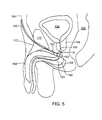
- FIG. 6 is a partial frontal view of a male patient.
- FIG. 7 is a simplified cross-sectional view of the external urinary sphincter and urethra of a patient with a probe inserted in the urethra.
- FIG. 8 is a simplified cross-sectional view of the external urinary sphincter and the urethra of a patient illustrating the insertion of a distal end of a stimulation lead in accordance with embodiments of the invention.
- FIG. 9 is a simplified cross-sectional view of the external urinary sphincter and the urethra of a patient taken generally along line 9 - 9 of FIG. 8 .
- FIGS. 10-12 Illustrate various steps of a method of implanting an electrode of an electronic stimulator device in an external urinary sphincter of a male patient, in accordance with embodiments of the invention.
- Embodiments of the present invention are directed to a method of implanting an electrode of an electronic stimulator device into a pelvic floor muscular structure of a male or female patient.
- the patient is a male patient.
- exemplary implantable electronic stimulator devices will be described with reference to FIGS. 1 and 2 .
- FIG. 1 is a schematic, pictorial view of an exemplary implantable electronic stimulator device 100 , in accordance with embodiments of the invention.
- Device 100 is configured for implantation into the pelvic region of a patient, as described in detail below.
- the device 100 can be used to provide muscle and/or nerve stimulation to control and/or treat a pelvic condition, such as pelvic pain, urinary incontinence and other pelvic conditions.
- the device 100 comprises a control unit 102 and one or more electrodes, generally referred to as 104 , such as electrodes 104 A and 104 B. Electrodes 104 are coupled to the control unit 102 by leads 106 .
- the device 100 includes at least one physiological sensor 108 , such as a miniature ultrasound transducer, one or more accelerometers, a pressure transducer or other sensors known in the art.
- control unit 102 comprises circuitry for senses electrical signals received by the electrodes 104 , such as electromyogram (EMG) signals, along with circuitry for processing the signals from the sensor 108 .
- control unit 104 comprises circuitry for applying electrical stimulation waveforms (i.e., electrical signals) to one or more of the electrodes 104 .
- the electrical stimulation waveforms are designed to control and/or treat the desired condition of the pelvic region.
- control unit 102 and the electrodes 104 are as described in the above-referenced patents, in PCT Patent Publication WO 00-19940, entitled “Incontinence Treatment Device,” and/or in PCT Patent Publication WO 00-19939, entitled “Control of Urge Incontinence,” with appropriate changes as are otherwise indicated by clinical and engineering considerations that are clear to those skilled in the art.
- the electrodes 104 are flexible intramuscular-type wire electrodes, approximately 1-35 millimeters long and 50-100 microns in diameter, in order to minimize patient discomfort.
- the electrodes 104 comprise a spiral hook, as known in the art, so that they can be easily and permanently anchored in a pelvic muscle of a patient.
- the wire, from which the electrodes 104 are made, comprises a suitable conductive material, such as a biocompatible metal such as silver, a platinum/iridium alloy (90-10) or a nickel-chromium alloy.
- the leads 106 have a length that is suitable for the application, such as 5-10 centimeters long, and are surrounded by an insolating jacket 110 typically comprising silicone, polyurethane or and other flexible, biocompatible insolating material.
- An optional additional wire (not shown) inside the jacket 110 can serve as an antenna for the purpose of wireless communications with the device 100 , in accordance with known methods.
- the control unit 102 comprises a circuitry for processing electrical signals received from the electrodes 104 and/or for applying an electrical waveform to one or both of the electrodes 104 .
- the circuitry is contained in a case 112 made of titanium or other suitable biocompatible metal.
- the case 112 is about 20 millimeters in diameter and 4 millimeters thick.
- the case 112 serves as a ground electrode for the electrodes 104 when they are sensing or stimulating in a monopolar mode.
- the case 112 may comprise metal coated with a layer of biocompatible plastic, such as polymethyl methacrylate (PMMA) or silicon.
- PMMA polymethyl methacrylate
- FIG. 2 is a side plan view of an exemplary electronic stimulator device 100 , in accordance with another embodiment of the invention. Except with respect to the difference described below, the embodiment of the device 100 shown in FIG. 2 is generally similar to the embodiments shown in FIG. 1 , and techniques described herein with respect to one of these configurations can generally be applied to the other configuration. Accordingly, elements in FIG. 2 that are labeled with the same or similar numbers as that used in FIG. 1 , generally correspond to the same or similar elements.
- One embodiment of the device 100 shown in FIG. 2 comprises a control unit 102 , at least one electrode 104 and a lead 106 connecting electrode 104 to the control unit 102 .
- the lead 106 includes a proximal end 114 that is coupled to the control unit 102 via a connector 115 and a distal end 116 at which the electrode 104 is located. Additional leads 106 or sensors 108 may be coupled to the control unit 102 at a suitable interface, such as interface 118 .
- the electrode 104 can be anchored to a pelvic floor muscle of the patient (e.g., the external urinary or anal sphincter), by means of a fixation element 120 , such as a helix, spiral hook, polypropylene mesh, or other anchor known in the art, as shown in the magnified schematic illustration of the distal end 116 of the lead 106 provided in FIG. 2 .
- the helix or spiral hook 120 can be embedded within the external urinary sphincter to anchor the lead 106 thereto.
- the fixation element 120 operates to provide electrical contact between the muscle and one or more stimulation electrodes 104 A and 104 B disposed on a silicone casing 122 of the lead 106 .
- the electrodes are approximately 3 millimeters in length, but can be much longer, such as less than about 80 millimeters in length, for example.
- the electrodes 104 are typically separated by approximately 3 millimeters along the length of the lead 106 .
- a tip 124 of an EMG wire 126 may protrude approximately 100 microns through the casing 124 , for those applications in which EMG sensing is desirable.
- the diameter of the wire 126 is approximately 50 microns, and the diameter of the casing 124 is approximately 1.5 millimeters.
- one embodiment of the device 100 illustrated in FIG. 2 comprises circuitry for applying electrical stimulation waveforms to the muscular tissue, in which the fixation element 120 is embedded and/or for sensing electrical signals received by the electrodes 104 , in accordance with conventional implantable electronic stimulator devices known in the art.
- Embodiments of the invention are directed to a method of installing the distal end 116 of the stimulation lead 106 into a pelvic floor muscular structure of a patient that is deep to, or below the pubic symphysis.
- the pelvic floor muscular structure being targeted by the distal end 116 of the stimulation lead 106 is the external urinary sphincter of the patient, as described in detail below.
- Other exemplary pelvic floor muscular structures that may be implanted with the distal end of the stimulation lead using embodiments of the method include Puborectalis, Pubococcygeus, and Iliococcygeus (individual elements of the Lavator ani muscular complex).
- FIG. 3 is a flowchart illustrating steps of the method, embodiments of which will initially be discussed with reference to FIGS. 4 and 5 , which are cross-sectional views of the pelvic region of a male patient.
- FIGS. 4 and 5 illustrate certain pelvic anatomy including the bladder 150 that connects to a proximal end 152 of the urethra 154 , at the bladder neck 156 .
- the urethra 154 extends distally from the bladder 150 through the prostate 158 and the external urinary sphincter 160 and below the prostate 158 through the perineal membrane 162 .
- the portion of the urethra 154 located distally relative to the perineal membrane 162 is referred to as the “bulbar urethra” 164 .
- Tissue below the perineal membrane 162 and the bulbar urethra 164 includes the corpus spongiosum 166 and the bulbospongiosus muscle 168 . Also illustrated is the pubic symphysis 170 and the suspensory ligament 172 located adjacent the notch in the posterior edge of the pubic symphysis 170 . While the drawings are of a male patient and some of the method steps are only applicable to male patients, it is understood that embodiments of the present invention may be applied to female patients.
- a stimulation lead such as the lead 106 ( FIGS. 1 and 2 ) in accordance with one or more of the embodiments described above.
- the stimulation lead 106 has proximal end 114 and a distal end 116 .
- the distal end 116 comprises one or more electrodes 104 , as illustrated in FIG. 2 .
- the distal end 116 of the stimulation lead 106 is fed along a path 184 , which is adjacent the pubic symphysis 170 , toward the pelvic floor muscular structure of the patient, such as the depicted external urinary sphincter 160 , as illustrated in FIG. 4 .
- the path 184 is positioned between the ventral face 186 of the pubic symphysis 170 and the penis 188 .
- the path 184 preferably avoids contacting the vessels and nerves across the top of the penis 188 during the feeding step 182 and subsequent steps of the method.
- a conventional introducer can bee used to feed the distal end 116 of the stimulation lead 106 along the path 184 .
- an incision 190 is initially made in the patient adjacent the notch in the posterior edge of the pubic symphysis 170 through which the distal end 116 of the stimulation lead 106 is fed into the patient along the path 184 in step 182 , as illustrated in the frontal view of the patient provided in FIG. 6 .
- the incision is approximately 1.5-2 cm lateral to the notch in the pubic symphysis 170 .
- the notch and the adjacent suspensory ligament 172 can be located, for example, by placing the penis 188 of the male patient on a stretch and palpating for the notch in the posterior edge of the pubic symphysis 170 .
- the distal end 116 of the stimulation lead 106 is fed along the path 184 and into the desired pelvic floor muscular structure of the patient, such as the depicted external urinary sphincter 160 , at an insertion point 194 to place the one or more electrodes 104 in contact with the external urinary sphincter 160 or another desired pelvic floor muscular structure, as illustrated in FIG. 5 .
- FIG. 7 is a simplified cross-sectional view of the external urinary sphincter 160 and urethra 154 of the patient.
- the bladder side of the probe 196 is indicated at 198 .
- the probe 194 comprises an ultrasound probe, which can be used to approximate the location of the distal end 116 of the stimulation lead 106 relative to the probe 196 and, thus, determine the location of the distal end 116 of the stimulation lead 106 relative to the urethra 154 and external urinary sphincter 160 of the patient.
- the surgeon palpates for the probe 196 through the rectum 200 ( FIGS. 4 and 5 ) to feel the location of the probe 196 within the urethra 154 adjacent to the desired insertion point 194 of the external urinary sphincter 160 .
- surgeon may palpate for the prostate 158 of the patient, from which the location of the external urinary sphincter 160 can be approximated.
- the path 184 passes through the perineal membrane 162 of the patient.
- the distal end 116 of the stimulation lead 106 or an introducer used to feed the stimulation lead 106 , encounters the perineal membrane 162 and an increase in resistance to the feeding of the distal end 116 can be sensed by the surgeon. This increase in resistance is followed by a decrease in resistance upon piercing the perineal membrane 162 . Accordingly, the sensed decrease in resistance indicates that the distal end 116 of the stimulation lead 106 is positioned slightly beyond the perineal membrane 162 .
- the surgeon feeds the distal end 116 of the stimulation lead 106 approximately 1 cm to place the distal end 116 and the one or more electrodes 104 within the external urinary sphincter 160 .
- the feeding step 192 comprises feeding the distal end 116 of the stimulation lead 106 into the external urinary sphincter 160 at the insertion point 194 at an angle 202 that is oblique to a plane 204 extending perpendicular to a central axis 206 of the urethra 154 and through the insertion point 194 , as illustrated in FIG. 8 , which is a simplified cross-sectional view of the external urinary sphincter 160 and the urethra 154 of the patient.
- the central axis 206 is defined as the approximate center of the urethra 154 aligned parallel to the general flow of fluid through the urethra 154 .
- the angle 202 is measured from the plane 204 to a central axis 207 of the stimulation lead 106 . In one embodiment, the angle 202 is in the range of 0-45 degrees.
- the feeding step 192 comprises feeding the distal end 116 of the stimulation lead 106 into the external urinary sphincter 160 along a path 208 that does not intersect the urethra 154 , as illustrated in FIG. 9 , which is a simplified cross-sectional view of the external urinary sphincter 160 and the urethra 154 of the patient taken generally along line 9 - 9 of FIG. 8 . This ensures that the distal end 116 of the stimulation lead 106 does not pierce the urethra 154 .
- a helical coil, a suture, or other conventional anchor or fixation element 120 may be attached to the distal end 116 of the stimulation lead 106 and embedded in the external urinary sphincter 160 or other desired pelvic floor muscular structure of the patient.
- the anchor 120 operates to resist relative movement between the distal end 116 of the lead 106 and the sphincter tissue.
- the one or more electrodes 104 at the distal end 116 of the lead 106 can be used to sense signals conducted through the pelvic floor muscular structure and/or apply electrical signals to the pelvic floor muscular structure when the proximal end 114 of the stimulation lead 106 is connected to the control unit 102 .
- the one or more electrodes 104 can be used to sense signals conducted through the external urinary sphincter 130 and/or apply electrical signals to the external urinary sphincter 130 , when the proximal end 114 of the stimulation lead 106 is connected to the control unit 102 .
- Additional embodiments of the invention are directed to connecting the control unit 102 to the lead 106 and implanting the control unit 102 in the patient.
- the proximal end 114 of the stimulation lead 106 is connected to the control unit 102 .
- the control unit 102 generates electrical signals that are applied to the external urinary sphincter 160 through the one or more electrodes 104 at the distal end 116 of the stimulation lead 106 that are embedded in the pelvic floor muscular structure to treat pelvic pain, urinary incontinence and/or another pelvic condition of the patient.
- control unit 102 receives signals from the one ore more electrodes 104 embedded in the pelvic floor muscular structure, such as the external urinary sphincter 160 , or from a sensor 108 ( FIG. 1 ), and applies the electrical signals to the pelvic floor muscular structure through one or more of the electrodes 104 of the stimulation lead 106 responsive to the received signals.
- an incision 220 is formed in the patient, as shown in the frontal view of the patient provided in FIG. 10 .
- the incision 220 is formed in the abdomen of the patient.
- the incision 220 comprises a 2-3 centimeter long, horizontal skin incision that is approximately 6 centimeters lateral to and 4 centimeters cephalad to the pubic symphysis bone 170 of the patient.
- the incision 220 is formed in the subcutaneous tissue adjacent to the fascia of the abdomen of the patient using blunt dissection to form an abdominal pocket 222 .
- the abdominal pocket 222 is sized to accommodate a control unit 102 of an implantable electronic stimulator device, such as one of the exemplary control units 102 described above.
- the abdominal pocket 222 may comprise a depth of approximately 4 centimeters in order to accommodate the control unit 102 .
- An antibiotic soaked pad may be placed in the abdominal pocket 222 .
- a subcutaneous tunnel 224 is formed between the incision 190 and the incision 220 using, for example, a suitable introducer, such as those described in U.S. patent application Ser. No. 11/961,615 filed Dec. 20, 2007, which is incorporated herein by reference in its entirety.
- the proximal end 114 of the stimulation lead 106 is then fed through the tunnel 224 and out the incision 220 , as illustrated in FIG. 10 , which is a partial frontal view of the patient.
- the feeding of the proximal end 114 of the stimulation lead 106 through the tunnel 224 can be accomplished by pushing the proximal end 114 through a tube (not shown) installed in the tunnel 224 , pulling the proximal end 114 through the tunnel 224 or other conventional technique. Additionally, it is understood that the feeding of the stimulation lead 106 through the tunnel 224 may be accomplished prior to the feeding of the distal end 116 of the stimulation lead 106 into the external urinary sphincter of the patient. In such a case, the distal end 116 of the stimulation lead 106 may be fed (i.e., pulled or pushed) through the tunnel 224 from the incision 220 and out the incision 190 . Once the stimulation lead 106 is fed through the tunnel 224 , slack in the stimulation lead is eliminated and the incision 190 can be closed.
- the connecting portion 115 at the proximal end 114 is installed in a corresponding socket 226 of the control unit 102 ( FIG. 2 ), to complete the assembly of the electrical stimulation device 100 .
- a seal is formed between the socket 226 and the connector 115 using an o-ring or other suitable component, to prevent fluids from entering the socket 226 .
- the device 100 is preferably tested to ensure that it is working properly including, for example, generating the desired electrical waveforms and applying the electrical waveforms or signals to the external urinary sphincter 160 through the one or more electrodes 104 . If the device 100 is operating properly, the lead 106 can be tucked into the abdominal pocket 222 and sutures 228 are looped through suture holes 230 ( FIG. 2 ) and used to secure the control unit 102 to the fascia within the pocket, as illustrated in FIG. 11 . In one embodiment, the control unit 102 is located 4 centimeters or less from the surface of the skin in the subcutaneous tissue to allow the unit 102 to receive programming signals. Once the control unit 102 is secured within the abdominal pocket 222 , the incision 220 is sealed to complete the implantation of the device 100 in the patient.
- control unit 102 is not connected to the connector 115 of the lead 106 following the feeding of the proximal end 114 through the tunnel 224 and out the incision 220 , and the control unit is not initially installed in the abdominal pocket 222 .
- an incision 240 such as a horizontal incision approximately 1 centimeter long, is made on the contralateral side of the patient's abdomen, as shown in FIG. 12 .
- the incision 240 is approximately 15 centimeters from the initial abdomen incision 220 .
- a subcutaneous tunnel is formed between the incision 220 and the incision 240 , in accordance with conventional methods.
- An extension lead 242 is fed through the tunnel such that the ends of the lead 242 extend outside the incisions 240 and 220 .
- a distal 244 end of the extension lead 242 is attached to the proximal end 114 of the stimulation lead 106 , as illustrated in FIG. 12 .
- the distal end 244 of the extension lead 242 includes a socket 246 to which the connector 115 at the proximal end 114 of the lead 106 is attached.
- the socket 246 receives the connector 115 and set screws are used to secure the connection.
- a seal is preferably formed between the socket 246 and the connector 115 using an o-ring or other suitable component to prevent fluids from entering the socket 246 in accordance with conventional methods.
- the socket 246 of the extension lead 242 and the proximal end 114 of the lead 106 are packed into the abdominal pocket 222 , as illustrated in FIG. 12 .
- the incision 220 is then closed over the lead extension socket 246 in two layers.
- a connector 248 at the proximal end 250 of the extension lead 242 is then installed in the socket 226 of the control unit 102 or other testing unit that is external to the patient.
- the connection of the control unit 102 to the extension lead 242 via connector 248 , and the extension lead 242 to the lead 106 via the installation of the connector 115 in the socket 246 allows the control unit 102 or testing device to send electrical signals to, and/or receive electrical signals from, the one or more electrodes 104 embedded in the pelvic floor muscular structure of the patient. Testing of the device 100 can then commence.
- the control unit can be worn on a belt outside of the body of the patient, if desired.
- the incision 220 can be reopened to expose the socket 246 and the connector 115 .
- the connector 115 is disconnected from the socket 246 and the extension lead 242 is pulled through the incision 240 .
- the connector 115 can then be installed directly in the socket 226 of the control unit and installed in the abdominal pocket of the patient as described above to complete the implantation of the device 100 in the patient.
Abstract
Description
Claims (20)
Priority Applications (1)
| Application Number | Priority Date | Filing Date | Title |
|---|---|---|---|
| US12/406,434 US9539433B1 (en) | 2009-03-18 | 2009-03-18 | Electrode implantation in a pelvic floor muscular structure |
Applications Claiming Priority (1)
| Application Number | Priority Date | Filing Date | Title |
|---|---|---|---|
| US12/406,434 US9539433B1 (en) | 2009-03-18 | 2009-03-18 | Electrode implantation in a pelvic floor muscular structure |
Publications (1)
| Publication Number | Publication Date |
|---|---|
| US9539433B1 true US9539433B1 (en) | 2017-01-10 |
Family
ID=57705795
Family Applications (1)
| Application Number | Title | Priority Date | Filing Date |
|---|---|---|---|
| US12/406,434 Expired - Fee Related US9539433B1 (en) | 2009-03-18 | 2009-03-18 | Electrode implantation in a pelvic floor muscular structure |
Country Status (1)
| Country | Link |
|---|---|
| US (1) | US9539433B1 (en) |
Cited By (2)
| Publication number | Priority date | Publication date | Assignee | Title |
|---|---|---|---|---|
| US20180231436A1 (en) * | 2017-02-13 | 2018-08-16 | City University Of Hong Kong | Apparatus and method for testing performance of an electrosurgical tool |
| WO2020231440A1 (en) * | 2019-05-16 | 2020-11-19 | Board Of Regents, The University Of Texas System | Devices and methods for neuromodulation |
Citations (229)
| Publication number | Priority date | Publication date | Assignee | Title |
|---|---|---|---|---|
| US3628538A (en) | 1968-09-18 | 1971-12-21 | Nat Res Dev | Apparatus for stimulating muscles controlled by the same muscles |
| US3640284A (en) | 1970-01-05 | 1972-02-08 | Philip A De Langis | Apparatus for electrotherapy of the pubococcygeus |
| US3646940A (en) | 1969-07-15 | 1972-03-07 | Univ Minnesota | Implantable electronic stimulator electrode and method |
| US3650276A (en) | 1969-03-26 | 1972-03-21 | Inst Demedicina Si Farmacie | Method and apparatus, including a flexible electrode, for the electric neurostimulation of the neurogenic bladder |
| US3662758A (en) | 1969-06-30 | 1972-05-16 | Mentor Corp | Stimulator apparatus for muscular organs with external transmitter and implantable receiver |
| US3667477A (en) | 1966-11-25 | 1972-06-06 | Canadian Patents Dev | Implantable vesical stimulator |
| US3866613A (en) | 1972-07-13 | 1975-02-18 | Devices Implants Limited | Pessary ring electrode system |
| US3870051A (en) | 1972-04-27 | 1975-03-11 | Nat Res Dev | Urinary control |
| US3926178A (en) | 1975-01-17 | 1975-12-16 | Alvin N Feldzamen | Apparatus for aiding the voluntary exercising of sphincter muscles |
| US3941136A (en) | 1973-11-21 | 1976-03-02 | Neuronyx Corporation | Method for artificially inducing urination, defecation, or sexual excitation |
| US3983881A (en) | 1975-05-21 | 1976-10-05 | Telectronics Pty. Limited | Muscle stimulator |
| US3983865A (en) | 1975-02-05 | 1976-10-05 | Shepard Richard S | Method and apparatus for myofunctional biofeedback |
| US4010758A (en) | 1975-09-03 | 1977-03-08 | Medtronic, Inc. | Bipolar body tissue electrode |
| US4023574A (en) | 1974-10-18 | 1977-05-17 | Hans Nemec | Electrostimulation method and apparatus |
| US4030509A (en) | 1975-09-30 | 1977-06-21 | Mieczyslaw Mirowski | Implantable electrodes for accomplishing ventricular defibrillation and pacing and method of electrode implantation and utilization |
| US4044774A (en) | 1976-02-23 | 1977-08-30 | Medtronic, Inc. | Percutaneously inserted spinal cord stimulation lead |
| US4106511A (en) | 1976-04-21 | 1978-08-15 | Svenska Utvecklingsaktiebolaget | Electrical stimulator in remedy of incontinence |
| US4136684A (en) | 1977-02-07 | 1979-01-30 | Scattergood Mark G | Linear electromyographic biofeedback system |
| US4139006A (en) | 1977-03-18 | 1979-02-13 | Corey Arthur E | Female incontinence device |
| US4153059A (en) | 1977-10-25 | 1979-05-08 | Minnesota Mining And Manufacturing Company | Urinary incontinence stimulator system |
| US4157087A (en) | 1978-03-06 | 1979-06-05 | Med General, Inc. | Peripheral nerve stimulator |
| US4165750A (en) | 1978-03-18 | 1979-08-28 | Aleev Leonid S | Bioelectrically controlled electric stimulator of human muscles |
| US4177819A (en) | 1978-03-30 | 1979-12-11 | Kofsky Harvey I | Muscle stimulating apparatus |
| US4222377A (en) | 1977-06-27 | 1980-09-16 | American Medical Systems, Inc. | Pressure regulated artificial sphincter systems |
| US4290420A (en) | 1980-06-09 | 1981-09-22 | Alberto Manetta | Stress incontinence diagnostic and treatment device |
| US4387719A (en) | 1980-10-23 | 1983-06-14 | Gorenje Tovarna Gospodinjske Opreme N.Sol.O. Velenje | Control circuit of a therapeutic stimulator for the urinary incontinence |
| US4402328A (en) | 1981-04-28 | 1983-09-06 | Telectronics Pty. Limited | Crista terminalis atrial electrode lead |
| US4406288A (en) | 1981-04-06 | 1983-09-27 | Hugh P. Cash | Bladder control device and method |
| US4414986A (en) | 1982-01-29 | 1983-11-15 | Medtronic, Inc. | Biomedical stimulation lead |
| US4431001A (en) | 1980-09-17 | 1984-02-14 | Crafon Medical Ab | Stimulator system |
| US4457299A (en) | 1981-02-06 | 1984-07-03 | Cornwell George H I | Incontinence control devices |
| US4492233A (en) | 1982-09-14 | 1985-01-08 | Wright State University | Method and apparatus for providing feedback-controlled muscle stimulation |
| US4515167A (en) | 1983-02-28 | 1985-05-07 | Hochman Joel S | Device for the development, training and rehabilitation of the pubococcygeal and related perineal musculature of the female |
| US4542753A (en) | 1982-12-22 | 1985-09-24 | Biosonics, Inc. | Apparatus and method for stimulating penile erectile tissue |
| US4568339A (en) | 1982-11-05 | 1986-02-04 | Craig Medical Products, Limited | Female incontinence device |
| US4569351A (en) | 1984-12-20 | 1986-02-11 | University Of Health Sciences/The Chicago Medical School | Apparatus and method for stimulating micturition and certain muscles in paraplegic mammals |
| US4571749A (en) | 1982-09-21 | 1986-02-25 | The Johns Hopkins University | Manually actuated hydraulic sphincter |
| US4580578A (en) | 1983-05-06 | 1986-04-08 | Richard Wolf Gmbh | Device for the treatment of female urinary incontinence |
| US4585005A (en) | 1984-04-06 | 1986-04-29 | Regents Of University Of California | Method and pacemaker for stimulating penile erection |
| US4602624A (en) | 1984-10-11 | 1986-07-29 | Case Western Reserve University | Implantable cuff, method of manufacture, and method of installation |
| US4607639A (en) | 1984-05-18 | 1986-08-26 | Regents Of The University Of California | Method and system for controlling bladder evacuation |
| US4628942A (en) | 1984-10-11 | 1986-12-16 | Case Western Reserve University | Asymmetric shielded two electrode cuff |
| US4688575A (en) | 1982-03-12 | 1987-08-25 | Duvall Wilbur E | Muscle contraction stimulation |
| US4703755A (en) | 1984-05-18 | 1987-11-03 | The Regents Of The University Of California | Control system for the stimulation of two bodily functions |
| US4731083A (en) | 1982-09-21 | 1988-03-15 | The Johns Hopkins University | Manually actuated hydraulic sphincter |
| US4735205A (en) | 1986-02-24 | 1988-04-05 | Medtronic, Inc. | Method and apparatus including a sliding insulation lead for cardiac assistance |
| US4739764A (en) | 1984-05-18 | 1988-04-26 | The Regents Of The University Of California | Method for stimulating pelvic floor muscles for regulating pelvic viscera |
| US4750494A (en) | 1981-05-12 | 1988-06-14 | Medtronic, Inc. | Automatic implantable fibrillation preventer |
| US4771779A (en) | 1984-05-18 | 1988-09-20 | The Regents Of The University Of California | System for controlling bladder evacuation |
| US4785828A (en) | 1986-10-06 | 1988-11-22 | Empi, Inc. | Vaginal stimulator for controlling urinary incontinence in women |
| US4881526A (en) | 1988-05-27 | 1989-11-21 | Empi, Inc. | Intravaginal electrode and stimulation system for controlling female urinary incontinence |
| US4913164A (en) | 1988-09-27 | 1990-04-03 | Intermedics, Inc. | Extensible passive fixation mechanism for lead assembly of an implantable cardiac stimulator |
| US4941874A (en) | 1987-08-11 | 1990-07-17 | Hoechst Aktiengesellschaft | Device for the administration of implants |
| EP0245547B1 (en) | 1986-05-12 | 1990-08-22 | The Regents Of The University Of California | Electronic control system for controlling pelvic viscera via neuro-electrical stimulation |
| WO1990012617A1 (en) | 1989-04-26 | 1990-11-01 | B.V. Optische Industrie 'de Oude Delft' | Electrode for stimulating and/or detecting the muscle activity of muscles or muscle groups of a patient which are accessible through a body orifice |
| US5013292A (en) | 1989-02-24 | 1991-05-07 | R. Laborie Medical Corporation | Surgical correction of female urinary stress incontinence and kit therefor |
| US5019032A (en) | 1990-04-03 | 1991-05-28 | Robertson Jack R | Refined suspension procedure with implement for treating female stress incontinence |
| US5082006A (en) | 1987-09-15 | 1992-01-21 | Linda Jonasson | Device for preventing involuntary micturition |
| US5094242A (en) | 1988-11-07 | 1992-03-10 | Regents Of The University Of California | Implantable nerve stimulation device |
| US5103835A (en) | 1990-05-02 | 1992-04-14 | Nihon Kohden Corporation | Impedance monitoring device for preventing urinary incontinence |
| US5112344A (en) | 1988-10-04 | 1992-05-12 | Petros Peter E | Surgical instrument and method of utilization of such |
| US5193539A (en) | 1991-12-18 | 1993-03-16 | Alfred E. Mann Foundation For Scientific Research | Implantable microstimulator |
| US5193540A (en) | 1991-12-18 | 1993-03-16 | Alfred E. Mann Foundation For Scientific Research | Structure and method of manufacture of an implantable microstimulator |
| US5199430A (en) | 1991-03-11 | 1993-04-06 | Case Western Reserve University | Micturitional assist device |
| US5285781A (en) | 1990-05-26 | 1994-02-15 | Stiwell S. A. | Electrical neuromuscular stimulation device |
| US5291902A (en) | 1993-01-11 | 1994-03-08 | Brent Carman | Incontinence treatment |
| US5312439A (en) | 1991-12-12 | 1994-05-17 | Loeb Gerald E | Implantable device having an electrolytic storage electrode |
| US5324323A (en) | 1992-09-09 | 1994-06-28 | Telectronics Pacing Systems, Inc. | Multiple channel cardiosynchronous myoplasty apparatus |
| US5324324A (en) | 1992-10-13 | 1994-06-28 | Siemens Pacesetter, Inc. | Coated implantable stimulation electrode and lead |
| US5330507A (en) | 1992-04-24 | 1994-07-19 | Medtronic, Inc. | Implantable electrical vagal stimulation for prevention or interruption of life threatening arrhythmias |
| US5358514A (en) | 1991-12-18 | 1994-10-25 | Alfred E. Mann Foundation For Scientific Research | Implantable microdevice with self-attaching electrodes |
| US5366493A (en) | 1991-02-04 | 1994-11-22 | Case Western Reserve University | Double helix functional stimulation electrode |
| US5370670A (en) | 1993-12-13 | 1994-12-06 | Thomas Jefferson University | Detrusor myoplasty and neuromuscular electrical stimulation of the urinary bladder |
| US5385577A (en) | 1992-11-12 | 1995-01-31 | Empi, Inc. | Electrode for activating pelvic reflexes |
| US5417226A (en) | 1994-06-09 | 1995-05-23 | Juma; Saad | Female anti-incontinence device |
| US5423329A (en) | 1994-04-15 | 1995-06-13 | Rehab Centers Of America, Inc. | Method of treatment for urinary incontinence |
| US5425751A (en) | 1993-07-30 | 1995-06-20 | Medtronic, Inc. | Method and apparatus for optimum positioning of a muscle stimulating implant |
| US5452719A (en) | 1991-07-23 | 1995-09-26 | Eisman; Eugene | Multiple electrode myographic probe and method |
| US5484445A (en) | 1993-10-12 | 1996-01-16 | Medtronic, Inc. | Sacral lead anchoring system |
| US5518504A (en) | 1993-12-28 | 1996-05-21 | American Medical Systems, Inc. | Implantable sphincter system utilizing lifting means |
| WO1996004955A3 (en) | 1994-08-16 | 1996-05-23 | Cortrak Medical Inc | Polymer matrix drug delivery apparatus and method |
| US5520606A (en) | 1990-10-18 | 1996-05-28 | Schoolman; Arnold | Mechanical urinary sphincter device |
| US5562717A (en) | 1992-05-23 | 1996-10-08 | Axelgaard Manufacturing Company, Ltd. | Electrical stimulation for treatment of incontinence and other neuromuscular disorders |
| WO1996032916A1 (en) | 1995-04-21 | 1996-10-24 | Multicept Aps | A vibrator |
| US5569351A (en) | 1994-11-14 | 1996-10-29 | Cms Gilbreth Packaging Systems, Inc. | Banding machine having improved film registration system |
| US5571148A (en) | 1994-08-10 | 1996-11-05 | Loeb; Gerald E. | Implantable multichannel stimulator |
| US5611768A (en) | 1994-07-01 | 1997-03-18 | Tutrone, Jr.; Ronald F. | Inflatable vaginal pessary |
| US5611515A (en) | 1991-12-03 | 1997-03-18 | Boston Scientic Corporation | Bladder neck suspension procedure |
| US5634462A (en) | 1993-10-15 | 1997-06-03 | Case Western Reserve University | Corrugated inter-fascicular nerve cuff method and apparatus |
| GB2309388A (en) | 1996-01-24 | 1997-07-30 | Shantha Jayatilake Ba Lenadora | Implantable device for incontinence relief in the female |
| WO1998017190A2 (en) | 1996-10-23 | 1998-04-30 | Oratec Interventions, Inc. | Method and apparatus for treating intervertebral discs |
| US5766229A (en) | 1996-04-15 | 1998-06-16 | Pacesetter, Inc. | Capture verification method and apparatus for implantable pacemaker utilizing heart rhythm stability measurements to minimize the likelihood of fusion |
| US5785666A (en) | 1995-10-31 | 1998-07-28 | Ergonomic Technologies Corporation | Portable electronic data collection apparatus for monitoring musculoskeletal stresses |
| US5807397A (en) | 1995-01-04 | 1998-09-15 | Plexus, Inc. | Implantable stimulator with replenishable, high value capacitive power source and method therefor |
| US5824027A (en) | 1997-08-14 | 1998-10-20 | Simon Fraser University | Nerve cuff having one or more isolated chambers |
| US5833595A (en) | 1994-09-06 | 1998-11-10 | Lin; Vernon Wen-Hau | Treatment of excretory problems |
| US5836994A (en) | 1997-04-30 | 1998-11-17 | Medtronic, Inc. | Method and apparatus for electrical stimulation of the gastrointestinal tract |
| US5842478A (en) | 1991-12-03 | 1998-12-01 | Boston Scientific Technology, Inc. | Method of securing a bone anchor |
| US5876353A (en) | 1997-01-31 | 1999-03-02 | Medtronic, Inc. | Impedance monitor for discerning edema through evaluation of respiratory rate |
| US5899909A (en) | 1994-08-30 | 1999-05-04 | Medscand Medical Ab | Surgical instrument for treating female urinary incontinence |
| US5927282A (en) | 1991-01-10 | 1999-07-27 | Uromed Corporation | Controlling urinary incontinence |
| US5941903A (en) | 1998-04-30 | 1999-08-24 | Cardiac Pacemakers, Inc | Pacemaker for detection of evoked response |
| US5954761A (en) | 1997-03-25 | 1999-09-21 | Intermedics Inc. | Implantable endocardial lead assembly having a stent |
| US5957965A (en) | 1997-03-03 | 1999-09-28 | Medtronic, Inc. | Sacral medical electrical lead |
| US5957920A (en) | 1997-08-28 | 1999-09-28 | Isothermix, Inc. | Medical instruments and techniques for treatment of urinary incontinence |
| US5963097A (en) | 1998-01-27 | 1999-10-05 | Garachtchenko; Alexander Viktorovich | Low-noise amplifier |
| US5978712A (en) | 1996-10-30 | 1999-11-02 | Nihon Kohden Corporation | Stimulating apparatus for preventing urinary incontinence |
| US5984854A (en) | 1996-02-15 | 1999-11-16 | Nihon Kohden Corporation | Method for treating urinary incontinence and an apparatus therefor |
| US6002964A (en) | 1998-07-15 | 1999-12-14 | Feler; Claudio A. | Epidural nerve root stimulation |
| WO2000000082A1 (en) | 1998-06-29 | 2000-01-06 | The Procter & Gamble Company | Disposable article having proactive sensor |
| US6026326A (en) | 1997-01-13 | 2000-02-15 | Medtronic, Inc. | Apparatus and method for treating chronic constipation |
| US6027456A (en) | 1998-07-10 | 2000-02-22 | Advanced Neuromodulation Systems, Inc. | Apparatus and method for positioning spinal cord stimulation leads |
| US6038463A (en) | 1997-09-26 | 2000-03-14 | Medtronic, Inc. | Medical electrical lead |
| US6039686A (en) | 1997-03-18 | 2000-03-21 | Kovac; S. Robert | System and a method for the long term cure of recurrent urinary female incontinence |
| US6042534A (en) | 1997-02-13 | 2000-03-28 | Scimed Life Systems, Inc. | Stabilization sling for use in minimally invasive pelvic surgery |
| WO2000019940A1 (en) | 1998-10-06 | 2000-04-13 | Bio Control Medical, Ltd. | Incontinence treatment device |
| US6051017A (en) | 1996-02-20 | 2000-04-18 | Advanced Bionics Corporation | Implantable microstimulator and systems employing the same |
| US6055456A (en) | 1999-04-29 | 2000-04-25 | Medtronic, Inc. | Single and multi-polar implantable lead for sacral nerve electrical stimulation |
| US6061596A (en) | 1995-11-24 | 2000-05-09 | Advanced Bionics Corporation | Method for conditioning pelvic musculature using an implanted microstimulator |
| US6078840A (en) | 1997-04-30 | 2000-06-20 | Medtronic, Inc. | Medical electrical lead having improved fixation |
| US6104960A (en) | 1998-07-13 | 2000-08-15 | Medtronic, Inc. | System and method for providing medical electrical stimulation to a portion of the nervous system |
| US6104955A (en) | 1997-12-15 | 2000-08-15 | Medtronic, Inc. | Method and apparatus for electrical stimulation of the gastrointestinal tract |
| US6110101A (en) | 1998-08-13 | 2000-08-29 | Conticare Medical, Inc. | Bladder sling |
| US6128536A (en) | 1995-03-13 | 2000-10-03 | Medizintechnik Dipl.-Ing. Heise Vertriebs Gmbh | Therapy apparatus for the functional electromyostimulation of smooth muscle cells |
| US6135945A (en) | 1997-08-04 | 2000-10-24 | Sultan; Hashem | Anti-incontinence device |
| WO2000001320A9 (en) | 1998-07-06 | 2000-11-02 | Advanced Bionics Corp | Implantable stimulator system and method for treatment of urinary incontinence |
| US6161029A (en) | 1999-03-08 | 2000-12-12 | Medtronic, Inc. | Apparatus and method for fixing electrodes in a blood vessel |
| US6178356B1 (en) | 1998-02-20 | 2001-01-23 | Cardiac Pacemakers, Inc. | Coronary venous lead having fixation mechanism |
| US6185452B1 (en) | 1997-02-26 | 2001-02-06 | Joseph H. Schulman | Battery-powered patient implantable device |
| US6208894B1 (en) | 1997-02-26 | 2001-03-27 | Alfred E. Mann Foundation For Scientific Research And Advanced Bionics | System of implantable devices for monitoring and/or affecting body parameters |
| US6240315B1 (en) | 1998-02-25 | 2001-05-29 | Seung Kee Mo | Electrical apparatus for medical treatment using EMG envelope signal |
| US6240316B1 (en) | 1998-08-14 | 2001-05-29 | Advanced Bionics Corporation | Implantable microstimulation system for treatment of sleep apnea |
| US20010002441A1 (en) | 1998-10-26 | 2001-05-31 | Boveja Birinder R. | Electrical stimulation adjunct (add-on) therapy for urinary incontinence and urological disorders using an external stimulator |
| US6243607B1 (en) | 1996-09-05 | 2001-06-05 | University Technologies International Inc. | Gastro-intestinal electrical pacemaker |
| US20010003799A1 (en) | 1998-10-26 | 2001-06-14 | Boveja Birinder Bob | Apparatus and method for adjunct (add-on) therapy for depression, migraine, neuropsychiatric disorders, partial complex epilepsy, generalized epilepsy and involuntary movement disorders utilizing an external stimulator |
| US6266557B1 (en) | 1998-06-29 | 2001-07-24 | The Procter & Gamble Company | Biofeedback device for an incontinent person |
| US6266564B1 (en) | 1998-04-30 | 2001-07-24 | Medtronic, Inc. | Method and device for electronically controlling the beating of a heart |
| US20010018549A1 (en) | 2000-01-21 | 2001-08-30 | Victor Scetbon | Percutaneous device and method for treating urinary stress incontinence in women using a sub-urethral tape |
| US6341236B1 (en) | 1999-04-30 | 2002-01-22 | Ivan Osorio | Vagal nerve stimulation techniques for treatment of epileptic seizures |
| US6354991B1 (en) | 1998-10-06 | 2002-03-12 | Bio Control Medical Ltd | Incontinence treatment device |
| US6366814B1 (en) | 1998-10-26 | 2002-04-02 | Birinder R. Boveja | External stimulator for adjunct (add-on) treatment for neurological, neuropsychiatric, and urological disorders |
| US6382214B1 (en) | 1998-04-24 | 2002-05-07 | American Medical Systems, Inc. | Methods and apparatus for correction of urinary and gynecological pathologies including treatment of male incontinence and female cystocele |
| US20020055761A1 (en) | 1998-07-06 | 2002-05-09 | Mann Carla M. | Implantable stimulator systems and methods for treatment of incontinence and pain |
| WO2002039890A2 (en) | 2000-11-20 | 2002-05-23 | Ethicon, Inc. | Surgical instrument and method for treating female urinary incontinence |
| US6397109B1 (en) | 1998-12-23 | 2002-05-28 | Avio Maria Perna | Single pass multiple chamber implantable electro-catheter for multi-site electrical therapy of up to four cardiac chambers, indicated in the treatment of such pathologies as atrial fibrillation and congestive/dilate cardio myopathy |
| US6418930B1 (en) | 2000-11-14 | 2002-07-16 | Mayo Foundation For Medical Education And Research | Anatomic incontinence pessary |
| US20020099259A1 (en) | 2001-01-23 | 2002-07-25 | Anderson Kimberly A. | Surgical instrument and method |
| US20020161382A1 (en) | 2001-03-29 | 2002-10-31 | Neisz Johann J. | Implant inserted without bone anchors |
| US20020165566A1 (en) | 1995-10-09 | 2002-11-07 | Ulf Ulmsten | Surgical instrument and method for treating female urinary incontinence |
| WO2003002192A1 (en) | 2001-06-28 | 2003-01-09 | Surgical Development Ag | Urinary dysfunction treatment apparatus |
| US20030018365A1 (en) | 2001-07-20 | 2003-01-23 | Loeb Gerald E. | Method and apparatus for the treatment of urinary tract dysfunction |
| US20030023296A1 (en) | 2001-07-25 | 2003-01-30 | Osypka Thomas P. | Implantable coronary sinus lead with mapping capabilities |
| WO2002069781A3 (en) | 2000-11-22 | 2003-02-06 | Ethicon Inc | Improved surgical instrument and method for treating female urinary incontinence |
| US20030028232A1 (en) | 2000-01-20 | 2003-02-06 | Medtronic, Inc. | Method of lmplanting a medical electrical lead |
| WO2002078592A3 (en) | 2001-03-30 | 2003-03-06 | Univ Case Western Reserve | Systems and methods for selectively stimulating components in, on, or near the pudendal nerve or its branches to achieve selective physiologic responses |
| US20030100930A1 (en) | 2001-11-29 | 2003-05-29 | Biocontrol Medical Ltd. | Pelvic disorder treatment device |
| US6582441B1 (en) | 2000-02-24 | 2003-06-24 | Advanced Bionics Corporation | Surgical insertion tool |
| US6600956B2 (en) | 2001-08-21 | 2003-07-29 | Cyberonics, Inc. | Circumneural electrode assembly |
| US6612977B2 (en) | 2001-01-23 | 2003-09-02 | American Medical Systems Inc. | Sling delivery system and method of use |
| US20030171644A1 (en) | 2002-03-07 | 2003-09-11 | Anderson Kimberly A. | Transobturator surgical articles and methods |
| US20030199961A1 (en) | 2002-04-03 | 2003-10-23 | Bjorklund Vicki L. | Method and apparatus for fixating a pacing lead of an implantable medical device |
| US20030212305A1 (en) | 2002-03-07 | 2003-11-13 | Anderson Kimberly A. | Transobturator surgical articles and methods |
| US6650943B1 (en) | 2000-04-07 | 2003-11-18 | Advanced Bionics Corporation | Fully implantable neurostimulator for cavernous nerve stimulation as a therapy for erectile dysfunction and other sexual dysfunction |
| US6652499B1 (en) | 1998-09-04 | 2003-11-25 | Sca Hygiene Products | Absorbent product with laterally movable portions |
| US6658297B2 (en) | 2000-09-07 | 2003-12-02 | Alfred E. Mann Institute For Biomedical Engineering At The University Of Southern California | Method and apparatus for control of bowel function |
| US6659936B1 (en) | 1999-08-04 | 2003-12-09 | University Of Melbourne | Method and apparatus for treating incontinence |
| US20030236558A1 (en) | 2002-06-20 | 2003-12-25 | Whitehurst Todd K. | Vagus nerve stimulation via unidirectional propagation of action potentials |
| US20030236557A1 (en) | 2002-06-20 | 2003-12-25 | Whitehurst Todd K. | Cavernous nerve stimulation via unidirectional propagation of action potentials |
| US20040015057A1 (en) | 2001-01-23 | 2004-01-22 | Ams Research Corporation | Sling assembly with secure and convenient attachment |
| US20040015205A1 (en) | 2002-06-20 | 2004-01-22 | Whitehurst Todd K. | Implantable microstimulators with programmable multielectrode configuration and uses thereof |
| US20040015204A1 (en) | 2002-06-20 | 2004-01-22 | Whitehurst Todd K. | Implantable microstimulators and methods for unidirectional propagation of action potentials |
| US20040039453A1 (en) | 2001-07-27 | 2004-02-26 | Anderson Kimberly A. | Pelvic health implants and methods |
| US20040059392A1 (en) | 2002-06-28 | 2004-03-25 | Jordi Parramon | Microstimulator having self-contained power source |
| US6712772B2 (en) | 2001-11-29 | 2004-03-30 | Biocontrol Medical Ltd. | Low power consumption implantable pressure sensor |
| US20040068203A1 (en) | 2002-10-03 | 2004-04-08 | Scimed Life Systems, Inc. | Sensing pressure |
| US6735474B1 (en) | 1998-07-06 | 2004-05-11 | Advanced Bionics Corporation | Implantable stimulator system and method for treatment of incontinence and pain |
| US20040093053A1 (en) | 1999-04-29 | 2004-05-13 | Medtronic, Inc. | Single and multi-polar implantable lead for sacral nerve electrical stimulation |
| US20040242956A1 (en) | 2002-07-29 | 2004-12-02 | Scorvo Sean K. | System for controlling fluid in a body |
| US20040248979A1 (en) | 2003-06-03 | 2004-12-09 | Dynogen Pharmaceuticals, Inc. | Method of treating lower urinary tract disorders |
| US6836684B1 (en) | 1998-10-30 | 2004-12-28 | Neurocon Aps | Method to control an overactive bladder |
| US20050038489A1 (en) | 2003-08-14 | 2005-02-17 | Grill Warren M. | Electrode array for use in medical stimulation and methods thereof |
| US20050043580A1 (en) | 2003-08-22 | 2005-02-24 | American Medical Systems | Surgical article and methods for treating female urinary incontinence |
| US20050065395A1 (en) | 2003-09-22 | 2005-03-24 | Ams Research Corporation | Prolapse repair |
| US20050113877A1 (en) | 2003-03-31 | 2005-05-26 | Medtronic, Inc. | Method, system and device for treating disorders of the pelvic floor by means of electrical stimulation of the pudenal and associated nerves, and the optional delivery of drugs in association therewith |
| US20050149156A1 (en) | 2003-12-24 | 2005-07-07 | Imad Libbus | Lead for stimulating the baroreceptors in the pulmonary artery |
| US20050216069A1 (en) | 2001-11-29 | 2005-09-29 | Biocontrol Medical Ltd. | Pelvic disorder treatment device |
| US6952613B2 (en) | 2001-01-31 | 2005-10-04 | Medtronic, Inc. | Implantable gastrointestinal lead with active fixation |
| US20050228346A1 (en) | 2004-04-09 | 2005-10-13 | Cook Vascular Incorporated | Modular hemostatic valve |
| US20050245874A1 (en) | 2002-11-20 | 2005-11-03 | Vygon | Device for locoregional anesthesia and method for making the cannula of said device |
| US20050245787A1 (en) | 2004-04-30 | 2005-11-03 | Ams Research Corporation | Method and apparatus for treating pelvic organ prolapse |
| US20050250977A1 (en) | 2004-05-07 | 2005-11-10 | Ams Research Corporation | Method and apparatus for cystocele repair |
| US6964699B1 (en) | 2002-06-05 | 2005-11-15 | The United States Of America As Represented By The Secretary Of The Navy | Rocket motor exhaust scrubber |
| US6964643B2 (en) | 1998-11-18 | 2005-11-15 | Nugyn, Inc. | Devices and methods for treatment of incontinence |
| US6971393B1 (en) | 2000-11-15 | 2005-12-06 | George Mamo | Minimally invasive method for implanting a sacral stimulation lead |
| US20050283235A1 (en) | 2002-04-26 | 2005-12-22 | Torax Medical, Inc. | Methods and apparatus for treating body tissue sphincters and the like |
| WO2005122954A1 (en) | 2004-06-14 | 2005-12-29 | Boston Scientific Scimed, Inc. | Systems, methods and devices relating to implantable supportive slings |
| US20060004429A1 (en) * | 2004-02-12 | 2006-01-05 | Ndi Medical, Inc. | Lead and electrode structures sized and configured for implantation in adipose tissue and associated methods of implantation |
| US20060004421A1 (en) | 2004-02-12 | 2006-01-05 | Bennett Maria E | Systems and methods for bilateral stimulation of left and right branches of the dorsal genital nerves to treat dysfunctions, such as urinary incontinence |
| WO2006047833A1 (en) | 2004-11-08 | 2006-05-11 | Continence Control Systems International Pty Ltd | An implantable electrode arrangement |
| US7054689B1 (en) | 2000-08-18 | 2006-05-30 | Advanced Bionics Corporation | Fully implantable neurostimulator for autonomic nerve fiber stimulation as a therapy for urinary and bowel dysfunction |
| EP1661600A1 (en) | 2004-11-30 | 2006-05-31 | Pacesetter, Inc. | Passive fixation mechanism for epicardial sensing and stimulation lead |
| US20060149345A1 (en) | 2003-09-12 | 2006-07-06 | Ndi Medical, Llc | Neuromodulation stimulation for the restoration of sexual function |
| US7079882B1 (en) | 2000-01-22 | 2006-07-18 | Richard Schmidt | Method and apparatus for quantifying nerve and neural-muscular integrity related to pelvic organs or pelvic floor functions |
| US7120499B2 (en) | 2004-02-12 | 2006-10-10 | Ndi Medical, Llc | Portable percutaneous assemblies, systems and methods for providing highly selective functional or therapeutic neuromuscular stimulation |
| US20060241733A1 (en) | 2005-04-25 | 2006-10-26 | Cardiac Pacemakers, Inc. | Atrial pacing lead |
| US20060287571A1 (en) | 2005-02-04 | 2006-12-21 | Christian Gozzi | Transobturator methods for installing sling to treat incontinence, and related devices |
| US20070043416A1 (en) | 2005-08-19 | 2007-02-22 | Cardiac Pacemakers, Inc. | Implantable electrode array |
| US20070123952A1 (en) | 2004-02-12 | 2007-05-31 | Ndi Medical, Llc | Portable assemblies, systems, and methods for providing functional or therapeutic neurostimulation |
| US20070179559A1 (en) | 2006-01-31 | 2007-08-02 | Medtronic, Inc. | Electrical stimulation to alleviate chronic pelvic pain |
| US20070185541A1 (en) | 2004-02-11 | 2007-08-09 | Diubaldi Anthony | Conductive mesh for neurostimulation |
| WO2007097994A2 (en) | 2006-02-16 | 2007-08-30 | Ams Research Corporation | Surgical articles and methods for treating pelvic conditions |
| US20070239224A1 (en) | 2004-02-12 | 2007-10-11 | Ndi Medical, Inc. | Systems and methods for bilateral stimulation of left and right branches of the dorsal genital nerves to treat urologic dysfunctions |
| US20070253998A1 (en) | 2006-04-28 | 2007-11-01 | Medtronic, Inc. | Drug delivery to iliohypogastric nerve to alleviate chronic pelvic pain |
| US20070255333A1 (en) | 2006-04-28 | 2007-11-01 | Medtronic, Inc. | Neuromodulation therapy for perineal or dorsal branch of pudendal nerve |
| WO2007126632A2 (en) | 2006-04-04 | 2007-11-08 | Ams Research Corporation | A tunneling instrument for and method of subcutaneously passing a medical electrical lead |
| US20070260288A1 (en) | 2006-03-03 | 2007-11-08 | Yossi Gross | Apparatus for treating stress and urge incontinence |
| WO2007145913A1 (en) | 2006-06-05 | 2007-12-21 | Ams Research Corporation | Electrical muscle stimulation to treat fecal incontinence and/or pelvic prolapse |
| US20080009914A1 (en) | 2006-07-10 | 2008-01-10 | Ams Research Corporation | Systems and Methods for Implanting Tissue Stimulation Electrodes in the Pelvic Region |
| US7330764B2 (en) | 2001-08-31 | 2008-02-12 | Medtronic, Inc. | Implantable medical electrical stimulation lead fixation method and apparatus |
| US20080071321A1 (en) | 2004-06-10 | 2008-03-20 | Ndi Medical, Inc. | Systems and methods of neuromodulation stimulation for the restoration of sexual function |
| US7376467B2 (en) | 2004-02-12 | 2008-05-20 | Ndi Medical, Inc. | Portable assemblies, systems and methods for providing functional or therapeutic neuromuscular stimulation |
| US20080132969A1 (en) | 2004-02-12 | 2008-06-05 | Ndi Medical, Inc. | Systems and methods for bilateral stimulation of left and right branches of the dorsal genital nerves to treat urologic dysfunctions |
| US20090012592A1 (en) | 2006-07-10 | 2009-01-08 | Ams Research Corporation | Tissue anchor |
| US20090157091A1 (en) | 2006-04-04 | 2009-06-18 | Ams Research Corporation | Apparatus for Implanting Neural Stimulation Leads |
| US7628795B2 (en) | 1997-09-24 | 2009-12-08 | Atrium Medical Corporation | Tunneling device for use with a graft |
| US7647113B2 (en) | 2006-12-21 | 2010-01-12 | Ams Research Corporation | Electrode implantation in male external urinary sphincter |
| US20100049289A1 (en) | 2007-07-10 | 2010-02-25 | Ams Research Corporation | Tissue anchor |
| US7771345B1 (en) | 2002-12-03 | 2010-08-10 | O'donnell Pat D | Surgical instrument for treating female urinary stress incontinence |
| US7890176B2 (en) | 1998-07-06 | 2011-02-15 | Boston Scientific Neuromodulation Corporation | Methods and systems for treating chronic pelvic pain |
-
2009
- 2009-03-18 US US12/406,434 patent/US9539433B1/en not_active Expired - Fee Related
Patent Citations (265)
| Publication number | Priority date | Publication date | Assignee | Title |
|---|---|---|---|---|
| US3667477A (en) | 1966-11-25 | 1972-06-06 | Canadian Patents Dev | Implantable vesical stimulator |
| US3628538A (en) | 1968-09-18 | 1971-12-21 | Nat Res Dev | Apparatus for stimulating muscles controlled by the same muscles |
| US3650276A (en) | 1969-03-26 | 1972-03-21 | Inst Demedicina Si Farmacie | Method and apparatus, including a flexible electrode, for the electric neurostimulation of the neurogenic bladder |
| US3662758A (en) | 1969-06-30 | 1972-05-16 | Mentor Corp | Stimulator apparatus for muscular organs with external transmitter and implantable receiver |
| US3646940A (en) | 1969-07-15 | 1972-03-07 | Univ Minnesota | Implantable electronic stimulator electrode and method |
| US3640284A (en) | 1970-01-05 | 1972-02-08 | Philip A De Langis | Apparatus for electrotherapy of the pubococcygeus |
| US3870051A (en) | 1972-04-27 | 1975-03-11 | Nat Res Dev | Urinary control |
| US3866613A (en) | 1972-07-13 | 1975-02-18 | Devices Implants Limited | Pessary ring electrode system |
| US3941136A (en) | 1973-11-21 | 1976-03-02 | Neuronyx Corporation | Method for artificially inducing urination, defecation, or sexual excitation |
| US4023574A (en) | 1974-10-18 | 1977-05-17 | Hans Nemec | Electrostimulation method and apparatus |
| US3926178A (en) | 1975-01-17 | 1975-12-16 | Alvin N Feldzamen | Apparatus for aiding the voluntary exercising of sphincter muscles |
| US3983865A (en) | 1975-02-05 | 1976-10-05 | Shepard Richard S | Method and apparatus for myofunctional biofeedback |
| US3983881A (en) | 1975-05-21 | 1976-10-05 | Telectronics Pty. Limited | Muscle stimulator |
| US4010758A (en) | 1975-09-03 | 1977-03-08 | Medtronic, Inc. | Bipolar body tissue electrode |
| US4030509A (en) | 1975-09-30 | 1977-06-21 | Mieczyslaw Mirowski | Implantable electrodes for accomplishing ventricular defibrillation and pacing and method of electrode implantation and utilization |
| US4044774A (en) | 1976-02-23 | 1977-08-30 | Medtronic, Inc. | Percutaneously inserted spinal cord stimulation lead |
| US4106511A (en) | 1976-04-21 | 1978-08-15 | Svenska Utvecklingsaktiebolaget | Electrical stimulator in remedy of incontinence |
| US4136684A (en) | 1977-02-07 | 1979-01-30 | Scattergood Mark G | Linear electromyographic biofeedback system |
| US4139006A (en) | 1977-03-18 | 1979-02-13 | Corey Arthur E | Female incontinence device |
| US4222377A (en) | 1977-06-27 | 1980-09-16 | American Medical Systems, Inc. | Pressure regulated artificial sphincter systems |
| US4153059A (en) | 1977-10-25 | 1979-05-08 | Minnesota Mining And Manufacturing Company | Urinary incontinence stimulator system |
| US4157087A (en) | 1978-03-06 | 1979-06-05 | Med General, Inc. | Peripheral nerve stimulator |
| US4165750A (en) | 1978-03-18 | 1979-08-28 | Aleev Leonid S | Bioelectrically controlled electric stimulator of human muscles |
| US4177819A (en) | 1978-03-30 | 1979-12-11 | Kofsky Harvey I | Muscle stimulating apparatus |
| US4290420A (en) | 1980-06-09 | 1981-09-22 | Alberto Manetta | Stress incontinence diagnostic and treatment device |
| US4431001A (en) | 1980-09-17 | 1984-02-14 | Crafon Medical Ab | Stimulator system |
| US4387719A (en) | 1980-10-23 | 1983-06-14 | Gorenje Tovarna Gospodinjske Opreme N.Sol.O. Velenje | Control circuit of a therapeutic stimulator for the urinary incontinence |
| US4457299A (en) | 1981-02-06 | 1984-07-03 | Cornwell George H I | Incontinence control devices |
| US4406288A (en) | 1981-04-06 | 1983-09-27 | Hugh P. Cash | Bladder control device and method |
| US4402328A (en) | 1981-04-28 | 1983-09-06 | Telectronics Pty. Limited | Crista terminalis atrial electrode lead |
| US4750494A (en) | 1981-05-12 | 1988-06-14 | Medtronic, Inc. | Automatic implantable fibrillation preventer |
| US4414986A (en) | 1982-01-29 | 1983-11-15 | Medtronic, Inc. | Biomedical stimulation lead |
| US4688575A (en) | 1982-03-12 | 1987-08-25 | Duvall Wilbur E | Muscle contraction stimulation |
| US4492233A (en) | 1982-09-14 | 1985-01-08 | Wright State University | Method and apparatus for providing feedback-controlled muscle stimulation |
| US4571749A (en) | 1982-09-21 | 1986-02-25 | The Johns Hopkins University | Manually actuated hydraulic sphincter |
| US4731083A (en) | 1982-09-21 | 1988-03-15 | The Johns Hopkins University | Manually actuated hydraulic sphincter |
| US4568339A (en) | 1982-11-05 | 1986-02-04 | Craig Medical Products, Limited | Female incontinence device |
| US4542753A (en) | 1982-12-22 | 1985-09-24 | Biosonics, Inc. | Apparatus and method for stimulating penile erectile tissue |
| US4515167A (en) | 1983-02-28 | 1985-05-07 | Hochman Joel S | Device for the development, training and rehabilitation of the pubococcygeal and related perineal musculature of the female |
| US4580578A (en) | 1983-05-06 | 1986-04-08 | Richard Wolf Gmbh | Device for the treatment of female urinary incontinence |
| US4585005A (en) | 1984-04-06 | 1986-04-29 | Regents Of University Of California | Method and pacemaker for stimulating penile erection |
| US4607639A (en) | 1984-05-18 | 1986-08-26 | Regents Of The University Of California | Method and system for controlling bladder evacuation |
| US4703755A (en) | 1984-05-18 | 1987-11-03 | The Regents Of The University Of California | Control system for the stimulation of two bodily functions |
| US4739764A (en) | 1984-05-18 | 1988-04-26 | The Regents Of The University Of California | Method for stimulating pelvic floor muscles for regulating pelvic viscera |
| US4771779A (en) | 1984-05-18 | 1988-09-20 | The Regents Of The University Of California | System for controlling bladder evacuation |
| US4628942A (en) | 1984-10-11 | 1986-12-16 | Case Western Reserve University | Asymmetric shielded two electrode cuff |
| US4602624A (en) | 1984-10-11 | 1986-07-29 | Case Western Reserve University | Implantable cuff, method of manufacture, and method of installation |
| US4569351A (en) | 1984-12-20 | 1986-02-11 | University Of Health Sciences/The Chicago Medical School | Apparatus and method for stimulating micturition and certain muscles in paraplegic mammals |
| US4735205A (en) | 1986-02-24 | 1988-04-05 | Medtronic, Inc. | Method and apparatus including a sliding insulation lead for cardiac assistance |
| EP0245547B1 (en) | 1986-05-12 | 1990-08-22 | The Regents Of The University Of California | Electronic control system for controlling pelvic viscera via neuro-electrical stimulation |
| US4785828A (en) | 1986-10-06 | 1988-11-22 | Empi, Inc. | Vaginal stimulator for controlling urinary incontinence in women |
| US4941874A (en) | 1987-08-11 | 1990-07-17 | Hoechst Aktiengesellschaft | Device for the administration of implants |
| US5082006A (en) | 1987-09-15 | 1992-01-21 | Linda Jonasson | Device for preventing involuntary micturition |
| US4881526A (en) | 1988-05-27 | 1989-11-21 | Empi, Inc. | Intravaginal electrode and stimulation system for controlling female urinary incontinence |
| US4913164A (en) | 1988-09-27 | 1990-04-03 | Intermedics, Inc. | Extensible passive fixation mechanism for lead assembly of an implantable cardiac stimulator |
| US5112344A (en) | 1988-10-04 | 1992-05-12 | Petros Peter E | Surgical instrument and method of utilization of such |
| US5094242A (en) | 1988-11-07 | 1992-03-10 | Regents Of The University Of California | Implantable nerve stimulation device |
| US5013292A (en) | 1989-02-24 | 1991-05-07 | R. Laborie Medical Corporation | Surgical correction of female urinary stress incontinence and kit therefor |
| WO1990012617A1 (en) | 1989-04-26 | 1990-11-01 | B.V. Optische Industrie 'de Oude Delft' | Electrode for stimulating and/or detecting the muscle activity of muscles or muscle groups of a patient which are accessible through a body orifice |
| US5019032A (en) | 1990-04-03 | 1991-05-28 | Robertson Jack R | Refined suspension procedure with implement for treating female stress incontinence |
| US5103835A (en) | 1990-05-02 | 1992-04-14 | Nihon Kohden Corporation | Impedance monitoring device for preventing urinary incontinence |
| US5285781A (en) | 1990-05-26 | 1994-02-15 | Stiwell S. A. | Electrical neuromuscular stimulation device |
| US5520606A (en) | 1990-10-18 | 1996-05-28 | Schoolman; Arnold | Mechanical urinary sphincter device |
| US6131575A (en) | 1991-01-10 | 2000-10-17 | The Procter & Gamble Company | Urinary incontinence device |
| US5927282A (en) | 1991-01-10 | 1999-07-27 | Uromed Corporation | Controlling urinary incontinence |
| US5366493A (en) | 1991-02-04 | 1994-11-22 | Case Western Reserve University | Double helix functional stimulation electrode |
| US5199430A (en) | 1991-03-11 | 1993-04-06 | Case Western Reserve University | Micturitional assist device |
| US5452719A (en) | 1991-07-23 | 1995-09-26 | Eisman; Eugene | Multiple electrode myographic probe and method |
| US5860425A (en) | 1991-12-03 | 1999-01-19 | Boston Scientific Technology, Inc. | Bladder neck suspension procedure |
| US5842478A (en) | 1991-12-03 | 1998-12-01 | Boston Scientific Technology, Inc. | Method of securing a bone anchor |
| US5611515A (en) | 1991-12-03 | 1997-03-18 | Boston Scientic Corporation | Bladder neck suspension procedure |
| US5312439A (en) | 1991-12-12 | 1994-05-17 | Loeb Gerald E | Implantable device having an electrolytic storage electrode |
| US5324316A (en) | 1991-12-18 | 1994-06-28 | Alfred E. Mann Foundation For Scientific Research | Implantable microstimulator |
| US5358514A (en) | 1991-12-18 | 1994-10-25 | Alfred E. Mann Foundation For Scientific Research | Implantable microdevice with self-attaching electrodes |
| US5193539A (en) | 1991-12-18 | 1993-03-16 | Alfred E. Mann Foundation For Scientific Research | Implantable microstimulator |
| US5405367A (en) | 1991-12-18 | 1995-04-11 | Alfred E. Mann Foundation For Scientific Research | Structure and method of manufacture of an implantable microstimulator |
| US5193540A (en) | 1991-12-18 | 1993-03-16 | Alfred E. Mann Foundation For Scientific Research | Structure and method of manufacture of an implantable microstimulator |
| US5330507A (en) | 1992-04-24 | 1994-07-19 | Medtronic, Inc. | Implantable electrical vagal stimulation for prevention or interruption of life threatening arrhythmias |
| US5562717A (en) | 1992-05-23 | 1996-10-08 | Axelgaard Manufacturing Company, Ltd. | Electrical stimulation for treatment of incontinence and other neuromuscular disorders |
| US5702428A (en) | 1992-05-23 | 1997-12-30 | Axelgaard Manufacturing Company, Ltd. | Electrical stimulation for treatment of incontinence and other neuro-muscular disorders |
| US5324323A (en) | 1992-09-09 | 1994-06-28 | Telectronics Pacing Systems, Inc. | Multiple channel cardiosynchronous myoplasty apparatus |
| US5324324A (en) | 1992-10-13 | 1994-06-28 | Siemens Pacesetter, Inc. | Coated implantable stimulation electrode and lead |
| US5385577A (en) | 1992-11-12 | 1995-01-31 | Empi, Inc. | Electrode for activating pelvic reflexes |
| US5411548A (en) | 1993-01-11 | 1995-05-02 | Carman; Brent | Method of varying appropriate muscle strength of a person to alleviate urinary or fecal urgency or incontinence or vaginal or bladder spasms |
| US5291902A (en) | 1993-01-11 | 1994-03-08 | Brent Carman | Incontinence treatment |
| US5425751A (en) | 1993-07-30 | 1995-06-20 | Medtronic, Inc. | Method and apparatus for optimum positioning of a muscle stimulating implant |
| US5484445A (en) | 1993-10-12 | 1996-01-16 | Medtronic, Inc. | Sacral lead anchoring system |
| US5634462A (en) | 1993-10-15 | 1997-06-03 | Case Western Reserve University | Corrugated inter-fascicular nerve cuff method and apparatus |
| US5370670A (en) | 1993-12-13 | 1994-12-06 | Thomas Jefferson University | Detrusor myoplasty and neuromuscular electrical stimulation of the urinary bladder |
| US5752978A (en) | 1993-12-13 | 1998-05-19 | Thomas Jefferson University | Detrusor myoplasty and neuro-muscular electrical stimulation |
| US5518504A (en) | 1993-12-28 | 1996-05-21 | American Medical Systems, Inc. | Implantable sphincter system utilizing lifting means |
| US5423329A (en) | 1994-04-15 | 1995-06-13 | Rehab Centers Of America, Inc. | Method of treatment for urinary incontinence |
| US5417226A (en) | 1994-06-09 | 1995-05-23 | Juma; Saad | Female anti-incontinence device |
| US5611768A (en) | 1994-07-01 | 1997-03-18 | Tutrone, Jr.; Ronald F. | Inflatable vaginal pessary |
| US5571148A (en) | 1994-08-10 | 1996-11-05 | Loeb; Gerald E. | Implantable multichannel stimulator |
| WO1996004955A3 (en) | 1994-08-16 | 1996-05-23 | Cortrak Medical Inc | Polymer matrix drug delivery apparatus and method |
| US5899909A (en) | 1994-08-30 | 1999-05-04 | Medscand Medical Ab | Surgical instrument for treating female urinary incontinence |
| US5833595A (en) | 1994-09-06 | 1998-11-10 | Lin; Vernon Wen-Hau | Treatment of excretory problems |
| US5569351A (en) | 1994-11-14 | 1996-10-29 | Cms Gilbreth Packaging Systems, Inc. | Banding machine having improved film registration system |
| US5807397A (en) | 1995-01-04 | 1998-09-15 | Plexus, Inc. | Implantable stimulator with replenishable, high value capacitive power source and method therefor |
| US6128536A (en) | 1995-03-13 | 2000-10-03 | Medizintechnik Dipl.-Ing. Heise Vertriebs Gmbh | Therapy apparatus for the functional electromyostimulation of smooth muscle cells |
| WO1996032916A1 (en) | 1995-04-21 | 1996-10-24 | Multicept Aps | A vibrator |
| US20020165566A1 (en) | 1995-10-09 | 2002-11-07 | Ulf Ulmsten | Surgical instrument and method for treating female urinary incontinence |
| US5785666A (en) | 1995-10-31 | 1998-07-28 | Ergonomic Technologies Corporation | Portable electronic data collection apparatus for monitoring musculoskeletal stresses |
| US6061596A (en) | 1995-11-24 | 2000-05-09 | Advanced Bionics Corporation | Method for conditioning pelvic musculature using an implanted microstimulator |
| GB2309388A (en) | 1996-01-24 | 1997-07-30 | Shantha Jayatilake Ba Lenadora | Implantable device for incontinence relief in the female |
| US5984854A (en) | 1996-02-15 | 1999-11-16 | Nihon Kohden Corporation | Method for treating urinary incontinence and an apparatus therefor |
| US6051017A (en) | 1996-02-20 | 2000-04-18 | Advanced Bionics Corporation | Implantable microstimulator and systems employing the same |
| US5766229A (en) | 1996-04-15 | 1998-06-16 | Pacesetter, Inc. | Capture verification method and apparatus for implantable pacemaker utilizing heart rhythm stability measurements to minimize the likelihood of fusion |
| US6243607B1 (en) | 1996-09-05 | 2001-06-05 | University Technologies International Inc. | Gastro-intestinal electrical pacemaker |
| WO1998017190A2 (en) | 1996-10-23 | 1998-04-30 | Oratec Interventions, Inc. | Method and apparatus for treating intervertebral discs |
| US5978712A (en) | 1996-10-30 | 1999-11-02 | Nihon Kohden Corporation | Stimulating apparatus for preventing urinary incontinence |
| US6026326A (en) | 1997-01-13 | 2000-02-15 | Medtronic, Inc. | Apparatus and method for treating chronic constipation |
| US6238423B1 (en) | 1997-01-13 | 2001-05-29 | Medtronic, Inc. | Apparatus and method for treating chronic constipation |
| US5876353A (en) | 1997-01-31 | 1999-03-02 | Medtronic, Inc. | Impedance monitor for discerning edema through evaluation of respiratory rate |
| US6042534A (en) | 1997-02-13 | 2000-03-28 | Scimed Life Systems, Inc. | Stabilization sling for use in minimally invasive pelvic surgery |
| US6208894B1 (en) | 1997-02-26 | 2001-03-27 | Alfred E. Mann Foundation For Scientific Research And Advanced Bionics | System of implantable devices for monitoring and/or affecting body parameters |
| US6185452B1 (en) | 1997-02-26 | 2001-02-06 | Joseph H. Schulman | Battery-powered patient implantable device |
| US5957965A (en) | 1997-03-03 | 1999-09-28 | Medtronic, Inc. | Sacral medical electrical lead |
| US6328686B1 (en) | 1997-03-18 | 2001-12-11 | American Medical Systems, Inc. | Transvaginal system and method for treating female urinary incontinence |
| US6641524B2 (en) | 1997-03-18 | 2003-11-04 | Ams Research Corporation | Sling system for treating incontinence |
| US6039686A (en) | 1997-03-18 | 2000-03-21 | Kovac; S. Robert | System and a method for the long term cure of recurrent urinary female incontinence |
| US5954761A (en) | 1997-03-25 | 1999-09-21 | Intermedics Inc. | Implantable endocardial lead assembly having a stent |
| US6078840A (en) | 1997-04-30 | 2000-06-20 | Medtronic, Inc. | Medical electrical lead having improved fixation |
| US5836994A (en) | 1997-04-30 | 1998-11-17 | Medtronic, Inc. | Method and apparatus for electrical stimulation of the gastrointestinal tract |
| US6135945A (en) | 1997-08-04 | 2000-10-24 | Sultan; Hashem | Anti-incontinence device |
| US5824027A (en) | 1997-08-14 | 1998-10-20 | Simon Fraser University | Nerve cuff having one or more isolated chambers |
| US5957920A (en) | 1997-08-28 | 1999-09-28 | Isothermix, Inc. | Medical instruments and techniques for treatment of urinary incontinence |
| US7628795B2 (en) | 1997-09-24 | 2009-12-08 | Atrium Medical Corporation | Tunneling device for use with a graft |
| US6038463A (en) | 1997-09-26 | 2000-03-14 | Medtronic, Inc. | Medical electrical lead |
| US6104955A (en) | 1997-12-15 | 2000-08-15 | Medtronic, Inc. | Method and apparatus for electrical stimulation of the gastrointestinal tract |
| US5963097A (en) | 1998-01-27 | 1999-10-05 | Garachtchenko; Alexander Viktorovich | Low-noise amplifier |
| US6178356B1 (en) | 1998-02-20 | 2001-01-23 | Cardiac Pacemakers, Inc. | Coronary venous lead having fixation mechanism |
| US6240315B1 (en) | 1998-02-25 | 2001-05-29 | Seung Kee Mo | Electrical apparatus for medical treatment using EMG envelope signal |
| US6382214B1 (en) | 1998-04-24 | 2002-05-07 | American Medical Systems, Inc. | Methods and apparatus for correction of urinary and gynecological pathologies including treatment of male incontinence and female cystocele |
| US6266564B1 (en) | 1998-04-30 | 2001-07-24 | Medtronic, Inc. | Method and device for electronically controlling the beating of a heart |
| US5941903A (en) | 1998-04-30 | 1999-08-24 | Cardiac Pacemakers, Inc | Pacemaker for detection of evoked response |
| US6407308B1 (en) | 1998-06-29 | 2002-06-18 | The Procter & Gamble Company | Disposable article having sensor to detect impending elimination of bodily waste |
| US6266557B1 (en) | 1998-06-29 | 2001-07-24 | The Procter & Gamble Company | Biofeedback device for an incontinent person |
| WO2000000082A1 (en) | 1998-06-29 | 2000-01-06 | The Procter & Gamble Company | Disposable article having proactive sensor |
| WO2000001320A9 (en) | 1998-07-06 | 2000-11-02 | Advanced Bionics Corp | Implantable stimulator system and method for treatment of urinary incontinence |
| US7890176B2 (en) | 1998-07-06 | 2011-02-15 | Boston Scientific Neuromodulation Corporation | Methods and systems for treating chronic pelvic pain |
| US6735474B1 (en) | 1998-07-06 | 2004-05-11 | Advanced Bionics Corporation | Implantable stimulator system and method for treatment of incontinence and pain |
| US6941171B2 (en) | 1998-07-06 | 2005-09-06 | Advanced Bionics Corporation | Implantable stimulator methods for treatment of incontinence and pain |
| US20020055761A1 (en) | 1998-07-06 | 2002-05-09 | Mann Carla M. | Implantable stimulator systems and methods for treatment of incontinence and pain |
| US6027456A (en) | 1998-07-10 | 2000-02-22 | Advanced Neuromodulation Systems, Inc. | Apparatus and method for positioning spinal cord stimulation leads |
| US6104960A (en) | 1998-07-13 | 2000-08-15 | Medtronic, Inc. | System and method for providing medical electrical stimulation to a portion of the nervous system |
| US6002964A (en) | 1998-07-15 | 1999-12-14 | Feler; Claudio A. | Epidural nerve root stimulation |
| US6110101A (en) | 1998-08-13 | 2000-08-29 | Conticare Medical, Inc. | Bladder sling |
| US6240316B1 (en) | 1998-08-14 | 2001-05-29 | Advanced Bionics Corporation | Implantable microstimulation system for treatment of sleep apnea |
| US6652499B1 (en) | 1998-09-04 | 2003-11-25 | Sca Hygiene Products | Absorbent product with laterally movable portions |
| WO2000019940A1 (en) | 1998-10-06 | 2000-04-13 | Bio Control Medical, Ltd. | Incontinence treatment device |
| US7582053B2 (en) | 1998-10-06 | 2009-09-01 | Ams Research Corporation | Control of urge incontinence |
| EP1119314B1 (en) | 1998-10-06 | 2006-06-07 | Bio Control Medical, Ltd. | Control of urge incontinence |
| US6652449B1 (en) | 1998-10-06 | 2003-11-25 | Bio Control Medical, Ltd. | Control of urge incontinence |
| US20080242918A1 (en) | 1998-10-06 | 2008-10-02 | Ams Research Corporation | Incontinence Treatment Device |
| US6896651B2 (en) | 1998-10-06 | 2005-05-24 | Biocontrol Medical Ltd. | Mechanical and electrical sensing for incontinence treatment |
| US6354991B1 (en) | 1998-10-06 | 2002-03-12 | Bio Control Medical Ltd | Incontinence treatment device |
| US7387603B2 (en) | 1998-10-06 | 2008-06-17 | Ams Research Corporation | Incontinence treatment device |
| US8083663B2 (en) | 1998-10-06 | 2011-12-27 | Ams Research Corporation | Pelvic disorder treatment |
| US6366814B1 (en) | 1998-10-26 | 2002-04-02 | Birinder R. Boveja | External stimulator for adjunct (add-on) treatment for neurological, neuropsychiatric, and urological disorders |
| US20010002441A1 (en) | 1998-10-26 | 2001-05-31 | Boveja Birinder R. | Electrical stimulation adjunct (add-on) therapy for urinary incontinence and urological disorders using an external stimulator |
| US20010003799A1 (en) | 1998-10-26 | 2001-06-14 | Boveja Birinder Bob | Apparatus and method for adjunct (add-on) therapy for depression, migraine, neuropsychiatric disorders, partial complex epilepsy, generalized epilepsy and involuntary movement disorders utilizing an external stimulator |
| US6836684B1 (en) | 1998-10-30 | 2004-12-28 | Neurocon Aps | Method to control an overactive bladder |
| US6964643B2 (en) | 1998-11-18 | 2005-11-15 | Nugyn, Inc. | Devices and methods for treatment of incontinence |
| US6397109B1 (en) | 1998-12-23 | 2002-05-28 | Avio Maria Perna | Single pass multiple chamber implantable electro-catheter for multi-site electrical therapy of up to four cardiac chambers, indicated in the treatment of such pathologies as atrial fibrillation and congestive/dilate cardio myopathy |
| US6161029A (en) | 1999-03-08 | 2000-12-12 | Medtronic, Inc. | Apparatus and method for fixing electrodes in a blood vessel |
| US20040093053A1 (en) | 1999-04-29 | 2004-05-13 | Medtronic, Inc. | Single and multi-polar implantable lead for sacral nerve electrical stimulation |
| US6055456A (en) | 1999-04-29 | 2000-04-25 | Medtronic, Inc. | Single and multi-polar implantable lead for sacral nerve electrical stimulation |
| US6360750B1 (en) | 1999-04-29 | 2002-03-26 | Medtronic, Inc. | Minimally invasive surgical techniques for implanting devices that deliver stimulant to the nervous system |
| US6341236B1 (en) | 1999-04-30 | 2002-01-22 | Ivan Osorio | Vagal nerve stimulation techniques for treatment of epileptic seizures |
| US6659936B1 (en) | 1999-08-04 | 2003-12-09 | University Of Melbourne | Method and apparatus for treating incontinence |
| US20050119710A1 (en) | 1999-08-04 | 2005-06-02 | Furness John B. | Method and apparatus for treating incontinence |
| US20030028232A1 (en) | 2000-01-20 | 2003-02-06 | Medtronic, Inc. | Method of lmplanting a medical electrical lead |
| US20010018549A1 (en) | 2000-01-21 | 2001-08-30 | Victor Scetbon | Percutaneous device and method for treating urinary stress incontinence in women using a sub-urethral tape |
| US7079882B1 (en) | 2000-01-22 | 2006-07-18 | Richard Schmidt | Method and apparatus for quantifying nerve and neural-muscular integrity related to pelvic organs or pelvic floor functions |
| US6582441B1 (en) | 2000-02-24 | 2003-06-24 | Advanced Bionics Corporation | Surgical insertion tool |
| US6650943B1 (en) | 2000-04-07 | 2003-11-18 | Advanced Bionics Corporation | Fully implantable neurostimulator for cavernous nerve stimulation as a therapy for erectile dysfunction and other sexual dysfunction |
| US7054689B1 (en) | 2000-08-18 | 2006-05-30 | Advanced Bionics Corporation | Fully implantable neurostimulator for autonomic nerve fiber stimulation as a therapy for urinary and bowel dysfunction |
| US6658297B2 (en) | 2000-09-07 | 2003-12-02 | Alfred E. Mann Institute For Biomedical Engineering At The University Of Southern California | Method and apparatus for control of bowel function |
| US6418930B1 (en) | 2000-11-14 | 2002-07-16 | Mayo Foundation For Medical Education And Research | Anatomic incontinence pessary |
| US6971393B1 (en) | 2000-11-15 | 2005-12-06 | George Mamo | Minimally invasive method for implanting a sacral stimulation lead |
| WO2002039890A2 (en) | 2000-11-20 | 2002-05-23 | Ethicon, Inc. | Surgical instrument and method for treating female urinary incontinence |
| WO2002069781A3 (en) | 2000-11-22 | 2003-02-06 | Ethicon Inc | Improved surgical instrument and method for treating female urinary incontinence |
| US6802807B2 (en) | 2001-01-23 | 2004-10-12 | American Medical Systems, Inc. | Surgical instrument and method |
| US6652450B2 (en) | 2001-01-23 | 2003-11-25 | American Medical Systems, Inc. | Implantable article and method for treating urinary incontinence using means for repositioning the implantable article |
| US20040015057A1 (en) | 2001-01-23 | 2004-01-22 | Ams Research Corporation | Sling assembly with secure and convenient attachment |
| US6612977B2 (en) | 2001-01-23 | 2003-09-02 | American Medical Systems Inc. | Sling delivery system and method of use |
| US20070021650A1 (en) | 2001-01-23 | 2007-01-25 | Ams Research Corporation | Sling Assembly with Secure and Convenient Attachment |
| US20020099259A1 (en) | 2001-01-23 | 2002-07-25 | Anderson Kimberly A. | Surgical instrument and method |
| US6952613B2 (en) | 2001-01-31 | 2005-10-04 | Medtronic, Inc. | Implantable gastrointestinal lead with active fixation |
| US20020161382A1 (en) | 2001-03-29 | 2002-10-31 | Neisz Johann J. | Implant inserted without bone anchors |
| WO2002078592A3 (en) | 2001-03-30 | 2003-03-06 | Univ Case Western Reserve | Systems and methods for selectively stimulating components in, on, or near the pudendal nerve or its branches to achieve selective physiologic responses |
| WO2003002192A1 (en) | 2001-06-28 | 2003-01-09 | Surgical Development Ag | Urinary dysfunction treatment apparatus |
| US20030018365A1 (en) | 2001-07-20 | 2003-01-23 | Loeb Gerald E. | Method and apparatus for the treatment of urinary tract dysfunction |
| US20030023296A1 (en) | 2001-07-25 | 2003-01-30 | Osypka Thomas P. | Implantable coronary sinus lead with mapping capabilities |
| US20040039453A1 (en) | 2001-07-27 | 2004-02-26 | Anderson Kimberly A. | Pelvic health implants and methods |
| US6600956B2 (en) | 2001-08-21 | 2003-07-29 | Cyberonics, Inc. | Circumneural electrode assembly |
| US7330764B2 (en) | 2001-08-31 | 2008-02-12 | Medtronic, Inc. | Implantable medical electrical stimulation lead fixation method and apparatus |
| US20050049648A1 (en) | 2001-11-29 | 2005-03-03 | Biocontrol Medical Ltd. | Pelvic disorder treatment device |
| US20090036946A1 (en) | 2001-11-29 | 2009-02-05 | American Medical Systems, Inc. | Pelvic disorder treatments |
| US7613516B2 (en) | 2001-11-29 | 2009-11-03 | Ams Research Corporation | Pelvic disorder treatment device |
| US6862480B2 (en) | 2001-11-29 | 2005-03-01 | Biocontrol Medical Ltd. | Pelvic disorder treatment device |
| US6712772B2 (en) | 2001-11-29 | 2004-03-30 | Biocontrol Medical Ltd. | Low power consumption implantable pressure sensor |
| US20030100930A1 (en) | 2001-11-29 | 2003-05-29 | Biocontrol Medical Ltd. | Pelvic disorder treatment device |
| US20050216069A1 (en) | 2001-11-29 | 2005-09-29 | Biocontrol Medical Ltd. | Pelvic disorder treatment device |
| US6911003B2 (en) | 2002-03-07 | 2005-06-28 | Ams Research Corporation | Transobturator surgical articles and methods |
| US20050143618A1 (en) | 2002-03-07 | 2005-06-30 | Anderson Kimberly A. | Transobturator surgical articles and methods |
| US20030212305A1 (en) | 2002-03-07 | 2003-11-13 | Anderson Kimberly A. | Transobturator surgical articles and methods |
| US20030171644A1 (en) | 2002-03-07 | 2003-09-11 | Anderson Kimberly A. | Transobturator surgical articles and methods |
| US20030199961A1 (en) | 2002-04-03 | 2003-10-23 | Bjorklund Vicki L. | Method and apparatus for fixating a pacing lead of an implantable medical device |
| US20050283235A1 (en) | 2002-04-26 | 2005-12-22 | Torax Medical, Inc. | Methods and apparatus for treating body tissue sphincters and the like |
| US6964699B1 (en) | 2002-06-05 | 2005-11-15 | The United States Of America As Represented By The Secretary Of The Navy | Rocket motor exhaust scrubber |
| US20030236557A1 (en) | 2002-06-20 | 2003-12-25 | Whitehurst Todd K. | Cavernous nerve stimulation via unidirectional propagation of action potentials |
| US20040015204A1 (en) | 2002-06-20 | 2004-01-22 | Whitehurst Todd K. | Implantable microstimulators and methods for unidirectional propagation of action potentials |
| US20040015205A1 (en) | 2002-06-20 | 2004-01-22 | Whitehurst Todd K. | Implantable microstimulators with programmable multielectrode configuration and uses thereof |
| US20030236558A1 (en) | 2002-06-20 | 2003-12-25 | Whitehurst Todd K. | Vagus nerve stimulation via unidirectional propagation of action potentials |
| US20040059392A1 (en) | 2002-06-28 | 2004-03-25 | Jordi Parramon | Microstimulator having self-contained power source |
| US20040242956A1 (en) | 2002-07-29 | 2004-12-02 | Scorvo Sean K. | System for controlling fluid in a body |
| US20040068203A1 (en) | 2002-10-03 | 2004-04-08 | Scimed Life Systems, Inc. | Sensing pressure |
| US20050245874A1 (en) | 2002-11-20 | 2005-11-03 | Vygon | Device for locoregional anesthesia and method for making the cannula of said device |
| US7771345B1 (en) | 2002-12-03 | 2010-08-10 | O'donnell Pat D | Surgical instrument for treating female urinary stress incontinence |
| US7328068B2 (en) | 2003-03-31 | 2008-02-05 | Medtronic, Inc. | Method, system and device for treating disorders of the pelvic floor by means of electrical stimulation of the pudendal and associated nerves, and the optional delivery of drugs in association therewith |
| US20050113877A1 (en) | 2003-03-31 | 2005-05-26 | Medtronic, Inc. | Method, system and device for treating disorders of the pelvic floor by means of electrical stimulation of the pudenal and associated nerves, and the optional delivery of drugs in association therewith |
| US20040248979A1 (en) | 2003-06-03 | 2004-12-09 | Dynogen Pharmaceuticals, Inc. | Method of treating lower urinary tract disorders |
| US20050038489A1 (en) | 2003-08-14 | 2005-02-17 | Grill Warren M. | Electrode array for use in medical stimulation and methods thereof |
| US20050043580A1 (en) | 2003-08-22 | 2005-02-24 | American Medical Systems | Surgical article and methods for treating female urinary incontinence |
| US20060149345A1 (en) | 2003-09-12 | 2006-07-06 | Ndi Medical, Llc | Neuromodulation stimulation for the restoration of sexual function |
| US20050065395A1 (en) | 2003-09-22 | 2005-03-24 | Ams Research Corporation | Prolapse repair |
| US20050149156A1 (en) | 2003-12-24 | 2005-07-07 | Imad Libbus | Lead for stimulating the baroreceptors in the pulmonary artery |
| US20070185541A1 (en) | 2004-02-11 | 2007-08-09 | Diubaldi Anthony | Conductive mesh for neurostimulation |
| US20070239224A1 (en) | 2004-02-12 | 2007-10-11 | Ndi Medical, Inc. | Systems and methods for bilateral stimulation of left and right branches of the dorsal genital nerves to treat urologic dysfunctions |
| US20070123952A1 (en) | 2004-02-12 | 2007-05-31 | Ndi Medical, Llc | Portable assemblies, systems, and methods for providing functional or therapeutic neurostimulation |
| US20060004421A1 (en) | 2004-02-12 | 2006-01-05 | Bennett Maria E | Systems and methods for bilateral stimulation of left and right branches of the dorsal genital nerves to treat dysfunctions, such as urinary incontinence |
| US7120499B2 (en) | 2004-02-12 | 2006-10-10 | Ndi Medical, Llc | Portable percutaneous assemblies, systems and methods for providing highly selective functional or therapeutic neuromuscular stimulation |
| US20060004429A1 (en) * | 2004-02-12 | 2006-01-05 | Ndi Medical, Inc. | Lead and electrode structures sized and configured for implantation in adipose tissue and associated methods of implantation |
| US20080132969A1 (en) | 2004-02-12 | 2008-06-05 | Ndi Medical, Inc. | Systems and methods for bilateral stimulation of left and right branches of the dorsal genital nerves to treat urologic dysfunctions |
| US7376467B2 (en) | 2004-02-12 | 2008-05-20 | Ndi Medical, Inc. | Portable assemblies, systems and methods for providing functional or therapeutic neuromuscular stimulation |
| US7343202B2 (en) | 2004-02-12 | 2008-03-11 | Ndi Medical, Llc. | Method for affecting urinary function with electrode implantation in adipose tissue |
| US20050228346A1 (en) | 2004-04-09 | 2005-10-13 | Cook Vascular Incorporated | Modular hemostatic valve |
| US20050245787A1 (en) | 2004-04-30 | 2005-11-03 | Ams Research Corporation | Method and apparatus for treating pelvic organ prolapse |
| US20050250977A1 (en) | 2004-05-07 | 2005-11-10 | Ams Research Corporation | Method and apparatus for cystocele repair |
| US20080071321A1 (en) | 2004-06-10 | 2008-03-20 | Ndi Medical, Inc. | Systems and methods of neuromodulation stimulation for the restoration of sexual function |
| WO2005122954A1 (en) | 2004-06-14 | 2005-12-29 | Boston Scientific Scimed, Inc. | Systems, methods and devices relating to implantable supportive slings |
| WO2006047833A1 (en) | 2004-11-08 | 2006-05-11 | Continence Control Systems International Pty Ltd | An implantable electrode arrangement |
| EP1661600A1 (en) | 2004-11-30 | 2006-05-31 | Pacesetter, Inc. | Passive fixation mechanism for epicardial sensing and stimulation lead |
| US7319905B1 (en) | 2004-11-30 | 2008-01-15 | Pacesetter, Inc. | Passive fixation mechanism for epicardial sensing and stimulation lead placed through pericardial access |
| US20060287571A1 (en) | 2005-02-04 | 2006-12-21 | Christian Gozzi | Transobturator methods for installing sling to treat incontinence, and related devices |
| US20060241733A1 (en) | 2005-04-25 | 2006-10-26 | Cardiac Pacemakers, Inc. | Atrial pacing lead |
| US20070043416A1 (en) | 2005-08-19 | 2007-02-22 | Cardiac Pacemakers, Inc. | Implantable electrode array |
| US20070179559A1 (en) | 2006-01-31 | 2007-08-02 | Medtronic, Inc. | Electrical stimulation to alleviate chronic pelvic pain |
| WO2007097994A2 (en) | 2006-02-16 | 2007-08-30 | Ams Research Corporation | Surgical articles and methods for treating pelvic conditions |
| US20070260288A1 (en) | 2006-03-03 | 2007-11-08 | Yossi Gross | Apparatus for treating stress and urge incontinence |
| US20090043356A1 (en) | 2006-03-03 | 2009-02-12 | Ams Research Corporation | Electrode Sling for Treating Stress and Urge Incontinence |
| WO2007126632A2 (en) | 2006-04-04 | 2007-11-08 | Ams Research Corporation | A tunneling instrument for and method of subcutaneously passing a medical electrical lead |
| US20090157091A1 (en) | 2006-04-04 | 2009-06-18 | Ams Research Corporation | Apparatus for Implanting Neural Stimulation Leads |
| US20070255333A1 (en) | 2006-04-28 | 2007-11-01 | Medtronic, Inc. | Neuromodulation therapy for perineal or dorsal branch of pudendal nerve |
| US20070253998A1 (en) | 2006-04-28 | 2007-11-01 | Medtronic, Inc. | Drug delivery to iliohypogastric nerve to alleviate chronic pelvic pain |
| US20100076254A1 (en) | 2006-06-05 | 2010-03-25 | Ams Research Corporation | Electrical muscle stimulation to treat fecal incontinence and/or pelvic prolapse |
| WO2007145913A1 (en) | 2006-06-05 | 2007-12-21 | Ams Research Corporation | Electrical muscle stimulation to treat fecal incontinence and/or pelvic prolapse |
| US20090012592A1 (en) | 2006-07-10 | 2009-01-08 | Ams Research Corporation | Tissue anchor |
| US20080009914A1 (en) | 2006-07-10 | 2008-01-10 | Ams Research Corporation | Systems and Methods for Implanting Tissue Stimulation Electrodes in the Pelvic Region |
| US7647113B2 (en) | 2006-12-21 | 2010-01-12 | Ams Research Corporation | Electrode implantation in male external urinary sphincter |
| US20100049289A1 (en) | 2007-07-10 | 2010-02-25 | Ams Research Corporation | Tissue anchor |
| WO2010107751A2 (en) | 2009-03-17 | 2010-09-23 | Ams Research Corporation | Implantable device and tissue anchor |
Non-Patent Citations (35)
| Title |
|---|
| A first Communication issued by the European Patent Office for European Patent Application No. 07795734.8, dated Nov. 17, 2010. |
| A supplementary European Search Report for European Patent Application No. 02793278.9, dated Feb. 14, 2011. |
| Caldwell, K.P.S. "Electrical Stimulation.", Sphincter Research Unit, Royal Devon and Exeter Hospital, Exeter (England), Urol. Int. 29: 225, 1974.(1 page). |
| Caldwell, K.P.S. "The Use of Electrical Stimulation in Urinary Retention and Incontinence [Abridged]." Section of Urology, vol. 61, pp. 35-39, Jul. 1968. |
| Caldwell, K.P.S. et al. "Stress Incontinence in Females: Report on 31 Cases Treated by Electrical Implant." J. Obstet. Gynaec. Brit. Cwlth vol. 75, pp. 777-780, Jul. 1968. |
| Caldwell, K.P.S. et al. "Urethral Pressure Recordings in Male Incontinents Under Electrical Stimulation." Investigative Urology vol. 5, No. 6, pp. 572-579, May 1968. |
| Caraballo et al., "Sacral Nerve Stimulation as a Treatment for Urge Incontinence and Associated Pelvic Floor Disorders at a Pelvic Floor Center: A Follow-up Study", Urology, 57(6 Suppl 1), p. 121, Jun. 2001. |
| Chai et al., "Percutaneous Sacral Third Nerve Root Neurostimulation Improves Symptoms and Normalizes Urinary HB-EGF Levels and Antiproliferative Activity in Patients with Interstitial Cystitis", Urology, 55(5), pp. 643-646, May 2000. |
| Dietz et al., Mechanical Properties of Urogynecologic Implant Materials, Int. Urogynecol J. (2003) 14:239-243. |
| European Search Report and Written Opinion of 06011641.5 completed Aug. 21, 2006. |
| Extended European Search Report from European Patent Application No. 10176162.5, mailed Apr. 28, 2011. |
| Fall et al., "Electrical Stimulation in Interstitial Cystitis", Journal of Urology, 123(2), pp. 192-195, Feb. 1980. |
| http://www.obgyn.net/cpp/images/voiding-image2.gif. * |
| Iglesia et al., "The Use of Mesh in Gynecologic Surgery", Int. Urogynecol J. (1997) 8:105-115. |
| International Preliminary Report on Patentability and Written Opinion of PCT/US2007/013190, filed Jun. 5, 2007. |
| International Search Report and Written Opinion from PCT/US2011/023677 dated Apr. 21, 2011. |
| International Search Report and Written Opinion of PCT/US2007/000112 filed Jan. 3, 2007. |
| International Search Report and Written Opinion of PCT/US2007/004474 filed Feb. 22, 2007. |
| Medtronic®'s "InterStim Therapy for Urinary . . . for People with Bladder Control Problem", 1997, Medtronic, Inc., Spring Lake Park, MN, 2 pages. (http://webprod1.medtronic.com/neuro/interstim/1types.html). |
| Medtronic®'s "InterStim Therapy for Urinary Control-Patient Stories", 1997, Medtronic, Inc., Spring Lake Park, MN 2 pages.(http://webprod1.medtronic.com/neuro/interstim/4Bsize.html). |
| Merrill Daniel C. et al., "Treatment with Electrical Stimulation of the Pelvic Floor", Urology, Jan. 1975, vol. V, No. 1, pp. 67-72. |
| Notification of a First Office Action from Chinese Patent Application No. 200780021028.1, issued Jun. 1, 2010. |
| P.D., O'Donnell ed., Urinary Incontinence, Chap. 26, 1997, Mosby Publishers, St. Louis, MI pp. 197-2002. |
| Partial European Search Report from European Patent Application No. 10176162.5, mailed Jan. 21, 2011. |
| Summary of Safety and Effectiveness of Medtronic® InterStim® Sacral Nerve Stimulation(SNS)TM System, Sep. 1997, Medtronic, Inc., Spring Lake Park, MN, 2 pages. |
| U.S. Appl. No. 11/746,476, filed May 9, 2007. |
| U.S. Appl. No. 12/558,143, filed Sep. 11, 2009. |
| U.S. Appl. No. 60/578,742, filed Jun. 10, 2004. |
| U.S. Appl. No. 60/779,219, filed Mar. 3, 2006. |
| U.S. Appl. No. 60/803,954, filed Jun. 5, 2006. |
| U.S. Appl. No. 60/805,036, filed Jun. 16, 2006. |
| U.S. Appl. No. 61/160,765, filed Mar. 17, 2009. |
| Yamamoto et al., "Optimal parameters for effective electrical stimulation of the anal sphincters in a child with fecal incontinence: preliminary report," Pediatr Surg Int (1993) 8:132-137. |
| Yamanishi et al., "Electrical Stimulation for Stress Incontinence", Int. Urogynecol J (1998) 9:281-290 Springer-Verlag London Ltd. |
| Zermann et al., "Sacral Nerve Stimulation for Pain Relief in Interstitial Cystitis", Urol. Int., 65(2), pp. 120-121, 2000. |
Cited By (4)
| Publication number | Priority date | Publication date | Assignee | Title |
|---|---|---|---|---|
| US20180231436A1 (en) * | 2017-02-13 | 2018-08-16 | City University Of Hong Kong | Apparatus and method for testing performance of an electrosurgical tool |
| US10551282B2 (en) * | 2017-02-13 | 2020-02-04 | City University Of Hong Kong | Apparatus and method for testing performance of an electrosurgical tool |
| WO2020231440A1 (en) * | 2019-05-16 | 2020-11-19 | Board Of Regents, The University Of Texas System | Devices and methods for neuromodulation |
| EP3969102A4 (en) * | 2019-05-16 | 2022-12-28 | Board of Regents, The University of Texas System | Devices and methods for neuromodulation |
Similar Documents
| Publication | Publication Date | Title |
|---|---|---|
| US7647113B2 (en) | Electrode implantation in male external urinary sphincter | |
| US8160710B2 (en) | Systems and methods for implanting tissue stimulation electrodes in the pelvic region | |
| EP2032203B1 (en) | Electrical muscle stimulation to treat fecal incontinence and/or pelvic prolapse | |
| AU2007243788B2 (en) | A tunneling instrument for and method of subcutaneously passing a medical electrical lead | |
| US8301265B2 (en) | Selective depth electrode deployment for electrical stimulation | |
| US7343202B2 (en) | Method for affecting urinary function with electrode implantation in adipose tissue | |
| EP1761303B1 (en) | Systems and methods for bilateral stimulation of left and right branches of the dorsal genital nerves to treat dysfunctions, such as urinary incontinence | |
| US6055456A (en) | Single and multi-polar implantable lead for sacral nerve electrical stimulation | |
| US8145323B2 (en) | Implantable medical electrical stimulation lead fixation method and apparatus | |
| US20050055063A1 (en) | Method and apparatus for the treatment of urinary tract dysfunction | |
| US20100274310A1 (en) | Systems and methods for the treatment of bladder dysfunctions using neuromodulation | |
| EP2569050B1 (en) | Implantable mechanical support | |
| US20070261115A1 (en) | Implantable medical electrical stimulation lead fixation method and apparatus | |
| US20070255367A1 (en) | Implantable Medical Electrical Stimulation Lead Fixation Method and Apparatus | |
| US9539433B1 (en) | Electrode implantation in a pelvic floor muscular structure | |
| US20170043156A1 (en) | System and method for implantation of lead and electrodes to the endopelvic portion of the pelvic nerves and connection cable for electrode with direction marker | |
| US20140228643A1 (en) | System and method for implantation of lead and electrodes to the endopelvic portion of the pelvic nerves to treat pelvic floor/organ dysfunctions and pelvic neuropathic pain | |
| WO2005007237A1 (en) | Method and apparatus for the treatment of urinary tract dysfunction | |
| WO2011003072A2 (en) | Electrode implantation in the urinary sphincter |
Legal Events
| Date | Code | Title | Description |
|---|---|---|---|
| AS | Assignment |
Owner name: AMS RESEARCH CORPORATION, MINNESOTA Free format text: ASSIGNMENT OF ASSIGNORS INTEREST;ASSIGNORS:WIRBISKY, ALAN G.;VANDEWEGHE, ANDREW P.;REEL/FRAME:022644/0835 Effective date: 20090217 |
|
| AS | Assignment |
Owner name: MORGAN STANLEY SENIOR FUNDING, INC., AS ADMINISTRA Free format text: SECURITY AGREEMENT;ASSIGNOR:AMS RESEARCH CORPORATION;REEL/FRAME:026632/0535 Effective date: 20110617 |
|
| AS | Assignment |
Owner name: AMS RESEARCH CORPORATION, MINNESOTA Free format text: RELEASE OF PATENT SECURITY INTEREST;ASSIGNOR:MORGAN STANLEY SENIOR FUNDING, INC., AS ADMINISTRATIVE AGENT;REEL/FRAME:032380/0053 Effective date: 20140228 |
|
| AS | Assignment |
Owner name: DEUTSCHE BANK AG NEW YORK BRANCH, AS COLLATERAL AGENT, NEW YORK Free format text: GRANT OF SECURITY INTEREST IN PATENTS;ASSIGNORS:ENDO PHARMACEUTICALS SOLUTIONS, INC.;ENDO PHARMACEUTICALS, INC.;AMS RESEARCH CORPORATION;AND OTHERS;REEL/FRAME:032491/0440 Effective date: 20140228 Owner name: DEUTSCHE BANK AG NEW YORK BRANCH, AS COLLATERAL AG Free format text: GRANT OF SECURITY INTEREST IN PATENTS;ASSIGNORS:ENDO PHARMACEUTICALS SOLUTIONS, INC.;ENDO PHARMACEUTICALS, INC.;AMS RESEARCH CORPORATION;AND OTHERS;REEL/FRAME:032491/0440 Effective date: 20140228 |
|
| AS | Assignment |
Owner name: AMS RESEARCH, LLC, MINNESOTA Free format text: RELEASE BY SECURED PARTY;ASSIGNOR:DEUTSCHE BANK AG NEW YORK BRANCH;REEL/FRAME:036285/0146 Effective date: 20150803 Owner name: LASERSCOPE, CALIFORNIA Free format text: RELEASE BY SECURED PARTY;ASSIGNOR:DEUTSCHE BANK AG NEW YORK BRANCH;REEL/FRAME:036285/0146 Effective date: 20150803 Owner name: AMERICAN MEDICAL SYSTEMS, LLC, MINNESOTA Free format text: RELEASE BY SECURED PARTY;ASSIGNOR:DEUTSCHE BANK AG NEW YORK BRANCH;REEL/FRAME:036285/0146 Effective date: 20150803 |
|
| AS | Assignment |
Owner name: AMS RESEARCH, LLC, MINNESOTA Free format text: CHANGE OF NAME;ASSIGNOR:AMS RESEARCH CORPATION;REEL/FRAME:037300/0607 Effective date: 20141217 |
|
| AS | Assignment |
Owner name: APHRODITE WOMEN'S HEALTH, LLC, MINNESOTA Free format text: ASSIGNMENT OF ASSIGNORS INTEREST;ASSIGNOR:AMS RESEARCH, LLC;REEL/FRAME:037473/0425 Effective date: 20150227 Owner name: ASTORA WOMEN'S HEALTH, LLC, MINNESOTA Free format text: CHANGE OF NAME;ASSIGNOR:APHRODITE WOMEN'S HEALTH, LLC;REEL/FRAME:037473/0535 Effective date: 20150929 Owner name: AMS RESEARCH, LLC, MINNESOTA Free format text: CORRECTIVE ASSIGNMENT TO CORRECT THE CONVEYING PARTY NAME PREVIOUSLY RECORDED AT REEL: 037300 FRAME: 0607. ASSIGNOR(S) HEREBY CONFIRMS THE ASSIGNMENT;ASSIGNOR:AMS RESEARCH CORPORATION;REEL/FRAME:037514/0402 Effective date: 20141217 |
|
| STCF | Information on status: patent grant |
Free format text: PATENTED CASE |
|
| AS | Assignment |
Owner name: WILMINGTON TRUST, NATIONAL ASSOCIATION, AS COLLATERAL TRUSTEE, DELAWARE Free format text: SECURITY INTEREST;ASSIGNOR:ASTORA WOMEN'S HEALTH, LLC;REEL/FRAME:042743/0278 Effective date: 20170427 Owner name: WILMINGTON TRUST, NATIONAL ASSOCIATION, AS COLLATE Free format text: SECURITY INTEREST;ASSIGNOR:ASTORA WOMEN'S HEALTH, LLC;REEL/FRAME:042743/0278 Effective date: 20170427 |
|
| AS | Assignment |
Owner name: BOSTON SCIENTIFIC CORPORATION, MASSACHUSETTS Free format text: ASSIGNMENT OF ASSIGNORS INTEREST;ASSIGNORS:ASTORA WOMEN'S HEALTH, LLC;ENDO HEALTH SOLUTIONS INC.;ASTORA WOMEN'S HEALTH HOLDINGS, LLC;REEL/FRAME:043777/0895 Effective date: 20161222 Owner name: BOSTON SCIENTIFIC SCIMED, INC., MINNESOTA Free format text: ASSIGNMENT OF ASSIGNORS INTEREST;ASSIGNOR:BOSTON SCIENTIFIC CORPORATION;REEL/FRAME:043778/0167 Effective date: 20161222 |
|
| FEPP | Fee payment procedure |
Free format text: MAINTENANCE FEE REMINDER MAILED (ORIGINAL EVENT CODE: REM.); ENTITY STATUS OF PATENT OWNER: LARGE ENTITY |
|
| LAPS | Lapse for failure to pay maintenance fees |
Free format text: PATENT EXPIRED FOR FAILURE TO PAY MAINTENANCE FEES (ORIGINAL EVENT CODE: EXP.); ENTITY STATUS OF PATENT OWNER: LARGE ENTITY |
|
| STCH | Information on status: patent discontinuation |
Free format text: PATENT EXPIRED DUE TO NONPAYMENT OF MAINTENANCE FEES UNDER 37 CFR 1.362 |
|
| FP | Lapsed due to failure to pay maintenance fee |
Effective date: 20210110 |