US9125906B2 - Treatment of peripheral vascular disease using umbilical cord tissue-derived cells - Google Patents
Treatment of peripheral vascular disease using umbilical cord tissue-derived cells Download PDFInfo
- Publication number
- US9125906B2 US9125906B2 US13/206,913 US201113206913A US9125906B2 US 9125906 B2 US9125906 B2 US 9125906B2 US 201113206913 A US201113206913 A US 201113206913A US 9125906 B2 US9125906 B2 US 9125906B2
- Authority
- US
- United States
- Prior art keywords
- cells
- cell
- vascular
- umbilical cord
- tissue
- Prior art date
- Legal status (The legal status is an assumption and is not a legal conclusion. Google has not performed a legal analysis and makes no representation as to the accuracy of the status listed.)
- Expired - Fee Related, expires
Links
- 210000003954 umbilical cord Anatomy 0.000 title claims abstract description 193
- 208000018262 Peripheral vascular disease Diseases 0.000 title claims abstract description 49
- 238000011282 treatment Methods 0.000 title claims description 45
- 210000001519 tissue Anatomy 0.000 claims abstract description 290
- 238000000034 method Methods 0.000 claims abstract description 114
- 108010080379 Fibrin Tissue Adhesive Proteins 0.000 claims abstract description 65
- 230000000302 ischemic effect Effects 0.000 claims abstract description 59
- 210000002027 skeletal muscle Anatomy 0.000 claims abstract description 58
- 239000000203 mixture Substances 0.000 claims abstract description 47
- 230000017531 blood circulation Effects 0.000 claims abstract description 37
- 230000002093 peripheral effect Effects 0.000 claims abstract description 23
- 210000004027 cell Anatomy 0.000 claims description 759
- 210000000130 stem cell Anatomy 0.000 claims description 83
- 108090000623 proteins and genes Proteins 0.000 claims description 68
- -1 CD31 Proteins 0.000 claims description 58
- 241000282414 Homo sapiens Species 0.000 claims description 57
- 210000003668 pericyte Anatomy 0.000 claims description 52
- 239000007924 injection Substances 0.000 claims description 51
- 238000002347 injection Methods 0.000 claims description 51
- 239000008194 pharmaceutical composition Substances 0.000 claims description 47
- 108010073385 Fibrin Proteins 0.000 claims description 45
- 102000009123 Fibrin Human genes 0.000 claims description 45
- BWGVNKXGVNDBDI-UHFFFAOYSA-N Fibrin monomer Chemical compound CNC(=O)CNC(=O)CN BWGVNKXGVNDBDI-UHFFFAOYSA-N 0.000 claims description 44
- 229950003499 fibrin Drugs 0.000 claims description 43
- 210000002464 muscle smooth vascular Anatomy 0.000 claims description 40
- 239000003795 chemical substances by application Substances 0.000 claims description 39
- 230000005012 migration Effects 0.000 claims description 37
- 238000013508 migration Methods 0.000 claims description 37
- 108090000190 Thrombin Proteins 0.000 claims description 35
- 210000003989 endothelium vascular Anatomy 0.000 claims description 35
- 229960004072 thrombin Drugs 0.000 claims description 35
- 210000003205 muscle Anatomy 0.000 claims description 34
- 230000035755 proliferation Effects 0.000 claims description 31
- 102000004169 proteins and genes Human genes 0.000 claims description 31
- 238000000338 in vitro Methods 0.000 claims description 29
- 108010049003 Fibrinogen Proteins 0.000 claims description 28
- 102000008946 Fibrinogen Human genes 0.000 claims description 28
- 229940012952 fibrinogen Drugs 0.000 claims description 28
- 210000004369 blood Anatomy 0.000 claims description 26
- 239000008280 blood Substances 0.000 claims description 26
- 230000001228 trophic effect Effects 0.000 claims description 25
- 210000003556 vascular endothelial cell Anatomy 0.000 claims description 25
- 210000002950 fibroblast Anatomy 0.000 claims description 24
- 206010034576 Peripheral ischaemia Diseases 0.000 claims description 23
- 230000002792 vascular Effects 0.000 claims description 23
- 102100024616 Platelet endothelial cell adhesion molecule Human genes 0.000 claims description 20
- 210000002901 mesenchymal stem cell Anatomy 0.000 claims description 20
- 210000002363 skeletal muscle cell Anatomy 0.000 claims description 18
- 210000004509 vascular smooth muscle cell Anatomy 0.000 claims description 18
- 230000001965 increasing effect Effects 0.000 claims description 17
- 102000004890 Interleukin-8 Human genes 0.000 claims description 15
- 108090001007 Interleukin-8 Proteins 0.000 claims description 15
- 208000037265 diseases, disorders, signs and symptoms Diseases 0.000 claims description 15
- 239000003937 drug carrier Substances 0.000 claims description 15
- 101001008874 Homo sapiens Mast/stem cell growth factor receptor Kit Proteins 0.000 claims description 14
- 102100027754 Mast/stem cell growth factor receptor Kit Human genes 0.000 claims description 14
- 102100031573 Hematopoietic progenitor cell antigen CD34 Human genes 0.000 claims description 13
- 101000777663 Homo sapiens Hematopoietic progenitor cell antigen CD34 Proteins 0.000 claims description 13
- 101000738771 Homo sapiens Receptor-type tyrosine-protein phosphatase C Proteins 0.000 claims description 13
- 102100037422 Receptor-type tyrosine-protein phosphatase C Human genes 0.000 claims description 13
- 210000002798 bone marrow cell Anatomy 0.000 claims description 13
- 201000010099 disease Diseases 0.000 claims description 13
- 102100022464 5'-nucleotidase Human genes 0.000 claims description 12
- 102100022749 Aminopeptidase N Human genes 0.000 claims description 12
- 102100032912 CD44 antigen Human genes 0.000 claims description 12
- 101000678236 Homo sapiens 5'-nucleotidase Proteins 0.000 claims description 12
- 101000757160 Homo sapiens Aminopeptidase N Proteins 0.000 claims description 12
- 101000868273 Homo sapiens CD44 antigen Proteins 0.000 claims description 12
- 101000800116 Homo sapiens Thy-1 membrane glycoprotein Proteins 0.000 claims description 12
- 102000003729 Neprilysin Human genes 0.000 claims description 12
- 108090000028 Neprilysin Proteins 0.000 claims description 12
- 102100033523 Thy-1 membrane glycoprotein Human genes 0.000 claims description 12
- 239000007927 intramuscular injection Substances 0.000 claims description 12
- 102100022647 Reticulon-1 Human genes 0.000 claims description 11
- 101710122684 Reticulon-1 Proteins 0.000 claims description 11
- 229940096397 interleukin-8 Drugs 0.000 claims description 11
- XKTZWUACRZHVAN-VADRZIEHSA-N interleukin-8 Chemical compound C([C@H](NC(=O)[C@H](CC(O)=O)NC(=O)[C@H](CC=1C2=CC=CC=C2NC=1)NC(=O)[C@@H](NC(C)=O)CCSC)C(=O)N[C@@H](CC(O)=O)C(=O)N[C@@H](CC(O)=O)C(=O)N[C@@H](CC(C)C)C(=O)N[C@@H](CC(N)=O)C(=O)N[C@@H](CC=1C=CC=CC=1)C(=O)N[C@@H]([C@@H](C)O)C(=O)NCC(=O)N[C@@H](CCSC)C(=O)N1[C@H](CCC1)C(=O)N1[C@H](CCC1)C(=O)N[C@@H](C)C(=O)N[C@H](CC(O)=O)C(=O)N[C@H](CCC(O)=O)C(=O)N[C@H](CC(O)=O)C(=O)N[C@H](CC=1C=CC(O)=CC=1)C(=O)N[C@H](CO)C(=O)N1[C@H](CCC1)C(N)=O)C1=CC=CC=C1 XKTZWUACRZHVAN-VADRZIEHSA-N 0.000 claims description 11
- 230000002424 anti-apoptotic effect Effects 0.000 claims description 9
- 238000010255 intramuscular injection Methods 0.000 claims description 9
- 239000003446 ligand Substances 0.000 claims description 9
- 102000009410 Chemokine receptor Human genes 0.000 claims description 8
- 108050000299 Chemokine receptor Proteins 0.000 claims description 8
- 102000050083 Class E Scavenger Receptors Human genes 0.000 claims description 8
- 230000003399 chemotactic effect Effects 0.000 claims description 8
- 210000003714 granulocyte Anatomy 0.000 claims description 8
- 108091005418 scavenger receptor class E Proteins 0.000 claims description 8
- 230000002519 immonomodulatory effect Effects 0.000 claims description 7
- 239000003018 immunosuppressive agent Substances 0.000 claims description 7
- 210000002894 multi-fate stem cell Anatomy 0.000 claims description 7
- 210000001778 pluripotent stem cell Anatomy 0.000 claims description 7
- 229940125721 immunosuppressive agent Drugs 0.000 claims description 6
- 239000003527 fibrinolytic agent Substances 0.000 claims description 5
- 230000001023 pro-angiogenic effect Effects 0.000 claims description 5
- 230000006378 damage Effects 0.000 abstract description 30
- 230000008439 repair process Effects 0.000 abstract description 14
- 230000033115 angiogenesis Effects 0.000 abstract description 12
- 230000000694 effects Effects 0.000 description 56
- 210000002889 endothelial cell Anatomy 0.000 description 55
- 241000699670 Mus sp. Species 0.000 description 46
- 230000010412 perfusion Effects 0.000 description 46
- 239000001963 growth medium Substances 0.000 description 45
- 238000007918 intramuscular administration Methods 0.000 description 45
- 238000003501 co-culture Methods 0.000 description 44
- 241001465754 Metazoa Species 0.000 description 41
- 210000003141 lower extremity Anatomy 0.000 description 41
- 210000003414 extremity Anatomy 0.000 description 40
- 239000003981 vehicle Substances 0.000 description 40
- 102000003745 Hepatocyte Growth Factor Human genes 0.000 description 39
- 108090000100 Hepatocyte Growth Factor Proteins 0.000 description 39
- 102000018233 Fibroblast Growth Factor Human genes 0.000 description 37
- 108050007372 Fibroblast Growth Factor Proteins 0.000 description 37
- 229940126864 fibroblast growth factor Drugs 0.000 description 37
- 102000010834 Extracellular Matrix Proteins Human genes 0.000 description 32
- 108010037362 Extracellular Matrix Proteins Proteins 0.000 description 32
- 210000002744 extracellular matrix Anatomy 0.000 description 32
- 230000012010 growth Effects 0.000 description 29
- 208000028867 ischemia Diseases 0.000 description 29
- 239000013592 cell lysate Substances 0.000 description 28
- 208000014674 injury Diseases 0.000 description 28
- 208000027418 Wounds and injury Diseases 0.000 description 27
- 239000000047 product Substances 0.000 description 27
- 239000003102 growth factor Substances 0.000 description 26
- 239000002609 medium Substances 0.000 description 25
- 108091003079 Bovine Serum Albumin Proteins 0.000 description 24
- 108010073929 Vascular Endothelial Growth Factor A Proteins 0.000 description 24
- 102000005789 Vascular Endothelial Growth Factors Human genes 0.000 description 24
- 108010019530 Vascular Endothelial Growth Factors Proteins 0.000 description 24
- 239000012091 fetal bovine serum Substances 0.000 description 24
- 235000018102 proteins Nutrition 0.000 description 24
- 239000000463 material Substances 0.000 description 23
- 230000003472 neutralizing effect Effects 0.000 description 23
- 108010017842 Telomerase Proteins 0.000 description 20
- 230000014509 gene expression Effects 0.000 description 20
- 239000011159 matrix material Substances 0.000 description 19
- 239000002953 phosphate buffered saline Substances 0.000 description 19
- 239000003636 conditioned culture medium Substances 0.000 description 18
- LOKCTEFSRHRXRJ-UHFFFAOYSA-I dipotassium trisodium dihydrogen phosphate hydrogen phosphate dichloride Chemical compound P(=O)(O)(O)[O-].[K+].P(=O)(O)([O-])[O-].[Na+].[Na+].[Cl-].[K+].[Cl-].[Na+] LOKCTEFSRHRXRJ-UHFFFAOYSA-I 0.000 description 18
- 230000003511 endothelial effect Effects 0.000 description 18
- 230000004069 differentiation Effects 0.000 description 17
- 239000003814 drug Substances 0.000 description 17
- 238000002360 preparation method Methods 0.000 description 17
- 238000002054 transplantation Methods 0.000 description 17
- 102000004190 Enzymes Human genes 0.000 description 16
- 108090000790 Enzymes Proteins 0.000 description 16
- 241000508269 Psidium Species 0.000 description 16
- 210000004271 bone marrow stromal cell Anatomy 0.000 description 16
- 229940088598 enzyme Drugs 0.000 description 16
- 238000004519 manufacturing process Methods 0.000 description 16
- 210000004204 blood vessel Anatomy 0.000 description 15
- 210000002826 placenta Anatomy 0.000 description 15
- FAPWRFPIFSIZLT-UHFFFAOYSA-M Sodium chloride Chemical compound [Na+].[Cl-] FAPWRFPIFSIZLT-UHFFFAOYSA-M 0.000 description 14
- 238000004458 analytical method Methods 0.000 description 14
- 238000004113 cell culture Methods 0.000 description 14
- 239000007788 liquid Substances 0.000 description 14
- 239000011780 sodium chloride Substances 0.000 description 14
- 101100111164 Arabidopsis thaliana BAC2 gene Proteins 0.000 description 13
- 238000001727 in vivo Methods 0.000 description 13
- 239000007787 solid Substances 0.000 description 13
- 102000003951 Erythropoietin Human genes 0.000 description 12
- 108090000394 Erythropoietin Proteins 0.000 description 12
- 108010041111 Thrombopoietin Proteins 0.000 description 12
- 102000036693 Thrombopoietin Human genes 0.000 description 12
- 230000001413 cellular effect Effects 0.000 description 12
- 229920001577 copolymer Polymers 0.000 description 12
- 229940079593 drug Drugs 0.000 description 12
- 229940105423 erythropoietin Drugs 0.000 description 12
- 239000000017 hydrogel Substances 0.000 description 12
- OXCMYAYHXIHQOA-UHFFFAOYSA-N potassium;[2-butyl-5-chloro-3-[[4-[2-(1,2,4-triaza-3-azanidacyclopenta-1,4-dien-5-yl)phenyl]phenyl]methyl]imidazol-4-yl]methanol Chemical compound [K+].CCCCC1=NC(Cl)=C(CO)N1CC1=CC=C(C=2C(=CC=CC=2)C2=N[N-]N=N2)C=C1 OXCMYAYHXIHQOA-UHFFFAOYSA-N 0.000 description 12
- 108010035532 Collagen Proteins 0.000 description 11
- 102000008186 Collagen Human genes 0.000 description 11
- 229920001436 collagen Polymers 0.000 description 11
- 238000002474 experimental method Methods 0.000 description 11
- 238000011580 nude mouse model Methods 0.000 description 11
- 230000003169 placental effect Effects 0.000 description 11
- 239000000243 solution Substances 0.000 description 11
- 241000699660 Mus musculus Species 0.000 description 10
- 239000002870 angiogenesis inducing agent Substances 0.000 description 10
- 239000002299 complementary DNA Substances 0.000 description 10
- 238000012258 culturing Methods 0.000 description 10
- UCSJYZPVAKXKNQ-HZYVHMACSA-N streptomycin Chemical compound CN[C@H]1[C@H](O)[C@@H](O)[C@H](CO)O[C@H]1O[C@@H]1[C@](C=O)(O)[C@H](C)O[C@H]1O[C@@H]1[C@@H](NC(N)=N)[C@H](O)[C@@H](NC(N)=N)[C@H](O)[C@H]1O UCSJYZPVAKXKNQ-HZYVHMACSA-N 0.000 description 10
- 238000012360 testing method Methods 0.000 description 10
- XLYOFNOQVPJJNP-UHFFFAOYSA-N water Chemical compound O XLYOFNOQVPJJNP-UHFFFAOYSA-N 0.000 description 10
- 102000003972 Fibroblast growth factor 7 Human genes 0.000 description 9
- 108090000385 Fibroblast growth factor 7 Proteins 0.000 description 9
- 102100037362 Fibronectin Human genes 0.000 description 9
- 108010010803 Gelatin Proteins 0.000 description 9
- 238000003556 assay Methods 0.000 description 9
- 230000010261 cell growth Effects 0.000 description 9
- 150000001875 compounds Chemical class 0.000 description 9
- 108010007093 dispase Proteins 0.000 description 9
- 229920000159 gelatin Polymers 0.000 description 9
- 235000019322 gelatine Nutrition 0.000 description 9
- 235000011852 gelatine desserts Nutrition 0.000 description 9
- 238000003384 imaging method Methods 0.000 description 9
- 239000003112 inhibitor Substances 0.000 description 9
- 208000037906 ischaemic injury Diseases 0.000 description 9
- 238000001356 surgical procedure Methods 0.000 description 9
- 102100034608 Angiopoietin-2 Human genes 0.000 description 8
- 108010048036 Angiopoietin-2 Proteins 0.000 description 8
- IJGRMHOSHXDMSA-UHFFFAOYSA-N Atomic nitrogen Chemical compound N#N IJGRMHOSHXDMSA-UHFFFAOYSA-N 0.000 description 8
- 108060005980 Collagenase Proteins 0.000 description 8
- 102000029816 Collagenase Human genes 0.000 description 8
- 108020004414 DNA Proteins 0.000 description 8
- 206010028851 Necrosis Diseases 0.000 description 8
- QVGXLLKOCUKJST-UHFFFAOYSA-N atomic oxygen Chemical compound [O] QVGXLLKOCUKJST-UHFFFAOYSA-N 0.000 description 8
- 238000009472 formulation Methods 0.000 description 8
- 239000008273 gelatin Substances 0.000 description 8
- 210000005260 human cell Anatomy 0.000 description 8
- 239000007943 implant Substances 0.000 description 8
- 238000011534 incubation Methods 0.000 description 8
- 238000001990 intravenous administration Methods 0.000 description 8
- 238000002955 isolation Methods 0.000 description 8
- 230000008774 maternal effect Effects 0.000 description 8
- 108020004999 messenger RNA Proteins 0.000 description 8
- 230000017074 necrotic cell death Effects 0.000 description 8
- 239000001301 oxygen Substances 0.000 description 8
- 229910052760 oxygen Inorganic materials 0.000 description 8
- 230000002829 reductive effect Effects 0.000 description 8
- 210000002966 serum Anatomy 0.000 description 8
- 238000010186 staining Methods 0.000 description 8
- 239000000126 substance Substances 0.000 description 8
- 239000000758 substrate Substances 0.000 description 8
- 230000001225 therapeutic effect Effects 0.000 description 8
- 238000012546 transfer Methods 0.000 description 8
- 210000005166 vasculature Anatomy 0.000 description 8
- DGVVWUTYPXICAM-UHFFFAOYSA-N β‐Mercaptoethanol Chemical compound OCCS DGVVWUTYPXICAM-UHFFFAOYSA-N 0.000 description 8
- 108010067306 Fibronectins Proteins 0.000 description 7
- 101000871708 Homo sapiens Proheparin-binding EGF-like growth factor Proteins 0.000 description 7
- KZSNJWFQEVHDMF-BYPYZUCNSA-N L-valine Chemical compound CC(C)[C@H](N)C(O)=O KZSNJWFQEVHDMF-BYPYZUCNSA-N 0.000 description 7
- JGSARLDLIJGVTE-MBNYWOFBSA-N Penicillin G Chemical compound N([C@H]1[C@H]2SC([C@@H](N2C1=O)C(O)=O)(C)C)C(=O)CC1=CC=CC=C1 JGSARLDLIJGVTE-MBNYWOFBSA-N 0.000 description 7
- 102100033762 Proheparin-binding EGF-like growth factor Human genes 0.000 description 7
- 230000008901 benefit Effects 0.000 description 7
- 238000005119 centrifugation Methods 0.000 description 7
- 229960002424 collagenase Drugs 0.000 description 7
- 238000002513 implantation Methods 0.000 description 7
- 229920000642 polymer Polymers 0.000 description 7
- 230000008569 process Effects 0.000 description 7
- 230000008929 regeneration Effects 0.000 description 7
- 238000011069 regeneration method Methods 0.000 description 7
- 230000010410 reperfusion Effects 0.000 description 7
- 238000000926 separation method Methods 0.000 description 7
- 239000013598 vector Substances 0.000 description 7
- 102000007469 Actins Human genes 0.000 description 6
- 108010085238 Actins Proteins 0.000 description 6
- 241000699666 Mus <mouse, genus> Species 0.000 description 6
- 229920000954 Polyglycolide Polymers 0.000 description 6
- 108090000631 Trypsin Proteins 0.000 description 6
- 102000004142 Trypsin Human genes 0.000 description 6
- 239000013543 active substance Substances 0.000 description 6
- 230000004663 cell proliferation Effects 0.000 description 6
- 239000003599 detergent Substances 0.000 description 6
- 238000011156 evaluation Methods 0.000 description 6
- 238000000684 flow cytometry Methods 0.000 description 6
- RWSXRVCMGQZWBV-WDSKDSINSA-N glutathione Chemical compound OC(=O)[C@@H](N)CCC(=O)N[C@@H](CS)C(=O)NCC(O)=O RWSXRVCMGQZWBV-WDSKDSINSA-N 0.000 description 6
- 230000006872 improvement Effects 0.000 description 6
- 238000001802 infusion Methods 0.000 description 6
- 239000006166 lysate Substances 0.000 description 6
- 108010082117 matrigel Proteins 0.000 description 6
- 238000005259 measurement Methods 0.000 description 6
- 230000007246 mechanism Effects 0.000 description 6
- 230000001404 mediated effect Effects 0.000 description 6
- 239000012528 membrane Substances 0.000 description 6
- 238000010172 mouse model Methods 0.000 description 6
- 210000000056 organ Anatomy 0.000 description 6
- 230000037361 pathway Effects 0.000 description 6
- 229920000747 poly(lactic acid) Polymers 0.000 description 6
- 238000003752 polymerase chain reaction Methods 0.000 description 6
- 230000028327 secretion Effects 0.000 description 6
- 230000004936 stimulating effect Effects 0.000 description 6
- 229960005322 streptomycin Drugs 0.000 description 6
- 230000004083 survival effect Effects 0.000 description 6
- 239000012588 trypsin Substances 0.000 description 6
- 210000001113 umbilicus Anatomy 0.000 description 6
- 108010081589 Becaplermin Proteins 0.000 description 5
- 108010014258 Elastin Proteins 0.000 description 5
- 229940121710 HMGCoA reductase inhibitor Drugs 0.000 description 5
- 101000669513 Homo sapiens Metalloproteinase inhibitor 1 Proteins 0.000 description 5
- 108090000723 Insulin-Like Growth Factor I Proteins 0.000 description 5
- 108090001005 Interleukin-6 Proteins 0.000 description 5
- 102000004889 Interleukin-6 Human genes 0.000 description 5
- PIWKPBJCKXDKJR-UHFFFAOYSA-N Isoflurane Chemical compound FC(F)OC(Cl)C(F)(F)F PIWKPBJCKXDKJR-UHFFFAOYSA-N 0.000 description 5
- PWKSKIMOESPYIA-BYPYZUCNSA-N L-N-acetyl-Cysteine Chemical compound CC(=O)N[C@@H](CS)C(O)=O PWKSKIMOESPYIA-BYPYZUCNSA-N 0.000 description 5
- 108010085895 Laminin Proteins 0.000 description 5
- 102000007547 Laminin Human genes 0.000 description 5
- 102100039364 Metalloproteinase inhibitor 1 Human genes 0.000 description 5
- 229930182555 Penicillin Natural products 0.000 description 5
- 102100035071 Vimentin Human genes 0.000 description 5
- 108010065472 Vimentin Proteins 0.000 description 5
- 230000002491 angiogenic effect Effects 0.000 description 5
- 230000015572 biosynthetic process Effects 0.000 description 5
- 230000008081 blood perfusion Effects 0.000 description 5
- 239000003153 chemical reaction reagent Substances 0.000 description 5
- 230000004087 circulation Effects 0.000 description 5
- 210000004748 cultured cell Anatomy 0.000 description 5
- 230000029087 digestion Effects 0.000 description 5
- 238000005516 engineering process Methods 0.000 description 5
- 210000003754 fetus Anatomy 0.000 description 5
- 238000005755 formation reaction Methods 0.000 description 5
- 239000000499 gel Substances 0.000 description 5
- 239000002471 hydroxymethylglutaryl coenzyme A reductase inhibitor Substances 0.000 description 5
- 229960002725 isoflurane Drugs 0.000 description 5
- 210000004379 membrane Anatomy 0.000 description 5
- 229940049954 penicillin Drugs 0.000 description 5
- 230000001172 regenerating effect Effects 0.000 description 5
- 230000004044 response Effects 0.000 description 5
- 230000009758 senescence Effects 0.000 description 5
- 239000011550 stock solution Substances 0.000 description 5
- 230000009182 swimming Effects 0.000 description 5
- 208000024891 symptom Diseases 0.000 description 5
- 230000009885 systemic effect Effects 0.000 description 5
- 229960001005 tuberculin Drugs 0.000 description 5
- 210000005048 vimentin Anatomy 0.000 description 5
- APKFDSVGJQXUKY-KKGHZKTASA-N Amphotericin-B Natural products O[C@H]1[C@@H](N)[C@H](O)[C@@H](C)O[C@H]1O[C@H]1C=CC=CC=CC=CC=CC=CC=C[C@H](C)[C@@H](O)[C@@H](C)[C@H](C)OC(=O)C[C@H](O)C[C@H](O)CC[C@@H](O)[C@H](O)C[C@H](O)C[C@](O)(C[C@H](O)[C@H]2C(O)=O)O[C@H]2C1 APKFDSVGJQXUKY-KKGHZKTASA-N 0.000 description 4
- 102100036153 C-X-C motif chemokine 6 Human genes 0.000 description 4
- OYPRJOBELJOOCE-UHFFFAOYSA-N Calcium Chemical compound [Ca] OYPRJOBELJOOCE-UHFFFAOYSA-N 0.000 description 4
- 108010008951 Chemokine CXCL12 Proteins 0.000 description 4
- 238000002965 ELISA Methods 0.000 description 4
- 238000012286 ELISA Assay Methods 0.000 description 4
- 108090000379 Fibroblast growth factor 2 Proteins 0.000 description 4
- 102100024785 Fibroblast growth factor 2 Human genes 0.000 description 4
- WQZGKKKJIJFFOK-GASJEMHNSA-N Glucose Natural products OC[C@H]1OC(O)[C@H](O)[C@@H](O)[C@@H]1O WQZGKKKJIJFFOK-GASJEMHNSA-N 0.000 description 4
- 241000282412 Homo Species 0.000 description 4
- 101000924488 Homo sapiens Atrial natriuretic peptide receptor 3 Proteins 0.000 description 4
- 101000599951 Homo sapiens Insulin-like growth factor I Proteins 0.000 description 4
- 108010003272 Hyaluronate lyase Proteins 0.000 description 4
- 102000001974 Hyaluronidases Human genes 0.000 description 4
- 102100037852 Insulin-like growth factor I Human genes 0.000 description 4
- XUJNEKJLAYXESH-REOHCLBHSA-N L-Cysteine Chemical compound SC[C@H](N)C(O)=O XUJNEKJLAYXESH-REOHCLBHSA-N 0.000 description 4
- 102000002452 NPR3 Human genes 0.000 description 4
- 102000035092 Neutral proteases Human genes 0.000 description 4
- 108091005507 Neutral proteases Proteins 0.000 description 4
- 108010038512 Platelet-Derived Growth Factor Proteins 0.000 description 4
- 102000010780 Platelet-Derived Growth Factor Human genes 0.000 description 4
- 102000013275 Somatomedins Human genes 0.000 description 4
- 102100021669 Stromal cell-derived factor 1 Human genes 0.000 description 4
- 102000007000 Tenascin Human genes 0.000 description 4
- 108010008125 Tenascin Proteins 0.000 description 4
- 108010000499 Thromboplastin Proteins 0.000 description 4
- 102100030859 Tissue factor Human genes 0.000 description 4
- 108091009550 Vesicle-associated membrane protein 5 Proteins 0.000 description 4
- 102100035140 Vitronectin Human genes 0.000 description 4
- 108010031318 Vitronectin Proteins 0.000 description 4
- 239000012190 activator Substances 0.000 description 4
- 230000000735 allogeneic effect Effects 0.000 description 4
- APKFDSVGJQXUKY-INPOYWNPSA-N amphotericin B Chemical compound O[C@H]1[C@@H](N)[C@H](O)[C@@H](C)O[C@H]1O[C@H]1/C=C/C=C/C=C/C=C/C=C/C=C/C=C/[C@H](C)[C@@H](O)[C@@H](C)[C@H](C)OC(=O)C[C@H](O)C[C@H](O)CC[C@@H](O)[C@H](O)C[C@H](O)C[C@](O)(C[C@H](O)[C@H]2C(O)=O)O[C@H]2C1 APKFDSVGJQXUKY-INPOYWNPSA-N 0.000 description 4
- 229960003942 amphotericin b Drugs 0.000 description 4
- 230000003110 anti-inflammatory effect Effects 0.000 description 4
- 239000003963 antioxidant agent Substances 0.000 description 4
- 235000006708 antioxidants Nutrition 0.000 description 4
- 210000002565 arteriole Anatomy 0.000 description 4
- 230000009286 beneficial effect Effects 0.000 description 4
- 229920002988 biodegradable polymer Polymers 0.000 description 4
- 239000004621 biodegradable polymer Substances 0.000 description 4
- 239000011575 calcium Substances 0.000 description 4
- 229910052791 calcium Inorganic materials 0.000 description 4
- 239000006143 cell culture medium Substances 0.000 description 4
- 239000011248 coating agent Substances 0.000 description 4
- 238000000576 coating method Methods 0.000 description 4
- 238000011161 development Methods 0.000 description 4
- 230000018109 developmental process Effects 0.000 description 4
- 230000002255 enzymatic effect Effects 0.000 description 4
- 239000006260 foam Substances 0.000 description 4
- 239000012737 fresh medium Substances 0.000 description 4
- 238000012239 gene modification Methods 0.000 description 4
- 230000005017 genetic modification Effects 0.000 description 4
- 235000013617 genetically modified food Nutrition 0.000 description 4
- 229960002773 hyaluronidase Drugs 0.000 description 4
- 230000028993 immune response Effects 0.000 description 4
- 230000001506 immunosuppresive effect Effects 0.000 description 4
- 230000001976 improved effect Effects 0.000 description 4
- 201000002818 limb ischemia Diseases 0.000 description 4
- 239000003550 marker Substances 0.000 description 4
- 230000001617 migratory effect Effects 0.000 description 4
- 229910052757 nitrogen Inorganic materials 0.000 description 4
- 229940021182 non-steroidal anti-inflammatory drug Drugs 0.000 description 4
- 229920001610 polycaprolactone Polymers 0.000 description 4
- 239000013641 positive control Substances 0.000 description 4
- 239000002243 precursor Substances 0.000 description 4
- 102000004196 processed proteins & peptides Human genes 0.000 description 4
- 108090000765 processed proteins & peptides Proteins 0.000 description 4
- 238000003753 real-time PCR Methods 0.000 description 4
- 238000011160 research Methods 0.000 description 4
- 238000012552 review Methods 0.000 description 4
- 230000002103 transcriptional effect Effects 0.000 description 4
- 210000003606 umbilical vein Anatomy 0.000 description 4
- KIUKXJAPPMFGSW-DNGZLQJQSA-N (2S,3S,4S,5R,6R)-6-[(2S,3R,4R,5S,6R)-3-Acetamido-2-[(2S,3S,4R,5R,6R)-6-[(2R,3R,4R,5S,6R)-3-acetamido-2,5-dihydroxy-6-(hydroxymethyl)oxan-4-yl]oxy-2-carboxy-4,5-dihydroxyoxan-3-yl]oxy-5-hydroxy-6-(hydroxymethyl)oxan-4-yl]oxy-3,4,5-trihydroxyoxane-2-carboxylic acid Chemical compound CC(=O)N[C@H]1[C@H](O)O[C@H](CO)[C@@H](O)[C@@H]1O[C@H]1[C@H](O)[C@@H](O)[C@H](O[C@H]2[C@@H]([C@@H](O[C@H]3[C@@H]([C@@H](O)[C@H](O)[C@H](O3)C(O)=O)O)[C@H](O)[C@@H](CO)O2)NC(C)=O)[C@@H](C(O)=O)O1 KIUKXJAPPMFGSW-DNGZLQJQSA-N 0.000 description 3
- 108091032973 (ribonucleotides)n+m Proteins 0.000 description 3
- LCSKNASZPVZHEG-UHFFFAOYSA-N 3,6-dimethyl-1,4-dioxane-2,5-dione;1,4-dioxane-2,5-dione Chemical compound O=C1COC(=O)CO1.CC1OC(=O)C(C)OC1=O LCSKNASZPVZHEG-UHFFFAOYSA-N 0.000 description 3
- QTBSBXVTEAMEQO-UHFFFAOYSA-N Acetic acid Chemical compound CC(O)=O QTBSBXVTEAMEQO-UHFFFAOYSA-N 0.000 description 3
- 108090000715 Brain-derived neurotrophic factor Proteins 0.000 description 3
- 102000004219 Brain-derived neurotrophic factor Human genes 0.000 description 3
- 102100021943 C-C motif chemokine 2 Human genes 0.000 description 3
- 102100032367 C-C motif chemokine 5 Human genes 0.000 description 3
- 102100031650 C-X-C chemokine receptor type 4 Human genes 0.000 description 3
- 108010055166 Chemokine CCL5 Proteins 0.000 description 3
- 102100032765 Chordin-like protein 1 Human genes 0.000 description 3
- 101710173231 Chordin-like protein 1 Proteins 0.000 description 3
- 102000004127 Cytokines Human genes 0.000 description 3
- 108090000695 Cytokines Proteins 0.000 description 3
- 102000016911 Deoxyribonucleases Human genes 0.000 description 3
- 108010053770 Deoxyribonucleases Proteins 0.000 description 3
- IAZDPXIOMUYVGZ-UHFFFAOYSA-N Dimethylsulphoxide Chemical compound CS(C)=O IAZDPXIOMUYVGZ-UHFFFAOYSA-N 0.000 description 3
- 102000016942 Elastin Human genes 0.000 description 3
- 102100033167 Elastin Human genes 0.000 description 3
- 108010071289 Factor XIII Proteins 0.000 description 3
- CEAZRRDELHUEMR-URQXQFDESA-N Gentamicin Chemical compound O1[C@H](C(C)NC)CC[C@@H](N)[C@H]1O[C@H]1[C@H](O)[C@@H](O[C@@H]2[C@@H]([C@@H](NC)[C@@](C)(O)CO2)O)[C@H](N)C[C@@H]1N CEAZRRDELHUEMR-URQXQFDESA-N 0.000 description 3
- 229930182566 Gentamicin Natural products 0.000 description 3
- 108010024636 Glutathione Proteins 0.000 description 3
- 108010017080 Granulocyte Colony-Stimulating Factor Proteins 0.000 description 3
- 102000004269 Granulocyte Colony-Stimulating Factor Human genes 0.000 description 3
- 102100028972 HLA class I histocompatibility antigen, A alpha chain Human genes 0.000 description 3
- 108010075704 HLA-A Antigens Proteins 0.000 description 3
- 102000006354 HLA-DR Antigens Human genes 0.000 description 3
- 108010058597 HLA-DR Antigens Proteins 0.000 description 3
- 101000922348 Homo sapiens C-X-C chemokine receptor type 4 Proteins 0.000 description 3
- 101000763314 Homo sapiens Thrombomodulin Proteins 0.000 description 3
- HEFNNWSXXWATRW-UHFFFAOYSA-N Ibuprofen Chemical compound CC(C)CC1=CC=C(C(C)C(O)=O)C=C1 HEFNNWSXXWATRW-UHFFFAOYSA-N 0.000 description 3
- 108010016731 PPAR gamma Proteins 0.000 description 3
- 208000002193 Pain Diseases 0.000 description 3
- 102100038825 Peroxisome proliferator-activated receptor gamma Human genes 0.000 description 3
- 239000002202 Polyethylene glycol Substances 0.000 description 3
- 239000004365 Protease Substances 0.000 description 3
- 102100026966 Thrombomodulin Human genes 0.000 description 3
- 102000004887 Transforming Growth Factor beta Human genes 0.000 description 3
- 108090001012 Transforming Growth Factor beta Proteins 0.000 description 3
- GLNADSQYFUSGOU-GPTZEZBUSA-J Trypan blue Chemical compound [Na+].[Na+].[Na+].[Na+].C1=C(S([O-])(=O)=O)C=C2C=C(S([O-])(=O)=O)C(/N=N/C3=CC=C(C=C3C)C=3C=C(C(=CC=3)\N=N\C=3C(=CC4=CC(=CC(N)=C4C=3O)S([O-])(=O)=O)S([O-])(=O)=O)C)=C(O)C2=C1N GLNADSQYFUSGOU-GPTZEZBUSA-J 0.000 description 3
- 229960004308 acetylcysteine Drugs 0.000 description 3
- 238000010171 animal model Methods 0.000 description 3
- 229940121363 anti-inflammatory agent Drugs 0.000 description 3
- 239000002260 anti-inflammatory agent Substances 0.000 description 3
- 210000003719 b-lymphocyte Anatomy 0.000 description 3
- 210000002469 basement membrane Anatomy 0.000 description 3
- 230000003115 biocidal effect Effects 0.000 description 3
- 239000003181 biological factor Substances 0.000 description 3
- 230000015556 catabolic process Effects 0.000 description 3
- 230000032823 cell division Effects 0.000 description 3
- 230000012292 cell migration Effects 0.000 description 3
- 230000006041 cell recruitment Effects 0.000 description 3
- 238000002659 cell therapy Methods 0.000 description 3
- 230000010094 cellular senescence Effects 0.000 description 3
- 239000002131 composite material Substances 0.000 description 3
- 210000004351 coronary vessel Anatomy 0.000 description 3
- 230000007423 decrease Effects 0.000 description 3
- 229940119679 deoxyribonucleases Drugs 0.000 description 3
- 238000001514 detection method Methods 0.000 description 3
- 230000009429 distress Effects 0.000 description 3
- 238000012377 drug delivery Methods 0.000 description 3
- 229920002549 elastin Polymers 0.000 description 3
- 229940012444 factor xiii Drugs 0.000 description 3
- 210000004700 fetal blood Anatomy 0.000 description 3
- 238000004108 freeze drying Methods 0.000 description 3
- 230000006870 function Effects 0.000 description 3
- 229960002518 gentamicin Drugs 0.000 description 3
- 239000008103 glucose Substances 0.000 description 3
- 229960003180 glutathione Drugs 0.000 description 3
- 238000010438 heat treatment Methods 0.000 description 3
- 210000003958 hematopoietic stem cell Anatomy 0.000 description 3
- 229920002674 hyaluronan Polymers 0.000 description 3
- 229960003160 hyaluronic acid Drugs 0.000 description 3
- 238000011065 in-situ storage Methods 0.000 description 3
- 230000006698 induction Effects 0.000 description 3
- 238000011081 inoculation Methods 0.000 description 3
- JVTAAEKCZFNVCJ-UHFFFAOYSA-N lactic acid Chemical compound CC(O)C(O)=O JVTAAEKCZFNVCJ-UHFFFAOYSA-N 0.000 description 3
- 108010046018 leukocyte inhibitory factor Proteins 0.000 description 3
- 210000001161 mammalian embryo Anatomy 0.000 description 3
- 239000011859 microparticle Substances 0.000 description 3
- 238000010899 nucleation Methods 0.000 description 3
- 229960000988 nystatin Drugs 0.000 description 3
- VQOXZBDYSJBXMA-NQTDYLQESA-N nystatin A1 Chemical compound O[C@H]1[C@@H](N)[C@H](O)[C@@H](C)O[C@H]1O[C@H]1/C=C/C=C/C=C/C=C/CC/C=C/C=C/[C@H](C)[C@@H](O)[C@@H](C)[C@H](C)OC(=O)C[C@H](O)C[C@H](O)C[C@H](O)CC[C@@H](O)[C@H](O)C[C@](O)(C[C@H](O)[C@H]2C(O)=O)O[C@H]2C1 VQOXZBDYSJBXMA-NQTDYLQESA-N 0.000 description 3
- 230000036407 pain Effects 0.000 description 3
- 239000008188 pellet Substances 0.000 description 3
- 239000013612 plasmid Substances 0.000 description 3
- 229920003023 plastic Polymers 0.000 description 3
- 239000004033 plastic Substances 0.000 description 3
- 229920001223 polyethylene glycol Polymers 0.000 description 3
- 108010055896 polyornithine Proteins 0.000 description 3
- 229920002714 polyornithine Polymers 0.000 description 3
- 238000002600 positron emission tomography Methods 0.000 description 3
- 230000007115 recruitment Effects 0.000 description 3
- CXMXRPHRNRROMY-UHFFFAOYSA-N sebacic acid Chemical compound OC(=O)CCCCCCCCC(O)=O CXMXRPHRNRROMY-UHFFFAOYSA-N 0.000 description 3
- 241000894007 species Species 0.000 description 3
- 230000000638 stimulation Effects 0.000 description 3
- 239000006228 supernatant Substances 0.000 description 3
- 230000008961 swelling Effects 0.000 description 3
- ZRKFYGHZFMAOKI-QMGMOQQFSA-N tgfbeta Chemical compound C([C@H](NC(=O)[C@H](C(C)C)NC(=O)CNC(=O)[C@H](CCC(O)=O)NC(=O)[C@H](CCCNC(N)=N)NC(=O)[C@H](CC(N)=O)NC(=O)[C@H](CC(C)C)NC(=O)[C@H]([C@@H](C)O)NC(=O)[C@H](CCC(O)=O)NC(=O)[C@H]([C@@H](C)O)NC(=O)[C@H](CC(C)C)NC(=O)CNC(=O)[C@H](C)NC(=O)[C@H](CO)NC(=O)[C@H](CCC(N)=O)NC(=O)[C@@H](NC(=O)[C@H](C)NC(=O)[C@H](C)NC(=O)[C@@H](NC(=O)[C@H](CC(C)C)NC(=O)[C@@H](N)CCSC)C(C)C)[C@@H](C)CC)C(=O)N[C@@H]([C@@H](C)O)C(=O)N[C@@H](C(C)C)C(=O)N[C@@H](CC=1C=CC=CC=1)C(=O)N[C@@H](C)C(=O)N1[C@@H](CCC1)C(=O)N[C@@H]([C@@H](C)O)C(=O)N[C@@H](CC(N)=O)C(=O)N[C@@H](CCC(O)=O)C(=O)N[C@@H](C)C(=O)N[C@@H](CC=1C=CC=CC=1)C(=O)N[C@@H](CCCNC(N)=N)C(=O)N[C@@H](C)C(=O)N[C@@H](CC(C)C)C(=O)N1[C@@H](CCC1)C(=O)N1[C@@H](CCC1)C(=O)N[C@@H](CCCNC(N)=N)C(=O)N[C@@H](CCC(O)=O)C(=O)N[C@@H](CCCNC(N)=N)C(=O)N[C@@H](CO)C(=O)N[C@@H](CCCNC(N)=N)C(=O)N[C@@H](CC(C)C)C(=O)N[C@@H](CC(C)C)C(O)=O)C1=CC=C(O)C=C1 ZRKFYGHZFMAOKI-QMGMOQQFSA-N 0.000 description 3
- 210000001364 upper extremity Anatomy 0.000 description 3
- VBEQCZHXXJYVRD-GACYYNSASA-N uroanthelone Chemical compound C([C@@H](C(=O)N[C@H](C(=O)N[C@@H](CS)C(=O)N[C@@H](CC(N)=O)C(=O)N[C@@H](CS)C(=O)N[C@H](C(=O)N[C@@H]([C@@H](C)CC)C(=O)NCC(=O)N[C@@H](CC=1C=CC(O)=CC=1)C(=O)N[C@@H](CO)C(=O)NCC(=O)N[C@@H](CC(O)=O)C(=O)N[C@@H](CCCNC(N)=N)C(=O)N[C@@H](CS)C(=O)N[C@@H](CCC(N)=O)C(=O)N[C@@H]([C@@H](C)O)C(=O)N[C@@H](CCCNC(N)=N)C(=O)N[C@@H](CC(O)=O)C(=O)N[C@@H](CC(C)C)C(=O)N[C@@H](CCCNC(N)=N)C(=O)N[C@@H](CC=1C2=CC=CC=C2NC=1)C(=O)N[C@@H](CC=1C2=CC=CC=C2NC=1)C(=O)N[C@@H](CCC(O)=O)C(=O)N[C@@H](CC(C)C)C(=O)N[C@@H](CCCNC(N)=N)C(O)=O)C(C)C)[C@@H](C)O)NC(=O)[C@H](CO)NC(=O)[C@H](CC(O)=O)NC(=O)[C@H](CC(C)C)NC(=O)[C@H](CO)NC(=O)[C@H](CCC(O)=O)NC(=O)[C@@H](NC(=O)[C@H](CC=1NC=NC=1)NC(=O)[C@H](CCSC)NC(=O)[C@H](CS)NC(=O)[C@@H](NC(=O)CNC(=O)CNC(=O)[C@H](CC(N)=O)NC(=O)[C@H](CC(C)C)NC(=O)[C@H](CS)NC(=O)[C@H](CC=1C=CC(O)=CC=1)NC(=O)CNC(=O)[C@H](CC(O)=O)NC(=O)[C@H](CC=1C=CC(O)=CC=1)NC(=O)[C@H](CO)NC(=O)[C@H](CO)NC(=O)[C@H]1N(CCC1)C(=O)[C@H](CS)NC(=O)CNC(=O)[C@H]1N(CCC1)C(=O)[C@H](CC=1C=CC(O)=CC=1)NC(=O)[C@H](CO)NC(=O)[C@@H](N)CC(N)=O)C(C)C)[C@@H](C)CC)C1=CC=C(O)C=C1 VBEQCZHXXJYVRD-GACYYNSASA-N 0.000 description 3
- 229960004295 valine Drugs 0.000 description 3
- 230000007998 vessel formation Effects 0.000 description 3
- 230000035899 viability Effects 0.000 description 3
- 239000013603 viral vector Substances 0.000 description 3
- 235000019154 vitamin C Nutrition 0.000 description 3
- 239000011718 vitamin C Substances 0.000 description 3
- 235000019165 vitamin E Nutrition 0.000 description 3
- 239000011709 vitamin E Substances 0.000 description 3
- GVJHHUAWPYXKBD-UHFFFAOYSA-N (±)-α-Tocopherol Chemical compound OC1=C(C)C(C)=C2OC(CCCC(C)CCCC(C)CCCC(C)C)(C)CCC2=C1C GVJHHUAWPYXKBD-UHFFFAOYSA-N 0.000 description 2
- 108020004463 18S ribosomal RNA Proteins 0.000 description 2
- UMCMPZBLKLEWAF-BCTGSCMUSA-N 3-[(3-cholamidopropyl)dimethylammonio]propane-1-sulfonate Chemical compound C([C@H]1C[C@H]2O)[C@H](O)CC[C@]1(C)[C@@H]1[C@@H]2[C@@H]2CC[C@H]([C@@H](CCC(=O)NCCC[N+](C)(C)CCCS([O-])(=O)=O)C)[C@@]2(C)[C@@H](O)C1 UMCMPZBLKLEWAF-BCTGSCMUSA-N 0.000 description 2
- 102100024394 Adipocyte enhancer-binding protein 1 Human genes 0.000 description 2
- 101710105604 Adipocyte enhancer-binding protein 1 Proteins 0.000 description 2
- 229920000936 Agarose Polymers 0.000 description 2
- 102100024090 Aldo-keto reductase family 1 member C3 Human genes 0.000 description 2
- 102100040743 Alpha-crystallin B chain Human genes 0.000 description 2
- 108010039627 Aprotinin Proteins 0.000 description 2
- CIWBSHSKHKDKBQ-JLAZNSOCSA-N Ascorbic acid Chemical compound OC[C@H](O)[C@H]1OC(=O)C(O)=C1O CIWBSHSKHKDKBQ-JLAZNSOCSA-N 0.000 description 2
- 102100034605 Atrial natriuretic peptide receptor 3 Human genes 0.000 description 2
- 102100037140 BCL2/adenovirus E1B 19 kDa protein-interacting protein 3-like Human genes 0.000 description 2
- 108090000145 Bacillolysin Proteins 0.000 description 2
- 102000004954 Biglycan Human genes 0.000 description 2
- 108090001138 Biglycan Proteins 0.000 description 2
- 101710155857 C-C motif chemokine 2 Proteins 0.000 description 2
- 102100036189 C-X-C motif chemokine 3 Human genes 0.000 description 2
- 101710085504 C-X-C motif chemokine 6 Proteins 0.000 description 2
- 108010076667 Caspases Proteins 0.000 description 2
- 102000011727 Caspases Human genes 0.000 description 2
- 102000016950 Chemokine CXCL1 Human genes 0.000 description 2
- 108010014419 Chemokine CXCL1 Proteins 0.000 description 2
- 108010010786 Cholesterol 25-hydroxylase Proteins 0.000 description 2
- 102100024337 Collagen alpha-1(VIII) chain Human genes 0.000 description 2
- 108010079362 Core Binding Factor Alpha 3 Subunit Proteins 0.000 description 2
- 102100026533 Cytochrome P450 1A2 Human genes 0.000 description 2
- 101710088434 Cytochrome c oxidase subunit 7A1, mitochondrial Proteins 0.000 description 2
- 102100025629 Cytochrome c oxidase subunit 7A1, mitochondrial Human genes 0.000 description 2
- 102100038493 Cytokine receptor-like factor 1 Human genes 0.000 description 2
- 101710194728 Cytokine receptor-like factor 1 Proteins 0.000 description 2
- 102000053602 DNA Human genes 0.000 description 2
- 102100032883 DNA-binding protein SATB2 Human genes 0.000 description 2
- 102100028556 Disheveled-associated activator of morphogenesis 2 Human genes 0.000 description 2
- 101710093421 Disheveled-associated activator of morphogenesis 2 Proteins 0.000 description 2
- 239000006144 Dulbecco’s modified Eagle's medium Substances 0.000 description 2
- KCXVZYZYPLLWCC-UHFFFAOYSA-N EDTA Chemical compound OC(=O)CN(CC(O)=O)CCN(CC(O)=O)CC(O)=O KCXVZYZYPLLWCC-UHFFFAOYSA-N 0.000 description 2
- 102100031814 EGF-containing fibulin-like extracellular matrix protein 1 Human genes 0.000 description 2
- 101710176517 EGF-containing fibulin-like extracellular matrix protein 1 Proteins 0.000 description 2
- 102100021717 Early growth response protein 3 Human genes 0.000 description 2
- 108010055191 EphA3 Receptor Proteins 0.000 description 2
- 102100030324 Ephrin type-A receptor 3 Human genes 0.000 description 2
- 101800003838 Epidermal growth factor Proteins 0.000 description 2
- 102100039676 Frizzled-7 Human genes 0.000 description 2
- 101710115997 Gamma-tubulin complex component 2 Proteins 0.000 description 2
- AEMRFAOFKBGASW-UHFFFAOYSA-N Glycolic acid Chemical compound OCC(O)=O AEMRFAOFKBGASW-UHFFFAOYSA-N 0.000 description 2
- 229920002683 Glycosaminoglycan Polymers 0.000 description 2
- 102100028967 HLA class I histocompatibility antigen, alpha chain G Human genes 0.000 description 2
- 108010024164 HLA-G Antigens Proteins 0.000 description 2
- 102100039165 Heat shock protein beta-1 Human genes 0.000 description 2
- 101710100504 Heat shock protein beta-1 Proteins 0.000 description 2
- HTTJABKRGRZYRN-UHFFFAOYSA-N Heparin Chemical compound OC1C(NC(=O)C)C(O)OC(COS(O)(=O)=O)C1OC1C(OS(O)(=O)=O)C(O)C(OC2C(C(OS(O)(=O)=O)C(OC3C(C(O)C(O)C(O3)C(O)=O)OS(O)(=O)=O)C(CO)O2)NS(O)(=O)=O)C(C(O)=O)O1 HTTJABKRGRZYRN-UHFFFAOYSA-N 0.000 description 2
- 101800001649 Heparin-binding EGF-like growth factor Proteins 0.000 description 2
- 102400001369 Heparin-binding EGF-like growth factor Human genes 0.000 description 2
- 102000052396 Hephaestin Human genes 0.000 description 2
- 108700038053 Hephaestin Proteins 0.000 description 2
- 102100022373 Homeobox protein DLX-5 Human genes 0.000 description 2
- 102100037102 Homeobox protein MOX-2 Human genes 0.000 description 2
- 102100029279 Homeobox protein SIX1 Human genes 0.000 description 2
- 102100027332 Homeobox protein SIX2 Human genes 0.000 description 2
- 101000891982 Homo sapiens Alpha-crystallin B chain Proteins 0.000 description 2
- 101000740545 Homo sapiens BCL2/adenovirus E1B 19 kDa protein-interacting protein 3-like Proteins 0.000 description 2
- 101000947193 Homo sapiens C-X-C motif chemokine 3 Proteins 0.000 description 2
- 101000947177 Homo sapiens C-X-C motif chemokine 6 Proteins 0.000 description 2
- 101000909492 Homo sapiens Collagen alpha-1(VIII) chain Proteins 0.000 description 2
- 101000655236 Homo sapiens DNA-binding protein SATB2 Proteins 0.000 description 2
- 101000896450 Homo sapiens Early growth response protein 3 Proteins 0.000 description 2
- 101001027128 Homo sapiens Fibronectin Proteins 0.000 description 2
- 101000885797 Homo sapiens Frizzled-7 Proteins 0.000 description 2
- 101000901627 Homo sapiens Homeobox protein DLX-5 Proteins 0.000 description 2
- 101000955037 Homo sapiens Homeobox protein MOX-2 Proteins 0.000 description 2
- 101000634171 Homo sapiens Homeobox protein SIX1 Proteins 0.000 description 2
- 101000651912 Homo sapiens Homeobox protein SIX2 Proteins 0.000 description 2
- 101001044940 Homo sapiens Insulin-like growth factor-binding protein 2 Proteins 0.000 description 2
- 101001015037 Homo sapiens Integrin beta-7 Proteins 0.000 description 2
- 101000997670 Homo sapiens Integrin beta-8 Proteins 0.000 description 2
- 101000976713 Homo sapiens Integrin beta-like protein 1 Proteins 0.000 description 2
- 101001003147 Homo sapiens Interleukin-11 receptor subunit alpha Proteins 0.000 description 2
- 101001026236 Homo sapiens Intermediate conductance calcium-activated potassium channel protein 4 Proteins 0.000 description 2
- 101000602237 Homo sapiens Neuroblastoma suppressor of tumorigenicity 1 Proteins 0.000 description 2
- 101000904196 Homo sapiens Pancreatic secretory granule membrane major glycoprotein GP2 Proteins 0.000 description 2
- 101000612134 Homo sapiens Procollagen C-endopeptidase enhancer 1 Proteins 0.000 description 2
- 101000736906 Homo sapiens Protein prune homolog 2 Proteins 0.000 description 2
- 101000880310 Homo sapiens SH3 and cysteine-rich domain-containing protein Proteins 0.000 description 2
- 101000703741 Homo sapiens Short stature homeobox protein 2 Proteins 0.000 description 2
- 101000914484 Homo sapiens T-lymphocyte activation antigen CD80 Proteins 0.000 description 2
- 101000626125 Homo sapiens Tetranectin Proteins 0.000 description 2
- 101000830570 Homo sapiens Tumor necrosis factor alpha-induced protein 3 Proteins 0.000 description 2
- 101000808011 Homo sapiens Vascular endothelial growth factor A Proteins 0.000 description 2
- 102100034980 ICOS ligand Human genes 0.000 description 2
- 206010062016 Immunosuppression Diseases 0.000 description 2
- 206010061218 Inflammation Diseases 0.000 description 2
- 102000048143 Insulin-Like Growth Factor II Human genes 0.000 description 2
- 108090001117 Insulin-Like Growth Factor II Proteins 0.000 description 2
- 102100022710 Insulin-like growth factor-binding protein 2 Human genes 0.000 description 2
- 102100033016 Integrin beta-7 Human genes 0.000 description 2
- 102100033336 Integrin beta-8 Human genes 0.000 description 2
- 102100023481 Integrin beta-like protein 1 Human genes 0.000 description 2
- 108010002352 Interleukin-1 Proteins 0.000 description 2
- 102100020787 Interleukin-11 receptor subunit alpha Human genes 0.000 description 2
- 102100037441 Intermediate conductance calcium-activated potassium channel protein 4 Human genes 0.000 description 2
- 206010022562 Intermittent claudication Diseases 0.000 description 2
- 102100023529 Iroquois-class homeodomain protein IRX-5 Human genes 0.000 description 2
- 101710136127 Iroquois-class homeodomain protein IRX-5 Proteins 0.000 description 2
- 239000004201 L-cysteine Substances 0.000 description 2
- 235000013878 L-cysteine Nutrition 0.000 description 2
- 241000829100 Macaca mulatta polyomavirus 1 Species 0.000 description 2
- 102000005741 Metalloproteases Human genes 0.000 description 2
- 108010006035 Metalloproteases Proteins 0.000 description 2
- 241001529936 Murinae Species 0.000 description 2
- 101100407308 Mus musculus Pdcd1lg2 gene Proteins 0.000 description 2
- 101150066297 NPR3 gene Proteins 0.000 description 2
- 102100037142 Neuroblastoma suppressor of tumorigenicity 1 Human genes 0.000 description 2
- 239000004677 Nylon Substances 0.000 description 2
- 102100024019 Pancreatic secretory granule membrane major glycoprotein GP2 Human genes 0.000 description 2
- 241001631646 Papillomaviridae Species 0.000 description 2
- 102000035195 Peptidases Human genes 0.000 description 2
- 108091005804 Peptidases Proteins 0.000 description 2
- 102000013566 Plasminogen Human genes 0.000 description 2
- 108010051456 Plasminogen Proteins 0.000 description 2
- 229920001710 Polyorthoester Polymers 0.000 description 2
- 229920001213 Polysorbate 20 Polymers 0.000 description 2
- 102100033237 Pro-epidermal growth factor Human genes 0.000 description 2
- 102100041026 Procollagen C-endopeptidase enhancer 1 Human genes 0.000 description 2
- 102100038931 Proenkephalin-A Human genes 0.000 description 2
- 108700030875 Programmed Cell Death 1 Ligand 2 Proteins 0.000 description 2
- 102100024213 Programmed cell death 1 ligand 2 Human genes 0.000 description 2
- 108010065942 Prostaglandin-F synthase Proteins 0.000 description 2
- 102100036040 Protein prune homolog 2 Human genes 0.000 description 2
- 241000283984 Rodentia Species 0.000 description 2
- 102100025369 Runt-related transcription factor 3 Human genes 0.000 description 2
- 102100037646 SH3 and cysteine-rich domain-containing protein Human genes 0.000 description 2
- 102100031976 Short stature homeobox protein 2 Human genes 0.000 description 2
- 108020004459 Small interfering RNA Proteins 0.000 description 2
- 229920002472 Starch Polymers 0.000 description 2
- QAOWNCQODCNURD-UHFFFAOYSA-N Sulfuric acid Chemical compound OS(O)(=O)=O QAOWNCQODCNURD-UHFFFAOYSA-N 0.000 description 2
- 208000002220 Supravalvular aortic stenosis Diseases 0.000 description 2
- 210000001744 T-lymphocyte Anatomy 0.000 description 2
- 102100027222 T-lymphocyte activation antigen CD80 Human genes 0.000 description 2
- 206010043276 Teratoma Diseases 0.000 description 2
- 102100024554 Tetranectin Human genes 0.000 description 2
- 102000011117 Transforming Growth Factor beta2 Human genes 0.000 description 2
- 101800000304 Transforming growth factor beta-2 Proteins 0.000 description 2
- 102100024596 Tumor necrosis factor alpha-induced protein 3 Human genes 0.000 description 2
- 102100031988 Tumor necrosis factor ligand superfamily member 6 Human genes 0.000 description 2
- 102000033162 Vesicle-associated membrane protein 5 Human genes 0.000 description 2
- 102100031484 Vesicle-associated membrane protein 5 Human genes 0.000 description 2
- 206010049644 Williams syndrome Diseases 0.000 description 2
- 201000001305 Williams-Beuren syndrome Diseases 0.000 description 2
- 210000004504 adult stem cell Anatomy 0.000 description 2
- 230000002411 adverse Effects 0.000 description 2
- 229920000615 alginic acid Polymers 0.000 description 2
- 235000010443 alginic acid Nutrition 0.000 description 2
- 150000001371 alpha-amino acids Chemical class 0.000 description 2
- 235000008206 alpha-amino acids Nutrition 0.000 description 2
- 238000002266 amputation Methods 0.000 description 2
- 230000001857 anti-mycotic effect Effects 0.000 description 2
- 230000001028 anti-proliverative effect Effects 0.000 description 2
- 239000003429 antifungal agent Substances 0.000 description 2
- 239000002543 antimycotic Substances 0.000 description 2
- 230000006907 apoptotic process Effects 0.000 description 2
- 229960004405 aprotinin Drugs 0.000 description 2
- 229920000249 biocompatible polymer Polymers 0.000 description 2
- 238000006065 biodegradation reaction Methods 0.000 description 2
- 239000012620 biological material Substances 0.000 description 2
- 230000033228 biological regulation Effects 0.000 description 2
- 229960000074 biopharmaceutical Drugs 0.000 description 2
- 230000036770 blood supply Effects 0.000 description 2
- 239000000872 buffer Substances 0.000 description 2
- 229940046731 calcineurin inhibitors Drugs 0.000 description 2
- 230000024245 cell differentiation Effects 0.000 description 2
- 238000012512 characterization method Methods 0.000 description 2
- 210000001136 chorion Anatomy 0.000 description 2
- 210000000349 chromosome Anatomy 0.000 description 2
- 238000010367 cloning Methods 0.000 description 2
- ACTIUHUUMQJHFO-UPTCCGCDSA-N coenzyme Q10 Chemical compound COC1=C(OC)C(=O)C(C\C=C(/C)CC\C=C(/C)CC\C=C(/C)CC\C=C(/C)CC\C=C(/C)CC\C=C(/C)CC\C=C(/C)CC\C=C(/C)CC\C=C(/C)CCC=C(C)C)=C(C)C1=O ACTIUHUUMQJHFO-UPTCCGCDSA-N 0.000 description 2
- 239000003246 corticosteroid Substances 0.000 description 2
- 229960001334 corticosteroids Drugs 0.000 description 2
- 229960002433 cysteine Drugs 0.000 description 2
- 230000034994 death Effects 0.000 description 2
- 230000007547 defect Effects 0.000 description 2
- 230000007850 degeneration Effects 0.000 description 2
- 238000006731 degradation reaction Methods 0.000 description 2
- 238000013461 design Methods 0.000 description 2
- 230000001079 digestive effect Effects 0.000 description 2
- 239000003085 diluting agent Substances 0.000 description 2
- 238000010790 dilution Methods 0.000 description 2
- 239000012895 dilution Substances 0.000 description 2
- 208000035475 disorder Diseases 0.000 description 2
- 238000010494 dissociation reaction Methods 0.000 description 2
- 230000005593 dissociations Effects 0.000 description 2
- 239000002552 dosage form Substances 0.000 description 2
- 230000002500 effect on skin Effects 0.000 description 2
- 238000001962 electrophoresis Methods 0.000 description 2
- 210000002308 embryonic cell Anatomy 0.000 description 2
- 210000001671 embryonic stem cell Anatomy 0.000 description 2
- 230000009771 endothelial network formation Effects 0.000 description 2
- 229940116977 epidermal growth factor Drugs 0.000 description 2
- 229940011871 estrogen Drugs 0.000 description 2
- 239000000262 estrogen Substances 0.000 description 2
- 230000007717 exclusion Effects 0.000 description 2
- 239000000284 extract Substances 0.000 description 2
- 238000000605 extraction Methods 0.000 description 2
- 210000002219 extraembryonic membrane Anatomy 0.000 description 2
- 210000001105 femoral artery Anatomy 0.000 description 2
- 239000000835 fiber Substances 0.000 description 2
- 238000001914 filtration Methods 0.000 description 2
- 238000007667 floating Methods 0.000 description 2
- 239000012530 fluid Substances 0.000 description 2
- 238000001943 fluorescence-activated cell sorting Methods 0.000 description 2
- NBVXSUQYWXRMNV-UHFFFAOYSA-N fluoromethane Chemical compound FC NBVXSUQYWXRMNV-UHFFFAOYSA-N 0.000 description 2
- 239000012634 fragment Substances 0.000 description 2
- 210000001654 germ layer Anatomy 0.000 description 2
- 230000035876 healing Effects 0.000 description 2
- FUZZWVXGSFPDMH-UHFFFAOYSA-N hexanoic acid Chemical compound CCCCCC(O)=O FUZZWVXGSFPDMH-UHFFFAOYSA-N 0.000 description 2
- 102000058223 human VEGFA Human genes 0.000 description 2
- 230000001900 immune effect Effects 0.000 description 2
- 230000005847 immunogenicity Effects 0.000 description 2
- 238000003364 immunohistochemistry Methods 0.000 description 2
- 238000000099 in vitro assay Methods 0.000 description 2
- 238000010348 incorporation Methods 0.000 description 2
- 230000001939 inductive effect Effects 0.000 description 2
- 230000004054 inflammatory process Effects 0.000 description 2
- ZPNFWUPYTFPOJU-LPYSRVMUSA-N iniprol Chemical compound C([C@H]1C(=O)NCC(=O)NCC(=O)N[C@H]2CSSC[C@H]3C(=O)N[C@@H](CCCCN)C(=O)N[C@@H](C)C(=O)N[C@@H](CCCNC(N)=N)C(=O)N[C@H](C(N[C@H](C(=O)N[C@@H](CCCNC(N)=N)C(=O)N[C@@H](CC=4C=CC(O)=CC=4)C(=O)N[C@@H](CC=4C=CC=CC=4)C(=O)N[C@@H](CC=4C=CC(O)=CC=4)C(=O)N[C@@H](CC(N)=O)C(=O)N[C@@H](C)C(=O)N[C@@H](CCCCN)C(=O)N[C@@H](C)C(=O)NCC(=O)N[C@@H](CC(C)C)C(=O)N[C@@H](CSSC[C@H](NC(=O)[C@H](CC(O)=O)NC(=O)[C@H](CCC(O)=O)NC(=O)[C@H](C)NC(=O)[C@H](CO)NC(=O)[C@H](CCCCN)NC(=O)[C@H](CC=4C=CC=CC=4)NC(=O)[C@H](CC(N)=O)NC(=O)[C@H](CC(N)=O)NC(=O)[C@H](CCCNC(N)=N)NC(=O)[C@H](CCCCN)NC(=O)[C@H](C)NC(=O)[C@H](CCCNC(N)=N)NC2=O)C(=O)N[C@@H](CCSC)C(=O)N[C@@H](CCCNC(N)=N)C(=O)N[C@@H]([C@@H](C)O)C(=O)N[C@@H](CSSC[C@H](NC(=O)[C@H](CC=2C=CC=CC=2)NC(=O)[C@H](CC(O)=O)NC(=O)[C@H]2N(CCC2)C(=O)[C@@H](N)CCCNC(N)=N)C(=O)N[C@@H](CC(C)C)C(=O)N[C@@H](CCC(O)=O)C(=O)N2[C@@H](CCC2)C(=O)N2[C@@H](CCC2)C(=O)N[C@@H](CC=2C=CC(O)=CC=2)C(=O)N[C@@H]([C@@H](C)O)C(=O)NCC(=O)N2[C@@H](CCC2)C(=O)N3)C(=O)NCC(=O)NCC(=O)N[C@@H](C)C(O)=O)C(=O)N[C@@H](CCC(N)=O)C(=O)N[C@H](C(=O)N[C@@H](CC=2C=CC=CC=2)C(=O)N[C@H](C(=O)N1)C(C)C)[C@@H](C)O)[C@@H](C)CC)=O)[C@@H](C)CC)C1=CC=C(O)C=C1 ZPNFWUPYTFPOJU-LPYSRVMUSA-N 0.000 description 2
- NOESYZHRGYRDHS-UHFFFAOYSA-N insulin Chemical compound N1C(=O)C(NC(=O)C(CCC(N)=O)NC(=O)C(CCC(O)=O)NC(=O)C(C(C)C)NC(=O)C(NC(=O)CN)C(C)CC)CSSCC(C(NC(CO)C(=O)NC(CC(C)C)C(=O)NC(CC=2C=CC(O)=CC=2)C(=O)NC(CCC(N)=O)C(=O)NC(CC(C)C)C(=O)NC(CCC(O)=O)C(=O)NC(CC(N)=O)C(=O)NC(CC=2C=CC(O)=CC=2)C(=O)NC(CSSCC(NC(=O)C(C(C)C)NC(=O)C(CC(C)C)NC(=O)C(CC=2C=CC(O)=CC=2)NC(=O)C(CC(C)C)NC(=O)C(C)NC(=O)C(CCC(O)=O)NC(=O)C(C(C)C)NC(=O)C(CC(C)C)NC(=O)C(CC=2NC=NC=2)NC(=O)C(CO)NC(=O)CNC2=O)C(=O)NCC(=O)NC(CCC(O)=O)C(=O)NC(CCCNC(N)=N)C(=O)NCC(=O)NC(CC=3C=CC=CC=3)C(=O)NC(CC=3C=CC=CC=3)C(=O)NC(CC=3C=CC(O)=CC=3)C(=O)NC(C(C)O)C(=O)N3C(CCC3)C(=O)NC(CCCCN)C(=O)NC(C)C(O)=O)C(=O)NC(CC(N)=O)C(O)=O)=O)NC(=O)C(C(C)CC)NC(=O)C(CO)NC(=O)C(C(C)O)NC(=O)C1CSSCC2NC(=O)C(CC(C)C)NC(=O)C(NC(=O)C(CCC(N)=O)NC(=O)C(CC(N)=O)NC(=O)C(NC(=O)C(N)CC=1C=CC=CC=1)C(C)C)CC1=CN=CN1 NOESYZHRGYRDHS-UHFFFAOYSA-N 0.000 description 2
- 238000007913 intrathecal administration Methods 0.000 description 2
- 235000014655 lactic acid Nutrition 0.000 description 2
- 210000003041 ligament Anatomy 0.000 description 2
- 239000003589 local anesthetic agent Substances 0.000 description 2
- 229960005015 local anesthetics Drugs 0.000 description 2
- 210000004698 lymphocyte Anatomy 0.000 description 2
- 229940124302 mTOR inhibitor Drugs 0.000 description 2
- 239000003628 mammalian target of rapamycin inhibitor Substances 0.000 description 2
- 230000010534 mechanism of action Effects 0.000 description 2
- 239000002829 mitogen activated protein kinase inhibitor Substances 0.000 description 2
- 238000007799 mixed lymphocyte reaction assay Methods 0.000 description 2
- 230000000510 mucolytic effect Effects 0.000 description 2
- 230000001016 myotrophic effect Effects 0.000 description 2
- 210000000822 natural killer cell Anatomy 0.000 description 2
- 229920001778 nylon Polymers 0.000 description 2
- 238000005457 optimization Methods 0.000 description 2
- 230000003204 osmotic effect Effects 0.000 description 2
- 230000036542 oxidative stress Effects 0.000 description 2
- 102000002574 p38 Mitogen-Activated Protein Kinases Human genes 0.000 description 2
- 108010068338 p38 Mitogen-Activated Protein Kinases Proteins 0.000 description 2
- 230000007170 pathology Effects 0.000 description 2
- 229960004439 pemirolast Drugs 0.000 description 2
- HIANJWSAHKJQTH-UHFFFAOYSA-N pemirolast Chemical compound CC1=CC=CN(C2=O)C1=NC=C2C=1N=NNN=1 HIANJWSAHKJQTH-UHFFFAOYSA-N 0.000 description 2
- 210000003819 peripheral blood mononuclear cell Anatomy 0.000 description 2
- 230000035699 permeability Effects 0.000 description 2
- 239000000825 pharmaceutical preparation Substances 0.000 description 2
- 210000002381 plasma Anatomy 0.000 description 2
- 210000004623 platelet-rich plasma Anatomy 0.000 description 2
- 229920001308 poly(aminoacid) Polymers 0.000 description 2
- 229920000515 polycarbonate Polymers 0.000 description 2
- 239000004417 polycarbonate Substances 0.000 description 2
- 239000000256 polyoxyethylene sorbitan monolaurate Substances 0.000 description 2
- 235000010486 polyoxyethylene sorbitan monolaurate Nutrition 0.000 description 2
- 229920002635 polyurethane Polymers 0.000 description 2
- 239000004814 polyurethane Substances 0.000 description 2
- 210000003137 popliteal artery Anatomy 0.000 description 2
- 229960003912 probucol Drugs 0.000 description 2
- FYPMFJGVHOHGLL-UHFFFAOYSA-N probucol Chemical compound C=1C(C(C)(C)C)=C(O)C(C(C)(C)C)=CC=1SC(C)(C)SC1=CC(C(C)(C)C)=C(O)C(C(C)(C)C)=C1 FYPMFJGVHOHGLL-UHFFFAOYSA-N 0.000 description 2
- 108010041071 proenkephalin Proteins 0.000 description 2
- 230000002062 proliferating effect Effects 0.000 description 2
- 230000004224 protection Effects 0.000 description 2
- 210000003314 quadriceps muscle Anatomy 0.000 description 2
- ZAHRKKWIAAJSAO-UHFFFAOYSA-N rapamycin Natural products COCC(O)C(=C/C(C)C(=O)CC(OC(=O)C1CCCCN1C(=O)C(=O)C2(O)OC(CC(OC)C(=CC=CC=CC(C)CC(C)C(=O)C)C)CCC2C)C(C)CC3CCC(O)C(C3)OC)C ZAHRKKWIAAJSAO-UHFFFAOYSA-N 0.000 description 2
- 238000011084 recovery Methods 0.000 description 2
- 230000009467 reduction Effects 0.000 description 2
- 229940116176 remicade Drugs 0.000 description 2
- 230000003362 replicative effect Effects 0.000 description 2
- 230000002441 reversible effect Effects 0.000 description 2
- 239000012266 salt solution Substances 0.000 description 2
- 229960002930 sirolimus Drugs 0.000 description 2
- QFJCIRLUMZQUOT-HPLJOQBZSA-N sirolimus Chemical compound C1C[C@@H](O)[C@H](OC)C[C@@H]1C[C@@H](C)[C@H]1OC(=O)[C@@H]2CCCCN2C(=O)C(=O)[C@](O)(O2)[C@H](C)CC[C@H]2C[C@H](OC)/C(C)=C/C=C/C=C/[C@@H](C)C[C@@H](C)C(=O)[C@H](OC)[C@H](O)/C(C)=C/[C@@H](C)C(=O)C1 QFJCIRLUMZQUOT-HPLJOQBZSA-N 0.000 description 2
- 239000004055 small Interfering RNA Substances 0.000 description 2
- 210000002460 smooth muscle Anatomy 0.000 description 2
- 239000011343 solid material Substances 0.000 description 2
- 238000000527 sonication Methods 0.000 description 2
- 239000003381 stabilizer Substances 0.000 description 2
- 238000010561 standard procedure Methods 0.000 description 2
- 235000019698 starch Nutrition 0.000 description 2
- 238000007619 statistical method Methods 0.000 description 2
- 210000001768 subcellular fraction Anatomy 0.000 description 2
- 238000007920 subcutaneous administration Methods 0.000 description 2
- 238000007801 sublethal irradiation Methods 0.000 description 2
- 230000009469 supplementation Effects 0.000 description 2
- 230000008093 supporting effect Effects 0.000 description 2
- 239000000725 suspension Substances 0.000 description 2
- 230000002459 sustained effect Effects 0.000 description 2
- 238000013268 sustained release Methods 0.000 description 2
- 239000012730 sustained-release form Substances 0.000 description 2
- 210000002504 synaptic vesicle Anatomy 0.000 description 2
- XYKWNRUXCOIMFZ-UHFFFAOYSA-N tepoxalin Chemical compound C1=CC(OC)=CC=C1N1C(C=2C=CC(Cl)=CC=2)=CC(CCC(=O)N(C)O)=N1 XYKWNRUXCOIMFZ-UHFFFAOYSA-N 0.000 description 2
- 229950009638 tepoxalin Drugs 0.000 description 2
- 239000004753 textile Substances 0.000 description 2
- 201000002341 thymus lymphoma Diseases 0.000 description 2
- 230000008467 tissue growth Effects 0.000 description 2
- 230000017423 tissue regeneration Effects 0.000 description 2
- UPSPUYADGBWSHF-UHFFFAOYSA-N tolmetin Chemical compound C1=CC(C)=CC=C1C(=O)C1=CC=C(CC(O)=O)N1C UPSPUYADGBWSHF-UHFFFAOYSA-N 0.000 description 2
- 229960001017 tolmetin Drugs 0.000 description 2
- 238000013518 transcription Methods 0.000 description 2
- 230000035897 transcription Effects 0.000 description 2
- 238000000108 ultra-filtration Methods 0.000 description 2
- 210000005167 vascular cell Anatomy 0.000 description 2
- 230000000982 vasogenic effect Effects 0.000 description 2
- 210000003462 vein Anatomy 0.000 description 2
- 230000003612 virological effect Effects 0.000 description 2
- 239000012130 whole-cell lysate Substances 0.000 description 2
- 238000002689 xenotransplantation Methods 0.000 description 2
- DIGQNXIGRZPYDK-WKSCXVIASA-N (2R)-6-amino-2-[[2-[[(2S)-2-[[2-[[(2R)-2-[[(2S)-2-[[(2R,3S)-2-[[2-[[(2S)-2-[[2-[[(2S)-2-[[(2S)-2-[[(2R)-2-[[(2S,3S)-2-[[(2R)-2-[[(2S)-2-[[(2S)-2-[[(2S)-2-[[2-[[(2S)-2-[[(2R)-2-[[2-[[2-[[2-[(2-amino-1-hydroxyethylidene)amino]-3-carboxy-1-hydroxypropylidene]amino]-1-hydroxy-3-sulfanylpropylidene]amino]-1-hydroxyethylidene]amino]-1-hydroxy-3-sulfanylpropylidene]amino]-1,3-dihydroxypropylidene]amino]-1-hydroxyethylidene]amino]-1-hydroxypropylidene]amino]-1,3-dihydroxypropylidene]amino]-1,3-dihydroxypropylidene]amino]-1-hydroxy-3-sulfanylpropylidene]amino]-1,3-dihydroxybutylidene]amino]-1-hydroxy-3-sulfanylpropylidene]amino]-1-hydroxypropylidene]amino]-1,3-dihydroxypropylidene]amino]-1-hydroxyethylidene]amino]-1,5-dihydroxy-5-iminopentylidene]amino]-1-hydroxy-3-sulfanylpropylidene]amino]-1,3-dihydroxybutylidene]amino]-1-hydroxy-3-sulfanylpropylidene]amino]-1,3-dihydroxypropylidene]amino]-1-hydroxyethylidene]amino]-1-hydroxy-3-sulfanylpropylidene]amino]-1-hydroxyethylidene]amino]hexanoic acid Chemical compound C[C@@H]([C@@H](C(=N[C@@H](CS)C(=N[C@@H](C)C(=N[C@@H](CO)C(=NCC(=N[C@@H](CCC(=N)O)C(=NC(CS)C(=N[C@H]([C@H](C)O)C(=N[C@H](CS)C(=N[C@H](CO)C(=NCC(=N[C@H](CS)C(=NCC(=N[C@H](CCCCN)C(=O)O)O)O)O)O)O)O)O)O)O)O)O)O)O)N=C([C@H](CS)N=C([C@H](CO)N=C([C@H](CO)N=C([C@H](C)N=C(CN=C([C@H](CO)N=C([C@H](CS)N=C(CN=C(C(CS)N=C(C(CC(=O)O)N=C(CN)O)O)O)O)O)O)O)O)O)O)O)O DIGQNXIGRZPYDK-WKSCXVIASA-N 0.000 description 1
- RKDVKSZUMVYZHH-UHFFFAOYSA-N 1,4-dioxane-2,5-dione Chemical compound O=C1COC(=O)CO1 RKDVKSZUMVYZHH-UHFFFAOYSA-N 0.000 description 1
- CMCBDXRRFKYBDG-UHFFFAOYSA-N 1-dodecoxydodecane Chemical compound CCCCCCCCCCCCOCCCCCCCCCCCC CMCBDXRRFKYBDG-UHFFFAOYSA-N 0.000 description 1
- IPVYMXZYXFFDGW-UHFFFAOYSA-N 1-methylpiperidin-4-ol;hydrochloride Chemical compound Cl.CN1CCC(O)CC1 IPVYMXZYXFFDGW-UHFFFAOYSA-N 0.000 description 1
- WEEMDRWIKYCTQM-UHFFFAOYSA-N 2,6-dimethoxybenzenecarbothioamide Chemical compound COC1=CC=CC(OC)=C1C(N)=S WEEMDRWIKYCTQM-UHFFFAOYSA-N 0.000 description 1
- IEQAICDLOKRSRL-UHFFFAOYSA-N 2-[2-[2-[2-[2-[2-[2-[2-[2-[2-[2-[2-[2-[2-[2-[2-[2-[2-[2-[2-[2-[2-(2-dodecoxyethoxy)ethoxy]ethoxy]ethoxy]ethoxy]ethoxy]ethoxy]ethoxy]ethoxy]ethoxy]ethoxy]ethoxy]ethoxy]ethoxy]ethoxy]ethoxy]ethoxy]ethoxy]ethoxy]ethoxy]ethoxy]ethoxy]ethanol Chemical compound CCCCCCCCCCCCOCCOCCOCCOCCOCCOCCOCCOCCOCCOCCOCCOCCOCCOCCOCCOCCOCCOCCOCCOCCOCCOCCOCCO IEQAICDLOKRSRL-UHFFFAOYSA-N 0.000 description 1
- LHYQAEFVHIZFLR-UHFFFAOYSA-L 4-(4-diazonio-3-methoxyphenyl)-2-methoxybenzenediazonium;dichloride Chemical compound [Cl-].[Cl-].C1=C([N+]#N)C(OC)=CC(C=2C=C(OC)C([N+]#N)=CC=2)=C1 LHYQAEFVHIZFLR-UHFFFAOYSA-L 0.000 description 1
- FZPBXNLCWHEGFO-UHFFFAOYSA-N 4-[2-(4-carboxyphenoxy)propan-2-yloxy]benzoic acid Chemical compound C(=O)(O)C1=CC=C(OC(C)(C)OC2=CC=C(C=C2)C(=O)O)C=C1 FZPBXNLCWHEGFO-UHFFFAOYSA-N 0.000 description 1
- 101150079978 AGRN gene Proteins 0.000 description 1
- 229920001817 Agar Polymers 0.000 description 1
- 102100040026 Agrin Human genes 0.000 description 1
- 108700019743 Agrin Proteins 0.000 description 1
- 229920000856 Amylose Polymers 0.000 description 1
- 206010002091 Anaesthesia Diseases 0.000 description 1
- 108020005544 Antisense RNA Proteins 0.000 description 1
- 239000004475 Arginine Substances 0.000 description 1
- 102100026189 Beta-galactosidase Human genes 0.000 description 1
- 238000009010 Bradford assay Methods 0.000 description 1
- 210000003771 C cell Anatomy 0.000 description 1
- 102100023698 C-C motif chemokine 17 Human genes 0.000 description 1
- 101100298998 Caenorhabditis elegans pbs-3 gene Proteins 0.000 description 1
- 102100035882 Catalase Human genes 0.000 description 1
- 108010053835 Catalase Proteins 0.000 description 1
- 108090000994 Catalytic RNA Proteins 0.000 description 1
- 102000053642 Catalytic RNA Human genes 0.000 description 1
- 102000019034 Chemokines Human genes 0.000 description 1
- 108010012236 Chemokines Proteins 0.000 description 1
- 241000819038 Chichester Species 0.000 description 1
- 229920002101 Chitin Polymers 0.000 description 1
- 229920001661 Chitosan Polymers 0.000 description 1
- 229920001287 Chondroitin sulfate Polymers 0.000 description 1
- 208000032064 Chronic Limb-Threatening Ischemia Diseases 0.000 description 1
- 108090000317 Chymotrypsin Proteins 0.000 description 1
- 206010053567 Coagulopathies Diseases 0.000 description 1
- 229930182843 D-Lactic acid Natural products 0.000 description 1
- ZZZCUOFIHGPKAK-UHFFFAOYSA-N D-erythro-ascorbic acid Natural products OCC1OC(=O)C(O)=C1O ZZZCUOFIHGPKAK-UHFFFAOYSA-N 0.000 description 1
- JVTAAEKCZFNVCJ-UWTATZPHSA-N D-lactic acid Chemical compound C[C@@H](O)C(O)=O JVTAAEKCZFNVCJ-UWTATZPHSA-N 0.000 description 1
- 208000034423 Delivery Diseases 0.000 description 1
- 108091027757 Deoxyribozyme Proteins 0.000 description 1
- 229920000045 Dermatan sulfate Polymers 0.000 description 1
- 229920002307 Dextran Polymers 0.000 description 1
- 102100031107 Disintegrin and metalloproteinase domain-containing protein 11 Human genes 0.000 description 1
- 101710121366 Disintegrin and metalloproteinase domain-containing protein 11 Proteins 0.000 description 1
- 239000012983 Dulbecco’s minimal essential medium Substances 0.000 description 1
- 241000196324 Embryophyta Species 0.000 description 1
- 241000283073 Equus caballus Species 0.000 description 1
- LFQSCWFLJHTTHZ-UHFFFAOYSA-N Ethanol Chemical compound CCO LFQSCWFLJHTTHZ-UHFFFAOYSA-N 0.000 description 1
- 108010080865 Factor XII Proteins 0.000 description 1
- 102000000429 Factor XII Human genes 0.000 description 1
- 238000012413 Fluorescence activated cell sorting analysis Methods 0.000 description 1
- 108700028146 Genetic Enhancer Elements Proteins 0.000 description 1
- 102000053187 Glucuronidase Human genes 0.000 description 1
- 108010060309 Glucuronidase Proteins 0.000 description 1
- SXRSQZLOMIGNAQ-UHFFFAOYSA-N Glutaraldehyde Chemical compound O=CCCCC=O SXRSQZLOMIGNAQ-UHFFFAOYSA-N 0.000 description 1
- 102100031181 Glyceraldehyde-3-phosphate dehydrogenase Human genes 0.000 description 1
- 102000003886 Glycoproteins Human genes 0.000 description 1
- 108090000288 Glycoproteins Proteins 0.000 description 1
- 241000193159 Hathewaya histolytica Species 0.000 description 1
- 102000002812 Heat-Shock Proteins Human genes 0.000 description 1
- 108010004889 Heat-Shock Proteins Proteins 0.000 description 1
- 229920002971 Heparan sulfate Polymers 0.000 description 1
- 101000978362 Homo sapiens C-C motif chemokine 17 Proteins 0.000 description 1
- 101001052035 Homo sapiens Fibroblast growth factor 2 Proteins 0.000 description 1
- 101001066129 Homo sapiens Glyceraldehyde-3-phosphate dehydrogenase Proteins 0.000 description 1
- 101000898034 Homo sapiens Hepatocyte growth factor Proteins 0.000 description 1
- 101001076292 Homo sapiens Insulin-like growth factor II Proteins 0.000 description 1
- 101000979760 Homo sapiens Protein NDNF Proteins 0.000 description 1
- 241000701044 Human gammaherpesvirus 4 Species 0.000 description 1
- 206010020772 Hypertension Diseases 0.000 description 1
- 206010061598 Immunodeficiency Diseases 0.000 description 1
- 102000004877 Insulin Human genes 0.000 description 1
- 108090001061 Insulin Proteins 0.000 description 1
- 102000004218 Insulin-Like Growth Factor I Human genes 0.000 description 1
- 102100025947 Insulin-like growth factor II Human genes 0.000 description 1
- 239000007760 Iscove's Modified Dulbecco's Medium Substances 0.000 description 1
- 108010076876 Keratins Proteins 0.000 description 1
- 102000011782 Keratins Human genes 0.000 description 1
- OUYCCCASQSFEME-QMMMGPOBSA-N L-tyrosine Chemical compound OC(=O)[C@@H](N)CC1=CC=C(O)C=C1 OUYCCCASQSFEME-QMMMGPOBSA-N 0.000 description 1
- GUBGYTABKSRVRQ-QKKXKWKRSA-N Lactose Natural products OC[C@H]1O[C@@H](O[C@H]2[C@H](O)[C@@H](O)C(O)O[C@@H]2CO)[C@H](O)[C@@H](O)[C@H]1O GUBGYTABKSRVRQ-QKKXKWKRSA-N 0.000 description 1
- 108010052014 Liberase Proteins 0.000 description 1
- 101710091439 Major capsid protein 1 Proteins 0.000 description 1
- 241000124008 Mammalia Species 0.000 description 1
- 102000018697 Membrane Proteins Human genes 0.000 description 1
- 108010052285 Membrane Proteins Proteins 0.000 description 1
- 102000003792 Metallothionein Human genes 0.000 description 1
- 108090000157 Metallothionein Proteins 0.000 description 1
- 206010049565 Muscle fatigue Diseases 0.000 description 1
- 206010028980 Neoplasm Diseases 0.000 description 1
- 101100042258 Neurospora crassa (strain ATCC 24698 / 74-OR23-1A / CBS 708.71 / DSM 1257 / FGSC 987) sem-1 gene Proteins 0.000 description 1
- 101710163270 Nuclease Proteins 0.000 description 1
- 229920002201 Oxidized cellulose Polymers 0.000 description 1
- 108010067372 Pancreatic elastase Proteins 0.000 description 1
- 102000016387 Pancreatic elastase Human genes 0.000 description 1
- 108090000526 Papain Proteins 0.000 description 1
- 108010069381 Platelet Endothelial Cell Adhesion Molecule-1 Proteins 0.000 description 1
- 239000004952 Polyamide Substances 0.000 description 1
- 229920002732 Polyanhydride Polymers 0.000 description 1
- 108010039918 Polylysine Proteins 0.000 description 1
- 239000004721 Polyphenylene oxide Substances 0.000 description 1
- 239000004793 Polystyrene Substances 0.000 description 1
- 102100024983 Protein NDNF Human genes 0.000 description 1
- 238000002123 RNA extraction Methods 0.000 description 1
- 239000012980 RPMI-1640 medium Substances 0.000 description 1
- 108700008625 Reporter Genes Proteins 0.000 description 1
- 108010081750 Reticulin Proteins 0.000 description 1
- 108010022999 Serine Proteases Proteins 0.000 description 1
- 102000012479 Serine Proteases Human genes 0.000 description 1
- 108020004682 Single-Stranded DNA Proteins 0.000 description 1
- DBMJMQXJHONAFJ-UHFFFAOYSA-M Sodium laurylsulphate Chemical compound [Na+].CCCCCCCCCCCCOS([O-])(=O)=O DBMJMQXJHONAFJ-UHFFFAOYSA-M 0.000 description 1
- 208000005392 Spasm Diseases 0.000 description 1
- 238000000692 Student's t-test Methods 0.000 description 1
- QAOWNCQODCNURD-UHFFFAOYSA-L Sulfate Chemical compound [O-]S([O-])(=O)=O QAOWNCQODCNURD-UHFFFAOYSA-L 0.000 description 1
- 229920001963 Synthetic biodegradable polymer Polymers 0.000 description 1
- 102100032938 Telomerase reverse transcriptase Human genes 0.000 description 1
- 108090001109 Thermolysin Proteins 0.000 description 1
- 108010073925 Vascular Endothelial Growth Factor B Proteins 0.000 description 1
- 108010073923 Vascular Endothelial Growth Factor C Proteins 0.000 description 1
- 108010073919 Vascular Endothelial Growth Factor D Proteins 0.000 description 1
- 102100038217 Vascular endothelial growth factor B Human genes 0.000 description 1
- 102100038232 Vascular endothelial growth factor C Human genes 0.000 description 1
- 102100038234 Vascular endothelial growth factor D Human genes 0.000 description 1
- 206010047139 Vasoconstriction Diseases 0.000 description 1
- 241000251539 Vertebrata <Metazoa> Species 0.000 description 1
- 229930003268 Vitamin C Natural products 0.000 description 1
- 229930003427 Vitamin E Natural products 0.000 description 1
- 210000002593 Y chromosome Anatomy 0.000 description 1
- 239000002253 acid Substances 0.000 description 1
- 230000009692 acute damage Effects 0.000 description 1
- 230000001154 acute effect Effects 0.000 description 1
- 230000002293 adipogenic effect Effects 0.000 description 1
- 239000012574 advanced DMEM Substances 0.000 description 1
- 238000012382 advanced drug delivery Methods 0.000 description 1
- 239000008272 agar Substances 0.000 description 1
- 235000010419 agar Nutrition 0.000 description 1
- 150000001298 alcohols Chemical class 0.000 description 1
- 150000001335 aliphatic alkanes Chemical class 0.000 description 1
- 229920003232 aliphatic polyester Polymers 0.000 description 1
- 125000002877 alkyl aryl group Chemical group 0.000 description 1
- 210000001643 allantois Anatomy 0.000 description 1
- 208000026935 allergic disease Diseases 0.000 description 1
- 238000011316 allogeneic transplantation Methods 0.000 description 1
- 229940024606 amino acid Drugs 0.000 description 1
- 235000001014 amino acid Nutrition 0.000 description 1
- 150000001413 amino acids Chemical class 0.000 description 1
- 125000003277 amino group Chemical group 0.000 description 1
- 229960002684 aminocaproic acid Drugs 0.000 description 1
- 210000001691 amnion Anatomy 0.000 description 1
- 230000037005 anaesthesia Effects 0.000 description 1
- 210000000648 angioblast Anatomy 0.000 description 1
- 238000002399 angioplasty Methods 0.000 description 1
- 230000002168 angioprotective effect Effects 0.000 description 1
- 210000004102 animal cell Anatomy 0.000 description 1
- 239000003242 anti bacterial agent Substances 0.000 description 1
- 230000000692 anti-sense effect Effects 0.000 description 1
- 229940088710 antibiotic agent Drugs 0.000 description 1
- 239000003146 anticoagulant agent Substances 0.000 description 1
- 229940127219 anticoagulant drug Drugs 0.000 description 1
- 238000003782 apoptosis assay Methods 0.000 description 1
- ODKSFYDXXFIFQN-UHFFFAOYSA-N arginine Natural products OC(=O)C(N)CCCNC(N)=N ODKSFYDXXFIFQN-UHFFFAOYSA-N 0.000 description 1
- 238000003491 array Methods 0.000 description 1
- 210000001367 artery Anatomy 0.000 description 1
- 230000003143 atherosclerotic effect Effects 0.000 description 1
- 239000012298 atmosphere Substances 0.000 description 1
- 239000012752 auxiliary agent Substances 0.000 description 1
- 230000004888 barrier function Effects 0.000 description 1
- 239000007640 basal medium Substances 0.000 description 1
- 239000011324 bead Substances 0.000 description 1
- 108010005774 beta-Galactosidase Proteins 0.000 description 1
- 210000002459 blastocyst Anatomy 0.000 description 1
- 210000001124 body fluid Anatomy 0.000 description 1
- 230000037396 body weight Effects 0.000 description 1
- 210000001185 bone marrow Anatomy 0.000 description 1
- 210000004556 brain Anatomy 0.000 description 1
- 239000007853 buffer solution Substances 0.000 description 1
- 201000011510 cancer Diseases 0.000 description 1
- 239000002775 capsule Substances 0.000 description 1
- 150000001720 carbohydrates Chemical class 0.000 description 1
- 235000014633 carbohydrates Nutrition 0.000 description 1
- 125000004432 carbon atom Chemical group C* 0.000 description 1
- 238000009960 carding Methods 0.000 description 1
- 230000001269 cardiogenic effect Effects 0.000 description 1
- 229940105657 catalase Drugs 0.000 description 1
- 238000012832 cell culture technique Methods 0.000 description 1
- 210000000170 cell membrane Anatomy 0.000 description 1
- 239000006285 cell suspension Substances 0.000 description 1
- 230000036755 cellular response Effects 0.000 description 1
- 210000003850 cellular structure Anatomy 0.000 description 1
- 230000008859 change Effects 0.000 description 1
- 238000010382 chemical cross-linking Methods 0.000 description 1
- 238000006243 chemical reaction Methods 0.000 description 1
- 230000000973 chemotherapeutic effect Effects 0.000 description 1
- 230000002648 chondrogenic effect Effects 0.000 description 1
- 229940094517 chondroitin 4-sulfate Drugs 0.000 description 1
- KXKPYJOVDUMHGS-OSRGNVMNSA-N chondroitin sulfate Chemical compound CC(=O)N[C@H]1[C@H](O)O[C@H](OS(O)(=O)=O)[C@H](O)[C@@H]1O[C@H]1[C@H](O)[C@@H](O)[C@H](O)[C@@H](C(O)=O)O1 KXKPYJOVDUMHGS-OSRGNVMNSA-N 0.000 description 1
- 229960002376 chymotrypsin Drugs 0.000 description 1
- 208000024980 claudication Diseases 0.000 description 1
- 230000035602 clotting Effects 0.000 description 1
- 239000003086 colorant Substances 0.000 description 1
- 238000004891 communication Methods 0.000 description 1
- 230000000052 comparative effect Effects 0.000 description 1
- 239000003184 complementary RNA Substances 0.000 description 1
- 230000001143 conditioned effect Effects 0.000 description 1
- 238000011109 contamination Methods 0.000 description 1
- 238000013270 controlled release Methods 0.000 description 1
- 238000002788 crimping Methods 0.000 description 1
- 229920006037 cross link polymer Polymers 0.000 description 1
- 230000002338 cryopreservative effect Effects 0.000 description 1
- 230000001186 cumulative effect Effects 0.000 description 1
- 238000005520 cutting process Methods 0.000 description 1
- 125000004122 cyclic group Chemical group 0.000 description 1
- 229940022769 d- lactic acid Drugs 0.000 description 1
- 210000005151 decidua basalis Anatomy 0.000 description 1
- 230000003247 decreasing effect Effects 0.000 description 1
- 229920006237 degradable polymer Polymers 0.000 description 1
- 238000012217 deletion Methods 0.000 description 1
- 230000037430 deletion Effects 0.000 description 1
- 239000012649 demethylating agent Substances 0.000 description 1
- 238000009795 derivation Methods 0.000 description 1
- AVJBPWGFOQAPRH-FWMKGIEWSA-L dermatan sulfate Chemical compound CC(=O)N[C@H]1[C@H](O)O[C@H](CO)[C@H](OS([O-])(=O)=O)[C@@H]1O[C@H]1[C@H](O)[C@@H](O)[C@H](O)[C@H](C([O-])=O)O1 AVJBPWGFOQAPRH-FWMKGIEWSA-L 0.000 description 1
- 229940051593 dermatan sulfate Drugs 0.000 description 1
- 239000008121 dextrose Substances 0.000 description 1
- 206010012601 diabetes mellitus Diseases 0.000 description 1
- 238000002405 diagnostic procedure Methods 0.000 description 1
- 235000014113 dietary fatty acids Nutrition 0.000 description 1
- 239000000539 dimer Substances 0.000 description 1
- 230000003292 diminished effect Effects 0.000 description 1
- 230000003467 diminishing effect Effects 0.000 description 1
- 229940042399 direct acting antivirals protease inhibitors Drugs 0.000 description 1
- 238000002845 discoloration Methods 0.000 description 1
- 239000012153 distilled water Substances 0.000 description 1
- 238000009826 distribution Methods 0.000 description 1
- 238000007878 drug screening assay Methods 0.000 description 1
- 239000000975 dye Substances 0.000 description 1
- 230000003503 early effect Effects 0.000 description 1
- 210000003981 ectoderm Anatomy 0.000 description 1
- 210000004177 elastic tissue Anatomy 0.000 description 1
- 238000004520 electroporation Methods 0.000 description 1
- 230000008030 elimination Effects 0.000 description 1
- 238000003379 elimination reaction Methods 0.000 description 1
- 201000011025 embryonal testis carcinoma Diseases 0.000 description 1
- 239000003995 emulsifying agent Substances 0.000 description 1
- 210000001900 endoderm Anatomy 0.000 description 1
- 230000009762 endothelial cell differentiation Effects 0.000 description 1
- 230000010595 endothelial cell migration Effects 0.000 description 1
- 239000003623 enhancer Substances 0.000 description 1
- 230000006862 enzymatic digestion Effects 0.000 description 1
- 210000001339 epidermal cell Anatomy 0.000 description 1
- 230000003203 everyday effect Effects 0.000 description 1
- 238000011124 ex vivo culture Methods 0.000 description 1
- 230000001747 exhibiting effect Effects 0.000 description 1
- 238000013401 experimental design Methods 0.000 description 1
- 239000013613 expression plasmid Substances 0.000 description 1
- 210000000646 extraembryonic cell Anatomy 0.000 description 1
- 239000000194 fatty acid Substances 0.000 description 1
- 229930195729 fatty acid Natural products 0.000 description 1
- 230000004720 fertilization Effects 0.000 description 1
- 230000001605 fetal effect Effects 0.000 description 1
- 210000000604 fetal stem cell Anatomy 0.000 description 1
- 239000012467 final product Substances 0.000 description 1
- 239000010419 fine particle Substances 0.000 description 1
- GNBHRKFJIUUOQI-UHFFFAOYSA-N fluorescein Chemical compound O1C(=O)C2=CC=CC=C2C21C1=CC=C(O)C=C1OC1=CC(O)=CC=C21 GNBHRKFJIUUOQI-UHFFFAOYSA-N 0.000 description 1
- MKXKFYHWDHIYRV-UHFFFAOYSA-N flutamide Chemical compound CC(C)C(=O)NC1=CC=C([N+]([O-])=O)C(C(F)(F)F)=C1 MKXKFYHWDHIYRV-UHFFFAOYSA-N 0.000 description 1
- 238000005194 fractionation Methods 0.000 description 1
- BTCSSZJGUNDROE-UHFFFAOYSA-N gamma-aminobutyric acid Chemical compound NCCCC(O)=O BTCSSZJGUNDROE-UHFFFAOYSA-N 0.000 description 1
- WIGCFUFOHFEKBI-UHFFFAOYSA-N gamma-tocopherol Natural products CC(C)CCCC(C)CCCC(C)CCCC1CCC2C(C)C(O)C(C)C(C)C2O1 WIGCFUFOHFEKBI-UHFFFAOYSA-N 0.000 description 1
- 239000007789 gas Substances 0.000 description 1
- 238000001641 gel filtration chromatography Methods 0.000 description 1
- 238000003633 gene expression assay Methods 0.000 description 1
- 238000011223 gene expression profiling Methods 0.000 description 1
- 238000003197 gene knockdown Methods 0.000 description 1
- 238000001415 gene therapy Methods 0.000 description 1
- 150000004676 glycans Chemical class 0.000 description 1
- 108020004445 glyceraldehyde-3-phosphate dehydrogenase Proteins 0.000 description 1
- 239000008187 granular material Substances 0.000 description 1
- 230000005484 gravity Effects 0.000 description 1
- 230000003394 haemopoietic effect Effects 0.000 description 1
- 210000003566 hemangioblast Anatomy 0.000 description 1
- 229920000669 heparin Polymers 0.000 description 1
- 229960002897 heparin Drugs 0.000 description 1
- 238000004128 high performance liquid chromatography Methods 0.000 description 1
- 230000006801 homologous recombination Effects 0.000 description 1
- 238000002744 homologous recombination Methods 0.000 description 1
- 229920001519 homopolymer Polymers 0.000 description 1
- 229940088597 hormone Drugs 0.000 description 1
- 239000005556 hormone Substances 0.000 description 1
- 102000047486 human GAPDH Human genes 0.000 description 1
- 102000057308 human HGF Human genes 0.000 description 1
- 229920001477 hydrophilic polymer Polymers 0.000 description 1
- 229920003063 hydroxymethyl cellulose Polymers 0.000 description 1
- 229940031574 hydroxymethyl cellulose Drugs 0.000 description 1
- 230000002631 hypothermal effect Effects 0.000 description 1
- 210000003090 iliac artery Anatomy 0.000 description 1
- 210000002865 immune cell Anatomy 0.000 description 1
- 238000003365 immunocytochemistry Methods 0.000 description 1
- 230000002480 immunoprotective effect Effects 0.000 description 1
- 229960003444 immunosuppressant agent Drugs 0.000 description 1
- 230000001861 immunosuppressant effect Effects 0.000 description 1
- 230000006057 immunotolerant effect Effects 0.000 description 1
- 238000007901 in situ hybridization Methods 0.000 description 1
- 230000000415 inactivating effect Effects 0.000 description 1
- 230000002779 inactivation Effects 0.000 description 1
- 230000028709 inflammatory response Effects 0.000 description 1
- 230000005764 inhibitory process Effects 0.000 description 1
- 230000000977 initiatory effect Effects 0.000 description 1
- 238000003780 insertion Methods 0.000 description 1
- 230000037431 insertion Effects 0.000 description 1
- 229940125396 insulin Drugs 0.000 description 1
- 208000021156 intermittent vascular claudication Diseases 0.000 description 1
- 210000000936 intestine Anatomy 0.000 description 1
- 238000001361 intraarterial administration Methods 0.000 description 1
- 230000003834 intracellular effect Effects 0.000 description 1
- 238000007912 intraperitoneal administration Methods 0.000 description 1
- 238000004255 ion exchange chromatography Methods 0.000 description 1
- 230000007794 irritation Effects 0.000 description 1
- 230000002147 killing effect Effects 0.000 description 1
- 239000004310 lactic acid Substances 0.000 description 1
- JJTUDXZGHPGLLC-UHFFFAOYSA-N lactide Chemical compound CC1OC(=O)C(C)OC1=O JJTUDXZGHPGLLC-UHFFFAOYSA-N 0.000 description 1
- 239000008101 lactose Substances 0.000 description 1
- 239000002502 liposome Substances 0.000 description 1
- 230000033001 locomotion Effects 0.000 description 1
- 230000007774 longterm Effects 0.000 description 1
- 239000000314 lubricant Substances 0.000 description 1
- 210000004072 lung Anatomy 0.000 description 1
- 230000002934 lysing effect Effects 0.000 description 1
- 229920002521 macromolecule Polymers 0.000 description 1
- 238000002595 magnetic resonance imaging Methods 0.000 description 1
- 230000035800 maturation Effects 0.000 description 1
- 239000012092 media component Substances 0.000 description 1
- 238000011177 media preparation Methods 0.000 description 1
- 239000013028 medium composition Substances 0.000 description 1
- 230000003340 mental effect Effects 0.000 description 1
- 210000003716 mesoderm Anatomy 0.000 description 1
- 229910052751 metal Inorganic materials 0.000 description 1
- 239000002184 metal Substances 0.000 description 1
- 150000002739 metals Chemical class 0.000 description 1
- 229920000609 methyl cellulose Polymers 0.000 description 1
- 239000001923 methylcellulose Substances 0.000 description 1
- 238000010603 microCT Methods 0.000 description 1
- 230000000813 microbial effect Effects 0.000 description 1
- 239000004005 microsphere Substances 0.000 description 1
- 210000004088 microvessel Anatomy 0.000 description 1
- 238000002156 mixing Methods 0.000 description 1
- 230000004048 modification Effects 0.000 description 1
- 238000012986 modification Methods 0.000 description 1
- 239000000178 monomer Substances 0.000 description 1
- 230000000877 morphologic effect Effects 0.000 description 1
- 210000000663 muscle cell Anatomy 0.000 description 1
- 210000003098 myoblast Anatomy 0.000 description 1
- 210000000107 myocyte Anatomy 0.000 description 1
- 230000001114 myogenic effect Effects 0.000 description 1
- 229920005615 natural polymer Polymers 0.000 description 1
- 239000013642 negative control Substances 0.000 description 1
- 230000002988 nephrogenic effect Effects 0.000 description 1
- 230000001272 neurogenic effect Effects 0.000 description 1
- 238000010984 neurological examination Methods 0.000 description 1
- 210000002569 neuron Anatomy 0.000 description 1
- 239000004745 nonwoven fabric Substances 0.000 description 1
- 231100000862 numbness Toxicity 0.000 description 1
- UYDLBVPAAFVANX-UHFFFAOYSA-N octylphenoxy polyethoxyethanol Chemical compound CC(C)(C)CC(C)(C)C1=CC=C(OCCOCCOCCOCCO)C=C1 UYDLBVPAAFVANX-UHFFFAOYSA-N 0.000 description 1
- 239000003921 oil Substances 0.000 description 1
- 230000002188 osteogenic effect Effects 0.000 description 1
- 229940107304 oxidized cellulose Drugs 0.000 description 1
- 229940055729 papain Drugs 0.000 description 1
- 235000019834 papain Nutrition 0.000 description 1
- 238000007911 parenteral administration Methods 0.000 description 1
- 239000002245 particle Substances 0.000 description 1
- 230000032696 parturition Effects 0.000 description 1
- 230000001575 pathological effect Effects 0.000 description 1
- 230000007310 pathophysiology Effects 0.000 description 1
- 229940056360 penicillin g Drugs 0.000 description 1
- 239000000137 peptide hydrolase inhibitor Substances 0.000 description 1
- 238000003359 percent control normalization Methods 0.000 description 1
- 239000000546 pharmaceutical excipient Substances 0.000 description 1
- 230000000144 pharmacologic effect Effects 0.000 description 1
- 229920001982 poly(ester urethane) Polymers 0.000 description 1
- 229920001691 poly(ether urethane) Polymers 0.000 description 1
- 239000005014 poly(hydroxyalkanoate) Substances 0.000 description 1
- 229920001606 poly(lactic acid-co-glycolic acid) Polymers 0.000 description 1
- 229920002463 poly(p-dioxanone) polymer Polymers 0.000 description 1
- 229920002627 poly(phosphazenes) Polymers 0.000 description 1
- 230000008488 polyadenylation Effects 0.000 description 1
- 229920002647 polyamide Polymers 0.000 description 1
- 239000000622 polydioxanone Substances 0.000 description 1
- 229920000728 polyester Polymers 0.000 description 1
- 229920000570 polyether Polymers 0.000 description 1
- 229920000656 polylysine Polymers 0.000 description 1
- 238000006116 polymerization reaction Methods 0.000 description 1
- 102000040430 polynucleotide Human genes 0.000 description 1
- 108091033319 polynucleotide Proteins 0.000 description 1
- 239000002157 polynucleotide Substances 0.000 description 1
- 229920001184 polypeptide Polymers 0.000 description 1
- 229920001299 polypropylene fumarate Polymers 0.000 description 1
- 229920001282 polysaccharide Polymers 0.000 description 1
- 239000005017 polysaccharide Substances 0.000 description 1
- 229940068977 polysorbate 20 Drugs 0.000 description 1
- 229920002223 polystyrene Polymers 0.000 description 1
- 229920006316 polyvinylpyrrolidine Polymers 0.000 description 1
- 239000011148 porous material Substances 0.000 description 1
- 230000003389 potentiating effect Effects 0.000 description 1
- 230000035935 pregnancy Effects 0.000 description 1
- 239000003755 preservative agent Substances 0.000 description 1
- 238000012545 processing Methods 0.000 description 1
- 230000005522 programmed cell death Effects 0.000 description 1
- 230000009696 proliferative response Effects 0.000 description 1
- 235000019419 proteases Nutrition 0.000 description 1
- 230000001681 protective effect Effects 0.000 description 1
- 238000003498 protein array Methods 0.000 description 1
- 230000006920 protein precipitation Effects 0.000 description 1
- 101150036383 rad16 gene Proteins 0.000 description 1
- 150000003254 radicals Chemical class 0.000 description 1
- 239000000700 radioactive tracer Substances 0.000 description 1
- 108700005467 recombinant KCB-1 Proteins 0.000 description 1
- 238000002278 reconstructive surgery Methods 0.000 description 1
- 230000001105 regulatory effect Effects 0.000 description 1
- 230000008943 replicative senescence Effects 0.000 description 1
- 238000002271 resection Methods 0.000 description 1
- 238000010993 response surface methodology Methods 0.000 description 1
- 238000012340 reverse transcriptase PCR Methods 0.000 description 1
- 238000010839 reverse transcription Methods 0.000 description 1
- 108091092562 ribozyme Proteins 0.000 description 1
- 229920002477 rna polymer Polymers 0.000 description 1
- 150000003839 salts Chemical class 0.000 description 1
- 238000007790 scraping Methods 0.000 description 1
- 239000006152 selective media Substances 0.000 description 1
- 239000012056 semi-solid material Substances 0.000 description 1
- 239000012679 serum free medium Substances 0.000 description 1
- 239000004017 serum-free culture medium Substances 0.000 description 1
- 208000018655 severe necrosis Diseases 0.000 description 1
- 230000011664 signaling Effects 0.000 description 1
- 210000004683 skeletal myoblast Anatomy 0.000 description 1
- 210000003491 skin Anatomy 0.000 description 1
- 210000001626 skin fibroblast Anatomy 0.000 description 1
- 210000000329 smooth muscle myocyte Anatomy 0.000 description 1
- 239000001590 sorbitan monolaureate Substances 0.000 description 1
- 235000011067 sorbitan monolaureate Nutrition 0.000 description 1
- 238000001179 sorption measurement Methods 0.000 description 1
- 108010048090 soybean lectin Proteins 0.000 description 1
- 230000000920 spermatogeneic effect Effects 0.000 description 1
- 239000012192 staining solution Substances 0.000 description 1
- 239000008107 starch Substances 0.000 description 1
- 230000003068 static effect Effects 0.000 description 1
- 239000008223 sterile water Substances 0.000 description 1
- 238000003860 storage Methods 0.000 description 1
- 229960002385 streptomycin sulfate Drugs 0.000 description 1
- QTENRWWVYAAPBI-YCRXJPFRSA-N streptomycin sulfate Chemical compound OS(O)(=O)=O.OS(O)(=O)=O.OS(O)(=O)=O.CN[C@H]1[C@H](O)[C@@H](O)[C@H](CO)O[C@H]1O[C@@H]1[C@](C=O)(O)[C@H](C)O[C@H]1O[C@@H]1[C@@H](N=C(N)N)[C@H](O)[C@@H](N=C(N)N)[C@H](O)[C@H]1O.CN[C@H]1[C@H](O)[C@@H](O)[C@H](CO)O[C@H]1O[C@@H]1[C@](C=O)(O)[C@H](C)O[C@H]1O[C@@H]1[C@@H](N=C(N)N)[C@H](O)[C@@H](N=C(N)N)[C@H](O)[C@H]1O QTENRWWVYAAPBI-YCRXJPFRSA-N 0.000 description 1
- 238000013337 sub-cultivation Methods 0.000 description 1
- KDYFGRWQOYBRFD-UHFFFAOYSA-L succinate(2-) Chemical compound [O-]C(=O)CCC([O-])=O KDYFGRWQOYBRFD-UHFFFAOYSA-L 0.000 description 1
- 229920001059 synthetic polymer Polymers 0.000 description 1
- 238000007910 systemic administration Methods 0.000 description 1
- 239000003826 tablet Substances 0.000 description 1
- 108010057210 telomerase RNA Proteins 0.000 description 1
- 102000055501 telomere Human genes 0.000 description 1
- 108091035539 telomere Proteins 0.000 description 1
- 210000003411 telomere Anatomy 0.000 description 1
- 206010062123 testicular embryonal carcinoma Diseases 0.000 description 1
- 238000002560 therapeutic procedure Methods 0.000 description 1
- 230000000451 tissue damage Effects 0.000 description 1
- 231100000827 tissue damage Toxicity 0.000 description 1
- 230000009772 tissue formation Effects 0.000 description 1
- 239000002407 tissue scaffold Substances 0.000 description 1
- 238000003325 tomography Methods 0.000 description 1
- 231100000331 toxic Toxicity 0.000 description 1
- 230000002588 toxic effect Effects 0.000 description 1
- 230000008791 toxic response Effects 0.000 description 1
- 231100000419 toxicity Toxicity 0.000 description 1
- 230000001988 toxicity Effects 0.000 description 1
- 229960005342 tranilast Drugs 0.000 description 1
- NZHGWWWHIYHZNX-CSKARUKUSA-N tranilast Chemical compound C1=C(OC)C(OC)=CC=C1\C=C\C(=O)NC1=CC=CC=C1C(O)=O NZHGWWWHIYHZNX-CSKARUKUSA-N 0.000 description 1
- 230000001052 transient effect Effects 0.000 description 1
- 230000010474 transient expression Effects 0.000 description 1
- 230000005945 translocation Effects 0.000 description 1
- 230000008733 trauma Effects 0.000 description 1
- 238000011269 treatment regimen Methods 0.000 description 1
- GPRLSGONYQIRFK-MNYXATJNSA-N triton Chemical compound [3H+] GPRLSGONYQIRFK-MNYXATJNSA-N 0.000 description 1
- 238000007492 two-way ANOVA Methods 0.000 description 1
- OUYCCCASQSFEME-UHFFFAOYSA-N tyrosine Natural products OC(=O)C(N)CC1=CC=C(O)C=C1 OUYCCCASQSFEME-UHFFFAOYSA-N 0.000 description 1
- 241001430294 unidentified retrovirus Species 0.000 description 1
- 230000004862 vasculogenesis Effects 0.000 description 1
- 230000025033 vasoconstriction Effects 0.000 description 1
- 230000024883 vasodilation Effects 0.000 description 1
- 229940046009 vitamin E Drugs 0.000 description 1
- 239000011534 wash buffer Substances 0.000 description 1
- 230000004580 weight loss Effects 0.000 description 1
- 230000036642 wellbeing Effects 0.000 description 1
- 239000000080 wetting agent Substances 0.000 description 1
- 210000001325 yolk sac Anatomy 0.000 description 1
- PAPBSGBWRJIAAV-UHFFFAOYSA-N ε-Caprolactone Chemical compound O=C1CCCCCO1 PAPBSGBWRJIAAV-UHFFFAOYSA-N 0.000 description 1
Images


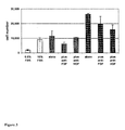


Classifications
-
- A—HUMAN NECESSITIES
- A61—MEDICAL OR VETERINARY SCIENCE; HYGIENE
- A61K—PREPARATIONS FOR MEDICAL, DENTAL OR TOILETRY PURPOSES
- A61K35/00—Medicinal preparations containing materials or reaction products thereof with undetermined constitution
- A61K35/12—Materials from mammals; Compositions comprising non-specified tissues or cells; Compositions comprising non-embryonic stem cells; Genetically modified cells
- A61K35/48—Reproductive organs
- A61K35/51—Umbilical cord; Umbilical cord blood; Umbilical stem cells
-
- A—HUMAN NECESSITIES
- A61—MEDICAL OR VETERINARY SCIENCE; HYGIENE
- A61K—PREPARATIONS FOR MEDICAL, DENTAL OR TOILETRY PURPOSES
- A61K35/00—Medicinal preparations containing materials or reaction products thereof with undetermined constitution
- A61K35/12—Materials from mammals; Compositions comprising non-specified tissues or cells; Compositions comprising non-embryonic stem cells; Genetically modified cells
- A61K35/14—Blood; Artificial blood
- A61K35/15—Cells of the myeloid line, e.g. granulocytes, basophils, eosinophils, neutrophils, leucocytes, monocytes, macrophages or mast cells; Myeloid precursor cells; Antigen-presenting cells, e.g. dendritic cells
-
- A—HUMAN NECESSITIES
- A61—MEDICAL OR VETERINARY SCIENCE; HYGIENE
- A61K—PREPARATIONS FOR MEDICAL, DENTAL OR TOILETRY PURPOSES
- A61K35/00—Medicinal preparations containing materials or reaction products thereof with undetermined constitution
- A61K35/12—Materials from mammals; Compositions comprising non-specified tissues or cells; Compositions comprising non-embryonic stem cells; Genetically modified cells
- A61K35/34—Muscles; Smooth muscle cells; Heart; Cardiac stem cells; Myoblasts; Myocytes; Cardiomyocytes
-
- A—HUMAN NECESSITIES
- A61—MEDICAL OR VETERINARY SCIENCE; HYGIENE
- A61K—PREPARATIONS FOR MEDICAL, DENTAL OR TOILETRY PURPOSES
- A61K35/00—Medicinal preparations containing materials or reaction products thereof with undetermined constitution
- A61K35/12—Materials from mammals; Compositions comprising non-specified tissues or cells; Compositions comprising non-embryonic stem cells; Genetically modified cells
- A61K35/44—Vessels; Vascular smooth muscle cells; Endothelial cells; Endothelial progenitor cells
-
- A—HUMAN NECESSITIES
- A61—MEDICAL OR VETERINARY SCIENCE; HYGIENE
- A61K—PREPARATIONS FOR MEDICAL, DENTAL OR TOILETRY PURPOSES
- A61K35/00—Medicinal preparations containing materials or reaction products thereof with undetermined constitution
- A61K35/12—Materials from mammals; Compositions comprising non-specified tissues or cells; Compositions comprising non-embryonic stem cells; Genetically modified cells
- A61K35/48—Reproductive organs
- A61K35/54—Ovaries; Ova; Ovules; Embryos; Foetal cells; Germ cells
- A61K35/545—Embryonic stem cells; Pluripotent stem cells; Induced pluripotent stem cells; Uncharacterised stem cells
-
- A—HUMAN NECESSITIES
- A61—MEDICAL OR VETERINARY SCIENCE; HYGIENE
- A61K—PREPARATIONS FOR MEDICAL, DENTAL OR TOILETRY PURPOSES
- A61K38/00—Medicinal preparations containing peptides
- A61K38/16—Peptides having more than 20 amino acids; Gastrins; Somatostatins; Melanotropins; Derivatives thereof
- A61K38/17—Peptides having more than 20 amino acids; Gastrins; Somatostatins; Melanotropins; Derivatives thereof from animals; from humans
- A61K38/36—Blood coagulation or fibrinolysis factors
- A61K38/363—Fibrinogen
-
- A—HUMAN NECESSITIES
- A61—MEDICAL OR VETERINARY SCIENCE; HYGIENE
- A61K—PREPARATIONS FOR MEDICAL, DENTAL OR TOILETRY PURPOSES
- A61K38/00—Medicinal preparations containing peptides
- A61K38/16—Peptides having more than 20 amino acids; Gastrins; Somatostatins; Melanotropins; Derivatives thereof
- A61K38/43—Enzymes; Proenzymes; Derivatives thereof
- A61K38/46—Hydrolases (3)
- A61K38/48—Hydrolases (3) acting on peptide bonds (3.4)
- A61K38/482—Serine endopeptidases (3.4.21)
- A61K38/4833—Thrombin (3.4.21.5)
-
- A—HUMAN NECESSITIES
- A61—MEDICAL OR VETERINARY SCIENCE; HYGIENE
- A61K—PREPARATIONS FOR MEDICAL, DENTAL OR TOILETRY PURPOSES
- A61K45/00—Medicinal preparations containing active ingredients not provided for in groups A61K31/00 - A61K41/00
- A61K45/06—Mixtures of active ingredients without chemical characterisation, e.g. antiphlogistics and cardiaca
-
- A—HUMAN NECESSITIES
- A61—MEDICAL OR VETERINARY SCIENCE; HYGIENE
- A61L—METHODS OR APPARATUS FOR STERILISING MATERIALS OR OBJECTS IN GENERAL; DISINFECTION, STERILISATION OR DEODORISATION OF AIR; CHEMICAL ASPECTS OF BANDAGES, DRESSINGS, ABSORBENT PADS OR SURGICAL ARTICLES; MATERIALS FOR BANDAGES, DRESSINGS, ABSORBENT PADS OR SURGICAL ARTICLES
- A61L27/00—Materials for grafts or prostheses or for coating grafts or prostheses
- A61L27/36—Materials for grafts or prostheses or for coating grafts or prostheses containing ingredients of undetermined constitution or reaction products thereof, e.g. transplant tissue, natural bone, extracellular matrix
- A61L27/38—Materials for grafts or prostheses or for coating grafts or prostheses containing ingredients of undetermined constitution or reaction products thereof, e.g. transplant tissue, natural bone, extracellular matrix containing added animal cells
- A61L27/3804—Materials for grafts or prostheses or for coating grafts or prostheses containing ingredients of undetermined constitution or reaction products thereof, e.g. transplant tissue, natural bone, extracellular matrix containing added animal cells characterised by specific cells or progenitors thereof, e.g. fibroblasts, connective tissue cells, kidney cells
- A61L27/3834—Cells able to produce different cell types, e.g. hematopoietic stem cells, mesenchymal stem cells, marrow stromal cells, embryonic stem cells
-
- A—HUMAN NECESSITIES
- A61—MEDICAL OR VETERINARY SCIENCE; HYGIENE
- A61L—METHODS OR APPARATUS FOR STERILISING MATERIALS OR OBJECTS IN GENERAL; DISINFECTION, STERILISATION OR DEODORISATION OF AIR; CHEMICAL ASPECTS OF BANDAGES, DRESSINGS, ABSORBENT PADS OR SURGICAL ARTICLES; MATERIALS FOR BANDAGES, DRESSINGS, ABSORBENT PADS OR SURGICAL ARTICLES
- A61L27/00—Materials for grafts or prostheses or for coating grafts or prostheses
- A61L27/36—Materials for grafts or prostheses or for coating grafts or prostheses containing ingredients of undetermined constitution or reaction products thereof, e.g. transplant tissue, natural bone, extracellular matrix
- A61L27/38—Materials for grafts or prostheses or for coating grafts or prostheses containing ingredients of undetermined constitution or reaction products thereof, e.g. transplant tissue, natural bone, extracellular matrix containing added animal cells
- A61L27/3839—Materials for grafts or prostheses or for coating grafts or prostheses containing ingredients of undetermined constitution or reaction products thereof, e.g. transplant tissue, natural bone, extracellular matrix containing added animal cells characterised by the site of application in the body
-
- A—HUMAN NECESSITIES
- A61—MEDICAL OR VETERINARY SCIENCE; HYGIENE
- A61L—METHODS OR APPARATUS FOR STERILISING MATERIALS OR OBJECTS IN GENERAL; DISINFECTION, STERILISATION OR DEODORISATION OF AIR; CHEMICAL ASPECTS OF BANDAGES, DRESSINGS, ABSORBENT PADS OR SURGICAL ARTICLES; MATERIALS FOR BANDAGES, DRESSINGS, ABSORBENT PADS OR SURGICAL ARTICLES
- A61L27/00—Materials for grafts or prostheses or for coating grafts or prostheses
- A61L27/36—Materials for grafts or prostheses or for coating grafts or prostheses containing ingredients of undetermined constitution or reaction products thereof, e.g. transplant tissue, natural bone, extracellular matrix
- A61L27/38—Materials for grafts or prostheses or for coating grafts or prostheses containing ingredients of undetermined constitution or reaction products thereof, e.g. transplant tissue, natural bone, extracellular matrix containing added animal cells
- A61L27/3886—Materials for grafts or prostheses or for coating grafts or prostheses containing ingredients of undetermined constitution or reaction products thereof, e.g. transplant tissue, natural bone, extracellular matrix containing added animal cells comprising two or more cell types
-
- A—HUMAN NECESSITIES
- A61—MEDICAL OR VETERINARY SCIENCE; HYGIENE
- A61L—METHODS OR APPARATUS FOR STERILISING MATERIALS OR OBJECTS IN GENERAL; DISINFECTION, STERILISATION OR DEODORISATION OF AIR; CHEMICAL ASPECTS OF BANDAGES, DRESSINGS, ABSORBENT PADS OR SURGICAL ARTICLES; MATERIALS FOR BANDAGES, DRESSINGS, ABSORBENT PADS OR SURGICAL ARTICLES
- A61L27/00—Materials for grafts or prostheses or for coating grafts or prostheses
- A61L27/36—Materials for grafts or prostheses or for coating grafts or prostheses containing ingredients of undetermined constitution or reaction products thereof, e.g. transplant tissue, natural bone, extracellular matrix
- A61L27/38—Materials for grafts or prostheses or for coating grafts or prostheses containing ingredients of undetermined constitution or reaction products thereof, e.g. transplant tissue, natural bone, extracellular matrix containing added animal cells
- A61L27/3895—Materials for grafts or prostheses or for coating grafts or prostheses containing ingredients of undetermined constitution or reaction products thereof, e.g. transplant tissue, natural bone, extracellular matrix containing added animal cells using specific culture conditions, e.g. stimulating differentiation of stem cells, pulsatile flow conditions
-
- A—HUMAN NECESSITIES
- A61—MEDICAL OR VETERINARY SCIENCE; HYGIENE
- A61P—SPECIFIC THERAPEUTIC ACTIVITY OF CHEMICAL COMPOUNDS OR MEDICINAL PREPARATIONS
- A61P9/00—Drugs for disorders of the cardiovascular system
-
- A—HUMAN NECESSITIES
- A61—MEDICAL OR VETERINARY SCIENCE; HYGIENE
- A61P—SPECIFIC THERAPEUTIC ACTIVITY OF CHEMICAL COMPOUNDS OR MEDICINAL PREPARATIONS
- A61P9/00—Drugs for disorders of the cardiovascular system
- A61P9/10—Drugs for disorders of the cardiovascular system for treating ischaemic or atherosclerotic diseases, e.g. antianginal drugs, coronary vasodilators, drugs for myocardial infarction, retinopathy, cerebrovascula insufficiency, renal arteriosclerosis
-
- C—CHEMISTRY; METALLURGY
- C12—BIOCHEMISTRY; BEER; SPIRITS; WINE; VINEGAR; MICROBIOLOGY; ENZYMOLOGY; MUTATION OR GENETIC ENGINEERING
- C12N—MICROORGANISMS OR ENZYMES; COMPOSITIONS THEREOF; PROPAGATING, PRESERVING, OR MAINTAINING MICROORGANISMS; MUTATION OR GENETIC ENGINEERING; CULTURE MEDIA
- C12N5/00—Undifferentiated human, animal or plant cells, e.g. cell lines; Tissues; Cultivation or maintenance thereof; Culture media therefor
- C12N5/06—Animal cells or tissues; Human cells or tissues
- C12N5/0602—Vertebrate cells
- C12N5/0603—Embryonic cells ; Embryoid bodies
- C12N5/0605—Cells from extra-embryonic tissues, e.g. placenta, amnion, yolk sac, Wharton's jelly
-
- A—HUMAN NECESSITIES
- A61—MEDICAL OR VETERINARY SCIENCE; HYGIENE
- A61K—PREPARATIONS FOR MEDICAL, DENTAL OR TOILETRY PURPOSES
- A61K2300/00—Mixtures or combinations of active ingredients, wherein at least one active ingredient is fully defined in groups A61K31/00 - A61K41/00
-
- C—CHEMISTRY; METALLURGY
- C12—BIOCHEMISTRY; BEER; SPIRITS; WINE; VINEGAR; MICROBIOLOGY; ENZYMOLOGY; MUTATION OR GENETIC ENGINEERING
- C12N—MICROORGANISMS OR ENZYMES; COMPOSITIONS THEREOF; PROPAGATING, PRESERVING, OR MAINTAINING MICROORGANISMS; MUTATION OR GENETIC ENGINEERING; CULTURE MEDIA
- C12N2510/00—Genetically modified cells
Definitions
- the invention relates to the field of cell based or regenerative therapy for peripheral vascular disease patients, especially those with peripheral ischemia.
- the invention provides cells derived from umbilical cord tissue having the capability to stimulate and support angiogenesis, to improve blood flow, to regenerate, repair, and improve skeletal muscle damaged by a peripheral ischemic event, and to protect skeletal muscle from ischemic damage.
- PVD Peripheral vascular disease
- iliac blood vessels femoral and popliteal blood vessels, and subclavian blood vessels, and its effects can be exacerbated by thrombi, emboli, or trauma. It is estimated that approximately 8 to 12 million individuals in the United States, especially among the elderly population and those with diabetes, are afflicted with PVD.
- PVD Common symptoms of PVD include cramping in the upper and lower limbs and extremities, numbness, weakness, muscle fatigue, pain in the limbs and extremities, hypothermia in the limbs and extremities, discoloration of the extremities, dry or scaly skin, and hypertension.
- the most common symptom is claudication or feelings of pain, tightness, and fatigue in muscles downstream of the occluded blood vessel that occurs during some form of exercise such as walking, but self-resolve after a period of rest.
- the occluded blood vessels cause ischemia of tissues at the site of and distal to the obstruction.
- This ischemia is generally referred to as peripheral ischemia, meaning that it occurs in locations distal to the heart.
- the severity of the ischemia is a function of the size and number of obstructions, whether the obstruction is near a muscle or organ, and whether there is sufficient redundant vasculature. In more severe cases, the ischemia results in death of the affected tissues, and can result in amputation of affected limbs, or even death of the patient.
- stem cells which can divide and differentiate, or muscles cells from other sources, including smooth and skeletal muscles cells, to assist the in the repair or reversal of tissue damage.
- Transplantation of stem cells can be utilized as a clinical tool for reconstituting a target tissue, thereby restoring physiologic and anatomic functionality.
- the application of stem cell technology is wide-ranging, including tissue engineering, gene therapy delivery, and cell therapeutics, i.e., delivery of biotherapeutic agents to a target location via exogenously supplied living cells or cellular components that produce or contain those agents (For a review, see Tresco, P. A. et al., (2000) Advanced Drug Delivery Reviews 42:2-37).
- the identification of stem cells has stimulated research aimed at the selective generation of specific cell types for regenerative medicine.
- Embryonic, or fetal tissue is one source of stem cells.
- Embryonic stem and progenitor cells have been isolated from a number of mammalian species, including humans, and several such cell types have been shown capable of self-renewal and expansion, as well differentiation into a number of different cell lineages.
- the derivation of stem cells from embryonic or fetal sources has raised many ethical and moral issues that are desirable to avoid by identifying other sources of multipotent or pluripotent cells.
- Postpartum tissues such as the umbilical cord and placenta
- stem cells have generated interest as an alternative source of stem cells.
- methods for recovery of stem cells by perfusion of the placenta or collection from umbilical cord blood or tissue have been described.
- a limitation of stem cell procurement from these methods has been an inadequate volume of cord blood or quantity of cells obtained, as well as heterogeneity in, or lack of characterization of, the populations of cells obtained from those sources.
- a reliable, well-characterized and plentiful supply of substantially homogeneous populations of such cells having the ability to differentiate into an array of skeletal muscle, pericyte, or vascular lineages would be an advantage in a variety of diagnostic and therapeutic applications for skeletal muscle repair, regeneration, and improvement, for the stimulation and/or support of angiogenesis, and for the improvement of blood flow subsequent to a peripheral ischemic event, particularly in PVD patients.
- One aspect of the invention features method of treating a patient having peripheral vascular disease, the method comprising administering to the patient umbilical cord tissue-derived cells in an amount effective to treat the peripheral vascular disease, wherein the umbilical cord tissue-derived cells are derived from human umbilical cord tissue substantially free of blood, wherein the cells are capable of self-renewal and expansion in culture and have the potential to differentiate into cells of at least a skeletal muscle, vascular smooth muscle, pericyte, or vascular endothelium phenotype; wherein the cells require L-valine for growth and can grow in at least about 5% oxygen; wherein the cells further comprise at least one of the following characteristics: (a) potential for at least about 40 doublings in culture; (b) attachment and expansion on a coated or uncoated tissue culture vessel, wherein the coated tissue culture vessel comprises a coating of gelatin, laminin, collagen, polyornithine, vitronectin, or fibronectin; (c) production of at least one of tissue factor, vimentin, and
- the peripheral vascular disease is peripheral ischemia.
- the cells are induced in vitro to differentiate into a skeletal muscle, vascular smooth muscle, pericyte, or vascular endothelium lineage cells prior to administration.
- the cells are genetically engineered to produce a gene product that promotes treatment of peripheral vascular disease.
- cells are administered with at least one other cell type, which may include skeletal muscle cells, skeletal muscle progenitor cells, vascular smooth muscle cells, vascular smooth muscle progenitor cells, pericytes, vascular endothelial cells, vascular endothelium progenitor cells, or other multipotent or pluripotent stem cells.
- the other cell type can administered simultaneously with, or before, or after, the umbilical cord tissue-derived cells.
- the cells are administered with at least one other agent, which may be an antithrombogenic agent, an anti-inflammatory agent, an immunosuppressive agent, an immunomodulatory agent, pro-angiogenic, or an antiapoptotic agent, for example.
- the other agent can be administered simultaneously with, or before, or after, the umbilical cord tissue-derived cells.
- the cells are preferably administered at or proximal to the sites of the peripheral ischemia, but can also be administered at sites distal to the peripheral ischemia. They can be administered by injection, infusion, a device implanted in the patient, or by implantation of a matrix or scaffold containing the cells.
- the cells may exert a trophic effect, such as proliferation, on the skeletal muscle, vascular smooth muscle or vascular endothelium of the patient.
- the cells may induce migration of skeletal muscle cells, vascular smooth muscle cells, vascular endothelial cells, skeletal muscle progenitor cells, pericytes, vascular smooth muscle progenitor cells, or vascular endothelium progenitor cells to the site or sites of peripheral vascular disease, such as peripheral ischemia.
- compositions and kits for treating a patient having a peripheral vascular disease comprising a pharmaceutically acceptable carrier and the umbilical cord tissue-derived cells described above or preparations made from such umbilical cord tissue-derived cells.
- the preparations comprise FGF and HGF.
- the pharmaceutical compositions and kits are designed and/or formulated for practicing the methods of the invention as outlined above.
- the above-described methods may be practiced using a preparation made from the umbilical cord tissue-derived cells, wherein the preparation comprises a cell lysate of the umbilical cord tissue-derived cells, an extracellular matrix of the umbilical cord tissue-derived cells or a conditioned medium in which the umbilical cord tissue-derived cells were grown. It is preferred that such preparations comprise FGF and HGF.
- compositions and kits containing preparations comprising cell lysates, extracellular matrices or conditioned media of the umbilical cord tissue-derived cells.
- One embodiment of the invention is a method of treating a patient having a peripheral vascular disease, comprising administering a pharmaceutical composition comprising a fibrin glue and an isolated homogenous population of cells obtained from human umbilical cord tissue in an amount effective to treat the disease, wherein the umbilical cord tissue is substantially free of blood, and wherein isolated homogenous population of cells is capable of self-renewal and expansion in culture, has the potential to differentiate and does not express CD117 and/or telomerase.
- the isolated population of cells may also have one or more of the following characteristics:
- (c) expresses, relative to a human fibroblast, mesenchymal stem cell, or iliac crest bone marrow cell, increased levels of interleukin 8 or reticulon 1;
- (d) has the potential to differentiate into cells of at least a skeletal muscle, vascular smooth muscle, pericyte or vascular endothelium phenotype
- (d) expresses CD10, CD13, CD44, CD73, and CD90.
- the peripheral vascular disease is peripheral ischemia.
- the pharmaceutical composition is administered at the sites of peripheral ischemia.
- the pharmaceutical composition is administered locally.
- the pharmaceutical composition is administered by injection, infusion, a device implanted in a patient, or by implantation of a matrix or scaffold containing the pharmaceutical composition.
- the pharmaceutical composition is administered by intramuscular injection and injection into adipose depots in muscle.
- the pharmaceutical composition is administered by injection into interstitial spaces so as not to enter directly into circulation.
- the isolated population of cells may be induced in vitro to differentiate into a skeletal muscle, vascular muscle, pericyte or vascular endothelium lineage prior to administration.
- the population of cells may also be genetically engineered to produce a gene product that promotes treatment of peripheral vascular disease.
- the composition further comprises an agent selected from the group consisting of an antithrombogenic agent, an immunosuppressive agent, an immunomodulatory agent, a pro-angiogenic, an antiapoptotic agent and mixtures thereof.
- the composition further comprises at least one other cell type (such as e.g. a skeletal muscle cell, a skeletal muscle progenitor cell, a vascular smooth muscle cell, a vascular smooth muscle progenitor cell, a pericyte, a vascular endothelial cell, a vascular endothelium progenitor cell or other multipotent or pluripotent stem cell).
- the pharmaceutical composition exerts a trophic effect (such as e.g. proliferation of vascular endothelial cells).
- the pharmaceutical composition induces migration of vascular endothelial cells and/or vascular endothelium progenitor cells to the sites of the peripheral disease.
- the pharmaceutical composition induces migration of vascular smooth muscle cells and/or vascular smooth muscle progenitor cells to the sites of the peripheral disease.
- the pharmaceutical composition induces migration of pericytes to the sites of the peripheral vascular disease.
- the fibrin glue comprises fibrinogen and thrombin. In another embodiment, the fibrin glue comprises from about 16 to about 24 IU/ml of thrombin and from about 39.3 to about 60.7 mg/ml of fibrinogen.
- Another embodiment of the invention is a method of treating a patient having a peripheral vascular disease, comprising administering a fibrin glue (e.g. a composition comprising fibrinogen and thrombin) and an isolated homogenous population of cells obtained from human umbilical cord tissue in an amount effective to treat the disease, wherein the umbilical cord tissue is substantially free of blood, and wherein isolated homogenous population of the cells is capable of self-renewal and expansion in culture, has the potential to differentiate and does not express CD117 and/or telomerase.
- a fibrin glue e.g. a composition comprising fibrinogen and thrombin
- isolated homogenous population of cells obtained from human umbilical cord tissue in an amount effective to treat the disease, wherein the umbilical cord tissue is substantially free of blood, and wherein isolated homogenous population of the cells is capable of self-renewal and expansion in culture, has the potential to differentiate and does not express CD117 and/or telomerase.
- the isolated population of cells may have other characteristics, including
- (c) express, relative to a human fibroblast, mesenchymal stem cell, or iliac crest bone marrow cell, increased levels of interleukin 8 or reticulon 1;
- (d) has the potential to differentiate into cells of at least a skeletal muscle, vascular smooth muscle, pericyte or vascular endothelium phenotype
- (d) expresses CD10, CD13, CD44, CD73, and CD90.
- the peripheral vascular disease is peripheral ischemia and, optionally, the fibrin glue and the population of cells are administered at the sites of peripheral ischemia.
- Various routes of administration may be used including administered by injection, infusion, a device implanted in a patient, or by implantation of a matrix or scaffold containing the cells.
- the population of cells and fibrin glue are administered locally (such as e.g. by intramuscular injection and injection into adipose depots in muscle).
- the cells and fibrin glue are administered by injection into interstitial spaces so as not to directly enter into circulation.
- the isolated population of cells is induced in vitro to differentiate into a skeletal muscle, vascular muscle, pericyte or vascular endothelium lineage prior to administration.
- the population of cells may also be genetically engineered to produce a gene product that promotes treatment of peripheral vascular disease.
- the method further comprises administration of an agent selected from the group consisting of an antithrombogenic agent, an immunosuppressive agent, an immunomodulatory agent, a pro-angiogenic, an antiapoptotic agent and mixtures thereof.
- the method further comprises administration of at least one other cell type (such as e.g.
- the population of cells exerts a trophic effect (e.g. proliferation of vascular endothelial cells).
- the population of cells may induce migration of vascular endothelial cells and/or vascular endothelium progenitor cells to the sites of the peripheral disease.
- the population of cells may induce migration of vascular smooth muscle cells and/or vascular smooth muscle progenitor cells to the sites of the peripheral disease.
- the population of cells also may induce migration of pericytes to the sites of the peripheral vascular disease.
- the fibrin glue may comprise fibrinogen and thrombin.
- the fibrin glue is administered simultaneously with, or before, or after, the isolated homogenous population of cells obtained from human umbilical cord tissue.
- the fibrin glue comprises from about 16 to about 24 IU/ml of thrombin and from about 39.3 to about 60.7 mg/ml of fibrinogen.
- kits for treating a patient having a peripheral vascular disease comprising fibrinogen, thrombin and an isolated homogenous population of cells obtained from human umbilical cord tissue in an amount effective to treat the disease, wherein the umbilical cord tissue is substantially free of blood, and wherein said isolated homogenous population of the cells is capable of self-renewal and expansion in culture, has the potential to differentiate and do not express CD117 and/or telomerase.
- the kit may further comprise instructions for use.
- the fibrinogen and isolated homogenous population of cells are in provided in a composition to which thrombin may be added immediately prior to use.
- the isolated population of cells may have other characteristics, including one or more of the following:
- (c) express, relative to a human fibroblast, mesenchymal stem cell, or iliac crest bone marrow cell, increased levels of interleukin 8 or reticulon 1;
- (d) has the potential to differentiate into cells of at least a skeletal muscle, vascular smooth muscle, pericyte or vascular endothelium phenotype
- (d) expresses CD10, CD13, CD44, CD73, and CD90.
- the kit comprises from about 16 to about 24 IU/ml of thrombin and from about 39.3 to about 60.7 mg/ml of fibrinogen. In one embodiment, the kit comprises a fibrinogen component comprising fibrin and factor XII and a thrombin component comprising thrombin and calcium.
- FIG. 1 shows the effect of hUTC lot#120304, MSCs, and fibroblasts on the proliferation of endothelial cells.
- Endothelial cells were seeded onto the bottom of a 24-well tissue culture dish at a density of 5000 cells/cm 2 (10,000 cells/well) and hUTC lot#120304, MSCs, or fibroblasts inside transwell inserts at a density of 5000 cells/cm 2 (1,650 cells/insert) in co-culture media (Hayflick 80%+EGM-2MV 20% or Hayflick 50%+EGM-2MV 50%). After 7 days of co-culture, cells were harvested and counted using a Guava® instrument. Endothelial cells were also maintained in EGM-2MV media as positive control.
- A HUVECs.
- B HCAECs.
- C HIAECs.
- FIG. 2 shows the effect of hUTC lot#120304 and neutralizing antibodies on the proliferation of endothelial cells.
- HUVECs or HCAECs were seeded onto the bottom of a 24-well tissue culture dish at a density of 5000 cells/cm 2 (10,000 cells/well) and hUTC lot#120304 inside transwell inserts at a density of 5000 cells/cm 2 (1,650 cells/insert) in co-culture media (Hayflick 50%+EGM-2MV 50%).
- Neutralizing antibodies to FGF (7 ⁇ g/ml), HGF (1 ⁇ g/ml), or VEGF (1 ⁇ g/ml) were also added at this time.
- FIG. 3 shows the effect of hUTC lot#120304 cell lysate and neutralizing antibodies on proliferation of HUVECs.
- HUVECs were seeded onto the bottom of a 24-well tissue culture dish at a density of 5000 cells/cm 2 (10,000 cells/well) in EGM-2MV media for 8 h. Cells were then serum-starved by overnight incubation in 0.5 ml of EGM-2MV media containing 0.5% FBS and without growth factors. Afterwards, FBS, freshly prepared hUTC lot#120304 cell lysates, and neutralizing antibodies to FGF (7 ⁇ g/ml) or HGF (1 ⁇ g/ml) were added. After 4 days of culture, cells were harvested and counted using a Guava® instrument. Light grey bars, media controls. Medium grey bars, HUVECs incubated with lysate containing 62.5 ⁇ g of protein. Dark grey bars, HUVECs incubated with lysate containing 125 ⁇ g of protein.
- FIG. 4 shows the effect of hUTCs and MSCs on the migration of endothelial cells.
- HUVECs or HCAECs were seeded inside transwell inserts at a density of 5000 cells/cm 2 (23,000 cells/insert) and hUTC lot#120304 or MSCs onto the bottom of a E-well tissue culture dish at a density of 5000 cells/cm 2 (48,000 cells/well) in co-culture media (Hayflick 50%+EGM-2MV 50%). After 7 days of co-culture, cells that were on the underside of the transwell insert were harvested and counted using a Guava® instrument. Endothelial cells were also maintained in EGM-2MV media as control.
- A HUVECs.
- B HCAECs.
- FIG. 5 shows the effect of hUTC lot#120304 and neutralizing antibodies on the migration of endothelial cells.
- HUVECs or HCAECs were seeded inside transwell inserts at a density of 5000 cells/cm 2 (23,000 cells/insert) and hUTC lot#120304 onto the bottom of a 6-well tissue culture dish at a density of 5000 cells/cm 2 (48,000 cells/well) in co-culture media (Hayflick 50%+EGM-2MV 50%).
- Neutralizing antibodies to FGF (7 ng/ml) or HGF (1 ng/ml) were added at this time.
- endothelial cells were also maintained in EGM-2MV media as control.
- FIG. 6 shows the laser doppler perfusion data from the experiment with NSG mice for the study disclosed in Example 5. Data are expressed as mean ⁇ sem. The identity of data points is displayed in the legend. Numbers in parentheses are (1) P ⁇ 0.001 compared to appropriate control; (2) P ⁇ 0.05 compared to hUTC cells without fibrin.
- FIG. 7 shows the laser doppler perfusion data from the experiment with nude mice for the study disclosed in Example 5. Data are expressed as mean ⁇ sem. The identity of data points is displayed in the legend. Numbers in parentheses are (1) P ⁇ 0.001 compared to appropriate control; (2) P ⁇ 0.05 compared to hUTC cells without fibrin.
- FIG. 8 shows laser doppler perfusion data comparing systemic (IV), local (IM), and local+fibrin glue delivery for the study disclosed in Example 6. Data are expressed as mean ⁇ sem.
- FIG. 9 shows laser doppler perfusion data showing different doses of hUTC in fibrin glue delivered locally (IM) for the study disclosed in Example 6. Data are expressed as mean ⁇ sem.
- FIG. 10 shows laser doppler perfusion data comparing systemic (IV), local (IM), and local+fibrin glue delivery at 14 days post-injury for the study disclosed in Example 6. Data shown as mean for clarity.
- FIG. 11 shows laser doppler perfusion for the study disclosed in Example 7. The identity of data points is displayed in the legend. *, P ⁇ 0.05; ***, P ⁇ 0.001.
- FIG. 12 shows capillary density of ischemic limbs compared to non-ischemic limbs of mice surviving to 21 days for the study disclosed in Example 7.
- FIG. 13 shows arteriole density ischemic limbs compared to non-ischemic limbs of mice surviving to 21 days for the study disclosed in Example 7.
- Stem cells are undifferentiated cells defined by the ability of a single cell both to self-renew and to differentiate to produce progeny cells, including self-renewing progenitors, non-renewing progenitors, and terminally differentiated cells. Stem cells are also characterized by their ability to differentiate in vitro into functional cells of various cell lineages from multiple germ layers (endoderm, mesoderm and ectoderm), as well as to give rise to tissues of multiple germ layers following transplantation, and to contribute substantially to most, if not all, tissues following injection into blastocysts.
- Stem cells are classified according to their developmental potential as: (1) totipotent; (2) pluripotent; (3) multipotent; (4) oligopotent; and (5) unipotent.
- Totipotent cells are able to give rise to all embryonic and extraembryonic cell types.
- Pluripotent cells are able to give rise to all embryonic cell types.
- Mutt/potent cells include those able to give rise to a subset of cell lineages, but all within a particular tissue, organ, or physiological system.
- hematopoietic stem cells can produce progeny that include HSC (self-renewal), blood cell-restricted oligopotent progenitors, and all cell types and elements (e.g., platelets) that are normal components of the blood.
- HSC self-renewal
- oligopotent progenitors can give rise to a more restricted subset of cell lineages than multipotent stem cells.
- Cells that are unipotent are able to give rise to a single cell lineage (e.g., spermatogenic stem cells).
- Stem cells are also categorized on the basis of the source from which they are obtained.
- An adult stem cell is generally a multipotent undifferentiated cell found in tissue comprising multiple differentiated cell types. The adult stem cell can renew itself. Under normal circumstances, it can also differentiate to yield the specialized cell types of the tissue from which it originated, and possibly other tissue types.
- An embryonic stem cell is a pluripotent cell from the inner cell mass of a blastocyst-stage embryo.
- a fetal stem cell is one that originates from fetal tissues or membranes.
- a postpartum stem cell is a multipotent or pluripotent cell that originates substantially from extraembryonic tissue available after birth, namely, the placenta and the umbilical cord.
- Postpartum stem cells may be blood-derived (e.g., as are those obtained from umbilical cord blood) or non-blood-derived (e.g., as obtained from the non-blood tissues of the umbilical cord and placenta).
- Embryonic tissue is typically defined as tissue originating from the embryo (which in humans refers to the period from fertilization to about six weeks of development. Fetal tissue refers to tissue originating from the fetus, which in humans refers to the period from about six weeks of development to parturition. Extraembryonic tissue is tissue associated with, but not originating from, the embryo or fetus. Extraembryonic tissues include extraembryonic membranes (chorion, amnion, yolk sac and allantois), umbilical cord and placenta (which itself forms from the chorion and the maternal decidua basalis).
- Differentiation is the process by which an unspecialized (“uncommitted”) or less specialized cell acquires the features of a specialized cell, such as a nerve cell or a muscle cell, for example.
- a differentiated cell is one that has taken on a more specialized (“committed”) position within the lineage of a cell.
- the term committed, when applied to the process of differentiation, refers to a cell that has proceeded in the differentiation pathway to a point where, under normal circumstances, it will continue to differentiate into a specific cell type or subset of cell types, and cannot, under normal circumstances, differentiate into a different cell type or revert to a less differentiated cell type.
- De-differentiation refers to the process by which a cell reverts to a less specialized (or committed) position within the lineage of a cell.
- the lineage of a cell defines the heredity of the cell, i.e. which cells it came from and what cells it can give rise to.
- the lineage of a cell places the cell within a hereditary scheme of development and differentiation.
- a progenitor cell is a cell that has the capacity to create progeny that are more differentiated than itself, and yet retains the capacity to replenish the pool of progenitors.
- stem cells themselves are also progenitor cells, as are the more immediate precursors to terminally differentiated cells.
- this broad definition of progenitor cell may be used.
- a progenitor cell is often defined as a cell that is intermediate in the differentiation pathway, i.e., it arises from a stem cell and is intermediate in the production of a mature cell type or subset of cell types. This type of progenitor cell is generally not able to self-renew. Accordingly, if this type of cell is referred to herein, it will be referred to as a non-renewing progenitor cell or as an intermediate progenitor or precursor cell.
- the phrase differentiates into a mesodermal, ectodermal or endodermal lineage refers to a cell that becomes committed to a specific mesodermal, ectodermal or endodermal lineage, respectively.
- Examples of cells that differentiate into a mesodermal lineage or give rise to specific mesodermal cells include, but are not limited to, cells that are adipogenic, chondrogenic, cardiogenic, dermatogenic, hematopoietic, hemangiogenic, myogenic, nephrogenic, urogenitogenic, osteogenic, pericardiogenic, or stromal.
- Examples of cells that differentiate into ectodermal lineage include, but are not limited to epidermal cells, neurogenic cells, and neurogliagenic cells.
- Examples of cells that differentiate into endodermal lineage include, but are not limited to, pleurigenic cells, hepatogenic cells, cells that give rise to the lining of the intestine, and cells that give rise to pancreogenic and splanchogenic cells.
- the cells used in the present invention are generally referred to as referred to as umbilical cord tissue-derived cells (UTC(s) or hUTC(s)). They also may sometimes be referred to as umbilicus-derived cells (UDCs).
- UDCs umbilicus-derived cells
- the cells may be described as being stem or progenitor cells, the latter term being used in the broad sense.
- the term derived is used to indicate that the cells have been obtained from their biological source and grown or otherwise manipulated in vitro (e.g., cultured in a Growth Medium to expand the population and/or to produce a cell line). The in vitro manipulations of umbilical stem cells and the unique features of the umbilicus-derived cells of the present invention are described in detail below.
- Pericytes also known in the art as Rouget cells or mural cells, refers to the cells typically found embedded within the vascular basement membrane of blood microvessels (Armulik A et al. (2005) Circ. Res. 97:512-23), that are believed to play a role in, among other things, communication/signalling with endothelial cells, vasoconstriction, vasodilation, the regulation of blood flow, blood vasculature formation and development, angiogenesis, and endothelial differentiation and growth arrest (Bergers G et al. (2005) Neuro - Oncology 7:452-64).
- Cell culture refers generally to cells taken from a living organism and grown under controlled condition (“in culture” or “cultured”).
- a primary cell culture is a culture of cells, tissues, or organs taken directly from an organism(s) before the first subculture.
- Cells are expanded in culture when they are placed in a Growth Medium under conditions that facilitate cell growth and/or division, resulting in a larger population of the cells.
- the rate of cell proliferation is sometimes measured by the amount of time needed for the cells to double in number. This is referred to as doubling time.
- a cell line is a population of cells formed by one or more subcultivations of a primary cell culture. Each round of subculturing is referred to as a passage. When cells are subcultured, they are referred to as having been passaged. A specific population of cells, or a cell line, is sometimes referred to or characterized by the number of times it has been passaged. For example, a cultured cell population that has been passaged ten times may be referred to as a P10 culture.
- the primary culture i.e., the first culture following the isolation of cells from tissue, is designated P0. Following the first subculture, the cells are described as a secondary culture (P1 or passage 1).
- the cells After the second subculture, the cells become a tertiary culture (P2 or passage 2), and so on. It will be understood by those of skill in the art that there may be many population doublings during the period of passaging; therefore the number of population doublings of a culture is greater than the passage number.
- the expansion of cells (i.e., the number of population doublings) during the period between passaging depends on many factors, including but not limited to the seeding density, substrate, medium, growth conditions, and time between passaging.
- a conditioned medium is a medium in which a specific cell or population of cells has been cultured, and then removed. When cells are cultured in a medium, they may secrete cellular factors that can provide trophic support to other cells. Such trophic factors include, but are not limited to hormones, cytokines, extracellular matrix (ECM), proteins, vesicles, antibodies, and granules.
- the medium containing the cellular factors is the conditioned medium.
- a trophic factor is defined as a substance that promotes survival, growth, proliferation and/or maturation of a cell, or stimulates increased activity of a cell.
- senescence also replicative senescence or cellular senescence refers to a property attributable to finite cell cultures; namely, their inability to grow beyond a finite number of population doublings (sometimes referred to as Hayflick's limit).
- cellular senescence was first described using fibroblast-like cells, most normal human cell types that can be grown successfully in culture undergo cellular senescence.
- the in vitro lifespan of different cell types varies, but the maximum lifespan is typically fewer than 100 population doublings (this is the number of doublings for all the cells in the culture to become senescent and thus render the culture unable to divide).
- Senescence does not depend on chronological time, but rather is measured by the number of cell divisions, or population doublings, the culture has undergone. Thus, cells made quiescent by removing essential growth factors are able to resume growth and division when the growth factors are re-introduced, and thereafter carry out the same number of doublings as equivalent cells grown continuously. Similarly, when cells are frozen in liquid nitrogen after various numbers of population doublings and then thawed and cultured, they undergo substantially the same number of doublings as cells maintained unfrozen in culture. Senescent cells are not dead or dying cells; they are actually resistant to programmed cell death (apoptosis), and have been maintained in their nondividing state for as long as three years. These cells are very much alive and metabolically active, but they do not divide. The nondividing state of senescent cells has not yet been found to be reversible by any biological, chemical, or viral agent.
- the term growth medium generally refers to a medium sufficient for the culturing of umbilical cord tissue-derived cells.
- one presently preferred medium for the culturing of the cells of the invention in comprises Dulbecco's Modified Essential Media (DMEM).
- DMEM Dulbecco's Modified Essential Media
- Particularly preferred is DMEM-low glucose (DMEM-LG) (Invitrogen, Carlsbad, Calif.).
- the DMEM-LG is preferably supplemented with serum, most preferably fetal bovine serum or human serum. Typically, 15% (v/v) fetal bovine serum (e.g.
- fetal bovine serum Hyclone, Logan Utah
- antibiotics/antimycotics (preferably 100 Unit/milliliter penicillin, 100 milligrams/milliliter streptomycin, and 0.25 microgram/milliliter amphotericin B; Invitrogen, Carlsbad, Calif.)
- 0.001% (v/v) 2-mercaptoethanol Sigma, St. Louis Mo.
- different growth media are used, or different supplementations are provided, and these are normally indicated in the text as supplementations to Growth Medium.
- the cells may be grown without serum present at all. In such cases, the cells may require certain growth factors, which can be added to the medium to support and sustain the cells.
- Presently preferred factors to be added for growth on serum-free media include one or more of bFGF, EGF, IGF-I, and PDGF. In more preferred embodiments, two, three or all four of the factors are add to serum free or chemically defined media. In other embodiments, LIF is added to serum-free medium to support or improve growth of the cells.
- standard growth conditions refers to culturing of cells at 37° C., in a standard atmosphere comprising 5% CO 2 . Relative humidity is maintained at about 100%. While the foregoing conditions are useful for culturing, it is to be understood that such conditions are capable of being varied by the skilled artisan who will appreciate the options available in the art for culturing cells.
- an effective amount refers to a concentration or amount of a compound, material, or composition, as described herein, that is effective to achieve a particular biological result. Such results include, but are not limited to, the regeneration, repair, or improvement of skeletal tissue, the improvement of blood flow, and/or the stimulation and/or support of angiogenesis in peripheral ischemia patients. Such effective activity may be achieved, for example, by administering the cells and/or compositions of the present invention to peripheral ischemia patients. With respect to umbilical cord tissue-derived cells as administered to a patient in vivo, an effective amount may range from as few as several hundred or fewer to as many as several million or more. In specific embodiments, an effective amount may range from 10 3 -10 11 , more specifically at least about 10 4 cells.
- the number of cells to be administered will vary depending on the specifics of the disorder to be treated, including but not limited to size or total volume/surface area to be treated, and proximity of the site of administration to the location of the region to be treated, among other factors familiar to the medicinal biologist.
- treat, treating or treatment refer to any success or indicia of success in the attenuation or amelioration of an injury, pathology or condition, including any objective or subjective parameter such as abatement, remission, diminishing of symptoms or making the injury, pathology, or condition more tolerable to the patient, slowing in the rate of degeneration or decline, making the final point of degeneration less debilitating, improving a subject's physical or mental well-being, or prolonging the length of survival.
- the treatment or amelioration of symptoms can be based on objective or subjective parameters; including the results of a physical examination, neurological examination, and/or psychiatric evaluations.
- effective period and effective conditions refer to a period of time or other controllable conditions (e.g., temperature, humidity for in vitro methods), necessary or preferred for an agent or pharmaceutical composition to achieve its intended result.
- controllable conditions e.g., temperature, humidity for in vitro methods
- patient or subject refers to animals, preferably mammals, and more preferably humans, who are treated with the pharmaceutical or therapeutic compositions or in accordance with the methods described herein.
- Ischemia refers to any decrease or stoppage in the blood supply to any bodily organ, tissue, or part caused by any constriction or obstruction of the vasculature.
- Ischemic episode or ischemic event are used interchangeably herein and refer to any transient or permanent period of ischemia.
- Peripheral ischemia refers to any decrease or stoppage in the blood supply to any bodily organ, tissue, or part, excluding the heart, caused by any constriction or obstruction of the vasculature.
- Peripheral vascular disease refers to diseases of the blood vessels outside the heart and brain.
- PVD also encompasses Raynouds, intermittent claudication and critical limb ischemia.
- pharmaceutically acceptable carrier or medium refers to reagents, cells, compounds, materials, compositions, and/or dosage forms that are not only compatible with the cells and other agents to be administered therapeutically, but also are, within the scope of sound medical judgment, suitable for use in contact with the tissues of human beings and animals without excessive toxicity, irritation, allergic response, or other complication commensurate with a reasonable benefit/risk ratio.
- pharmaceutically acceptable carriers suitable for use in the present invention include liquids, semi-solid (e.g., gels) and solid materials (e.g., cell scaffolds and matrices, tubes sheets and other such materials as known in the art and described in greater detail herein).
- biodegradable materials may be designed to resist degradation within the body (non-biodegradable) or they may be designed to degrade within the body (biodegradable, bioerodable).
- a biodegradable material may further be bioresorbable or bioabsorbable, i.e., it may be dissolved and absorbed into bodily fluids (water-soluble implants are one example), or degraded and ultimately eliminated from the body, either by conversion into other materials or breakdown and elimination through natural pathways.
- the biodegradation rate can vary according to the desired release rate once implanted in the body.
- the matrix desirably also acts as a temporary scaffold until replaced by newly grown skeletal muscle, pericytes, vascular smooth muscle, or vascular endothelial tissue.
- the matrix provides for sustained release of the other agents used in conjunction with the umbilical cord tissue-derived cells and may provide a structure for developing tissue growth in the patient.
- the matrix simply provides a temporary scaffold for the developing tissue.
- the matrix can be in particulate form (macroparticles greater than 10 microns in diameter or microparticles less than 10 microns in diameter), or can be in the form of a structurally stable, three-dimensional implant (e.g., a scaffold).
- the implant can be, for example, a cube, cylinder, tube, block, film, sheet, or an appropriate anatomical form.
- the terms autologous transfer, autologous transplantation, autograft and the like refer to transplantation wherein the transplant donor is also the transplant recipient.
- the terms allogeneic transfer, allogeneic transplantation, allograft and the like refer to transplantation wherein the transplant donor is of the same species as the transplant recipient, but is not the recipient.
- a cell transplant in which the donor cells have been histocompatibly matched with a recipient is sometimes referred to as a syngeneic transfer.
- the terms xenogeneic transfer, xenogeneic transplantation, xenograft and the like refer to transplantation wherein the transplant donor is of a different species than the transplant recipient.
- the present invention features methods and pharmaceutical compositions for treatment of peripheral vascular disease that utilize progenitor cells and cell populations derived from umblicus tissue. These methods and pharmaceutical compositions are designed to stimulate and support angiogenesis, to improve blood flow, to regenerate, repair, and improve skeletal muscle damaged by a peripheral ischemic event, and/or to protect skeletal muscle from ischemic damage.
- the cells, cell populations and preparations comprising cell lysates, conditioned media and the like, used in the pharmaceutical preparations and methods of the present invention are described in detail in US Patent Publication Nos. 2005/0058631 and 2005/0054098, and also herein below.
- One embodiment of the invention is a method of treating a peripheral vascular disease with an umbilical cord tissue-derived cell as described in herein.
- the cells are provided as part of a pharmaceutical composition.
- the method of treating a peripheral vascular disease utilizes fibrin glue (also known as a fibrin sealant).
- fibrin glue shall encompass any biological or synthetic substance used to create a fibrin clot.
- the fibrin glue is scaffold for cell implantation.
- the fibrin glue has the ability to withstand, for a sufficient period of time, its degradation inside the body.
- the fibrin glue comprises fibrinogen (factor I), such as e.g. recombinant fibrinogen or fibrinogen purified from blood, and thrombin.
- the fibrin glue comprises fibrinogen, thrombin, factor XIII and optionally one or more of calcium, aprotinin, fibronectin and plasminogen.
- the fibrin glue comprises fibrinogen, thrombin and optionally one or more of factor XIII, anti-fribinolytic agents (e.g. transexamic acid), stabilizers (e.g. arginine hydrocholoride), calcium, aprotinin, fibronectin and plasminogen.
- the fibrin glue is substantially free of added protease inhibitors.
- the fibrin glue comprises BAC2 (fibrinogen) and thrombin.
- the fibrin glue is EVICEL® fibrin glue (EVICEL® Fibrin sealant (Human), Omrix Pharmaceuticals) (thrombin and BAC2 (fibrinogen)).
- the fibrin glue may be provided as a multi-component system, which is mixed prior to use, with one component comprising fibrin (and optionally factor XIII) and another component comprising thrombin (and optionally calcium).
- the fibrin glue is a scaffold as described in U.S. Provisional Application No. 61/372,929 (filed Aug. 12, 2010), the disclosure of which is incorporated by reference in its entirety as it pertains to the description, characterization and use of fibrin scaffolds.
- the fibrin glue may be administered simultaneously with, or before, or after umbilical-cord tissue derived cells as described herein (umbilical-derived cells).
- the fibrin glue and umbilical-derived cells as described herein are provided in the form of a composition such as e.g. a pharmaceutical composition.
- the composition is administered locally (such as e.g. via intramuscular injection or injection into adipose depots in muscle).
- the fibrin glue and umbilical-derived cells as described herein are administered locally (such as e.g. via intramuscular injection or injection into adipose depots in muscle).
- the composition, or the cells and fibrin glue are administered by injection into interstitial spaces so as not to directly enter into circulation.
- the method comprises providing the cells in fibrinogen to which thrombin is added immediately prior to local delivery.
- the fibrin glue comprises from about 16 to about 24 IU/ml, alternatively from about 18 to 22 IU/ml of thrombin and from about 39.3 to about 60.7 mg/ml, alternatively from about 45 to about 60 mg/ml, alternatively from about 40 to about 55 mg/ml, alternatively from about 45 to about 55 mg/ml of fibrinogen (e.g. BAC2).
- the fibrin glue comprises about 16, 17, 18, 19, 20, 21, 22, 23 or 24 IU/ml of thrombin and about 40, 43, 45, 48, 50, 52, 53, 58 or 60 mg/ml of fibrinogen. In one embodiment, about 1 ⁇ 10 6 cells are used with the fibrin glue.
- a mammalian umbilical cord is recovered upon or shortly after termination of either a full-term or pre-term pregnancy, for example, after expulsion of after birth.
- the umbilical cord tissue may be transported from the birth site to a laboratory in a sterile container such as a flask, beaker, culture dish, or bag.
- the container may have a solution or medium, including but not limited to a salt solution, such as Dulbecco's Modified Eagle's Medium (DMEM) (also known as Dulbecco's Minimal Essential Medium) or phosphate buffered saline (PBS), or any solution used for transportation of organs used for transplantation, such as University of Wisconsin solution or perfluorochemical solution.
- DMEM Dulbecco's Modified Eagle's Medium
- PBS phosphate buffered saline
- antibiotic and/or antimycotic agents such as but not limited to penicillin, streptomycin, amphotericin B, gentamicin, and nystatin, may be added to the medium or buffer.
- the tissue may be rinsed with an anticoagulant solution such as heparin-containing solution. It is preferable to keep the tissue at about 4-10° C. prior to extraction of umbilical cord tissue-derived cells. It is even more preferable that the tissue not be frozen prior to extraction of umbilical cord tissue-derived cells.
- Isolation of umbilical-derived cells preferably occurs in an aseptic environment.
- the umbilical cord may be separated from the placenta by means known in the art. Blood and debris are preferably removed from the tissue prior to isolation of umbilical cord tissue-derived cells.
- the umbilical tissue may be washed with buffer solution, including but not limited to phosphate buffered saline.
- the wash buffer also may comprise one or more antimycotic and/or antibiotic agents, including but not limited to penicillin, streptomycin, amphotericin B, gentamicin, and nystatin.
- Umbilical tissue comprising a whole umbilical cord or a fragment or section thereof is disaggregated by mechanical force (mincing or shear forces).
- the isolation procedure also utilizes an enzymatic digestion process.
- Many enzymes are known in the art to be useful for the isolation of individual cells from complex tissue matrices to facilitate growth in culture. Digestion enzymes range from weakly digestive (e.g. deoxyribonucleases and the neutral protease, dispase) to strongly digestive (e.g. papain and trypsin), and are available commercially.
- a nonexhaustive list of enzymes compatible herewith includes mucolytic enzyme activities, metalloproteases, neutral proteases, serine proteases (such as trypsin, chymotrypsin, or elastase), and deoxyribonucleases.
- enzyme activities selected from metalloproteases, neutral proteases and mucolytic activities.
- collagenases are known to be useful for isolating various cells from tissues.
- Deoxyribonucleases can digest single-stranded DNA and can minimize cell-clumping during isolation.
- Preferred methods involve enzymatic treatment with for example collagenase and dispase, or collagenase, dispase, and hyaluronidase.
- a mixture of collagenase and the neutral protease dispase are used in the dissociating step. More specific embodiments employ digestion in the presence of at least one collagenase from Clostridium histolyticum , and either of the protease activities, dispase and thermolysin. Still other embodiments employ digestion with both collagenase and dispase enzyme activities. Also utilized are methods that include digestion with a hyaluronidase activity in addition to collagenase and dispase activities. The skilled artisan will appreciate that many such enzyme treatments are known in the art for isolating cells from various tissue sources.
- the enzyme blends for tissue disassociation sold under the trade name LIBERASE® (Roche, Indianapolis, Ind.) are suitable for use in the instant methods.
- Other sources of enzymes are known, and the skilled artisan may also obtain such enzymes directly from their natural sources.
- the skilled artisan is also well-equipped to assess new or additional enzymes or enzyme combinations for their utility in isolating the cells of the invention.
- Preferred enzyme treatments are 0.5, 1, 1.5, or 2 hours long or longer.
- the tissue is incubated at 37° C. during the enzyme treatment of the dissociation step.
- umbilical tissue is separated into sections comprising various aspects of the tissue, such as neonatal, neonatal/maternal, and maternal aspects of the placenta, for instance.
- the separated sections then are dissociated by mechanical and/or enzymatic dissociation according to the methods described herein.
- Cells of neonatal or maternal lineage may be identified by any means known in the art, for example, by karyotype analysis or in situ hybridization for a Y chromosome.
- Isolated cells or umbilical tissue from which cells are derived may be used to initiate, or seed, cell cultures. Isolated cells are transferred to sterile tissue culture vessels either uncoated or coated with extracellular matrix or ligands such as laminin, collagen (native, denatured or crosslinked), gelatin, fibronectin, and other extracellular matrix proteins.
- extracellular matrix or ligands such as laminin, collagen (native, denatured or crosslinked), gelatin, fibronectin, and other extracellular matrix proteins.
- Umbilical cord tissue-derived cells are cultured in any culture medium capable of sustaining growth of the cells such as, but not limited to, DMEM (high or low glucose), advanced DMEM, DMEM/MCDB 201, Eagle's basal medium, Ham's F10 medium (F10), Ham's F-12 medium (F12), Iscove's modified Dulbecco's medium, Mesenchymal Stem Cell Growth Medium (MSCGM), DMEM/F12, RPMI 1640, and serum/media free medium sold under the trade name CELL-GRO-FREE® (Mediatech, Inc., Herndon, Va.).
- the culture medium may be supplemented with one or more components including, for example, fetal bovine serum (FBS), preferably about 2-15% (v/v); equine serum (ES); human serum (HS); beta-mercaptoethanol (BME or 2-ME), preferably about 0.001% (v/v); one or more growth factors, for example, platelet-derived growth factor (PDGF), epidermal growth factor (EGF), fibroblast growth factor (FGF), vascular endothelial growth factor (VEGF), insulin-like growth factor-1 (IGF-1), leukocyte inhibitory factor (LIF) and erythropoietin (EPO); amino acids, including L-valine; and one or more antibiotic and/or antimycotic agents to control microbial contamination, such as penicillin G, streptomycin sulfate, amphotericin B, gentamicin, and nystatin, either alone or in combination.
- the culture medium preferably comprises Growth Medium as defined in the Examples below.
- the cells are seeded in culture vessels at a density to allow cell growth.
- the cells are cultured at about 0 to about 5 percent by volume CO 2 in air.
- the cells are cultured at about 2 to about 25 percent O 2 in air, preferably about 5 to about 20 percent O 2 in air.
- the cells preferably are cultured at a temperature of about 25 to about 40° C. and more preferably are cultured at 37° C.
- the cells are preferably cultured in an incubator.
- the medium in the culture vessel can be static or agitated, for example, using a bioreactor.
- Umbilical cord tissue-derived cells preferably are grown under low oxidative stress (e.g., with addition of glutathione, Vitamin C, Catalase, Vitamin E, N-Acetylcysteine). “Low oxidative stress,” as used herein, refers to conditions of no or minimal free radical damage to the cultured cells.
- umbilical cord tissue-derived cells After culturing the isolated cells or tissue fragments for a sufficient period of time, umbilical cord tissue-derived cells will have grown out, either as a result of migration from the umbilical tissue or cell division, or both.
- umbilical cord tissue-derived cells are passaged, or removed to a separate culture vessel containing fresh medium of the same or a different type as that used initially, where the population of cells can be mitotically expanded.
- the cells of the invention may be used at any point between passage 0 and senescence.
- the cells preferably are passaged between about 3 and about 25 times, more preferably are passaged about 4 to about 12 times, and preferably are passaged 10 or 11 times. Cloning and/or subcloning may be performed to confirm that a clonal population of cells has been isolated.
- the different cell types present in umbilical tissue are fractionated into subpopulations from which the umbilical cord tissue-derived cells can be isolated. Fractionation or selection may be accomplished using standard techniques for cell separation including, but not limited to, enzymatic treatment to dissociate umbilical tissue into its component cells, followed by cloning and selection of specific cell types, including but not limited to selection based on morphological and/or biochemical markers; selective growth of desired cells (positive selection), selective destruction of unwanted cells (negative selection); separation based upon differential cell agglutinability in the mixed population as, for example, with soybean agglutinin; freeze-thaw procedures; differential adherence properties of the cells in the mixed population; filtration; conventional and zonal centrifugation; centrifugal elutriation (counter-streaming centrifugation); unit gravity separation; countercurrent distribution; electrophoresis; and fluorescence activated cell sorting (FACS).
- FACS fluorescence activated cell sorting
- the culture medium is changed as necessary, for example, by carefully aspirating the medium from the dish, for example, with a pipette, and replenishing with fresh medium. Incubation is continued until a sufficient number or density of cells accumulates in the dish.
- the original explanted tissue sections may be removed and the remaining cells trypsinized using standard techniques or using a cell scraper. After trypsinization, the cells are collected, removed to fresh medium and incubated as above. In some embodiments, the medium is changed at least once at approximately 24 hours post-trypsinization to remove any floating cells.
- the cells remaining in culture are considered to be umbilical cord tissue-derived cells.
- Umbilical cord tissue-derived cells may be cryopreserved. Accordingly, in a preferred embodiment described in greater detail below, umbilical cord tissue-derived cells for autologous transfer (for either the mother or child) may be derived from appropriate umbilical tissues following the birth of a child, then cryopreserved so as to be available in the event they are later needed for transplantation.
- Umbilical cord tissue-derived cells may be characterized, for example, by growth characteristics (e.g., population doubling capability, doubling time, passages to senescence), karyotype analysis (e.g., normal karyotype; maternal or neonatal lineage), flow cytometry (e.g., FACS analysis), immunohistochemistry and/or immunocytochemistry (e.g., for detection of epitopes), gene expression profiling (e.g., gene chip arrays; polymerase chain reaction (for example, reverse transcriptase PCR, real time PCR, and conventional PCR)), protein arrays, protein secretion (e.g., by plasma clotting assay or analysis of PDC-conditioned medium, for example, by Enzyme Linked ImmunoSorbent Assay (ELISA)), mixed lymphocyte reaction (e.g., as measure of stimulation of PBMCs), and/or other methods known in the art.
- growth characteristics e.g., population doubling capability, doubling time, passages to
- Examples of cells derived from umbilicus tissue were deposited with the American Type Culture Collection (ATCC, 10801 University Boulevard., Manassas, Va.) on Jun. 10, 2004, and assigned ATCC Accession Numbers as follows: (1) strain designation UMB 022803 (P7) was assigned Accession No. PTA-6067; and (2) strain designation UMB 022803 (P17) was assigned Accession No. PTA-6068.
- ATCC American Type Culture Collection
- the umbilical cord tissue-derived cells possess one or more of the following growth features (1) they require L-valine for growth in culture; (2) they are capable of growth in atmospheres containing oxygen from about 5% to at least about 20% (3) they have the potential for at least about 40 doublings in culture before reaching senescence; and (4) they attach and expand on a coated or uncoated tissue culture vessel, wherein the coated tissue culture vessel comprises a coating of gelatin, laminin, collagen, polyornithine, vitronectin or fibronectin.
- the umbilical cord tissue-derived cells possess a normal karyotype, which is maintained as the cells are passaged.
- Karyotyping is particularly useful for identifying and distinguishing neonatal from maternal cells derived from placenta. Methods for karyotyping are available and known to those of skill in the art.
- the umbilical cord tissue-derived cells may be characterized by production of certain proteins, including (1) production of at least one of tissue factor, vimentin, and alpha-smooth muscle actin; and (2) production of at least one of CD10, CD13, CD44, CD73, CD90, PDGFr-alpha, PD-L2 and HLA-A,B,C cell surface markers, as detected by flow cytometry.
- the umbilical cord tissue-derived cells may be characterized by lack of production of at least one of CD31, CD34, CD45, CD80, CD86, CD117, CD141, CD178, B7-H2, HLA-G, and HLA-DR,DP,DQ cell surface markers, as detected by flow cytometry.
- Particularly preferred are cells that produce at least two of tissue factor, vimentin, and alpha-smooth muscle actin. More preferred are those cells producing all three of the proteins tissue factor, vimentin, and alpha-smooth muscle actin.
- the umbilical cord tissue-derived cells may be characterized by gene expression, which relative to a human cell that is a fibroblast, a mesenchymal stem cell, or an iliac crest bone marrow cell, is increased for a gene encoding at least one of interleukin 8; reticulon 1; chemokine (C-X-C motif) ligand 1 (melonoma growth stimulating activity, alpha); chemokine (C-X-C motif) ligand 6 (granulocyte chemotactic protein 2); chemokine (C-X-C motif) ligand 3; and tumor necrosis factor, alpha-induced protein 3.
- interleukin 8 reticulon 1
- chemokine (C-X-C motif) ligand 1 melonoma growth stimulating activity, alpha
- chemokine (C-X-C motif) ligand 6 granulocyte chemotactic protein 2
- chemokine (C-X-C motif) ligand 3
- the umbilical cord tissue-derived cells may be characterized by gene expression, which relative to a human cell that is a fibroblast, a mesenchymal stem cell, or an iliac crest bone marrow cell, is reduced for a gene encoding at least one of: short stature homeobox 2; heat shock 27 kDa protein 2; chemokine (C-X-C motif) ligand 12 (stromal cell-derived factor 1); elastin (supravalvular aortic stenosis, Williams-Beuren syndrome); Homo sapiens mRNA; cDNA DKFZp586M2022 (from clone DKFZp586M2022); mesenchyme homeo box 2 (growth arrest-specific homeo box); sine oculis homeobox homolog 1 (Drosophila); crystallin, alpha B; disheveled associated activator of morphogenesis 2; DKFZP586B2420 protein; similar
- the umbilical cord tissue-derived cells may be characterized by secretion of at least one of MCP-1, IL-6, IL-8, GCP-2, HGF, KGF, FGF, HB-EGF, BDNF, TPO, MIP1b, RANTES, and TIMP1.
- the umbilical cord tissue-derived cells may be characterized by lack of secretion of at least one of TGF-beta2, ANG2, PDGFbb, MIP1a and VEGF, as detected by ELISA.
- the cells are derived from umbilical cord tissue substantially free of blood, are capable of self-renewal and expansion in culture, require L-valine for growth, can grow in at least about 5% oxygen, and comprise at least one of the following characteristics: potential for at least about 40 doublings in culture; attachment and expansion on a coated or uncoated tissue culture vessel that comprises a coating of gelatin, laminin, collagen, polyornithine, vitronectin, or fibronectin; production of vimentin and alpha-smooth muscle actin; production of CD10, CD13, CD44, CD73, and CD90; and, expression of a gene, which relative to a human cell that is a fibroblast, a mesenchymal stem cell, or an iliac crest bone marrow cell, is increased for a gene encoding interleukin 8 and reticulon 1.
- such cells do not produce CD45 and CD117.
- the cells as described in this paragraph can be used in methods for treating a patient having peripheral vascular disease, can be used in pharmaceutical compositions for treating peripheral vascular disease, for example, wherein such compositions comprise the cells having these characteristics and a pharmaceutically acceptable carrier, and can be used in kits for making, using, and practicing such methods and pharmaceutical compositions as described and exemplified herein.
- the cells as described in this paragraph can be used to generate conditioned cell culture media or to make preparations such as cell extracts and subcellular fractions that can be used for making, using, and practicing such methods and pharmaceutical compositions as described and exemplified herein.
- the cells do not express telomerase (hTert).
- telomerase telomerase
- one embodiment of the invention is umbilical-derived cells that do not express telomerase (hTert) and that have one or more of the characteristics disclosed herein.
- the cells are isolated from human umbilical cord tissue substantially free of blood, capable of self-renewal and expansion in culture and lack the production of CD117 and/or telomerase.
- the cells optionally (i) express oxidized low density lipoprotein receptor 1, reticulon, chemokine receptor ligand 3, and/or granulocyte chemotactic protein; and/or (ii) do not express CD31, CD34 or CD45; and/or (iii) express, relative to a human fibroblast, mesenchymal stem cell, or iliac crest bone marrow cell, increased levels of interleukin 8 or reticulon 1; and/or (iv) have the potential to differentiate into cells of at least a skeletal muscle, vascular smooth muscle, pericyte or vascular endothelium phenotype; and/or (v) express CD10, CD13, CD44, CD73, and CD90.
- the cells are isolated from human umbilical cord tissue substantially free of blood, capable of self-renewal and expansion in culture and lack the production of CD117, CD34, CD31 and/or telomerase. In yet another embodiment of the invention, the cells are isolated from human umbilical cord tissue substantially free of blood, capable of self-renewal and expansion in culture and lack the production of CD117, CD45, CD34, CD31 and/or telomerase.
- umbilical cord tissue-derived cells having the characteristics described above and more particularly those wherein the cells have normal karyotypes and maintain normal karyotypes with passaging, and further wherein the cells express each of the markers CD10, CD13, CD44, CD73, CD90, PDGFr-alpha, and HLA-A,B,C, wherein the cells produce the immunologically-detectable proteins which correspond to the listed markers. Still more preferred are those cells which in addition to the foregoing do not produce proteins corresponding to any of the markers CD31, CD34, CD45, CD117, CD141, or HLA-DR,DP,DQ, as detected by flow cytometry.
- Certain cells having the potential to differentiate along lines leading to various phenotypes are unstable and thus can spontaneously differentiate.
- Presently preferred for use with the invention are cells that do not spontaneously differentiate, for example along myoblast, skeletal muscle, vascular smooth muscle, pericyte, hemangiogenic, angiogenic, vasculogenic, or vascular endothelial lines.
- Preferred cells, when grown in Growth Medium are substantially stable with respect to the cell markers produced on their surface, and with respect to the expression pattern of various genes, for example as determined using a medical diagnostic test sold under the trade name GENECHIP (Affymetrix, Inc., Santa Clara, Calif.). The cells remain substantially constant, for example in their surface marker characteristics over passaging, through multiple population doublings.
- the cell population is heterogeneous.
- a heterogeneous cell population of the invention may comprise at least about 5%, 10%, 20%, 30%, 40%, 50%, 60%, 70%, 80%, 90%, or 95% umbilical cord tissue-derived cells of the invention.
- the heterogeneous cell populations of the invention may further comprise stem cells or other progenitor cells, such as myoblasts or other muscle progenitor cells, hemangioblasts, or blood vessel precursor cells, or it may further comprise fully differentiated skeletal muscle cells, smooth muscle cells, pericytes, or blood vessel endothelial cells.
- the population is substantially homogeneous, i.e., comprises substantially only umbilical cord tissue-derived cells (preferably at least about 96%, 97%, 98%, 99% or more umbilical cord tissue-derived cells).
- the homogeneous cell population of the invention may comprise umbilicus- or placenta-derived cells. Homogeneous populations of umbilicus-derived cells are preferably free of cells of maternal lineage. Homogeneous populations of placenta-derived cells may be of neonatal or maternal lineage. Homogeneity of a cell population may be achieved by any method known in the art, for example, by cell sorting (e.g., flow cytometry) or by clonal expansion in accordance with known methods. Thus, preferred homogeneous cell populations may comprise a clonal cell line of umbilical cord tissue-derived cells. Such populations are particularly useful when a cell clone with highly desirable functionality has been isolated.
- factors are known in the art and the skilled artisan will appreciate that determination of suitable conditions for differentiation can be accomplished with routine experimentation. Optimization of such conditions can be accomplished by statistical experimental design and analysis, for example response surface methodology allows simultaneous optimization of multiple variables, for example in a biological culture.
- Presently preferred factors include, but are not limited to growth or trophic factors, chemokines, cytokines, cellular products, demethylating agents, and other stimuli which are now known or later determined to stimulate differentiation, for example, of stem cells along angiogenic, hemangiogenic, vasculogenic, skeletal muscle, vascular smooth muscle, pericyte, or vascular endothelial pathways or lineages.
- Umbilical cord tissue-derived cells may also be genetically modified to produce therapeutically useful gene products, to produce angiogenic agents to facilitate or support additional blood vessel formation or growth, or to produce factors to recruit endothelial progenitor cells to the area of ischemic damage. Endothelial progenitor cells facilitate vasculogenesis and blood flow, particularly following an ischemic event (Urbich C and Dimmeler S (2004) Circ. Res. 95:343-53).
- Factors that play a role in endothelial cell recruitment include, but are not limited to VEGF, stromal derived factor-1 (SDF-1), erythropoietin (EPO), G-CSF, statins, strogen, PPAR ⁇ , CXCR4, FGF, and HGF.
- Genetic modification may be accomplished using any of a variety of vectors including, but not limited to, integrating viral vectors, e.g., retrovirus vector or adeno-associated viral vectors; non-integrating replicating vectors, e.g., papilloma virus vectors, SV40 vectors, adenoviral vectors; or replication-defective viral vectors.
- Other methods of introducing DNA into cells include the use of liposomes, electroporation, a particle gun, or by direct DNA injection.
- Hosts cells are preferably transformed or transfected with DNA controlled by or in operative association with, one or more appropriate expression control elements such as promoter or enhancer sequences, transcription terminators, polyadenylation sites, among others, and a selectable marker.
- Any promoter may be used to drive the expression of the inserted gene.
- viral promoters include, but are not limited to, the CMV promoter/enhancer, SV40, papillomavirus, Epstein-Barr virus or elastin gene promoter.
- the control elements used to control expression of the gene of interest can allow for the regulated expression of the gene so that the product is synthesized only when needed in vivo.
- constitutive promoters are preferably used in a non-integrating and/or replication-defective vector.
- inducible promoters could be used to drive the expression of the inserted gene when necessary.
- Inducible promoters include, but are not limited to, those associated with metallothionein and heat shock proteins.
- engineered cells may be allowed to grow in enriched media and then switched to selective media.
- the selectable marker in the foreign DNA confers resistance to the selection and allows cells to stably integrate the foreign DNA as, for example, on a plasmid, into their chromosomes and grow to form foci which, in turn, can be cloned and expanded into cell lines. This method can be advantageously used to engineer cell lines that express the gene product.
- the cells of the invention may be genetically engineered to “knock out” or “knock down” expression of factors that promote inflammation or rejection at the implant site. Negative modulatory techniques for the reduction of target gene expression levels or target gene product activity levels are discussed below. “Negative modulation,” as used herein, refers to a reduction in the level and/or activity of target gene product relative to the level and/or activity of the target gene product in the absence of the modulatory treatment.
- a gene native to a skeletal muscle cell, vascular smooth muscle cell, pericyte, vascular endothelial cell, or progenitor cells thereof can be reduced or knocked out using a number of techniques including, for example, inhibition of expression by inactivating the gene using the homologous recombination technique.
- an exon encoding an important region of the protein (or an exon 5′ to that region) is interrupted by a positive selectable marker, e.g., neo, preventing the production of normal mRNA from the target gene and resulting in inactivation of the gene.
- a gene may also be inactivated by creating a deletion in part of a gene, or by deleting the entire gene.
- the invention utilizes cell lysates and cell soluble fractions prepared from umbilical cord tissue-derived cells, or heterogeneous or homogeneous cell populations comprising umbilical cord tissue-derived cells, as well as umbilical cord tissue-derived cells or populations thereof that have been genetically modified or that have been stimulated to differentiate along a skeletal muscle, vascular smooth muscle, pericyte, or vascular endothelium pathway.
- Such lysates and fractions thereof have many utilities.
- cell lysate soluble fraction i.e., substantially free of membranes
- Methods of lysing cells include various means of mechanical disruption, enzymatic disruption, or chemical disruption, or combinations thereof.
- Such cell lysates may be prepared from cells directly in their Growth Medium and thus containing secreted growth factors and the like, or may be prepared from cells washed free of medium in, for example, PBS or other solution. Washed cells may be resuspended at concentrations greater than the original population density if preferred.
- whole cell lysates are prepared, e.g., by disrupting cells without subsequent separation of cell fractions.
- a cell membrane fraction is separated from a soluble fraction of the cells by routine methods known in the art, e.g., centrifugation, filtration, or similar methods.
- Cell lysates or cell soluble fractions prepared from populations of umbilical cord tissue-derived cells may be used as is, further concentrated, by for example, ultrafiltration or lyophilization, or even dried, partially purified, combined with pharmaceutically-acceptable carriers or diluents as are known in the art, or combined with other compounds such as biologicals, for example pharmaceutically useful protein compositions.
- Cell lysates or fractions thereof may be used in vitro or in vivo, alone or for example, with autologous or syngeneic live cells.
- the lysates, if introduced in vivo may be introduced locally at a site of treatment, or remotely to provide, for example needed cellular growth factors to a patient.
- umbilical cord tissue-derived cells can be cultured in vitro to produce biological products in high yield.
- Umbilical cord tissue-derived cells that either naturally produce a particular biological product of interest (e.g., a trophic factor), or that have been genetically engineered to produce a biological product, can be clonally expanded using the culture techniques described herein.
- cells may be expanded in a medium that induces differentiation to a skeletal muscle, vascular smooth muscle, pericyte, or vascular endothelial lineage.
- biological products produced by the cell and secreted into the medium can be readily isolated from the conditioned medium using standard separation techniques, e.g., such as differential protein precipitation, ion-exchange chromatography, gel filtration chromatography, electrophoresis, and HPLC, to name a few.
- a “bioreactor” may be used to take advantage of the flow method for feeding, for example, a three-dimensional culture in vitro. Essentially, as fresh media is passed through the three-dimensional culture, the biological product is washed out of the culture and may then be isolated from the outflow, as above.
- a biological product of interest may remain within the cell and, thus, its collection may require that the cells be lysed, as described above.
- the biological product may then be purified using any one or more of the above-listed techniques.
- the invention utilizes conditioned medium from cultured umbilical cord tissue-derived cells for use in vitro and in vivo as described below.
- Use of the cell conditioned medium allows the beneficial trophic factors secreted by the umbilical cord tissue-derived cells to be used allogeneically in a patient without introducing intact cells that could trigger rejection, or other adverse immunological responses.
- Conditioned medium is prepared by culturing cells in a culture medium, then removing the cells from the medium.
- Conditioned medium prepared from populations of umbilical cord tissue-derived cells may be used as is, further concentrated, for example, by ultrafiltration or lyophilization, or even dried, partially purified, combined with pharmaceutically-acceptable carriers or diluents as are known in the art, or combined with other compounds such as biologicals, for example pharmaceutically useful protein compositions.
- Conditioned medium may be used in vitro or in vivo, alone or combined with autologous or syngeneic live cells, for example.
- the conditioned medium, if introduced in vivo may be introduced locally at a site of treatment, or remotely to provide needed cellular growth or trophic factors to a patient.
- an extracellular matrix (ECM) produced by culturing umbilical cord tissue-derived cells on liquid, solid or semi-solid substrates is prepared, collected and utilized as an alternative to implanting live cells into a subject in need of tissue repair or replacement.
- Umbilical cord tissue-derived cells are cultured in vitro, on a three dimensional framework as described elsewhere herein, under conditions such that a desired amount of ECM is secreted onto the framework.
- the cells comprising the new tissue are removed, and the ECM processed for further use, for example, as an injectable preparation.
- cells on the framework are killed and any cellular debris removed from the framework.
- This process may be carried out in a number of different ways.
- the living tissue can be flash-frozen in liquid nitrogen without a cryopreservative, or the tissue can be immersed in sterile distilled water so that the cells burst in response to osmotic pressure.
- the cellular membranes may be disrupted and cellular debris removed by treatment with a mild detergent rinse, such as Ethylenediaminetetraacetic acid (EDTA), 3-[(3-cholamidopropyl)dimethylammonio]-1-propanesulfonate (CHAPS) or a zwitterionic detergent.
- EDTA Ethylenediaminetetraacetic acid
- CHAPS 3-[(3-cholamidopropyl)dimethylammonio]-1-propanesulfonate
- a zwitterionic detergent e.g., the tissue can be enzymatically digested and/or extracted with reagents that break down cellular membranes and allow removal of cell contents.
- enzymes include, but are not limited to, hyaluronidase, dispase, proteases, and nucleases.
- detergents include non-ionic detergents such as, for example, alkylaryl polyether alcohol (TRITON X-100), octylphenoxy polyethoxy-ethanol (Rohm and Haas, Philadelphia, Pa.), BRIJ-35, a polyethoxyethanol lauryl ether (Atlas Chemical Co., San Diego, Calif.), polysorbate 20 (TWEEN 20), a polyethoxyethanol sorbitan monolaureate (Rohm and Haas, Philadelphia, Pa.), polyethylene lauryl ether (Rohm and Haas, Philadelphia, Pa.); and ionic detergents such as sodium dodecyl sulfate, sulfated higher aliphatic alcohols, sulfonated alkanes and sulfonated alkylarenes containing 7 to 22 carbon atoms in a branched or unbranched chain.
- non-ionic detergents such as, for example, alkylaryl polyether alcohol (TRITON
- the collection of the ECM can be accomplished in a variety of ways, depending at least in part on whether the new tissue has been formed on a three-dimensional framework that is biodegradable or non-biodegradable, as in the case of metals.
- the framework is non-biodegradable
- the ECM can be removed by subjecting the framework to sonication, high pressure water jets, mechanical scraping, or mild treatment with detergents or enzymes, or any combination of the above.
- the ECM can be collected, for example, by allowing the framework to degrade or dissolve in solution.
- the biodegradable framework is composed of a material that can itself be injected along with the ECM, the framework and the ECM can be processed in toto for subsequent injection.
- the ECM can be removed from the biodegradable framework by any of the methods described above for collection of ECM from a non-biodegradable framework. All collection processes are preferably designed so as not to denature the ECM.
- the ECM may be processed further.
- the ECM can be homogenized to fine particles using techniques well known in the art such as by sonication, so that it can pass through a surgical needle.
- the components of the ECM can also be crosslinked, if desired, by gamma irradiation.
- the ECM can be irradiated between 0.25 to 2 mega rads to sterilize and crosslink the ECM.
- the amounts and/or ratios of proteins may be adjusted by mixing the ECM produced by the cells of the invention with ECM of one or more other cell types.
- biologically active substances such as proteins, growth factors and/or drugs, can be incorporated into the ECM.
- tissue growth factors such as TGF-beta, and the like, which promote healing and tissue repair at the site of the injection.
- additional agents may be utilized in any of the embodiments described herein above, e.g., with whole cell lysates, soluble cell fractions, or further purified components and products produced by the umbilical cord tissue-derived cells.
- the invention provides pharmaceutical compositions that utilize the umbilical cord tissue-derived cells, umbilical cord tissue-derived cell populations, components and products of umbilical cord tissue-derived cells in various methods for the treatment of injury or damage caused by a peripheral ischemic episode.
- Certain embodiments encompass pharmaceutical compositions comprising live cells (umbilical cord tissue-derived cells alone or admixed with other cell types).
- compositions comprising umbilical cord tissue-derived cell cellular components (e.g., cell lysates, soluble cell fractions, conditioned medium, ECM, or components of any of the foregoing) or products (e.g., trophic and other biological factors produced naturally by umbilical cord tissue-derived cells or through genetic modification, conditioned medium from umbilical cord tissue-derived cell culture).
- the pharmaceutical composition may further comprise other active agents, such as anti-inflammatory agents, anti-apoptotic agents, antioxidants, growth factors, myotrophic factors or myoregenerative or myoprotective drugs as known in the art.
- Examples of other components that may be added to the pharmaceutical compositions include, but are not limited to: (1) other myobeneficial or myoprotective drugs, or angiobeneficial or angioprotective drugs; (2) selected extracellular matrix components, such as one or more types of collagen known in the art, and/or growth factors, platelet-rich plasma, and drugs (alternatively, umbilical cord tissue-derived cells may be genetically engineered to express and produce growth factors); (3) anti-apoptotic agents (e.g., erythropoietin (EPO), EPO mimetibody, thrombopoietin, insulin-like growth factor (IGF)-I, IGF-II, hepatocyte growth factor, caspase inhibitors); (4) anti-inflammatory compounds (e.g., p38 MAP kinase inhibitors, TGF-beta inhibitors, statins, IL-6 and IL-1 inhibitors, Pemirolast, Tranilast, REMICADE® (Centocor, Inc., Malver
- compositions of the invention comprise umbilical cord tissue-derived cells, components or products thereof, including preparations made from umbilical cord tissue-derived cells, formulated with a pharmaceutically acceptable carrier or medium.
- suitable pharmaceutically acceptable carriers include water, salt solution (such as Ringer's solution), alcohols, oils, gelatins, polyvinyl pyrrolidine, carbohydrates (such as lactose, amylose, or starch, fatty acid esters), and hydroxymethylcellulose.
- Such preparations can be sterilized, and if desired, mixed with auxiliary agents such as lubricants, preservatives, stabilizers, wetting agents, emulsifiers, salts for influencing osmotic pressure, buffers, and coloring agents.
- Pharmaceutical carriers suitable for use in the present invention are known in the art and are described, for example, in Pharmaceutical Sciences (17 th Ed., Mack Pub. Co., Easton, Pa.) and WO 96/05309.
- compositions comprising umbilical cord tissue-derived cell components or products, but not live cells, are formulated as liquids (or as solid tablets, capsules and the like, when oral delivery is appropriate). These may be formulated for administration by any acceptable route known in the art to achieve delivery of drugs and biological molecules to the target skeletal muscle, vascular smooth muscle, pericyte, or vascular endothelial tissue, including, but not limited to, oral, nasal, ophthalmic and parenteral, including intravenous. Particular routes of parenteral administration include, but are not limited to, intramuscular, subcutaneous, intraperitoneal, intrathecal, intracisternal, or via syringes with needles or catheters with or without pump devices.
- compositions comprising live umbilical cord tissue-derived cells are typically formulated as liquids, semisolids (e.g., gels (including fibrin glue)) or solids (e.g., matrices, scaffolds and the like, as appropriate for vascular or skeletal muscle tissue engineering).
- Liquid compositions are formulated for administration by any acceptable route known in the art to achieve delivery of live cells to the target vascular or skeletal muscle tissues. Typically, these include injection or infusion, either in a diffuse fashion, or targeted to the site of peripheral ischemic injury, damage, or distress, by a route of administration including, but not limited to, intramuscular, intravenous, or intra-arterial delivery via syringes with needles and/or catheters with or without pump devices.
- compositions comprising live cells in a semi-solid or solid carrier are typically formulated for surgical implantation at the site of ischemic injury, damage, or distress. It will be appreciated that liquid compositions also may be administered by surgical procedures.
- semi-solid or solid pharmaceutical compositions may comprise semi-permeable gels, lattices, cellular scaffolds and the like, which may be non-biodegradable or biodegradable.
- it may be desirable or appropriate to sequester the exogenous cells from their surroundings, yet enable the cells to secrete and deliver biological molecules (e.g. myotrophic factors, angiotrophic factors, or endothelial progenitor cell recruitment factors) to surrounding skeletal muscle or vascular cells.
- cells may be formulated as autonomous implants comprising living umbilical cord tissue-derived cells or cell population comprising umbilical cord tissue-derived cells surrounded by a non-degradable, selectively permeable barrier that physically separates the transplanted cells from host tissue.
- Such implants are sometimes referred to as “immunoprotective,” as they have the capacity to prevent immune cells and macromolecules from killing the transplanted cells in the absence of pharmacologically induced immunosuppression (for a review of such devices and methods, see, e.g., P. A. Tresco et al., (2000) Adv. Drug Delivery Rev. 42:3-27).
- degradable materials particularly suitable for sustained release formulations include biocompatible polymers, such as poly(lactic acid), poly (lactic acid-co-glycolic acid), methylcellulose, hyaluronic acid, collagen, and the like.
- biocompatible polymers such as poly(lactic acid), poly (lactic acid-co-glycolic acid), methylcellulose, hyaluronic acid, collagen, and the like.
- the structure, selection and use of degradable polymers in drug delivery vehicles have been reviewed in several publications, including, A. Domb et al., 1992, Polymers for Advanced Technologies 3:279-292.
- a biodegradable, preferably bioresorbable or bioabsorbable, scaffold or matrix typically three-dimensional biomaterials contain the living cells attached to the scaffold, dispersed within the scaffold, or incorporated in an extracellular matrix entrapped in the scaffold. Once implanted into the target region of the body, these implants become integrated with the host tissue, wherein the transplanted cells gradually become established (see, e.g., Tresco, Pa., et al. (2000) supra; see also Hutraum, D W (2001) J. Biomater. Sci. Polymer Edn. 12:107-174).
- the biocompatible matrix may be comprised of natural, modified natural or synthetic biodegradable polymers, including homopolymers, copolymers and block polymers, as well as combinations thereof. It is noted that a polymer is generally named based on the monomer from which it is synthesized.
- biodegradable polymers or polymer classes include fibrin, collagen, elastin, gelatin, vitronectin, fibronectin, laminin, thrombin, poly(aminoacid), oxidized cellulose, tropoelastin, silk, ribonucleic acids, deoxyribonucleic acids; proteins, polynucleotides, reconstituted basement membrane matrices, starches, dextrans, alginates, hyaluron, chitin, chitosan, agarose, polysaccharides, hyaluronic acid, poly(lactic acid), poly(glycolic acid), polyethylene glycol, decellularized tissue, self-assembling peptides, polypeptides, glycosaminoglycans, their derivatives and mixtures thereof.
- an intermediate cyclic dimer is typically prepared and purified prior to polymerization. These intermediate dimers are called glycolide and lactide, respectively.
- Other useful biodegradable polymers or polymer classes include, without limitation, aliphatic polyesters, poly(alkylene oxalates), tyrosine derived polycarbonates, polyiminocarbonates, polyorthoesters, polyoxaesters, polyamidoesters, polyoxaesters containing amine groups, poly(propylene fumarate), polydioxanones, polycarbonates, polyoxalates, poly(alpha-hydroxyacids), poly(esters), polyurethane, poly(ester urethane), poly(ether urethane), polyanhydrides, polyacetates, polycaprolactones, poly(orthoesters), polyamino acids, polyamides and blends and copolymers thereof.
- Additional useful biodegradable polymers include, without limitation stereopolymers of L- and D-lactic acid, copolymers of bis(para-carboxyphenoxy) propane and sebacic acid, sebacic acid copolymers, copolymers of caprolactone, poly(lactic acid)/poly(glycolic acid)/polyethyleneglycol copolymers, copolymers of polyurethane and poly(lactic acid), copolymers of alpha-amino acids, copolymers of alpha-amino acids and caproic acid, copolymers of alpha-benzyl glutamate and polyethylene glycol, copolymers of succinate and poly(glycols), polyphosphazene, poly(hydroxyalkanoates) and mixtures thereof. Binary and ternary systems also are contemplated.
- a suitable biodegradable polymer for use as the matrix is desirably configured so that it has mechanical properties that are suitable for the intended application, remains sufficiently intact until tissue has in-grown and healed, does not invoke an inflammatory or toxic response, is metabolized in the body after fulfilling its purpose, is easily processed into the desired final product to be formed, demonstrates acceptable shelf-life, and is easily sterilized.
- the biocompatible polymer used to form the matrix is in the form of a hydrogel.
- the hydrogel comprisese a fibrin glue.
- hydrogels are cross-linked polymeric materials that can absorb more than 20% of their weight in water while maintaining a distinct three-dimensional structure. This definition includes dry cross-linked polymers that will swell in aqueous environments, as well as water-swollen materials.
- a host of hydrophilic polymers can be cross-linked to produce hydrogels, whether the polymer is of biological origin, semi-synthetic, or wholly synthetic.
- the hydrogel may be produced from a synthetic polymeric material.
- the matrices may include hydrogels formed from self assembling peptides, as those discussed in U.S. Pat. Nos. 5,670,483 and 5,955,343, U.S. Pub. App. No. 2002/0160471, and PCT Publication No. WO 02/062969.
- hydrogels Properties that make hydrogels valuable in drug delivery applications include the equilibrium swelling degree, sorption kinetics, solute permeability, and their in vivo performance characteristics. Permeability to compounds depends in part upon the swelling degree or water content and the rate of biodegradation. Since the mechanical strength of a gel declines in direct proportion to the swelling degree, it is also well within the contemplation of the present invention that the hydrogel can be attached to a substrate so that the composite system enhances mechanical strength. In some embodiments, the hydrogel can be impregnated within a porous substrate, so as to gain the mechanical strength of the substrate, along with the useful delivery properties of the hydrogel.
- Non-limiting examples of scaffold or matrix that may be used in the present invention include textile structures such as weaves, knits, braids, meshes, non-wovens, and warped knits; porous foams, semi-porous foams, perforated films or sheets, microparticles, beads, and spheres and composite structures being a combination of the above structures.
- Nonwoven mats may, for example, be formed using fibers comprised of a synthetic absorbable copolymer of glycolic and lactic acids (PGA/PLA), sold under the tradename VICRYL sutures (Ethicon, Inc., Somerville, N.J.).
- Foams composed of, for example, poly(epsilon-caprolactone)/poly(glycolic acid) (PCL/PGA) copolymer, formed by processes such as freeze-drying, or lyophilized, as discussed in U.S. Pat. No. 6,355,699, also may be utilized.
- Hydrogels such as self-assembling peptides (e.g., RAD16) may also be used.
- In situ-forming degradable networks are also suitable for use in the invention (see, e.g., Anseth, K S et al. (2002) J. Controlled Release 78:199-209; Wang, D. et al., (2003) Biomaterials 24:3969-3980; U.S.
- Patent Publication 2002/0022676 to He et al. are formulated as fluids suitable for injection, and then may be induced to form a hydrogel by a variety of means such as change in temperature, pH, and exposure to light in situ or in vivo.
- the framework is a felt, which can be composed of a multifilament yarn made from a bioabsorbable material, e.g., PGA, PLA, PCL copolymers or blends, or hyaluronic acid.
- the yarn is made into a felt using standard textile processing techniques consisting of crimping, cutting, carding and needling.
- cells are seeded onto foam scaffolds that may be composite structures.
- the framework may be molded into a useful shape, such as that of a blood vessel.
- umbilical cord tissue-derived cells may be cultured on pre-formed, non-degradable surgical or implantable devices, e.g., in a manner corresponding to that used for preparing fibroblast-containing GDC endovascular coils, for instance (Marx, W F et al., (2001) Am. J. Neuroradiol. 22:323-333).
- the matrix, scaffold or device may be treated prior to inoculation of cells in order to enhance cell attachment.
- nylon matrices can be treated with 0.1 molar acetic acid and incubated in polylysine, PBS, and/or collagen to coat the nylon.
- Polystyrene can be similarly treated using sulfuric acid.
- the external surfaces of a framework may also be modified to improve the attachment or growth of cells and differentiation of tissue, such as by plasma coating the framework or addition of one or more proteins (e.g., collagens, elastic fibers, reticular fibers), glycoproteins, glycosaminoglycans (e.g., heparin sulfate, chondroitin-4-sulfate, chondroitin-6-sulfate, dermatan sulfate, keratin sulfate), genetic materials such as cytokines and growth factors, a cellular matrix, and/or other materials, including but not limited to, gelatin, alginates, agar, agarose, and plant gums, among other factors affecting cell survival and differentiation.
- proteins e.g., collagens, elastic fibers, reticular fibers
- glycoproteins e.g., glycoproteins, glycosaminoglycans (e.g., heparin sulfate, chondroitin-4-
- hUTC-containing frameworks are prepared according to methods known in the art. For example, cells can be grown freely in a culture vessel to sub-confluency or confluency, lifted from the culture and inoculated onto the framework. Growth factors may be added to the culture medium prior to, during, or subsequent to inoculation of the cells to trigger differentiation and tissue formation, if desired. Alternatively, the frameworks themselves may be modified so that the growth of cells thereon is enhanced, or so that the risk of rejection of the implant is reduced. Thus, one or more biologically active compounds, including, but not limited to, anti-inflammatory compounds, immunosuppressants or growth factors, may be added to the framework for local release.
- biologically active compounds including, but not limited to, anti-inflammatory compounds, immunosuppressants or growth factors, may be added to the framework for local release.
- Umbilical cord tissue-derived cells, parts of umbilical cord tissue-derived cells, or cell populations comprising umbilical cord tissue-derived cells, or components of or products produced by umbilical cord tissue-derived cells may be used in a variety of ways to support and facilitate the repair, regeneration, and improvement of skeletal muscle cells and tissues, to improve blood flow, and to stimulate and/or support angiogenesis, especially in peripheral vascular disease patients. Such utilities encompass in vitro, ex vivo and in vivo methods.
- umbilical cord tissue-derived cells can be cultured in vitro to produce biological products that are either naturally produced by the cells, or produced by the cells when induced to differentiate into skeletal muscle, vascular smooth muscle, pericyte, or vascular endothelial lineages, or produced by the cells via genetic modification.
- TIMP1, TPO, KGF, HGF, FGF, HBEGF, BDNF, MIP1b, MCP1, RANTES, I309, TARC, MDC, and IL-8 were found to be secreted from umbilicus-derived cells grown in Growth Medium.
- VEGF endothelial progenitor cell recruitment
- SDF-1 vascular endothelial progenitor cell proliferation
- EPO vascular endothelial progenitor cell proliferation
- G-CSF vascular endothelial progenitor cell proliferation
- another embodiment of the invention features use of umbilical cord tissue-derived cells for production of conditioned medium, either from undifferentiated umbilical cord tissue-derived cells or from umbilical cord tissue-derived cells incubated under conditions that stimulate differentiation into a skeletal muscle or vascular lineage.
- Such conditioned media are contemplated for use in in vitro or ex vivo culture of skeletal muscle, vascular smooth muscle, pericyte, or vascular endothelial precursor cells, or in vivo to support transplanted cells comprising homogeneous populations of umbilical cord tissue-derived cells or heterogeneous populations comprising umbilical cord tissue-derived cells and skeletal muscle, vascular smooth muscle, pericyte, or vascular endothelial progenitors, or to recruit endothelial progenitor cells to the site of ischemic injury, for example.
- Yet another embodiment comprises the use of hUTC cell lysates, soluble cell fractions or components thereof, or ECM or components thereof, for a variety of purposes. As mentioned above, some of these components may be used in pharmaceutical compositions.
- a cell lysate or ECM is used to coat or otherwise treat substances or devices to be used surgically, or for implantation, or for ex vivo purposes, to promote healing or survival of cells or tissues contacted in the course of such treatments.
- preparations made from umbilical cord tissue-derived cells comprise FGF and HGF.
- umbilical cord tissue-derived cells are used advantageously in co-cultures in vitro to provide trophic support to other cells, in particular, skeletal muscle cells, skeletal muscle progenitor cells, vascular smooth muscle cells, vascular smooth muscle progenitor cells, pericytes, vascular endothelial cells, or vascular endothelium progenitor cells.
- the trophic support is proliferation of the cells.
- the umbilical cord tissue-derived cells can first be grown to confluence, and then will serve as a substrate for the second desired cell type in culture.
- the cells may further be physically separated, e.g., by a membrane or similar device, such that the other cell type may be removed and used separately, following the co-culture period.
- Use of umbilical cord tissue-derived cells in co-culture to promote expansion and differentiation of skeletal muscle or vascular cell types may find applicability in research and in clinical/therapeutic areas.
- umbilical cord tissue-derived cell co-culture may be utilized to facilitate growth and differentiation of skeletal muscle, vascular smooth muscle, pericytes, or vascular endothelial cells in culture, for basic research purposes or for use in drug screening assays, for example.
- Umbilical cord tissue-derived cell co-culture may also be utilized for ex vivo expansion of skeletal muscle, vascular smooth muscle, pericyte, or vascular endothelium progenitors for later administration for therapeutic purposes.
- skeletal muscle, vascular smooth muscle, pericyte, or vascular endothelium progenitor cells may be harvested from an individual, expanded ex vivo in co-culture with umbilical cord tissue-derived cells, then returned to that individual (autologous transfer) or another individual (syngeneic or allogeneic transfer).
- autologous transfer or another individual (syngeneic or allogeneic transfer).
- the mixed population of cells comprising the umbilical cord tissue-derived cells and skeletal muscle, vascular smooth muscle, pericyte, or vascular endothelium progenitors could be administered to a patient in need of treatment.
- the co-cultured cell populations may be physically separated in culture, enabling removal of the autologous skeletal muscle, vascular smooth muscle, or vascular endothelium progenitors for administration to the patient.
- umbilical cord tissue-derived cells have been shown to be effectively transplanted into the body, and to improve blood flow and reduce tissue necrosis in an accepted animal model.
- the umbilical cord tissue-derived cells are transplanted into a target location in the body, especially at or proximal to the location of the ischemic episode, where the umbilical cord tissue-derived cells can differentiate into one or more of skeletal muscle, vascular smooth muscle, pericyte, or vascular endothelium phenotypes, the umbilical cord tissue-derived cells can provide trophic support for skeletal muscle cell, vascular smooth muscle cell, pericyte, or vascular endothelial cell progenitors and/or skeletal muscle cells, vascular smooth muscle cells, pericytes, or vascular endothelial cells in situ, the umbilical cord tissue-derived cells can produce factors to recruit endothelial progenitor cells to the site of the ischemic injury, or the umbilical cord tissue-derived cells can exert a beneficial effect in two or more of those fashions, among others.
- Umbilical cord tissue-derived cells secrete trophic factors including, but not limited to GFGFm, IL-6, IL-8, HGF, IGF-1, TPO, and the like.
- Umbilical cord tissue-derived cells can aid in the recruitment of vascular progenitor cells such as angioblasts to stimulate new blood vessel formation.
- Umbilical cord tissue-derived cells can exert trophic effects in the body of the patient to which they are administered.
- umbilical cord tissue-derived cells can exert trophic effects on skeletal muscle cells, vascular smooth muscle cells, vascular endothelial cells, pericytes, or progenitor cells thereof.
- the trophic effect is the proliferation of such cells.
- Umbilical cord tissue-derived cells can also induce migration of cells in the body of the patient to which they are administered. Such migration can facilitate the repair, regeneration, and treatment of peripheral vascular disease such as peripheral ischemia.
- umbilical cord tissue-derived cells administered at or near a site of peripheral vascular disease can induce migration of cells to the site of peripheral vascular disease in order to repair, regenerate, or otherwise treat the diseased tissue and its surroundings.
- Umbilical cord tissue-derived cells so administered can induce migration of skeletal muscle cells, vascular smooth muscle cells, vascular endothelial cells, pericytes, or progenitor cells thereof.
- umbilical cord tissue-derived cells induce migration of vascular endothelial cells and/or vascular endothelium progenitor cells to the site, or at least near to the site of the peripheral vascular disease.
- migration is induced or supported by FGF and/or HGF, preferably FGF and HGF expressed by the umbilical cord tissue-derived cells.
- FGF and/or HGF preferably FGF and HGF expressed by the umbilical cord tissue-derived cells.
- Preparations made from umbilical cord tissue-derived cells including cell lysates, subcellular fractions, and the like, can also be used to treat peripheral vascular disease.
- Such preparations can be formulated with pharmaceutically acceptable carriers such as those described and exemplified herein, and administered to patients in amounts effective to treat peripheral vascular disease.
- preparations made from umbilical cord tissue-derived cells comprise FGF and HGF.
- Specific embodiments of the invention are directed to the direct repair, regeneration, replacement of, or the support of the repair, regeneration, or replacement of blood vessels for the treatment of peripheral ischemic injury or damage.
- Umbilical cord tissue-derived cells may be administered alone (e.g., as substantially homogeneous populations) or as admixtures with other cells. As described above, umbilical cord tissue-derived cells may be administered as formulated in a pharmaceutical preparation with a matrix or scaffold, or with conventional pharmaceutically acceptable carriers. Where umbilical cord tissue-derived cells are administered with other cells, they may be administered simultaneously or sequentially with the other cells (either before or after the other cells).
- Cells that may be administered in conjunction with umbilical cord tissue-derived cells include, but are not limited to, myocytes, skeletal muscle cells, skeletal muscle progenitor cells, vascular smooth muscle cells, vascular smooth muscle progenitor cells, pericytes, vascular endothelial cells, or vascular endothelium progenitor cells, and/or other multipotent or pluripotent stem cells.
- the cells of different types may be admixed with the umbilical cord tissue-derived cells immediately or shortly prior to administration, or they may be co-cultured together for a period of time prior to administration.
- the umbilical cord tissue-derived cells may be administered with other beneficial drugs or biological molecules, or other active agents, such as anti-inflammatory agents, anti-apoptotic agents, antioxidants, growth factors, angiogenic factors, or myoregenerative or myooprotective drugs as known in the art.
- beneficial drugs or biological molecules such as anti-inflammatory agents, anti-apoptotic agents, antioxidants, growth factors, angiogenic factors, or myoregenerative or myooprotective drugs as known in the art.
- active agents such as anti-inflammatory agents, anti-apoptotic agents, antioxidants, growth factors, angiogenic factors, or myoregenerative or myooprotective drugs as known in the art.
- agents may be administered together in a single pharmaceutical composition, or in separate pharmaceutical compositions, simultaneously or sequentially with the other agents (either before or after administration of the other agents).
- the other agents may be a part of a treatment regimen that begins either before transplantation and continuing throughout the course of recovery, or may be initiated at the time of transplantation, or even
- umbilical cord tissue-derived cells examples include, but are not limited to: (1) other angiogenic factors, angiogenic drugs, or myoregenerative or myooprotective factors or drugs; (2) selected extracellular matrix components, such as one or more types of collagen known in the art, and/or growth factors, platelet-rich plasma, and drugs (alternatively, umbilical cord tissue-derived cells may be genetically engineered to express and produce growth factors); (3) anti-apoptotic agents (e.g., erythropoietin (EPO), EPO mimetibody, thrombopoietin, insulin-like growth factor (IGF)-I, IGF-II, hepatocyte growth factor, caspase inhibitors); (4) anti-inflammatory compounds (e.g., p38 MAP kinase inhibitors, TGF-beta inhibitors, statins, IL-6 and IL-1 inhibitors, Pemirolast, Tranliast, REMICADE®
- EPO erythro
- umbilical cord tissue-derived cells are administered as undifferentiated cells, i.e., as cultured in Growth Medium.
- umbilical cord tissue-derived cells may be administered following exposure in culture to conditions that stimulate differentiation toward a desired skeletal muscle, vascular smooth muscle, pericyte, or vascular endothelium phenotype.
- the cells of the invention may be surgically implanted, injected, delivered (e.g., by way of a catheter, syringe, shunt, stent, microcatheter, or pump), or otherwise administered directly or indirectly to the site of ischemic injury, damage, or distress.
- Routes of administration of the cells of the invention or compositions thereof include, but are not limited to, intravenous, intramuscular, subcutaneous, intranasal, intrathecal, intracisternal, or via syringes with needles or catheters with or without pump devices.
- Liquid or fluid pharmaceutical compositions may be administered through the blood, or directly into affected muscle tissue (e.g., throughout a diffusely affected area, such as would be the case for diffuse ischemic injury).
- the migration of the umbilical cord tissue-derived cells can be guided by chemical signals, growth factors, or calpains.
- the umbilical cord tissue-derived cells or compositions and/or matrices comprising the umbilical cord tissue-derived cells may be delivered to the site via a micro catheter, intracatheterization, or via a mini-pump.
- the vehicle excipient or carrier can be any of those known to be pharmaceutically acceptable for administration to a patient, particularly locally at the site at which cellular differentiation is to be induced. Examples include liquid media, for example, Dulbeccos Modified Eagle's Medium (DMEM), sterile saline, sterile phosphate buffered saline, Leibovitz's medium (L15, Invitrogen, Carlsbad, Calif.), dextrose in sterile water, and any other physiologically acceptable liquid.
- DMEM Dulbeccos Modified Eagle's Medium
- sterile saline sterile phosphate buffered saline
- Leibovitz's medium L15, Invitrogen, Carlsbad, Calif.
- compositions comprising a pharmaceutically acceptable carrier and umbilical cord tissue-derived cell cellular components (e.g., cell lysates or components thereof) or products (e.g., trophic and other biological factors produced naturally by umbilical cord tissue-derived cells or through genetic modification, conditioned medium from umbilical cord tissue-derived cell culture), or umbilical cord tissue-derived cell growth medium or products purified from growth medium.
- the biological factors are FGF and HGF.
- methods may further comprise administering other active agents, such as growth factors, angiogenic factors or myoregenerative or myoprotective drugs as known in the art.
- Dosage forms and regimes for administering umbilical cord tissue-derived cells or any of the other therapeutic or pharmaceutical compositions described herein are developed in accordance with good medical practice, taking into account the condition of the individual patient, e.g., nature and extent of the injury or damage from the peripheral ischemic event, age, sex, body weight and general medical condition, and other factors known to medical practitioners.
- the effective amount of a pharmaceutical composition to be administered to a patient is determined by these considerations as known in the art.
- Umbilical cord tissue-derived cells have been shown not to stimulate allogeneic PBMCs in a mixed lymphocyte reaction. Accordingly, transplantation with allogeneic, or even xenogeneic, umbilical cord tissue-derived cells may be tolerated in some instances. In some embodiments, the umbilical cord tissue-derived cells themselves provide an immunosuppressant effect, thereby preventing host rejection of the transplanted umbilical cord tissue-derived cells. In such instances, pharmacological immunosuppression during cell therapy may not be necessary.
- Survival of transplanted umbilical cord tissue-derived cells in a living patient can be determined through the use of a variety of scanning techniques, e.g., computerized axial tomography (CAT or CT) scan, magnetic resonance imaging (MRI) or positron emission tomography (PET) scans. Determination of transplant survival can also be done post mortem by removing the skeletal muscle or vascular tissue, and examining it visually or through a microscope. Alternatively, cells can be treated with stains that are specific for skeletal muscle cells, vascular smooth muscle cells, pericytes, or vascular endothelial cells.
- CAT or CT computerized axial tomography
- MRI magnetic resonance imaging
- PET positron emission tomography
- Transplanted cells can also be identified by prior incorporation of tracer dyes such as rhodamine- or fluorescein-labeled microspheres, fast blue, ferric microparticles, bisbenzamide or genetically introduced reporter gene products, such as beta-galactosidase or beta-glucuronidase.
- tracer dyes such as rhodamine- or fluorescein-labeled microspheres, fast blue, ferric microparticles, bisbenzamide or genetically introduced reporter gene products, such as beta-galactosidase or beta-glucuronidase.
- kits that utilize the umbilical cord tissue-derived cells, umbilical cord tissue-derived cell populations, components and products of umbilical cord tissue-derived cells in various methods for stimulating and/or supporting angiogenesis, for improving blood flow, for regenerating, repairing, and improving skeletal muscle injured or damaged by a peripheral ischemic event, as described above.
- the kits may include one or more cell populations, including at least umbilical cord tissue-derived cells and a pharmaceutically acceptable carrier (liquid, semi-solid or solid).
- the kits also optionally may include a means of administering the cells, for example by injection.
- the kits further may include instructions for use of the cells.
- Kits prepared for field hospital use may include full-procedure supplies including tissue scaffolds, surgical sutures, and the like, where the cells are to be used in conjunction with repair of acute injuries.
- Kits for assays and in vitro methods as described herein may contain one or more of (1) umbilical cord tissue-derived cells or components or products of umbilical cord tissue-derived cells, (2) reagents for practicing the in vitro method, (3) other cells or cell populations, as appropriate, and (4) instructions for conducting the in vitro method.
- Umbilical Cell Culture and Isolation Umbilicus-derived cells (UDCs) were prepared as described in U.S. Patent Publications 2005/0058631 or 2005/0054098. Cells were cultured to passage 10 or 11 (approximately 20 to 25 population doublings) and then cryogenically preserved.
- Group 1 PBS negative control 2 Expression plasmid for vascular endothelial growth factor(pVEGF), positive control 3 cell line #1 cells, 5 ⁇ 10 5 cells total 4 cell line #1 cells, 1 ⁇ 10 6 cells total 5 cell line #2 cells, 1 ⁇ 10 6 cells total 6 cell line #1 cells, cultured, 1 ⁇ 10 6 cells total cell line 1: U120304 p10, cell line 2: U072804A p11
- mice were anesthetized for laser Doppler imaging analysis of the plantar region. While mice were still under anesthesia, cells were injected at 5 sites in the left (ischemic) limb: (1) 20 ⁇ l into the tibilias anterior; (2) 2 ⁇ 20 ⁇ l into gastrocnemius; and (3) 2 ⁇ 20 ⁇ l into rectus femoris of quadriceps bundle.
- Laser Doppler imaging was performed at days 1, 4, 8, 14 and 21. At 21 days, mice were sacrificed and tibilias anterior (TA), gastrocnemius and quadriceps muscles were excised and cryofixed for thin sectioning and immunhistochemical staining with CD31 antibody. MicroCT/PET analysis using fluoromethane gas to determine perfusion status of muscles was performed at 8 days. These mice were sacrificed immediately after and hindlimb muscles were processed for CD31 immunohistochemistry on cryofixed thin sections.
- TA tibilias anterior
- gastrocnemius and quadriceps muscles were excised and cryofixed for thin sectioning and immunhistochemical staining with CD31 antibody.
- MicroCT/PET analysis using fluoromethane gas to determine perfusion status of muscles was performed at 8 days. These mice were sacrificed immediately after and hindlimb muscles were processed for CD31 immunohistochemistry on cryofixed thin sections.
- mice exhibiting severe toe necrosis at day 1 following surgery were excluded from the study before injections. Mice were also excluded at any time in the study due to severe necrosis (e.g., total necrosis of the foot) or if they experienced severe weight loss or otherwise exhibited signs of extreme pain.
- Results presented in FIG. 1 show that the UDCs conferred a benefit on the mice, as there was improved perfusion in the animals treated with the cultured cells at Day 4 and 8, while blood flow was also improved in the animals treated with the 120304 cells thawed immediately before injection at Day 8.
- the cells 072804A did not show a benefit at any time point, suggesting a difference between these two lots of cells.
- the animals showed improvement over time indicating that this strain of animals has some degree of native repair capability. These animals were also relatively young which may be a factor in their innate regenerative capabilities.
- the TA muscles were collected at the end of the study, and sections were probed with an anti CD31 antibody to detect vascular endothelial cells. Representative results are shown in FIG. 2 .
- the results show that the PBS control animals presented gross necrosis and limited vasculature in the ischemic limb, (for example mouse #26 & #43) whereas the UDC-treated limbs showed higher relative levels of CD31 staining and reduced levels of necrosis.
- the results also suggest that the animals treated with cultured UDCs showed improved vasculature as compared to controls—(PBS control and in some cases, the normal (uninjured) limb). Increased CD 31 staining was observed in the ischemic but treated limb as compared to the normal limb.
- the animals treated with VEGF plasmid and Umb072804A showed similar results as PBS control.
- the cells may exert their effect by stimulating the growth of new blood vessels or protecting the muscle tissue from the progression of the damage, for example, by protection from apoptosis or recruitment of endogenous active agents. Additional studies are necessary to investigate the precise mechanism of action.
- Angiogenesis or the formation of new vasculature, is necessary for the growth of new tissue. Induction of angiogenesis is an important therapeutic goal in many pathological conditions.
- MATRIGEL BD Discovery Labware, Bedford, Mass.
- a basement membrane extract Nicosia and Ottinetti (1990) In Vitro Cell Dev. Biol. 26(2):119-28 was followed.
- Treating endothelial cells on MATRIGEL (BD Discovery Labware, Bedford, Mass.) with angiogenic factors will stimulate the cells to form a network that is similar to capillaries.
- This is a common in vitro assay for testing stimulators and inhibitors of blood vessel formation (Ito et al. (1996) Int. J. Cancer 67(1):148-52).
- the experiments made use of a co-culture system with the umbilical cord tissue-derived cells seeded onto culture well inserts. These permeable inserts allow for the passive exchange of media components between the endothelial and the umbilical cord tissue-derived cell culture media.
- Actively growing cells were trypsinized, counted, and seeded onto COSTAR TRANSWELL 6.5 millimeter diameter tissue culture inserts (Corning, Corning, N.Y.) at 15,000 cells per insert. Cells were cultured on the inserts for 48 to 72 hours in Growth medium at 37° C. under standard growth conditions.
- hMSC Human Mesenchymal Stem Cells
- hMSCs were purchased from Cambrex (Walkersville, Md.) and cultured in MSCGM (Cambrex). The cultures were incubated under standard growth conditions.
- Actively growing MSCs were trypsinized and counted and seeded onto COSTAR TRANSWELL 6.5 millimeter diameter tissue culture inserts (Corning, Corning, N.Y.) at 15,000 cells per insert. Cells were cultured on the inserts for 48-72 hours in Growth medium under standard growth conditions.
- HUVEC were obtained from Cambrex (Walkersville, Md.). Cells were grown in separate cultures in either EBM or EGM endothelial cell media (Cambrex). Cells were grown on standard tissue-cultured plastic under standard growth conditions. Cells used in the assay were between passages 4 and 10.
- HCAEC Human Coronary Artery Endothelial Cells
- HCAEC were purchased from Cambrex Incorporated (Walkersville, Md.). These cells were also maintained in separate cultures in either the EBM or EGM media formulations. Cells were grown on standard tissue cultured plastic under standard growth conditions. Cells used for experiments were between passages 4 and 8.
- MATRIGEL Endothelial Network Formation
- Endothelial cell cultures were then treated with either 10 nanomolar human bFGF (Peprotech, Rocky Hill, N.J.) or 10 nanomolar human VEGF (Peprotech, Rocky Hill, N.J.) to serve as a positive control for endothelial cell response.
- Transwell inserts seeded with umbilical cord tissue-derived cells were added to appropriate wells with Growth medium with 2% FBS in the insert chamber. Cultures were incubated at 37° C. with 5% CO 2 for approximately 24 hours. The well plate was removed from the incubator, and images of the endothelial cell cultures were collected with an Olympus inverted microscope (Olympus, Melville, N.Y.).
- HUVEC form cell networks (data not shown). HUVEC cells form limited cell networks in co-culture experiments with hMSCs and with 10 nanomolar bFGF (not shown). HUVEC cells without any treatment showed very little or no network formation (data not shown). These results suggest that the umbilical cord tissue-derived cells release angiogenic factors that stimulate the HUVEC.
- CAECs form cell networks (data not shown).
- Table 2-1 shows levels of known angiogenic factors released by the placental- and umbilical cord tissue-derived cells in Growth medium. Placental- and umbilical cord tissue-derived cells were seeded onto inserts as described above. The cells were cultured at 37° C. in atmospheric oxygen for 48 hours on the inserts and then switched to a 2% FBS media and returned at 37° C. for 24 hours. Media was removed, immediately frozen and stored at ⁇ 80° C., and analyzed by the SearchLight multiplex ELISA assay (Pierce Chemical Company, Rockford, Ill.). Results shown are the averages of duplicate measurements.
- the cells do release measurable quantities of tissue inhibitor of metallinoprotease-1 (TIMP-1), angiopoietin 2 (ANG2), thrombopoietin (TPO), keratinocyte growth factor (KGF), hepatocyte growth factor (HGF), fibroblast growth factor (FGF), and vascular endothelial growth factor (VEGF).
- TMP-1 tissue inhibitor of metallinoprotease-1
- ANG2 angiopoietin 2
- TPO thrombopoietin
- KGF keratinocyte growth factor
- HGF hepatocyte growth factor
- FGF fibroblast growth factor
- VEGF vascular endothelial growth factor
- Results are the means of a duplicate analysis. Values are concentrations in the media reported in picograms per milliliter of culture media. Plac: placenta derived cells; Umb cord: umbilical cord derived cells.
- Table 2-2 shows levels of known angiogenic factors released by the umbilical- and placental-derived cells.
- Umbilical- and placental-derived cells were seeded onto inserts as described above. The cells were cultured in Growth medium at 5% oxygen for 48 hours on the inserts and then switched to a 2% FBS medium and returned to 5% O 2 incubation for 24 hours. Media was removed, immediately frozen, and stored at ⁇ 80° C., and analyzed by the SearchLight multiplex ELISA assay (Pierce Chemical Company, Rockford, Ill.). Results shown are the averages of duplicate measurements.
- the cells do release measurable quantities of tissue inhibitor of metallinoprotease-1 (TIMP-1), angiopoietin 2 (ANG2), thrombopoietin (TPO), keratinocyte growth factor (KGF), hepatocyte growth factor (HGF), fibroblast growth factor (FGF), and vascular endothelial growth factor (VEGF).
- TMP-1 tissue inhibitor of metallinoprotease-1
- ANG2 angiopoietin 2
- TPO thrombopoietin
- KGF keratinocyte growth factor
- HGF hepatocyte growth factor
- FGF fibroblast growth factor
- VEGF vascular endothelial growth factor
- TIMP1 ANG2 PDGF-BB TPO KGF HGF FGF VEGF HBEGF (pg/ml) (pg/ml) (pg/ml) (pg/ml) (pg/ml) (pg/ml) (pg/ml) (pg/ml) (pg/ml) (pg/ml) (pg/ml) (pg/ml) (pg/ml) (pg/ml) (pg/ml) (pg/ml) (pg/ml) (pg/ml) (pg/ml) (pg/ml) (pg/ml) (pg/ml) (pg/ml) (pg/ml) (pg/ml) (pg/ml) (pg/ml) (pg/ml) (pg/ml) (pg/ml) (pg/ml) (pg/ml) (pg/m
- umbilical- and placental-derived cells can stimulate both human umbilical vein and coronary artery endothelial cells to form networks in an in vitro MATRIGEL (BD Discovery Labware, Bedford, Mass.) assay. This effect is similar to that seen with known angiogenic factors in this assay system. These results suggest that the umbilical- and placental-derived cells are useful for stimulating angiogenesis in vivo.
- MATRIGEL BD Discovery Labware, Bedford, Mass.
- hUTCs human umbilical tissue-derived cells
- FGF fibroblast growth factor
- HGF hepatocyte growth factor
- hUTCs human umbilical tissue-derived cells lot#120304 were thawed at passage 8-9 and seeded onto gelatin-coated flasks and cultured in Hayflick growth media (DMEM—low glucose (Gibco, catalog number11885-084), 15% v/v fetal bovine serum (FBS, Hyclone, catalog number SH30070.03), 0.001% v/v beta-mercaptoethanol (Sigma, catalog number M7154), and 50 U/ml penicillin and 50 micrograms/ml streptomycin (Gibco, catalog number 3810-74-0)).
- DMEM low glucose
- FBS Hyclone, catalog number SH30070.03
- beta-mercaptoethanol Sigma, catalog number M7154
- 50 U/ml penicillin and 50 micrograms/ml streptomycin Gabco, catalog number 3810-74-0
- Human umbilical vein endothelial cells (HUVECs, catalog number C2517A), human coronary artery endothelial cells (HCAECs, catalog number CC2585), and human iliac artery endothelial cells (HIAECs, catalog number CC2545) were obtained from Cambrex and were cultured in endothelial growth medium (EGM-2MV, catalog number 3202) according to manufacturer's recommendations.
- Human mesenchymal stem cells (MSCs, catalog number PT-2501) were also purchased from Cambrex and were maintained in mesenchymal stem cell growth medium (MSCGM, catalog number PT-3001) according to manufacturer's recommendations.
- Human dermal fibroblasts (CCD9) were from ATCC and were maintained in DMEM/F12 media containing 10% FBS and 1 U/ml penicillin-streptomycin.
- Recombinant human basic fibroblast growth factor (bFGF, catalog number 100-18B) and recombinant human hepatocyte growth factor (HGF, catalog number 100-39) were from Peprotech and recombinant human vascular endothelial growth factor (VEGF, catalog number 293-VE) was from R and D Systems.
- Antibodies to bFGF (catalog number ab11937), HGF (catalog number ab10678), and VEGF (catalog number ab9570) were purchased from Abcam (Cambridge, Mass.).
- hUTC lot#120304 were cultured for 4 days, harvested by trypsinization, and pelleted by centrifugation. Cells were then washed with PBS 3 times and resuspended in PBS at 1 ⁇ 10 7 cells/ml. Aliquots of 1 ml suspensions were placed into 1.5 ml sterile siliconized microcentrifuge tubes and centrifuged at 300 rcf for 5 minutes. PBS was aspirated and cell pellets stored at ⁇ 80° C. until use.
- HUVECs were seeded onto 24-well tissue culture dishes at a density of 10,000 cells/well in EGM-2MV media for 8 hours. Cells were then serum-starved by overnight incubation in 0.5 ml of EGM-2MV media containing 0.5% FBS and without growth factors. Afterwards, FBS, freshly prepared hUTC lot#120304 cell lysate containing 62.5 ⁇ g or 125 ⁇ g of protein, and neutralizing antibodies to FGF (7 ⁇ g/ml) or HGF (1 ⁇ g/ml) were added. After 4 days of culture, cells were harvested and counted using a Guava® instrument.
- hUTC-mediated increase in endothelial cell proliferation neutralizing antibodies to FGF (7 ng/ml), HGF (1 ng/ml), and VEGF (1 ng/ml) were included in co-cultures of HUVECs and HCAECs with hUTCs.
- the antibodies were added to the cell culture media when the cells were initially plated. After 7 days of co-culture, cells were harvested and counted using a Guava® instrument.
- a 6-well transwell (Corning catalog number 3428) set-up was used. Cells were seeded directly into the indicated media formulation at a density of 5000 cells/cm 2 . Endothelial cells were seeded inside the transwell inserts (23,000 cells/transwell insert) and hUTC lot#120304 or MSCs plated onto the bottom of the well (48,000 cells/well). Migration was assessed after 7 days of co-culture by counting the number of cells on the underside of the transwell. Briefly, transwells were transferred to a clean well and washed once with PBS. Cells from the underside of the well were harvested by adding trypsin to the bottom of the well. Trypsin was inhibited by the addition of complete growth media and cells collected by centrifugation. Cells were then resuspended in 25 ⁇ l of media and 20 ⁇ l of this used to obtain cell counts using a Guava® instrument.
- a co-culture system was utilized to study the effects of hUTCs on the proliferation of endothelial cells. This was performed using a transwell set-up with endothelial cells plated on the bottom of a 24-well tissue culture dish and hUTCs plated inside the transwell inserts. In these experiments, two different media formulations were used (media composition detailed in Materials and Methods): (1) Hayflick 80%+EGM-2MV 20% (H80) or (2) Hayflick 50%+EGM-2MV 50% (H50). After 6 or 7 days of co-culture, the transwell inserts were removed, endothelial cells harvested by trypsinization, and counted using the Guava® instrument.
- FIG. 1 The effect of hUTC lot#120304 on the proliferation of endothelial cells cultured in H80 compared with H50 is shown in FIG. 1 .
- the proliferation of HUVECs maintained in H50 was higher than those kept in H80 ( FIG. 1A ) while HCAECs and HIAECs exhibited similar growth in these co-culture media formulations ( FIG. 1B and FIG. 1C ).
- co-culture of endothelial cells with hUTC lot#120304 resulted in significant increases in cell number after 7 days. All subsequent co-culture studies of hUTCs and endothelial cells were performed in the Hayflick 50%+EGM-2MV 50% (H50) media formulation.
- MSCs and fibroblasts were also tested in co-cultures with endothelial cells to determine whether other cell types have the ability to influence the proliferation of endothelial cells.
- FIG. 1A there was no difference in the proliferation of HUVECs in co-culture media (H50 or H80) and those that were co-cultured with MSCs or with fibroblasts.
- HCAECs FIG. 1B
- HIAECs FIG. 1C
- a neutralizing antibody to VEGF did not have a significant effect on the cell proliferation induced by co-culture of both HUVECs ( FIGS. 2A and 2B ) and HCAECs ( FIGS. 2C and 2D ) with hUTC lot#120304.
- the proliferation of hUTC lot#120304 was not affected by the addition of neutralizing antibodies to FGF and VEGF to the culture media (data not shown).
- HUVECs were seeded onto 24-well plates in EGM-2MV media for 8 h at a density of 5000 cells/cm 2 . The cells were then serum-starved by an overnight incubation in 0.5 ml of EGM-2MV media containing 0.5% fetal bovine serum (FBS) and without growth factors. Following the incubation, varying concentrations of freshly prepared hUTC lot#120304 cell lysate were added. In some instances, FGF, HGF, and neutralizing antibodies were also included. After 4 days of culture, HUVECs were harvested and counted using a Guava® instrument.
- FBS fetal bovine serum
- FIG. 3 shows that the addition of cell lysates led to an increase in HUVECs cell number compared to cells kept in low serum (0.5% FBS) and the increase in cell number was proportional to the amount of added cell lysate.
- the lower concentration of cell lysate used (62.5 mg/ml) resulted in a cell number comparable to cells incubated in optimal media condition (10% FBS).
- the addition of a neutralizing antibody to either FGF or HGF moderated the increase in cell number induced by the 2 different concentrations of cell lysate.
- FIG. 4A shows the migration of HUVECs that were co-cultured with hUTCs and MSCs.
- hUTC lot#120304 induced the movement of HUVECs to the underside of the transwell while MSCs did not ( FIG. 4A ).
- FIG. 4B The same result was observed with HCAECs where co-culture with hUTC lot#120304 resulted in more cells migrating through the transwell relative to media control ( FIG. 4B ).
- hUTC lot#120304 The effect of hUTC lot#120304 on the migratory behavior of HUVECs and HCAECs was further tested with the use of neutralizing antibodies to FGF and HGF. As shown in FIG. 5A , these antibodies reduced the migration of HUVECs induced by hUTC lot#120304. In co-cultures of HCAECs with hUTC lot#120304, a neutralizing antibody to HGF blocked hUTC lot#120304-mediated increase in cell migration while a neutralizing antibody to FGF did not ( FIG. 5B ).
- the results outlined here describe the effects of hUTCs on the proliferative and migratory behavior of endothelial cells in vitro.
- the studies were performed using co-cultures of hUTC lot#120304 and endothelial cells or direct incubation of endothelial cells with cell lysate prepared from hUTC lot#120304.
- hUTC lot#120304 For studies of proliferation, the effects of hUTC lot#120304 were tested and three endothelial cell types from different vascular beds were used as responder cells. Co-culture with hUTCs resulted in enhanced proliferation of endothelial cells. Co-culture with MSCs or fibroblasts resulted in cell numbers comparable to media controls.
- the proliferative response of HUVECs to hUTC lot#120304 was dampened by the addition of neutralizing antibodies to FGF and HGF, but not by neutralizing antibody to VEGF. This implies that the induction of proliferation by hUTC lot#120304 is mediated by FGF and HGF. It is worth noting that incubation of HUVECs with hUTC lot#120304 lysate mirrored the effect observed with co-cultures.
- Telomerase functions to synthesize telomere repeats that serve to protect the integrity of chromosomes and to prolong the replicative life span of cells (Liu, K, et al., PNAS, 1999; 96:5147-5152). Telomerase consists of two components, telomerase RNA template (hTER) and telomerase reverse transcriptase (hTERT). Regulation of telomerase is determined by transcription of hTERT but not hTER. Real-time polymerase chain reaction (PCR) for hTERT mRNA thus is an accepted method for determining telomerase activity of cells.
- PCR Real-time polymerase chain reaction
- Human umbilical cord tissue-derived cells were prepared in accordance the examples set forth above. Generally, umbilical cords obtained from National Disease Research Interchange (Philadelphia, Pa.) following a normal delivery were washed to remove blood and debris and mechanically dissociated. The tissue was then incubated with digestion enzymes including collagenase, dispase and hyaluronidase in culture medium at 37° C. Human umbilical cord tissue-derived cells were cultured according to the methods set forth in the examples above. Mesenchymal stem cells and normal dermal skin fibroblasts (cc-2509 lot #9F0844) were obtained from Cambrex, Walkersville, Md.
- nTera-2 cl.D1 A pluripotent human testicular embryonal carcinoma (teratoma) cell line nTera-2 cells (NTERA-2 cl.D1), (see, Plaia et al., Stem Cells, 2006; 24(3):531-546) was purchased from ATCC (Manassas, Va.) and was cultured according to the methods set forth above.
- PCR was performed on cDNA samples using the Applied Biosystems Assays-On-DemandTM (also known as TaqMan® Gene Expression Assays) according to the manufacturer's specifications (Applied Biosystems).
- This commercial kit is widely used to assay for telomerase in human cells. Briefly, hTert (human telomerase gene (Hs00162669) and human GAPDH (an internal control) were mixed with cDNA and TaqMan® Universal PCR master mix using a 7000 sequence detection system with ABI prism 7000 SDS software (Applied Biosystems). Thermal cycle conditions were initially 50° C. for 2 minutes and 95° C. for 10 minutes followed by 40 cycles of 95° C. for 15 seconds and 60° C. for 1 minute. PCR data was analyzed according to the manufacturer's specifications.
- Human umbilical cord tissue-derived cells (ATCC Accession No. PTA-6067), fibroblasts, and mesenchymal stem cells were assayed for hTert and 18S RNA. As shown in Table 4-1, hTert, and hence telomerase, was not detected in human umbilical cord tissue-derived cells.
- Human umbilical cord tissue-derived cells isolated 022803, ATCC Accession No. PTA-6067
- nTera-2 cells were assayed and the results showed no expression of the telomerase in two lots of human umbilical cord tissue-derived cells while the teratoma cell line revealed high level of expression (Table 4-2).
- the human umbilical tissue-derived cells of the present invention do not express telomerase.
- This example evaluates the efficacy of intramuscular injection of hUTC and hUTC in fibrin glue in a mouse model of peripheral hindlimb ischemia (unilateral hindlimb ischemia model) Immunocompromised nude and NOD/scid IL2r ⁇ ⁇ / ⁇ (NSG) strains of mice were used.
- the NSG mouse strain has generated interest for xenotransplantation studies because of its multiple immunological defects including absence of mature lymphocytes, T cells, B cells, and NK cells. These animals survive longer than 6 months and do not develop thymic lymphomas even after sublethal irradiation (Ito M. et al. (2002) Blood. 100: 3175-82).
- mice Unilateral hindlimb ischemia was created in these mice. Briefly, animals were anesthetized by isoflurane inhalation. An incision was made at the midline of the left hindlimb. The femoral artery and its branches were ligated, beginning from the inguinal ligament to the bifurcation of saphenous and popliteal arteries. The regions between the ligatures were excised and the incisions were closed with 5-0 silk Vicryl sutures (Ethicon).
- Frozen cell suspensions were provided by shipment in a dry shipper. Once received, the cells were transferred to liquid nitrogen for long-term storage. Cells were thawed immediately before injection. Cells were counted and viability was determined by trypan blue staining and counting on a hemocytometer. The entire dose was resuspended in PBS and loaded into a 0.3 ml tuberculin syringe with 28 gauge needle for injection.
- Fibrin glue (EVICEL® Fibrin sealant (Human), Omrix Pharmaceuticals) was used for these studies. The components were thawed prior to use and diluted to a final concentration of 16-24 IU/ml of thrombin and 39.3-60.7 mg/ml for BAC2 (fibrinogen). Stock solutions of thrombin (stock concentration of approximately 800-1200 IU/ml) and BAC2 (fibrinogen) (stock concentration of approximately 55 to 85 mg/ml) were diluted 1:50 for thrombin and 1:1.4 for BAC2, respectively.
- mice A total of forty-eight (48) nude mice (6 to 8-weeks old) and forty-eight (48) NOG/SCID (NSG) mice (6 to 11-weeks old), matched by date of birth, were randomized into the groups as detailed in Table 5-1 below.
- Endpoint Testing was conducted by measuring the following parameters: Evaluation of blood flow by laser Doppler Imaging on days 1, 7, 14, 21 and 28; assessment of capillary density by CD31 staining on day 7 (3 animals from each group) and day 28.
- hUTC injections the specified number of hUTC in vehicle or hUTC in fibrin glue was injected into the ischemic hindlimb muscle. There were three (3 ⁇ 20 ⁇ L) injections into the upper limbs and two (2 ⁇ 20 ⁇ L) into the lower limbs for a total dose of 100 ⁇ l. Control animals received vehicle in the same manner as the cells.
- hUTC in thrombin were transferred into the tube containing BAC2, mixed, and drawn into a 0.3 ml tuberculin syringe (with 28 gauge needle) and injected into the mouse hindlimb.
- the 100 ⁇ L was delivered in five 20 ⁇ l intramuscular (IM) injections-3 injections into the upper hindlimb and 2 into the lower hindlimb.
- Control animals received fibrin glue delivered in the same manner as the cells.
- mice hindlimbs Blood perfusion in mice hindlimbs was evaluated using laser Doppler imaging with a Moor LDI device. Animals were anesthetized by isoflurane inhalation and will be placed on a heating pad set at 37° C. To establish baseline ischemia, blood perfusion data in plantar region of both hindlimbs was collected at 24 hours after creation of injury. Serial perfusion assessment was performed at days 7, 14, 21 and 28. Data is reported as a ratio of perfusion values in the left (ischemic) versus right (non-ischemic) limbs.
- Direct intramuscular administration of hUTC 1 day after creating ischemia enhanced reperfusion of ischemic muscles in both NSG and nude mice. Delivery of the cells in a fibrin matrix to NSG mice appeared to produce a response which was superior to delivery of cells in vehicle alone. Animals treated with direct intramuscular administration of hUTC in a murine hindlimb model of peripheral ischemia showed an enhanced reperfusion of ischemic limbs in both NSG and nude mice. However, animals that were treated with hUTC in fibrin glue exhibited a more significant and sustained response compared to delivery of cells in vehicle alone in the NSG mice.
- This study evaluated the efficacy of hUTC in a mouse model of peripheral hindlimb ischemia (unilateral hindlimb ischemia model).
- the NOD/scid IL2r ⁇ ⁇ / ⁇ (NSG) strain of mice was used.
- the effect on restoration of blood flow was assessed when hUTC were delivered (1) locally (intramuscular) with vehicle, (2) locally (intramuscular) with fibrin glue, or (3) systemically (intravenous).
- the study also assessed the effect of hUTC administered intramuscularly at different doses with or without fibrin glue, on restoration of blood flow.
- NSG mice were used.
- the NSG mouse strain has generated interest for xenotransplantation studies because of its multiple immunological defects including absence of mature lymphocytes, T cells, B cells, and NK cells. These animals survive longer than 6 months and do not develop thymic lymphomas even after sublethal irradiation (Ito M. et al. (2002) Blood. 100: 3175-82).
- mice Unilateral hindlimb ischemia was created in these mice. Briefly, animals were anesthetized by isoflurane inhalation. An incision was made at the midline of the left hindlimb. The femoral artery and its branches were ligated, beginning from the inguinal ligament to the bifurcation of saphenous and popliteal arteries. The region between the ligatures was excised and the incision was closed with 5-0 silk Vicryl sutures.
- Cryopreserved hUTC were thawed immediately before injection. Cells were counted and viability was determined by trypan blue staining and counting on a hemocytometer. The entire dose was resuspended in either vehicle or fibrin glue and loaded into a 0.3 ml tuberculin syringe with 28 gauge needle for injection.
- Fibrin glue (EVICEL® Fibrin sealant [Human], Omrix Pharmaceuticals) was used. The components were thawed prior to use and diluted to a final concentration of 16 to 24 IU/ml of thrombin and 39.3 to 60.7 mg/ml for BAC2.
- Stock solutions of thrombin (stock concentration of approximately 800-1200 IU/ml) and BAC2 (fibrinogen) (stock concentration of approximately 55 to 85 mg/ml) were provided and diluted 1:50 for thrombin and 1:1.4 for BAC2, respectively.
- mice (6 to 11 weeks old), matched by date of birth, were randomized into the groups as detailed in Table 6-1 below:
- hUTC were injected either systemically or locally.
- the specified number of hUTC in 100 ⁇ L of vehicle was administered through the tail vein using a 0.3 cc insulin syringe and a 28-gauge needle. Cell injections were performed over a period of approximately 1 minute. Control animals received vehicle only.
- hUTC in vehicle or hUTC in fibrin glue was injected into the ischemic hindlimb muscle. Injections were made into 5 sites; each delivered 20 ⁇ l intramuscular (IM) injections. There were three injections (3 ⁇ 20 ⁇ l) injections into the upper limbs and two (2 ⁇ 20 ⁇ l) into the lower limbs for a total dose of 100 ⁇ L. Control animals received vehicle in the same manner as the cells.
- hUTC in fibrin glue For each injection of hUTC in fibrin glue, cells were resuspended in thrombin (final concentration of 16 to 24 IU/ml). BAC2 (fibrinogen; final concentration of 39.3 to 60.7 mg/ml) was aliquoted into a separate eppendorf tube. Immediately prior to injection, hUTC in thrombin were transferred into the tube containing BAC2, mixed, and drawn into a 0.3 ml tuberculin syringe (with 28 gauge needle) and injected into the mouse hindlimb. The 100 ⁇ l dose was delivered in five 20 ⁇ l intramuscular (IM) injections-3 injections into the upper hindlimb and 2 into the lower hindlimb. Control animals received fibrin glue delivered in the same manner as the cells.
- IM intramuscular
- mice hindlimbs Blood perfusion in mice hindlimbs was evaluated using laser Doppler imaging with a Moor LDI device. Animals were anesthetized by isoflurane inhalation and were placed on a heating pad set at 37° C. To establish baseline ischemia, blood perfusion data in plantar region of both hindlimbs was collected at 24 hours after creation of injury. Serial perfusion assessment was performed at days 7, 14, 21 and 28 post-injury. Data is reported as a ratio of perfusion values in the left (ischemic) versus right (non-ischemic) limbs.
- hUTC cell delivery restores blood flow in a mouse model of peripheral limb ischemia (unilateral hindlimb ischemia model).
- a comparison was made between two routes of administration—intravenous and intramuscular delivery; the latter which also included suspension of cells in a fibrin matrix.
- the treatment groups are shown in Table 7-1 below:
- mice Male immunotolerant nude mice (8 to 10 weeks old) underwent surgically-induced unilateral hindlimb ischemia. At 1 day after surgery, blood flow in both hindlimbs was evaluated by laser Doppler perfusion imaging (LDPI).
- LDPI laser Doppler perfusion imaging
- the route of administration was either IV injection of 100 ⁇ l through the tail vein or the same cumulative dose via 20 ⁇ l IM injection into the upper (3 sites) and lower (2 sites) skeletal muscle in the ischemic limb. In 2 groups receiving IM injections, a fibrin matrix was also included.
- Serial LDPI was performed at 1, 3, 7, 10, 14 and 21 days; the latter being the last day of the study.
- Swim endurance was evaluated on 3 days before surgery and again at 10 days after surgery. A mouse was judged to reach its limit for swim endurance when unable to rise to the surface within 5 seconds of submerging. The ratio of post-ischemia swim endurance time to the average endurance before ischemia was compared.
- Post-mortem gastrocnemius muscle tissue samples obtained from ischemic and normal limbs of 5 mice from each group having reached day 21 of the study, were processed for histological staining of capillaries (CD31/PECAM-1) and arterioles (smooth muscle a actin). Vessel densities were quantitated from digitized images of immuno-stained slides.
- mice hindlimb Blood flow in mice hindlimb was evaluated using laser Doppler imaging with a Moor LDI device. Animals were anesthetized by isoflurane inhalation and will be placed on a heating pad set at 37° C. To establish baseline ischemia, blood perfusion data in plantar region of both hindlimbs will be collected at 24 hours after creation of injury. Serial perfusion assessment was performed at days 5, 10, 15 and 20. Data was reported as a ratio of perfusion values in the left (ischemic) versus right (non-ischemic) limbs.
- mice were also monitored for ability to swim or stay afloat in a swimming chamber. To do this, mice were trained to stay afloat in the swimming chamber. Mice were trained everyday for 3 days. At the end of this period, mice were assessed according to the length of time they stay afloat until fatigue, defined as the failure to rise to the surface of the water to breathe within 7-10s (baseline, ⁇ 3 days). At day 0, mice were subjected to unilateral hindlimb injury and cells were administered 24 hours post-injury. The animals were then assessed for their swimming ability/floating endurance on days 10 and 15.
- mice in each group that were removed from the study were: Group 1 (2); Group 2 (1); Group 3 (2); Group 4 (2); Group 5 (1); and Group 6 (2).
- mice The capacity of mice to swim against a laminar flow current was assessed before surgery and again at 10 days after surgery. The total time of swimming was recorded before induction of ischemia each session and compared. There was no significant difference between controls and treatment groups in the functional assessment.
- the results show that intravenous administration of hUTC lead to restoration of blood flow in ischemic muscles on day 3, day 10 and day 21 post-injury.
- intravenous administration of hUTC 1 day after creating ischemia resulted in accelerated reperfusion of ischemic muscles and a greater level of relative perfusion at the end of the experiment (21 days).
- Other treatments did not have an apparent affect by any of the measures used in this study.
- the mechanism by which IV delivered hUTC enhanced reperfusion was not apparent based on the analysis of vessel re-growth in the ischemic region. It is possible that other mechanisms may explain the observed effects.
Abstract
Description
| |
|
| 1 | PBS, |
| 2 | Expression plasmid for vascular endothelial |
| growth factor(pVEGF), |
|
| 3 | |
| 4 | |
| 5 | |
| 6 | |
| cell line 1: U120304 p10, | |
| cell line 2: U072804A p11 | |
- 1) Rehman, J. et al. (2004) Circulation 109:1292-1298.
| TABLE 2-1 |
| Potential angiogenic factors released from umbilical and placental-derived cells. |
| TIMP1 | ANG2 | PDGFBB | TPO | KGF | HGF | FGF | VEGF | HBEGF | ||
| (pg/ml) | (pg/ml) | (pg/ml) | (pg/ml) | (pg/ml) | (pg/ml) | (pg/ml) | (pg/ml) | (pg/ml) | ||
| Plac (P4) | 91655.3 | 175.5 | <2.0 | 275.5 | 3.0 | 58.3 | 7.5 | 644.6 | <1.2 |
| Plac (P11) | 1592832.4 | 28.1 | <2.0 | 1273.1 | 193.3 | 5960.3 | 34.8 | 12361.1 | 1.7 |
| Umb cord (P4) | 81831.7 | <9.8 | <2.0 | 365.9 | 14.1 | 200.2 | 5.8 | <4.0 | <1.2 |
| Media alone | <9.8 | 25.1 | <2.0 | <6.4 | <2.0 | <3.2 | <5.4 | <4.0 | <1.2 |
Umbilical- and placental-derived cells were cultured in 24 hours in media with 2% FBS in atmospheric oxygen. Media was removed and assayed by the SearchLight multiplex ELISA assay (Pierce). Results are the means of a duplicate analysis. Values are concentrations in the media reported in picograms per milliliter of culture media. Plac: placenta derived cells; Umb cord: umbilical cord derived cells.
| TABLE 2-2 |
| Potential angiogenic factors released from umbilical- and placental-derived cells. |
| TIMP1 | ANG2 | PDGF-BB | TPO | KGF | HGF | FGF | VEGF | HBEGF | ||
| (pg/ml) | (pg/ml) | (pg/ml) | (pg/ml) | (pg/ml) | (pg/ml) | (pg/ml) | (pg/ml) | (pg/ml) | ||
| Plac (P4) | 72972.5 | 253.6 | <2.0 | 743.1 | 2.5 | 30.2 | 15.1 | 1495.1 | <1.2 |
| Plac (P11) | 458023.1 | 55.1 | <2.0 | 2562.2 | 114.2 | 2138.0 | 295.1 | 7521.3 | 1.8 |
| Umb cord (P4) | 50244.7 | <9.8 | <2.0 | 403.3 | 10.7 | 156.8 | 5.7 | <4.0 | <1.2 |
| Media alone | <9.8 | 25.1 | <2.0 | <6.4 | <2.0 | <3.2 | <5.4 | <4.0 | <1.2 |
Umbilical- and placental-derived cells were cultured in 24 hours in media with 2% FBS in 5% oxygen. Media was removed and assayed by the SearchLight multiplex ELISA assay (Pierce). Results are the means of a duplicate analysis. Values are concentrations in the media reported in picograms per milliter of culture media. Plac: placenta derived cells; Umb cord: umbilical cord derived cells.
| TABLE 4-1 | |||
| hTert | 18S RNA | ||
| Umbilical cells (022803) | ND | + | ||
| Fibroblasts | ND | + | ||
| ND—not detected; | ||||
| + signal detected | ||||
| TABLE 4-2 | ||||
| hTert | GAPDH | |||
| Cell type | Exp. 1 | Exp. 2 | Exp. 1 | Exp. 2 | hTert norm |
| nTera2 | 25.85 | 27.31 | 16.41 | 16.31 | 0.61 |
| 022803 | — | — | 22.97 | 22.79 | — |
| TABLE 5-1 | ||
| Endpoint Testing | ||
| (Days Post-Injury) | ||
| Grp | Test | # of | Cell | Blood Flow | Capillary | |||
| # | Animal | Animals | Test Material | Dose | Necrosis | by | density | |
| 1 | Nude | 12 | hUTC vehicle | N/A | 1-7 | 1, 7, 14, 21, | 7 and 28 | |
| and 28 | ||||||||
| 2 | Nude | 12 | |
1 × 10⁶ | 1-7 | 1, 7, 14, 21, | 7 and 28 | |
| and 28 | ||||||||
| 3 | Nude | 12 | Saline + Fibrin | N/A | 1-7 | 1, 7, 14, 21, | 7 and 28 | |
| and 28 | ||||||||
| 4 | Nude | 12 | Saline + Fibrin + |
1 × 10⁶ | 1-7 | 1, 7, 14, 21, | 7 and 28 | |
| and 28 | ||||||||
| 5 | NSG | 12 | hUTC Vehicle | N/A | 1-7 | 1, 7, 14, 21, | 7 and 28 | |
| and 28 | ||||||||
| 6 | NSG | 12 | |
1 × 10⁶ | 1-7 | 1, 7, 14, 21, | 7 and 28 | |
| and 28 | ||||||||
| 7 | NSG | 12 | Saline + Fibrin | N/A | 1-7 | 1, 7, 14, 21, | 7 and 28 | |
| and 28 | ||||||||
| 8 | NSG | 12 | Saline + Fibrin + |
1 × 10⁶ | 1-7 | 1, 7, 14, 21, | 7 and 28 | |
| and 28 | ||||||||
| TABLE 5-2 |
| Means ± sems for relative perfusion values in NSG mice. |
| |
| Agent |
| 1 | 7 | 14 | 21 | 28 | |
| vehicle | 16.5 ± 0.9 | 25.4 ± 1.7 | 27.5 ± 1.9 | 25.6 ± 1.8 | 23.7 ± 1.3 |
| hUTC | 15.7 ± 1.1 | 30.8 ± 3.3 | 30.6 ± 3.1 | 31.7 ± 3.1 | 30.0 ± 2.3 |
| saline + | 17.0 ± 1.1 | 26.5 ± 2.4 | 24.6 ± 2.3 | 22.6 ± 2.3 | 22.5 ± 2.0 |
| fibrin | |||||
| Fibrin + | 14.9 ± 1.2 | 29.9 ± 3.4 | 39.6 ± 7.5 | 40.3 ± 2.4 | 41.8 ± 4.3 |
| hUTC | |||||
| Average ± sem |
| TABLE 5-3 |
| Means ± sems for relative perfusion values in nude mice. |
| |
| Agent |
| 1 | 7 | 14 | 21 | 28 | |
| vehicle | 13.9 ± 1.1 | 32.0 ± 5.1 | 40.6 ± 1.7 | 43.7 ± 7.4 | 47.1 ± 4.9 |
| hUTC | 13.7 ± 0.6 | 38.4 ± 5.1 | 46.1 ± 3.0 | 64.0 ± 6.3 | 57.0 ± 3.5 |
| saline + | 14.5 ± 0.84 | 29.9 ± 3.5 | 39.2 ± 2.7 | 40.1 ± 3.7 | 40.8 ± 4.3 |
| fibrin | |||||
| Fibrin + | 13.4 ± 0.69 | 33.0 ± 3.7 | 54.9 ± 4.7 | 53.4 ± 3.2 | 52.0 ± 5.8 |
| hUTC | |||||
| Average ± sem |
| TABLE 6-1 | |||||
| Test | Total Cells | Blood Flow | |||
| Group | #/Group | Material | Delivered | ROA | by |
| 1 | 12 | Vehicle | N/ | IM | 1, 7, 14, 21, | |||||
| and 28 | ||||||||||
| 2 | 12 | |
1 × 10⁶ | |
1, 7, 14, 21, | |||||
| and 28 | ||||||||||
| 3 | 12 | hUTC | 0.5 × 10⁶ | |
1, 7, 14, 21, | |||||
| and 28 | ||||||||||
| 4 | 12 | Vehicle + | N/ | IM | 1, 7, 14, 21, | |||||
| Fibrin | and 28 | |||||||||
| 5 | 12 | Vehicle + | 1 × 10⁶ | |
1, 7, 14, 21, | |||||
| Fibrin + | and 28 | |||||||||
| |
||||||||||
| 6 | 12 | Vehicle + | 0.5 × 10⁶ | |
1, 7, 14, 21, | |||||
| Fibrin + | and 28 | |||||||||
| |
||||||||||
| 7 | 12 | Vehicle + | 0.25 × 10⁶ | |
1, 7, 14, 21, | |||||
| Fibrin + | and 28 | |||||||||
| hUTC | ||||||||||
| 8 | 12 | Vehicle + | 0.125 × 10⁶ | |
1, 7, 14, 21, | |||||
| Fibrin + | and 28 | |||||||||
| hUTC | ||||||||||
| 9 | 12 | Vehicle | N/ | IV | 1, 7, 14, 21, | |||||
| and 28 | ||||||||||
| 10 | 12 | |
1 × 10⁶ | |
1, 7, 14, 21, | |||||
| and 28 | ||||||||||
| TABLE 6-2 |
| Summary of laser Doppler imaging data. |
| |
| 1 | 7 | 14 | 21 | 28 |
| Group | Treatment | Cell Dose | ROA | mean | sem | mean | sem | mean | sem | mean | sem | mean | |
| 1 | Vehicle | N/A | IM | 11.4 | 1.0 | 22.8 | 2.2 | 25.4 | 1.9 | 26.8 | 2.8 | 28.1 | 3.3 |
| 2 | |
1 × 10⁶ | IM | 11.9 | 0.9 | 24.4 | 2.0 | 35.1 | 5.4 |
|
5.3 |
|
5.8 |
| 3 | hUTC | 0.5 × 10⁶ | IM | 12.4 | 1.3 | 26.7 | 2.4 | 26.7 | 2.0 | 35.3 | 4.0 | 36.7 | 3.1 |
| 4 | Vehicle + Fibrin | N/A | IM | 11.8 | 1.8 | 25.3 | 1.9 | 23.7 | 2.7 | 17.8 | 1.4 | 24.1 | 3.7 |
| 5 | Vehicle + Fibrin + |
1 × 10⁶ | IM | 11.1 | 1.0 | 26.7 | 2.4 | 35.8 | 6.0 |
|
1.8 |
|
7.6 |
| 6 | Vehicle + Fibrin + hUTC | 0.5 × 10⁶ | IM | 11.8 | 0.8 | 28.1 | 3.6 | 34.8 | 2.8 |
|
6.1 | 38.5 | 6.5 |
| 7 | Vehicle + Fibrin + hUTC | 0.25 × 10⁶ | IM | 10.3 | 0.8 | 25.5 | 1.7 | 32.1 | 3.7 |
|
7.3 | 35.4 | 6.3 |
| 8 | Vehicle + Fibrin + hUTC | 0.125 × 10⁶ | IM | 11.6 | 1.2 | 28.6 | 1.6 | 28.7 | 2.6 |
|
5.7 | 39.8 | 11.4 |
| 9 | Vehicle | N/A | IV | 11.7 | 1.0 | 25.5 | 2.7 | 25.2 | 2.3 | 22.9 | 2.4 | 23.3 | 1.5 |
| 10 | |
1 × 10⁶ | IV | 11.9 | 1.1 | 28.1 | 2.3 | 33.9 | 2.3 |
|
3.0 |
|
3.4 |
| Mean ± standard errors (sem) for the measurements for specific days (post-injury) are shown. | |||||||||||||
| Bold and italicized numbers indicate statistical significance compared to control. | |||||||||||||
| TABLE 7-1 |
| Treatment Groups |
| # of | ||||||
| Grp # | Test Animal | Animals | Test Material | | Cell Dose | |
| 1 | Nude | 12 | Saline | IV | N/ |
|
| 2 | Nude | 12 | | IV | 1 × 10⁶ | |
| 3 | Nude | 12 | Saline | IM | N/ |
|
| 4 | Nude | 12 | Saline + | IM | 1 × 10⁶ | |
| 5 | Nude | 12 | Saline + Fibrin | IM | N/ |
|
| 6 | Nude | 12 | Saline + Fibrin + | |
1 × 10⁶ | |
| hUTC | ||||||
| TABLE 7-2 |
| Fibrin glue formations for |
| Group |
| 5 | |
|||
| | None | 1 × 10⁶ | ||
| Saline (PBS) | 0.075 ml | 0.075 ml | ||
| Solution A (thrombin) | 0.0125 ml | 0.0125 ml | ||
| Solution B (fibrinogen) | 0.0125 ml | 0.0125 ml | ||
| All amounts shown are per animal (0.1 ml per animal) | ||||
| Thrombin = 1:5000 final dilution of stock solution (stock solution contains 1.6 μl thrombin (800-1200 IU/ml)) + 998.4 μl PBS | ||||
| Fibrinogen = 1:8 dilution (stock solution is 55-85 mg/ml) |
| TABLE 7-3 |
| Means ± sems for relative perfusion values |
| |
| Dose |
| 1 | 3 | 7 | 10 | 14 | 21 | |
| saline iv | 14.41 ± 0.85 | 1363 ± 1.16 | 23.57 ± 3.39 | 32.07 ± 2.80 | 57.29 ± 6.64* | 49.53 ± 2.86 |
| cells iv | 14.19 ± 2.18 | 16.63 ± 2.18 | 41.07 ± 3.34 | 59.46 ± 5.10 | 59.92 ± 3.93 | 67.24 ± 3.18 |
| saline im | 1422 ± 1.39 | 16.98 ± 2.39 | 28.82 ± 3.99 | 33.66 ± 5.29 | 39.34 ± 8.14 | 36.21 ± 6.14 |
| cells im | 12.98 ± 0.71 | 25.32 ± 4.33 | 31.13 ± 2.58 | 42.95 ± 2.00 | 46.34 ± 1.66 | 55.43 ± 9.84 |
| fibrin im | 13.15 ± 1.01 | 15.16 ± 2.08 | 25.64 ± 2.82 | 35.99 ± 2.51 | 50.35 ± 3.03 | 44.20 ± 3.4 |
| fibrin + cells | 12.47 ± 0.70 | 16.34 ± 1.73 | 27.49 ± 1.73 | 43.76 ± 5.84 | 44.73 ± 3.12 | 52.84 ± 3.9 |
| im | ||||||
| *values for 2 mice excluded based on ischemic limb value >100% control limb |
Claims (28)
Priority Applications (2)
| Application Number | Priority Date | Filing Date | Title |
|---|---|---|---|
| US13/206,913 US9125906B2 (en) | 2005-12-28 | 2011-08-10 | Treatment of peripheral vascular disease using umbilical cord tissue-derived cells |
| US14/846,573 US9585918B2 (en) | 2005-12-28 | 2015-09-04 | Treatment of peripheral vascular disease using umbilical cord tissue-derived cells |
Applications Claiming Priority (3)
| Application Number | Priority Date | Filing Date | Title |
|---|---|---|---|
| US75436605P | 2005-12-28 | 2005-12-28 | |
| US11/617,346 US20070160588A1 (en) | 2005-12-28 | 2006-12-28 | Treatment Of Peripheral Vascular Disease Using Postpartum-Derived Cells |
| US13/206,913 US9125906B2 (en) | 2005-12-28 | 2011-08-10 | Treatment of peripheral vascular disease using umbilical cord tissue-derived cells |
Related Parent Applications (1)
| Application Number | Title | Priority Date | Filing Date |
|---|---|---|---|
| US11/617,346 Continuation-In-Part US20070160588A1 (en) | 2005-12-28 | 2006-12-28 | Treatment Of Peripheral Vascular Disease Using Postpartum-Derived Cells |
Related Child Applications (1)
| Application Number | Title | Priority Date | Filing Date |
|---|---|---|---|
| US14/846,573 Continuation US9585918B2 (en) | 2005-12-28 | 2015-09-04 | Treatment of peripheral vascular disease using umbilical cord tissue-derived cells |
Publications (2)
| Publication Number | Publication Date |
|---|---|
| US20120213743A1 US20120213743A1 (en) | 2012-08-23 |
| US9125906B2 true US9125906B2 (en) | 2015-09-08 |
Family
ID=46652905
Family Applications (2)
| Application Number | Title | Priority Date | Filing Date |
|---|---|---|---|
| US13/206,913 Expired - Fee Related US9125906B2 (en) | 2005-12-28 | 2011-08-10 | Treatment of peripheral vascular disease using umbilical cord tissue-derived cells |
| US14/846,573 Active US9585918B2 (en) | 2005-12-28 | 2015-09-04 | Treatment of peripheral vascular disease using umbilical cord tissue-derived cells |
Family Applications After (1)
| Application Number | Title | Priority Date | Filing Date |
|---|---|---|---|
| US14/846,573 Active US9585918B2 (en) | 2005-12-28 | 2015-09-04 | Treatment of peripheral vascular disease using umbilical cord tissue-derived cells |
Country Status (1)
| Country | Link |
|---|---|
| US (2) | US9125906B2 (en) |
Cited By (8)
| Publication number | Priority date | Publication date | Assignee | Title |
|---|---|---|---|---|
| US9572840B2 (en) | 2003-06-27 | 2017-02-21 | DePuy Synthes Products, Inc. | Regeneration and repair of neural tissue using postpartum-derived cells |
| US9585918B2 (en) | 2005-12-28 | 2017-03-07 | DePuy Synthes Products, Inc. | Treatment of peripheral vascular disease using umbilical cord tissue-derived cells |
| US9592258B2 (en) | 2003-06-27 | 2017-03-14 | DePuy Synthes Products, Inc. | Treatment of neurological injury by administration of human umbilical cord tissue-derived cells |
| US9717763B2 (en) | 2003-06-27 | 2017-08-01 | DePuy Synthes Products, Inc. | Postpartum cells derived from umbilical cord tissue, and methods of making and using the same |
| US10179900B2 (en) | 2008-12-19 | 2019-01-15 | DePuy Synthes Products, Inc. | Conditioned media and methods of making a conditioned media |
| US10557116B2 (en) | 2008-12-19 | 2020-02-11 | DePuy Synthes Products, Inc. | Treatment of lung and pulmonary diseases and disorders |
| US10767164B2 (en) | 2017-03-30 | 2020-09-08 | The Research Foundation For The State University Of New York | Microenvironments for self-assembly of islet organoids from stem cells differentiation |
| WO2023141638A1 (en) * | 2022-01-21 | 2023-07-27 | The Methodist Hospital System | Methods for vascular regeneration and wound treatment |
Families Citing this family (9)
| Publication number | Priority date | Publication date | Assignee | Title |
|---|---|---|---|---|
| AU2010229651B2 (en) | 2009-03-26 | 2014-05-08 | Advanced Technologies And Regenerative Medicine, Llc | Human umbilical cord tissue cells as therapy for Alzheimer' s disease |
| US8796315B2 (en) | 2009-06-25 | 2014-08-05 | Darlene E. McCord | Methods for improved wound closure employing olivamine and human umbilical vein endothelial cells |
| US9144555B2 (en) | 2012-11-30 | 2015-09-29 | Darlene E. McCord | Hydroxytyrosol and oleuropein compositions for induction of DNA damage, cell death and LSD1 inhibition |
| CA2901653C (en) * | 2013-03-01 | 2019-01-08 | Gunther Eissner | Protection of the vascular endothelium from immunologically mediated cytotoxic reactions with human cd34-negative progenitor cells |
| WO2014138710A1 (en) * | 2013-03-07 | 2014-09-12 | Neostem, Inc. | Stem cell compositions and methods for wound healing |
| RU2618621C1 (en) * | 2016-03-22 | 2017-05-04 | Федеральное государственное бюджетное образовательное учреждение высшего профессионального образования "Курский государственный университет" | Method for skeletal muscle experimental ischemia pharmacological correction using sildenafil and pentoxifylline in combined therapy |
| RU2618622C1 (en) * | 2016-03-22 | 2017-05-04 | Федеральное государственное бюджетное образовательное учреждение высшего профессионального образования "Курский государственный университет" | Method for skeletal muscle experimental ischemia pharmacological correction using vardenafil and pentoxifylline in combined therapy |
| DE102020201661A1 (en) * | 2020-02-11 | 2021-08-12 | Leibniz-Institut Für Nutztierbiologie | Meat production in vitro |
| RU2735838C1 (en) * | 2020-03-21 | 2020-11-09 | Федеральное государственное бюджетное образовательное учреждение высшего образования "Курский государственный университет" | Method for pharmacological correction of experimental critical skeletal muscle ischemia with a combination of simvastatin and mononuclear fraction of autologous bone marrow |
Citations (236)
| Publication number | Priority date | Publication date | Assignee | Title |
|---|---|---|---|---|
| US4352883A (en) | 1979-03-28 | 1982-10-05 | Damon Corporation | Encapsulation of biological material |
| US4882162A (en) | 1987-06-26 | 1989-11-21 | Dow Corning Kabushiki Kaisha | Artificial skin |
| US4925677A (en) | 1988-08-31 | 1990-05-15 | Theratech, Inc. | Biodegradable hydrogel matrices for the controlled release of pharmacologically active agents |
| WO1990011354A1 (en) | 1989-03-20 | 1990-10-04 | Institut Pasteur | Process for the specific replacement of a copy of a gene present in the receiver genome via the integration of a gene |
| US4963489A (en) | 1987-04-14 | 1990-10-16 | Marrow-Tech, Inc. | Three-dimensional cell and tissue culture system |
| US5004681A (en) | 1987-11-12 | 1991-04-02 | Biocyte Corporation | Preservation of fetal and neonatal hematopoietic stem and progenitor cells of the blood |
| US5047055A (en) | 1990-12-21 | 1991-09-10 | Pfizer Hospital Products Group, Inc. | Hydrogel intervertebral disc nucleus |
| WO1992003917A1 (en) | 1990-08-29 | 1992-03-19 | Genpharm International | Homologous recombination in mammalian cells |
| EP0529751A1 (en) | 1991-08-09 | 1993-03-03 | W.R. Grace & Co.-Conn. | Cell culture substrate, test material for cell culture and preparations thereof |
| WO1993004169A1 (en) | 1991-08-20 | 1993-03-04 | Genpharm International, Inc. | Gene targeting in animal cells using isogenic dna constructs |
| US5192553A (en) | 1987-11-12 | 1993-03-09 | Biocyte Corporation | Isolation and preservation of fetal and neonatal hematopoietic stem and progenitor cells of the blood and methods of therapeutic use |
| EP0552380A1 (en) | 1991-08-08 | 1993-07-28 | Kao Corporation | Cell culture support, production thereof, and production of cell cluster using same |
| EP0333328B1 (en) | 1988-02-17 | 1993-12-08 | Genethics Limited | Clinical developments using amniotic cells |
| US5286632A (en) | 1991-01-09 | 1994-02-15 | Jones Douglas H | Method for in vivo recombination and mutagenesis |
| US5320962A (en) | 1992-07-22 | 1994-06-14 | Duke University | DNA encoding the human A1 adenosine receptor |
| US5342761A (en) | 1990-10-01 | 1994-08-30 | Research Development Foundation | Oncofetal gene, gene product and uses therefor |
| WO1994025584A1 (en) | 1993-04-28 | 1994-11-10 | Johns Hopkins University School Of Medicine | Chronic endothelial cell culture under flow |
| WO1995017911A1 (en) | 1993-12-30 | 1995-07-06 | Cell Genesys, Inc. | Homologous recombination for universal donor cells and chimeric mammalian hosts |
| US5437994A (en) | 1989-06-15 | 1995-08-01 | Regents Of The University Of Michigan | Method for the ex vivo replication of stem cells, for the optimization of hematopoietic progenitor cell cultures, and for increasing the metabolism, GM-CSF secretion and/or IL-6 secretion of human stromal cells |
| US5443950A (en) | 1986-04-18 | 1995-08-22 | Advanced Tissue Sciences, Inc. | Three-dimensional cell and tissue culture system |
| WO1995023216A1 (en) | 1994-02-24 | 1995-08-31 | KLINIKUM DER ALBERT-LUDWIGS-UNIVERSITäT FREIBURG | Method for preparing clonogenic fibroblasts, method for gene transfection of fibroblasts and gene-transfected fibroblasts so obtained |
| US5466233A (en) | 1994-04-25 | 1995-11-14 | Escalon Ophthalmics, Inc. | Tack for intraocular drug delivery and method for inserting and removing same |
| WO1996001316A1 (en) | 1994-07-01 | 1996-01-18 | Biopharm Gesellschaft Zur Biotechnologischen Entwicklung Von Pharmaka Mbh | NOVEL GROWTH OR DIFFERENTIATION FACTOR OF THE TGF-β FAMILY |
| US5486359A (en) | 1990-11-16 | 1996-01-23 | Osiris Therapeutics, Inc. | Human mesenchymal stem cells |
| WO1996005309A3 (en) | 1994-08-17 | 1996-03-21 | Univ Rockefeller | Modulators of body weight, corresponding nucleic acids and proteins, and diagnostic and therapeutic uses thereof |
| US5589376A (en) | 1992-07-27 | 1996-12-31 | California Institute Of Technology | Mammalian neural crest stem cells |
| US5670483A (en) | 1992-12-28 | 1997-09-23 | Massachusetts Insititute Of Technology | Stable macroscopic membranes formed by self-assembly of amphiphilic peptides and uses therefor |
| US5677181A (en) | 1992-10-29 | 1997-10-14 | The Australian National University | Angiogenesis inhibitory antibodies |
| US5698518A (en) | 1994-03-30 | 1997-12-16 | Oklahoma Medical Research Foundation | Method for regulating inflammation and tumor growth with calmodulin, calmodulin analogues or calmodulin antagonists |
| US5707643A (en) | 1993-02-26 | 1998-01-13 | Santen Pharmaceutical Co., Ltd. | Biodegradable scleral plug |
| US5736516A (en) | 1995-11-29 | 1998-04-07 | Amgen Inc. | Methods for treating photoreceptors using glial cell line-derived neurotrophic factor (GDNF) protein protein |
| WO1998017791A1 (en) | 1996-10-23 | 1998-04-30 | Advanced Tissue Sciences, Inc. | Production of cartilage tissue using cells isolated from wharton's jelly |
| WO1998033515A1 (en) | 1997-02-05 | 1998-08-06 | Case Western Reserve University | STIMULATORY EFFECTS OF bFGF AND BMP-2 ON OSTEOGENIC DIFFERENTIATION OF MESENCHYMAL STEM CELLS |
| US5811094A (en) | 1990-11-16 | 1998-09-22 | Osiris Therapeutics, Inc. | Connective tissue regeneration using human mesenchymal stem cell preparations |
| US5827735A (en) | 1992-06-22 | 1998-10-27 | Morphogen Pharmaceuticals, Inc. | Pluripotent mesenchymal stem cells and methods of use thereof |
| US5834308A (en) | 1994-04-28 | 1998-11-10 | University Of Florida Research Foundation, Inc. | In vitro growth of functional islets of Langerhans |
| WO1998051317A1 (en) | 1997-05-13 | 1998-11-19 | Osiris Therapeutics, Inc. | Osteoarthritis cartilage regeneration using human mesenchymal stem cells |
| US5840580A (en) | 1990-05-01 | 1998-11-24 | Becton Dickinson And Company | Phenotypic characterization of the hematopoietic stem cell |
| US5843780A (en) | 1995-01-20 | 1998-12-01 | Wisconsin Alumni Research Foundation | Primate embryonic stem cells |
| US5842477A (en) | 1996-02-21 | 1998-12-01 | Advanced Tissue Sciences, Inc. | Method for repairing cartilage |
| WO1999003973A1 (en) | 1997-07-14 | 1999-01-28 | Osiris Therapeutics, Inc. | Cardiac muscle regeneration using mesenchymal stem cells |
| US5869079A (en) | 1995-06-02 | 1999-02-09 | Oculex Pharmaceuticals, Inc. | Formulation for controlled release of drugs by combining hydrophilic and hydrophobic agents |
| US5902741A (en) | 1986-04-18 | 1999-05-11 | Advanced Tissue Sciences, Inc. | Three-dimensional cartilage cultures |
| US5902598A (en) | 1997-08-28 | 1999-05-11 | Control Delivery Systems, Inc. | Sustained release drug delivery devices |
| US5906934A (en) | 1995-03-14 | 1999-05-25 | Morphogen Pharmaceuticals, Inc. | Mesenchymal stem cells for cartilage repair |
| US5942225A (en) | 1995-01-24 | 1999-08-24 | Case Western Reserve University | Lineage-directed induction of human mesenchymal stem cell differentiation |
| US5955343A (en) | 1992-12-28 | 1999-09-21 | Massachusetts Institute Of Technology | Stable macroscopic membranes formed by self-assembly of amphiphilic peptides and uses therefor |
| US5994094A (en) | 1993-08-10 | 1999-11-30 | Biopharm Gesellschaft Zur Biotechnologischen | Growth/differentiation factor of the TGF-β family |
| US6001647A (en) | 1994-04-28 | 1999-12-14 | Ixion Biotechnology, Inc. | In vitro growth of functional islets of Langerhans and in vivo uses thereof |
| US6022743A (en) | 1986-04-18 | 2000-02-08 | Advanced Tissue Sciences, Inc. | Three-dimensional culture of pancreatic parenchymal cells cultured living stromal tissue prepared in vitro |
| US6059968A (en) | 1998-01-20 | 2000-05-09 | Baxter International Inc. | Systems for processing and storing placenta/umbilical cord blood |
| WO2000009666A9 (en) | 1998-08-10 | 2000-07-13 | Us Gov Health & Human Serv | Differentiation of non-insulin producing cells into insulin producing cells by glp-1 or exendin-4 and uses thereof |
| US6171610B1 (en) | 1998-04-24 | 2001-01-09 | University Of Massachusetts | Guided development and support of hydrogel-cell compositions |
| WO2001011011A2 (en) | 1999-08-05 | 2001-02-15 | Mcl Llc | Multipotent adult stem cells and methods for isolation |
| US6200606B1 (en) | 1996-01-16 | 2001-03-13 | Depuy Orthopaedics, Inc. | Isolation of precursor cells from hematopoietic and nonhematopoietic tissues and their use in vivo bone and cartilage regeneration |
| US6221904B1 (en) | 1997-05-21 | 2001-04-24 | Sloan-Kettering Institute For Cancer Research | Method for increasing the concentration of ascorbic acid in brain tissues of a subject |
| WO2000073421A3 (en) | 1999-06-02 | 2001-04-26 | Lifebank Services L L C | Methods of isolation, cryopreservation, and therapeutic use of human amniotic epithelial cells |
| WO2001019379A3 (en) | 1999-09-14 | 2001-05-10 | Children S Medical Ct Corp The | Methods for treating muscular dystrophy with bone marrow cells |
| WO2001034775A1 (en) | 1999-11-10 | 2001-05-17 | Universiteit Leiden | Mesenchymal stem cells and/or progenitor cells, their isolation and use |
| US6251090B1 (en) | 1994-12-12 | 2001-06-26 | Robert Logan Avery | Intravitreal medicine delivery |
| US6291240B1 (en) | 1998-01-29 | 2001-09-18 | Advanced Tissue Sciences, Inc. | Cells or tissues with increased protein factors and methods of making and using same |
| US20010024824A1 (en) | 1999-12-06 | 2001-09-27 | Moss Peter Ian | Stem cells and their use in transplantation |
| US20010031256A1 (en) | 1998-08-25 | 2001-10-18 | Diacrin, Inc. | Neural retinal cells and retinal pigment epithelium cells and their use in treatment of retinal disorders |
| US6323188B1 (en) | 1998-07-01 | 2001-11-27 | Donald L. Weissman | Treatment and prevention of cardiovascular diseases, heart attack, and stroke, primary and subsequent, with help of aspirin and certain vitamins |
| US6326201B1 (en) | 1999-02-10 | 2001-12-04 | Curis, Inc. | Pancreatic progenitor cells, methods and uses related thereto |
| US6331313B1 (en) | 1999-10-22 | 2001-12-18 | Oculex Pharmaceticals, Inc. | Controlled-release biocompatible ocular drug delivery implant devices and methods |
| US6333029B1 (en) | 1999-06-30 | 2001-12-25 | Ethicon, Inc. | Porous tissue scaffoldings for the repair of regeneration of tissue |
| US20020022676A1 (en) | 1999-04-16 | 2002-02-21 | Shulin He | Poly(Propylene Fumarate) cross linked with Poly(Ethylene Glycol) |
| US20020028510A1 (en) | 2000-03-09 | 2002-03-07 | Paul Sanberg | Human cord blood as a source of neural tissue for repair of the brain and spinal cord |
| US6355699B1 (en) | 1999-06-30 | 2002-03-12 | Ethicon, Inc. | Process for manufacturing biomedical foams |
| US6355239B1 (en) | 1998-03-13 | 2002-03-12 | Osiris Therapeutics, Inc. | Uses for non-autologous mesenchymal stem cells |
| US6358737B1 (en) | 1996-07-31 | 2002-03-19 | Board Of Regents, The University Of Texas System | Osteocyte cell lines |
| US6372494B1 (en) | 1999-05-14 | 2002-04-16 | Advanced Tissue Sciences, Inc. | Methods of making conditioned cell culture medium compositions |
| US6375972B1 (en) | 2000-04-26 | 2002-04-23 | Control Delivery Systems, Inc. | Sustained release drug delivery devices, methods of use, and methods of manufacturing thereof |
| US6387367B1 (en) | 1998-05-29 | 2002-05-14 | Osiris Therapeutics, Inc. | Human mesenchymal stem cells |
| US6391297B1 (en) | 1997-12-02 | 2002-05-21 | Artecel Sciences, Inc. | Differentiation of adipose stromal cells into osteoblasts and uses thereof |
| US20020062151A1 (en) | 1999-05-14 | 2002-05-23 | Gregory Altman | Bioengineered anterior cruciate ligament |
| US20020081725A1 (en) | 2000-06-30 | 2002-06-27 | Wen-Ghih Tsang | Culturing pancreatic stem cells having a specified, intermediate stage of development |
| US20020098584A1 (en) | 2000-11-06 | 2002-07-25 | Palmer Theo D. | Postmortem stem cells |
| US6429013B1 (en) | 1999-08-19 | 2002-08-06 | Artecel Science, Inc. | Use of adipose tissue-derived stromal cells for chondrocyte differentiation and cartilage repair |
| US6436704B1 (en) | 2000-04-10 | 2002-08-20 | Raven Biotechnologies, Inc. | Human pancreatic epithelial progenitor cells and methods of isolation and use thereof |
| WO2002063962A1 (en) | 2001-02-14 | 2002-08-22 | Hariri Robert J | Renovation and repopulation of decellularized tissues and cadaveric organs by stem cells |
| US20020119565A1 (en) | 2000-08-03 | 2002-08-29 | Regents Of The University Of Michigan | Isolation and use of solid tumor stem cells |
| US20020123141A1 (en) | 2000-12-06 | 2002-09-05 | Hariri Robert J. | Method of collecting placental stem cells |
| US20020151056A1 (en) | 2000-05-16 | 2002-10-17 | Yoshiki Sasai | Novel differentiation inducing process of embryonic stem cell to ectodermal cell and its use |
| WO2002062969A9 (en) | 2001-02-06 | 2002-10-17 | Massachusetts Inst Technology | Cellular reprogramming in peptide hydrogel and uses thereof |
| US20020150986A1 (en) | 1996-03-15 | 2002-10-17 | Munin Corporation | Extracellular matrix signalling molecules |
| US20020160471A1 (en) | 2001-02-06 | 2002-10-31 | John Kisiday | Peptide scaffold encapsulation of tissue cells and uses thereof |
| US20020164307A1 (en) | 1999-12-06 | 2002-11-07 | Habener Joel F. | Stem cells of the islets of langerhans and their use in treating diabetes mellitus |
| US20020164791A1 (en) | 2000-09-29 | 2002-11-07 | Van Der Kooy Derek | Primitive neural stem cells and method for differentiation of stem cells to neural cells |
| US20020168763A1 (en) | 2000-11-30 | 2002-11-14 | Yan Wen Liang | Isolated homozygous stem cells, differentiated cells derived therefrom, and materials and methods for making and using same |
| US20020182728A1 (en) | 2001-03-29 | 2002-12-05 | Vijayakumar Ramiya | Method for transdifferentiation of non pancreatic stem cells to the pancreatic differentiation pathway |
| US20020187550A1 (en) | 1993-11-03 | 2002-12-12 | Diacrin, Inc. | Embryonic stem cells capable of differentiating into desired cell lines |
| WO2002064755A3 (en) | 2001-02-14 | 2002-12-19 | Anthrogenesis Corp | Post-partum mammalian placenta, its use and placental stem cells therefrom |
| US6497875B1 (en) | 1996-04-26 | 2002-12-24 | Case Western Reserve University | Multilayer skin or dermal equivalent having a layer containing mesenchymal stem cells |
| US20030003574A1 (en) | 2000-01-24 | 2003-01-02 | Jean Toma | Multipotent stem cells from peripheral tissues and uses thereof |
| US20030007954A1 (en) * | 1999-04-12 | 2003-01-09 | Gail K. Naughton | Methods for using a three-dimensional stromal tissue to promote angiogenesis |
| US6511511B1 (en) | 1997-05-30 | 2003-01-28 | Osteobiologics, Inc. | Fiber-reinforced, porous, biodegradable implant device |
| US20030022369A1 (en) | 2001-05-18 | 2003-01-30 | Helen Fillmore | Differentiation of specialized dermal and epidermal cells into neuronal cells |
| US20030032183A1 (en) | 2001-05-25 | 2003-02-13 | Sheridan Steven D. | Stem cell differentiation |
| US20030032178A1 (en) | 1988-08-04 | 2003-02-13 | Williams Robert Lindsay | In vitro propagation of embryonic stem cells |
| US6528245B2 (en) | 1998-05-07 | 2003-03-04 | University Of South Florida | Bone marrow cells as a source of neurons for brain and spinal cord repair |
| US20030049837A1 (en) | 1991-07-08 | 2003-03-13 | Samuel Weiss | In vitro and in vivo proliferation and use of multipotent neural stem cells and their progeny |
| WO2003023020A1 (en) | 2001-09-07 | 2003-03-20 | Mcgill University | Pancreatic small cells and uses thereof |
| US20030059939A1 (en) | 2001-08-27 | 2003-03-27 | Raymond Page | Trans-differentiation and re-differentiation of somatic cells and production of cells for cell therapies |
| US6541028B1 (en) | 1997-01-17 | 2003-04-01 | Celadon Science, Llc | Methods for promoting healing of corneal resurfacing wounds |
| WO2003029445A1 (en) | 2001-09-27 | 2003-04-10 | Cardion Ag | Process for the production of cells exhibiting an islet-beta-cell-like state |
| WO2003029443A1 (en) | 2001-09-28 | 2003-04-10 | Es Cell International Pte Ltd | Methods of derivation and propagation of undifferentiated human embryonic stem (hes) cells on feeder-free matrices and human feeder layers |
| WO2002064748A8 (en) | 2001-02-14 | 2003-04-24 | Leo T Furcht | Multipotent adult stem cells, sources thereof, methods of obtaining and maintaining same, methods of differentiation thereof, methods of use thereof and cells derived thereof |
| US6555374B1 (en) | 1999-08-19 | 2003-04-29 | Artecel Sciences, Inc. | Multiple mesodermal lineage differentiation potentials for adipose tissue-derived stromal cells and uses thereof |
| US20030082160A1 (en) | 2001-10-25 | 2003-05-01 | Yu John S. | Differentiation of whole bone marrow |
| US20030082155A1 (en) | 1999-12-06 | 2003-05-01 | Habener Joel F. | Stem cells of the islets of langerhans and their use in treating diabetes mellitus |
| US20030096409A1 (en) | 2001-11-22 | 2003-05-22 | Shigeru Yasumoto | Cultured skin and method of manufacturing the same |
| US20030104997A1 (en) | 2001-09-05 | 2003-06-05 | Black Ira B. | Multi-lineage directed induction of bone marrow stromal cell differentiation |
| US20030109036A1 (en) | 2001-12-06 | 2003-06-12 | The Regents Of The University Of California | Method for differentiating islet precursor cells into beta cells |
| US20030113910A1 (en) | 2001-12-18 | 2003-06-19 | Mike Levanduski | Pluripotent stem cells derived without the use of embryos or fetal tissue |
| US20030118566A1 (en) | 2001-08-08 | 2003-06-26 | Toomas Neuman | Compositions and methods for isolation, propagation, and differentiation of human stem cells and uses thereof |
| US20030124721A1 (en) | 2001-11-09 | 2003-07-03 | Bentley Cheatham | Endocrine pancreas differentiation of adipose tissue-derived stromal cells and uses thereof |
| WO2002059278A3 (en) | 2001-01-24 | 2003-07-10 | Us Gov Health & Human Serv | Differentiation of stem cells to pancreatic endocrine cells |
| US20030138948A1 (en) | 2001-12-07 | 2003-07-24 | Fisk Gregory J. | Islet cells from human embryonic stem cells |
| WO2002086107A3 (en) | 2001-04-19 | 2003-07-24 | Develogen Ag | A method for differentiating stem cells into insulin-producing cells |
| US20030138951A1 (en) | 2001-10-18 | 2003-07-24 | Li Yin | Conversion of liver stem and progenitor cells to pancreatic functional cells |
| US6599323B2 (en) | 2000-12-21 | 2003-07-29 | Ethicon, Inc. | Reinforced tissue implants and methods of manufacture and use |
| US20030148513A1 (en) | 2002-01-14 | 2003-08-07 | Kiminobu Sugaya | Novel mammalian multipotent neural stem cells and compositions, methods of preparation and methods of administration thereof |
| US6610535B1 (en) | 2000-02-10 | 2003-08-26 | Es Cell International Pte Ltd. | Progenitor cells and methods and uses related thereto |
| JP2003235549A (en) | 2001-12-12 | 2003-08-26 | Japan Science & Technology Corp | Precursor cell of adult or postnatal tissue derived from placenta or the like |
| US20030162290A1 (en) | 2002-01-25 | 2003-08-28 | Kazutomo Inoue | Method for inducing differentiation of embryonic stem cells into functioning cells |
| US20030161818A1 (en) | 2002-02-25 | 2003-08-28 | Kansas State University Research Foundation | Cultures, products and methods using stem cells |
| WO2003070922A1 (en) | 2002-02-19 | 2003-08-28 | Medipost Co., Ltd. | Isolation and culture-expansion methods of mesenchymal stem/progenitor cells from umbilical cord blood, and differentiation method of umbilical cord blood-derived mesenchymal stem/progenitor cells into various mesenchymal tissues |
| US20030170215A1 (en) | 2001-12-21 | 2003-09-11 | Amcyte, Inc. | In situ maturation of cultured pancreatic stem cells having a specified, intermediate stage of development |
| US20030175963A1 (en) | 1999-02-04 | 2003-09-18 | Mcgill University | Platform for the differentiation of cells |
| US20030180269A1 (en) | 2002-02-13 | 2003-09-25 | Hariri Robert J. | Embryonic-like stem cells derived from post-partum mammalian placenta, and uses and methods of treatment using said cells |
| US20030186439A1 (en) | 2002-03-06 | 2003-10-02 | Hiromitsu Nakauchi | Self-renewing pluripotent hepatic stem cells |
| WO2003080822A1 (en) | 2002-03-27 | 2003-10-02 | Nipro Corporation | Placenta-origin mesenchymal cells and medicinal use thereof |
| US20030199447A1 (en) | 2002-02-15 | 2003-10-23 | Goldman Steven A. | Enhancing neurotrophin-induced neurogenesis by endogenous neural progenitor cells by concurrent overexpression of brain derived neurotrophic factor and an inhibitor of a pro-gliogenic bone morphogenetic protein |
| US20030203484A1 (en) | 2000-02-11 | 2003-10-30 | Philadelphia Health And Education Corporation | Differentiation of bone marrow cells into neuronal cells and uses therefor |
| US20030203483A1 (en) | 2000-06-05 | 2003-10-30 | Beerelli Seshi | Human mesenchymal progenitor cell |
| US20030207450A1 (en) | 2000-02-11 | 2003-11-06 | Young Michael J. | Isolation and transplantation of retinal stem cells |
| US20030211087A1 (en) | 2000-01-18 | 2003-11-13 | Goldman Steven A. | Neutral progenitor cells from hippocampal tissue and a method for isolating and purifying them |
| US20030211605A1 (en) | 2001-05-01 | 2003-11-13 | Lee Sang-Hun | Derivation of midbrain dopaminergic neurons from embryonic stem cells |
| US20030211603A1 (en) | 2001-08-14 | 2003-11-13 | Earp David J. | Reprogramming cells for enhanced differentiation capacity using pluripotent stem cells |
| US20030212024A1 (en) | 2000-05-12 | 2003-11-13 | Keating Mark T | Compositions and methods for cell dedifferentiation and tissue regeneration |
| US20030219894A1 (en) | 2002-04-17 | 2003-11-27 | Jcr Pharmaceuticals Co., Ltd. | Induction of insulin-producing cells |
| WO2003100038A1 (en) | 2002-05-28 | 2003-12-04 | Novocell, Inc. | Methods and compositions for expanding and differentiating insulin-producing cells |
| US20030228295A1 (en) | 2002-04-25 | 2003-12-11 | Svendsen Clive N. | Use of human neural stem cells secreting GDNF for treatment of parkinson's and other neurodegenerative diseases |
| WO2003042405A3 (en) | 2001-11-15 | 2003-12-18 | Childrens Medical Center | Methods of isolation, expansion and differentiation of fetal stem cells from chorionic villus, amniotic fluid, and placenta and therapeutic uses thereof |
| WO2003025149A3 (en) | 2001-09-21 | 2003-12-18 | Neuronyx Inc | Cell populations which co-express cd49c and cd90 |
| WO2003104442A1 (en) | 2002-06-07 | 2003-12-18 | Es Cell International Pte Ltd | Methods of regulating differentiation in stem cells |
| WO2003066832A3 (en) | 2002-02-07 | 2003-12-24 | Univ New York State Res Found | Generation of new insulin cells from progenitor cells present in adult pancreatic islets |
| US20030235909A1 (en) | 2002-04-12 | 2003-12-25 | Hariri Robert J. | Modulation of stem and progenitor cell differentiation, assays, and uses thereof |
| US20030235563A1 (en) | 2002-04-19 | 2003-12-25 | Strom Stephen C. | Placental derived stem cells and uses thereof |
| US6673606B1 (en) | 2000-04-12 | 2004-01-06 | The Children's Hospital Of Philadelphia | Therapeutic uses for mesenchymal stromal cells |
| WO2002036751A3 (en) | 2000-11-03 | 2004-01-08 | Kourion Therapeutics Gmbh | Human cord blood derived unrestricted somatic stem cells (ussc) |
| US20040005704A1 (en) | 1998-11-18 | 2004-01-08 | California Institute Of Technology | Low oxygen culturing of central nervous system progenitor cells |
| US20040009593A1 (en) | 2002-07-11 | 2004-01-15 | Keirstead Hans S. | Oligodendrocytes derived from human embryonic stem cells for remyelination and treatment of spinal cord injury |
| WO2003072728A3 (en) | 2002-02-22 | 2004-01-15 | Univ Florida | Cellular trans-differentiation |
| WO2003055992A3 (en) | 2001-12-28 | 2004-01-15 | Cell Therapeutics Scandinavia | A method for the establishment of a pluripotent human blastocyst-derived stem cell line |
| US6680198B1 (en) | 1998-08-14 | 2004-01-20 | The Children's Medical Center Corporation | Engraftable human neural stem cells |
| US20040014662A1 (en) | 2002-05-08 | 2004-01-22 | Per Lindquist | Modulation of neural stem cells and neural progenitor cells |
| US20040014210A1 (en) | 2002-07-16 | 2004-01-22 | Jessell Thomas M. | Methods for inducing differentiation of embryonic stem cells and uses thereof |
| US20040014211A1 (en) | 2002-06-11 | 2004-01-22 | Ogle Roy C. | Meningeal-derived stem cells |
| US20040014206A1 (en) | 1999-10-28 | 2004-01-22 | Robl James M. | Gynogenetic or androgenetic production of pluripotent cells and cell lines, and use thereof to produce differentiated cells and tissues |
| US6686198B1 (en) | 1993-10-14 | 2004-02-03 | President And Fellows Of Harvard College | Method of inducing and maintaining neuronal cells |
| US20040029269A1 (en) | 2002-05-07 | 2004-02-12 | Goldman Steven A | Promoter-based isolation, purification, expansion, and transplantation of neuronal progenitor cells, oligodendrocyte progenitor cells, or neural stem cells from a population of embryonic stem cells |
| US20040028660A1 (en) | 2002-05-30 | 2004-02-12 | Anthrogenesis Corporation | Methods of using JNK or MKK inhibitors to modulate cell differentiation and to treat myeloproliferative disorders and myelodysplastic syndromes |
| US20040033597A1 (en) | 1999-08-29 | 2004-02-19 | Jean Toma | Multipotent neural stemcells from peripheral tissues and uses thereof |
| WO2004007532A9 (en) | 2002-07-15 | 2004-02-26 | Univ Leeds | Beta-sheet forming peptides and material made thereof |
| US20040037818A1 (en) | 1998-07-30 | 2004-02-26 | Brand Stephen J. | Treatment for diabetes |
| US6703017B1 (en) | 1994-04-28 | 2004-03-09 | Ixion Biotechnology, Inc. | Reversal of insulin-dependent diabetes by islet-producing stem cells, islet progenitor cells and islet-like structures |
| WO2004011012A3 (en) | 2002-07-29 | 2004-03-25 | Asahi Chemical Ind | Stem cells for treating pancreatic damage |
| US20040058412A1 (en) | 2002-09-20 | 2004-03-25 | Neuronyx, Inc. | Cell populations which co-express CD49c and CD90 |
| WO2003054146A3 (en) | 2001-11-14 | 2004-04-01 | Univ Northwestern | Self-assembly and mineralization of peptide-amphiphile nanofibers |
| US20040063202A1 (en) | 2002-08-28 | 2004-04-01 | Petersen Bryon E. | Neurogenesis from hepatic stem cells |
| WO2003048336A3 (en) | 2001-12-04 | 2004-04-01 | Organogenesis Inc | Cultured cells from pancreatic islets |
| EP1405649A1 (en) | 2002-09-27 | 2004-04-07 | Ethicon, Inc. | Composite scaffolds seeded with mammalian cells |
| WO2003068937A3 (en) | 2002-02-13 | 2004-04-22 | Anthrogenesis Corp | Embryonic-like stem cells derived from post-partum mammalian placenta and uses and methods of treatment using said cells |
| WO2003064601A8 (en) | 2002-01-28 | 2004-04-29 | Univ Louisville Res Found | Adult human olfactory stem cell |
| WO2004011621A3 (en) | 2002-07-29 | 2004-07-08 | Es Cell Int Pte Ltd | Multi-step method for the differentiation of insulin positive, glucose |
| US20040136967A1 (en) | 2002-02-25 | 2004-07-15 | Kansas State University Research Foundation | Cultures, products and methods using stem cells |
| JP2004210713A (en) | 2002-12-27 | 2004-07-29 | Asahi Kasei Corp | Medical cell preparation derived from placenta |
| WO2004072273A1 (en) | 2003-02-11 | 2004-08-26 | Davies John E | Progenitor cells from wharton's jelly of human umbilical cord |
| JP2004254682A (en) | 2003-02-07 | 2004-09-16 | Norio Sakuragawa | Side population cell derived from human amnion and use of the same |
| WO2003087333A8 (en) | 2002-04-12 | 2004-09-30 | Celgene Corp | Modulation of stem and progenitor cell differentiation, assays, and uses thereof |
| EP1216718B1 (en) | 2000-12-21 | 2004-10-06 | Ethicon, Inc. | Reinforced foam implants for soft tissue repair and regeneration |
| US20040230309A1 (en) | 2003-02-14 | 2004-11-18 | Depuy Spine, Inc. | In-situ formed intervertebral fusion device and method |
| US20040258670A1 (en) * | 2002-12-05 | 2004-12-23 | Case Western Reserve University | Cell-based therapies for ischemia |
| WO2004016747A3 (en) | 2002-08-14 | 2004-12-23 | Univ Florida | Bone marrow cell differentiation |
| WO2004111207A1 (en) | 2003-06-09 | 2004-12-23 | Centelion | Multicellular in vitro model of angiogenesis and methods for using the same |
| WO2005001077A2 (en) | 2003-06-27 | 2005-01-06 | Ethicon, Incorporated | Repair and regeneration of ocular tissue using postpartum-derived cells |
| WO2005007176A1 (en) | 2003-06-27 | 2005-01-27 | Renomedix Institute Inc. | Remedy for internal administration against cranial nerve diseases containing mesenchymal cells as the active ingredient |
| WO2005021738A1 (en) | 2003-08-29 | 2005-03-10 | Regents Of The University Of Minnesota | Kidney derived stem cells and methods for their isolation, differentiation and use |
| US20050074435A1 (en) | 2001-12-21 | 2005-04-07 | Robert Casper | Cellular compositions and methods of making and using them |
| WO2004023100A3 (en) | 2002-09-06 | 2005-04-07 | Amcyte Inc | Cd56 positive human adult pancreatic endocrine progenitor cells |
| US20050118144A1 (en) * | 2002-05-02 | 2005-06-02 | Jianyi Zhang | Fibrin-based biomatrix |
| US20050177249A1 (en) | 2004-02-09 | 2005-08-11 | Kladakis Stephanie M. | Scaffolds with viable tissue |
| US20050249731A1 (en) | 2002-07-16 | 2005-11-10 | Hadi Aslan | Methods of implating mesenchymal stem cells for tissue repair and formation |
| EP1316322B1 (en) | 2001-11-30 | 2006-01-25 | Ethicon, Inc. | Porous tissue scaffolds for the repair and regeneration of dermal tissue |
| US20060094113A1 (en) | 2004-10-29 | 2006-05-04 | David Epstein | Chemically defined media compositions |
| WO2005042703A3 (en) | 2003-10-22 | 2006-06-22 | M Of Higher Education Universi | Placental stem cells and uses thereof |
| US20060153815A1 (en) | 2004-12-21 | 2006-07-13 | Agnieszka Seyda | Tissue engineering devices for the repair and regeneration of tissue |
| US20060166361A1 (en) | 2004-12-21 | 2006-07-27 | Agnieszka Seyda | Postpartum cells derived from placental tissue, and methods of making, culturing, and using the same |
| US20060171930A1 (en) | 2004-12-21 | 2006-08-03 | Agnieszka Seyda | Postpartum cells derived from umbilical cord tissue, and methods of making, culturing, and using the same |
| WO2006071778A3 (en) | 2004-12-23 | 2006-08-17 | Ethicon Inc | Treatment of parkinson's disease and related disorders using postpartum derived cells |
| US20060223177A1 (en) | 2003-06-27 | 2006-10-05 | Ethicon Inc. | Postpartum cells derived from umbilical cord tissue, and methods of making and using the same |
| US20060233765A1 (en) | 2003-06-27 | 2006-10-19 | Ethicon, Incorporated | Treatment of stroke and other acute neuraldegenerative disorders using postpartum derived cells |
| US20060251630A1 (en) | 2002-12-24 | 2006-11-09 | An-Go-Gen Inc. | Encapsulated cell therapy |
| US20060275273A1 (en) | 2004-02-20 | 2006-12-07 | Seyedin Mitchell S | Intervertebral Disc Repair, Methods and Devices Therefor |
| WO2006071773A3 (en) | 2004-12-23 | 2006-12-21 | Ethicon Incoporated | Treatment of osteochondral diseases using postpartum-derived cells and products thereof |
| WO2006071777A3 (en) | 2004-12-23 | 2007-03-01 | Ethicon Inc | Soft tissue repair and regeneration using postpartum-derived cells and cell products |
| WO2007070870A1 (en) | 2005-12-16 | 2007-06-21 | Ethicon, Inc. | Compositions and methods for inhibiting adverse immune response in histocompatibility-mismatched transplantation |
| US20070141700A1 (en) | 2005-12-19 | 2007-06-21 | Ethicon, Incorporated | In vitro expansion of postpartum-derived cells in roller bottles |
| US20070160588A1 (en) | 2005-12-28 | 2007-07-12 | Ethicon, Incorporated | Treatment Of Peripheral Vascular Disease Using Postpartum-Derived Cells |
| WO2007108003A2 (en) | 2006-03-23 | 2007-09-27 | Pluristem Ltd. | Methods for cell expansion and uses of cells and conditioned media produced thereby for therapy |
| US20070275362A1 (en) | 2000-12-06 | 2007-11-29 | James Edinger | Placental stem cell populations |
| US20080050349A1 (en) | 1999-03-26 | 2008-02-28 | Stewart Duncan J | Cell based therapy for the pulmonary system |
| WO2008036447A2 (en) | 2006-06-26 | 2008-03-27 | Lifescan, Inc. | Pluripotent stem cell culture |
| WO2008045498A1 (en) | 2006-10-12 | 2008-04-17 | Ethicon, Inc. | Kidney-derived cells and methods of use in tissue repair and regeneration |
| WO2008060541A2 (en) | 2006-11-13 | 2008-05-22 | Ethicon, Incorporated | In vitro expansion of postpartum-derived cells using microcarriers |
| US20080145934A1 (en) | 2004-09-28 | 2008-06-19 | Ian Ross Harris | Tissue-engineering scaffolds containing self-assembled-peptide hydrogels |
| WO2008008229A3 (en) | 2006-07-10 | 2008-09-18 | Univ Columbia | De novo formation and regeneration of vascularized tissue from tissue progenitor cells and vascular progenitor cells |
| WO2008085221A3 (en) | 2006-10-27 | 2008-11-27 | Caritas St Elizabeth Medical C | Therapeutic use of cd31 expressing cells |
| US20080305148A1 (en) | 2007-03-19 | 2008-12-11 | National Yang Ming University | Treatment of spinal injuries using human umbilical mesenchymal stem cells |
| WO2009046335A1 (en) | 2007-10-05 | 2009-04-09 | Ethicon, Incorporated | Repair and regeneration of renal tissue using human umbilical cord tissue-derived cells |
| US20090162411A1 (en) | 2007-12-20 | 2009-06-25 | Ethicon, Incorporated | Encapsulated kidney tissue |
| US20090166178A1 (en) | 2007-12-20 | 2009-07-02 | Ethicon, Incorporated | Methods for sterilizing materials containing biologically active agents |
| US20090169597A1 (en) | 2007-12-27 | 2009-07-02 | Ethicon, Incorporated | Treatment of intervertebral disc degeneration using human umbilical cord tissue-derived cells |
| WO2010006219A2 (en) | 2008-07-09 | 2010-01-14 | Baxter International Inc. | Use of scaffold comprising fibrin for delivery of stem cells |
| US20100158877A1 (en) | 2008-12-19 | 2010-06-24 | Ethicon, Incorporated | Treatment of lung and pulmonary diseases and disorders |
| WO2010071862A1 (en) | 2008-12-19 | 2010-06-24 | Ethicon, Incorporated | Umbilical cord tissue derived cells for treating neuropathic pain and spasticity |
| US20100159588A1 (en) | 2008-12-19 | 2010-06-24 | Ethicon, Incorporated | Conditioned media and methods of making a conditioned media |
| WO2010071863A1 (en) | 2008-12-19 | 2010-06-24 | Ethicon, Incorporated | Regeneration and repair of neural tissue following injury |
| WO2010111663A1 (en) | 2009-03-26 | 2010-09-30 | Ethicon, Inc. | Human umbilical cord tissue cells as therapy for alzheimer' s disease |
| US20110280838A1 (en) | 2007-12-27 | 2011-11-17 | Advanced Technologies And Regenerative Medicine, Llc | Treatment of intervertebral disc degeneration using human umbilical cord tissue-derived cells |
| WO2013022447A1 (en) | 2011-08-10 | 2013-02-14 | Advanced Technologies And Regenerative Medicine, Llc | Treatment of peripheral vascular disease using umbilical cord tissue-derived cells |
| US20140154226A1 (en) | 2003-06-27 | 2014-06-05 | DePuy Synthes Products, LLC | Regeneration and repair of neural tissue using postpartum-derived cells |
| US20150064781A1 (en) | 2003-06-27 | 2015-03-05 | Depuy Synthes Products Llc | Repair and regeneration of ocular tissue using postpartum-derived cells |
Family Cites Families (13)
| Publication number | Priority date | Publication date | Assignee | Title |
|---|---|---|---|---|
| US3665061A (en) | 1969-07-16 | 1972-05-23 | United States Banknote Corp | Process for producing collagen sponges |
| JPS5651747B2 (en) | 1973-05-31 | 1981-12-08 | ||
| US4216144A (en) | 1977-10-20 | 1980-08-05 | Ashmead H H | Soluble iron proteinates |
| JPS6040439B2 (en) | 1978-03-29 | 1985-09-11 | 大正製薬株式会社 | hydrocortisone derivatives |
| US4487865A (en) | 1983-12-15 | 1984-12-11 | Biomatrix, Inc. | Polymeric articles modified with hyaluronate |
| NL9100038A (en) | 1991-01-11 | 1992-08-03 | Stamicarbon | ENZYME-CATALYZED PREPARATION OF OPTICALLY ACTIVE CARBONIC ACIDS. |
| PT952792E (en) | 1994-06-06 | 2003-12-31 | Osiris Therapeutics Inc | BIOMATRIZ FOR REGENERATION OF FABRICS |
| KR100968165B1 (en) | 1999-03-10 | 2010-07-06 | 더 리전츠 오브 더 유니버시티 오브 캘리포니아 | Adipose-derived stem cells and lattices |
| US20080255408A1 (en) | 2000-10-06 | 2008-10-16 | Michael Dancu | Systems and methods of promoting endothelialization of a hybrid lower limb artery bypass vascular graft in a mammal |
| US20060128014A1 (en) | 2002-08-26 | 2006-06-15 | Johan Haggblad | Compositions and methods for culturing stem cells |
| WO2004039248A2 (en) | 2002-10-31 | 2004-05-13 | The General Hospital Corporation | Repairing or replacing tissues or organs |
| US7741311B2 (en) | 2005-01-03 | 2010-06-22 | Shaker Mousa | Composition and method for treating occlusive vascular diseases, nerve regeneration, and wound healing |
| US9125906B2 (en) | 2005-12-28 | 2015-09-08 | DePuy Synthes Products, Inc. | Treatment of peripheral vascular disease using umbilical cord tissue-derived cells |
-
2011
- 2011-08-10 US US13/206,913 patent/US9125906B2/en not_active Expired - Fee Related
-
2015
- 2015-09-04 US US14/846,573 patent/US9585918B2/en active Active
Patent Citations (316)
| Publication number | Priority date | Publication date | Assignee | Title |
|---|---|---|---|---|
| US4352883A (en) | 1979-03-28 | 1982-10-05 | Damon Corporation | Encapsulation of biological material |
| US6022743A (en) | 1986-04-18 | 2000-02-08 | Advanced Tissue Sciences, Inc. | Three-dimensional culture of pancreatic parenchymal cells cultured living stromal tissue prepared in vitro |
| US5443950A (en) | 1986-04-18 | 1995-08-22 | Advanced Tissue Sciences, Inc. | Three-dimensional cell and tissue culture system |
| US6140039A (en) | 1986-04-18 | 2000-10-31 | Advanced Tissue Sciences, Inc. | Three-dimensional filamentous tissue having tendon or ligament function |
| US5902741A (en) | 1986-04-18 | 1999-05-11 | Advanced Tissue Sciences, Inc. | Three-dimensional cartilage cultures |
| US5962325A (en) | 1986-04-18 | 1999-10-05 | Advanced Tissue Sciences, Inc. | Three-dimensional stromal tissue cultures |
| US4963489A (en) | 1987-04-14 | 1990-10-16 | Marrow-Tech, Inc. | Three-dimensional cell and tissue culture system |
| US4882162A (en) | 1987-06-26 | 1989-11-21 | Dow Corning Kabushiki Kaisha | Artificial skin |
| US5004681B1 (en) | 1987-11-12 | 2000-04-11 | Biocyte Corp | Preservation of fetal and neonatal hematopoietic stem and progenitor cells of the blood |
| US5004681A (en) | 1987-11-12 | 1991-04-02 | Biocyte Corporation | Preservation of fetal and neonatal hematopoietic stem and progenitor cells of the blood |
| US5192553A (en) | 1987-11-12 | 1993-03-09 | Biocyte Corporation | Isolation and preservation of fetal and neonatal hematopoietic stem and progenitor cells of the blood and methods of therapeutic use |
| EP0333328B1 (en) | 1988-02-17 | 1993-12-08 | Genethics Limited | Clinical developments using amniotic cells |
| US20030032178A1 (en) | 1988-08-04 | 2003-02-13 | Williams Robert Lindsay | In vitro propagation of embryonic stem cells |
| US4925677A (en) | 1988-08-31 | 1990-05-15 | Theratech, Inc. | Biodegradable hydrogel matrices for the controlled release of pharmacologically active agents |
| WO1990011354A1 (en) | 1989-03-20 | 1990-10-04 | Institut Pasteur | Process for the specific replacement of a copy of a gene present in the receiver genome via the integration of a gene |
| US5437994A (en) | 1989-06-15 | 1995-08-01 | Regents Of The University Of Michigan | Method for the ex vivo replication of stem cells, for the optimization of hematopoietic progenitor cell cultures, and for increasing the metabolism, GM-CSF secretion and/or IL-6 secretion of human stromal cells |
| US5840580A (en) | 1990-05-01 | 1998-11-24 | Becton Dickinson And Company | Phenotypic characterization of the hematopoietic stem cell |
| WO1992003917A1 (en) | 1990-08-29 | 1992-03-19 | Genpharm International | Homologous recombination in mammalian cells |
| US5342761A (en) | 1990-10-01 | 1994-08-30 | Research Development Foundation | Oncofetal gene, gene product and uses therefor |
| US5811094A (en) | 1990-11-16 | 1998-09-22 | Osiris Therapeutics, Inc. | Connective tissue regeneration using human mesenchymal stem cell preparations |
| US5486359A (en) | 1990-11-16 | 1996-01-23 | Osiris Therapeutics, Inc. | Human mesenchymal stem cells |
| US5047055A (en) | 1990-12-21 | 1991-09-10 | Pfizer Hospital Products Group, Inc. | Hydrogel intervertebral disc nucleus |
| US5286632A (en) | 1991-01-09 | 1994-02-15 | Jones Douglas H | Method for in vivo recombination and mutagenesis |
| US20030049837A1 (en) | 1991-07-08 | 2003-03-13 | Samuel Weiss | In vitro and in vivo proliferation and use of multipotent neural stem cells and their progeny |
| EP0552380A1 (en) | 1991-08-08 | 1993-07-28 | Kao Corporation | Cell culture support, production thereof, and production of cell cluster using same |
| EP0529751A1 (en) | 1991-08-09 | 1993-03-03 | W.R. Grace & Co.-Conn. | Cell culture substrate, test material for cell culture and preparations thereof |
| WO1993004169A1 (en) | 1991-08-20 | 1993-03-04 | Genpharm International, Inc. | Gene targeting in animal cells using isogenic dna constructs |
| US5827735A (en) | 1992-06-22 | 1998-10-27 | Morphogen Pharmaceuticals, Inc. | Pluripotent mesenchymal stem cells and methods of use thereof |
| US5320962A (en) | 1992-07-22 | 1994-06-14 | Duke University | DNA encoding the human A1 adenosine receptor |
| US5589376A (en) | 1992-07-27 | 1996-12-31 | California Institute Of Technology | Mammalian neural crest stem cells |
| US5677181A (en) | 1992-10-29 | 1997-10-14 | The Australian National University | Angiogenesis inhibitory antibodies |
| US5955343A (en) | 1992-12-28 | 1999-09-21 | Massachusetts Institute Of Technology | Stable macroscopic membranes formed by self-assembly of amphiphilic peptides and uses therefor |
| US5670483A (en) | 1992-12-28 | 1997-09-23 | Massachusetts Insititute Of Technology | Stable macroscopic membranes formed by self-assembly of amphiphilic peptides and uses therefor |
| US5707643A (en) | 1993-02-26 | 1998-01-13 | Santen Pharmaceutical Co., Ltd. | Biodegradable scleral plug |
| WO1994025584A1 (en) | 1993-04-28 | 1994-11-10 | Johns Hopkins University School Of Medicine | Chronic endothelial cell culture under flow |
| US5994094A (en) | 1993-08-10 | 1999-11-30 | Biopharm Gesellschaft Zur Biotechnologischen | Growth/differentiation factor of the TGF-β family |
| US6686198B1 (en) | 1993-10-14 | 2004-02-03 | President And Fellows Of Harvard College | Method of inducing and maintaining neuronal cells |
| US20020187550A1 (en) | 1993-11-03 | 2002-12-12 | Diacrin, Inc. | Embryonic stem cells capable of differentiating into desired cell lines |
| WO1995017911A1 (en) | 1993-12-30 | 1995-07-06 | Cell Genesys, Inc. | Homologous recombination for universal donor cells and chimeric mammalian hosts |
| WO1995023216A1 (en) | 1994-02-24 | 1995-08-31 | KLINIKUM DER ALBERT-LUDWIGS-UNIVERSITäT FREIBURG | Method for preparing clonogenic fibroblasts, method for gene transfection of fibroblasts and gene-transfected fibroblasts so obtained |
| US5698518A (en) | 1994-03-30 | 1997-12-16 | Oklahoma Medical Research Foundation | Method for regulating inflammation and tumor growth with calmodulin, calmodulin analogues or calmodulin antagonists |
| US5466233A (en) | 1994-04-25 | 1995-11-14 | Escalon Ophthalmics, Inc. | Tack for intraocular drug delivery and method for inserting and removing same |
| US5834308A (en) | 1994-04-28 | 1998-11-10 | University Of Florida Research Foundation, Inc. | In vitro growth of functional islets of Langerhans |
| US6001647A (en) | 1994-04-28 | 1999-12-14 | Ixion Biotechnology, Inc. | In vitro growth of functional islets of Langerhans and in vivo uses thereof |
| US6703017B1 (en) | 1994-04-28 | 2004-03-09 | Ixion Biotechnology, Inc. | Reversal of insulin-dependent diabetes by islet-producing stem cells, islet progenitor cells and islet-like structures |
| WO1996001316A1 (en) | 1994-07-01 | 1996-01-18 | Biopharm Gesellschaft Zur Biotechnologischen Entwicklung Von Pharmaka Mbh | NOVEL GROWTH OR DIFFERENTIATION FACTOR OF THE TGF-β FAMILY |
| WO1996005309A3 (en) | 1994-08-17 | 1996-03-21 | Univ Rockefeller | Modulators of body weight, corresponding nucleic acids and proteins, and diagnostic and therapeutic uses thereof |
| US6251090B1 (en) | 1994-12-12 | 2001-06-26 | Robert Logan Avery | Intravitreal medicine delivery |
| US5843780A (en) | 1995-01-20 | 1998-12-01 | Wisconsin Alumni Research Foundation | Primate embryonic stem cells |
| US6200806B1 (en) | 1995-01-20 | 2001-03-13 | Wisconsin Alumni Research Foundation | Primate embryonic stem cells |
| US5942225A (en) | 1995-01-24 | 1999-08-24 | Case Western Reserve University | Lineage-directed induction of human mesenchymal stem cell differentiation |
| US6214369B1 (en) | 1995-03-14 | 2001-04-10 | Morphogen Pharmaceuticals, Inc. | Mesenchymal stem cells for cartilage repair |
| US5906934A (en) | 1995-03-14 | 1999-05-25 | Morphogen Pharmaceuticals, Inc. | Mesenchymal stem cells for cartilage repair |
| US5869079A (en) | 1995-06-02 | 1999-02-09 | Oculex Pharmaceuticals, Inc. | Formulation for controlled release of drugs by combining hydrophilic and hydrophobic agents |
| US5736516A (en) | 1995-11-29 | 1998-04-07 | Amgen Inc. | Methods for treating photoreceptors using glial cell line-derived neurotrophic factor (GDNF) protein protein |
| US6200606B1 (en) | 1996-01-16 | 2001-03-13 | Depuy Orthopaedics, Inc. | Isolation of precursor cells from hematopoietic and nonhematopoietic tissues and their use in vivo bone and cartilage regeneration |
| US5842477A (en) | 1996-02-21 | 1998-12-01 | Advanced Tissue Sciences, Inc. | Method for repairing cartilage |
| US20020150986A1 (en) | 1996-03-15 | 2002-10-17 | Munin Corporation | Extracellular matrix signalling molecules |
| US6497875B1 (en) | 1996-04-26 | 2002-12-24 | Case Western Reserve University | Multilayer skin or dermal equivalent having a layer containing mesenchymal stem cells |
| US6358737B1 (en) | 1996-07-31 | 2002-03-19 | Board Of Regents, The University Of Texas System | Osteocyte cell lines |
| US5919702A (en) | 1996-10-23 | 1999-07-06 | Advanced Tissue Science, Inc. | Production of cartilage tissue using cells isolated from Wharton's jelly |
| WO1998017791A1 (en) | 1996-10-23 | 1998-04-30 | Advanced Tissue Sciences, Inc. | Production of cartilage tissue using cells isolated from wharton's jelly |
| US6541028B1 (en) | 1997-01-17 | 2003-04-01 | Celadon Science, Llc | Methods for promoting healing of corneal resurfacing wounds |
| WO1998033515A1 (en) | 1997-02-05 | 1998-08-06 | Case Western Reserve University | STIMULATORY EFFECTS OF bFGF AND BMP-2 ON OSTEOGENIC DIFFERENTIATION OF MESENCHYMAL STEM CELLS |
| WO1998051317A1 (en) | 1997-05-13 | 1998-11-19 | Osiris Therapeutics, Inc. | Osteoarthritis cartilage regeneration using human mesenchymal stem cells |
| US6221904B1 (en) | 1997-05-21 | 2001-04-24 | Sloan-Kettering Institute For Cancer Research | Method for increasing the concentration of ascorbic acid in brain tissues of a subject |
| US6511511B1 (en) | 1997-05-30 | 2003-01-28 | Osteobiologics, Inc. | Fiber-reinforced, porous, biodegradable implant device |
| WO1999003973A1 (en) | 1997-07-14 | 1999-01-28 | Osiris Therapeutics, Inc. | Cardiac muscle regeneration using mesenchymal stem cells |
| US5902598A (en) | 1997-08-28 | 1999-05-11 | Control Delivery Systems, Inc. | Sustained release drug delivery devices |
| US6391297B1 (en) | 1997-12-02 | 2002-05-21 | Artecel Sciences, Inc. | Differentiation of adipose stromal cells into osteoblasts and uses thereof |
| US6059968A (en) | 1998-01-20 | 2000-05-09 | Baxter International Inc. | Systems for processing and storing placenta/umbilical cord blood |
| US6291240B1 (en) | 1998-01-29 | 2001-09-18 | Advanced Tissue Sciences, Inc. | Cells or tissues with increased protein factors and methods of making and using same |
| US20020064519A1 (en) | 1998-03-13 | 2002-05-30 | Bruder Scott P. | Uses for non-autologous mesenchymal stem cells |
| US6355239B1 (en) | 1998-03-13 | 2002-03-12 | Osiris Therapeutics, Inc. | Uses for non-autologous mesenchymal stem cells |
| US6171610B1 (en) | 1998-04-24 | 2001-01-09 | University Of Massachusetts | Guided development and support of hydrogel-cell compositions |
| US6528245B2 (en) | 1998-05-07 | 2003-03-04 | University Of South Florida | Bone marrow cells as a source of neurons for brain and spinal cord repair |
| US6387367B1 (en) | 1998-05-29 | 2002-05-14 | Osiris Therapeutics, Inc. | Human mesenchymal stem cells |
| US6323188B1 (en) | 1998-07-01 | 2001-11-27 | Donald L. Weissman | Treatment and prevention of cardiovascular diseases, heart attack, and stroke, primary and subsequent, with help of aspirin and certain vitamins |
| US20040037818A1 (en) | 1998-07-30 | 2004-02-26 | Brand Stephen J. | Treatment for diabetes |
| WO2000009666A9 (en) | 1998-08-10 | 2000-07-13 | Us Gov Health & Human Serv | Differentiation of non-insulin producing cells into insulin producing cells by glp-1 or exendin-4 and uses thereof |
| US6680198B1 (en) | 1998-08-14 | 2004-01-20 | The Children's Medical Center Corporation | Engraftable human neural stem cells |
| US20010031256A1 (en) | 1998-08-25 | 2001-10-18 | Diacrin, Inc. | Neural retinal cells and retinal pigment epithelium cells and their use in treatment of retinal disorders |
| US20040005704A1 (en) | 1998-11-18 | 2004-01-08 | California Institute Of Technology | Low oxygen culturing of central nervous system progenitor cells |
| US6638765B1 (en) | 1999-02-04 | 2003-10-28 | Mcgill University | Platform for the differentiation of cells |
| US20030175963A1 (en) | 1999-02-04 | 2003-09-18 | Mcgill University | Platform for the differentiation of cells |
| US6326201B1 (en) | 1999-02-10 | 2001-12-04 | Curis, Inc. | Pancreatic progenitor cells, methods and uses related thereto |
| US20080050349A1 (en) | 1999-03-26 | 2008-02-28 | Stewart Duncan J | Cell based therapy for the pulmonary system |
| US20030007954A1 (en) * | 1999-04-12 | 2003-01-09 | Gail K. Naughton | Methods for using a three-dimensional stromal tissue to promote angiogenesis |
| US20020022676A1 (en) | 1999-04-16 | 2002-02-21 | Shulin He | Poly(Propylene Fumarate) cross linked with Poly(Ethylene Glycol) |
| US6372494B1 (en) | 1999-05-14 | 2002-04-16 | Advanced Tissue Sciences, Inc. | Methods of making conditioned cell culture medium compositions |
| US20020062151A1 (en) | 1999-05-14 | 2002-05-23 | Gregory Altman | Bioengineered anterior cruciate ligament |
| WO2000073421A3 (en) | 1999-06-02 | 2001-04-26 | Lifebank Services L L C | Methods of isolation, cryopreservation, and therapeutic use of human amniotic epithelial cells |
| US6333029B1 (en) | 1999-06-30 | 2001-12-25 | Ethicon, Inc. | Porous tissue scaffoldings for the repair of regeneration of tissue |
| US6355699B1 (en) | 1999-06-30 | 2002-03-12 | Ethicon, Inc. | Process for manufacturing biomedical foams |
| US6534084B1 (en) | 1999-06-30 | 2003-03-18 | Ethicon, Inc. | Porous tissue scaffoldings for the repair or regeneration of tissue |
| WO2001011011A2 (en) | 1999-08-05 | 2001-02-15 | Mcl Llc | Multipotent adult stem cells and methods for isolation |
| US6555374B1 (en) | 1999-08-19 | 2003-04-29 | Artecel Sciences, Inc. | Multiple mesodermal lineage differentiation potentials for adipose tissue-derived stromal cells and uses thereof |
| US6429013B1 (en) | 1999-08-19 | 2002-08-06 | Artecel Science, Inc. | Use of adipose tissue-derived stromal cells for chondrocyte differentiation and cartilage repair |
| US20040033597A1 (en) | 1999-08-29 | 2004-02-19 | Jean Toma | Multipotent neural stemcells from peripheral tissues and uses thereof |
| WO2001019379A3 (en) | 1999-09-14 | 2001-05-10 | Children S Medical Ct Corp The | Methods for treating muscular dystrophy with bone marrow cells |
| US6331313B1 (en) | 1999-10-22 | 2001-12-18 | Oculex Pharmaceticals, Inc. | Controlled-release biocompatible ocular drug delivery implant devices and methods |
| US20040014206A1 (en) | 1999-10-28 | 2004-01-22 | Robl James M. | Gynogenetic or androgenetic production of pluripotent cells and cell lines, and use thereof to produce differentiated cells and tissues |
| WO2001034775A1 (en) | 1999-11-10 | 2001-05-17 | Universiteit Leiden | Mesenchymal stem cells and/or progenitor cells, their isolation and use |
| US20010046489A1 (en) | 1999-12-06 | 2001-11-29 | Habener Joel E. | Stem cells of the islets of langerhans and their use in treating diabetes mellitus |
| US20010024824A1 (en) | 1999-12-06 | 2001-09-27 | Moss Peter Ian | Stem cells and their use in transplantation |
| US20020164307A1 (en) | 1999-12-06 | 2002-11-07 | Habener Joel F. | Stem cells of the islets of langerhans and their use in treating diabetes mellitus |
| US20030082155A1 (en) | 1999-12-06 | 2003-05-01 | Habener Joel F. | Stem cells of the islets of langerhans and their use in treating diabetes mellitus |
| US20030031657A1 (en) | 1999-12-06 | 2003-02-13 | Massachusetts General Hospital | Stem cells and their use in transplantation |
| US20030211087A1 (en) | 2000-01-18 | 2003-11-13 | Goldman Steven A. | Neutral progenitor cells from hippocampal tissue and a method for isolating and purifying them |
| US20030003574A1 (en) | 2000-01-24 | 2003-01-02 | Jean Toma | Multipotent stem cells from peripheral tissues and uses thereof |
| US6610535B1 (en) | 2000-02-10 | 2003-08-26 | Es Cell International Pte Ltd. | Progenitor cells and methods and uses related thereto |
| US20030207450A1 (en) | 2000-02-11 | 2003-11-06 | Young Michael J. | Isolation and transplantation of retinal stem cells |
| US20030203484A1 (en) | 2000-02-11 | 2003-10-30 | Philadelphia Health And Education Corporation | Differentiation of bone marrow cells into neuronal cells and uses therefor |
| US20020028510A1 (en) | 2000-03-09 | 2002-03-07 | Paul Sanberg | Human cord blood as a source of neural tissue for repair of the brain and spinal cord |
| US6436704B1 (en) | 2000-04-10 | 2002-08-20 | Raven Biotechnologies, Inc. | Human pancreatic epithelial progenitor cells and methods of isolation and use thereof |
| US20020192816A1 (en) | 2000-04-10 | 2002-12-19 | Roberts Penelope E. | Human pancreatic epithelial progenitor cells and methods of isolation and use thereof |
| US6673606B1 (en) | 2000-04-12 | 2004-01-06 | The Children's Hospital Of Philadelphia | Therapeutic uses for mesenchymal stromal cells |
| US6375972B1 (en) | 2000-04-26 | 2002-04-23 | Control Delivery Systems, Inc. | Sustained release drug delivery devices, methods of use, and methods of manufacturing thereof |
| US20030212024A1 (en) | 2000-05-12 | 2003-11-13 | Keating Mark T | Compositions and methods for cell dedifferentiation and tissue regeneration |
| US20020151056A1 (en) | 2000-05-16 | 2002-10-17 | Yoshiki Sasai | Novel differentiation inducing process of embryonic stem cell to ectodermal cell and its use |
| US20030203483A1 (en) | 2000-06-05 | 2003-10-30 | Beerelli Seshi | Human mesenchymal progenitor cell |
| US20020081725A1 (en) | 2000-06-30 | 2002-06-27 | Wen-Ghih Tsang | Culturing pancreatic stem cells having a specified, intermediate stage of development |
| US20020119565A1 (en) | 2000-08-03 | 2002-08-29 | Regents Of The University Of Michigan | Isolation and use of solid tumor stem cells |
| US20020164791A1 (en) | 2000-09-29 | 2002-11-07 | Van Der Kooy Derek | Primitive neural stem cells and method for differentiation of stem cells to neural cells |
| WO2002036751A3 (en) | 2000-11-03 | 2004-01-08 | Kourion Therapeutics Gmbh | Human cord blood derived unrestricted somatic stem cells (ussc) |
| US20020098584A1 (en) | 2000-11-06 | 2002-07-25 | Palmer Theo D. | Postmortem stem cells |
| US20020168763A1 (en) | 2000-11-30 | 2002-11-14 | Yan Wen Liang | Isolated homozygous stem cells, differentiated cells derived therefrom, and materials and methods for making and using same |
| US20030032179A1 (en) | 2000-12-06 | 2003-02-13 | Hariri Robert J. | Post-partum mammalian placenta, its use and placental stem cells therefrom |
| US20020123141A1 (en) | 2000-12-06 | 2002-09-05 | Hariri Robert J. | Method of collecting placental stem cells |
| US20040048372A1 (en) | 2000-12-06 | 2004-03-11 | Anthrogenesis Corporation | Post-partum mammalian placenta, its use and placental stem cells therefrom |
| US20070275362A1 (en) | 2000-12-06 | 2007-11-29 | James Edinger | Placental stem cell populations |
| WO2002046373A9 (en) | 2000-12-06 | 2003-01-09 | Robert J Hariri | Method of collecting placental stem cells |
| US6599323B2 (en) | 2000-12-21 | 2003-07-29 | Ethicon, Inc. | Reinforced tissue implants and methods of manufacture and use |
| EP1216718B1 (en) | 2000-12-21 | 2004-10-06 | Ethicon, Inc. | Reinforced foam implants for soft tissue repair and regeneration |
| WO2002059278A3 (en) | 2001-01-24 | 2003-07-10 | Us Gov Health & Human Serv | Differentiation of stem cells to pancreatic endocrine cells |
| WO2002062969A9 (en) | 2001-02-06 | 2002-10-17 | Massachusetts Inst Technology | Cellular reprogramming in peptide hydrogel and uses thereof |
| US20020160471A1 (en) | 2001-02-06 | 2002-10-31 | John Kisiday | Peptide scaffold encapsulation of tissue cells and uses thereof |
| WO2002063962A1 (en) | 2001-02-14 | 2002-08-22 | Hariri Robert J | Renovation and repopulation of decellularized tissues and cadaveric organs by stem cells |
| US20020160510A1 (en) | 2001-02-14 | 2002-10-31 | Hariri Robert J. | Renovation and repopulation of decellularized tissues and cadaveric organs by stem cells |
| WO2002064755A3 (en) | 2001-02-14 | 2002-12-19 | Anthrogenesis Corp | Post-partum mammalian placenta, its use and placental stem cells therefrom |
| WO2002064748A8 (en) | 2001-02-14 | 2003-04-24 | Leo T Furcht | Multipotent adult stem cells, sources thereof, methods of obtaining and maintaining same, methods of differentiation thereof, methods of use thereof and cells derived thereof |
| US20020182728A1 (en) | 2001-03-29 | 2002-12-05 | Vijayakumar Ramiya | Method for transdifferentiation of non pancreatic stem cells to the pancreatic differentiation pathway |
| WO2002086107A3 (en) | 2001-04-19 | 2003-07-24 | Develogen Ag | A method for differentiating stem cells into insulin-producing cells |
| US20030211605A1 (en) | 2001-05-01 | 2003-11-13 | Lee Sang-Hun | Derivation of midbrain dopaminergic neurons from embryonic stem cells |
| US20030022369A1 (en) | 2001-05-18 | 2003-01-30 | Helen Fillmore | Differentiation of specialized dermal and epidermal cells into neuronal cells |
| US20030032183A1 (en) | 2001-05-25 | 2003-02-13 | Sheridan Steven D. | Stem cell differentiation |
| US20030118566A1 (en) | 2001-08-08 | 2003-06-26 | Toomas Neuman | Compositions and methods for isolation, propagation, and differentiation of human stem cells and uses thereof |
| US20030211603A1 (en) | 2001-08-14 | 2003-11-13 | Earp David J. | Reprogramming cells for enhanced differentiation capacity using pluripotent stem cells |
| US20030059939A1 (en) | 2001-08-27 | 2003-03-27 | Raymond Page | Trans-differentiation and re-differentiation of somatic cells and production of cells for cell therapies |
| US20030104997A1 (en) | 2001-09-05 | 2003-06-05 | Black Ira B. | Multi-lineage directed induction of bone marrow stromal cell differentiation |
| WO2003023020A1 (en) | 2001-09-07 | 2003-03-20 | Mcgill University | Pancreatic small cells and uses thereof |
| WO2003025149A3 (en) | 2001-09-21 | 2003-12-18 | Neuronyx Inc | Cell populations which co-express cd49c and cd90 |
| WO2003029445A1 (en) | 2001-09-27 | 2003-04-10 | Cardion Ag | Process for the production of cells exhibiting an islet-beta-cell-like state |
| WO2003029443A1 (en) | 2001-09-28 | 2003-04-10 | Es Cell International Pte Ltd | Methods of derivation and propagation of undifferentiated human embryonic stem (hes) cells on feeder-free matrices and human feeder layers |
| US20030138951A1 (en) | 2001-10-18 | 2003-07-24 | Li Yin | Conversion of liver stem and progenitor cells to pancreatic functional cells |
| US20030082160A1 (en) | 2001-10-25 | 2003-05-01 | Yu John S. | Differentiation of whole bone marrow |
| US20030124721A1 (en) | 2001-11-09 | 2003-07-03 | Bentley Cheatham | Endocrine pancreas differentiation of adipose tissue-derived stromal cells and uses thereof |
| WO2003039489A3 (en) | 2001-11-09 | 2004-01-29 | Artecel Sciences Inc | Endocrine pancreas differentiation of adipose tissue-derived stromal cells and uses thereof |
| WO2003054146A3 (en) | 2001-11-14 | 2004-04-01 | Univ Northwestern | Self-assembly and mineralization of peptide-amphiphile nanofibers |
| WO2003042405A3 (en) | 2001-11-15 | 2003-12-18 | Childrens Medical Center | Methods of isolation, expansion and differentiation of fetal stem cells from chorionic villus, amniotic fluid, and placenta and therapeutic uses thereof |
| US20030096409A1 (en) | 2001-11-22 | 2003-05-22 | Shigeru Yasumoto | Cultured skin and method of manufacturing the same |
| US6916655B2 (en) | 2001-11-22 | 2005-07-12 | Nipro Corporation | Cultured skin and method of manufacturing the same |
| EP1316322B1 (en) | 2001-11-30 | 2006-01-25 | Ethicon, Inc. | Porous tissue scaffolds for the repair and regeneration of dermal tissue |
| WO2003048336A3 (en) | 2001-12-04 | 2004-04-01 | Organogenesis Inc | Cultured cells from pancreatic islets |
| US20030109036A1 (en) | 2001-12-06 | 2003-06-12 | The Regents Of The University Of California | Method for differentiating islet precursor cells into beta cells |
| US20030138948A1 (en) | 2001-12-07 | 2003-07-24 | Fisk Gregory J. | Islet cells from human embryonic stem cells |
| JP2003235549A (en) | 2001-12-12 | 2003-08-26 | Japan Science & Technology Corp | Precursor cell of adult or postnatal tissue derived from placenta or the like |
| US20030113910A1 (en) | 2001-12-18 | 2003-06-19 | Mike Levanduski | Pluripotent stem cells derived without the use of embryos or fetal tissue |
| US20030170215A1 (en) | 2001-12-21 | 2003-09-11 | Amcyte, Inc. | In situ maturation of cultured pancreatic stem cells having a specified, intermediate stage of development |
| US20050074435A1 (en) | 2001-12-21 | 2005-04-07 | Robert Casper | Cellular compositions and methods of making and using them |
| WO2003055992A3 (en) | 2001-12-28 | 2004-01-15 | Cell Therapeutics Scandinavia | A method for the establishment of a pluripotent human blastocyst-derived stem cell line |
| US20030148513A1 (en) | 2002-01-14 | 2003-08-07 | Kiminobu Sugaya | Novel mammalian multipotent neural stem cells and compositions, methods of preparation and methods of administration thereof |
| US20030162290A1 (en) | 2002-01-25 | 2003-08-28 | Kazutomo Inoue | Method for inducing differentiation of embryonic stem cells into functioning cells |
| US20040072344A1 (en) | 2002-01-25 | 2004-04-15 | Kazutomo Inoue | Method for inducing differentiation of embryonic stem cells into functioning cells |
| WO2003064601A8 (en) | 2002-01-28 | 2004-04-29 | Univ Louisville Res Found | Adult human olfactory stem cell |
| WO2003066832A3 (en) | 2002-02-07 | 2003-12-24 | Univ New York State Res Found | Generation of new insulin cells from progenitor cells present in adult pancreatic islets |
| US20030180269A1 (en) | 2002-02-13 | 2003-09-25 | Hariri Robert J. | Embryonic-like stem cells derived from post-partum mammalian placenta, and uses and methods of treatment using said cells |
| WO2003068937A3 (en) | 2002-02-13 | 2004-04-22 | Anthrogenesis Corp | Embryonic-like stem cells derived from post-partum mammalian placenta and uses and methods of treatment using said cells |
| US20030199447A1 (en) | 2002-02-15 | 2003-10-23 | Goldman Steven A. | Enhancing neurotrophin-induced neurogenesis by endogenous neural progenitor cells by concurrent overexpression of brain derived neurotrophic factor and an inhibitor of a pro-gliogenic bone morphogenetic protein |
| WO2003070922A1 (en) | 2002-02-19 | 2003-08-28 | Medipost Co., Ltd. | Isolation and culture-expansion methods of mesenchymal stem/progenitor cells from umbilical cord blood, and differentiation method of umbilical cord blood-derived mesenchymal stem/progenitor cells into various mesenchymal tissues |
| WO2003072728A3 (en) | 2002-02-22 | 2004-01-15 | Univ Florida | Cellular trans-differentiation |
| US20030161818A1 (en) | 2002-02-25 | 2003-08-28 | Kansas State University Research Foundation | Cultures, products and methods using stem cells |
| US20040136967A1 (en) | 2002-02-25 | 2004-07-15 | Kansas State University Research Foundation | Cultures, products and methods using stem cells |
| US20030186439A1 (en) | 2002-03-06 | 2003-10-02 | Hiromitsu Nakauchi | Self-renewing pluripotent hepatic stem cells |
| WO2003080822A1 (en) | 2002-03-27 | 2003-10-02 | Nipro Corporation | Placenta-origin mesenchymal cells and medicinal use thereof |
| US20030235909A1 (en) | 2002-04-12 | 2003-12-25 | Hariri Robert J. | Modulation of stem and progenitor cell differentiation, assays, and uses thereof |
| WO2003087333A8 (en) | 2002-04-12 | 2004-09-30 | Celgene Corp | Modulation of stem and progenitor cell differentiation, assays, and uses thereof |
| WO2003087392A9 (en) | 2002-04-12 | 2004-12-29 | Celgene Corp | Modulation of stem and progenitor cell differentiation, assays, and uses thereof |
| US20030219894A1 (en) | 2002-04-17 | 2003-11-27 | Jcr Pharmaceuticals Co., Ltd. | Induction of insulin-producing cells |
| WO2003089619A3 (en) | 2002-04-19 | 2004-06-03 | Univ Pittsburgh | Placental derived stem cells and uses thereof |
| US20030235563A1 (en) | 2002-04-19 | 2003-12-25 | Strom Stephen C. | Placental derived stem cells and uses thereof |
| US20030228295A1 (en) | 2002-04-25 | 2003-12-11 | Svendsen Clive N. | Use of human neural stem cells secreting GDNF for treatment of parkinson's and other neurodegenerative diseases |
| US20050118144A1 (en) * | 2002-05-02 | 2005-06-02 | Jianyi Zhang | Fibrin-based biomatrix |
| US20040029269A1 (en) | 2002-05-07 | 2004-02-12 | Goldman Steven A | Promoter-based isolation, purification, expansion, and transplantation of neuronal progenitor cells, oligodendrocyte progenitor cells, or neural stem cells from a population of embryonic stem cells |
| US20040014662A1 (en) | 2002-05-08 | 2004-01-22 | Per Lindquist | Modulation of neural stem cells and neural progenitor cells |
| WO2003102134A3 (en) | 2002-05-28 | 2005-12-01 | Becton Dickinson Co | Pancreatic acinar cells into insulin producing cells |
| WO2003100038A1 (en) | 2002-05-28 | 2003-12-04 | Novocell, Inc. | Methods and compositions for expanding and differentiating insulin-producing cells |
| WO2003102151A3 (en) | 2002-05-30 | 2005-03-03 | Celgene Corp | Modulating cell differentiation and treating myeloprolifertive disorders with jnk/mkk inhibitors |
| US20040028660A1 (en) | 2002-05-30 | 2004-02-12 | Anthrogenesis Corporation | Methods of using JNK or MKK inhibitors to modulate cell differentiation and to treat myeloproliferative disorders and myelodysplastic syndromes |
| WO2003104442A1 (en) | 2002-06-07 | 2003-12-18 | Es Cell International Pte Ltd | Methods of regulating differentiation in stem cells |
| US20040014211A1 (en) | 2002-06-11 | 2004-01-22 | Ogle Roy C. | Meningeal-derived stem cells |
| US20040009593A1 (en) | 2002-07-11 | 2004-01-15 | Keirstead Hans S. | Oligodendrocytes derived from human embryonic stem cells for remyelination and treatment of spinal cord injury |
| WO2004007532A9 (en) | 2002-07-15 | 2004-02-26 | Univ Leeds | Beta-sheet forming peptides and material made thereof |
| US20040014210A1 (en) | 2002-07-16 | 2004-01-22 | Jessell Thomas M. | Methods for inducing differentiation of embryonic stem cells and uses thereof |
| US20050249731A1 (en) | 2002-07-16 | 2005-11-10 | Hadi Aslan | Methods of implating mesenchymal stem cells for tissue repair and formation |
| WO2004011621A3 (en) | 2002-07-29 | 2004-07-08 | Es Cell Int Pte Ltd | Multi-step method for the differentiation of insulin positive, glucose |
| WO2004011012A3 (en) | 2002-07-29 | 2004-03-25 | Asahi Chemical Ind | Stem cells for treating pancreatic damage |
| WO2004016747A3 (en) | 2002-08-14 | 2004-12-23 | Univ Florida | Bone marrow cell differentiation |
| US20040063202A1 (en) | 2002-08-28 | 2004-04-01 | Petersen Bryon E. | Neurogenesis from hepatic stem cells |
| WO2004023100A3 (en) | 2002-09-06 | 2005-04-07 | Amcyte Inc | Cd56 positive human adult pancreatic endocrine progenitor cells |
| US20040058412A1 (en) | 2002-09-20 | 2004-03-25 | Neuronyx, Inc. | Cell populations which co-express CD49c and CD90 |
| EP1405649A1 (en) | 2002-09-27 | 2004-04-07 | Ethicon, Inc. | Composite scaffolds seeded with mammalian cells |
| US20040258670A1 (en) * | 2002-12-05 | 2004-12-23 | Case Western Reserve University | Cell-based therapies for ischemia |
| US20060251630A1 (en) | 2002-12-24 | 2006-11-09 | An-Go-Gen Inc. | Encapsulated cell therapy |
| JP2004210713A (en) | 2002-12-27 | 2004-07-29 | Asahi Kasei Corp | Medical cell preparation derived from placenta |
| JP2004254682A (en) | 2003-02-07 | 2004-09-16 | Norio Sakuragawa | Side population cell derived from human amnion and use of the same |
| WO2004072273A1 (en) | 2003-02-11 | 2004-08-26 | Davies John E | Progenitor cells from wharton's jelly of human umbilical cord |
| US20050148074A1 (en) | 2003-02-11 | 2005-07-07 | Davies John E. | Progenitor cells from wharton's jelly of human umbilical cord |
| US20040230309A1 (en) | 2003-02-14 | 2004-11-18 | Depuy Spine, Inc. | In-situ formed intervertebral fusion device and method |
| WO2004111207A1 (en) | 2003-06-09 | 2004-12-23 | Centelion | Multicellular in vitro model of angiogenesis and methods for using the same |
| WO2005038012A3 (en) | 2003-06-27 | 2005-09-15 | Ethicon Inc | Cartilage and bone repair and regeneration using postpartum-derived cells |
| US20130022585A1 (en) | 2003-06-27 | 2013-01-24 | Advanced Technologies And Regenerative Medicine, Llc | Regeneration and repair of neural tissue using postpartum-derived cells |
| WO2005001076A3 (en) | 2003-06-27 | 2005-03-31 | Ethicon Inc | Postpartum cells derived from placental tissue, and methods of making and using the same |
| WO2005001080A3 (en) | 2003-06-27 | 2005-03-24 | Ethicon Inc | Postpartum-derived cells for use in treatment of disease of the heart and circulatory system |
| WO2005001078A3 (en) | 2003-06-27 | 2005-04-21 | Ethicon Inc | Regeneration and repair of neural tissue using postpartum-derived cells |
| WO2005001079A3 (en) | 2003-06-27 | 2005-04-28 | Ethicon Inc | Soft tissue repair and regeneration using postpartum-derived cells |
| US20050058631A1 (en) | 2003-06-27 | 2005-03-17 | Kihm Anthony J. | Postpartum cells derived from placental tissue, and methods of making and using the same |
| US20050058629A1 (en) * | 2003-06-27 | 2005-03-17 | Harmon Alexander M. | Soft tissue repair and regeneration using postpartum-derived cells |
| US20050058630A1 (en) | 2003-06-27 | 2005-03-17 | Harris Ian Ross | Postpartum-derived cells for use in treatment of disease of the heart and circulatory system |
| US7413734B2 (en) | 2003-06-27 | 2008-08-19 | Ethicon, Incorporated | Treatment of retinitis pigmentosa with human umbilical cord cells |
| US7510873B2 (en) | 2003-06-27 | 2009-03-31 | Ethicon, Incorporated | Postpartum cells isolated from umbilical cord tissue, and methods of making and using the same |
| WO2005001077A2 (en) | 2003-06-27 | 2005-01-06 | Ethicon, Incorporated | Repair and regeneration of ocular tissue using postpartum-derived cells |
| US20050054098A1 (en) | 2003-06-27 | 2005-03-10 | Sanjay Mistry | Postpartum cells derived from umbilical cord tissue, and methods of making and using the same |
| US20050037491A1 (en) | 2003-06-27 | 2005-02-17 | Sanjay Mistry | Repair and regeneration of ocular tissue using postpartum-derived cells |
| US20050019865A1 (en) | 2003-06-27 | 2005-01-27 | Kihm Anthony J. | Cartilage and bone repair and regeneration using postpartum-derived cells |
| US7524489B2 (en) | 2003-06-27 | 2009-04-28 | Ethicon Incorporated | Regeneration and repair of neural tissue using postpartum-derived cells |
| US20150064781A1 (en) | 2003-06-27 | 2015-03-05 | Depuy Synthes Products Llc | Repair and regeneration of ocular tissue using postpartum-derived cells |
| US20060153817A1 (en) | 2003-06-27 | 2006-07-13 | Ethicon, Incorporated | Cartilage and bone repair and regeneration using postpartum-derived cells |
| US20060153816A1 (en) | 2003-06-27 | 2006-07-13 | Laura Brown | Soft tissue repair and regeneration using postpartum-derived cells and cell products |
| US20060153818A1 (en) | 2003-06-27 | 2006-07-13 | Ethicon, Incorporated | Cartilage and bone repair and regeneration using postpartum-derived cells |
| US20060154366A1 (en) | 2003-06-27 | 2006-07-13 | Laura Brown | Treatment of osteochondral diseases using postpartum-derived cells and products thereof |
| US20060154367A1 (en) | 2003-06-27 | 2006-07-13 | Ethicon, Incorporated | Cartilage and bone repair and regeneration using postpartum-derived cells |
| US8815587B2 (en) | 2003-06-27 | 2014-08-26 | DePuy Synthes Products, LLC | Postpartum cells derived from umbilical tissue and methods of making and using the same |
| US20140154226A1 (en) | 2003-06-27 | 2014-06-05 | DePuy Synthes Products, LLC | Regeneration and repair of neural tissue using postpartum-derived cells |
| US8703121B2 (en) | 2003-06-27 | 2014-04-22 | DePuy Synthes Products, LLC | Postpartum-derived cells for use in treatment of disease of the heart and circulatory system |
| WO2005003334A3 (en) | 2003-06-27 | 2005-04-07 | Ethicon Inc | Postpartum cells derived from umbilical cord tissue, and methods of making and using the same |
| US20060188983A1 (en) | 2003-06-27 | 2006-08-24 | Ethicon Incorporated | Postpartum-derived cells for use in treatment of disease of the heart and circulatory system |
| US20060223177A1 (en) | 2003-06-27 | 2006-10-05 | Ethicon Inc. | Postpartum cells derived from umbilical cord tissue, and methods of making and using the same |
| US20060234376A1 (en) | 2003-06-27 | 2006-10-19 | Ethicon Incorporated | Repair and regeneration of ocular tissue using postpartum-derived cells |
| US20120315251A1 (en) | 2003-06-27 | 2012-12-13 | Advanced Technologies And Regenerative Medicine, Llc | Postpartum cells derived from umbilical tissue and methods of making and using the same |
| US20060233765A1 (en) | 2003-06-27 | 2006-10-19 | Ethicon, Incorporated | Treatment of stroke and other acute neuraldegenerative disorders using postpartum derived cells |
| US20050032209A1 (en) | 2003-06-27 | 2005-02-10 | Messina Darin J. | Regeneration and repair of neural tissue using postpartum-derived cells |
| US7560276B2 (en) | 2003-06-27 | 2009-07-14 | Ethicon, Incorporated | Soft tissue repair and regeneration using postpartum-derived cells |
| US8318483B2 (en) | 2003-06-27 | 2012-11-27 | Advanced Technologies And Regenerative Medicine, Llc | Postpartum cells derived from umbilical cord tissue, and methods of making and using the same |
| WO2005007176A1 (en) | 2003-06-27 | 2005-01-27 | Renomedix Institute Inc. | Remedy for internal administration against cranial nerve diseases containing mesenchymal cells as the active ingredient |
| US20070009494A1 (en) | 2003-06-27 | 2007-01-11 | Ethicon, Incorporated | Postpartum cells derived from umbilical cord tissue, and methods of making and using the same |
| US20070014771A1 (en) | 2003-06-27 | 2007-01-18 | Ethicon, Incorporated | Postpartum cells derived from umbilical cord tissue, and methods of making and using the same |
| US8277796B2 (en) | 2003-06-27 | 2012-10-02 | Advanced Technologies And Regenerative Medicine, Llc | Regeneration and repair of neural tissue using postpartum-derived cells |
| US20070036767A1 (en) | 2003-06-27 | 2007-02-15 | Ethicon, Incorporated | Postpartum cells derived from umbilical cord tissue, and methods of making and using the same |
| US20100159025A1 (en) | 2003-06-27 | 2010-06-24 | Ethicon, Incorporated | Systemically and locally administered cells for neuropathic pain |
| US7875272B2 (en) | 2003-06-27 | 2011-01-25 | Ethicon, Incorporated | Treatment of stroke and other acute neuraldegenerative disorders using postpartum derived cells |
| US20100260843A1 (en) | 2003-06-27 | 2010-10-14 | Ethicon, Incorporated | Regeneration and repair of neural tissue using postpartum-derived cells |
| US20100215714A1 (en) | 2003-06-27 | 2010-08-26 | Ethicon, Incorporated | Treatment of stroke and other acute neural degenerative disorders using postpartum-derived cells |
| US20100210013A1 (en) | 2003-06-27 | 2010-08-19 | Ethicon, Incorporated | Postpartum cells derived from umbilical cord tissue, and methods of making and using the same |
| WO2005021738A1 (en) | 2003-08-29 | 2005-03-10 | Regents Of The University Of Minnesota | Kidney derived stem cells and methods for their isolation, differentiation and use |
| WO2005042703A3 (en) | 2003-10-22 | 2006-06-22 | M Of Higher Education Universi | Placental stem cells and uses thereof |
| US20050177249A1 (en) | 2004-02-09 | 2005-08-11 | Kladakis Stephanie M. | Scaffolds with viable tissue |
| US20060275273A1 (en) | 2004-02-20 | 2006-12-07 | Seyedin Mitchell S | Intervertebral Disc Repair, Methods and Devices Therefor |
| US8039258B2 (en) | 2004-09-28 | 2011-10-18 | Ethicon, Inc. | Tissue-engineering scaffolds containing self-assembled-peptide hydrogels |
| US20080145934A1 (en) | 2004-09-28 | 2008-06-19 | Ian Ross Harris | Tissue-engineering scaffolds containing self-assembled-peptide hydrogels |
| US20060094113A1 (en) | 2004-10-29 | 2006-05-04 | David Epstein | Chemically defined media compositions |
| WO2006083394A3 (en) | 2004-12-21 | 2006-12-28 | Ethicon Inc | Postpartum cells derived from placental tissue, and methods of making, culturing, and using the same |
| US20060153815A1 (en) | 2004-12-21 | 2006-07-13 | Agnieszka Seyda | Tissue engineering devices for the repair and regeneration of tissue |
| US20060171930A1 (en) | 2004-12-21 | 2006-08-03 | Agnieszka Seyda | Postpartum cells derived from umbilical cord tissue, and methods of making, culturing, and using the same |
| US20060166361A1 (en) | 2004-12-21 | 2006-07-27 | Agnieszka Seyda | Postpartum cells derived from placental tissue, and methods of making, culturing, and using the same |
| US20060233766A1 (en) | 2004-12-23 | 2006-10-19 | Messina Darin J | Treatment of Parkinson's disease and related disorders using postpartum derived cells |
| WO2006071773A3 (en) | 2004-12-23 | 2006-12-21 | Ethicon Incoporated | Treatment of osteochondral diseases using postpartum-derived cells and products thereof |
| WO2006071802A3 (en) | 2004-12-23 | 2006-08-24 | Ethicon Inc | Treatment of stroke and other acute neural degenerative disorders using postpartum derived cells |
| WO2006071778A3 (en) | 2004-12-23 | 2006-08-17 | Ethicon Inc | Treatment of parkinson's disease and related disorders using postpartum derived cells |
| WO2006071794A3 (en) | 2004-12-23 | 2007-01-25 | Ethicon Inc | Postpartum cells derived from umbilical cord tissue, and methods of making and using the same |
| US7875273B2 (en) | 2004-12-23 | 2011-01-25 | Ethicon, Incorporated | Treatment of Parkinson's disease and related disorders using postpartum derived cells |
| WO2006071777A3 (en) | 2004-12-23 | 2007-03-01 | Ethicon Inc | Soft tissue repair and regeneration using postpartum-derived cells and cell products |
| US20070264269A1 (en) | 2005-12-16 | 2007-11-15 | Ethicon, Incorporated | Compositions and methods for inhibiting adverse immune response in histocompatibility-mismatched transplantation |
| WO2007070870A1 (en) | 2005-12-16 | 2007-06-21 | Ethicon, Inc. | Compositions and methods for inhibiting adverse immune response in histocompatibility-mismatched transplantation |
| WO2007073552A1 (en) | 2005-12-19 | 2007-06-28 | Ethicon, Inc. | In vitro expansion of postpartum derived cells in roller bottles |
| US20070141700A1 (en) | 2005-12-19 | 2007-06-21 | Ethicon, Incorporated | In vitro expansion of postpartum-derived cells in roller bottles |
| WO2007076522A3 (en) | 2005-12-28 | 2007-10-11 | Du Pont | Treatment of peripheral vascular disease using postpartum-derived cells |
| US20070160588A1 (en) | 2005-12-28 | 2007-07-12 | Ethicon, Incorporated | Treatment Of Peripheral Vascular Disease Using Postpartum-Derived Cells |
| WO2007108003A2 (en) | 2006-03-23 | 2007-09-27 | Pluristem Ltd. | Methods for cell expansion and uses of cells and conditioned media produced thereby for therapy |
| WO2008036447A2 (en) | 2006-06-26 | 2008-03-27 | Lifescan, Inc. | Pluripotent stem cell culture |
| WO2008008229A3 (en) | 2006-07-10 | 2008-09-18 | Univ Columbia | De novo formation and regeneration of vascularized tissue from tissue progenitor cells and vascular progenitor cells |
| US20080112939A1 (en) | 2006-10-12 | 2008-05-15 | Ethicon, Inc. | Kidney-derived cells and methods of use in tissue repair and regeneration |
| WO2008045498A1 (en) | 2006-10-12 | 2008-04-17 | Ethicon, Inc. | Kidney-derived cells and methods of use in tissue repair and regeneration |
| WO2008085221A3 (en) | 2006-10-27 | 2008-11-27 | Caritas St Elizabeth Medical C | Therapeutic use of cd31 expressing cells |
| US20080166328A1 (en) | 2006-11-13 | 2008-07-10 | Ethicon, Inc. | In vitro expansion of postpartum-derived cells using microcarriers |
| WO2008060541A2 (en) | 2006-11-13 | 2008-05-22 | Ethicon, Incorporated | In vitro expansion of postpartum-derived cells using microcarriers |
| US20080305148A1 (en) | 2007-03-19 | 2008-12-11 | National Yang Ming University | Treatment of spinal injuries using human umbilical mesenchymal stem cells |
| US20090092653A1 (en) | 2007-10-05 | 2009-04-09 | Ethicon, Incorporated | Repair and regeneration of renal tissue using human umbilical cord tissue-derived cells |
| WO2009046335A1 (en) | 2007-10-05 | 2009-04-09 | Ethicon, Incorporated | Repair and regeneration of renal tissue using human umbilical cord tissue-derived cells |
| US20090162411A1 (en) | 2007-12-20 | 2009-06-25 | Ethicon, Incorporated | Encapsulated kidney tissue |
| US20090166178A1 (en) | 2007-12-20 | 2009-07-02 | Ethicon, Incorporated | Methods for sterilizing materials containing biologically active agents |
| US20110280838A1 (en) | 2007-12-27 | 2011-11-17 | Advanced Technologies And Regenerative Medicine, Llc | Treatment of intervertebral disc degeneration using human umbilical cord tissue-derived cells |
| US20090169597A1 (en) | 2007-12-27 | 2009-07-02 | Ethicon, Incorporated | Treatment of intervertebral disc degeneration using human umbilical cord tissue-derived cells |
| WO2009085860A1 (en) | 2007-12-27 | 2009-07-09 | Ethicon, Incorporated | Treatment of intervertebral disc degeneration using human umbilical cord tissue-derived cells |
| WO2010006219A2 (en) | 2008-07-09 | 2010-01-14 | Baxter International Inc. | Use of scaffold comprising fibrin for delivery of stem cells |
| WO2010071863A1 (en) | 2008-12-19 | 2010-06-24 | Ethicon, Incorporated | Regeneration and repair of neural tissue following injury |
| WO2010071864A1 (en) | 2008-12-19 | 2010-06-24 | Ethicon, Incorporated | Treatment of lung and pulmonary diseases and disorders |
| US20100159588A1 (en) | 2008-12-19 | 2010-06-24 | Ethicon, Incorporated | Conditioned media and methods of making a conditioned media |
| WO2010071862A1 (en) | 2008-12-19 | 2010-06-24 | Ethicon, Incorporated | Umbilical cord tissue derived cells for treating neuropathic pain and spasticity |
| US20100158877A1 (en) | 2008-12-19 | 2010-06-24 | Ethicon, Incorporated | Treatment of lung and pulmonary diseases and disorders |
| US20100158880A1 (en) | 2008-12-19 | 2010-06-24 | Ethicon, Incorporated | Regeneration and repair of neural tissue following injury |
| WO2010080364A2 (en) | 2008-12-19 | 2010-07-15 | Ethicon, Incorporated | Conditioned media and methods of making a conditioned media |
| US20100247499A1 (en) | 2009-03-26 | 2010-09-30 | Ethicon, Inc. | hUTC AS THERAPY FOR ALZHEIMER'S DISEASE |
| WO2010111663A1 (en) | 2009-03-26 | 2010-09-30 | Ethicon, Inc. | Human umbilical cord tissue cells as therapy for alzheimer' s disease |
| WO2012158952A1 (en) | 2011-05-19 | 2012-11-22 | Advanced Technologies And Regenerative Medicine, Llc | Treatment of intervertebral disc degeneration using human umbilical cord tissue-derived cells |
| WO2013022447A1 (en) | 2011-08-10 | 2013-02-14 | Advanced Technologies And Regenerative Medicine, Llc | Treatment of peripheral vascular disease using umbilical cord tissue-derived cells |
Non-Patent Citations (517)
| Title |
|---|
| "Cell Isolation Theory", in Tissue Dissociation Guide, Worthington Biochemical, accessible at http://www.tissuedissociation.com, accessed Aug. 8, 2007. |
| "Cell Lysis, p. 2" http://www.piercenet.com/objects/view.cfm?type=Page&ID=1904ED25-8FA4-475C-8068-C2EB13D5F4E7; accessed Aug. 7, 2008. |
| "Unigene Entry for Hs.522632, Homo sapiens TMP Metallopeptidase Inhibitor 1 (TIMP1)," printed from http://www.ncbi.nlm.nih.gov/UniGene on Oct. 12, 2006. |
| Abbas, A.K. et al., "Chapter 8-Activation of T Lymphocytes," Cellular and Molecular Immunology (5th Ed., 2003), Saunders, Philadelphia, p. 171 (plus book cover pp. 1 and 2)(3 pages). |
| Aboody, K.S. et al., "Neural Stem Cells Display Extensive Tropism for Pathology in Adult Brain: Evidence From Intracranial Gliomase," PNAS, 2000; 97(23):12846-12851. |
| Agbulut, O. et al., "Comparison of Human Skeletal Myoblasts and Bone Marrow-Derived CD133+ Progenitors for the Repair of Infarcted Myocardium," Journal of the American College of Cardiology, 2004; 44(2):458-463. |
| Age-Related Eye Disease Study Research Group, "A Randomized, Placebo-Controlled, Clinical Trial of High-Dose Supplementation With Vitamins C and E, Beta Carotene, and Zinc for Age-Related Macular Degeneration and Vision Loss," AREDS Report No. 8, Arch Ophthalmol., 2001; 119(10): 1417-1436. |
| Aggarwal et al., "Human Mesenchymal Stem Cells Modulate Allogeneic Immune Cell Responses," Blood, 2005; 105(4):1815-1822. |
| Aldskogius, H. et al., "Strategies for Repair of the Deafferented Spinal Cord," Brain Res. Rev., 2002; 40:301-308. |
| Al-Khaldi et al, Ann. Thorac. Surg. 75:204-209, 2003. * |
| Allcock, H.R. et al., "Synthesis of Poly[(Amino Acid Alkyl Ester)Phosphazenes]1-3," Macromolecules, 1977; 10(4):824-830. |
| Altman, G.H. et al., "Advanced Bioreactor With Controlled Application of Multi-Dimensional Strain for Tissue Engineering," J. Biomech. Eng., 2002; 124:742-749. |
| Altman, R.D. et al., "Radiographic Assessment of Progression in Osteoarthritis," Arthritis & Rheum., 1987; 30(11):1214-1225. |
| Anseth, K.S. et al., "In Situ Forming Degradable Networks and Their Application in Tissue Engineering and Drug Delivery," J. of Controlled Release, 2002; 78:199-209. |
| Armulik, A. et al., "Endothelial/Pericyte Interactions," Circ. Res., 2005; 97:512-523. |
| Aston, J. E., et al., "Repair of Articular Surfaces by Allografts of Articular and Growth-Plate Cartilage," Journal of Bone and Joint Surgery, 1986; 68-B(1):29-35. |
| Auda-Boucher, G. et al., "Staging of the Commitment of Murine Cardiac Cell Progenitors," Dev. Bio., 2000; 225(1):214-225. |
| Avital, I. et al., "Isolation, Characterization, and Transplantation of Bone Marrow-Derived Hepatocyte Stem Cells," Biochem. & Biophys. Res. Comm., 2001; 288:156-164. |
| Azizi, S.A. et al., "Engraftment and Migration of Human Bone Marrow Stromal Cells Implanted in the Brains of Albino Rats-Similarities to Astrocyte Grafts," Proc. Natl. Acad. Sci. USA, 1998; 95:3908-3913. |
| Bai, M., et al., "Dimerization of the Extracellular Calcium-sensing Receptor (CaR) on the Cell Surface of CaR-Transfected HEK293 Cells," J. Biol Chem., 1998; 273(36): 23605-23610. |
| Baker, K.A. et al., "Intrastriatal and Intranigral Grafting of hNT Neurons in the 6-OHDA Rat Model of Parkinson's Disease," Exper. Neurol., 2000; 162:350-360. |
| Balis, F. et al., "Central Nervous System Pharmacology of Antileukemic Drugs," Am. J. of Pediatric Hematol. Oncol., 1989; 11(1):74-86. |
| Balkema, G.W. et al., "Impaired Visual Thresholds in Hypopigmented Animals," Visual Neuroscience, 1991; 6:577-585. |
| Bao, Z.Z. et al., "Regulation of Chamber-Specific Gene Expression in the Developing Heart by IrX 4," Science, 1999; 283(5405):1161-1164 (Abstract 1 page). |
| Barberi, T. et al., "Neural Subtype Specification of Fertilization and Nuclear Transfer Embryonic Stem Cells and Application in Parkinsonian Mice," Nature Biotechnology, 2003; 21(10):1200-1207. |
| Bartholomew, A. et al., "Mesenchymal stem cells suppress lymphocyte proliferation in vitro and prolong skin graft survival in vivo," Exp Hematol, 2002, 30:42-48. |
| Beck, R.W. et al., "A Clinical Comparison of Visual Field Testing With a New Automated Perimeter, the Humphrey Field Analyzer, and the Goldmann Perimeter," Ophthalmology, 1985; 92(1):77-82. |
| Bergers, G. et al., "Cell Factories for Fighting Cancer," Nature Biotechnology, 2001; 19:20-21. |
| Bergers, G. et al., "The Role of Pericytes in Blood-Vessel Formation and Maintenance," Neuro-Oncology, 2005; 7:452-464. |
| Bhindi, R. et al., "Rat Models of Mycocardial Infarction," Thromb Haemost, 2006; 96:602-610. |
| Björklund, L.M. et al., "Embryonic Stem Cells Develop Into Functional Dopaminergic Neurons After Transplantation in a Parkinson Rat Model," PNAS, 2002; 99(4):2344-2349. |
| Blakemore et al., "Modelling Large Areas of Demyelination in the Rat Reveals the Potential and Possible Limitations of Transplanted Glial Cells for Remyelination in the CNS," GLIA, 2002; 38:155-168. |
| Bradley, B.A., "The Role of HLA Matching in Transplantation," Immunol. Lett., 1991; 29:55-59. |
| Brodsky, S.V., "Coagulation, Fibrinolysis and Angiogenesis: New Insights From Knockout Mice," Exp. Nephrol., 2002; 10:299-306. |
| Brookes, J. Bone and Joint Surgery (42-B):110-125, 1960. * |
| Brooks, P., "Inflammation as an Important Feature of Osteoarthritis," Bull. World Health Org., 2003; 81(9):689-690. |
| Brown, J.A. et al., "Blockade of Programmed Death-1 Ligands on Dendritic Cells Enhances T Cell Activation and Cytokine Production," J. Immunology, 2003; 170:1257-1266. |
| Bruder et al., "Mesenchymal Stem Cell Surface Antigen SB-10 Corresponds to Activated Leukocyte Cell Adhesion Molecule and Is Involved in Osteogenic Differentiation," Journal of Bone and Mineral Research, 1998; 13(4):655-663. |
| Burnstein, R.M. et al., "Differentiation and Migration of Long Term Expanded Human Neural Progenitors in a Partial Lesion Model of Parkinson's Disease," Intern. J. of Biochem. & Cell Biology, 2004; 36:702-713. |
| Bussolati et al., "Isolation of Renal Progenitor Cells from Adult Human Kidney," American Journal of Pathology, 2005; 166(2):545-555. |
| Caballero, S. et al., "The Many Possible Roles of Stem Cells in Age-Related Macular Degeneration," Graefe's Arch. Clin. Exp. Ophthalmol., 2004; 242:85-90. |
| Cai, J. et al "Stem cell and precursor cell therapy," NeuroMolecular Medicine, 2002; 3:233-249. |
| Campbell, I.K. et al., "Human Articular Cartilage and Chondrocytes Produce Hemopoietic Colony-Stimulating Factors in Culture in Response to IL-1," J. of Immun., 1991; 147(4):1238-1246. |
| Can et al., "Concise Review: Human Umbilical Cord Stroma with Regard to the Source of Fetus-Derived Stem Cells," Stem Cells, 2007; 25:2886-2895. |
| Cao, Q. et al., "Stem Cell Repair of Central Nervous System Injury," J. of Neuroscience Res., 2002; 68:501-510. |
| Caplan, A.I. et al., "Mesenchymal Stem Cells: Building Blocks for Molecular Medicine in the 21st Century," Trends in Molecular Med., 2001; 7(6):259-264. |
| Capoccia, B. J. et al, "Bone Marrow-Derived Aldehyde Dehdrogenase Expressing Cells Possess Endothelial Progenitor Function in Addition to Hematopoietic Repopulating Ability and Aid in Blood Flow Recovery after Acute Ischemic Injury," Blood, 2005; 106(11): 747A, Abstract No. 2663. |
| Carter, D. et al., "Characterization of MSC Potential to Treat GVHD Using Molecular Markers Linked to MSC-Mediated Immunosuppression in Vitro," Blood, 2005; 106(11) part 2, Abstract No. 4322, 160B. |
| Chagraoui, J. et al., "Fetal Liver Stroma Consists of Cells in Epithelial-To-Mesenchymal Transition," Blood, 2003; 101(8):2973-2982. |
| Chen K. et al., "Human umbilical cord mesenchymal stem cells hUC-MSCs exert immunosuppressive activities through a PGE2-dependent mechanism," Clinical Immunology, 2010, 135; 448-458. |
| Chen, D. et al. "Differential Roles for Bone Morphogenic Protein (BMP) Receptor Type IB and IA in Differentiation and Specification of Mesenchymal Precursor Cells to Osteoblast and Adipocyte Lineages," J. Cell Biol., 1998; 142(1):295-305. |
| Chen, H. et al., "The Effect of Hypothermia on Transient Middle Cerebral Artery Occlusion in the Rat," J. Cereb. Blood Flow Metab., 1992; 12(4):621-628. |
| Chen, J. et al., "Intravenous Administration of Human Umbilical cord Blood Reduces Behavioral Deficits After Stroke in Rats," Stroke, 2001; 32:2682-2688. |
| Chen, J. et al., "Therapeutic Benefit of Intravenous Administration of Bone Marrow Stromal Cells after Cerebral Ischemia in Rats," Stroke, 2001; 32(4):1005-1011. |
| Chen, ST. et al., "A model of focal ischemic stroke in the rat: reproducible extensive cortical infarction," Stroke, 1986; 17: 738-743. |
| Cheng, A. et al. "Nitric Oxide Acts in a Positive Feedback Loop With BDNF to Regulate Neural Progenitor Cell Proliferation and Differentiation in the Mammalian Brain," Dev. Biol., 2003; 258:319-333. |
| Cho, H. J. et al, "Regulation of endothelial cell and endothelial progenitor cell survival and vasculogenesis by integrin-linked kinase," Arterioscler Thromb Vasc Biol, 2005; 25: 1154-60. |
| Ciavarella, S. et al., "Umbilical Cord Mesenchymal Stem Cells: Role of Regulatory Genes in Their Differentiation to Osteoblasts," Stem Cells and Development, 2009; 18:1211-1220. |
| Constantini, S. et al., "The Effects of Methylprednisolone and the Ganglioside GM1 on Acute Spinal Cord Injury in Rats," J. Neurosurg., 1994; 80(1):97-111. |
| Coumans, B. et al., "Lymphoid Cell Apoptosis Induced by Trophoblastic Cells: A Model of Active Foeto-Placental Tolerance," J. of Immunological Methods, 1999; 224:185-196. |
| Curcio, C. et al., "Photoreceptor Loss in Age-Related Macular Degeneration", Investigative Ophthalmology & Visual Science, 1996; 37(7): 1236-1249. |
| Daley, G.Q. et al., "Realistic Prospects for Stem Cell Therapeutics," Hematol., 2003; 398-418. |
| Danon, D. et al., "Macrophage Treatment of Pressure Sores in Paraplegia," J. Wound Care, 1998; 7(6):281-283. |
| Danon, D. et al., "Treatment of Human Ulcers by Application of Macrophages Prepared From a Blood Unit," Exp. Gerontol., 1997; 32(6):633-641. |
| Dawson, T.M. et al., "Neuroprotective and Neurorestorative Strategies for Parkinson's Disease," Nat. Neurosci., 2002; 5 Suppl.:1058-1061. |
| D'Cruz, P.M. et al., "Mutation of the Receptor Tyrosine Kinase Gene Mertk in the Retinal Dystrophic RCS Rat," Hum. Mol. Genet., 2000; 9(4):645-651. |
| del Monte, F. et al., "Improvement in Survival and Cardiac Metabolism After Gene Transfer of Sarcoplasmic Reticulum Ca 2+-ATPase in a Rat Model of Heart Failure," Circulation, 2001;104:1424-1429. |
| Diao et al, "Human Umbilical Cord Mesenchymal Stem Cells: Osteogenesis In Vivo as Seed Cells for Bone Tissue Engineering," J. BioMed Mater Res., 2009; 91A:123-131. |
| Dickinson, A.M. et al., "Non-HLA Immunogenetics in Hematopoietic Stem Cell Transplantation," Curr. Opin. Immunol., 2005; 17(5):517-525. |
| Dimri, G.P. et al., "A Biomarker That Identifies Senescent Human Cells in Culture and in Aging Skin In Vivo," Proc. Natl. Acad. Sci. USA, 1995; 92:9363-9367. |
| Domb, A. et al., "Degradable Polymers for Site-Specific Drug Delivery," Polymers for Advanced Technologies, 1992; 3:279-292. |
| Doshi, S.N. et al., "Evolving Role of Tissue Factor and Its Pathway Inhibitor," Critical Care Med., 2002; 30(5):S241-S250. |
| Doyle, J., "Spiraling Complexity, Robustness, and Fragility in Biology," http://www.cds.caltech.edu/˜doyle/CmplxNets/Bio1.pdf, available online Feb. 28, 2004. |
| Draper et al., "Surface Antigens of Human Embryonic Stem Cells: Changes Upon Differentiation in Culture," J. Anat., 2002; 200:249-258. |
| Du, Y. et al., "Functional Reconstruction of Rabbit Corneal Epithelium by Human Limbal Cells Cultured on Amniotic Membrane," Molecular Vision, 2003; 9:635-643. |
| Dykens, J. et al., "Photoreceptor Preservation in the S334ter Model of Retinitis Pigmentosa by a Novel Estradiol Analog", Biochemical Pharmacology, 2004; 68: 1971-1984. |
| Eagle, H., "The Specific Amino Acid Requirements of a Mammalian Cell (Strain L) in Tissue Culture," J. Biol. Chem., 1955; 214:839-852. |
| Eblenkamp, M. et al., "Umbilical Cord Stromal Cells (UCSC). Cells Featuring Osteogenic Differentiation Potential," Der Orthopade, Dec. 2004; 33:1338-1345 (English abstract on p. 1339). |
| Edelstein, M. L. et al., "Gene Therapy Clinical Trials Worldwide 1989-2004-An Overview," J. Gene Med., 2004; 6(6):597-602. |
| Edlund, H., "Pancreatic Organogenesis-Developmental Mechanisms and Implications for Therapy," Nat. Rev. Genet., 2002; 3:524-532. |
| Efrat, S. et al., "Cell Replacement Therapy for Type 1 Diabetes," Trends in Molecular Medicine, 2002; 8(7):334-339. |
| Ehtesham, M. et al., "Induction of Glioblastoma Apoptosis Using Neural Stem Cell-Mediated Delivery of Tumor Necrosis Factor-Related Apoptosis-Inducing Ligand," Cancer Res., 2002; 62:7170-7174. |
| Ehtesham, M. et al., "The Use of Interleukin 12-Secreting Neural Stem Cells for the Treatment of Intracranial Glioma," Cancer Res., 2002; 5657-5663. |
| Eisenhofer, G.E. et al., "Tyrosinase: A Developmentally Specific Major Determinant of Peripheral Dopamine," FASEB J., 2003; 17:1248-1255. |
| Ende, N. et al., "Parkinson's Disease Mice and Human Umbilical Cord Blood," J. Med., 2002; 33(1-4):173-180. |
| Engstad, C.S. et al., "The Effect of Soluble β-1,3-Glucan and Lipopolysaccharide on Cytokine Production and Coagulation Activation in Whole Blood," Int. Immunopharmacol., 2002; 2:1585-1597. |
| Enzmann, V. et al., "Enhanced Induction of RPE Lineage Markers in Pluripotent Neural Stem Cells Engrafted Into the Adult Rat Subretinal Space," Investig. Ophthalmol. Visual Sci., 2003; 44:5417-5422. |
| Erices et al., Mesenchymal Progenitor Cells in Human Umbilical Cord Blood, Br. J. Haematol., 2000; 109(1):235-42. |
| Fauza, D., "Chapter 28 Fetal Tissue Engineering," Principles of Tissue Engineering, Lanza et al. editors (2nd ed., May 2002), Academic Press, New York, p. 353-355. |
| Fazleabas, A.T. et al., "Endometrial Function: Cell Specific Changes in the Uterine Environment," Mol. & Cellular. Endo., 2002; 186:143-147. |
| Fernandes, A.M. et al., "Mouse Embryonic Stem Cell Expansion in a Microcarrier-based Stirred Culture System," Journal of Biotechnology, 2007; 132(2): 227-236. |
| Fiegel, H.C. et al., "Liver-Specific Gene Expression in Cultured Human Hematopoietic Stem Cells," Stem Cells, 2003;21:98-104. |
| Fields, G.B.,"Induction of Protein-Like Molecular Architecture by Self-Assembly Processes," Bioorg. Med. Chem., 1999; 7(1):75-81. |
| Fischer, D. et al., "Lens-Injury-Stimulated Axonal Regeneration Throughout the Optic Pathway of Adult Rats," Exp. Neurol., 2001; 172:257-272. |
| Foley, A. et al., "Heart Induction: Embryology to Cardiomyocyte Regeneration," Trends Cardiovasc. Med., 2004; 14(3):121-125. |
| Franc, S. et al., "Microfibrillar Composition of Umbilical Cord Matrix : Characterization of Fibrillin, Collagen VI and Intact Collagen V," Placenta, 1988; 19:95-104. |
| Freed, C.R. et al., "Transplantation of Embryonic Dopamine Neurons for Severe Parkinson's Disease," N. Engl. J. Med., 2001; 344(10):710-719. |
| Frenkel, O. et al., "Activated Macrophages for Treating Skin Ulceration: Gene Expression in Human Monocytes After Hypo-Osmotic Shock," Clin. Exp. Immunol., 2002; 128:59-66. |
| Friedman, J.A. et al., "Biodegradable Polymer Grafts for Surgical Repair of the Injured Spinal Cord," Neurosurgery, 2002; 51(3):742-751. |
| Fukuchi, Y. et al., "Human Placenta-Derived Cells Have Mesenchymal Stem/Progenitor Cell Potential," Stem Cells, 2004; 22:649-658. |
| Fukuda, K., "Reprogramming of Bone Marrow Mesenchymal Stem Cells Into Cardiomyocytes," C.R. Biol., 2002; 325:1027-1038. |
| Gellersen, B. et al., "Cyclic AMP and Progesterone Receptor Cross-Talk in Human Endometrium: A Decidualizing Affair," J. Endocrinol., 2003; 178(3):357-372. |
| Gerdes, D. et al., "Cloning and Tissue Expression of Two Putative Steroid Membrane Receptors," Biol. Chem., 1998; 379:907-911. |
| Gökhan, S. et al., "Basic and Clinical Neuroscience Applications of Embryonic Stem Cells," Anat. Rec. (New Anat), 2001; 265:142-156. |
| Goodwin, H.S. et al., "Multilineage Differentiation Activity by Cells Isolated from Umbilical Cord Blood: Expression of Bone, Fat, and Neural Markers," Biology of Blood and Marrow Transplantation, 2001; 7: 581-588. |
| Gosiewska, A. et al., "Development of a Three-Dimensional Transmigration Assay for Testing Cell-Polymer Interactions for Tissue Engineering Applications," Tissue Eng., 2001; 7(3):267-277. |
| Gottleib, D.I. "Large-Scale Sources of Neural Stem Cells," Annu. Rev. Neurosci., 2002; 25:381-407. |
| Gröhn, P. et al., "Collagen-Coated BA2+-Alginate Microcarriers for the Culture of Anchorage-Dependent Mammalian Cells," Bio Techniques, 1997; 22(5): 970-975. |
| Gupta, S. et al., "Isolation and Characterization of Kidney-Derived Stem Cells," J. of Am. Soci. of Nephrol., 2006; 17(11):3028-3040. |
| Halvorsen, Y.C. et al., "Extracellular Matrix Mineralization and Osteoblast Gene Expression by Human Adipose Tissue-Derived Stromal Cells," Tissue Eng., 2001; 7(6):729-741. |
| Hanahan, D. "Heritable Formation of Pancreatic β-Cell Tumours in Transgenic Mice Expressing Recombinant Insulin/Simian Virus 40 Oncogenes," Nature, 1985; 315:115-122. |
| Hartgerink, J.D. et al., "Peptide-Amphiphile Nanofibers: A Versatile Scaffold for the Preparation of Self-Assembling Materials," PNAS, 2002; 99(8):5133-5138. |
| Haruta, M. et al., "In Vitro and In Vivo Characterization of Pigment Epithelial Cells Differentiated From Primate Embryonic Stem Cells," Investig. Ophthalmol. & Visual Sci., 2004; 45(3):1020-1025. |
| Hass, R. et al., "Different populations and sources of human mesenchymal stem cells (MSC): A comparison of adult and neonatal tissue-derived MSC," Cell Communication and Signaling, 2011; 9:12, p. 1-14. |
| Hayflick, L., "The Longevity of Cultured Human Cells," J. Am. Geriatr. Soc., 1974; 22(1):1-12. |
| Hayflick, L., "The Strategy of Senescence," Gerontologist, 1974; 14(1):37-45. |
| Haynesworth et al., "Cell Surface Antigens on Human Marrow-Derived Mesenchymal Cells are Detected by Monoclonal Antibodies," Bone, 1992; 13:69-80. |
| Herrera, M.B. et al., "Mesenchymal Stem Cells Contribute to the Renal Repair of Acute Tubular Epithelial Injury," Int. J. Mol. Med., 2004; 14(6):1035-1041. |
| Hill, D.P. et al., "Screening for Novel Pattern Formation Genes Using Gene Trap Approaches," Methods in Enzymology, 1993; 225:664-681. |
| Hill, M. et al., "Treatment for Swallowing Difficulties (Dysphagia) in Chronic Muscle Disease," The Cochrane Library Cochrane Database Syst Rev., 2004; 2:1-12. |
| Hishikawa, K. et al., "Musculin/MyoR is Expressed in Kidney Side Population Cells and Can Regulate Their Function," Journal of Cell Biology, 2005; 169(6):921-928. |
| Ho, A.D. et al., "Heterogeneity of mesenchymal stromal cell preparations," Cytotherapy, 2008; 10(4):320-30. |
| Holz, F.G. et al., "Intraocular Microablation of Choroidal Tissue by a 308 nm AIDA Excimer Laser for RPE-Transplantation in Patients With Age-Related Macular Degeneration," Biomed. Technik, (Berlin), 2003; 48:82-85. |
| Hongpaisan, J., "Inhibition of Proliferation of Contaminating Fibroblasts by D-Valine in Cultures of Smooth Muscle Cells From Human Myometrium," Cell Biol. Int., 2000; 24(1):1-7. |
| Hoynowski, S.M. et al., "Characterization and Differentiation of Equine Umbilical Cord-Derived Matrix Cells," Biochemical and Biophysical Research Communications, 2007; 362:347-353. |
| Hu, A. et al., "Hepatic Differentiation From Embryonic Stem Cells In Vitro," Chin. Med. J., 2003; 116(12):1893-1897. |
| Hughes, G.C. et al., "Therapeutic Angiogenesis in Chronically Ischemic Porcine Myocardium: Comparative Effects of BFGF and VEGF," Ann. Thorac. Surg., 2004; 77:812-818. |
| Hutmacher, D.W., "Scaffold Design and Fabrication Technologies for Engineering Tissues-State of the Art and Future Perspectives," J. Biomater. Sci. Polymer Edn., 2001;12(1):107-24. |
| Ikeda, Y. et al, "Development of angiogenic cell and gene therapy by transplantation of umbilical cord blood with vascular endothelial growth factor gene," Hypertens Res, 2004; 27:119-28. |
| In the U. S. Patent and Trademark Office, Advisory Action in re: U.S. Appl. No. 11/315,897 dated Jun. 30, 2009, 3 pages. |
| In the U. S. Patent and Trademark Office, Advisory Action in re: U.S. Appl. No. 11/315,969 dated Sep. 29, 2009, 9 pages. |
| In the U. S. Patent and Trademark Office, Advisory Action in re: U.S. Appl. No. 11/322,372 dated May 12, 2009, 10 pages. |
| In the U. S. Patent and Trademark Office, Final Action in re: U.S. Appl. No. 10/876,998 dated May 27, 2009, 14 pages. |
| In the U. S. Patent and Trademark Office, Final Action in re: U.S. Appl. No. 10/877,446 dated Jun. 4, 2010, 17 pages. |
| In the U. S. Patent and Trademark Office, Final Action in re: U.S. Appl. No. 11/322,003 dated Feb. 13, 2009, 17 pages. |
| In the U. S. Patent and Trademark Office, Final Action in re: U.S. Appl. No. 11/481,456 dated Oct. 11, 2011, 6 pages. |
| In the U. S. Patent and Trademark Office, Final Action in re: U.S. Appl. No. 11/481,480 dated Sep. 17, 2009, 12 pages. |
| In the U. S. Patent and Trademark Office, Final Action in re: U.S. Appl. No. 11/617,346 dated Apr. 15, 2010, 7 pages. |
| In the U. S. Patent and Trademark Office, Final Office Action in re: U.S. Appl. No. 10/876,998 dated Feb. 13, 2009, 10 pages. |
| In the U. S. Patent and Trademark Office, Final Office Action in re: U.S. Appl. No. 10/877,445 dated Aug. 25, 2009, 18 pages. |
| In the U. S. Patent and Trademark Office, Final Office Action in re: U.S. Appl. No. 10/877,445 dated Jul. 8, 2010, 20 pages. |
| In the U. S. Patent and Trademark Office, Final Office Action in re: U.S. Appl. No. 10/877,445, dated Mar. 19, 2009, 15 pages. |
| In the U. S. Patent and Trademark Office, Final Office Action in re: U.S. Appl. No. 11/304,091 dated Jul. 8, 2009, 11 pages. |
| In the U. S. Patent and Trademark Office, Final Office Action in re: U.S. Appl. No. 11/315,897 dated May 14, 2010, 13 pages. |
| In the U. S. Patent and Trademark Office, Final Office Action in re: U.S. Appl. No. 11/315,897, dated Mar. 20, 2009, 13 pages. |
| In the U. S. Patent and Trademark Office, Final Office Action in re: U.S. Appl. No. 11/315,898, dated Sep. 16, 2008, 8 pages. |
| In the U. S. Patent and Trademark Office, Final Office Action in re: U.S. Appl. No. 11/315,969 dated Aug. 31, 2010, 6 pages. |
| In the U. S. Patent and Trademark Office, Final Office Action in re: U.S. Appl. No. 11/315,969 dated May 13, 2009, 11 pages. |
| In the U. S. Patent and Trademark Office, Final Office Action in re: U.S. Appl. No. 11/315,969, dated Dec. 23, 2008, 11 pages. |
| In the U. S. Patent and Trademark Office, Final Office Action in re: U.S. Appl. No. 11/316,104 dated Mar. 24, 2010, 12 pages. |
| In the U. S. Patent and Trademark Office, Final Office Action in re: U.S. Appl. No. 11/317,574 dated Apr. 29, 2009, 21 pages. |
| In the U. S. Patent and Trademark Office, Final Office Action in re: U.S. Appl. No. 11/317,574 dated Oct. 6, 2010, 16 pages. |
| In the U. S. Patent and Trademark Office, Final Office Action in re: U.S. Appl. No. 11/321,863 dated Aug. 17, 2010, 15 pages. |
| In the U. S. Patent and Trademark Office, Final Office Action in re: U.S. Appl. No. 11/321,863 dated Aug. 7, 2009, 11 pages. |
| In the U. S. Patent and Trademark Office, Final Office Action in re: U.S. Appl. No. 11/321,863, dated Feb. 12, 2009, 15 pages. |
| In the U. S. Patent and Trademark Office, Final Office Action in re: U.S. Appl. No. 11/321,864 dated Aug. 17, 2009, 13 pages. |
| In the U. S. Patent and Trademark Office, Final Office Action in re: U.S. Appl. No. 11/321,864 dated Aug. 31, 2010, 7 pages. |
| In the U. S. Patent and Trademark Office, Final Office Action in re: U.S. Appl. No. 11/321,864, dated Jan. 8, 2009, 10 pages. |
| In the U. S. Patent and Trademark Office, Final Office Action in re: U.S. Appl. No. 11/322,003 dated Feb. 13, 2009, 17 pages. |
| In the U. S. Patent and Trademark Office, Final Office Action in re: U.S. Appl. No. 11/322,372 dated Aug. 31, 2010, 11 pages. |
| In the U. S. Patent and Trademark Office, Final Office Action in re: U.S. Appl. No. 11/322,372 dated Aug. 6, 2009, 12 pages. |
| In the U. S. Patent and Trademark Office, Final Office Action in re: U.S. Appl. No. 11/322,372 dated Feb. 13, 2009, 14 pages. |
| In the U. S. Patent and Trademark Office, Final Office Action in re: U.S. Appl. No. 11/481,456 dated Oct. 9, 2009, 11 pages. |
| In the U. S. Patent and Trademark Office, Final Office Action in re: U.S. Appl. No. 11/481,481 dated Sep. 18, 2009, 11 pages. |
| In the U. S. Patent and Trademark Office, Final Office Action in re: U.S. Appl. No. 11/612,872 dated May 26, 2010, 11 pages. |
| In the U. S. Patent and Trademark Office, Final Office Action in re: U.S. Appl. No. 11/617,346 dated Aug. 20, 2010, 12 pages. |
| In the U. S. Patent and Trademark Office, Final Office Action in re: U.S. Appl. No. 11/871,045 dated Jun. 3, 2010, 7 pages. |
| In the U. S. Patent and Trademark Office, Non-Final Action in re: U.S. Appl. No. 10/876,998 dated Feb. 1, 2011, 11 pages. |
| In the U. S. Patent and Trademark Office, Non-Final Action in re: U.S. Appl. No. 10/876,998 dated Nov. 24, 2009, 7 pages. |
| In the U. S. Patent and Trademark Office, Non-Final Action in re: U.S. Appl. No. 11/315,897 dated Sep. 2, 2009, 12 pages. |
| In the U. S. Patent and Trademark Office, Non-Final Action in re: U.S. Appl. No. 11/316,104 dated Oct. 31, 2008, 15 pages. |
| In the U. S. Patent and Trademark Office, Non-Final Action in re: U.S. Appl. No. 11/481,456 dated Feb. 3, 2011, 10 pages. |
| In the U. S. Patent and Trademark Office, Non-Final Action in re: U.S. Appl. No. 11/481,480 dated Feb. 3, 2011, 10 pages. |
| In the U. S. Patent and Trademark Office, Non-Final Action in re: U.S. Appl. No. 11/481,481 dated Feb. 3, 2011, 10 pages. |
| In the U. S. Patent and Trademark Office, Non-Final Action in re: U.S. Appl. No. 12/337,439 dated Jan. 6, 2011, 11 pages. |
| In the U. S. Patent and Trademark Office, Non-Final Action in re: U.S. Appl. No. 12/389,305 dated Feb. 8, 2011, 14 pages. |
| In the U. S. Patent and Trademark Office, Non-Final Office Action in re: U.S. Appl. No. 10/876,998 dated Aug. 3, 2010, 14 pages. |
| In the U. S. Patent and Trademark Office, Non-Final Office Action in re: U.S. Appl. No. 10/877,446 dated Jun. 12, 2009, 16 pages. |
| In the U. S. Patent and Trademark Office, Non-Final Office Action in re: U.S. Appl. No. 10/951,357, dated Nov. 26, 2008, 19 pages. |
| In the U. S. Patent and Trademark Office, Non-Final Office Action in re: U.S. Appl. No. 11/304,091 dated Feb. 27, 2009, 11 pages. |
| In the U. S. Patent and Trademark Office, Non-Final Office Action in re: U.S. Appl. No. 11/315,898 dated Feb. 18, 2009, 10 pages. |
| In the U. S. Patent and Trademark Office, Non-Final Office Action in re: U.S. Appl. No. 11/315,943 dated Feb. 19, 2010, 13 pages. |
| In the U. S. Patent and Trademark Office, Non-Final Office Action in re: U.S. Appl. No. 11/315,943, dated Feb. 20, 2009, 9 pages. |
| In the U. S. Patent and Trademark Office, Non-Final Office Action in re: U.S. Appl. No. 11/315,969 dated Jan. 27, 2010, 12 pages. |
| In the U. S. Patent and Trademark Office, Non-Final Office Action in re: U.S. Appl. No. 11/316,104 dated Sep. 21, 2010, 13 pages. |
| In the U. S. Patent and Trademark Office, Non-Final Office Action in re: U.S. Appl. No. 11/317,574 dated Dec. 28, 2009, 26 pages. |
| In the U. S. Patent and Trademark Office, Non-Final Office Action in re: U.S. Appl. No. 11/317,574, dated Sep. 30, 2008, 23 pages. |
| In the U. S. Patent and Trademark Office, Non-Final Office Action in re: U.S. Appl. No. 11/321,863 dated Jan. 7, 2010, 13 pages. |
| In the U. S. Patent and Trademark Office, Non-Final Office Action in re: U.S. Appl. No. 11/321,864 dated Jan. 27, 2010, 12 pages. |
| In the U. S. Patent and Trademark Office, Non-Final Office Action in re: U.S. Appl. No. 11/322,372 dated Jan. 21, 2010, 10 pages. |
| In the U. S. Patent and Trademark Office, Non-Final Office Action in re: U.S. Appl. No. 11/322,372 dated Sep. 3, 2008, 13 pages. |
| In the U. S. Patent and Trademark Office, Non-Final Office Action in re: U.S. Appl. No. 11/323,372, dates Sep. 3, 2008, 45 pages. |
| In the U. S. Patent and Trademark Office, Non-Final Office Action in re: U.S. Appl. No. 11/481,456 dated Apr. 16, 2009, 14 pages. |
| In the U. S. Patent and Trademark Office, Non-Final Office Action in re: U.S. Appl. No. 11/481,456 dated May 14, 2010, 9 pages. |
| In the U. S. Patent and Trademark Office, Non-Final Office Action in re: U.S. Appl. No. 11/481,480 dated May 17, 2010, 10 pages. |
| In the U. S. Patent and Trademark Office, Non-Final Office Action in re: U.S. Appl. No. 11/481,480, dated Mar. 20, 2009, 13 pages. |
| In the U. S. Patent and Trademark Office, Non-Final Office Action in re: U.S. Appl. No. 11/481,481 dated May 13, 2010, 9 pages. |
| In the U. S. Patent and Trademark Office, Non-Final Office Action in re: U.S. Appl. No. 11/481,481, dated Mar. 20, 2009, 12 pages. |
| In the U. S. Patent and Trademark Office, Non-Final Office Action in re: U.S. Appl. No. 11/612,872 dated Oct. 2, 2009, 9 pages. |
| In the U. S. Patent and Trademark Office, Non-Final Office Action in re: U.S. Appl. No. 11/617,346 dated Aug. 11, 2009, 12 pages. |
| In the U. S. Patent and Trademark Office, Non-Final Office Action in re: U.S. Appl. No. 11/871,045 dated Apr. 27, 2009, 7 pages. |
| In the U. S. Patent and Trademark Office, Non-Final Office Action in re: U.S. Appl. No. 11/871,045 dated Nov. 30, 2009, 7 pages. |
| In the U. S. Patent and Trademark Office, Non-Final Office Action in re: U.S. Appl. No. 11/939,360 dated Oct. 7, 2010, 4 pages. |
| In the U. S. Patent and Trademark Office, Non-Final Office Action in re: U.S. Appl. No. 12/054,718 dated Sep. 29, 2010, 18 pages. |
| In the U. S. Patent and Trademark Office, Non-Final Office Action in re: U.S. Appl. No. 12/245,571 dated Sep. 15, 2010, 8 pages. |
| In the U.S Patent and Trademark Office, Final Office Action in re: U.S. Appl. No. 12/642,773 dated Sep. 13, 2012, 21 pages. |
| In the U.S. Patent and Trademark Office Final Office Action in re: U.S. Appl. No. 12/389,305 dated Aug. 6, 2014, 57 pages. |
| In the U.S. Patent and Trademark Office, Advisory Office Action in re: U.S. Appl. No. 10/877,541, dated Apr. 18, 2007, 4 pages. |
| In the U.S. Patent and Trademark Office, Advisory Office Action in re: U.S. Appl. No. 11/317,574, dated Jun. 4, 2008, 3 pages. |
| In the U.S. Patent and Trademark Office, Final Action in re: U.S. Appl. No. 11/481,456 dated Nov. 2, 2011, 10 pages. |
| In the U.S. Patent and Trademark Office, Final Office Action in re: U.S. Appl. No. 10/876,998 dated Dec. 16, 2014, 19 pages. |
| In the U.S. Patent and Trademark Office, Final Office Action in re: U.S. Appl. No. 10/876,998, dated Feb. 27, 2008, 18 pages. |
| In the U.S. Patent and Trademark Office, Final Office Action in re: U.S. Appl. No. 10/876,998, dated Oct. 18, 2006, 29 pages. |
| In the U.S. Patent and Trademark Office, Final Office Action in re: U.S. Appl. No. 10/877,009, dated Jul. 25, 2007, 17 pages. |
| In the U.S. Patent and Trademark Office, Final Office Action in re: U.S. Appl. No. 10/877,012, dated Mar. 15, 2007, 13 pages. |
| In the U.S. Patent and Trademark Office, Final Office Action in re: U.S. Appl. No. 10/877,269, dated Aug. 14, 2007, 6 pages. |
| In the U.S. Patent and Trademark Office, Final Office Action in re: U.S. Appl. No. 10/877,445, dated Nov. 5, 2007, 17 pages. |
| In the U.S. Patent and Trademark Office, Final Office Action in re: U.S. Appl. No. 10/877,445, dated Sep. 11, 2006, 30 pages. |
| In the U.S. Patent and Trademark Office, Final Office Action in re: U.S. Appl. No. 10/877,446 dated Nov. 2, 2011, 12 pages. |
| In the U.S. Patent and Trademark Office, Final Office Action in re: U.S. Appl. No. 10/877,446, dated Jun. 27, 2007, 24 pages. |
| In the U.S. Patent and Trademark Office, Final Office Action in re: U.S. Appl. No. 10/877,541, dated Jan. 10, 2007, 19 pages. |
| In the U.S. Patent and Trademark Office, Final Office Action in re: U.S. Appl. No. 11/297,778, dated Apr. 11, 2008, 9 pages. |
| In the U.S. Patent and Trademark Office, Final Office Action in re: U.S. Appl. No. 11/304,091, dated Apr. 11, 2008, 11 pages. |
| In the U.S. Patent and Trademark Office, Final Office Action in re: U.S. Appl. No. 11/315,943, dated Aug. 20, 2008, 7 pages. |
| In the U.S. Patent and Trademark Office, Final Office Action in re: U.S. Appl. No. 11/315,969 dated Dec. 18, 2014, 30 pages. |
| In the U.S. Patent and Trademark Office, Final Office Action in re: U.S. Appl. No. 11/315,969, dated May 19, 2008, 9 pages. |
| In the U.S. Patent and Trademark Office, Final Office Action in re: U.S. Appl. No. 11/317,574 dated Apr. 1, 2015, 12 pages. |
| In the U.S. Patent and Trademark Office, Final Office Action in re: U.S. Appl. No. 11/317,574 dated Feb. 3, 2014, 12 pages |
| In the U.S. Patent and Trademark Office, Final Office Action in re: U.S. Appl. No. 11/317,574 dated Jul. 11, 2013, 29 pages. |
| In the U.S. Patent and Trademark Office, Final Office Action in re: U.S. Appl. No. 11/317,574, dated Aug. 10, 2007, 14 pages. |
| In the U.S. Patent and Trademark Office, Final Office Action in re: U.S. Appl. No. 11/317,574, dated Mar. 5, 2008, 10 pages. |
| In the U.S. Patent and Trademark Office, Final Office Action in re: U.S. Appl. No. 11/321,863 dated Jan. 31, 2014, 17 pages. |
| In the U.S. Patent and Trademark Office, Final Office Action in re: U.S. Appl. No. 11/321,864 dated Nov. 3, 2014, 10 pages. |
| In the U.S. Patent and Trademark Office, Final Office Action in re: U.S. Appl. No. 11/617,346 dated Apr. 15, 2015, 21 pages. |
| In the U.S. Patent and Trademark Office, Final Office Action in re: U.S. Appl. No. 12/389,305 dated Oct. 12, 2011, 12 pages. |
| In the U.S. Patent and Trademark Office, Final Office Action in re: U.S. Appl. No. 12/429,849 dated Mar. 20, 2012, 9 pages. |
| In the U.S. Patent and Trademark Office, Final Office Action in re: U.S. Appl. No. 13/111,933 dated May 28, 2013 , 14 pages. |
| In the U.S. Patent and Trademark Office, Final Office Action in re: U.S. Appl. No. 14/444,689 dated Mar. 24, 2015, 9 pages. |
| In the U.S. Patent and Trademark Office, Non-Final Office Action in re: U.S. Appl. No. 10/876,998, dated Jul. 13, 2007, 30 pages. |
| In the U.S. Patent and Trademark Office, Non-Final Office Action in re: U.S. Appl. No. 10/876,998, dated Jun. 25, 2008, 9 pages. |
| In the U.S. Patent and Trademark Office, Non-Final Office Action in re: U.S. Appl. No. 10/876,998, dated Mar. 30, 2006, 24 pages. |
| In the U.S. Patent and Trademark Office, Non-Final Office Action in re: U.S. Appl. No. 10/877,009 dated Jan. 9, 2008, 12 pages. |
| In the U.S. Patent and Trademark Office, Non-Final Office Action in re: U.S. Appl. No. 10/877,009, dated Nov. 21, 2006, 15 pages. |
| In the U.S. Patent and Trademark Office, Non-Final Office Action in re: U.S. Appl. No. 10/877,012, dated Jul. 18, 2006, 26 pages. |
| In the U.S. Patent and Trademark Office, Non-Final Office Action in re: U.S. Appl. No. 10/877,012, dated Sep. 24, 2007, 18 pages. |
| In the U.S. Patent and Trademark Office, Non-Final Office Action in re: U.S. Appl. No. 10/877,269, dated Jan. 17, 2008, 10 pages. |
| In the U.S. Patent and Trademark Office, Non-Final Office Action in re: U.S. Appl. No. 10/877,269, dated May 3, 2007, 12 pages. |
| In the U.S. Patent and Trademark Office, Non-Final Office Action in re: U.S. Appl. No. 10/877,445, dated Jul. 11, 2008, 12 pages. |
| In the U.S. Patent and Trademark Office, Non-Final Office Action in re: U.S. Appl. No. 10/877,445, dated Mar. 19, 2008, 12 pages. |
| In the U.S. Patent and Trademark Office, Non-Final Office Action in re: U.S. Appl. No. 10/877,445, dated May 17, 2007, 20 pages. |
| In the U.S. Patent and Trademark Office, Non-Final Office Action in re: U.S. Appl. No. 10/877,445, dated Nov. 21, 2005, 17 pages. |
| In the U.S. Patent and Trademark Office, Non-Final Office Action in re: U.S. Appl. No. 10/877,446, dated Feb. 28, 2008, 19 pages. |
| In the U.S. Patent and Trademark Office, Non-Final Office Action in re: U.S. Appl. No. 10/877,446, dated Nov. 20, 2006, 24 pages. |
| In the U.S. Patent and Trademark Office, Non-Final Office Action in re: U.S. Appl. No. 10/877,541, dated Feb. 22, 2006, 13 pages. |
| In the U.S. Patent and Trademark Office, Non-Final Office Action in re: U.S. Appl. No. 10/877,541, dated Jul. 25, 2007, 13 pages. |
| In the U.S. Patent and Trademark Office, Non-Final Office Action in re: U.S. Appl. No. 11/297,778, dated Feb. 22, 2007, 8 pages. |
| In the U.S. Patent and Trademark Office, Non-Final Office Action in re: U.S. Appl. No. 11/304,091, dated Feb. 23, 2007, 9 pages. |
| In the U.S. Patent and Trademark Office, Non-Final Office Action in re: U.S. Appl. No. 11/315,897, dated Jun. 13, 2008, 12 pages. |
| In the U.S. Patent and Trademark Office, Non-Final Office Action in re: U.S. Appl. No. 11/315,898, dated Feb. 13, 2008, 12 pages. |
| In the U.S. Patent and Trademark Office, Non-Final Office Action in re: U.S. Appl. No. 11/315,943, dated Feb. 12, 2008, 11 pages. |
| In the U.S. Patent and Trademark Office, Non-Final Office Action in re: U.S. Appl. No. 11/315,969, dated Nov. 1, 2007, 12 pages. |
| In the U.S. Patent and Trademark Office, Non-Final Office Action in re: U.S. Appl. No. 11/321,863, dated Aug. 19, 2008, 15 pages. |
| In the U.S. Patent and Trademark Office, Non-Final Office Action in re: U.S. Appl. No. 11/321,864, dated Apr. 21, 2008, 7 pages. |
| In the U.S. Patent and Trademark Office, Non-Final Office Action in re: U.S. Appl. No. 11/322,003, dated Jun. 2, 2008, 14 pages. |
| In the U.S. Patent and Trademark Office, Non-Final Office Action in re: U.S. Appl. No. 11/322,372 dated Nov. 25, 2014, 24 pages. |
| In the U.S. Patent and Trademark Office, Non-Final Office Action in re: U.S. Appl. No. 11/611,602 dated Oct. 9, 2014, 15 pages. |
| In the U.S. Patent and Trademark Office, Non-Final Office Action in re: U.S. Appl. No. 12/339,872 dated Oct. 7, 2011, 8 pages. |
| In the U.S. Patent and Trademark Office, Non-Final Office Action in re: U.S. Appl. No. 12/642,773 dated Mar. 29, 2012, 28 pages. |
| In the U.S. Patent and Trademark Office, Non-Final Office Action in re: U.S. Appl. No. 12/642,774 dated Jul. 17, 2012, 9 pages. |
| In the U.S. Patent and Trademark Office, Non-Final Office Action in re: U.S. Appl. No. 12/642,775 dated Nov. 21, 2012, 16 pages. |
| In the U.S. Patent and Trademark Office, Non-Final Office Action in re: U.S. Appl. No. 12/697,081 dated Apr. 2, 2012. |
| In the U.S. Patent and Trademark Office, Non-Final Office Action in re: U.S. Appl. No. 12/748,170 dated May 21, 2012, 7 pages. |
| In the U.S. Patent and Trademark Office, Non-Final Office Action in re: U.S. Appl. No. 13/111,933 dated Dec. 19, 2012, 17 pages. |
| In the U.S. Patent and Trademark Office, Non-Final Office Action in re: U.S. Appl. No. 13/605,716 dated Feb. 13, 2013, 13 pages. |
| In the U.S. Patent and Trademark Office, Non-Final Office Action in re: U.S. Appl. No. 14/152,649 dated Feb. 26, 2015, 9 pages. |
| In't Anker, P., et al., "Isolation of Mesenchymal Stem Cells of Fetal or Maternal Origin from Human Placenta," Stem Cells, 2004; 22:1338-1345. |
| Isacson, O., "The Production and Use of Cells as Therapeutic Agents in Neurodegenerative Diseases," The Lancet (Neurology), 2003; 2:417-424. |
| Isacson, O., et al., "Specific Axon Guidance Factors Persist in the Adult Brain as Demonstrated by Pig Neuroblasts Transplanted to the Rat," Neurosci., 1996; 75(3):827-837. |
| Ishii, M. et al., "Molecular Markers Distinguish Bone Marrow Mesenchymal Stem Cells From Fibroblasts," Biochemical and Biophysical Research Communications, 2005; 332:297-303. |
| Ito, Y. et al., "A Quantitative Assay Using Basement Membrane Extracts to Study Tumor Angiogenesis In Vivo," Int. J. Cancer, 1996; 67:148-152. |
| Iwasaki, T., "Recent Advances in the Treatment of Graft-Versus-Host Disease," Clin. Med. Res., 2004; 2(4):243-252. |
| Jackson, K.A. et al., "Regeneration of Ischemic Cardiac Muscle and Vascular Endothelium by Adult Stem Cells," J. Clin. Invest., 2001; 107:1395-1402. |
| Jaffe, E.A. et al., "Culture of Human Endothelial Cells Derived From Umbilical Veins; Identification by Morphologic and Immunologic Criteria" J Clin Invest, 1973; 52:2745-2756. |
| Jaiswal et al., Adult Human Mesenchymal Stem Cell Differentiation to the Osteogenic or Adipogenic Lineage is Regulated by Motogen-Activated Protein Kinase, J. Biol. Chem., 2000; 275(13):9645-9652. |
| Janderová, L. et al., "Human Mesenchymal Stem Cells as an In Vitro Model for Human Adipogenesis," Obes. Res., 2003; 11(1):65-74. |
| Jang, Y.K. et al., "Retinoic Acid-Mediated Induction of Neurons and Glial Cells From Human Umbilical Cord-Derived Hematopoietic Stem Cells," J. Neurosci. Res., 2004; 75(4):573-584. |
| Jeras, M., "The Role of In Vitro Alloreactive T-Cell Functional Tests in the Selection of HLA Matched and Mismatched Haematopoietic Stem Cell Donors," Transpl. Immunol., 2002; 10:205-214. |
| Jikuhara, T. et al., "Left Atrial Function as a Reliable Predictor of Exercise Capacity in Patients With Recent Myocardial Infarction," Chest, 1997; 111(4):922-928. |
| Jo, Y.W. et al., "Use of Pharmasep Unit for Processing Microspheres," AAPS PharmSciTech, 2001; 2(1):Technical Note 2. |
| Johe, K.K. et al., "Single Factors Direct the Differentiation of Stem Cells From the Fetal and Adult Central Nervous System," Genes & Devel., 1996; 10:3129-3140. |
| Johnstone, B. et al., "In Vitro Chondrogenesis of Bone-Marrow-Derived Mesenchymal Progenitor Cells," Exp. Cell Res., 1998; 238:265-272. |
| Jomura, S. et al., "Potential Treatment of Cerebral Global Ischemia with Oct-4+ Umbilical Cord Matrix Cells," Stem Cells, Sep. 7, 2006, AlphaMed Press, Downloaded from www.StemCells.com at Ethicon, Inc. on Sep. 11, 2006 and Supplemental Data: 2. |
| Jones, J. et al., "Insulin-Like Growth Factors and their Binding Proteins: Biological Actions," Endocrine Review, 1995; 16(1):3-34. |
| Jones-Villeneuve, E.M. et al., "Retinoic Acid-Induced Neural Differentiation of Embryonal Carcinoma Cells," Mol. & Cellu. Biol., 1983; 3(12):2271-2279. |
| Jørgensen, N.R. et al., "Intercellular Calcium Signaling Occurs Between Human Osteoblasts and Osteoclasts and Requires Activation of Osteoclast P2X7 Receptors," The Journal of Biological Chemistry, (2002); 277(9): 7574-7580. |
| Joussen, A.M. "Cell Transplantation in Age Related Macular Degeneration: Current Concepts and Future Hopes," Graefe's Arch. Clin. Exp. Ophthalmol., 2004; 242:1-2. |
| Kadiyala, S. et al., "Culture Expanded Canine Mesenchymal Stem Cells Possess Osteochondrogenic Potential In Vivo and In Vitro," Cell Transplant., 1997; 6(2):125-134. |
| Kawata, M. et al., "Transcriptional Control of HLA-A,B,C Antigen in Human Placental Cytotrophoblast Isolated Using Trophoblast- and HLA-Specific Monoclonal Antibodies and the Fluorescence-Activated Cell Sorter," J. Exp. Med., Sep. 1984; 160:633-651. |
| Kicic, A. et al., "Differentiation of Marrow Stromal Cells Into Photoreceptors in the Rat Eye," J. of Neurosci., 2003; 23(21):7742-7749. |
| Kim, J. et al., "Dopamine Neurons Derived From Embryonic Stem Cells Function in an Animal Model of Parkinson's Disease," Nature, 2002; 418:50-56. |
| Kim, J.Y. et al., "Ocular Surface Reconstruction: Limbal Stem Cell Transplantation," Ophthal. Clin. N. Am., 2003; 16:67-77. |
| Kim, S.K. et al., "Intercellular Signals Regulating Pancreas Development and Function," Genes Dev., 2001; 15:111-127. |
| Kirschstein, R. et al., "Can Stem Cells Repair a Damaged Heart?" Stem Cells: Scientific Progress and Future Research Directions, 2001; 87-92. |
| Kisiday, J. et al., "Self-Assembling Peptide Hydrogel Fosters Chondrocyte Extracellular Matrix Production and Cell Division: Implications for Cartilage Tissue Repair," PNAS, 2002; 99(15):9996-10001. |
| Kitamura, S. et al., "Establishment and Characterization of Renal Progenitor Like Cells from S3 Segment of Nephron in Rat Adult Kidney," The FASEB Journal: Official Publication of The Federation of American Societies for Experimental Biology, 2005; 19(13). |
| Klahr, S et al., "Obstructive Nephropathy and Renal Fibrosis," Am. J. Physiol. Renal. Physiol., 2002; 283(5):F861-875. |
| Klassen, H. et al., "Stem Cells and Retinal Repair," Prog. Retin. Eye Res., 2004; 23(2):149-181. |
| Klein, J., Immunology: The Science of Self-Nonself Discrimination, John Wiley & Sons, New York, 1982; 453-58. |
| Kokufuta, E. et al., "Effects of Surfactants on the Phase Transition of Poly(N-isopropylacrylamide) Gel," Macromolecules, 1993; 26:1053-1059. |
| Kope{hacek over (c)}ek, J., "Smart and Genetically Engineered Biomaterials and Drug Delivery Systems," European Journal of Pharmaceutical Sciences, 2003; 20:1-16. |
| Kurtz, A. et al., "Activity in Fetal Bovine Serum that Stimulates Erythroid Colony Formation in Fetal Mouse Livers is Insulinlike Growth Factor I," J. Clin. Invest., 1985; 76;1643-1648. |
| Kusama et al., "Growth and morphogenesis of mouse prostate epithelial cells in collagen gel matrix culture" Cell Biol Int Rep, 1989; 13:569-575. |
| Kushida, A., et al., "Decrease in Culture Temperature Releases Monolayer Endothelial Cell Sheets Together with Deposited Fibronectin Matrix from Temperature-Responsive Culture Surfaces," J. of Biomedical Materials Research, 1999; 45(4):355-362. |
| Laface, D. et al., "Gene Transfer Into Hematopoietic Progenitor Cells Mediated by an Adeno-Associated Virus Vector," Virology, 1988; 162:483-486. |
| Lang, K.J.D. et al., "Differentiation of Embryonic Stem Cells to a Neural Fate: A Route to Re-Building the Nervous System?" J. of Neurosci. Res., 2004; 76:184-92. |
| Langeggen, H. et al.,"HUVEC Take Up Opsonized Zymosan Particles and Secrete Cytokines IL-6 and IL-8 In Vitro," FEMS Immunol. Med. Microbiol., 2003; 36:55-61. |
| Le Belle, J.E. et al., "Stem Cells for Neurodegenerative Disorders: Where Can We Go From Here?," Biodrugs, 2002; 16(6):389-401. |
| Le Blanc, K. et al., "Treatment of Severe Acute Graft-Versus-Host Disease With Third Party Haploidentical Mesenchymal Stem Cells," Lancet, 2004; 363(9419):1439-1441. |
| Le Bouteiller, P. et al., "Soluble HLA-G1 at the Materno-Foetal Interface-A Review," Placenta, 2003; 24(Suppl. A):S10-S15. |
| Leventhal, C. et al. "Endothelial trophic support for neuronal production and recruitment from the adult mammalian subependyma," Molecular and Cellular Neuroscience, 1999; 13; 450-464. |
| Li, A. et al., "IL-8 Directly Enhanced Endothelial Cell Survival, Proliferation, and Matrix Metalloproteinases Production and Regulated Angiogenesis," J. Immunol., 2003; 170:3369-3376. |
| Li, C.D. et al, "Mesenchymal Stem Cells Derived From Human Placenta Suppress Allogeneic Umbilical Cord Blood Lymphocyte Proliferation," Cell Research, 2005; 15(7):539-547. |
| Li, L.X. et al., "Inherited Retinal Dystrophy in the RCS Rat: Prevention of Photoreceptor Degeneration by Pigment Epithelial Cell Transplantation," Exp. Eye Res., 1988; 47:911-917. |
| Li, Y. et al., "Intact, Injured, Necrotic and Apoptotic Cells after Focal Cerebral Ischemia in the Rat," J. Neurol. Sci., 1998; 156:119-132. |
| Li, Y. et al., "Intracerebral Transplantation of Bone Marrow Stromal Cells in a 1-Methyl-4-Phenyl-1,2,3,6-Tetrahydropyridine Mouse Model of Parkinson's Disease," Neuroscience Letts., 2001; 315:67-70. |
| Li, Y. et al., "Transplanted Olfactory Ensheathing Cells Promote Regeneration of Cut Adult at Optic Nerve Axons," J. of Neuro., 2003; 23(21):7783-7788. |
| Li, Y. et al., "Ultrastructural and Light Microscopic Evidence of Apoptosis after Middle Cerebral Artery Occlusion in the Rat," Am. J. Pathol., 1995; 146(5):1045-1051. |
| Liddiard, et al., "An Improved Method for the Preparation of Human Fetal and Adult Hepatocytes," Arch. Toxicol., 1980; 44(1-3):107-12. |
| Lindenlaub, T. et al., "Partial Sciatic Nerve Transection as a Model of Neuropathic Pain: A Qualitative and Quantitative Study," Pain, 2000; 89(1): 97-106. |
| Lindvall, O. et al., "Stem Cell Therapy for Human Neurodegenerative Disorders-How to Make It Work," Nature Medicine, 2004;10(Suppl.):S42-S50. |
| Linnes, M.P. et al., "A fibrinogen-based precision microporous scaffold for tissue engineering," Biomaterials, 2007; 28:5298-306. |
| Liu, K. et al, "Constitutive and Regulated Expression of Telomerase Reverse Transcriptase (hTERT) in Human Lymphocytes," Proc. Natl. Acad. Sci., 1999; 96:5147-5152. |
| Liu, Y. et al., "Molecular and Genetic Mechanisms of Obesity: Implications for Future Management," Curr. Mol. Med., 2003; 3(4):325-340. |
| Lockhart, D.J. et al., "Expression Monitoring by Hybridization to High-Density Oligonucleotide Arrays," Nat. Biotechnol., 1996;14:1675-1680. |
| Lodie et al, Tissue Engineering 8(5):739-751, 2002. * |
| Lodie, T.A. et al., "Systematic Analysis of Reportedly Distinct Populations of Mulitpotent Bone Marrow-Derived Stem Cells Reveals a Lack of Distinction," Tissue Engineering, 2002; 8(5):739-751. |
| Lund, R.D. et al., "Cell Transplantation as a Treatment for Retinal Disease," Progress in Retinal and Eye Research, 2001; 20(4):415-449. |
| Lund, R.D. et al., "Retinal Transplantation: Progress and Problems in Clinical Application," J. Leukocyte Biol., 2003; 74:151-160. |
| Lund, R.D. et al., "Subretinal Transplantation of Genetically Modified Human Cell Lines Attenuates Loss of Visual Function in Dystrophic Rats," PNAS, 2001; 98(17):9942-9997. |
| Luo, D. et al., "Synthetic DNA Delivery Systems," Nat. Biotechnol., 2000; 18:33-36. |
| Luyten, F.P. et al., "Skeletal Tissue Engineering: Opportunities and Challenges," Best Pract. Res. Clin. Rheumatol., 2001; 15(5):759-769. |
| Ma, L. et al., "Human Umbilical Cord Wharton's Jelly-Derived Mesenchymal Stem Cells Differentiation into Nerve-Like Cells," Chinese Med. Jour., 2005; 118(23):1987-1993. |
| Ma, P.X . et al., "Synthetic Nano-Scale Fibrous Extracellular Matrix ," J. Biomed Mater Res., 1999; 46(1):60-72. |
| MacDonald, R.J. "Expression of the Pancreatic Elastase I Gene in Transgenic Mice," Hepatology, 1987; 7(1):42S-51S. |
| Mackay, A.M. et al., "Chondrogenic Differentiation of Cultured Human Mesenchymal Stem Cells From Marrow," Tissue Engineering, 1998; 4(4):415-428. |
| Maeshima, A. et al., "Adult Kidney Tubular Cell Population Showing Phenotypic Plasticity, Tubulogenic Capacity, and Integration Capability into Developing Kidney," Journal of American Society of Nephrology, 2006; 17(1):188-198. |
| Makino et al., "Cardiomyocytes can be generated from marrow stromal cells in vitro," J. Clin. Invest., 1999; 103:697-70. |
| Marx, W.F. et al., "Endovascular Treatment of Experimental Aneurysms by Use of Biologically Modified Embolic Devices: Coil-Mediated Intraaneurysamal Delivery of Fibroblast Tissue Allografts," Am. J. Neuroradiol., 2001; 22:323-333. |
| Mason, A.J. et al., "The Hypogonadal Mouse: Reproductive Functions Restored by Gene Therapy," Science, 1986; 234:1372-1378. |
| Mattsson, J. et al. "Graft Failure after Allogenic Hematopoietic Cell Transplantation," Biol Blood Marrow Transplant, 2008; 14 (Supplement 1): 165-170. |
| Mayer-Proschel, M. et al., "Isolation of Lineage-Restricted Neuronal Precursors From Multipotent Neuroepithelial Stem Cells," Neuron., 1997; 19:773-785. |
| Medicetty, S. et al., "Transplantation of Human Umbilical Cord Matrix Stem Cells Alleviates Apomorphine-Induced Rotations in Parkinsonian Rats", 2003, XP-002383776, 1 page. |
| Melero-Martin, J. et al., "Optimal In-Vitro Expansion of Chondroprogenitor Cells in Monolayer Culture," Biotechnology and Bioengineering, 2006; 93(3):519-533. |
| Merriam-Webster Medline Plus Online Medical Dictionary, definitions of "iliac", "ilium" ileal/ileac and "ileum"; accessible online at http://www.nlm.nih.gov/medlineplus/mplusdictionary.html, last accessed Feb. 12, 2008. |
| Merriam-Webster Online Dictionary 10th Edition, Definition of "Scaffold" [retrieved on Sep. 12, 2008]. |
| Merx, M.W. et al., "Transplantation of Human Umbilical Vein Endothelial Cells Improves Left Ventricular Function in a Rat Model of Myocardial Infarction," Basic Res. Cardiol., 2005; 100:208-216. |
| Messina, D.J., et al., "Comparison of Pure and Mixed Populations of Human Fetal-Derived Neural Progenitors Transplanted Into Intact Adult Rat Brain," Exper. Neurol., 2003; 184:816-829. |
| Mitchell, K.E. et al., "Matrix Cells From Wharton's Jelly Form Neurons and Glia," Stem Cells, 2003; 21:50-60. |
| Moll, S. et al., "Monitoring Warfarin Therapy in Patients With Lupus Anticoagulants," Ann. Intern. Med., 1997; 127(3):177-185. |
| Mombaerts, P. et al., "Creation of a Large Genomic Deletion at the T-Cell Antigen Receptor β-Subunit Locus in Mouse Embryonic Stem Cells by Gene Targeting," Proc. Nat. Acad. Sci. USA, 1991; 88:3084-3087. |
| Moore, A.E. et al., "Parkinsonian Motor Deficits are Reflected by Proportional A9/A10 Dopamine Neuron Degeneration in the Rat," Exp. Neurol., 2001; 172(2):363-76. |
| Morgenstern, J.P. et al., "Advanced Mammalian Gene Transfer: High Titre Retroviral Vectors With Multiple Drug Selection Markers and a Complementary Helper-Free Packaging Cell Line," Nucleic Acids Res., 1990; 18(12):3587-3596. |
| Morigi, M. et al., "Mesenchymal Stem Cells are Renotropic, Helping to Repair the Kidney and Improve Function in Acute Renal Failure," J. Am. Soc. Nephrol., 2004; 15(7):1794-1804. |
| Morishima, Y. et al., "The Clinical Significance of Human Leukocyte Antigen (HLA) Allele Compatibility in Patients Receiving a Marrow Transplant from Serologically HLA-A, HLA-B, and HLA-DR Matched Unrelated Donors," Blood, 2002; 99(11):4200-4206. |
| Moulder, J.E., "Pharmacological Intervention to Prevent or Ameliorate Chronic Radiation Injuries," Semin. Radiat. Oncol., 2003; 13(1):73-84. |
| Murohara, T., "Therapeutic vasculogenesis using human cord blood-derived endothelial progenitors," Trends Cardiovasc Med, 2001; 11: 303-307. |
| Nakamura, T. et al., "Ocular Surface Reconstruction Using Cultivated Mucosal Epithelial Stem Cells," Cornea, 2003; 22(Supp. 1):S75-S80. |
| Naughton et al., "Cells isolated from Wharton's jelly of the human umbilical cord develop a cartilage phenotype when treated with TGF-b in vitro" 1997; FASEB J 11:A19 (Abstract 108). |
| Newman, K.D. et al.,"Poly(D,L lactic-co-glycolic acid) Microspheres as Biodegradable Microcarriers for Pluripotent Stem Cells," Biomaterials, 2004; 25:5763-5771. |
| Nicosia, R.F. et al., "Modulation of Microvascular Growth and Morphogenesis by Reconstituted Basement Membrane Gel in Three-Dimensional Cultures of Rat Aorta: A Comparative Study of Angiogenesis in Matrigal, Collagen, Fibrin, and Plasma Clot," In Vitro Cell Dev. Biol., 1990; 26:119-128. |
| Ninichuk, V. et al.,"Multipotent Mesenchymal Stem Cells Reduce Interstitial Fibrosis but Do Not Delay Progression of Chronic Kidney Disease in Collagen4A3-Deficient Mice," Kidney Int., 2006; 70:121-129. |
| Nishida, K. et al., "Functional Bioengineered Corneal Epithelial Sheet Grafts From Corneal Stem Cells Expanded Ex Vivo on a Temperature-Responsive Cell Culture Surface," Transplantation, 2004; 77(3):379-385. |
| Nishishita, T. et al., "A Potential Pro-Angiogenic Cell Therapy With Human Placenta-Derived Mesenchymal Cells," Biochemical and Biophysical Research Communications, 2004; 325:24-31. |
| Nixon, P.J. et al., "The Contribution of Cone Responses to Rat Electroretinograms," Clin. Experiment Ophthalmol., 2001; 29:193-196. |
| Nork, T.M. et al., "Swelling and Loss of Photoreceptors in Chronic Human and Experimental Glaucomas," Arch. Ophthalmol., 2000; 118:235-245. |
| Nowak, A.P. et al., "Rapidly Recovering Hydrogel Scaffolds From Self-Assembling Diblock Copolypeptide Amphilphiles," Nature, 2002; 417:424-428. |
| Nusinowitz, S. et al., "Rod Multifocal Electroretinograms in Mice," Invest Ophthalmol Vis. Sci., 1999; 40(12): 2848-2858. |
| Oh, S.H. et al., "Hepatocyte Growth Factor Induces Differentiation of Adult Rat Bone Marrow Cells Into a Hepatocyte Lineage In Vitro," Biochem. & Biophys. Res. Comm., 2000; 279:500-504. |
| Ohnishi, S. et al., "Mesenchymal stem cells attenuate cardiac fibroblast proliferation and collagen synthesis through paracrine actions" FEBS Letters, 2007;581:3961-6. |
| Okumoto, K. et al., "Differentiation of Bone Marrow Cells Into Cells That Express Liver-Specific Genes In Vitro: Implication of the Notch Signals in Differentiation," Biochem. & Biophys. Res. Commun., 2003; 304:691-695. |
| Oliver, J.A. et al., "The Renal Papilla is a Niche for Adult Kidney Stem Cells," J. Clin Invest., 2004, 114(6):795-804. |
| Orlic, D. et al., "Stem Cells for Myocardial Regeneration," Circ. Res., 2002; 91:1092-1102. |
| Ornitz, D.M. et al., "Elastase I Promoter Directs Expression of Human Growth Hormone and SV40 T Antigen Genes to Pancreatic Acinar Cells in Transgenic Mice," Cold Spring Harbor Symp. Quant. Biol., 1985; 50:399-409. |
| Ortiz, L.A. et al., "Interleukin 1 receptor antagonist mediates the antiiflammatory and antifibrotic effect of mesenchymal stem cells during lung injury," PNAS, 2007;104:11002-7. |
| Ortiz, L.A. et al., "Mesenchymal stem cell engraftment in lung is enhanced in response to bleomycin exposure and ameliorates its fibrotic effects," PNAS, 2003; 100:8407-8411. |
| Osborne, N.N. et al., "Some Current Ideas on the Pathogenesis and the Role of Neuroprotection in Glaucomatous Optic Neuropathy," Eur. J. Ophthalmol., 2003; 13(Supp. 3):S19-S26. |
| Otsuka, A. et al. "Lipopolysaccharide augments HLA-A,B,C molecule expression but inhibits interferon-gamma-induced HLA-DR molecule expression on cultured human endothelial cells," Immunology, 1991; 73; 428-432. |
| Palù, G. et al., "In Pursuit of New Developments for Gene Therapy of Human Diseases," J. Biotechnol., 1999; 68:1-13. |
| Panepucci, R.A. et al., "Comparison of Gene Expression of Umbilical Cord Vein and Bone Marrow-Derived Mesenchymal Stem Cells," Stem Cells, 2004; 22(7):1263-78. |
| Park, B-G et al., "Development of high density mammalian cell culture system for the production of tissue-type plasminogen activator," Biotechnology and Bioprocess Engineering, 2000; 5:123-129. |
| Patel, K.M. et al., "Mesenchymal Stem Cells Attenuate Hypoxic Pulmonary Vasoconstriction by a Paracrine Mechanism," J Surg Res., 2007; 143:281-5. |
| Pera, M.F. et al., "Human Embryonic Stem Cells", J. Cell Science, 2000; 113:5-10. |
| Pesce, M. et al., "Myoendothelial Differentiation of Human Umbilical Cord Blood-Derived Stem Cells in Ischemic Limb Tissues," Circulation Research, 2003; 93:e51-e62. |
| Petersdorf, E.W., "HLA Matching in Allogeneic Stem Cell Transplantation," Curr. Op. Hematol, 2004; 11:386-391. |
| Phipps, J.A. et al., "Paired-Flash Identification of Rod and Cone Dysfunction in the Diabetic Rat," Investigative Ophthalmology & Visual Science, 2004; 45:4592-4600. |
| Pisharodi, M. et al., "An Animal Model for Neuron-Specific Spinal Cord Lesions by the Microinjection of N-Methylaspartate, Kainic Acid, and Quisqualic Acid," Appl. Neurophysiology, 1985; 48:226-233. |
| Pittenger, M.F. et al., "Mesenchymal Stem Cells and Their Potential as Cardiac Therapeutics," Circ. Res., 2004; 95:9-20. |
| Pittenger, M.F. et al., "Multilineage Potential of Adult Human Mesenchymal Stem Cells," Science, 1999; 284:143-47 and seven pages of online supplementary material. |
| Pittenger, M.F. et al.; "Human mesenchymal stem cells: progenitor cells for cartilage, bone, fat and stroma," Current Topics in Microbiology and Immunology, 2000; 251:3-11. |
| Plaia, T., et al., "Characterization of a New NIH-Registered Variant Human Embryonic Stem Cell Line, BG01V: A Tool for Human Embryonic Stem Cell Research," Stem Cells, 2006: 24(3): 531-546. |
| Pountos, I. et al., "Mesenchymal Stem Cell Tissue Engineering: Techniques for Isolation, Expansion and Application," Injury, 2007; 38(Supp. 4):S23-33. |
| Quaini, F. et al., "Chimerism of the Transplanted Heart," NEJM, 2002; 346:5-15. |
| Rabbany, S.Y. et al., "Molecular Pathways Regulating Mobilization of Marrow-Derived Stem Cells for Tissue Revascularization," TRENDS in Molecular Med., 2003; 9(3):109-117. |
| Rachakatla, R. S. et al., "Development of Human Umbilical Cord Matrix Stem Cell-Based Gene Therapy for Experimental Lung Tumors," Cancer Gene Therapy, 2007; 14:828-835. |
| Rafii, S. et al., "Therapeutic Stem and Progenitor Cell Transplantation for Organ Vascularization and Regeneration," Nature Med., 2003; 9(6):702-712. |
| Rahman, Z. et al., "Isolation and Primary Culture Urothelial Cells from Normal Human Bladder," Urol. Research, 1987; 15:315-320. |
| Ramon-Cueto, A. et al., "Functional Recovery of Paraplegic Rats and Motor Axon Regeneration in Their Spinal Cords by Olfactory Ensheathing Glia," Neuron, 2000; 25:425-435. |
| Ratner, B.D., et al., "Biomaterials: Where We Have Been and Where we are Going," Annu. Rev. Biomed. Eng., 2004; 6:41-75. |
| Readhead, C. et al., "Expression of a Myelin Basic Protein Gene in Transgenic Shiverer Mice: Correction of the Dysmyelinating Phenotype," Cell, 1987; 48:703-712. |
| Refaie, A. et al., "Experimental Islet Cell Transplantation in Rats: Optimization of the Transplantation Site," Trans. Proc., 1998; 30:400-403. |
| Rehman, J. et al., "Secretion of Angiogenic and Antiapoptotic Factors by Human Adipose Stromal Cells," Circulation, 2004; 109:1292-1298. |
| Reubinoff, B.E. et al., "Neural Progenitors From Human Embryonic Stem Cells," Nature Biotechnology, 2001; 19:1134-1140. |
| Reyes, M. et al., "Purification and Ex Vivo Expansion of Postnatal Human Marrow Mesodermal Progenitor Cells," Blood, 2001; 98(9):2615-2625. |
| Rezai, K.A. et al., "Iris Pigment Epithelium Transplantation," Graefe's Arch. Clin. Ophthalmol., 1997; 235:558-562. |
| Rickard, D.J. et al., "Induction of Rapid Osteoblast Differentiation in Rat Bone Marrow Stromal Cell Cultures by Dexamethasone and BMP-2," Dev. Biol., 1994; 161:218-228. |
| Rios, M. et al., "Catecholamine Synthesis is Mediated by Tyrosinase in the Absence of Tyrosine Hydroxylase," J. Neurosci., 1999, 19(9):3519-3526. |
| Rojas, M. et al. "Bone Marrow-Derived Mesenchymal Stem Cells in Repair of the Injured Lung," Am J Respir Cell Mol Biol., 2005; 33:145-152. |
| Romanov et al, Stem Cells 21:105-110, 2003. * |
| Romanov, Y.A. et al., "Searching for Alternative Sources of Postnatal Human Mesenchymal Stem Cells: Candidate MSC-Like Cells from Umbilical Cord," Stem Cells, 2003; 21:105-110. |
| Rosen, E.M. et al., "HGF/SF in Angiogenesis," Ciba Found. Symp., 1997; 212:215-229. |
| Roskams, A.J. et al., "Directing Stem Cells and Progenitor Cells on the Stage of Spinal Cord Injury," Exp. Neurol., 2005; 193:267-272. |
| Russo, E., Cultivating Policy from Cell Types, The Scientist, 2001; 15(11):6 (printout is numbered 1-6). |
| Rutherford, A. et al., "Eyeing-Up Stem Cell Transplantation," Trends in Molecular Medicine, 2001; 7(1):11. |
| Ryadnov, M.G. et al., "Engineering the Morphology of a Self-Assembling Protein Fibre," Nat. Mater, 2003; 2:329-332. |
| Sagrinati, C. et al., "Isolation and Characterization of Multipotent Progenitor Cells from the Bowman's Capsule of Adult Human Kidney," Journal of American Society of Nephrology, 2006; 17(9)2443-56. |
| Sahn, D.J. et al., "Recommendations Regarding Quantitation in M-Mode Echocardiography: Results of a Survey of Echocardiographic Measurements," Circulation, 1978; 58(6):1072-83. |
| Sakariassen, K.S. et al., "Methods and Models to Evaluate Shear-Dependent and Surface Reactivity-Dependent Antithrombotic Efficacy," Thromb. Res., 2001; 104:149-174. |
| Salcedo, R. et al., "Human Endothelial Cells Express CCR2 and Respond to MCP-1: Direct Role of MCP-1 in Angiogenesis and Tumor Progression," Blood, 2000; 96(1):34-40. |
| Salgado, A.J. et al., "Bone Tissue Engineering: State of the Art and Future Trends," Macromol. Biosci., 2004; 4:743-765. |
| Sauvé, Y. et al., "The Relationship Between Full Field Electroretinogram and Perimetry-Like Visual Thresholds in RCS Rats During Photoreceptor Degeneration and Rescue by Cell Transplants," Vision Res., 2004; 44(1):9-18. |
| Schallert, T. et al., "Use-Dependent Structural Events in Recovery of Function," Brain Plasticity, Adv. Neurol., 1997; 73:229-238. |
| Schouten, J.W. et al., "A Review and Rationale for the Use of Cellular Transplantation as a Therapeutic Strategy for Traumatic Brain Injury," Journal of Neurotrauma, 2004; 21(11):1501-1538. |
| Schraermeyer, U. et al., "Subretinally Transplanted Embryonic Stem Cells Rescue Photoreceptor Cells From Degeneration in the RCS Rats," Cell Transplantation, 2001; 10:673-680. |
| Schreuder, G.M. et al., "The HLA Dictionary 1999: A Summary of HLA-A, -B, -C, -DRB1/3/4/5, -DQB1 Alleles and Their Association with Serologically Defined HLA-A, -B, -C, -DR and -DQ Antigens," Tissue Antigens, 1999; 54(4):409-37. |
| Schuler,JJ et al., "Efficacy of prostaglandin E1 in the treatment of lower extremity ischemic ulcers secondary to peripheral vascular occlusive disease. Results of a prospective randomized, double-blind, multicenter clinical trial," J. Vasc. Surg., 1984, 1(1):160-70. |
| Schwartz, R.E. et al.,"Multipotent Adult Progenitor Cells From Bone Marrow Differentiate Into Functional Hepatocyte-Like Cells," J. of Clin. Invest., 2002; 109:1291-1302. |
| Seaver et al. "The chick oviduct in tissue culture. I. Initial characterization of growing primary oviduct tissue cultures," Exp. Cell Res., 1984; 155: 241-251. |
| Sébire, G. et al., "In Vitro Production of IL-6, IL-β, and Tumor Necrosis Factor-α by Human Embryonic Microglial and Neural Cells," J. Immunol., 1993; 150(4):1517-1523. |
| Secco, M. et al., "Multipotent Stem Cells from Umbilical Cord: Cord is Richer than Blood!" Stem Cells, 2008; 26:146-150. |
| Seiji, T. et al., "Possibility of Regenerative Medicine Using Human Amniotic Cells," Regenerative Medicine, 2002; 1(2):79-85. (English Language Abstract). |
| Sethe, S. et al., "Aging of Mesenchymal Stem Cells," Ageing Research Reviews, 2006; 5:91-116. |
| Shani, M., "Tissue-Specific Expression of Rat Myosin Light-Chain 2 Gene in Transgenic Mice," Nature, 1985; 314:283-286. |
| Shimizu, T. et al., "Cell Sheet Engineering for Myocardial Tissue Reconstruction," Biomaterials, 2003; 24:2309-2316. |
| Shimizu, T. et al., "Fabrication of Pulsatile Cardiac Tissue Grafts Using a Novel 3-Dimensional Cell Sheet Manipulation Technique and Temperature-Responsive Cell Culture Surfaces," Circulation Research, 2002; 90:e40-e48. |
| Shuto, T. et al., "Dexamethasone Stimulates Osteoclast-Like Cell Formation by Inhibiting Granulocyte-Macrophage Colony-Stimulating Factor Production in Mouse Bone Marrow Cultures," Endocrinology, 1994; 134(3):1121-1126. |
| Siminoff, R. et al., "Properties of Reptilian Cutaneous Mechanoreceptors," Exp. Neurol., 1968; 20:403-414. |
| Solomon, D. E., "An in vitro examination of extracellular matrix scaffold for use in wound healing," Int. J. Path, 2002, 93: 209-216. |
| Sommer, et al., "Ocular Tissue Engineering," Ad. Exptl. Med. Biol., 2007, 585, pp. 413-429. |
| Song, H. et al., "Astroglia Induce Neurogenesis From Adult Neural Stem Cells," Nature, 2002; 417(6884):39-44. |
| Sordillo, L.M. et al., "Culture of Bovine Mammary Epithelial Cells in D-Valine Modified Medium: Selective Removal of Contaminating Fibroblasts," Cell Biol. Int. Rep., 1988; 12(5):354-365. |
| Spotnitz, Am. J. Surg. 182:8S-14S, 2001. * |
| Storch, T.G. "Oxygen Concentration Regulates 5-Azacytidine-Induced Myogenesis in C3H/10T1/2 Cultures," Biochim. Biophys. Acta, 1990; 1055:126-129. |
| Street, C.N. et al., "Stem Cells: A Promising Source of Pancreatic Islets for Transplantation in Type 1 Diabetes," Curr. Top Dev. Biol., 2003; 58:111-136. |
| Svendsen, C.N. "The Amazing Astrocyte," Nature, 2002; 417:29-32. |
| Svendsen, C.N. et al., "Long-Term Survival of Human Central Nervous System Progenitor Cells Transplanted Into a Rat Model of Parkinson's Disease," Experim. Neurol., 1997; 148:135-146. |
| Swanson, R.A. et al., "A Semiautomated Method for Measuring Brain Infarct Volume," J. Cereb. Blood Flow Metab., 1990; 10(2):290-293. |
| Swift, G.H. et al., "Tissue-Specific Expression of the Rat Pancreatic Elastase I Gene in Transgenic Mice," Cell, 1984; 38:639-646. |
| Tao, W., "Application of Encapsulated Cell Technology for Retinal Degenerative Disease", Expert. Opin. Biol. Ther., 2006; 6(7): 717-726. |
| Taylor, D.A. et al., "Cardiac Chimerism as a Mechanism for Self-Repair: Does It Happen and If So to What Degree?" Circulation, 2002; 106:2-4. |
| Taylor, D.A. et al., "Regenerating Functional Myocardium: Improved Performance After Skeletal Myoblast Transplantation," Nature Medicine, 1998; 4(8):929-1200). |
| Thorsby, E. et al., "Role of HLA Molecules in the Induction of Alloimmune Responses: Clinical Significance in the Cyclosporine Era," Transplant Proc., 2004; 36(Suppl 2S):16S-21S. |
| Timmermans, F. et al., "Stem Cells for the Heart, Are We There Yet?" Cardiology, 2003; 100:176-185. |
| Toma, C. et al., "Human Mesenchymal Stem Cells Differentiate to a Cardiomyocyte Phenotype in the Adult Murine Heart," Circulation 2002; 105:93-98. |
| Tomita, M. et al., "Bone Marrow-Derived Stem Cells Can Differentiate Into Retinal Cells in Injured Rat Retina," Stem Cells, 2002; 20:279-283. |
| Tremain, N. et al., "MicroSAGE Analysis of 2,353 Expressed Genes in a Single Cell-Derived Colony of Undifferentiated Human Mesenchymal Stem Cells Reveals mRNAs of Multiple Cell Lineages," Stem Cells, 2001; 19:408-418. |
| Trenka-Benthin, S., 2005, Testicular Lesion Histology Report and Photomicrograph Indexes. |
| Tresco, P.A. et al., "Cellular Transplants as Sources for Therapeutic Agents," Advanced Drug Delivery Reviews, 2000; 42:3-27. |
| Troyer, D. L. et al., "Concise Review: Wharton's Jelly-Derived Cells Are a Primitive Stromal Cell Population," Stem Cells, 2008; 26:591-599. |
| Tsonis, P.A. et al., "Lens and Retina Regeneration: Transdifferentiation, Stem Cells and Clinical Applications," Experim. Eye Res., 2004; 78:161-172. |
| Turner, D., "The Human Leucocyte Antigen (HLA) System," Vox Sang., 2004; 87(Suppl 1):S87-S90. |
| Tusher, V.G. et al., "Significance Analysis of Microarrays Applied to the Ionizing Radiation Response," PNAS, 2001; 98(9):5116-5121. |
| Ujike, H. et al., "Gene Expression Related to Synaptogenesis, Neuritogenesis, and MAP Kinase in Behavioral Sensitization to Psychostimulants," Ann. N.Y. Acad. Sci., 2002; 965:55-67. |
| Ulloa-Montoya, F. et al., "Culture Systems for Pluripotent Stem Cells," Journal of Bioscience and Bioengineering, 2005; 100(1):12-27. |
| Urbich, C. et al., "Endothelial Progenitor Cells Characterization and Role in Vascular Biology," Circ. Res., 2004; 95:343-353. |
| Urbich, C. et al., "Endothelial Progenitor Cells Characterization and Role in Vascular Biology,", Circ. Res., 2004; 95:343-353. |
| Vajsar, J. et al., "Walker-Warburg Syndrome," Orphanet Journal of Rare Diseases, 2006; 1:29. |
| Van Hoffelen, S.J. et al., "Incorporation of Murine Brain Progenitor Cells Into the Developing Mammalian Retina," Invest. Ophthalmol. Vis. Sci., 2003; 44(1):426-434. |
| Vassliopoulos, G. et al., "Transplanted Bone Marrow Regenerates Liver by Cell Fusion," Nature, 2003; 422:901-904. |
| Verma, I. M. et al., "Gene Therapy-Promises, Problems and Prospects," Nature, 1997; 389:239-242. |
| Vermot-Desroches, C. et al., "Heterogeneity of Antigen Expression Among Human Umbilical Cord Vascular Endothelial Cells: Identification of Cell Subsets by Co-Expression of Haemopoietic Antigens," Immunol. Lett., 1995; 48:1-9. |
| Villegas-Perez, M.P. et al., "Influences of Peripheral Nerve Grafts on the Survival and Regrowth of Axotomized Retinal Ganglion Cells in Adult Rats," J. Neurosci., 1988; 8(1):265-280. |
| Villegas-Perez, M.P. et al., "Rapid and Protracted Phases of Retinal Ganglion Cell Loss Follow Axotomy in the Optic Nerve of Adult Rats," J. Neurobiology, 1993; 24(1):23-36. |
| von Koskull, H. et al., "Induction of Cytokeratin Expression in Human Mesenchymal Cells," J. Cell Physiol., 1987; 133:321-329. |
| Walboomers, X .F. et al., "Cell and Tissue Behavior on Micro-Grooved Surfaces," Odontology, 2001; 89:2-11. |
| Wang, D. et al., "Synthesis and Characterization of a Novel Degradable Phosphate-Containing Hydrogel," Biomaterials, 2003; 24:3969-3980. |
| Wang, X . et al., "Cell Fusion Is the Principal Source of Bone-Marrow-Derived Hepatocytes," Nature, 2003; 422:897-900. |
| Wang, Y. et al., "Enhanced Recovery of Hematopoietic Progenitor and Stem Cells from Cultivated, Postpartum Human Placenta," Blood, 2001; 98(11): 183a (Abstract 769 ). |
| Webster, T.J. et al.,"Nanoceramic Surface Roughness Enhances Osteoblast and Osteoclast Functions for Improved Orthopaedic/Dental Implant Efficacy," Scripta Materialia, 2001; 44(8/9):1639-1642. |
| Wegman, A. et al., "Nonsteroidal Anti-Inflammatory Drugs or Acetaminophen for Osteoarthritis of the Hip or Knee? A Synstematic Review of Evidence and Guidelines," J. Rheumatol., 2004; 31(2):344-354. |
| Weiss, M.L. et al., "Human Umbilical Cord Matrix Stem Cells: Preliminary Characterization and Effect of Transplantation in a Rodent Model of Parkinson's Disease," Stem Cells, 2006; 24:781-792. |
| Weiss, M.L. et al., "Transplantation of Porcine Umbilical Cord Matrix Cells Into the Rat Brain," Exp. Neur., 2003; 182:288-299. |
| Wenning, G.K. et al., "Neural Transplantation in Animal Models of Multiple System Atrophy: A Review," J. Nueral Transm., 1999; Suppl.(55):103-113. |
| Wikipedia, Definition of "Iliac crest" provided by Wikipedia, the free encyclopedia; retrieved from the Internet at URL: http://en.wikipedia.org/wiki/Iliac-crest; downloaded on Dec. 18, 2007. |
| Williams, J.T. et al., "Cells Isolated From Adult Human Skeletal Muscle Capable of Differentiating Into Multiple Mesodermal Phenotypes," Am. Surg. Jan. 1999:65(I):22-26. |
| Wobus, A.M. et al., "Retinoic Acid Accelerates Embryonic Stem Cell-Derived Cardiac Differentiation and Enhances Development of Ventricular Cardiomyocytes," J. Mol. Cell Cardiol., 1997; 29:1525-1539. |
| Wolford, L.M. et al., "Considerations in Nerve Repair," BUMC Proceedings, 2003; 16:152-156. |
| Woodbury, D., et al., "Adult Rat and Human Bone Marrow Stromal Cells Differentiate Into Neurons," J. Neurosci. Res., 2000; 61:364-370. |
| Wulf, G.G. et al., "Mesengenic Progenitor Cells Derived From Human Placenta," Tissue Engineering, 2004; 10(7/8):1136-1147. |
| Xu, A. et al.,"Soft, Porous Poly(D,L lactide-co-glycotide) Microcarriers Designed for Ex Vivo Studies and for Transplantation of Adherent Cell Types including Progenitors," Annals of the New York Academy of Sciences, 2001, vol. 944: 144-159. |
| Xu, C. et al., "Characterization and Enrichment of Cardiomyocytes Derived From Human Embryonic Stem Cells," Circ. Res., 2002; 91:501-508. |
| Xu, Y. et al., "Dopamine, in the Presence of Tyrosinase, Covalently Modifies and Inactivates Tyrosine Hydroxylase," J. Neurosci. Res., 1998; 54:691-697. |
| Yamada, M. et al., "Bone Marrow-Derived Progenitor Cells Are Important for Lung Repair after Lipopolysaccharide-Induced Lung Injury," J Immunol., 2004; 172:1266-72; erratum J Immunol. 2004;173:4755. |
| Yamashima, T., "Implication of Cysteine Proteases Calpain, Cathepsin and Caspase in Ischemic Neuronal Death of Primates," Progress in Neurobiology, 2000; 62:273-295. |
| Yang, H. et al., "Region-Specific Differentiation of Neural Tube-Derived Neuronal Restricted Progenitor Cells After Heterotopic Transplantation," PNAS, 2000; 97(24):13366-13371. |
| Ye Q. et al., "Recovery of Placental-Derived Adherent Cells With Mesenchymal Stem Cell Characteristics", Blood, 2001; 98(11 Part 2):147B (Abstract No. 4260). |
| Yip, H.K., et al., "Axonal Regeneration of Retinal Ganglion Cells: Effect of Trophic Factors," Prog. Retin Eye Res., 2000; 19(5):559-575. |
| Yokoo, T. et al., "Stem Cell Gene Therapy for Chronic Renal Failure," Curr Gene Ther., 2003; 3:387-394. |
| Yu, M. et al., "Mid-Trimester Fetal Blood-Derived Adherent Cells Share Characteristics Similar to Mesenchymal Stem Cells but Full-Term Umbilical Cord Blood Does Not," British J. of Haematology, 2004; 124:666-675. |
| Zangani, D. et al., "Multiple Differentiation Pathways of Rat Mammary Stromal Cells In Vitro: Acquisition of a Fibroblast, Adipocyte or Endothelial Phenotype is Dependent on Hormonal and Extracellular Matrix Stimulation," Differentiation, 1999; 64:91-101. |
| Zeng, B.Y. et al., "Regenerative and Other Responses to Injury in the Retinal Stump of the Optic Nerve in Adult Albino Rats: Transection of the Intracranial Optic Nerve," J. Anat., 1995; 186:495-508. |
| Zhang, L. et al., "A Test for Detecting Long-Term Sensorimotor Dysfunction in the Mouse after Focal Cerebral Ischemia," J. Neurosci. Methods, 2002; 117:207-214. |
| Zhang, S. et al., "In Vitro Differentiation of Transplantable Neural Precursors From Human Embryonic Stem Cells," Nature Biotechnology, 2001; 19:1129-1133. |
| Zhang, X. et al., "Efficient Adeno-Associated Virus-Mediated Gene Expression in Human Placenta-Derived Mesenchymal Cells," Microbiol. Immunol., 2003; 47(1):109-116. |
| Zhang, Y. et al., "Comparison of Mesenchymal Stem Cells from Human Placenta and Bone Marrow," Chinese Medical Journal, 2004; 117:882-887. |
| Zhang, Z.G. et al., "Correlation of VEGF and Angiopoietin Expression with Disruption of Blood-Brain Barrier and Angiogenesis after Focal Cerebral Ischemia," J. Cereb. Blood Flow Metab., 2002; 22(4):379-392. |
| Zimmerman, S. et al., "Lack of Telomerase Activity in Human Mesenchymal Stem Cells," Leukemia, 2003; 17:1146-1149. |
| Zuloff-Shani, A. et al., "Macrophage Suspensions Prepared From a Blood Unit for Treatment of Refractory Human Ulcers," Transfus. Apheresis Sci., 2004; 30:163-167. |
Cited By (8)
| Publication number | Priority date | Publication date | Assignee | Title |
|---|---|---|---|---|
| US9572840B2 (en) | 2003-06-27 | 2017-02-21 | DePuy Synthes Products, Inc. | Regeneration and repair of neural tissue using postpartum-derived cells |
| US9592258B2 (en) | 2003-06-27 | 2017-03-14 | DePuy Synthes Products, Inc. | Treatment of neurological injury by administration of human umbilical cord tissue-derived cells |
| US9717763B2 (en) | 2003-06-27 | 2017-08-01 | DePuy Synthes Products, Inc. | Postpartum cells derived from umbilical cord tissue, and methods of making and using the same |
| US9585918B2 (en) | 2005-12-28 | 2017-03-07 | DePuy Synthes Products, Inc. | Treatment of peripheral vascular disease using umbilical cord tissue-derived cells |
| US10179900B2 (en) | 2008-12-19 | 2019-01-15 | DePuy Synthes Products, Inc. | Conditioned media and methods of making a conditioned media |
| US10557116B2 (en) | 2008-12-19 | 2020-02-11 | DePuy Synthes Products, Inc. | Treatment of lung and pulmonary diseases and disorders |
| US10767164B2 (en) | 2017-03-30 | 2020-09-08 | The Research Foundation For The State University Of New York | Microenvironments for self-assembly of islet organoids from stem cells differentiation |
| WO2023141638A1 (en) * | 2022-01-21 | 2023-07-27 | The Methodist Hospital System | Methods for vascular regeneration and wound treatment |
Also Published As
| Publication number | Publication date |
|---|---|
| US9585918B2 (en) | 2017-03-07 |
| US20150374758A1 (en) | 2015-12-31 |
| US20120213743A1 (en) | 2012-08-23 |
Similar Documents
| Publication | Publication Date | Title |
|---|---|---|
| US9585918B2 (en) | Treatment of peripheral vascular disease using umbilical cord tissue-derived cells | |
| EP1979050B1 (en) | Treatment of peripheral vascular disease using postpartum-derived cells | |
| CA2844619C (en) | Treatment of peripheral vascular disease using umbilical cord tissue-derived cells | |
| US10557116B2 (en) | Treatment of lung and pulmonary diseases and disorders | |
| EP2379089B1 (en) | Regeneration and repair of neural tissue following injury | |
| US20130302283A1 (en) | hUTC MODULATION OF PRO-INFLAMMATORY MEDIATORS OF LUNG AND PULMONARY DISEASES AND DISORDERS | |
| US9592258B2 (en) | Treatment of neurological injury by administration of human umbilical cord tissue-derived cells | |
| US20190054125A1 (en) | hUTC MODULATION OF PRO-INFLAMMATORY MEDIATORS OF LUNG AND PULMONARY DISEASES AND DISORDERS | |
| JP2016135791A (en) | Treatment of peripheral vascular disease using umbilical cord tissue-derived cells |
Legal Events
| Date | Code | Title | Description |
|---|---|---|---|
| AS | Assignment |
Owner name: ADVANCED TECHNOLOGIES AND REGENERATIVE MEDICINE, L Free format text: NUNC PRO TUNC ASSIGNMENT;ASSIGNORS:BUENSUCESO, CHARITO S;KIHM, ANTHONY J;DHANARAJ, SRIDEVI;AND OTHERS;SIGNING DATES FROM 20140226 TO 20140324;REEL/FRAME:032751/0629 |
|
| FEPP | Fee payment procedure |
Free format text: PAYOR NUMBER ASSIGNED (ORIGINAL EVENT CODE: ASPN); ENTITY STATUS OF PATENT OWNER: LARGE ENTITY |
|
| ZAAA | Notice of allowance and fees due |
Free format text: ORIGINAL CODE: NOA |
|
| ZAAB | Notice of allowance mailed |
Free format text: ORIGINAL CODE: MN/=. |
|
| AS | Assignment |
Owner name: HAND INNOVATIONS, LLC, FLORIDA Free format text: ASSIGNMENT OF ASSIGNORS INTEREST;ASSIGNOR:DEPUY SPINE, LLC;REEL/FRAME:036198/0701 Effective date: 20121230 Owner name: DEPUY ORTHOPAEDICS, INC., INDIANA Free format text: MERGER;ASSIGNOR:ADVANCED TECHNOLOGIES AND REGENERATIVE MEDICINE, LLC;REEL/FRAME:036198/0655 Effective date: 20121230 Owner name: DEPUY SPINE, INC., MASSACHUSETTS Free format text: ASSIGNMENT OF ASSIGNORS INTEREST;ASSIGNOR:DEPUY ORTHOPAEDICS, INC.;REEL/FRAME:036198/0673 Effective date: 20121230 Owner name: DEPUY SYNTHES PRODUCTS, INC., MASSACHUSETTS Free format text: CHANGE OF NAME;ASSIGNOR:DEPUY SYNTHES PRODUCTS, LLC;REEL/FRAME:036201/0885 Effective date: 20150106 Owner name: DEPUY SYNTHES PRODUCTS, LLC, MASSACHUSETTS Free format text: CHANGE OF NAME;ASSIGNOR:HAND INNOVATIONS, LLC;REEL/FRAME:036201/0881 Effective date: 20121221 Owner name: DEPUY SPINE, LLC, MASSACHUSETTS Free format text: CHANGE OF NAME;ASSIGNOR:DEPUY SPINE, INC.;REEL/FRAME:036201/0871 Effective date: 20121207 |
|
| STCF | Information on status: patent grant |
Free format text: PATENTED CASE |
|
| MAFP | Maintenance fee payment |
Free format text: PAYMENT OF MAINTENANCE FEE, 4TH YEAR, LARGE ENTITY (ORIGINAL EVENT CODE: M1551); ENTITY STATUS OF PATENT OWNER: LARGE ENTITY Year of fee payment: 4 |
|
| FEPP | Fee payment procedure |
Free format text: MAINTENANCE FEE REMINDER MAILED (ORIGINAL EVENT CODE: REM.); ENTITY STATUS OF PATENT OWNER: LARGE ENTITY |
|
| LAPS | Lapse for failure to pay maintenance fees |
Free format text: PATENT EXPIRED FOR FAILURE TO PAY MAINTENANCE FEES (ORIGINAL EVENT CODE: EXP.); ENTITY STATUS OF PATENT OWNER: LARGE ENTITY |
|
| STCH | Information on status: patent discontinuation |
Free format text: PATENT EXPIRED DUE TO NONPAYMENT OF MAINTENANCE FEES UNDER 37 CFR 1.362 |
|
| FP | Lapsed due to failure to pay maintenance fee |
Effective date: 20230908 |