US9095292B2 - Analyte concentration detection devices and methods - Google Patents
Analyte concentration detection devices and methods Download PDFInfo
- Publication number
- US9095292B2 US9095292B2 US13/562,129 US201213562129A US9095292B2 US 9095292 B2 US9095292 B2 US 9095292B2 US 201213562129 A US201213562129 A US 201213562129A US 9095292 B2 US9095292 B2 US 9095292B2
- Authority
- US
- United States
- Prior art keywords
- component
- reagent
- sample
- assay
- pad
- Prior art date
- Legal status (The legal status is an assumption and is not a legal conclusion. Google has not performed a legal analysis and makes no representation as to the accuracy of the status listed.)
- Expired - Lifetime
Links
- 239000012491 analyte Substances 0.000 title claims abstract description 37
- 238000000034 method Methods 0.000 title abstract description 26
- 238000001514 detection method Methods 0.000 title abstract description 16
- 239000003153 chemical reaction reagent Substances 0.000 claims abstract description 71
- 238000003556 assay Methods 0.000 claims abstract description 56
- 210000001124 body fluid Anatomy 0.000 claims abstract description 16
- 238000006243 chemical reaction Methods 0.000 claims description 23
- WQZGKKKJIJFFOK-GASJEMHNSA-N Glucose Natural products OC[C@H]1OC(O)[C@H](O)[C@@H](O)[C@@H]1O WQZGKKKJIJFFOK-GASJEMHNSA-N 0.000 claims description 11
- 239000008103 glucose Substances 0.000 claims description 11
- 238000004891 communication Methods 0.000 claims description 7
- 239000012528 membrane Substances 0.000 abstract description 47
- 238000010276 construction Methods 0.000 abstract description 34
- 230000003287 optical effect Effects 0.000 abstract description 11
- 238000011045 prefiltration Methods 0.000 abstract description 5
- 238000003892 spreading Methods 0.000 abstract description 4
- 230000007480 spreading Effects 0.000 abstract description 4
- 238000005286 illumination Methods 0.000 abstract description 3
- 230000005540 biological transmission Effects 0.000 abstract description 2
- 239000000463 material Substances 0.000 description 86
- 238000012360 testing method Methods 0.000 description 38
- 230000002209 hydrophobic effect Effects 0.000 description 21
- 239000010410 layer Substances 0.000 description 21
- 239000000126 substance Substances 0.000 description 21
- 239000000470 constituent Substances 0.000 description 15
- 230000008859 change Effects 0.000 description 14
- 238000004458 analytical method Methods 0.000 description 13
- RVTZCBVAJQQJTK-UHFFFAOYSA-N oxygen(2-);zirconium(4+) Chemical compound [O-2].[O-2].[Zr+4] RVTZCBVAJQQJTK-UHFFFAOYSA-N 0.000 description 13
- 229910001928 zirconium oxide Inorganic materials 0.000 description 13
- 239000011230 binding agent Substances 0.000 description 12
- 229920002125 Sokalan® Polymers 0.000 description 11
- 210000004369 blood Anatomy 0.000 description 11
- 239000008280 blood Substances 0.000 description 11
- 229920005596 polymer binder Polymers 0.000 description 11
- 239000002491 polymer binding agent Substances 0.000 description 11
- 239000002002 slurry Substances 0.000 description 11
- 230000000903 blocking effect Effects 0.000 description 10
- 238000003384 imaging method Methods 0.000 description 10
- 239000004677 Nylon Substances 0.000 description 9
- 210000003743 erythrocyte Anatomy 0.000 description 9
- 229920001778 nylon Polymers 0.000 description 9
- 239000000203 mixture Substances 0.000 description 8
- 239000002904 solvent Substances 0.000 description 8
- 239000010839 body fluid Substances 0.000 description 7
- 239000010408 film Substances 0.000 description 7
- 239000001866 hydroxypropyl methyl cellulose Substances 0.000 description 7
- 235000010979 hydroxypropyl methyl cellulose Nutrition 0.000 description 7
- 229920003088 hydroxypropyl methyl cellulose Polymers 0.000 description 7
- UFVKGYZPFZQRLF-UHFFFAOYSA-N hydroxypropyl methyl cellulose Chemical compound OC1C(O)C(OC)OC(CO)C1OC1C(O)C(O)C(OC2C(C(O)C(OC3C(C(O)C(O)C(CO)O3)O)C(CO)O2)O)C(CO)O1 UFVKGYZPFZQRLF-UHFFFAOYSA-N 0.000 description 7
- -1 interstitial fluid Substances 0.000 description 7
- 239000000047 product Substances 0.000 description 7
- 238000004528 spin coating Methods 0.000 description 7
- 239000010409 thin film Substances 0.000 description 7
- RLFWWDJHLFCNIJ-UHFFFAOYSA-N 4-aminoantipyrine Chemical compound CN1C(C)=C(N)C(=O)N1C1=CC=CC=C1 RLFWWDJHLFCNIJ-UHFFFAOYSA-N 0.000 description 6
- OKKJLVBELUTLKV-UHFFFAOYSA-N Methanol Chemical compound OC OKKJLVBELUTLKV-UHFFFAOYSA-N 0.000 description 6
- 239000011248 coating agent Substances 0.000 description 6
- 238000000576 coating method Methods 0.000 description 6
- 108010015776 Glucose oxidase Proteins 0.000 description 5
- 239000004366 Glucose oxidase Substances 0.000 description 5
- 238000001704 evaporation Methods 0.000 description 5
- 229940116332 glucose oxidase Drugs 0.000 description 5
- 235000019420 glucose oxidase Nutrition 0.000 description 5
- 238000012806 monitoring device Methods 0.000 description 5
- 239000004952 Polyamide Substances 0.000 description 4
- 239000000975 dye Substances 0.000 description 4
- 238000012544 monitoring process Methods 0.000 description 4
- 229920002647 polyamide Polymers 0.000 description 4
- 229920000178 Acrylic resin Polymers 0.000 description 3
- 239000004925 Acrylic resin Substances 0.000 description 3
- 235000010469 Glycine max Nutrition 0.000 description 3
- 244000068988 Glycine max Species 0.000 description 3
- VEXZGXHMUGYJMC-UHFFFAOYSA-N Hydrochloric acid Chemical compound Cl VEXZGXHMUGYJMC-UHFFFAOYSA-N 0.000 description 3
- 102000003992 Peroxidases Human genes 0.000 description 3
- YXFVVABEGXRONW-UHFFFAOYSA-N Toluene Chemical compound CC1=CC=CC=C1 YXFVVABEGXRONW-UHFFFAOYSA-N 0.000 description 3
- 125000002490 anilino group Chemical class [H]N(*)C1=C([H])C([H])=C([H])C([H])=C1[H] 0.000 description 3
- QVGXLLKOCUKJST-UHFFFAOYSA-N atomic oxygen Chemical compound [O] QVGXLLKOCUKJST-UHFFFAOYSA-N 0.000 description 3
- 230000001419 dependent effect Effects 0.000 description 3
- 229910052760 oxygen Inorganic materials 0.000 description 3
- 239000001301 oxygen Substances 0.000 description 3
- 108040007629 peroxidase activity proteins Proteins 0.000 description 3
- 108090000790 Enzymes Proteins 0.000 description 2
- 102000004190 Enzymes Human genes 0.000 description 2
- PPBRXRYQALVLMV-UHFFFAOYSA-N Styrene Chemical compound C=CC1=CC=CC=C1 PPBRXRYQALVLMV-UHFFFAOYSA-N 0.000 description 2
- 239000011942 biocatalyst Substances 0.000 description 2
- 230000008878 coupling Effects 0.000 description 2
- 238000010168 coupling process Methods 0.000 description 2
- 238000005859 coupling reaction Methods 0.000 description 2
- 238000009792 diffusion process Methods 0.000 description 2
- 238000005516 engineering process Methods 0.000 description 2
- 230000002255 enzymatic effect Effects 0.000 description 2
- 230000008020 evaporation Effects 0.000 description 2
- 210000003722 extracellular fluid Anatomy 0.000 description 2
- 238000001914 filtration Methods 0.000 description 2
- 238000011835 investigation Methods 0.000 description 2
- 230000035515 penetration Effects 0.000 description 2
- 239000012466 permeate Substances 0.000 description 2
- 229920000642 polymer Polymers 0.000 description 2
- 239000007787 solid Substances 0.000 description 2
- 239000002344 surface layer Substances 0.000 description 2
- IXPNQXFRVYWDDI-UHFFFAOYSA-N 1-methyl-2,4-dioxo-1,3-diazinane-5-carboximidamide Chemical compound CN1CC(C(N)=N)C(=O)NC1=O IXPNQXFRVYWDDI-UHFFFAOYSA-N 0.000 description 1
- CBECDWUDYQOTSW-UHFFFAOYSA-N 2-ethylbut-3-enal Chemical compound CCC(C=C)C=O CBECDWUDYQOTSW-UHFFFAOYSA-N 0.000 description 1
- NCGICGYLBXGBGN-UHFFFAOYSA-N 3-morpholin-4-yl-1-oxa-3-azonia-2-azanidacyclopent-3-en-5-imine;hydrochloride Chemical compound Cl.[N-]1OC(=N)C=[N+]1N1CCOCC1 NCGICGYLBXGBGN-UHFFFAOYSA-N 0.000 description 1
- 238000012935 Averaging Methods 0.000 description 1
- 108010010803 Gelatin Proteins 0.000 description 1
- 102000001554 Hemoglobins Human genes 0.000 description 1
- 108010054147 Hemoglobins Proteins 0.000 description 1
- 229920000663 Hydroxyethyl cellulose Polymers 0.000 description 1
- 239000004354 Hydroxyethyl cellulose Substances 0.000 description 1
- VYPSYNLAJGMNEJ-UHFFFAOYSA-N Silicium dioxide Chemical compound O=[Si]=O VYPSYNLAJGMNEJ-UHFFFAOYSA-N 0.000 description 1
- GWEVSGVZZGPLCZ-UHFFFAOYSA-N Titan oxide Chemical compound O=[Ti]=O GWEVSGVZZGPLCZ-UHFFFAOYSA-N 0.000 description 1
- MCMNRKCIXSYSNV-UHFFFAOYSA-N ZrO2 Inorganic materials O=[Zr]=O MCMNRKCIXSYSNV-UHFFFAOYSA-N 0.000 description 1
- 230000005856 abnormality Effects 0.000 description 1
- 238000010521 absorption reaction Methods 0.000 description 1
- 239000006117 anti-reflective coating Substances 0.000 description 1
- 238000000149 argon plasma sintering Methods 0.000 description 1
- 230000009286 beneficial effect Effects 0.000 description 1
- 229920002301 cellulose acetate Polymers 0.000 description 1
- 239000003795 chemical substances by application Substances 0.000 description 1
- 230000000295 complement effect Effects 0.000 description 1
- 239000012141 concentrate Substances 0.000 description 1
- 238000012937 correction Methods 0.000 description 1
- 230000009089 cytolysis Effects 0.000 description 1
- 230000007547 defect Effects 0.000 description 1
- 239000004744 fabric Substances 0.000 description 1
- 239000000835 fiber Substances 0.000 description 1
- 229920000159 gelatin Polymers 0.000 description 1
- 239000008273 gelatin Substances 0.000 description 1
- 235000019322 gelatine Nutrition 0.000 description 1
- 235000011852 gelatine desserts Nutrition 0.000 description 1
- 239000003365 glass fiber Substances 0.000 description 1
- 235000019447 hydroxyethyl cellulose Nutrition 0.000 description 1
- 239000007788 liquid Substances 0.000 description 1
- 238000004519 manufacturing process Methods 0.000 description 1
- 238000005259 measurement Methods 0.000 description 1
- 229910044991 metal oxide Inorganic materials 0.000 description 1
- 150000004706 metal oxides Chemical class 0.000 description 1
- 238000005497 microtitration Methods 0.000 description 1
- 238000012986 modification Methods 0.000 description 1
- 230000004048 modification Effects 0.000 description 1
- JTHNLKXLWOXOQK-UHFFFAOYSA-N n-propyl vinyl ketone Natural products CCCC(=O)C=C JTHNLKXLWOXOQK-UHFFFAOYSA-N 0.000 description 1
- 229920006393 polyether sulfone Polymers 0.000 description 1
- 229920002981 polyvinylidene fluoride Polymers 0.000 description 1
- 230000000717 retained effect Effects 0.000 description 1
- 239000004065 semiconductor Substances 0.000 description 1
- 230000035945 sensitivity Effects 0.000 description 1
- 238000000926 separation method Methods 0.000 description 1
- 229910052814 silicon oxide Inorganic materials 0.000 description 1
- 239000002356 single layer Substances 0.000 description 1
- 238000002791 soaking Methods 0.000 description 1
- 239000000661 sodium alginate Substances 0.000 description 1
- 235000010413 sodium alginate Nutrition 0.000 description 1
- 229940005550 sodium alginate Drugs 0.000 description 1
- 238000007619 statistical method Methods 0.000 description 1
- 238000003860 storage Methods 0.000 description 1
- 239000000758 substrate Substances 0.000 description 1
- 230000036962 time dependent Effects 0.000 description 1
- OGIDPMRJRNCKJF-UHFFFAOYSA-N titanium oxide Inorganic materials [Ti]=O OGIDPMRJRNCKJF-UHFFFAOYSA-N 0.000 description 1
- 230000001960 triggered effect Effects 0.000 description 1
- 238000009827 uniform distribution Methods 0.000 description 1
Images


Classifications
-
- A—HUMAN NECESSITIES
- A61—MEDICAL OR VETERINARY SCIENCE; HYGIENE
- A61B—DIAGNOSIS; SURGERY; IDENTIFICATION
- A61B5/00—Measuring for diagnostic purposes; Identification of persons
- A61B5/145—Measuring characteristics of blood in vivo, e.g. gas concentration, pH value; Measuring characteristics of body fluids or tissues, e.g. interstitial fluid, cerebral tissue
- A61B5/1455—Measuring characteristics of blood in vivo, e.g. gas concentration, pH value; Measuring characteristics of body fluids or tissues, e.g. interstitial fluid, cerebral tissue using optical sensors, e.g. spectral photometrical oximeters
- A61B5/1459—Measuring characteristics of blood in vivo, e.g. gas concentration, pH value; Measuring characteristics of body fluids or tissues, e.g. interstitial fluid, cerebral tissue using optical sensors, e.g. spectral photometrical oximeters invasive, e.g. introduced into the body by a catheter
-
- A—HUMAN NECESSITIES
- A61—MEDICAL OR VETERINARY SCIENCE; HYGIENE
- A61B—DIAGNOSIS; SURGERY; IDENTIFICATION
- A61B5/00—Measuring for diagnostic purposes; Identification of persons
- A61B5/145—Measuring characteristics of blood in vivo, e.g. gas concentration, pH value; Measuring characteristics of body fluids or tissues, e.g. interstitial fluid, cerebral tissue
- A61B5/14507—Measuring characteristics of blood in vivo, e.g. gas concentration, pH value; Measuring characteristics of body fluids or tissues, e.g. interstitial fluid, cerebral tissue specially adapted for measuring characteristics of body fluids other than blood
- A61B5/1451—Measuring characteristics of blood in vivo, e.g. gas concentration, pH value; Measuring characteristics of body fluids or tissues, e.g. interstitial fluid, cerebral tissue specially adapted for measuring characteristics of body fluids other than blood for interstitial fluid
- A61B5/14514—Measuring characteristics of blood in vivo, e.g. gas concentration, pH value; Measuring characteristics of body fluids or tissues, e.g. interstitial fluid, cerebral tissue specially adapted for measuring characteristics of body fluids other than blood for interstitial fluid using means for aiding extraction of interstitial fluid, e.g. microneedles or suction
-
- A—HUMAN NECESSITIES
- A61—MEDICAL OR VETERINARY SCIENCE; HYGIENE
- A61B—DIAGNOSIS; SURGERY; IDENTIFICATION
- A61B5/00—Measuring for diagnostic purposes; Identification of persons
- A61B5/145—Measuring characteristics of blood in vivo, e.g. gas concentration, pH value; Measuring characteristics of body fluids or tissues, e.g. interstitial fluid, cerebral tissue
- A61B5/14532—Measuring characteristics of blood in vivo, e.g. gas concentration, pH value; Measuring characteristics of body fluids or tissues, e.g. interstitial fluid, cerebral tissue for measuring glucose, e.g. by tissue impedance measurement
-
- A—HUMAN NECESSITIES
- A61—MEDICAL OR VETERINARY SCIENCE; HYGIENE
- A61B—DIAGNOSIS; SURGERY; IDENTIFICATION
- A61B5/00—Measuring for diagnostic purposes; Identification of persons
- A61B5/145—Measuring characteristics of blood in vivo, e.g. gas concentration, pH value; Measuring characteristics of body fluids or tissues, e.g. interstitial fluid, cerebral tissue
- A61B5/14546—Measuring characteristics of blood in vivo, e.g. gas concentration, pH value; Measuring characteristics of body fluids or tissues, e.g. interstitial fluid, cerebral tissue for measuring analytes not otherwise provided for, e.g. ions, cytochromes
-
- A—HUMAN NECESSITIES
- A61—MEDICAL OR VETERINARY SCIENCE; HYGIENE
- A61B—DIAGNOSIS; SURGERY; IDENTIFICATION
- A61B5/00—Measuring for diagnostic purposes; Identification of persons
- A61B5/15—Devices for taking samples of blood
- A61B5/150007—Details
- A61B5/150015—Source of blood
- A61B5/150022—Source of blood for capillary blood or interstitial fluid
-
- A—HUMAN NECESSITIES
- A61—MEDICAL OR VETERINARY SCIENCE; HYGIENE
- A61B—DIAGNOSIS; SURGERY; IDENTIFICATION
- A61B5/00—Measuring for diagnostic purposes; Identification of persons
- A61B5/15—Devices for taking samples of blood
- A61B5/150007—Details
- A61B5/150206—Construction or design features not otherwise provided for; manufacturing or production; packages; sterilisation of piercing element, piercing device or sampling device
- A61B5/150213—Venting means
-
- A—HUMAN NECESSITIES
- A61—MEDICAL OR VETERINARY SCIENCE; HYGIENE
- A61B—DIAGNOSIS; SURGERY; IDENTIFICATION
- A61B5/00—Measuring for diagnostic purposes; Identification of persons
- A61B5/15—Devices for taking samples of blood
- A61B5/150007—Details
- A61B5/150358—Strips for collecting blood, e.g. absorbent
-
- A—HUMAN NECESSITIES
- A61—MEDICAL OR VETERINARY SCIENCE; HYGIENE
- A61B—DIAGNOSIS; SURGERY; IDENTIFICATION
- A61B5/00—Measuring for diagnostic purposes; Identification of persons
- A61B5/15—Devices for taking samples of blood
- A61B5/150007—Details
- A61B5/150374—Details of piercing elements or protective means for preventing accidental injuries by such piercing elements
- A61B5/150381—Design of piercing elements
- A61B5/150389—Hollow piercing elements, e.g. canulas, needles, for piercing the skin
-
- A—HUMAN NECESSITIES
- A61—MEDICAL OR VETERINARY SCIENCE; HYGIENE
- A61B—DIAGNOSIS; SURGERY; IDENTIFICATION
- A61B5/00—Measuring for diagnostic purposes; Identification of persons
- A61B5/15—Devices for taking samples of blood
- A61B5/150007—Details
- A61B5/150374—Details of piercing elements or protective means for preventing accidental injuries by such piercing elements
- A61B5/150381—Design of piercing elements
- A61B5/150503—Single-ended needles
-
- A—HUMAN NECESSITIES
- A61—MEDICAL OR VETERINARY SCIENCE; HYGIENE
- A61B—DIAGNOSIS; SURGERY; IDENTIFICATION
- A61B5/00—Measuring for diagnostic purposes; Identification of persons
- A61B5/15—Devices for taking samples of blood
- A61B5/150007—Details
- A61B5/150755—Blood sample preparation for further analysis, e.g. by separating blood components or by mixing
-
- A—HUMAN NECESSITIES
- A61—MEDICAL OR VETERINARY SCIENCE; HYGIENE
- A61B—DIAGNOSIS; SURGERY; IDENTIFICATION
- A61B5/00—Measuring for diagnostic purposes; Identification of persons
- A61B5/15—Devices for taking samples of blood
- A61B5/151—Devices specially adapted for taking samples of capillary blood, e.g. by lancets, needles or blades
-
- A—HUMAN NECESSITIES
- A61—MEDICAL OR VETERINARY SCIENCE; HYGIENE
- A61B—DIAGNOSIS; SURGERY; IDENTIFICATION
- A61B5/00—Measuring for diagnostic purposes; Identification of persons
- A61B5/15—Devices for taking samples of blood
- A61B5/157—Devices characterised by integrated means for measuring characteristics of blood
-
- G—PHYSICS
- G01—MEASURING; TESTING
- G01N—INVESTIGATING OR ANALYSING MATERIALS BY DETERMINING THEIR CHEMICAL OR PHYSICAL PROPERTIES
- G01N21/00—Investigating or analysing materials by the use of optical means, i.e. using sub-millimetre waves, infrared, visible or ultraviolet light
- G01N21/01—Arrangements or apparatus for facilitating the optical investigation
- G01N21/03—Cuvette constructions
-
- G—PHYSICS
- G01—MEASURING; TESTING
- G01N—INVESTIGATING OR ANALYSING MATERIALS BY DETERMINING THEIR CHEMICAL OR PHYSICAL PROPERTIES
- G01N21/00—Investigating or analysing materials by the use of optical means, i.e. using sub-millimetre waves, infrared, visible or ultraviolet light
- G01N21/17—Systems in which incident light is modified in accordance with the properties of the material investigated
- G01N21/25—Colour; Spectral properties, i.e. comparison of effect of material on the light at two or more different wavelengths or wavelength bands
- G01N21/31—Investigating relative effect of material at wavelengths characteristic of specific elements or molecules, e.g. atomic absorption spectrometry
- G01N21/35—Investigating relative effect of material at wavelengths characteristic of specific elements or molecules, e.g. atomic absorption spectrometry using infrared light
- G01N21/359—Investigating relative effect of material at wavelengths characteristic of specific elements or molecules, e.g. atomic absorption spectrometry using infrared light using near infrared light
-
- G—PHYSICS
- G01—MEASURING; TESTING
- G01N—INVESTIGATING OR ANALYSING MATERIALS BY DETERMINING THEIR CHEMICAL OR PHYSICAL PROPERTIES
- G01N21/00—Investigating or analysing materials by the use of optical means, i.e. using sub-millimetre waves, infrared, visible or ultraviolet light
- G01N21/75—Systems in which material is subjected to a chemical reaction, the progress or the result of the reaction being investigated
- G01N21/77—Systems in which material is subjected to a chemical reaction, the progress or the result of the reaction being investigated by observing the effect on a chemical indicator
- G01N21/7703—Systems in which material is subjected to a chemical reaction, the progress or the result of the reaction being investigated by observing the effect on a chemical indicator using reagent-clad optical fibres or optical waveguides
-
- G—PHYSICS
- G01—MEASURING; TESTING
- G01N—INVESTIGATING OR ANALYSING MATERIALS BY DETERMINING THEIR CHEMICAL OR PHYSICAL PROPERTIES
- G01N21/00—Investigating or analysing materials by the use of optical means, i.e. using sub-millimetre waves, infrared, visible or ultraviolet light
- G01N21/75—Systems in which material is subjected to a chemical reaction, the progress or the result of the reaction being investigated
- G01N21/77—Systems in which material is subjected to a chemical reaction, the progress or the result of the reaction being investigated by observing the effect on a chemical indicator
- G01N21/78—Systems in which material is subjected to a chemical reaction, the progress or the result of the reaction being investigated by observing the effect on a chemical indicator producing a change of colour
-
- G—PHYSICS
- G01—MEASURING; TESTING
- G01N—INVESTIGATING OR ANALYSING MATERIALS BY DETERMINING THEIR CHEMICAL OR PHYSICAL PROPERTIES
- G01N21/00—Investigating or analysing materials by the use of optical means, i.e. using sub-millimetre waves, infrared, visible or ultraviolet light
- G01N21/84—Systems specially adapted for particular applications
- G01N21/8483—Investigating reagent band
-
- G—PHYSICS
- G01—MEASURING; TESTING
- G01N—INVESTIGATING OR ANALYSING MATERIALS BY DETERMINING THEIR CHEMICAL OR PHYSICAL PROPERTIES
- G01N33/00—Investigating or analysing materials by specific methods not covered by groups G01N1/00 - G01N31/00
- G01N33/48—Biological material, e.g. blood, urine; Haemocytometers
- G01N33/50—Chemical analysis of biological material, e.g. blood, urine; Testing involving biospecific ligand binding methods; Immunological testing
- G01N33/53—Immunoassay; Biospecific binding assay; Materials therefor
- G01N33/543—Immunoassay; Biospecific binding assay; Materials therefor with an insoluble carrier for immobilising immunochemicals
- G01N33/54366—Apparatus specially adapted for solid-phase testing
- G01N33/54373—Apparatus specially adapted for solid-phase testing involving physiochemical end-point determination, e.g. wave-guides, FETS, gratings
-
- A—HUMAN NECESSITIES
- A61—MEDICAL OR VETERINARY SCIENCE; HYGIENE
- A61B—DIAGNOSIS; SURGERY; IDENTIFICATION
- A61B5/00—Measuring for diagnostic purposes; Identification of persons
- A61B5/14—Devices for taking samples of blood ; Measuring characteristics of blood in vivo, e.g. gas concentration within the blood, pH-value of blood
- A61B5/1405—Devices for taking blood samples
- A61B5/1411—Devices for taking blood samples by percutaneous method, e.g. by lancet
-
- G—PHYSICS
- G01—MEASURING; TESTING
- G01N—INVESTIGATING OR ANALYSING MATERIALS BY DETERMINING THEIR CHEMICAL OR PHYSICAL PROPERTIES
- G01N21/00—Investigating or analysing materials by the use of optical means, i.e. using sub-millimetre waves, infrared, visible or ultraviolet light
- G01N21/01—Arrangements or apparatus for facilitating the optical investigation
- G01N21/03—Cuvette constructions
- G01N2021/0325—Cells for testing reactions, e.g. containing reagents
-
- G—PHYSICS
- G01—MEASURING; TESTING
- G01N—INVESTIGATING OR ANALYSING MATERIALS BY DETERMINING THEIR CHEMICAL OR PHYSICAL PROPERTIES
- G01N21/00—Investigating or analysing materials by the use of optical means, i.e. using sub-millimetre waves, infrared, visible or ultraviolet light
- G01N21/01—Arrangements or apparatus for facilitating the optical investigation
- G01N21/03—Cuvette constructions
- G01N2021/0346—Capillary cells; Microcells
-
- Y—GENERAL TAGGING OF NEW TECHNOLOGICAL DEVELOPMENTS; GENERAL TAGGING OF CROSS-SECTIONAL TECHNOLOGIES SPANNING OVER SEVERAL SECTIONS OF THE IPC; TECHNICAL SUBJECTS COVERED BY FORMER USPC CROSS-REFERENCE ART COLLECTIONS [XRACs] AND DIGESTS
- Y10—TECHNICAL SUBJECTS COVERED BY FORMER USPC
- Y10T—TECHNICAL SUBJECTS COVERED BY FORMER US CLASSIFICATION
- Y10T436/00—Chemistry: analytical and immunological testing
- Y10T436/14—Heterocyclic carbon compound [i.e., O, S, N, Se, Te, as only ring hetero atom]
- Y10T436/142222—Hetero-O [e.g., ascorbic acid, etc.]
- Y10T436/143333—Saccharide [e.g., DNA, etc.]
- Y10T436/144444—Glucose
Definitions
- the present invention is directed to techniques and devices for detection of the presence and/or concentration of an analyte.
- the state of the art has been advanced through the provision of devices and techniques such as those described further herein, for accurately, efficiently, and economically determining the presence and/or concentration of an analyte. According to the present invention, the state of the art has been advanced, especially, but not exclusively, within the context of personal glucose monitoring devices and techniques.
- the present invention provides a device for monitoring the concentration of an analyte present in bodily fluid, the device comprising a detector, the detector comprising a sensor, the sensor comprising a CMOS sensor, a CCD sensor, or a photodiode.
- the present invention provides a device for conducting an assay to determine the concentration of an analyte in a sample of bodily fluid, the device comprising: a sample collection channel having a bottom with at least one opening; an assay pad in communication with the at least one opening of the channel, the assay pad comprising a reagent adapted to produce a chemical reaction when exposed to the analyte, the chemical reaction producing a color change in the assay pad; and a linear array of CMOS optical detectors disposed relative to the assay pad so as to detect the color change.
- the present invention provides an assay pad construction comprising: a first component comprising a constituent to separate red blood cells from plasma and further comprising a diffuse reflective material constituent; a second component comprising a chemical reagent; and a third component comprising a polyamide-containing mesh.
- the present invention provides an assay pad comprising: a first component comprising a diffuse reflective material; a second component comprising a hydrophilic material; a third component comprising a reagent; and a fourth component comprising a mesh or a membrane.
- the present invention provides a method of performing an assay to determine the concentration of an analyte in a sample of bodily fluid, the method comprising: (i) providing a sample collection channel having a first volume; (ii) introducing a sample of bodily fluid into the channel, the sample introduced into the channel having a second volume which is less than the first volume; (iii) vertically conveying the sample from the channel onto an assay pad; (iv) reacting the analyte in the sample with a chemical reagent in the assay pad thereby producing a color change in the assay pad; and (v) detecting the color change with a linear array of CMOS sensors by detecting the color change with each individual CMOS sensor in the array which is incident upon the area of color change in the assay pad.
- the present invention provides a method of determining an estimated volume of a sample of bodily fluid being subjected to an assay, the method comprising: (i) providing an elongated sample collection channel having a volume directly proportional to its length; (ii) introducing a sample of bodily fluid into the channel; (iii) vertically conveying the sample from the channel onto an assay pad; (iv) reacting the analyte in the sample with a chemical reagent in the assay pad thereby producing a color change in the assay pad; (v) disposing a linear array of CMOS sensors incident to the assay pad for detecting the color change, the length of the CMOS array being commensurate with the length of the collection channel; (vi) detecting the color change with each individual CMOS sensor in the array which is incident upon the area of color change in the assay pad; and (v) calculating the estimated volume of the sample in the collection chamber based upon the number of sensors in the linear array which detect the change in color
- FIG. 1 is a schematic illustration of an analyte detection arrangement according to the present invention.
- FIG. 2 is a schematic illustration of an analyte detection arrangement according to another aspect of the present invention.
- FIG. 3 is a schematic illustration of an analyte detection arrangement according to a further aspect of the present invention.
- FIG. 4 is a schematic illustration of an analyte detection arrangement according to another aspect of the present invention.
- FIG. 5 is a schematic illustration of an analyte detection arrangement according to a further aspect of the present invention.
- FIG. 6 is a schematic illustration of an analyte detection according to another aspect of the present invention.
- FIG. 7 is a schematic illustration of an analyte detection arrangement according to a further aspect of the present invention.
- FIG. 8A is a schematic illustration of an assay arrangement and technique according to the present invention.
- FIG. 8B is a cross-section of FIG. 8A taken along line 8 B- 8 B.
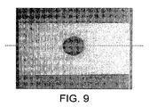
- FIG. 9 is picture of an exemplary reaction spot according to the present invention.
- FIG. 10 is a scan along the dashed line of FIG. 9 .
- FIG. 11 is a schematic illustration of a layered test strip constructed according to the present invention.
- FIG. 12 is a schematic illustration of another layered test strip construction according to the present invention.
- FIG. 13 is a picture of a mesh wherein at least a portion thereof includes retained reagent.
- FIG. 14 is a schematic illustration of a further layered test strip construction according to the present invention.
- FIG. 15 is a schematic illustration of yet another layered test strip construction according to the present invention.
- FIG. 16 is a schematic illustration of a further layered test strip construction according to the present invention.
- FIG. 1 is one such arrangement 100 for the optical detection of the presence and/or concentration of an analyte such as glucose.
- the arrangement 100 can generally be described as a diffuse transmission arrangement.
- the substance to be analyzed is transported via a conduit 12 .
- the conduit 12 can be in the form of a hollow member or needle.
- the needle can be of a very narrow gage, or a so-called microneedle. Such a needle typically having a size on the order of 40-100 micrometers.
- the microneedle may also act as a skin penetration member as well as a conduit.
- the skin penetration member may be in the form of a solid lancet (not shown), which acts to produce a sample of bodily fluid which is then transported via conduit, e.g.— 12 .
- the substance to be analyzed is transported via the conduit 12 to a chamber 14 .
- the chamber may have any suitable form.
- the chamber 14 is formed by a lower member 16 and an upper member 18 . These members 16 , 18 can be constructed to allow light to be transmitted therethrough.
- An assay pad 20 is provided within the chamber 14 .
- the pad 20 receives the substance to be analyzed.
- the pad 20 is provided with a substance that reacts with the analyte. The results of this reaction is detectable and the data generated through detection is used to determine the presence and/or concentration of the analyte.
- the pad 20 is provided with a construction to achieve this objective, and others. A discussion of the specifics of the pad 20 will be deferred.
- a light source 22 can be provided so as to produce light that is incident upon a first side of the pad 20 .
- the light source comprises a light emitting diode (LED).
- the LED or light source 22 can provide incident light of any suitable wavelength. Wavelengths on the order of 500-700 nm are suitable. For example, incident light with a wavelength on the order of 670 nm may be transmitted from light source 22 . At approximately 670 nm, absorption of the incident light by hemoglobin tends to be reduced.
- An optical detector 24 is positioned so as to receive light transmitted through a second side of the pad 20 , the second side being opposite the first side.
- the optical detector can be a digital imaging device, such as a complementary metal oxide semiconductor (CMOS) device, a charge couple device (CCD), or any photodiode.
- CMOS complementary metal oxide semiconductor
- CCD charge couple device
- the imaging device may comprise and array of individual detectors.
- the implementation of digital imaging devices permits the analysis of samples that are not of uniform volume. This is accomplished at least in part by the ability of individual detectors to take readings, which are then averaged or combined to generate an overall reading. The larger the sample, the larger the “spot” formed on the pad 20 , and the more individual detectors pick up a reading of the reaction occurring in the pad 20 .
- the assay of the present invention is not dependent upon the volume of the sample being analyzed. When an assay is incorporated into a monitoring device wherein the monitoring is performed in an automated fashion, the user is often unable to determine if a sample of body fluid of a particular volume has been obtained.
- the accuracy of the assay is directly linked to a specific volume of sample undergoing analysis (e.g.—“microtitration”).
- the present invention breaks this dependence, thereby providing robust and reliable devices, such as personal glucose monitoring devices, that can function effectively over a range of potential sample volumes.
- Other details of the detector 24 , and associated components, will be deferred to later text.
- the arrangement 100 may be incorporated into a larger overall device. According to one embodiment, the arrangement 100 is incorporated into a personal glucose-monitoring device. According to one alternative, the device is ambulatory.
- the arrangement 100 can be incorporated into devices, such as the ones described in U.S. Patent Publication No. US 2002/0087056 or US 2003/0153900, both of which are incorporated herein by reference in their entirety.
- FIG. 2 An alternative arrangement 200 is depicted in FIG. 2 .
- the arrangement 200 can generally be described as a diffuse reflection arrangement.
- the light source 22 is provided such that it transmits light which is incident upon a first side of the assay pad 20 . At least a portion of this incident light is reflected off of the first side. This reflected light can then be collected by the optical detector 24 , which is disposed on the same side as the light source 22 , relative to the test pad 20 .
- the light source 22 and the optical detector 24 are disposed, relative to the reaction chamber, on a side which is opposite from the side of the reaction chamber into which the conduit 12 introduces the substance to be analyzed. This orientation is shown in FIG. 2 .
- the test pad 20 may be provided with a blocking layer 21 .
- a blocking layer 21 can be formed of any suitable material, such as zirconium oxide or titanium oxide.
- the reading taken by the detector 24 is insensitive to the contents of the chamber 14 since the contents of the chamber 14 are not in direct communication with the side of the test pad 20 being read.
- FIG. 3 Another alternative arrangement constructed according to the principles of the present invention is illustrated in FIG. 3 .
- the arrangement 300 can generally be described as an edge or waveguide illumination arrangement.
- a light source 22 is arranged such that a diverging light beam strikes an edge of a transparent waveguide member 32 , and couples the transmitted light inside the waveguide, as illustrated by the broken lines contained in FIG. 3 .
- ⁇ C critical angle of incidence
- ⁇ C sin ⁇ 1 (n 2 /n 1 )
- ⁇ c the angle of incidence that will cause incident light to become trapped in the waveguide
- n 2 and n 1 are the indices of refraction for a first material and a second material that define a boundary across which the light is attempting to travel (e.g.—air and test strip material).
- the coupling edge of the waveguide may be treated with an anti reflective coating or an index-matching medium.
- an anti reflective coating or an index-matching medium For example, one or more thin layers of silicon oxide may be applied to the coupling edge. A thickness of approximately 200 nm is an exemplary thickness.
- TIR total internal reflection
- the assay pad 20 is provided with a surface in communication with the reflected light inside the waveguide, which is light scattering.
- Light impinging on this surface 34 of a test pad 20 is scattered at all angles. As illustrated by the dotted lines in FIG. 3 , some of the light is scattered at a large angle relative to the normal of surface 34 . This light is also trapped inside the waveguide. However, some of the light is scattered at a relatively small angle compared to the normal of surface 34 , as illustrated by the solid double arrow lines of FIG. 3 , and is consequently transmitted outside of the waveguide and received by the detector 24 .
- FIG. 4 Another arrangement 400 of the type described above, is also illustrated in FIG. 4 .
- arrangements of this type are such that light provided by the light source 22 is incident upon the waveguide at a relatively small angle to the surface thereof, a larger amount of this incident light is transmitted through the waveguide, when compared with arrangements wherein the incident light is provided at a relatively steep angle to the incident surface.
- the light source is an LED-type source, less power is required in order to provide the necessary amount of incident light to perform the assay, and power consumption can be minimized.
- the light source 22 can be closely aligned with the edge of the waveguide 32 , and thus may provide for a compact configuration.
- the detector element 24 can be positioned in close proximity to the waveguide 32 , assay pad 20 and light source 22 ( FIG. 4 ), thereby enabling it to be more protected from stray light sources.
- an imaging lens 52 is provided and disposed such that it captures and focuses light reflected from the assay pad 20 toward the optical detector 24 .
- the lens 52 may comprise a refractive or defractive element (Fresnel lens).
- the lens 52 is utilized in connection with an edge illumination-type arrangement.
- the lens 52 is illustrated as being disposed at some distance from the waveguide 32 which forms the upper portion of the reaction chamber 14 , the lens member may also be integrated into either the surface of the detector 24 , or into the upper member (e.g.— 18 , FIG. 1 ) or waveguide 32 .
- FIG. 6 depicts an alternative arrangement 600 which comprises a diffuse reflection type arrangement which incorporates an imaging lens member 52 , as described above.
- the arrangement 700 includes an edge-illumination type arrangement including waveguide 32 .
- An imaging lens 54 is also included. In this arrangement, the imaging lens is integrated into either the waveguide 32 or the detector 24 .
- FIGS. 8 a and 8 b illustrate in further detail certain aspects of an additional embodiment of the present invention.
- FIGS. 8 a and 8 b illustrate and arrangement 80 which can be generally described as a vertical-flow assay.
- the arrangement 800 includes a channel member 82 having an open “bottom”, an assay pad 20 in communication with the open bottom of channel member 82 , and an optical detector, preferably in the form of an array 84 of digital imaging devices 85 .
- lens elements 86 are incorporated into the arrangement. Although lens elements 86 are illustrated as being in the form of discrete individual lenses in close proximity to the individual detector elements or pixels 85 , it is within the scope of the present invention that alternative constructions are possible. For example, the lenses 86 could be spaced from the detectors 85 , and/or a single integrated lens construction could be utilized instead of discrete lenses 86 .
- FIGS. 8 a and 8 b permit the reliable and effective analysis of relatively small amounts of substances, which is not dependent upon a particular repeatable volume.
- the channel member 82 is elongated.
- the substance being analyzed such as a sample of body fluid is introduced at one end 81 of the channel member 82 and flows laterally or axially therethrough.
- the substance being analyzed then permeates the test pad 20 disposed thereunder.
- the open channel member 82 defines a particular volume, it is not necessary to completely fill the channel member 82 in order to conduct an accurate analysis.
- a particular volume of substance to be tested may only partially fill channel member 82 , for example, it may only fill channel member 82 up to the point defined along line 83 .
- the independent detector pixels 85 of the array 84 on the left-hand side of line 83 are activated and will take a reading from the analysis side of the test pad 20 once the analyte present in substance under analysis reacts with the chemicals contained in the test pad 20 .
- Those individual detector elements of the array which are on the left-hand side of line 83 will generate a signal which is indicative of the presence of this reaction.
- Those individual detector elements 85 of array 84 which are on the right hand side of line 83 will not generate such a reading.
- the readings or data generated by each individual detector element 85 of the array 84 is taken and analyzed, such as by averaging, to generate an overall reading of the presence and/or concentration of a particular analyte contained in the substance under analysis.
- the volume of the channel member 82 is known, and the length of the detector element contained in the array 84 , as well as any correction due to the presence of lens elements 86 are known, the volume of the sample under analysis can be estimated based upon the number of pixels which detect the reaction between the analyte and the chemical reagents contained in test pad 20 .
- An exemplary, non-limiting embodiment will now be described to further illustrate the principles of the present invention.
- the time between the readings taken by the detectors 85 of the array 84 can also be monitored by the electronics of the arrangement.
- the specific means of implementing this monitoring being well within the skill of those in the art.
- the arrangement of the present invention can also analyze the kinetics of the reaction between the analyte and the chemical reagents, thereby offering the possibly to provide additional useful information that can be used to provide an insightful analysis of the sample.
- An arrangement 80 of the type depicted in FIGS. 8 a through 8 b can advantageously be utilized to analyze very small volumes of a substance to be tested.
- the arrangement can be utilized to assay 1-500 nanoliter (nL) volumes.
- the substance to be tested may include whole blood, interstitial fluid, or combinations thereof.
- a sample of body fluid is introduced along direction B 1 from the right hand side 81 of channel number 82 through any suitable means, such as conduit 14 (see, e.g.— FIGS. 1 through 2 ).
- the sample is extended laterally into the elongated channel member 82 .
- the sample is extended over a relatively long length, which can be on the order of a few millimeters.
- the channel is approximately 250 micrometers (width) ⁇ 140 micrometers (height) ⁇ 7,000 micrometers (length).
- the sample of body fluid then flows vertically along direction B 2 through the open bottom portion of channel 82 and into the assay pad 20 .
- a color change in the assay pad 20 may be produced by a chemical reaction triggered by the presence of a specific analyte contained in the sample of body fluid, such as glucose. This color change can be detected on the opposite of the assay pad from which the sample of body fluid is introduced. This color change is detected by a suitable detection device, such as the optical detector array 84 , which may be composed of a plurality of digital imaging detectors 85 . According to one aspect, these digital detectors comprise either a CMOS, CCD, or photo diode array.
- red blood cells can be filtered or blocked from the opposite side of the test pad 20 , which is generally viewed as being preferable so as to enable a more accurate reading of the levels of analyte present in the sample.
- the chemical reagents and dye products utilized in test pad 20 can be chosen from any number of well know substances. In any event, these chemistries should provide a discernable and readable reaction over an appropriate range of analyte concentration levels.
- these chemistries should be able to produce reactions which can then be detected to indicate a glucose concentration ranging from, for example, 40-500 mg/dL.
- the detector array 84 is formed from a plurality of individual CMOS-type detectors 85 .
- CMOS detectors are less costly than other types of digital imaging devices.
- various “clock” or time-driven electronic devices may be incorporated. Data can be taken from the detectors 85 and fed to such devices (directly or from storage) thereby enabling the monitoring and interpretation of the kinetics and other aspects of the reaction between the analyte and the reagent.
- uniformity and accuracy of the assay is also promoted. For example, readings are taken and utilized from multiple individual detector elements, or pixels, are less susceptible to variations in uniformity of the materials of the assay pad, or other localized abnormalities, which can be accounted for and corrected by the interpretation and manipulation of the data generated by the individual pixels 85 of the detector array 84 . For example, statistical analyses and associated algorithms may be used to correct for such localized defects, and therefore improve the accuracy of the overall reading generated by the device.
- FIGS. 9 and 10 represent a scan performed by an arrangement 80 such as the one depicted in FIGS. 8 a and 8 b .
- FIG. 9 is a picture of a test spot formed on a commercially-available assay pad.
- FIG. 10 is a plot of the signal generated by an array of detector elements 84 as they are scanned along the dashed line contained in FIG. 9 . As illustrated in FIG. 10 , the lower voltage readings correspond to the area occupied by the test spot of FIG. 9 along the dashed line.
- FIG. 11 is a schematic illustration of one possible construction for the assay pad 20 .
- the assay pad construction 1100 has a sample application side SA as well as an analysis side A.
- the assay pad 1100 is composed of at least 3 constituent components: a blocking component 1102 ; a membrane component 1104 ; and a chemical reagent component 1106 .
- a blocking component 1102 When the sample to be analyzed is composed at least in part of whole blood, red blood cells are separated from the plasma, which is then transported to the reagent component 1106 which includes enzymatic chemistries selected to produce a chemical reaction with one or more analytes present in the sample.
- the blocking component 1102 may include an agent to filter or pre-filter red blood cells.
- the blocking component 1102 may include zirconium oxide (ZrO 2 ).
- the membrane component 1104 can be formed of any suitable material.
- the membrane be a high light transmitting material, or optionally can be an opaque or substantially opaque material. Suitable materials may include styrene and/or nylon.
- the membrane component 1104 can be in woven or non-woven form. According to one aspect, the membrane is in the form of a woven mesh.
- the reagent component 1106 is chosen according to its ability to react with the analyte under investigation. Any suitable reagent chemistry can be utilized. Specific examples of suitable material include a glucose oxidase (e.g.—from Biocatalysts product G575P), a soybean peroxidase (e.g.—from Organic Technologies, product 73930.1 medical diagnostic grade), an amino antipyrine hydrochloride (e.g.—from TCI, product A0257) and an Aniline derivative dye for a “Trinder” reaction (see, e.g.—P. Trinder, “Determination of Glucose in Blood Using Glucose Oxidase with an Alternative Oxygen Acceptor”).
- a glucose oxidase e.g.—from Biocatalysts product G575P
- soybean peroxidase e.g.—from Organic Technologies, product 73930.1 medical diagnostic grade
- an amino antipyrine hydrochloride e.g
- the blocking component 1102 and/or the reagent component 1106 may be partially or entirely subsumed and contained within the membrane component 1104 .
- a surface “layer” of the blocking component 1102 and/or reagent component 1106 may be present.
- the blocking component 1102 and/or the reagent component 1106 may be entirely subsumed, contained or impregnated within the membrane component 1104 , such that no distinct blocking component “layer” or reagent component “layer” is present at all.
- the blocking component 1102 and/or reagent component 1106 may be applied to the membrane component 1104 in any suitable manner.
- the blocking component 1102 and/or reagent component 1106 may be coated upon the membrane 1104 .
- suitable coating techniques includes spin coating.
- FIG. 12 illustrates one possible alternative construction 1200 for the assay pad 20 .
- the assay pad construction 1200 is composed of at least 4 components: a prefilter and reflective component 1202 , a membrane component 1204 , a reagent component 1206 , and a mesh component 1208 .
- the red blood cells are separated from the plasma, so that components 1202 and/or 1204 retain the red blood cells without lysis.
- the plasma is then vertically transported to the reagent component 1206 which includes enzymatic chemistries.
- These substances may include any suitable material, which are generally known in the art.
- suitable materials include a glucose oxidase (e.g.—from Biocatalysts product G575P), a soybean peroxidase (e.g.—from Organic Technologies, product 73930.1 medical diagnostic grade), an amino antipyrine hydrochloride (e.g.—from TCI, product A0257) and an Aniline derivative dye for a “Trinder” reaction (see, e.g.—P. Trinder, “Determination of Glucose in Blood Using Glucose Oxidase with an Alternative Oxygen Acceptor”).
- a glucose oxidase e.g.—from Biocatalysts product G575P
- soybean peroxidase e.g.—from Organic Technologies, product 73930.1 medical diagnostic grade
- an amino antipyrine hydrochloride e.g.—from TCI, product A0257
- an Aniline derivative dye for a “Trinder” reaction see, e.g.—P. Trin
- the component 1204 may comprise any suitable blood-filtering component or material.
- suitable blood-filtering component or material Some materials which have been used to separate red blood cells from plasma and samples of whole blood include glass fibers, mesh cloth, unwoven mats, asymmetric membranes, and porous polymers.
- a suitable material is, a hydrophilic poly(vinylidene fluoride) unwoven membrane.
- Such membranes are commercially available from the Millipore Corporation (e.g.—with 0.1 micron openings).
- Other suitable materials include unwoven membranes comprising a polyamide.
- Other materials may be incorporated to achieve various performance objectives, such as poly(acrylic acid) (PAA), hydroxyethyl cellulose, sodium alginate, and gelatin.
- the pad 1200 may further include a component 1202 , such as a prefilter and/or diffuse reflection component.
- a component 1202 such as a prefilter and/or diffuse reflection component.
- the component 1202 when included, serves to improve separation of red blood cells from a sample containing whole blood, and/or increase the reflection of incident light upon the opposite side of the pad when undergoing optical analysis.
- Suitable materials for use as component 1202 include a zirconium oxide (ZrO 2 ), optionally combined with a polymer binder material.
- the component 1202 can be applied to the membrane, or component 1204 , by using any suitable technique.
- One such suitable technique is spin coating. Spin coating promotes uniform distribution of the component 1202 onto and/or within the membrane component 1204 .
- the component 1202 may be partially, or entirely contained within the membrane component 1204 , thus effectively combining these two components into a single unitary member, rather than the illustrated distinct layers.
- the component 1202 comprises a prefilter and diffuse reflection material and a polymer binder
- the presence of the polymer binder tends to form at least a thin layer or film over the membrane component 1204 .
- This thin film may have any suitable thickness, for example, the component 1202 may partially permeate the membrane component 1204 and leave a thin film having a thickness on the order of less than 5 micrometers on top of the membrane component 1204 .
- the component 1206 comprises a reagent which may be formed from any suitable composition, such as the chemicals described above, and may optionally be provided in a binder of poly(acrylic acid), hydroxypropylmethylcellulose (HPMC) or similar polymers that reduce lateral spreading of the substrate or sample undergoing analysis.
- a reagent which may be formed from any suitable composition, such as the chemicals described above, and may optionally be provided in a binder of poly(acrylic acid), hydroxypropylmethylcellulose (HPMC) or similar polymers that reduce lateral spreading of the substrate or sample undergoing analysis.
- the mesh component 1208 and the reagent 1206 component may also be integrated into a single layer or constituent element of the test pad 20 .
- reagent material 1206 can be coated onto the mesh component 1208 .
- Any suitable coating technique may be utilized.
- a spin coating technique is one such technique for providing the reagent on the mesh component 1208 which allows for precise and relatively thin coating of the reagent component 1206 to be applied to the mesh component 1208 . According to one construction, this “coating” is entirely absorbed and contained within mesh component 1208 .
- the mesh component 1208 should be permeable to oxygen in order to promote the reaction of the analyte with the chemical reagent component 1206 .
- the mesh component 1208 can be formed from a high reflectance woven mesh, such as nylon.
- mesh component 1208 is formed from an open polyamide or nylon mesh material. This nylon mesh material having a reagent incorporated therein is believed to increase the sensitivity of the assay.
- the open structure of the nylon mesh in conjunction with the controlled amount of the reagent component material 1206 incorporated therein, increases the diffuse reflection of light incident thereon. This increased diffuse reflection is believed to be caused by the multiple reflections and increased path lengths provided by the open nylon mesh structure. According to one exemplary embodiment shown in FIG.
- the mesh component 1208 has openings have a dimension which is on the order of 68 microns, and a filament diameter which is on the order of approximately 50 microns.
- the region indicated by circle A 1 illustrates the absence of reagent material within the mesh, wherein the reagent material has been consumed by chemical reaction with the analyte.
- Circle A 2 indicates an area of the mesh in which the reagent material is still disposed thereon.
- the cylindrical fibers of the nylon mesh may scatter the light onto the external exposed layer or surface thereby introducing multiple reflection and increased path links.
- the scattering of incident light is largely dependent upon a refractive index of the cylinders which form the filaments of the mesh.
- the refractive index of nylon is relative large, on the order of 1.53, and this is beneficial.
- the above-described application of the reagent component 1206 to the mesh component 1208 can simplify manufacture.
- test pad construction 1400 includes three basic components: a filter and/or diffuse reflective component 1402 , a chemical reagent component 1404 , and a membrane component 1406 .
- a sample to be analyzed is applied to the sample application side SA of test pad 1400 .
- red blood cells are separated from the plasma by component 1402 and are transported to the reagent component 1404 , which may be partially or entirely impregnated it the membrane component 1406 .
- the component 1402 may be formed from any suitable construction that provides filtration and/or diffuse reflection functions.
- suitable constructions have been described above.
- the component 1402 may comprise a zirconium oxide material combined with a permeable polymer binder material, such as HPMC, that limits lateral diffusion.
- a zirconium oxide material combined with a permeable polymer binder such as vinyl butyral. The zirconium oxide and polymer binder are combined in any suitable proportions, such as 20:80.
- the reagent component 1404 includes at least one chemical reagent chosen to provide a suitable reaction with the analyte(s) under investigation.
- Any suitable reagent can be chosen.
- the reagent may comprise a glucose oxidase, soy bean peroxidase, amino antipyrine hydrochloride, and/or aniline derivative dyes.
- the chemical reagent may optionally be combined with a binder component.
- the binder component is preferably formed from a material which reduces the tendency for lateral spreading of the sample within the test pad 1400 . Suitable examples binder material examples include HPMC and PAA.
- a suitable membrane component 1406 can also be provided.
- the membrane component 1406 can comprise a fibrous unwoven material.
- One specific example includes a cellulose acetate unwoven fibrous membrane material.
- Both the filter/diffuse reflective component 1402 , as well as the reagent component 1404 can be applied to the membrane component 1406 by any suitable technique.
- the reagent component 1404 is applied to the membrane component 1406 by a coating technique, such as spin coating.
- Reagent component 1404 may be partially, or entirely impregnated within the membrane component 1406 .
- a thin film or layer of reagent component 1404 may be present on the top surface of the membrane component 1406 (i.e.—the side closest to the sample application side SA of the test pad 1400 ). When present, this thin top layer can be on the order of a few microns in thickness.
- the reagent component may be entirely subsumed, contained or impregnated within the membrane component 1406 , such that no distinct layer is present on the top surface of the membrane component 1406 (i.e.—the surface of membrane 1406 which is closest to the surface application side SA of the pad 1400 ).
- the filter/diffuse reflective component 1402 can then be applied subsequent to application of the reagent component 1404 .
- the component 1402 can also be applied by any suitable technique, such as spin coating.
- a filter/diffuse reflective component comprising a zirconium oxide and hydrophobic permeable polymer binder material is spin coated at approximately 1000 rpm onto the membrane/reagent components 1406 / 1404 .
- the filter/diffuse reflective component 1402 may be partially impregnated within the membrane 1406 , leaving a thin film layer at the surface application side SA of the combined membrane and reagent components 1406 and 1404 . When present, this thin surface layer can be a few microns in thickness, for example, on the order of less than 5 microns in thickness.
- the filter/diffuse reflective component 1402 may be entirely subsumed, contained, or impregnated within the combined reagent and membrane components 1404 and 1406 .
- test pad construction 1500 is comprised of at least four basic components: a filter/diffuse reflective component 1502 , a permeable material component 1504 , a reagent component 1506 , and a membrane component 1508 .
- the general functionality of the test pad 1500 and the functionality of the individual components thereof are similar to that previously described.
- the component 1502 may comprise any suitable filter and/or diffuse reflective material.
- the component 1502 may comprise a zirconium oxide material with or without a polymer binder.
- the polymer binder is preferably a permeable material.
- suitable permeable polymer binders include HPMC and an acrylic resin, such as Elvacite®.
- the component 1504 is preferably comprised of a suitable permeable material which can be hydrophobic.
- the hydrophobic material can comprise any suitable form.
- the component 1504 may comprise a film of a hydrophobic polymeric material, such as PAA.
- the film can be formed by any suitable technique.
- a film of PAA is formed by a fast evaporating solvent/PAA mixture.
- the mixture may contain 10% by weight (relative to the weight of the solvent) of PAA.
- the component 1506 may comprise a suitable reagent material.
- the reagent material may be combined with a binder.
- the reagent component may be chosen from any suitable substance or combination of substances, as described above.
- the binder material comprises a permeable hydrophobic binder material.
- HPMC HPMC.
- the membrane component 1508 can be formed from any suitable material. According to one possible construction, the membrane component 1508 is formed from an unwoven fibrous mat.
- the reagent component 1506 can be spin coated upon the membrane component 1508 . As discussed above, the reagent component 1506 may be partially, or entirely, impregnated within the membrane component 1508 . Thus, the reagent component 1506 may or may not comprise a thin surface layer formed on top of the membrane component 1508 (the top side being closest to the surface application side SA of the test pad 1500 ).
- the hydrophobic material component 1504 can then be applied.
- the hydrophobic material can be formed as a slurry or mixture with a fast evaporating solvent, such as methanol.
- the mixture is then coated onto the combined membrane and reagent components 1508 and 1506 . Subsequent to evaporation, a thin film of the hydrophobic material is left on top of the combined reagent/membrane component.
- the component 1502 may then be applied to the previously described combined components.
- a combination of filter/diffuse reflective material, e.g.—ZrO 2 , and a polymer binder material, such as the ones described above may be combined in the form of a slurry or mixture and then coated upon the previously combined components.
- a slurry containing up to 10% by weight of ZrO 2 may be formed and then spin coated onto the previous combined constituent components 1504 , 1506 , and 1508 .
- the component 1502 may be partially or entirely impregnated or subsumed within the previously combined components.
- the component 1502 may be indistinguishable from the previously combined components or, may form a thin film or layer on top of the previously combined components.
- test pad construction 1600 formed according to the principles of the present invention is illustrated in FIG. 16 .
- the test pad construction 1600 illustrated therein is formed from at least three basic constituent components: a filter/diffuse reflective component 1602 , a permeable hydrophobic material component 1604 and a combined reagent/mesh component 1606 .
- the basic functionality of the individual as well as combined constituent components of the test pad construction 1600 is similar to that previously discussed.
- the filter/diffuse reflective component 1602 can be formed from any suitable material, and comprise any suitable construction.
- the component 1602 comprises a zirconium oxide material, which may optionally be combined with a binder material.
- the binder material is preferably a permeable hydrophobic binder material such as an acrylic resin. Such material is being commercially available, e.g.—sold as Elvacite®.
- the permeable hydrophobic material component 1604 can be comprised of any suitable material, and take any suitable form.
- the permeable hydrophobic material can comprise a film or layer.
- the permeable hydrophobic material may comprise PAA.
- the combined reagent/mesh component 1606 is preferably the combination of two distinct parts or constituents. Namely, a reagent material such as those previously described, as well as a mesh material constituent. Preferably, these constituents are combined into a single, integral component 1606 .
- the mesh material can be formed of any suitable material and take any suitable form.
- the mesh material can be formed from a woven polyamide material, such as a woven nylon mesh.
- the mesh may have openings of any suitable dimensions, such as either 30 microns openings or 41 micron openings.
- the reagent and the mesh component are combined or integrated by soaking the mesh material in a slurry formed from the reagent material.
- a slurry or mixture can be formed which comprises 20% by weight HPMC binder material plus a suitable reagent chemistry.
- the mesh material is soaked in this liquid slurry.
- the mesh material is then removed and dried, thereby leaving an integrated mesh/reagent component 1606 .
- the hydrophobic component 1604 can be similarly be applied by any suitable technique.
- the hydrophobic material can be applied as a film formed from a fast evaporating solvent, like methanol.
- a hydrophobic material such as PAA is combined with a solvent to form a slurry.
- the slurry may comprise 10% by weight of the PAA material.
- the solvent is then applied to the combined reagent/mesh component 1606 . Upon evaporation of the solvent, a thin film or layer of the hydrophobic material is left behind, thereby forming the hydrophobic material component 1604 .
- the filter/diffuse reflective component 1602 may similarly be applied by any suitable technique, such as those previously described.
- a zirconium oxide material is added to a hydrophobic binder to form a slurry in an evaporative solvent such as toluene.
- the slurry contains approximately 10% by weight of acrylic resin such as Elvacite®.
- This slurry is then coated onto the combined hydrophobic and reagent/mesh components 1604 and 1606 .
- Any suitable coating technique can be utilized, such as the aforementioned spin coating technique.
- the filter/diffuse reflective component 1602 is provided in the form of a thin layer or film on top of the combined hydrophobic material and reagent/mesh component 1604 and 1606 .
Abstract
Description
Claims (8)
Priority Applications (1)
| Application Number | Priority Date | Filing Date | Title |
|---|---|---|---|
| US13/562,129 US9095292B2 (en) | 2003-03-24 | 2012-07-30 | Analyte concentration detection devices and methods |
Applications Claiming Priority (4)
| Application Number | Priority Date | Filing Date | Title |
|---|---|---|---|
| US10/394,230 US7052652B2 (en) | 2003-03-24 | 2003-03-24 | Analyte concentration detection devices and methods |
| US11/125,107 US7427377B2 (en) | 2003-03-24 | 2005-05-10 | Analyte concentration detection devices and methods |
| US12/222,724 US8231832B2 (en) | 2003-03-24 | 2008-08-14 | Analyte concentration detection devices and methods |
| US13/562,129 US9095292B2 (en) | 2003-03-24 | 2012-07-30 | Analyte concentration detection devices and methods |
Related Parent Applications (1)
| Application Number | Title | Priority Date | Filing Date |
|---|---|---|---|
| US12/222,724 Continuation US8231832B2 (en) | 2003-03-24 | 2008-08-14 | Analyte concentration detection devices and methods |
Publications (2)
| Publication Number | Publication Date |
|---|---|
| US20120296179A1 US20120296179A1 (en) | 2012-11-22 |
| US9095292B2 true US9095292B2 (en) | 2015-08-04 |
Family
ID=32988327
Family Applications (4)
| Application Number | Title | Priority Date | Filing Date |
|---|---|---|---|
| US10/394,230 Expired - Lifetime US7052652B2 (en) | 2003-03-24 | 2003-03-24 | Analyte concentration detection devices and methods |
| US11/125,107 Expired - Lifetime US7427377B2 (en) | 2003-03-24 | 2005-05-10 | Analyte concentration detection devices and methods |
| US12/222,724 Expired - Lifetime US8231832B2 (en) | 2003-03-24 | 2008-08-14 | Analyte concentration detection devices and methods |
| US13/562,129 Expired - Lifetime US9095292B2 (en) | 2003-03-24 | 2012-07-30 | Analyte concentration detection devices and methods |
Family Applications Before (3)
| Application Number | Title | Priority Date | Filing Date |
|---|---|---|---|
| US10/394,230 Expired - Lifetime US7052652B2 (en) | 2003-03-24 | 2003-03-24 | Analyte concentration detection devices and methods |
| US11/125,107 Expired - Lifetime US7427377B2 (en) | 2003-03-24 | 2005-05-10 | Analyte concentration detection devices and methods |
| US12/222,724 Expired - Lifetime US8231832B2 (en) | 2003-03-24 | 2008-08-14 | Analyte concentration detection devices and methods |
Country Status (2)
| Country | Link |
|---|---|
| US (4) | US7052652B2 (en) |
| EP (1) | EP2322914B1 (en) |
Cited By (11)
| Publication number | Priority date | Publication date | Assignee | Title |
|---|---|---|---|---|
| US9380974B2 (en) | 2005-09-30 | 2016-07-05 | Intuity Medical, Inc. | Multi-site body fluid sampling and analysis cartridge |
| US9636051B2 (en) | 2008-06-06 | 2017-05-02 | Intuity Medical, Inc. | Detection meter and mode of operation |
| US9782114B2 (en) | 2011-08-03 | 2017-10-10 | Intuity Medical, Inc. | Devices and methods for body fluid sampling and analysis |
| US9833183B2 (en) | 2008-05-30 | 2017-12-05 | Intuity Medical, Inc. | Body fluid sampling device—sampling site interface |
| US9897610B2 (en) | 2009-11-30 | 2018-02-20 | Intuity Medical, Inc. | Calibration material delivery devices and methods |
| US10330667B2 (en) | 2010-06-25 | 2019-06-25 | Intuity Medical, Inc. | Analyte monitoring methods and systems |
| US10383556B2 (en) | 2008-06-06 | 2019-08-20 | Intuity Medical, Inc. | Medical diagnostic devices and methods |
| US10433780B2 (en) | 2005-09-30 | 2019-10-08 | Intuity Medical, Inc. | Devices and methods for facilitating fluid transport |
| US10729386B2 (en) | 2013-06-21 | 2020-08-04 | Intuity Medical, Inc. | Analyte monitoring system with audible feedback |
| US10772550B2 (en) | 2002-02-08 | 2020-09-15 | Intuity Medical, Inc. | Autonomous, ambulatory analyte monitor or drug delivery device |
| US11090228B2 (en) * | 2016-08-19 | 2021-08-17 | Becton Dickinson And Company | Adapter assembly for attachment to a bottle |
Families Citing this family (109)
| Publication number | Priority date | Publication date | Assignee | Title |
|---|---|---|---|---|
| US6036924A (en) | 1997-12-04 | 2000-03-14 | Hewlett-Packard Company | Cassette of lancet cartridges for sampling blood |
| US6391005B1 (en) | 1998-03-30 | 2002-05-21 | Agilent Technologies, Inc. | Apparatus and method for penetration with shaft having a sensor for sensing penetration depth |
| US8641644B2 (en) | 2000-11-21 | 2014-02-04 | Sanofi-Aventis Deutschland Gmbh | Blood testing apparatus having a rotatable cartridge with multiple lancing elements and testing means |
| US9427532B2 (en) | 2001-06-12 | 2016-08-30 | Sanofi-Aventis Deutschland Gmbh | Tissue penetration device |
| US9795747B2 (en) | 2010-06-02 | 2017-10-24 | Sanofi-Aventis Deutschland Gmbh | Methods and apparatus for lancet actuation |
| US7699791B2 (en) | 2001-06-12 | 2010-04-20 | Pelikan Technologies, Inc. | Method and apparatus for improving success rate of blood yield from a fingerstick |
| US7749174B2 (en) | 2001-06-12 | 2010-07-06 | Pelikan Technologies, Inc. | Method and apparatus for lancet launching device intergrated onto a blood-sampling cartridge |
| DE60234597D1 (en) | 2001-06-12 | 2010-01-14 | Pelikan Technologies Inc | DEVICE AND METHOD FOR REMOVING BLOOD SAMPLES |
| US7981056B2 (en) | 2002-04-19 | 2011-07-19 | Pelikan Technologies, Inc. | Methods and apparatus for lancet actuation |
| US7033371B2 (en) | 2001-06-12 | 2006-04-25 | Pelikan Technologies, Inc. | Electric lancet actuator |
| US8337419B2 (en) | 2002-04-19 | 2012-12-25 | Sanofi-Aventis Deutschland Gmbh | Tissue penetration device |
| US9226699B2 (en) | 2002-04-19 | 2016-01-05 | Sanofi-Aventis Deutschland Gmbh | Body fluid sampling module with a continuous compression tissue interface surface |
| US7344507B2 (en) | 2002-04-19 | 2008-03-18 | Pelikan Technologies, Inc. | Method and apparatus for lancet actuation |
| CA2448902C (en) | 2001-06-12 | 2010-09-07 | Pelikan Technologies, Inc. | Self optimizing lancing device with adaptation means to temporal variations in cutaneous properties |
| US7041068B2 (en) | 2001-06-12 | 2006-05-09 | Pelikan Technologies, Inc. | Sampling module device and method |
| US6989891B2 (en) * | 2001-11-08 | 2006-01-24 | Optiscan Biomedical Corporation | Device and method for in vitro determination of analyte concentrations within body fluids |
| US7371247B2 (en) | 2002-04-19 | 2008-05-13 | Pelikan Technologies, Inc | Method and apparatus for penetrating tissue |
| US7331931B2 (en) | 2002-04-19 | 2008-02-19 | Pelikan Technologies, Inc. | Method and apparatus for penetrating tissue |
| US7226461B2 (en) | 2002-04-19 | 2007-06-05 | Pelikan Technologies, Inc. | Method and apparatus for a multi-use body fluid sampling device with sterility barrier release |
| US7297122B2 (en) | 2002-04-19 | 2007-11-20 | Pelikan Technologies, Inc. | Method and apparatus for penetrating tissue |
| US7648468B2 (en) | 2002-04-19 | 2010-01-19 | Pelikon Technologies, Inc. | Method and apparatus for penetrating tissue |
| US7976476B2 (en) | 2002-04-19 | 2011-07-12 | Pelikan Technologies, Inc. | Device and method for variable speed lancet |
| US7229458B2 (en) | 2002-04-19 | 2007-06-12 | Pelikan Technologies, Inc. | Method and apparatus for penetrating tissue |
| US8579831B2 (en) | 2002-04-19 | 2013-11-12 | Sanofi-Aventis Deutschland Gmbh | Method and apparatus for penetrating tissue |
| US7547287B2 (en) | 2002-04-19 | 2009-06-16 | Pelikan Technologies, Inc. | Method and apparatus for penetrating tissue |
| US7232451B2 (en) | 2002-04-19 | 2007-06-19 | Pelikan Technologies, Inc. | Method and apparatus for penetrating tissue |
| US7291117B2 (en) | 2002-04-19 | 2007-11-06 | Pelikan Technologies, Inc. | Method and apparatus for penetrating tissue |
| US7892183B2 (en) | 2002-04-19 | 2011-02-22 | Pelikan Technologies, Inc. | Method and apparatus for body fluid sampling and analyte sensing |
| US7901362B2 (en) | 2002-04-19 | 2011-03-08 | Pelikan Technologies, Inc. | Method and apparatus for penetrating tissue |
| US7717863B2 (en) | 2002-04-19 | 2010-05-18 | Pelikan Technologies, Inc. | Method and apparatus for penetrating tissue |
| US9248267B2 (en) | 2002-04-19 | 2016-02-02 | Sanofi-Aventis Deustchland Gmbh | Tissue penetration device |
| US8702624B2 (en) | 2006-09-29 | 2014-04-22 | Sanofi-Aventis Deutschland Gmbh | Analyte measurement device with a single shot actuator |
| US7909778B2 (en) | 2002-04-19 | 2011-03-22 | Pelikan Technologies, Inc. | Method and apparatus for penetrating tissue |
| US7674232B2 (en) | 2002-04-19 | 2010-03-09 | Pelikan Technologies, Inc. | Method and apparatus for penetrating tissue |
| US8221334B2 (en) | 2002-04-19 | 2012-07-17 | Sanofi-Aventis Deutschland Gmbh | Method and apparatus for penetrating tissue |
| US7491178B2 (en) | 2002-04-19 | 2009-02-17 | Pelikan Technologies, Inc. | Method and apparatus for penetrating tissue |
| US8267870B2 (en) | 2002-04-19 | 2012-09-18 | Sanofi-Aventis Deutschland Gmbh | Method and apparatus for body fluid sampling with hybrid actuation |
| US9795334B2 (en) | 2002-04-19 | 2017-10-24 | Sanofi-Aventis Deutschland Gmbh | Method and apparatus for penetrating tissue |
| US9314194B2 (en) | 2002-04-19 | 2016-04-19 | Sanofi-Aventis Deutschland Gmbh | Tissue penetration device |
| US8784335B2 (en) | 2002-04-19 | 2014-07-22 | Sanofi-Aventis Deutschland Gmbh | Body fluid sampling device with a capacitive sensor |
| IES20020794A2 (en) * | 2002-10-04 | 2003-02-19 | Minroc Techn Promotions Ltd | A down-the-hole hammer |
| US8574895B2 (en) | 2002-12-30 | 2013-11-05 | Sanofi-Aventis Deutschland Gmbh | Method and apparatus using optical techniques to measure analyte levels |
| US7052652B2 (en) | 2003-03-24 | 2006-05-30 | Rosedale Medical, Inc. | Analyte concentration detection devices and methods |
| WO2004107964A2 (en) | 2003-06-06 | 2004-12-16 | Pelikan Technologies, Inc. | Blood harvesting device with electronic control |
| WO2006001797A1 (en) | 2004-06-14 | 2006-01-05 | Pelikan Technologies, Inc. | Low pain penetrating |
| WO2005033659A2 (en) | 2003-09-29 | 2005-04-14 | Pelikan Technologies, Inc. | Method and apparatus for an improved sample capture device |
| US9351680B2 (en) | 2003-10-14 | 2016-05-31 | Sanofi-Aventis Deutschland Gmbh | Method and apparatus for a variable user interface |
| EP1706026B1 (en) | 2003-12-31 | 2017-03-01 | Sanofi-Aventis Deutschland GmbH | Method and apparatus for improving fluidic flow and sample capture |
| US7822454B1 (en) | 2005-01-03 | 2010-10-26 | Pelikan Technologies, Inc. | Fluid sampling device with improved analyte detecting member configuration |
| EP1725867A4 (en) * | 2004-03-18 | 2009-04-08 | Fujifilm Corp | Analysis element for use in method of testing specimen |
| US20050221279A1 (en) * | 2004-04-05 | 2005-10-06 | The Regents Of The University Of California | Method for creating chemical sensors using contact-based microdispensing technology |
| US8828203B2 (en) | 2004-05-20 | 2014-09-09 | Sanofi-Aventis Deutschland Gmbh | Printable hydrogels for biosensors |
| US9820684B2 (en) | 2004-06-03 | 2017-11-21 | Sanofi-Aventis Deutschland Gmbh | Method and apparatus for a fluid sampling device |
| US7410614B2 (en) * | 2004-07-26 | 2008-08-12 | Kabushiki Kaisha Toshiba | Optical waveguide type iontophoresis sensor chip and method for packaging sensor chip |
| US20060078471A1 (en) * | 2004-10-12 | 2006-04-13 | Witty Thomas R | Apparatus and method for a precision flow assay |
| US8652831B2 (en) | 2004-12-30 | 2014-02-18 | Sanofi-Aventis Deutschland Gmbh | Method and apparatus for analyte measurement test time |
| US20060281187A1 (en) | 2005-06-13 | 2006-12-14 | Rosedale Medical, Inc. | Analyte detection devices and methods with hematocrit/volume correction and feedback control |
| US20070081920A1 (en) * | 2005-10-12 | 2007-04-12 | Murphy R S | Semi-disposable optoelectronic rapid diagnostic test system |
| JP4230527B2 (en) * | 2005-10-28 | 2009-02-25 | パナソニック株式会社 | Measuring device, measuring apparatus and measuring method |
| US9976192B2 (en) | 2006-03-10 | 2018-05-22 | Ldip, Llc | Waveguide-based detection system with scanning light source |
| US9528939B2 (en) | 2006-03-10 | 2016-12-27 | Indx Lifecare, Inc. | Waveguide-based optical scanning systems |
| US7846110B2 (en) * | 2006-08-03 | 2010-12-07 | Advanced Medical Products Gmbh | Self-contained test unit for testing body fluids |
| US8372015B2 (en) | 2006-08-28 | 2013-02-12 | Intuity Medical, Inc. | Body fluid sampling device with pivotable catalyst member |
| US20080077035A1 (en) * | 2006-09-25 | 2008-03-27 | Baker Clark R | Carbon dioxide-sensing airway products and technique for using the same |
| WO2008037068A1 (en) * | 2006-09-29 | 2008-04-03 | Ottawa Health Research Institute | Correlation technique for analysis of clinical condition |
| US8771714B2 (en) * | 2006-10-31 | 2014-07-08 | Musc Foundation For Research Development | Systems and methods for in vivo measurement of interstitial biological activity, processes and/or compositions |
| WO2009126900A1 (en) | 2008-04-11 | 2009-10-15 | Pelikan Technologies, Inc. | Method and apparatus for analyte detecting device |
| EP2130493B1 (en) * | 2008-06-07 | 2013-09-25 | Roche Diagnostics GmbH | Analysis system for detecting an analyte in a bodily fluid, cartridge for an analytic device and method for manufacturing a cartridge for an analysis system. |
| TR201905816T4 (en) * | 2008-11-07 | 2019-05-21 | Hoffmann La Roche | Fine-grained fillers for photometric reaction films. |
| US9375169B2 (en) | 2009-01-30 | 2016-06-28 | Sanofi-Aventis Deutschland Gmbh | Cam drive for managing disposable penetrating member actions with a single motor and motor and control system |
| KR20100109196A (en) * | 2009-03-31 | 2010-10-08 | 삼성전자주식회사 | A system and method of measuring object's concentration using for transmission or reflection of light |
| US8871148B2 (en) * | 2009-05-22 | 2014-10-28 | 3M Innovative Properties Company | Multilayer colorimetric sensors |
| EP2456355B1 (en) | 2009-07-20 | 2016-09-14 | Optiscan Biomedical Corporation | Adjustable connector and dead space reduction |
| US8932256B2 (en) | 2009-09-02 | 2015-01-13 | Medtronic Minimed, Inc. | Insertion device systems and methods |
| US8900190B2 (en) | 2009-09-02 | 2014-12-02 | Medtronic Minimed, Inc. | Insertion device systems and methods |
| US8998840B2 (en) | 2009-12-30 | 2015-04-07 | Medtronic Minimed, Inc. | Connection and alignment systems and methods |
| US11497850B2 (en) | 2009-12-30 | 2022-11-15 | Medtronic Minimed, Inc. | Connection and alignment detection systems and methods |
| US8435209B2 (en) | 2009-12-30 | 2013-05-07 | Medtronic Minimed, Inc. | Connection and alignment detection systems and methods |
| US20120215163A1 (en) | 2009-12-30 | 2012-08-23 | Medtronic Minimed, Inc. | Sensing systems and methods |
| US8965476B2 (en) | 2010-04-16 | 2015-02-24 | Sanofi-Aventis Deutschland Gmbh | Tissue penetration device |
| EP2417910B1 (en) * | 2010-08-11 | 2013-06-26 | Roche Diagnostics GmbH | Analytical test unit and test system |
| US20120045787A1 (en) * | 2010-08-19 | 2012-02-23 | Ulrich Boettiger | Flexible micro-carrier system |
| US9469871B2 (en) | 2011-04-14 | 2016-10-18 | Corporos Inc. | Methods and apparatus for point-of-care nucleic acid amplification and detection |
| CN106929401B (en) * | 2011-05-05 | 2020-06-09 | 安派科生物医学科技有限公司 | Tumor cell detector |
| WO2013105678A1 (en) * | 2012-01-11 | 2013-07-18 | 경원대학교 산학협력단 | Blood glucose measurement unit, blood glucose measurement system comprising same, and blood glucose measurement method |
| US9354159B2 (en) | 2012-05-02 | 2016-05-31 | Nanoscopia (Cayman), Inc. | Opto-fluidic system with coated fluid channels |
| WO2013170216A1 (en) * | 2012-05-11 | 2013-11-14 | Wellsense Inc. | Mobile analyte monitoring system |
| US9255245B2 (en) * | 2012-07-03 | 2016-02-09 | Agilent Technologies, Inc. | Sample probes and methods for sampling intracellular material |
| WO2014058922A1 (en) * | 2012-10-08 | 2014-04-17 | Colorado State University Research Foundation | Distance-based quantitative analysis using a capillarity-based analytical device |
| WO2015023302A1 (en) * | 2013-08-12 | 2015-02-19 | Kateeva, Inc. | Ester-based solvent systems for printable organic light-emitting diode ink formulations |
| GB2519132A (en) * | 2013-10-11 | 2015-04-15 | Univ Singapore | Disposable photometric measurement tip |
| CN105979871B (en) * | 2014-01-10 | 2020-03-03 | 格鲁科威斯塔公司 | Non-invasive system and method for measuring substance concentration, computer readable medium |
| US10101342B2 (en) | 2014-02-12 | 2018-10-16 | Church & Dwight Co., Inc. | Devices and methods for electronic analyte assaying |
| US10018566B2 (en) | 2014-02-28 | 2018-07-10 | Ldip, Llc | Partially encapsulated waveguide based sensing chips, systems and methods of use |
| WO2016138427A1 (en) | 2015-02-27 | 2016-09-01 | Indx Lifecare, Inc. | Waveguide-based detection system with scanning light source |
| US10183290B2 (en) | 2015-04-16 | 2019-01-22 | Advanced Technology Applications, Llc | Method for analyte examination |
| US9759664B2 (en) * | 2015-05-28 | 2017-09-12 | Hamilton Sundstrand Space Systems International, Inc. | Microgravity-compatible analysis |
| CN106546728A (en) * | 2015-09-16 | 2017-03-29 | 杨波 | A kind of new immunochromatography reagent bar and its technical scheme of detection |
| WO2017059425A1 (en) * | 2015-10-01 | 2017-04-06 | Ldip, Llc | Waveguide-based detection system with scanning light source |
| KR20170099737A (en) | 2016-02-23 | 2017-09-01 | 노을 주식회사 | Contact-type staining patch and staining method using the same |
| WO2017146505A1 (en) * | 2016-02-23 | 2017-08-31 | 노을 주식회사 | Substance-marking patch, and method and apparatus for tissue diagnosis using same |
| WO2017146506A1 (en) * | 2016-02-23 | 2017-08-31 | 노을 주식회사 | Blood staining patch, and method and apparatus for testing blood using same |
| US10371610B2 (en) | 2016-02-23 | 2019-08-06 | Noul Co., Ltd. | Contact-type patch, staining method using the same, and manufacturing method thereof |
| US11092590B2 (en) * | 2016-05-25 | 2021-08-17 | Samsung Electronics Co., Ltd | Strip module and method for acquiring information of a sample |
| CN106568948A (en) * | 2016-10-10 | 2017-04-19 | 广州瑞博奥生物科技有限公司 | Immunochromatographic detection device |
| WO2018112251A1 (en) * | 2016-12-14 | 2018-06-21 | Reliant Immune Diagnostics, LLC | Two-sided flow-through immunoassay |
| ES2856259T3 (en) | 2017-01-27 | 2021-09-27 | Becton Dickinson Co | Vertical flow test device for detecting glucose concentration in a fluid sample |
| US10856782B2 (en) * | 2018-12-19 | 2020-12-08 | Anand Kumar Chavakula | Multi-use monitor |
| AU2020328540A1 (en) * | 2019-08-12 | 2022-03-03 | Efferent Labs, Inc. | Wearable devices for monitoring physiological changes and methods of use |
Citations (441)
| Publication number | Priority date | Publication date | Assignee | Title |
|---|---|---|---|---|
| US842690A (en) | 1905-12-18 | 1907-01-29 | Eugene B Oswalt | Printing-type. |
| US2749797A (en) | 1950-03-21 | 1956-06-12 | Bowser Inc | Sample holder |
| US3092465A (en) | 1960-03-25 | 1963-06-04 | Miles Lab | Diagnostic test device for blood sugar |
| US3310002A (en) | 1965-10-18 | 1967-03-21 | Robbin Lab Inc | Pipette pump |
| US3620209A (en) | 1970-05-08 | 1971-11-16 | Harvey Kravitz | Device for reducing the pain of injections of medicines and other biologicals |
| US3623475A (en) | 1968-07-26 | 1971-11-30 | Micromedic Systems Inc | Blood collector device |
| US3626929A (en) | 1968-07-26 | 1971-12-14 | Micromedic Systems Inc | Apparatus for obtaining a percutaneous and digital blood sample |
| US3630957A (en) | 1966-11-22 | 1971-12-28 | Boehringer Mannheim Gmbh | Diagnostic agent |
| US3723064A (en) | 1971-07-26 | 1973-03-27 | L Liotta | Method and device for determining the concentration of a material in a liquid |
| US3741197A (en) | 1970-09-04 | 1973-06-26 | Micromedia Syst Inc | Percussion apparatus for blood sampling |
| US3961898A (en) | 1975-01-14 | 1976-06-08 | The United States Of America As Represented By The Secretary Of The Army | Comparator circuit for automatic analysis apparatus |
| US3992158A (en) | 1973-08-16 | 1976-11-16 | Eastman Kodak Company | Integral analytical element |
| US4014328A (en) | 1975-06-23 | 1977-03-29 | Cluff Kenneth C | Blood sampling and infusion chamber |
| US4042335A (en) | 1975-07-23 | 1977-08-16 | Eastman Kodak Company | Integral element for analysis of liquids |
| US4057394A (en) | 1976-05-24 | 1977-11-08 | Miles Laboratories, Inc. | Test device and method for determining blood hemoglobin |
| US4109655A (en) | 1975-10-16 | 1978-08-29 | Manufacture Francaise d'Armes et Cycles de Saint-Etienne Manufrance | Multi-penetration vaccination apparatus |
| US4254083A (en) | 1979-07-23 | 1981-03-03 | Eastman Kodak Company | Structural configuration for transport of a liquid drop through an ingress aperture |
| US4258001A (en) | 1978-12-27 | 1981-03-24 | Eastman Kodak Company | Element, structure and method for the analysis or transport of liquids |
| US4260257A (en) | 1979-05-29 | 1981-04-07 | Neeley William E | Flow cell |
| US4289459A (en) | 1979-08-13 | 1981-09-15 | Neeley William E | Proportioning pump |
| US4321397A (en) | 1979-01-31 | 1982-03-23 | Millipore Corporation | 4-Aminoantipyrine dye for the analytic determination of hydrogen peroxide |
| US4350762A (en) | 1980-02-04 | 1982-09-21 | Elvi S.P.A. | Aminopyrine improved Trinder's reagent and dosing process for hydrogen peroxide from enzymatic oxidation of metabolic substrata with the same |
| US4394512A (en) | 1980-02-05 | 1983-07-19 | Boehringer Mannheim Gmbh | 1-(Substituted phenyl) aminoantipyrin compounds |
| US4414975A (en) | 1981-05-15 | 1983-11-15 | Ryder International Corp. | Blood lancet |
| US4416279A (en) | 1981-06-19 | 1983-11-22 | Lindner James A | Capillary blood sampling device |
| US4418037A (en) | 1981-04-17 | 1983-11-29 | Fuji Photo Film Co., Ltd. | Color indicator composition and film for detecting hydrogen peroxide |
| US4422941A (en) | 1980-09-08 | 1983-12-27 | University Of Pittsburgh | Apparatus for liquid-solid column centrifugation chromatography and method |
| US4429700A (en) | 1980-02-04 | 1984-02-07 | Honeywell B.V. | Blood pressure measuring device |
| EP0103426A2 (en) | 1982-08-23 | 1984-03-21 | Block, Myron J. | Immunoassay apparatus and method |
| US4627445A (en) | 1985-04-08 | 1986-12-09 | Garid, Inc. | Glucose medical monitoring system |
| US4637406A (en) | 1984-08-09 | 1987-01-20 | Hesston Corporation | Chaff and straw spreading attachment for combines |
| US4653513A (en) | 1985-08-09 | 1987-03-31 | Dombrowski Mitchell P | Blood sampler |
| US4661319A (en) | 1984-04-06 | 1987-04-28 | Boehringer Mannheim Diagnostics, Inc. | Blood transfer assembly |
| US4702261A (en) | 1985-07-03 | 1987-10-27 | Sherwood Medical Company | Biopsy device and method |
| US4711250A (en) | 1986-09-09 | 1987-12-08 | Gilbaugh Jr James H | Hand-held medical syringe actuator device |
| EP0255338A2 (en) | 1986-07-30 | 1988-02-03 | Glyme Valley Technology Limited | A lancet device |
| US4737458A (en) | 1984-06-19 | 1988-04-12 | Boehringer Mannheim Gmbh | Aminopyrazolinones, reagent containing them and the use thereof in the enzymatic determinaton of hydrogen peroxide |
| US4767415A (en) | 1984-03-02 | 1988-08-30 | Institute For Industrial Research And Standards | Syringe and non-linear passageway reservoir for use therewith |
| US4774192A (en) | 1987-01-28 | 1988-09-27 | Technimed Corporation | A dry reagent delivery system with membrane having porosity gradient |
| US4790979A (en) | 1986-08-29 | 1988-12-13 | Technimed Corporation | Test strip and fixture |
| JPS63305841A (en) | 1987-06-09 | 1988-12-13 | Omron Tateisi Electronics Co | Digital electronic hemomanometer |
| US4794926A (en) | 1986-11-24 | 1989-01-03 | Invictus, Inc. | Lancet cartridge |
| US4815843A (en) | 1985-05-29 | 1989-03-28 | Oerlikon-Buhrle Holding Ag | Optical sensor for selective detection of substances and/or for the detection of refractive index changes in gaseous, liquid, solid and porous samples |
| US4829470A (en) | 1985-12-12 | 1989-05-09 | International Business Machines Corp. | Text flow around irregular shaped graphic objects |
| US4846785A (en) | 1987-01-22 | 1989-07-11 | Robert Cassou | Instrument for artificial insemination, embryo transfer or sampling follicular liquids in mammals |
| US4887306A (en) | 1987-11-04 | 1989-12-12 | Advanced Technology Laboratories, Inc. | Adaptive temporal filter for ultrasound imaging system |
| US4920977A (en) | 1988-10-25 | 1990-05-01 | Becton, Dickinson And Company | Blood collection assembly with lancet and microcollection tube |
| US4930525A (en) | 1989-03-28 | 1990-06-05 | Palestrant Aubrey M | Method for performing C.T. guided drainage and biopsy procedures |
| US4935346A (en) | 1986-08-13 | 1990-06-19 | Lifescan, Inc. | Minimum procedure system for the determination of analytes |
| US4953552A (en) | 1989-04-21 | 1990-09-04 | Demarzo Arthur P | Blood glucose monitoring system |
| US4966646A (en) | 1986-09-24 | 1990-10-30 | Board Of Trustees Of Leland Stanford University | Method of making an integrated, microminiature electric-to-fluidic valve |
| EP0396016A2 (en) | 1989-05-01 | 1990-11-07 | Spacelabs, Inc. | Contamination-free method and apparatus for measuring body fluid chemical parameters |
| US4995402A (en) | 1988-10-12 | 1991-02-26 | Thorne, Smith, Astill Technologies, Inc. | Medical droplet whole blood and like monitoring |
| JPH0393189A (en) | 1989-09-04 | 1991-04-18 | Matsushita Electron Corp | Manufacture of thin film el element |
| US5029583A (en) | 1986-07-22 | 1991-07-09 | Personal Diagnostics, Inc. | Optical analyzer |
| US5049487A (en) | 1986-08-13 | 1991-09-17 | Lifescan, Inc. | Automated initiation of timing of reflectance readings |
| WO1991014212A1 (en) | 1990-03-14 | 1991-09-19 | Boucheron | Watch with removable bracelet |
| US5050617A (en) | 1990-02-20 | 1991-09-24 | Eastman Kodak Company | Removable handle and means for attachment to a syringe or phlebotomy device |
| US5059394A (en) | 1986-08-13 | 1991-10-22 | Lifescan, Inc. | Analytical device for the automated determination of analytes in fluids |
| US5077199A (en) | 1984-01-27 | 1991-12-31 | A. Menarini S.A.S. | Ready to use liquid reagent for determining the glucose content in blood |
| US5094943A (en) | 1984-12-20 | 1992-03-10 | Boehringer Mannheim Gmbh | Process and reagent for the improved quantitative colorimetic determination of hydrogen peroxide |
| US5116759A (en) | 1990-06-27 | 1992-05-26 | Fiberchem Inc. | Reservoir chemical sensors |
| US5131404A (en) | 1991-07-15 | 1992-07-21 | Neeley William E | Capillary tube carrier with putty-filled cap |
| US5141868A (en) | 1984-06-13 | 1992-08-25 | Internationale Octrooi Maatschappij "Octropa" Bv | Device for use in chemical test procedures |
| US5153416A (en) | 1989-09-20 | 1992-10-06 | Neeley William E | Procedure and assembly for drawing blood |
| US5164575A (en) | 1991-04-23 | 1992-11-17 | Neeley William E | Blood sampling procedure and apparatus |
| US5166498A (en) | 1989-09-20 | 1992-11-24 | Neeley William E | Procedure and assembly for drawing blood |
| US5174291A (en) | 1987-10-05 | 1992-12-29 | Rijksuniversiteit Te Groningen | Process for using a measuring cell assembly for glucose determination |
| US5176632A (en) | 1989-05-29 | 1993-01-05 | Ampliscientifica S.R.L. | Wearable artificial pancreas |
| US5183741A (en) | 1983-10-13 | 1993-02-02 | Fuji Photo Film Co., Ltd. | Integral multilayer element for glucose analysis |
| US5196302A (en) | 1990-08-29 | 1993-03-23 | The United States Of America As Represented By The Sectetary Of The Navy | Enzymatic assays using superabsorbent materials |
| US5208163A (en) | 1990-08-06 | 1993-05-04 | Miles Inc. | Self-metering fluid analysis device |
| US5213966A (en) | 1988-09-26 | 1993-05-25 | Kone Oy | Quantitative determination of glucose utilizing enzyme cascade system |
| US5217480A (en) | 1992-06-09 | 1993-06-08 | Habley Medical Technology Corporation | Capillary blood drawing device |
| US5218966A (en) | 1987-06-12 | 1993-06-15 | Omron Tateisi Electronics Co. | Electronic blood pressure meter |
| US5223219A (en) | 1992-04-10 | 1993-06-29 | Biotrack, Inc. | Analytical cartridge and system for detecting analytes in liquid samples |
| US5234818A (en) | 1989-12-21 | 1993-08-10 | Boehringer Mannheim Gmbh | 3-aminopyrazolo heterocyclic derivatives, and use for colorimetric determinations |
| US5241969A (en) | 1992-06-10 | 1993-09-07 | Carson Jay W | Controlled and safe fine needle aspiration device |
| US5251126A (en) | 1990-10-29 | 1993-10-05 | Miles Inc. | Diabetes data analysis and interpretation method |
| USD341848S (en) | 1991-12-09 | 1993-11-30 | Microsoft Corporation | Typeface |
| US5275159A (en) | 1991-03-22 | 1994-01-04 | Madaus Schwarzer Medizintechnik Gmbh & Co. Kg | Method and apparatus for diagnosis of sleep disorders |
| US5278079A (en) | 1992-09-02 | 1994-01-11 | Enzymatics, Inc. | Sealing device and method for inhibition of flow in capillary measuring devices |
| US5288646A (en) | 1988-12-22 | 1994-02-22 | Radiometer A/S | Method of photometric in vitro determination of the content of an analyte in a sample of whole blood |
| US5299571A (en) | 1993-01-22 | 1994-04-05 | Eli Lilly And Company | Apparatus and method for implantation of sensors |
| US5302513A (en) | 1988-06-29 | 1994-04-12 | Kyowa Medex Co., Ltd. | Method for determination of components |
| US5301686A (en) | 1991-05-02 | 1994-04-12 | Cobe Laboratories, Inc. | Fluid sampling method |
| US5306623A (en) | 1989-08-28 | 1994-04-26 | Lifescan, Inc. | Visual blood glucose concentration test strip |
| US5308767A (en) | 1986-10-31 | 1994-05-03 | Fuji Photo Film Co., Ltd. | Method for control or calibration in a chemical analytical determination |
| US5320607A (en) | 1992-02-13 | 1994-06-14 | Kabushiki Kaisya Advance | Simple blood sampling device |
| WO1994013203A1 (en) | 1992-12-10 | 1994-06-23 | Novo Nordisk A/S | Method and apparatus for in vivo determination of the concentration in a body fluid of metabolically significant substances |
| US5354537A (en) | 1992-04-27 | 1994-10-11 | Akzo N.V. | Piercing and sampling probe |
| US5360595A (en) | 1993-08-19 | 1994-11-01 | Miles Inc. | Preparation of diagnostic test strips containing tetrazolium salt indicators |
| US5368047A (en) | 1993-04-28 | 1994-11-29 | Nissho Corporation | Suction-type blood sampler |
| US5383512A (en) | 1993-01-27 | 1995-01-24 | Midwest Research Institute | Method for fabricating a substrate having spaced apart microcapillaries thereon |
| US5390671A (en) | 1994-03-15 | 1995-02-21 | Minimed Inc. | Transcutaneous sensor insertion set |
| JPH0767861A (en) | 1989-12-27 | 1995-03-14 | 2 モスコフスキ ゴスダルストベンニ メディツィンスキ インスティテュト イメニ エヌ.イー.ピロゴバ | Disposable type blood laser irradiation device |
| US5399316A (en) | 1992-03-13 | 1995-03-21 | Olympus Optical Co., Ltd. | Reaction vessel for conducting an immunological assay |
| US5401110A (en) | 1991-07-15 | 1995-03-28 | Neeley; William E. | Custom label printer |
| US5402798A (en) | 1991-07-18 | 1995-04-04 | Swierczek; Remi | Disposable skin perforator and blood testing device |
| WO1995010223A2 (en) | 1993-10-13 | 1995-04-20 | Inomet, Inc. | Interstitial fluid collection and constituent measurement |
| US5441513A (en) | 1992-03-12 | 1995-08-15 | United States Surgical Corporation | Retracting tip trocar assembly |
| JPH07213925A (en) | 1993-12-09 | 1995-08-15 | Fuji Photo Film Co Ltd | Fixed volume blood sampling tool |
| US5451350A (en) | 1992-05-29 | 1995-09-19 | Boehringer Mannheim Gmbh | Test carrier for the determination of an analyte as well as a process for its production |
| US5458140A (en) | 1993-11-15 | 1995-10-17 | Non-Invasive Monitoring Company (Nimco) | Enhancement of transdermal monitoring applications with ultrasound and chemical enhancers |
| US5460968A (en) | 1991-10-21 | 1995-10-24 | Hitachi, Ltd. | Analytical method and analytical apparatus using test strips |
| US5460777A (en) | 1992-03-16 | 1995-10-24 | Fuji Photo Film Co., Ltd. | Analytical element for whole blood analysis |
| US5482473A (en) | 1994-05-09 | 1996-01-09 | Minimed Inc. | Flex circuit connector |
| WO1996004857A1 (en) | 1994-08-16 | 1996-02-22 | Specialized Health Products, Inc. | Multiple segment encapsulated medical lancing device |
| US5506200A (en) | 1992-02-06 | 1996-04-09 | Biomagnetic Technologies, Inc. | Compact superconducting magnetometer having no vacuum insulation |
| US5508200A (en) | 1992-10-19 | 1996-04-16 | Tiffany; Thomas | Method and apparatus for conducting multiple chemical assays |
| US5507288A (en) | 1994-05-05 | 1996-04-16 | Boehringer Mannheim Gmbh | Analytical system for monitoring a substance to be analyzed in patient-blood |
| US5510266A (en) | 1995-05-05 | 1996-04-23 | Bayer Corporation | Method and apparatus of handling multiple sensors in a glucose monitoring instrument system |
| WO1996014026A1 (en) | 1994-11-04 | 1996-05-17 | Elan Medical Technologies Limited | Analyte-controlled liquid delivery device and analyte monitor |
| WO1996025088A1 (en) | 1995-02-16 | 1996-08-22 | Minimed Inc. | Transcutaneous sensor insertion set |
| US5568806A (en) | 1995-02-16 | 1996-10-29 | Minimed Inc. | Transcutaneous sensor insertion set |
| US5575403A (en) | 1995-01-13 | 1996-11-19 | Bayer Corporation | Dispensing instrument for fluid monitoring sensors |
| US5577499A (en) | 1994-10-03 | 1996-11-26 | Teves; Leonides Y. | Blood analyzer |
| US5591139A (en) | 1994-06-06 | 1997-01-07 | The Regents Of The University Of California | IC-processed microneedles |
| WO1997004707A1 (en) | 1995-07-28 | 1997-02-13 | Apls Co., Ltd. | Assembly for adjusting piercing depth of lancet |
| US5611809A (en) | 1994-11-04 | 1997-03-18 | Owen Mumford Limited | Needle devices for medical use |
| US5624458A (en) | 1993-10-20 | 1997-04-29 | Anne Marie Varro | Lancet device |
| WO1997015227A1 (en) | 1995-10-26 | 1997-05-01 | Medtronic, Inc. | Continuous monitoring of diabetes-related blood constituents |
| US5630986A (en) | 1995-01-13 | 1997-05-20 | Bayer Corporation | Dispensing instrument for fluid monitoring sensors |
| US5632410A (en) | 1995-04-17 | 1997-05-27 | Bayer Corporation | Means of handling multiple sensors in a glucose monitoring instrument system |
| US5636632A (en) | 1990-02-23 | 1997-06-10 | Cygnus, Inc. | Ultrasound-enhanced sampling of materials through the skin |
| JPH09168530A (en) | 1995-10-17 | 1997-06-30 | Dainippon Printing Co Ltd | Body fluid collecting tool and body fluid analyzer using the same |
| US5647851A (en) | 1995-06-12 | 1997-07-15 | Pokras; Norman M. | Method and apparatus for vibrating an injection device |
| US5658515A (en) | 1995-09-25 | 1997-08-19 | Lee; Abraham P. | Polymer micromold and fabrication process |
| WO1997029847A1 (en) | 1996-02-13 | 1997-08-21 | Selfcare, Inc. | Improved glucose monitor and test strip containers for use in same |
| WO1997030344A1 (en) | 1996-02-14 | 1997-08-21 | Selfcare, Inc. | Disposable test strips for determination of blood analytes, and methods and compositions for making same |
| US5660791A (en) | 1996-06-06 | 1997-08-26 | Bayer Corporation | Fluid testing sensor for use in dispensing instrument |
| US5676850A (en) | 1991-01-31 | 1997-10-14 | Carnegie Mellon University | Micromechanical barb and method for making the same |
| JPH09266889A (en) | 1996-04-01 | 1997-10-14 | Dainippon Printing Co Ltd | Body fluid analyzer |
| US5682233A (en) | 1995-09-08 | 1997-10-28 | Integ, Inc. | Interstitial fluid sampler |
| US5681484A (en) | 1994-11-10 | 1997-10-28 | David Sarnoff Research Center, Inc. | Etching to form cross-over, non-intersecting channel networks for use in partitioned microelectronic and fluidic device arrays for clinical diagnostics and chemical synthesis |
| WO1997041421A1 (en) | 1996-04-30 | 1997-11-06 | Metrika, Inc. | Method and device for measuring reflected optical radiation |
| JPH09294737A (en) | 1996-05-07 | 1997-11-18 | Dainippon Printing Co Ltd | Body fluid analyzer |
| JPH09313465A (en) | 1996-05-28 | 1997-12-09 | Dainippon Printing Co Ltd | Humor analysis device |
| US5697901A (en) | 1989-12-14 | 1997-12-16 | Elof Eriksson | Gene delivery by microneedle injection |
| US5701181A (en) * | 1995-05-12 | 1997-12-23 | Bayer Corporation | Fiber optic diffuse light reflectance sensor utilized in the detection of occult blood |
| US5701910A (en) | 1995-04-13 | 1997-12-30 | Advanced Cytometric Inc. | Aspiration needle apparatus incorporating its own vacuum and method and adapter for use therewith |
| US5705018A (en) | 1995-12-13 | 1998-01-06 | Hartley; Frank T. | Micromachined peristaltic pump |
| US5708787A (en) | 1995-05-29 | 1998-01-13 | Matsushita Electric Industrial | Menu display device |
| JPH1024028A (en) | 1996-07-11 | 1998-01-27 | Dainippon Printing Co Ltd | Device for analyzing humor |
| US5715417A (en) | 1994-09-12 | 1998-02-03 | Gardien; Paulus F. L. | Automatically temporarily enabling a displayed menu option |
| US5735273A (en) | 1995-09-12 | 1998-04-07 | Cygnus, Inc. | Chemical signal-impermeable mask |
| US5736103A (en) | 1996-08-09 | 1998-04-07 | Lifescan, Inc. | Remote-dosing analyte concentration meter |
| US5746720A (en) | 1995-10-18 | 1998-05-05 | Stouder, Jr.; Albert E. | Method and apparatus for insertion of a cannula and trocar |
| US5757666A (en) | 1993-04-23 | 1998-05-26 | Boehringer Mannheim Gmbh | System for analyzing compounds contained liquid samples |
| US5759364A (en) | 1997-05-02 | 1998-06-02 | Bayer Corporation | Electrochemical biosensor |
| US5766066A (en) | 1996-06-05 | 1998-06-16 | Ranniger; Richard L. | Skinning machine |
| US5771890A (en) | 1994-06-24 | 1998-06-30 | Cygnus, Inc. | Device and method for sampling of substances using alternating polarity |
| WO1998031275A1 (en) | 1997-01-20 | 1998-07-23 | Carpe Diem Salud, S.L. | Apparatus and system for the telematic control of physiological parameters of patients |
| WO1998035225A1 (en) | 1997-02-06 | 1998-08-13 | E. Heller & Company | Small volume in vitro analyte sensor |
| US5797693A (en) | 1995-05-02 | 1998-08-25 | Asulab S.A. | Apparatus intended for dispensing successive zones of a disposable strip |
| US5801057A (en) | 1996-03-22 | 1998-09-01 | Smart; Wilson H. | Microsampling device and method of construction |
| US5840020A (en) | 1996-02-12 | 1998-11-24 | Nokia Mobile Phones, Ltd. | Monitoring method and a monitoring equipment |
| US5841126A (en) | 1994-01-28 | 1998-11-24 | California Institute Of Technology | CMOS active pixel sensor type imaging system on a chip |
| US5846837A (en) | 1996-07-23 | 1998-12-08 | Boehringer Mannheim Gmbh | Volume-independent diagnostic test carrier and methods in which it is used to determine an analyte |
| US5856195A (en) | 1996-10-30 | 1999-01-05 | Bayer Corporation | Method and apparatus for calibrating a sensor element |
| US5858194A (en) | 1996-07-18 | 1999-01-12 | Beckman Instruments, Inc. | Capillary, interface and holder |
| USD403975S (en) | 1997-06-17 | 1999-01-12 | Mercury Diagnostics, Inc. | Test strip device |
| US5866281A (en) | 1996-11-27 | 1999-02-02 | Wisconsin Alumni Research Foundation | Alignment method for multi-level deep x-ray lithography utilizing alignment holes and posts |
| US5871494A (en) | 1997-12-04 | 1999-02-16 | Hewlett-Packard Company | Reproducible lancing for sampling blood |
| US5879310A (en) | 1995-09-08 | 1999-03-09 | Integ, Inc. | Body fluid sampler |
| US5879367A (en) | 1995-09-08 | 1999-03-09 | Integ, Inc. | Enhanced interstitial fluid collection |
| US5879326A (en) | 1995-05-22 | 1999-03-09 | Godshall; Ned Allen | Method and apparatus for disruption of the epidermis |
| WO1999012008A1 (en) | 1997-09-04 | 1999-03-11 | Avocet Medical, Inc. | Verification device for optical clinical assay systems |
| US5891053A (en) | 1995-05-25 | 1999-04-06 | Kabushiki Kaisya Advance | Blood-collecting device |
| US5893870A (en) | 1997-07-21 | 1999-04-13 | Actilife L.L.C. | Device and method for restricting venous flow for improved blood sampling |
| US5912139A (en) | 1996-07-23 | 1999-06-15 | Unitika, Ltd. | Test strip |
| US5911711A (en) | 1998-06-29 | 1999-06-15 | Becton, Dickinson And Company | Lubricant system for hypodermic needles and method for its application |
| US5911737A (en) | 1997-02-28 | 1999-06-15 | The Regents Of The University Of California | Microfabricated therapeutic actuators |
| US5925021A (en) | 1994-03-09 | 1999-07-20 | Visionary Medical Products, Inc. | Medication delivery device with a microprocessor and characteristic monitor |
| US5928207A (en) | 1997-06-30 | 1999-07-27 | The Regents Of The University Of California | Microneedle with isotropically etched tip, and method of fabricating such a device |
| US5930873A (en) | 1996-10-11 | 1999-08-03 | Lascor S.P.A. | System for attaching a wristlet to watch case |
| US5938679A (en) | 1997-10-14 | 1999-08-17 | Hewlett-Packard Company | Apparatus and method for minimally invasive blood sampling |
| US5945678A (en) | 1996-05-21 | 1999-08-31 | Hamamatsu Photonics K.K. | Ionizing analysis apparatus |
| WO1999044508A1 (en) | 1998-03-06 | 1999-09-10 | Spectrx, Inc. | Integrated poration, harvesting and analysis device, and method therefor |
| US5951493A (en) | 1997-05-16 | 1999-09-14 | Mercury Diagnostics, Inc. | Methods and apparatus for expressing body fluid from an incision |
| US5951492A (en) * | 1996-05-17 | 1999-09-14 | Mercury Diagnostics, Inc. | Methods and apparatus for sampling and analyzing body fluid |
| US5954685A (en) | 1996-05-24 | 1999-09-21 | Cygnus, Inc. | Electrochemical sensor with dual purpose electrode |
| US5962215A (en) | 1996-04-05 | 1999-10-05 | Mercury Diagnostics, Inc. | Methods for testing the concentration of an analyte in a body fluid |
| US5968765A (en) | 1996-11-08 | 1999-10-19 | Mercury Diagnostics, Inc. | Opaque reaction matrix for the analysis of the whole blood |
| US5971941A (en) | 1997-12-04 | 1999-10-26 | Hewlett-Packard Company | Integrated system and method for sampling blood and analysis |
| US5972294A (en) | 1996-04-04 | 1999-10-26 | Lifescan, Inc. | Reagent test strip for determination of blood glucose |
| US5986754A (en) | 1997-12-08 | 1999-11-16 | Lifescan, Inc. | Medical diagnostic apparatus using a Fresnel reflector |
| WO1999058051A1 (en) | 1998-05-13 | 1999-11-18 | Cygnus, Inc. | Monitoring of physiological analytes |
| US5989409A (en) | 1995-09-11 | 1999-11-23 | Cygnus, Inc. | Method for glucose sensing |
| US5993189A (en) | 1996-11-26 | 1999-11-30 | Jenoptik Aktiengesellschaft | Apparatus for molding microsystem structures |
| US6001067A (en) | 1997-03-04 | 1999-12-14 | Shults; Mark C. | Device and method for determining analyte levels |
| US6005545A (en) | 1995-01-17 | 1999-12-21 | Sega Enterprise, Ltd. | Image processing method and electronic device |
| US6010463A (en) | 1993-10-28 | 2000-01-04 | I-Stat | Fluid sample collection and introduction device and method |
| US6010519A (en) | 1996-09-24 | 2000-01-04 | International Technidyne Corporation | Incision device capable of automatic assembly and a method of assembly |
| US6014135A (en) | 1997-04-04 | 2000-01-11 | Netscape Communications Corp. | Collaboration centric document processing environment using an information centric visual user interface and information presentation method |
| US6014577A (en) | 1995-12-19 | 2000-01-11 | Abbot Laboratories | Device for the detection of analyte and administration of a therapeutic substance |
| US6027459A (en) | 1996-12-06 | 2000-02-22 | Abbott Laboratories | Method and apparatus for obtaining blood for diagnostic tests |
| WO2000009184A1 (en) | 1998-08-13 | 2000-02-24 | Imprint Pharmaceuticals Limited | Apparatus for delivering a substance having one or more needles driven at high velocity |
| US6030827A (en) | 1998-01-23 | 2000-02-29 | I-Stat Corporation | Microfabricated aperture-based sensor |
| US6036924A (en) | 1997-12-04 | 2000-03-14 | Hewlett-Packard Company | Cassette of lancet cartridges for sampling blood |
| WO2000013573A1 (en) | 1998-09-04 | 2000-03-16 | Powderject Research Limited | Monitoring methods using particle delivery methods |
| WO2000014535A1 (en) | 1998-09-09 | 2000-03-16 | Amira Medical | Interstitial fluid methods and devices for determination of an analyte in the body |
| WO2000014269A1 (en) | 1998-09-10 | 2000-03-16 | Amira Medical | Chromogen combinations for (per)oxidase-based determinations |
| US6041253A (en) | 1995-12-18 | 2000-03-21 | Massachusetts Institute Of Technology | Effect of electric field and ultrasound for transdermal drug delivery |
| WO2000018449A2 (en) | 1998-09-30 | 2000-04-06 | Minimed Inc. | Communication station and software for interfacing with an infusion pump, analyte monitor, analyte meter, or the like |
| US6050988A (en) | 1997-12-11 | 2000-04-18 | Alza Corporation | Device for enhancing transdermal agent flux |
| US6056701A (en) | 1996-05-17 | 2000-05-02 | Amira Medical | Body fluid sampling device and methods of use |
| US6058321A (en) | 1995-09-07 | 2000-05-02 | Swayze; Claude R. | Instrument for continuously monitoring fetal heart rate and intermittently monitoring fetal blood pH and method of use |
| US6056734A (en) | 1997-02-07 | 2000-05-02 | Sarcos Lc | Method for automatic dosing of drugs |
| JP2000126161A (en) | 1998-10-22 | 2000-05-09 | Nissho Corp | Blood inspecting tool with puncture needle |
| US6063039A (en) | 1996-12-06 | 2000-05-16 | Abbott Laboratories | Method and apparatus for obtaining blood for diagnostic tests |
| US6071294A (en) | 1997-12-04 | 2000-06-06 | Agilent Technologies, Inc. | Lancet cartridge for sampling blood |
| US6077660A (en) | 1998-06-10 | 2000-06-20 | Abbott Laboratories | Diagnostic assay requiring a small sample of biological fluid |
| JP2000168754A (en) | 1998-12-01 | 2000-06-20 | Osaka Sealing Printing Co Ltd | Device and method for affixing label |
| WO2000036400A1 (en) | 1998-12-17 | 2000-06-22 | Beckman Coulter, Inc. | Device and method for preliminary testing a neat serum sample in a primary collection tube |
| US6083196A (en) | 1997-12-11 | 2000-07-04 | Alza Corporation | Device for enhancing transdermal agent flux |
| US6086544A (en) | 1999-03-31 | 2000-07-11 | Ethicon Endo-Surgery, Inc. | Control apparatus for an automated surgical biopsy device |
| US6090790A (en) | 1989-12-14 | 2000-07-18 | Eriksson; Elof | Gene delivery by microneedle injection |
| US6091975A (en) | 1998-04-01 | 2000-07-18 | Alza Corporation | Minimally invasive detecting device |
| WO2000042422A1 (en) | 1999-01-12 | 2000-07-20 | Inverness Medical Technology, Inc. | Disposable test strips with integrated reagent/blood separation layer |
| US6097831A (en) | 1997-10-14 | 2000-08-01 | Chiron Corporation | Non-contract method for assay reagent volume dispense verification |
| US6099484A (en) | 1996-05-17 | 2000-08-08 | Amira Medical | Methods and apparatus for sampling and analyzing body fluid |
| US6100107A (en) | 1998-08-06 | 2000-08-08 | Industrial Technology Research Institute | Microchannel-element assembly and preparation method thereof |
| US6103197A (en) | 1996-07-12 | 2000-08-15 | Avl Medical Instruments Ag | Method and apparatus for optically determining total hemoglobin concentration |
| US6103033A (en) | 1998-03-04 | 2000-08-15 | Therasense, Inc. | Process for producing an electrochemical biosensor |
| US6106751A (en) | 1998-03-18 | 2000-08-22 | The Regents Of The University Of California | Method for fabricating needles via conformal deposition in two-piece molds |
| US6118126A (en) | 1997-10-31 | 2000-09-12 | Sarnoff Corporation | Method for enhancing fluorescence |
| JP2000254111A (en) | 1999-03-10 | 2000-09-19 | Terumo Corp | Device for measuring body fluid component |
| US6123861A (en) | 1996-07-02 | 2000-09-26 | Massachusetts Institute Of Technology | Fabrication of microchip drug delivery devices |
| US6126899A (en) | 1996-04-03 | 2000-10-03 | The Perkins-Elmer Corporation | Device for multiple analyte detection |
| US6132449A (en) | 1999-03-08 | 2000-10-17 | Agilent Technologies, Inc. | Extraction and transportation of blood for analysis |
| US6139562A (en) | 1998-03-30 | 2000-10-31 | Agilent Technologies, Inc. | Apparatus and method for incising |
| US6142939A (en) | 1993-11-15 | 2000-11-07 | Spectrx, Inc. | Microporation of human skin for drug delivery and monitoring applications |
| DE19922413A1 (en) | 1999-05-14 | 2000-11-23 | Volkswagen Ag | Watch mounting arrangement in which the watch casing is held by two semi-circular brackets that are held together by a threaded rod that is mounted in the ends of the brackets |
| US6152942A (en) | 1999-06-14 | 2000-11-28 | Bayer Corporation | Vacuum assisted lancing device |
| WO2000074763A2 (en) | 1999-06-04 | 2000-12-14 | Georgia Tech Research Corporation | Devices and methods for enhanced microneedle penetration of biological barriers |
| US6162639A (en) | 1997-12-19 | 2000-12-19 | Amira Medical | Embossed test strip system |
| WO2000078208A1 (en) | 1999-06-18 | 2000-12-28 | Spectrx, Inc. | System and method for monitoring glucose to assist in weight management and fitness training |
| US6175752B1 (en) | 1998-04-30 | 2001-01-16 | Therasense, Inc. | Analyte monitoring device and methods of use |
| US6183434B1 (en) | 1996-07-03 | 2001-02-06 | Spectrx, Inc. | Multiple mechanical microporation of skin or mucosa |
| US6192891B1 (en) | 1999-04-26 | 2001-02-27 | Becton Dickinson And Company | Integrated system including medication delivery pen, blood monitoring device, and lancer |
| WO2001016575A1 (en) | 1999-08-27 | 2001-03-08 | M-Biotech, Inc. | Glucose biosensor |
| US6200296B1 (en) | 1996-09-26 | 2001-03-13 | Becton Dickinson And Company | 5mm injection needle |
| US6214626B1 (en) | 1996-12-19 | 2001-04-10 | Dade Behring Marburg Gmbh | Apparatus (cuvette) for taking up and storing liquids and for carrying out optical measurements |
| US6219574B1 (en) | 1996-06-18 | 2001-04-17 | Alza Corporation | Device and method for enchancing transdermal sampling |
| US6228100B1 (en) | 1999-10-25 | 2001-05-08 | Steven Schraga | Multi-use lancet device |
| US20010001034A1 (en) | 1997-09-30 | 2001-05-10 | Douglas Joel S. | Device for determination of an analyte in a body fluid using small sample sizes |
| US6231531B1 (en) | 1999-04-09 | 2001-05-15 | Agilent Technologies, Inc. | Apparatus and method for minimizing pain perception |
| US6245215B1 (en) | 1998-09-30 | 2001-06-12 | Amira Medical | Membrane based electrochemical test device and related methods |
| JP2001159618A (en) | 1999-12-03 | 2001-06-12 | Matsushita Electric Ind Co Ltd | Biosensor |
| US6251083B1 (en) | 1999-09-07 | 2001-06-26 | Amira Medical | Interstitial fluid methods and devices for determination of an analyte in the body |
| US6251260B1 (en) | 1998-08-24 | 2001-06-26 | Therasense, Inc. | Potentiometric sensors for analytic determination |
| US6256533B1 (en) | 1999-06-09 | 2001-07-03 | The Procter & Gamble Company | Apparatus and method for using an intracutaneous microneedle array |
| US6255061B1 (en) | 1998-12-15 | 2001-07-03 | Fuji Photo Film Co., Ltd. | Analytical element for the analysis of whole blood |
| US6254586B1 (en) | 1998-09-25 | 2001-07-03 | Minimed Inc. | Method and kit for supplying a fluid to a subcutaneous placement site |
| WO2001052727A1 (en) | 2000-01-20 | 2001-07-26 | Minimed Inc. | Handheld personal data assistant (pda) with a medical device and method of using the same |
| US6271045B1 (en) | 1997-06-17 | 2001-08-07 | Amira Medical | Device for determination of an analyte in a body fluid |
| US6272364B1 (en) | 1998-05-13 | 2001-08-07 | Cygnus, Inc. | Method and device for predicting physiological values |
| WO2001064105A1 (en) | 2000-03-02 | 2001-09-07 | Inverness Medical Technology, Inc. | Combined lancet and electrochemical analyte-testing apparatus |
| US6289230B1 (en) | 1998-07-07 | 2001-09-11 | Lightouch Medical, Inc. | Tissue modulation process for quantitative noninvasive in vivo spectroscopic analysis of tissues |
| WO2001072220A1 (en) | 2000-03-27 | 2001-10-04 | Usf Filtration And Separations Group, Inc. | Method and device for sampling and analyzing interstitial fluid and whole blood samples |
| US20010027328A1 (en) | 1999-03-08 | 2001-10-04 | Paul Lum | Multiple lancet device |
| US6299578B1 (en) | 1995-12-28 | 2001-10-09 | Cygnus, Inc. | Methods for monitoring a physiological analyte |
| JP2001305096A (en) | 2000-04-27 | 2001-10-31 | Matsushita Electric Ind Co Ltd | Biosensor |
| WO2001080728A1 (en) | 2000-04-26 | 2001-11-01 | SIMONS, Hans-Jürgen | Device for automatically determining glucose content in blood |
| US6312612B1 (en) | 1999-06-09 | 2001-11-06 | The Procter & Gamble Company | Apparatus and method for manufacturing an intracutaneous microneedle array |
| US6312888B1 (en) | 1998-06-10 | 2001-11-06 | Abbott Laboratories | Diagnostic assay for a sample of biological fluid |
| US6315738B1 (en) * | 1999-01-04 | 2001-11-13 | Terumo Kabushiki Kaisha | Assembly having lancet and means for collecting and detecting body fluid |
| WO2001085233A2 (en) | 2000-05-08 | 2001-11-15 | Sterling Medivations, Inc. | Micro infusion drug delivery device |
| USD450711S1 (en) | 2001-01-25 | 2001-11-20 | Digeo, Inc. | User interface for a television display screen |
| US6322808B1 (en) | 1997-12-11 | 2001-11-27 | Alza Corporation | Device for enhancing transdermal agent flux |
| JP2001330581A (en) | 2000-05-19 | 2001-11-30 | Matsushita Electric Ind Co Ltd | Substrate concentration determination method |
| WO2001091634A2 (en) | 2000-05-26 | 2001-12-06 | Abbott Laboratories | Apparatus and method for obtaining blood for diagnostic tests |
| US6329161B1 (en) | 1993-12-02 | 2001-12-11 | Therasense, Inc. | Subcutaneous glucose electrode |
| US6331266B1 (en) | 1999-09-29 | 2001-12-18 | Becton Dickinson And Company | Process of making a molded device |
| US20010053891A1 (en) | 1999-12-30 | 2001-12-20 | Ackley Donald E. | Stacked microneedle systems |
| US6332871B1 (en) | 1996-05-17 | 2001-12-25 | Amira Medical | Blood and interstitial fluid sampling device |
| US6334856B1 (en) | 1998-06-10 | 2002-01-01 | Georgia Tech Research Corporation | Microneedle devices and methods of manufacture and use thereof |
| WO2002000101A2 (en) | 2000-06-27 | 2002-01-03 | Rosedale Medical, Inc. | Analyte monitor |
| US20020002344A1 (en) | 1996-05-17 | 2002-01-03 | Douglas Joel S. | Methods and apparatus for sampling and analyzing body fluid |
| US20020004640A1 (en) | 1998-05-13 | 2002-01-10 | Cygnus, Inc. | Collection assemblies, laminates, and autosensor assemblies for use in transdermal sampling systems |
| US20020006355A1 (en) | 2000-07-11 | 2002-01-17 | Bayer Corporation | Hollow microneedle patch |
| JP2002502045A (en) | 1998-02-03 | 2002-01-22 | アデザ・バイオメデイカル・コーポレイシヨン | Point-of-care diagnostic system |
| US20020016568A1 (en) | 2000-01-21 | 2002-02-07 | Lebel Ronald J. | Microprocessor controlled ambulatory medical apparatus with hand held communication device |
| US6350273B1 (en) | 1998-03-11 | 2002-02-26 | Nec Corporation | Corneum puncture needle |
| US20020023852A1 (en) | 1999-02-25 | 2002-02-28 | Minimed Inc. | Glucose sensor package system |
| US6358265B1 (en) | 2000-07-18 | 2002-03-19 | Specialized Health Products, Inc. | Single-step disposable safety lancet apparatus and methods |
| US20020042594A1 (en) | 1998-03-30 | 2002-04-11 | Paul Lum | Apparatus and method for penetration with shaft having a sensor for sensing penetration depth |
| US6375626B1 (en) | 1999-03-12 | 2002-04-23 | Integ, Inc. | Collection well for body fluid tester |
| US6375627B1 (en) | 2000-03-02 | 2002-04-23 | Agilent Technologies, Inc. | Physiological fluid extraction with rapid analysis |
| US6379969B1 (en) | 2000-03-02 | 2002-04-30 | Agilent Technologies, Inc. | Optical sensor for sensing multiple analytes |
| US20020052618A1 (en) | 2000-10-31 | 2002-05-02 | Hans-Peter Haar | Analytical device with integrated lancet |
| US6409679B2 (en) | 2000-06-13 | 2002-06-25 | Pacific Paragon Investment Fund Ltd. | Apparatus and method for collecting bodily fluid |
| WO2002049509A2 (en) | 2000-12-21 | 2002-06-27 | Insulet Corporation | Medical apparatus remote control and method |
| WO2002049507A1 (en) | 2000-12-19 | 2002-06-27 | Inverness Medical Limited | Analyte measurement |
| US6428664B1 (en) | 2000-06-19 | 2002-08-06 | Roche Diagnostics Corporation | Biosensor |
| US6449608B1 (en) | 1997-11-10 | 2002-09-10 | Hitachi, Ltd. | Video searching method and apparatus, video information producing method, and storage medium for storing processing program thereof |
| US20020137998A1 (en) | 2001-03-26 | 2002-09-26 | Wilson Smart | Silicon microprobe with integrated biosensor |
| US20020136667A1 (en) | 2001-03-26 | 2002-09-26 | Kumar Subramanian | Silicon nitride window for microsampling device and method of construction |
| WO2002082052A2 (en) | 2001-04-06 | 2002-10-17 | B. Braun Medical Inc. | Blood testing device |
| US20020160520A1 (en) | 2001-03-16 | 2002-10-31 | Phoenix Bioscience | Silicon nano-collection analytic device |
| US20020169394A1 (en) | 1993-11-15 | 2002-11-14 | Eppstein Jonathan A. | Integrated tissue poration, fluid harvesting and analysis device, and method therefor |
| US20020168290A1 (en) | 2002-05-09 | 2002-11-14 | Yuzhakov Vadim V. | Physiological sample collection devices and methods of using the same |
| US20020169411A1 (en) | 2001-05-11 | 2002-11-14 | The Procter & Gamble Co. | Portable interstitial fluid monitoring system |
| WO2002093144A1 (en) | 2001-05-10 | 2002-11-21 | Regents Of The University Of Minnesota | Imaging of biological samples using electronic light detector |
| US20020177761A1 (en) | 2001-04-26 | 2002-11-28 | Phoenix Bioscience | Integrated lancing and analytic device |
| US20020183102A1 (en) | 2001-04-21 | 2002-12-05 | Withers James G. | RBDS method and device for processing promotional opportunities |
| US20020188223A1 (en) | 2001-06-08 | 2002-12-12 | Edward Perez | Devices and methods for the expression of bodily fluids from an incision |
| EP1266607A2 (en) | 2001-06-12 | 2002-12-18 | Lifescan, Inc. | Percutaneous biological fluid constituent sampling and measurement devices and methods |
| WO2002101359A2 (en) | 2001-06-12 | 2002-12-19 | Pelikan Technologies, Inc. | Integrated blood sampling analysis system with multi-use sampling module |
| WO2002100251A2 (en) | 2001-06-12 | 2002-12-19 | Pelikan Technologies, Inc. | Self optimizing lancing device with adaptation means to temporal variations in cutaneous properties |
| US20020198444A1 (en) | 1999-12-13 | 2002-12-26 | Takatoshi Uchigaki | Body fluid measuring apparatus with lancet and lancet holder used for the measuring apparatus |
| US6500134B1 (en) | 2000-07-19 | 2002-12-31 | Alphonse Cassone | Method for treating circulatory disorders with acoustic waves |
| US20030012693A1 (en) | 2000-08-24 | 2003-01-16 | Imego Ab | Systems and methods for localizing and analyzing samples on a bio-sensor chip |
| US20030028125A1 (en) | 2001-08-06 | 2003-02-06 | Yuzhakov Vadim V. | Physiological sample collection devices and methods of using the same |
| US20030028087A1 (en) | 2001-08-01 | 2003-02-06 | Yuzhakov Vadim Vladimirovich | Devices for analyte concentration determination and methods of using the same |
| US6520973B1 (en) | 2000-08-30 | 2003-02-18 | Ethicon Endo-Surgery, Inc. | Anastomosis device having an improved needle driver |
| US20030039587A1 (en) | 2001-08-22 | 2003-02-27 | Volker Niermann | Transfer device |
| US6530892B1 (en) | 2001-03-07 | 2003-03-11 | Helen V. Kelly | Automatic skin puncturing system |
| US6537243B1 (en) | 2000-10-12 | 2003-03-25 | Abbott Laboratories | Device and method for obtaining interstitial fluid from a patient for diagnostic tests |
| US20030060730A1 (en) * | 2001-08-29 | 2003-03-27 | Edward Perez | Wicking methods and structures for use in sampling bodily fluids |
| US6544475B1 (en) | 1996-10-30 | 2003-04-08 | Amira Medical | Synchronized analyte testing system |
| WO2003030984A1 (en) | 2001-10-05 | 2003-04-17 | Becton, Dickinson And Company | Microdevice and method of delivering or withdrawing a substance through the skin of an animal |
| US6555061B1 (en) | 2000-10-05 | 2003-04-29 | Lifescan, Inc. | Multi-layer reagent test strip |
| US20030083686A1 (en) * | 2001-06-12 | 2003-05-01 | Freeman Dominique M. | Tissue penetration device |
| US6558624B1 (en) | 2000-07-25 | 2003-05-06 | General Electric Company | Method and analytical system for rapid screening of combinatorial libraries |
| US6579690B1 (en) | 1997-12-05 | 2003-06-17 | Therasense, Inc. | Blood analyte monitoring through subcutaneous measurement |
| US20030116596A1 (en) | 2001-12-21 | 2003-06-26 | Dai Terasawa | Wearable electronic device |
| JP2003180417A (en) | 2001-12-21 | 2003-07-02 | Seiko Instruments Inc | Wearing mechanism with positioning function for arm- wearing electronic equipment |
| US20030135166A1 (en) | 2001-09-28 | 2003-07-17 | Gonnelli Robert R. | Switchable microneedle arrays and systems and methods relating to same |
| US20030143746A1 (en) * | 2002-01-31 | 2003-07-31 | Sage Burton H. | Self-calibrating body anayte monitoring system |
| WO2003066128A2 (en) | 2002-02-08 | 2003-08-14 | Rosedale Medical, Inc. | Autonomous, ambulatory analyte monitor or drug delivery device |
| US20030153844A1 (en) | 2001-12-04 | 2003-08-14 | Dave Smith | Integral sample collection tip |
| WO2003070099A1 (en) | 2002-02-21 | 2003-08-28 | Facet Technologies, Llc | Blood analyzer and pricking device for use in blood analysis |
| US6616616B2 (en) | 2000-09-26 | 2003-09-09 | Roche Diagnostics Corporation | Lancet system |
| US20030175987A1 (en) | 2002-03-18 | 2003-09-18 | Edward Verdonk | Biochemical assay with programmable array detection |
| US6626874B1 (en) | 1998-03-19 | 2003-09-30 | Portex, Inc. | Anticoagulant internally coated needle |
| US20030206302A1 (en) | 2002-05-01 | 2003-11-06 | Pugh Jerry T. | Apparatuses and methods for analyte concentration determination |
| US20030208113A1 (en) | 2001-07-18 | 2003-11-06 | Mault James R | Closed loop glycemic index system |
| US20030207441A1 (en) | 2002-05-01 | 2003-11-06 | Eyster Curt R. | Devices and methods for analyte concentration determination |
| EP1360934A1 (en) | 2002-05-09 | 2003-11-12 | Lifescan, Inc. | Devices and methods for accessing and analyzing physiological fluid |
| US20030212344A1 (en) | 2002-05-09 | 2003-11-13 | Vadim Yuzhakov | Physiological sample collection devices and methods of using the same |
| US20030211619A1 (en) | 2002-05-09 | 2003-11-13 | Lorin Olson | Continuous strip of fluid sampling and testing devices and methods of making, packaging and using the same |
| US6656167B2 (en) | 2000-01-07 | 2003-12-02 | Nipro Corporation | Piercing needle |
| US20040010207A1 (en) | 2002-07-15 | 2004-01-15 | Flaherty J. Christopher | Self-contained, automatic transcutaneous physiologic sensing system |
| US6679852B1 (en) | 2000-05-26 | 2004-01-20 | Roche Diagnostics Corporation | System for withdrawing body fluid |
| US20040030353A1 (en) | 2000-05-26 | 2004-02-12 | Guenther Schmelzeisen-Redeker | System for withdrawing body fluid |
| US20040049219A1 (en) | 2002-04-19 | 2004-03-11 | Pelikan Technologies, Inc. | Methods and apparatus for lancet actuation |
| US6706049B2 (en) | 2000-06-09 | 2004-03-16 | Inverness Medical Limited | Cap for a lancing device |
| US6706000B2 (en) | 1997-11-21 | 2004-03-16 | Amira Medical | Methods and apparatus for expressing body fluid from an incision |
| US20040059256A1 (en) | 2001-09-26 | 2004-03-25 | Edward Perez | Method and apparatus for sampling bodily fluid |
| US20040073140A1 (en) | 1996-05-17 | 2004-04-15 | Douglas Joel S. | Methods and apparatus for expressing body fluid from an incision |
| US20040092995A1 (en) | 2002-04-19 | 2004-05-13 | Pelikan Technologies, Inc. | Method and apparatus for body fluid sampling with improved sensing |
| US20040098009A1 (en) | 2002-04-19 | 2004-05-20 | Pelikan Technologies, Inc. | Method and apparatus for body fluid sampling and analyte sensing |
| US6740800B1 (en) | 2003-02-13 | 2004-05-25 | Robert Felder Cunningham | Portable keyboard tremolo musical instrument |
| US20040102803A1 (en) | 2002-04-19 | 2004-05-27 | Pelikan Technologies, Inc. | Method and apparatus for a multi-use body fluid sampling device |
| WO2004045375A2 (en) | 2002-11-15 | 2004-06-03 | Lifescan, Inc. | Cap for a dermal tissue lancing device |
| US6748275B2 (en) | 1999-05-05 | 2004-06-08 | Respironics, Inc. | Vestibular stimulation system and method |
| US6753187B2 (en) | 2001-05-09 | 2004-06-22 | Lifescan, Inc. | Optical component based temperature measurement in analyte detection devices |
| US20040122339A1 (en) | 2002-12-24 | 2004-06-24 | Roe Steven N. | Sampling devices and methods utilizing biased capillary action |
| US20040132167A1 (en) | 2003-01-06 | 2004-07-08 | Peter Rule | Cartridge lance |
| US20040138588A1 (en) | 2002-11-06 | 2004-07-15 | Saikley Charles R | Automatic biological analyte testing meter with integrated lancing device and methods of use |
| US6766817B2 (en) | 2001-07-25 | 2004-07-27 | Tubarc Technologies, Llc | Fluid conduction utilizing a reversible unsaturated siphon with tubarc porosity action |
| JP2004522500A (en) | 2001-01-22 | 2004-07-29 | エフ ホフマン−ラ ロッシュ アクチェン ゲゼルシャフト | Lancet device with capillary action |
| WO2004062499A1 (en) | 2003-01-06 | 2004-07-29 | Becton Dickinson And Company | Flashback blood collection needle |
| DE10302501A1 (en) | 2003-01-23 | 2004-08-05 | Roche Diagnostics Gmbh | Device and method for absorbing a body fluid for analysis purposes |
| US20040155084A1 (en) | 2002-11-18 | 2004-08-12 | Worth Brown | Wearable personal item carrier |
| US20040178218A1 (en) | 2001-06-15 | 2004-09-16 | Jurgen Schomakers | Dosing stick containing rod-shaped tablets |
| US20040186394A1 (en) | 2003-01-29 | 2004-09-23 | Roe Steven N. | Integrated lancing test strip |
| JP2004528936A (en) | 2001-06-12 | 2004-09-24 | ペリカン テクノロジーズ インコーポレイテッド | Blood sampling apparatus and method |
| US20040191119A1 (en) | 2003-03-24 | 2004-09-30 | Zanzucchi Peter J. | Analyte concentration detection devices and methods |
| WO2004085995A2 (en) | 2003-03-24 | 2004-10-07 | Rosedale Medical Inc. | Analyte concentration detection devices and methods |
| US20040236251A1 (en) | 2002-12-27 | 2004-11-25 | Roe Steven N. | Precision depth control lancing tip |
| US20040242982A1 (en) * | 2001-09-11 | 2004-12-02 | Tetsuya Sakata | Measuring instrument, installation body, and density measurer |
| US20040238675A1 (en) | 2003-05-28 | 2004-12-02 | Banaszkiewicz Gregory G. | Adjustable reel assembly |
| EP1486766A1 (en) | 2003-05-29 | 2004-12-15 | Bayer Healthcare, LLC | Diagnostic test strip for collecting and detecting an analyte in a fluid sample and method for using same |
| US20040259180A1 (en) | 2003-06-20 | 2004-12-23 | Burke David W. | System and method for analyte measurement employing maximum dosing time delay |
| US6836678B2 (en) | 2003-02-13 | 2004-12-28 | Xiang Zheng Tu | Non-invasive blood glucose monitor |
| US20050010134A1 (en) | 1996-05-17 | 2005-01-13 | Douglas Joel S. | Blood and interstitial fluid sampling device |
| WO2005006939A2 (en) | 2003-06-11 | 2005-01-27 | Pelikan Technologies, Inc. | Method and apparatus for body fluid sampling and analyte sensing |
| WO2005009238A1 (en) | 2003-07-29 | 2005-02-03 | Htl Strefa Spolka Zo.O. | Puncturing device |
| WO2005013824A1 (en) | 2003-07-16 | 2005-02-17 | Roche Diagnostics Gmbh | System for withdrawing body fluid |
| WO2005018709A2 (en) | 2003-08-20 | 2005-03-03 | Facet Technologies, Llc | Lancing device with replaceable multi-lancet carousel |
| JP2005087613A (en) | 2003-09-19 | 2005-04-07 | Fujikura Ltd | Needle with built-in sensor, sample collecting instrument, and sample examination system |
| US20050096686A1 (en) | 2003-10-31 | 2005-05-05 | Allen John J. | Lancing device with trigger mechanism for penetration depth control |
| EP1529489A1 (en) | 2003-11-06 | 2005-05-11 | Becton, Dickinson and Company | Plunger rod for arterial blood collection syringes |
| US20050106713A1 (en) | 2003-09-03 | 2005-05-19 | Phan Brigitte C. | Personal diagnostic devices and related methods |
| US6919960B2 (en) | 1997-05-05 | 2005-07-19 | Chemometec A/S | Method and a system for determination of particles in a liquid sample |
| US20050159678A1 (en) | 2003-01-17 | 2005-07-21 | Yuko Taniike | Apparatus for measuring biological component |
| US20050187532A1 (en) | 2004-02-24 | 2005-08-25 | Medex, Inc. | Diaphragm-based reservoir for a closed blood sampling system |
| JP2005237938A (en) | 2004-02-29 | 2005-09-08 | Ogawa Hiroteru | Device and method to collect blood |
| WO2005085995A1 (en) | 2004-03-08 | 2005-09-15 | International Business Machines Corporation | Method, system, and program for invoking methods between computer languages |
| WO2005084546A2 (en) | 2004-03-06 | 2005-09-15 | Roche Diagnostics Gmbh | Body fluid sampling device |
| US20050202733A1 (en) | 2004-03-09 | 2005-09-15 | Brother Kogyo Kabushiki Kaisha | Test object receptacle, test apparatus, and test method |
| US20050209518A1 (en) * | 2004-03-17 | 2005-09-22 | Therafuse, Inc. | Self-calibrating body analyte monitoring system |
| US20050215923A1 (en) | 2004-03-26 | 2005-09-29 | Wiegel Christopher D | Fingertip conforming fluid expression cap |
| US20050245844A1 (en) | 2004-05-03 | 2005-11-03 | Mace Chad H | Analyte test device |
| US20050277972A1 (en) | 2004-06-10 | 2005-12-15 | Daniel Wong | Expression pad |
| US6988996B2 (en) | 2001-06-08 | 2006-01-24 | Roche Diagnostics Operatons, Inc. | Test media cassette for bodily fluid testing device |
| US20060036134A1 (en) | 2002-09-18 | 2006-02-16 | E-San Limited | Telemedicine system |
| US7011630B2 (en) | 2001-06-22 | 2006-03-14 | Animas Technologies, Llc | Methods for computing rolling analyte measurement values, microprocessors comprising programming to control performance of the methods, and analyte monitoring devices employing the methods |
| US20060122536A1 (en) | 2003-05-22 | 2006-06-08 | Hans-Peter Haar | Direct monitoring of interstitial fluid composition |
| US20060117616A1 (en) | 2004-12-03 | 2006-06-08 | Jones Robert A | Promotional badge holding apparatus |
| US20060135873A1 (en) | 2004-12-20 | 2006-06-22 | Omron Healthcare Company Ltd. | Cuff for blood pressure monitor, and blood pressure monitor having the same |
| US7066890B1 (en) | 2005-08-31 | 2006-06-27 | Lam Phillip L | Combined computer mouse and blood pressure sphygmomanometer |
| US20060155317A1 (en) | 2004-12-10 | 2006-07-13 | Hans List | Lancet device for generating a puncture wound, and lancet drive assembly |
| US20060189908A1 (en) | 2003-01-31 | 2006-08-24 | Jay Kennedy | Pelvic traction harness |
| US20060204399A1 (en) | 2002-12-30 | 2006-09-14 | Freeman Dominique M | Method and apparatus using optical techniques to measure analyte levels |
| US20060229533A1 (en) | 2003-06-06 | 2006-10-12 | Joachim Hoenes | Integrated test element |
| US20060241517A1 (en) | 2004-07-26 | 2006-10-26 | James Fowler | Lancet Sensor Assembly and Meter |
| US20060257993A1 (en) | 2004-02-27 | 2006-11-16 | Mcdevitt John T | Integration of fluids and reagents into self-contained cartridges containing sensor elements |
| US20060259102A1 (en) | 2001-12-10 | 2006-11-16 | Michael Slatkine | Method and apparatus for vacuum-assisted light-based treatments of the skin |
| US20060281187A1 (en) | 2005-06-13 | 2006-12-14 | Rosedale Medical, Inc. | Analyte detection devices and methods with hematocrit/volume correction and feedback control |
| US7156809B2 (en) | 1999-12-17 | 2007-01-02 | Q-Tec Systems Llc | Method and apparatus for health and disease management combining patient data monitoring with wireless internet connectivity |
| US20070016104A1 (en) | 2004-01-20 | 2007-01-18 | Paul Jansen | Analysis appliance for analysis of blood samples |
| US20070017824A1 (en) | 2005-07-19 | 2007-01-25 | Rippeth John J | Biosensor and method of manufacture |
| JP2007014381A (en) | 2005-07-05 | 2007-01-25 | National Institute Of Advanced Industrial & Technology | Biosensor integrated with puncturing instrument |
| US7192061B2 (en) | 2003-10-16 | 2007-03-20 | Martin Judy A | Birthday calendar |
| US20070078358A1 (en) | 2005-09-30 | 2007-04-05 | Rosedale Medical, Inc. | Devices and methods for facilitating fluid transport |
| US20070078313A1 (en) | 2005-09-30 | 2007-04-05 | Rosedale Medical, Inc. | Fluid sample transport devices and methods |
| USD540343S1 (en) | 2006-08-16 | 2007-04-10 | Microsoft Corporation | Icon for a portion of a display screen |
| US20070083131A1 (en) | 2005-09-30 | 2007-04-12 | Rosedale Medical, Inc. | Catalysts for body fluid sample extraction |
| US20070112281A1 (en) | 2005-11-17 | 2007-05-17 | Olson Lorin P | Cap with revolving body for a dermal tissue lancing device |
| US7225008B1 (en) | 2003-05-21 | 2007-05-29 | Isense Corporation | Multiple use analyte sensing assembly |
| US7223365B2 (en) | 2000-01-25 | 2007-05-29 | Volker Freiherr Von Der Goltz | Device and method for detecting the coagulation functions of global, especially primary hemostasis |
| US7258673B2 (en) | 2003-06-06 | 2007-08-21 | Lifescan, Inc | Devices, systems and methods for extracting bodily fluid and monitoring an analyte therein |
| USD551243S1 (en) | 2006-06-30 | 2007-09-18 | Microsoft Corporation | Graphical user interface for a display screen |
| US20070255302A1 (en) | 2004-03-02 | 2007-11-01 | Facet Technologies, Llc | Compact Multi-Use Lancing Device |
| US20070255181A1 (en) | 2006-04-27 | 2007-11-01 | Lifescan Scotland, Ltd. | Lancing device with integrated light source |
| JP2007537804A (en) | 2004-05-21 | 2007-12-27 | エフ ホフマン−ラ ロッシュ アクチェン ゲゼルシャフト | Apparatus and method for positioning a body part |
| US20080046831A1 (en) | 2006-08-16 | 2008-02-21 | Sony Ericsson Mobile Communications Japan, Inc. | Information processing apparatus, information processing method, information processing program |
| US20080077048A1 (en) | 2006-08-28 | 2008-03-27 | Rosedale Medical, Inc. | Body fluid monitoring and sampling devices and methods |
| US7379167B2 (en) | 2003-02-11 | 2008-05-27 | International Technidyne Corporation | Hemoglobin test strip and analysis system |
| US20080194934A1 (en) | 2007-02-09 | 2008-08-14 | Pinaki Ray | Method of ensuring date and time on a test meter is accurate |
| US20090156923A1 (en) | 2005-08-05 | 2009-06-18 | Power Barry D | Meter Having Post-Meal Test-Time Alarm |
| USD599373S1 (en) | 2007-10-31 | 2009-09-01 | Brother Industries, Ltd. | Computer icon image for a portion of a display screen |
| USD601578S1 (en) | 2007-12-04 | 2009-10-06 | Somfy Sas | Icon for a portion of a display screen |
| US20100021948A1 (en) | 2008-06-06 | 2010-01-28 | Intuity Medical, Inc. | Medical diagnostic devices and methods |
| US20100095229A1 (en) | 2008-09-18 | 2010-04-15 | Abbott Diabetes Care, Inc. | Graphical user interface for glucose monitoring system |
| USD642191S1 (en) | 2010-06-24 | 2011-07-26 | Microsoft Corporation | Display screen with transitional user interface |
| US20120166090A1 (en) | 2010-06-25 | 2012-06-28 | Intuity Medical, Inc. | Analyte Monitoring Methods and Systems |
Family Cites Families (1)
| Publication number | Priority date | Publication date | Assignee | Title |
|---|---|---|---|---|
| JPS5484498A (en) * | 1977-12-19 | 1979-07-05 | Hattori Masahiro | Signal for blind person |
-
2003
- 2003-03-24 US US10/394,230 patent/US7052652B2/en not_active Expired - Lifetime
-
2004
- 2004-03-24 EP EP10180848.3A patent/EP2322914B1/en not_active Revoked
-
2005
- 2005-05-10 US US11/125,107 patent/US7427377B2/en not_active Expired - Lifetime
-
2008
- 2008-08-14 US US12/222,724 patent/US8231832B2/en not_active Expired - Lifetime
-
2012
- 2012-07-30 US US13/562,129 patent/US9095292B2/en not_active Expired - Lifetime
Patent Citations (591)
| Publication number | Priority date | Publication date | Assignee | Title |
|---|---|---|---|---|
| US842690A (en) | 1905-12-18 | 1907-01-29 | Eugene B Oswalt | Printing-type. |
| US2749797A (en) | 1950-03-21 | 1956-06-12 | Bowser Inc | Sample holder |
| US3092465A (en) | 1960-03-25 | 1963-06-04 | Miles Lab | Diagnostic test device for blood sugar |
| US3310002A (en) | 1965-10-18 | 1967-03-21 | Robbin Lab Inc | Pipette pump |
| US3630957A (en) | 1966-11-22 | 1971-12-28 | Boehringer Mannheim Gmbh | Diagnostic agent |
| US3623475A (en) | 1968-07-26 | 1971-11-30 | Micromedic Systems Inc | Blood collector device |
| US3626929A (en) | 1968-07-26 | 1971-12-14 | Micromedic Systems Inc | Apparatus for obtaining a percutaneous and digital blood sample |
| US3620209A (en) | 1970-05-08 | 1971-11-16 | Harvey Kravitz | Device for reducing the pain of injections of medicines and other biologicals |
| US3741197A (en) | 1970-09-04 | 1973-06-26 | Micromedia Syst Inc | Percussion apparatus for blood sampling |
| US3723064A (en) | 1971-07-26 | 1973-03-27 | L Liotta | Method and device for determining the concentration of a material in a liquid |
| US3992158A (en) | 1973-08-16 | 1976-11-16 | Eastman Kodak Company | Integral analytical element |
| US3961898A (en) | 1975-01-14 | 1976-06-08 | The United States Of America As Represented By The Secretary Of The Army | Comparator circuit for automatic analysis apparatus |
| US4014328A (en) | 1975-06-23 | 1977-03-29 | Cluff Kenneth C | Blood sampling and infusion chamber |
| US4042335A (en) | 1975-07-23 | 1977-08-16 | Eastman Kodak Company | Integral element for analysis of liquids |
| US4109655A (en) | 1975-10-16 | 1978-08-29 | Manufacture Francaise d'Armes et Cycles de Saint-Etienne Manufrance | Multi-penetration vaccination apparatus |
| US4057394A (en) | 1976-05-24 | 1977-11-08 | Miles Laboratories, Inc. | Test device and method for determining blood hemoglobin |
| US4258001A (en) | 1978-12-27 | 1981-03-24 | Eastman Kodak Company | Element, structure and method for the analysis or transport of liquids |
| US4321397A (en) | 1979-01-31 | 1982-03-23 | Millipore Corporation | 4-Aminoantipyrine dye for the analytic determination of hydrogen peroxide |
| US4260257A (en) | 1979-05-29 | 1981-04-07 | Neeley William E | Flow cell |
| US4254083A (en) | 1979-07-23 | 1981-03-03 | Eastman Kodak Company | Structural configuration for transport of a liquid drop through an ingress aperture |
| US4289459A (en) | 1979-08-13 | 1981-09-15 | Neeley William E | Proportioning pump |
| US4350762A (en) | 1980-02-04 | 1982-09-21 | Elvi S.P.A. | Aminopyrine improved Trinder's reagent and dosing process for hydrogen peroxide from enzymatic oxidation of metabolic substrata with the same |
| US4429700A (en) | 1980-02-04 | 1984-02-07 | Honeywell B.V. | Blood pressure measuring device |
| US4394512A (en) | 1980-02-05 | 1983-07-19 | Boehringer Mannheim Gmbh | 1-(Substituted phenyl) aminoantipyrin compounds |
| US4422941A (en) | 1980-09-08 | 1983-12-27 | University Of Pittsburgh | Apparatus for liquid-solid column centrifugation chromatography and method |
| US4418037A (en) | 1981-04-17 | 1983-11-29 | Fuji Photo Film Co., Ltd. | Color indicator composition and film for detecting hydrogen peroxide |
| US4414975A (en) | 1981-05-15 | 1983-11-15 | Ryder International Corp. | Blood lancet |
| US4416279A (en) | 1981-06-19 | 1983-11-22 | Lindner James A | Capillary blood sampling device |
| EP0103426A2 (en) | 1982-08-23 | 1984-03-21 | Block, Myron J. | Immunoassay apparatus and method |
| US5183741A (en) | 1983-10-13 | 1993-02-02 | Fuji Photo Film Co., Ltd. | Integral multilayer element for glucose analysis |
| US5077199A (en) | 1984-01-27 | 1991-12-31 | A. Menarini S.A.S. | Ready to use liquid reagent for determining the glucose content in blood |
| US4767415A (en) | 1984-03-02 | 1988-08-30 | Institute For Industrial Research And Standards | Syringe and non-linear passageway reservoir for use therewith |
| US4661319A (en) | 1984-04-06 | 1987-04-28 | Boehringer Mannheim Diagnostics, Inc. | Blood transfer assembly |
| US5141868A (en) | 1984-06-13 | 1992-08-25 | Internationale Octrooi Maatschappij "Octropa" Bv | Device for use in chemical test procedures |
| US4737458A (en) | 1984-06-19 | 1988-04-12 | Boehringer Mannheim Gmbh | Aminopyrazolinones, reagent containing them and the use thereof in the enzymatic determinaton of hydrogen peroxide |
| US4637406A (en) | 1984-08-09 | 1987-01-20 | Hesston Corporation | Chaff and straw spreading attachment for combines |
| US5094943A (en) | 1984-12-20 | 1992-03-10 | Boehringer Mannheim Gmbh | Process and reagent for the improved quantitative colorimetic determination of hydrogen peroxide |
| US4627445A (en) | 1985-04-08 | 1986-12-09 | Garid, Inc. | Glucose medical monitoring system |
| US4637403A (en) * | 1985-04-08 | 1987-01-20 | Garid, Inc. | Glucose medical monitoring system |
| US4815843A (en) | 1985-05-29 | 1989-03-28 | Oerlikon-Buhrle Holding Ag | Optical sensor for selective detection of substances and/or for the detection of refractive index changes in gaseous, liquid, solid and porous samples |
| US4702261A (en) | 1985-07-03 | 1987-10-27 | Sherwood Medical Company | Biopsy device and method |
| US4653513A (en) | 1985-08-09 | 1987-03-31 | Dombrowski Mitchell P | Blood sampler |
| US4829470A (en) | 1985-12-12 | 1989-05-09 | International Business Machines Corp. | Text flow around irregular shaped graphic objects |
| US5029583A (en) | 1986-07-22 | 1991-07-09 | Personal Diagnostics, Inc. | Optical analyzer |
| EP0255338A2 (en) | 1986-07-30 | 1988-02-03 | Glyme Valley Technology Limited | A lancet device |
| US6268162B1 (en) | 1986-08-13 | 2001-07-31 | Lifescan, Inc. | Reflectance measurement of analyte concentration with automatic initiation of timing |
| US5843692A (en) | 1986-08-13 | 1998-12-01 | Lifescan, Inc. | Automatic initiation of a time interval for measuring glucose concentration in a sample of whole blood |
| US5059394A (en) | 1986-08-13 | 1991-10-22 | Lifescan, Inc. | Analytical device for the automated determination of analytes in fluids |
| US5179005A (en) | 1986-08-13 | 1993-01-12 | Lifescan, Inc. | Minimum procedure system for the determination of analytes |
| US4935346A (en) | 1986-08-13 | 1990-06-19 | Lifescan, Inc. | Minimum procedure system for the determination of analytes |
| US5968760A (en) | 1986-08-13 | 1999-10-19 | Lifescan, Inc. | Temperature-Independent Blood Glucose Measurement |
| US5304468A (en) | 1986-08-13 | 1994-04-19 | Lifescan, Inc. | Reagent test strip and apparatus for determination of blood glucose |
| US5049487A (en) | 1986-08-13 | 1991-09-17 | Lifescan, Inc. | Automated initiation of timing of reflectance readings |
| US4790979A (en) | 1986-08-29 | 1988-12-13 | Technimed Corporation | Test strip and fixture |
| US4711250A (en) | 1986-09-09 | 1987-12-08 | Gilbaugh Jr James H | Hand-held medical syringe actuator device |
| US4966646A (en) | 1986-09-24 | 1990-10-30 | Board Of Trustees Of Leland Stanford University | Method of making an integrated, microminiature electric-to-fluidic valve |
| US5308767A (en) | 1986-10-31 | 1994-05-03 | Fuji Photo Film Co., Ltd. | Method for control or calibration in a chemical analytical determination |
| US4794926A (en) | 1986-11-24 | 1989-01-03 | Invictus, Inc. | Lancet cartridge |
| US4846785A (en) | 1987-01-22 | 1989-07-11 | Robert Cassou | Instrument for artificial insemination, embryo transfer or sampling follicular liquids in mammals |
| US4774192A (en) | 1987-01-28 | 1988-09-27 | Technimed Corporation | A dry reagent delivery system with membrane having porosity gradient |
| JPS63305841A (en) | 1987-06-09 | 1988-12-13 | Omron Tateisi Electronics Co | Digital electronic hemomanometer |
| US5218966A (en) | 1987-06-12 | 1993-06-15 | Omron Tateisi Electronics Co. | Electronic blood pressure meter |
| US5174291A (en) | 1987-10-05 | 1992-12-29 | Rijksuniversiteit Te Groningen | Process for using a measuring cell assembly for glucose determination |
| US4887306A (en) | 1987-11-04 | 1989-12-12 | Advanced Technology Laboratories, Inc. | Adaptive temporal filter for ultrasound imaging system |
| US5302513A (en) | 1988-06-29 | 1994-04-12 | Kyowa Medex Co., Ltd. | Method for determination of components |
| US5213966A (en) | 1988-09-26 | 1993-05-25 | Kone Oy | Quantitative determination of glucose utilizing enzyme cascade system |
| US4995402A (en) | 1988-10-12 | 1991-02-26 | Thorne, Smith, Astill Technologies, Inc. | Medical droplet whole blood and like monitoring |
| US4920977A (en) | 1988-10-25 | 1990-05-01 | Becton, Dickinson And Company | Blood collection assembly with lancet and microcollection tube |
| US5525518A (en) * | 1988-12-22 | 1996-06-11 | Radiometer Medical A/S | Method of photometric in vitro determination of a blood gas parameter in a blood sample |
| US5288646A (en) | 1988-12-22 | 1994-02-22 | Radiometer A/S | Method of photometric in vitro determination of the content of an analyte in a sample of whole blood |
| US4930525A (en) | 1989-03-28 | 1990-06-05 | Palestrant Aubrey M | Method for performing C.T. guided drainage and biopsy procedures |
| US4953552A (en) | 1989-04-21 | 1990-09-04 | Demarzo Arthur P | Blood glucose monitoring system |
| EP0396016A3 (en) | 1989-05-01 | 1992-01-29 | Spacelabs, Inc. | Contamination-free method and apparatus for measuring body fluid chemical parameters |
| EP0396016A2 (en) | 1989-05-01 | 1990-11-07 | Spacelabs, Inc. | Contamination-free method and apparatus for measuring body fluid chemical parameters |
| US5145565A (en) | 1989-05-01 | 1992-09-08 | Spacelabs, Inc. | Contamination-free method and apparatus for measuring body fluid chemical parameters |
| JPH0363570A (en) | 1989-05-01 | 1991-03-19 | Spacelabs Inc | Method and apparatus for collecting and analyzing body fluid |
| US5176632A (en) | 1989-05-29 | 1993-01-05 | Ampliscientifica S.R.L. | Wearable artificial pancreas |
| US5306623A (en) | 1989-08-28 | 1994-04-26 | Lifescan, Inc. | Visual blood glucose concentration test strip |
| JPH0393189A (en) | 1989-09-04 | 1991-04-18 | Matsushita Electron Corp | Manufacture of thin film el element |
| US5166498A (en) | 1989-09-20 | 1992-11-24 | Neeley William E | Procedure and assembly for drawing blood |
| US5153416A (en) | 1989-09-20 | 1992-10-06 | Neeley William E | Procedure and assembly for drawing blood |
| US6090790A (en) | 1989-12-14 | 2000-07-18 | Eriksson; Elof | Gene delivery by microneedle injection |
| US5697901A (en) | 1989-12-14 | 1997-12-16 | Elof Eriksson | Gene delivery by microneedle injection |
| US5234818A (en) | 1989-12-21 | 1993-08-10 | Boehringer Mannheim Gmbh | 3-aminopyrazolo heterocyclic derivatives, and use for colorimetric determinations |
| JPH0767861A (en) | 1989-12-27 | 1995-03-14 | 2 モスコフスキ ゴスダルストベンニ メディツィンスキ インスティテュト イメニ エヌ.イー.ピロゴバ | Disposable type blood laser irradiation device |
| US5050617A (en) | 1990-02-20 | 1991-09-24 | Eastman Kodak Company | Removable handle and means for attachment to a syringe or phlebotomy device |
| US5636632A (en) | 1990-02-23 | 1997-06-10 | Cygnus, Inc. | Ultrasound-enhanced sampling of materials through the skin |
| US5146437A (en) | 1990-03-14 | 1992-09-08 | Boucheron | Watch with removable bracelet |
| WO1991014212A1 (en) | 1990-03-14 | 1991-09-19 | Boucheron | Watch with removable bracelet |
| US5116759A (en) | 1990-06-27 | 1992-05-26 | Fiberchem Inc. | Reservoir chemical sensors |
| US5208163A (en) | 1990-08-06 | 1993-05-04 | Miles Inc. | Self-metering fluid analysis device |
| US5196302A (en) | 1990-08-29 | 1993-03-23 | The United States Of America As Represented By The Sectetary Of The Navy | Enzymatic assays using superabsorbent materials |
| US5251126A (en) | 1990-10-29 | 1993-10-05 | Miles Inc. | Diabetes data analysis and interpretation method |
| US5676850A (en) | 1991-01-31 | 1997-10-14 | Carnegie Mellon University | Micromechanical barb and method for making the same |
| US5275159A (en) | 1991-03-22 | 1994-01-04 | Madaus Schwarzer Medizintechnik Gmbh & Co. Kg | Method and apparatus for diagnosis of sleep disorders |
| US5164575A (en) | 1991-04-23 | 1992-11-17 | Neeley William E | Blood sampling procedure and apparatus |
| US5301686A (en) | 1991-05-02 | 1994-04-12 | Cobe Laboratories, Inc. | Fluid sampling method |
| US5401110A (en) | 1991-07-15 | 1995-03-28 | Neeley; William E. | Custom label printer |
| US5131404A (en) | 1991-07-15 | 1992-07-21 | Neeley William E | Capillary tube carrier with putty-filled cap |
| US5402798A (en) | 1991-07-18 | 1995-04-04 | Swierczek; Remi | Disposable skin perforator and blood testing device |
| US5460968A (en) | 1991-10-21 | 1995-10-24 | Hitachi, Ltd. | Analytical method and analytical apparatus using test strips |
| USD341848S (en) | 1991-12-09 | 1993-11-30 | Microsoft Corporation | Typeface |
| US5506200A (en) | 1992-02-06 | 1996-04-09 | Biomagnetic Technologies, Inc. | Compact superconducting magnetometer having no vacuum insulation |
| US5320607A (en) | 1992-02-13 | 1994-06-14 | Kabushiki Kaisya Advance | Simple blood sampling device |
| US5441513A (en) | 1992-03-12 | 1995-08-15 | United States Surgical Corporation | Retracting tip trocar assembly |
| US5399316A (en) | 1992-03-13 | 1995-03-21 | Olympus Optical Co., Ltd. | Reaction vessel for conducting an immunological assay |
| US5460777A (en) | 1992-03-16 | 1995-10-24 | Fuji Photo Film Co., Ltd. | Analytical element for whole blood analysis |
| US5223219A (en) | 1992-04-10 | 1993-06-29 | Biotrack, Inc. | Analytical cartridge and system for detecting analytes in liquid samples |
| US5354537A (en) | 1992-04-27 | 1994-10-11 | Akzo N.V. | Piercing and sampling probe |
| US5451350A (en) | 1992-05-29 | 1995-09-19 | Boehringer Mannheim Gmbh | Test carrier for the determination of an analyte as well as a process for its production |
| US5217480A (en) | 1992-06-09 | 1993-06-08 | Habley Medical Technology Corporation | Capillary blood drawing device |
| US5241969A (en) | 1992-06-10 | 1993-09-07 | Carson Jay W | Controlled and safe fine needle aspiration device |
| US5278079A (en) | 1992-09-02 | 1994-01-11 | Enzymatics, Inc. | Sealing device and method for inhibition of flow in capillary measuring devices |
| US5508200A (en) | 1992-10-19 | 1996-04-16 | Tiffany; Thomas | Method and apparatus for conducting multiple chemical assays |
| WO1994013203A1 (en) | 1992-12-10 | 1994-06-23 | Novo Nordisk A/S | Method and apparatus for in vivo determination of the concentration in a body fluid of metabolically significant substances |
| US5680858A (en) | 1992-12-10 | 1997-10-28 | Novo Nordisk A/S | Method and apparatus for in vivo determination of the concentration in a body fluid of metabolically significant substances |
| US5299571A (en) | 1993-01-22 | 1994-04-05 | Eli Lilly And Company | Apparatus and method for implantation of sensors |
| US5383512A (en) | 1993-01-27 | 1995-01-24 | Midwest Research Institute | Method for fabricating a substrate having spaced apart microcapillaries thereon |
| US5757666A (en) | 1993-04-23 | 1998-05-26 | Boehringer Mannheim Gmbh | System for analyzing compounds contained liquid samples |
| US5368047A (en) | 1993-04-28 | 1994-11-29 | Nissho Corporation | Suction-type blood sampler |
| US5360595A (en) | 1993-08-19 | 1994-11-01 | Miles Inc. | Preparation of diagnostic test strips containing tetrazolium salt indicators |
| WO1995010223A2 (en) | 1993-10-13 | 1995-04-20 | Inomet, Inc. | Interstitial fluid collection and constituent measurement |
| WO1995010223A3 (en) | 1993-10-13 | 1995-05-11 | Inomet Inc | Interstitial fluid collection and constituent measurement |
| US6080116A (en) | 1993-10-13 | 2000-06-27 | Integ Incorporated | Interstitial fluid collection and constituent measurement |
| US6602205B1 (en) | 1993-10-13 | 2003-08-05 | Integ, Inc. | Interstitial fluid collection and constituent measurement |
| US5746217A (en) | 1993-10-13 | 1998-05-05 | Integ Incorporated | Interstitial fluid collection and constituent measurement |
| US5820570A (en) | 1993-10-13 | 1998-10-13 | Integ Incorporated | Interstitial fluid collection and constituent measurement |
| US5582184A (en) | 1993-10-13 | 1996-12-10 | Integ Incorporated | Interstitial fluid collection and constituent measurement |
| US5624458A (en) | 1993-10-20 | 1997-04-29 | Anne Marie Varro | Lancet device |
| US6010463A (en) | 1993-10-28 | 2000-01-04 | I-Stat | Fluid sample collection and introduction device and method |
| US6142939A (en) | 1993-11-15 | 2000-11-07 | Spectrx, Inc. | Microporation of human skin for drug delivery and monitoring applications |
| US20020169394A1 (en) | 1993-11-15 | 2002-11-14 | Eppstein Jonathan A. | Integrated tissue poration, fluid harvesting and analysis device, and method therefor |
| US5458140A (en) | 1993-11-15 | 1995-10-17 | Non-Invasive Monitoring Company (Nimco) | Enhancement of transdermal monitoring applications with ultrasound and chemical enhancers |
| US6329161B1 (en) | 1993-12-02 | 2001-12-11 | Therasense, Inc. | Subcutaneous glucose electrode |
| US5569287A (en) | 1993-12-09 | 1996-10-29 | Fuji Photo Film Co., Ltd. | Means for collecting and spotting small amount of blood |
| JPH07213925A (en) | 1993-12-09 | 1995-08-15 | Fuji Photo Film Co Ltd | Fixed volume blood sampling tool |
| US5841126A (en) | 1994-01-28 | 1998-11-24 | California Institute Of Technology | CMOS active pixel sensor type imaging system on a chip |
| US5925021A (en) | 1994-03-09 | 1999-07-20 | Visionary Medical Products, Inc. | Medication delivery device with a microprocessor and characteristic monitor |
| US5390671A (en) | 1994-03-15 | 1995-02-21 | Minimed Inc. | Transcutaneous sensor insertion set |
| US5507288B1 (en) | 1994-05-05 | 1997-07-08 | Boehringer Mannheim Gmbh | Analytical system for monitoring a substance to be analyzed in patient-blood |
| US5507288A (en) | 1994-05-05 | 1996-04-16 | Boehringer Mannheim Gmbh | Analytical system for monitoring a substance to be analyzed in patient-blood |
| US5482473A (en) | 1994-05-09 | 1996-01-09 | Minimed Inc. | Flex circuit connector |
| US5855801A (en) | 1994-06-06 | 1999-01-05 | Lin; Liwei | IC-processed microneedles |
| US5591139A (en) | 1994-06-06 | 1997-01-07 | The Regents Of The University Of California | IC-processed microneedles |
| US6023629A (en) | 1994-06-24 | 2000-02-08 | Cygnus, Inc. | Method of sampling substances using alternating polarity of iontophoretic current |
| US5771890A (en) | 1994-06-24 | 1998-06-30 | Cygnus, Inc. | Device and method for sampling of substances using alternating polarity |
| US6298254B2 (en) | 1994-06-24 | 2001-10-02 | Cygnus, Inc. | Device for sampling substances using alternating polarity of iontophoretic current |
| US5514152A (en) | 1994-08-16 | 1996-05-07 | Specialized Health Products, Inc. | Multiple segment encapsulated medical lancing device |
| WO1996004857A1 (en) | 1994-08-16 | 1996-02-22 | Specialized Health Products, Inc. | Multiple segment encapsulated medical lancing device |
| US5715417A (en) | 1994-09-12 | 1998-02-03 | Gardien; Paulus F. L. | Automatically temporarily enabling a displayed menu option |
| US5577499A (en) | 1994-10-03 | 1996-11-26 | Teves; Leonides Y. | Blood analyzer |
| US5611809A (en) | 1994-11-04 | 1997-03-18 | Owen Mumford Limited | Needle devices for medical use |
| US5807375A (en) | 1994-11-04 | 1998-09-15 | Elan Medical Technologies Limited | Analyte-controlled liquid delivery device and analyte monitor |
| WO1996014026A1 (en) | 1994-11-04 | 1996-05-17 | Elan Medical Technologies Limited | Analyte-controlled liquid delivery device and analyte monitor |
| US5800420A (en) * | 1994-11-04 | 1998-09-01 | Elan Medical Technologies Limited | Analyte-controlled liquid delivery device and analyte monitor |
| US5681484A (en) | 1994-11-10 | 1997-10-28 | David Sarnoff Research Center, Inc. | Etching to form cross-over, non-intersecting channel networks for use in partitioned microelectronic and fluidic device arrays for clinical diagnostics and chemical synthesis |
| US5575403A (en) | 1995-01-13 | 1996-11-19 | Bayer Corporation | Dispensing instrument for fluid monitoring sensors |
| US5630986A (en) | 1995-01-13 | 1997-05-20 | Bayer Corporation | Dispensing instrument for fluid monitoring sensors |
| US6005545A (en) | 1995-01-17 | 1999-12-21 | Sega Enterprise, Ltd. | Image processing method and electronic device |
| US5568806A (en) | 1995-02-16 | 1996-10-29 | Minimed Inc. | Transcutaneous sensor insertion set |
| US5586553A (en) | 1995-02-16 | 1996-12-24 | Minimed Inc. | Transcutaneous sensor insertion set |
| WO1996025088A1 (en) | 1995-02-16 | 1996-08-22 | Minimed Inc. | Transcutaneous sensor insertion set |
| US5854074A (en) | 1995-03-14 | 1998-12-29 | Bayer Corporation | Dispensing instrument for fluid monitoring sensors |
| US5701910A (en) | 1995-04-13 | 1997-12-30 | Advanced Cytometric Inc. | Aspiration needle apparatus incorporating its own vacuum and method and adapter for use therewith |
| US5632410A (en) | 1995-04-17 | 1997-05-27 | Bayer Corporation | Means of handling multiple sensors in a glucose monitoring instrument system |
| US5797693A (en) | 1995-05-02 | 1998-08-25 | Asulab S.A. | Apparatus intended for dispensing successive zones of a disposable strip |
| US5510266A (en) | 1995-05-05 | 1996-04-23 | Bayer Corporation | Method and apparatus of handling multiple sensors in a glucose monitoring instrument system |
| US5701181A (en) * | 1995-05-12 | 1997-12-23 | Bayer Corporation | Fiber optic diffuse light reflectance sensor utilized in the detection of occult blood |
| US5879326A (en) | 1995-05-22 | 1999-03-09 | Godshall; Ned Allen | Method and apparatus for disruption of the epidermis |
| US5891053A (en) | 1995-05-25 | 1999-04-06 | Kabushiki Kaisya Advance | Blood-collecting device |
| US5708787A (en) | 1995-05-29 | 1998-01-13 | Matsushita Electric Industrial | Menu display device |
| US5647851A (en) | 1995-06-12 | 1997-07-15 | Pokras; Norman M. | Method and apparatus for vibrating an injection device |
| US5730753A (en) | 1995-07-28 | 1998-03-24 | Apls Co., Ltd. | Assembly for adjusting pricking depth of lancet |
| WO1997004707A1 (en) | 1995-07-28 | 1997-02-13 | Apls Co., Ltd. | Assembly for adjusting piercing depth of lancet |
| JP3638958B2 (en) | 1995-07-28 | 2005-04-13 | アプルス株式会社 | Assembly for adjusting the penetration depth of the lancet |
| US6058321A (en) | 1995-09-07 | 2000-05-02 | Swayze; Claude R. | Instrument for continuously monitoring fetal heart rate and intermittently monitoring fetal blood pH and method of use |
| US5879310A (en) | 1995-09-08 | 1999-03-09 | Integ, Inc. | Body fluid sampler |
| US5879367A (en) | 1995-09-08 | 1999-03-09 | Integ, Inc. | Enhanced interstitial fluid collection |
| US5682233A (en) | 1995-09-08 | 1997-10-28 | Integ, Inc. | Interstitial fluid sampler |
| US5989409A (en) | 1995-09-11 | 1999-11-23 | Cygnus, Inc. | Method for glucose sensing |
| US5735273A (en) | 1995-09-12 | 1998-04-07 | Cygnus, Inc. | Chemical signal-impermeable mask |
| US5827183A (en) | 1995-09-12 | 1998-10-27 | Cygnus, Inc. | Method of measuring chemical concentration iontophoretically using impermeable mask |
| US5658515A (en) | 1995-09-25 | 1997-08-19 | Lee; Abraham P. | Polymer micromold and fabrication process |
| JPH09168530A (en) | 1995-10-17 | 1997-06-30 | Dainippon Printing Co Ltd | Body fluid collecting tool and body fluid analyzer using the same |
| US5746720A (en) | 1995-10-18 | 1998-05-05 | Stouder, Jr.; Albert E. | Method and apparatus for insertion of a cannula and trocar |
| WO1997015227A1 (en) | 1995-10-26 | 1997-05-01 | Medtronic, Inc. | Continuous monitoring of diabetes-related blood constituents |
| US5741211A (en) | 1995-10-26 | 1998-04-21 | Medtronic, Inc. | System and method for continuous monitoring of diabetes-related blood constituents |
| US5705018A (en) | 1995-12-13 | 1998-01-06 | Hartley; Frank T. | Micromachined peristaltic pump |
| US6041253A (en) | 1995-12-18 | 2000-03-21 | Massachusetts Institute Of Technology | Effect of electric field and ultrasound for transdermal drug delivery |
| US6014577A (en) | 1995-12-19 | 2000-01-11 | Abbot Laboratories | Device for the detection of analyte and administration of a therapeutic substance |
| US6032059A (en) | 1995-12-19 | 2000-02-29 | Abbott Laboratories | Device for the detection of analyte and administration of a therapeutic substance |
| US6309351B1 (en) | 1995-12-28 | 2001-10-30 | Cygnus, Inc. | Methods for monitoring a physiological analyte |
| US6299578B1 (en) | 1995-12-28 | 2001-10-09 | Cygnus, Inc. | Methods for monitoring a physiological analyte |
| US5840020A (en) | 1996-02-12 | 1998-11-24 | Nokia Mobile Phones, Ltd. | Monitoring method and a monitoring equipment |
| WO1997029847A1 (en) | 1996-02-13 | 1997-08-21 | Selfcare, Inc. | Improved glucose monitor and test strip containers for use in same |
| WO1997030344A1 (en) | 1996-02-14 | 1997-08-21 | Selfcare, Inc. | Disposable test strips for determination of blood analytes, and methods and compositions for making same |
| US6241862B1 (en) | 1996-02-14 | 2001-06-05 | Inverness Medical Technology, Inc. | Disposable test strips with integrated reagent/blood separation layer |
| US5801057A (en) | 1996-03-22 | 1998-09-01 | Smart; Wilson H. | Microsampling device and method of construction |
| JPH09266889A (en) | 1996-04-01 | 1997-10-14 | Dainippon Printing Co Ltd | Body fluid analyzer |
| US6126899A (en) | 1996-04-03 | 2000-10-03 | The Perkins-Elmer Corporation | Device for multiple analyte detection |
| US5972294A (en) | 1996-04-04 | 1999-10-26 | Lifescan, Inc. | Reagent test strip for determination of blood glucose |
| US5962215A (en) | 1996-04-05 | 1999-10-05 | Mercury Diagnostics, Inc. | Methods for testing the concentration of an analyte in a body fluid |
| US6242207B1 (en) | 1996-04-05 | 2001-06-05 | Amira Medical | Diagnostic compositions and devices utilizing same |
| WO1997041421A1 (en) | 1996-04-30 | 1997-11-06 | Metrika, Inc. | Method and device for measuring reflected optical radiation |
| JPH09294737A (en) | 1996-05-07 | 1997-11-18 | Dainippon Printing Co Ltd | Body fluid analyzer |
| US6332871B1 (en) | 1996-05-17 | 2001-12-25 | Amira Medical | Blood and interstitial fluid sampling device |
| US20040073140A1 (en) | 1996-05-17 | 2004-04-15 | Douglas Joel S. | Methods and apparatus for expressing body fluid from an incision |
| US6183489B1 (en) | 1996-05-17 | 2001-02-06 | Amira Medical | Disposable element for use in a body fluid sampling device |
| US20050010134A1 (en) | 1996-05-17 | 2005-01-13 | Douglas Joel S. | Blood and interstitial fluid sampling device |
| US6099484A (en) | 1996-05-17 | 2000-08-08 | Amira Medical | Methods and apparatus for sampling and analyzing body fluid |
| US6056701A (en) | 1996-05-17 | 2000-05-02 | Amira Medical | Body fluid sampling device and methods of use |
| US5951492A (en) * | 1996-05-17 | 1999-09-14 | Mercury Diagnostics, Inc. | Methods and apparatus for sampling and analyzing body fluid |
| US20020002344A1 (en) | 1996-05-17 | 2002-01-03 | Douglas Joel S. | Methods and apparatus for sampling and analyzing body fluid |
| US6352514B1 (en) | 1996-05-17 | 2002-03-05 | Amira Medical | Methods and apparatus for sampling and analyzing body fluid |
| US6793633B2 (en) | 1996-05-17 | 2004-09-21 | Roche Diagnostics Operations, Inc. | Blood and interstitial fluid sampling device |
| US5945678A (en) | 1996-05-21 | 1999-08-31 | Hamamatsu Photonics K.K. | Ionizing analysis apparatus |
| US5954685A (en) | 1996-05-24 | 1999-09-21 | Cygnus, Inc. | Electrochemical sensor with dual purpose electrode |
| JPH09313465A (en) | 1996-05-28 | 1997-12-09 | Dainippon Printing Co Ltd | Humor analysis device |
| US5766066A (en) | 1996-06-05 | 1998-06-16 | Ranniger; Richard L. | Skinning machine |
| US5660791A (en) | 1996-06-06 | 1997-08-26 | Bayer Corporation | Fluid testing sensor for use in dispensing instrument |
| US6219574B1 (en) | 1996-06-18 | 2001-04-17 | Alza Corporation | Device and method for enchancing transdermal sampling |
| US6230051B1 (en) | 1996-06-18 | 2001-05-08 | Alza Corporation | Device for enhancing transdermal agent delivery or sampling |
| US6123861A (en) | 1996-07-02 | 2000-09-26 | Massachusetts Institute Of Technology | Fabrication of microchip drug delivery devices |
| US6183434B1 (en) | 1996-07-03 | 2001-02-06 | Spectrx, Inc. | Multiple mechanical microporation of skin or mucosa |
| JPH1024028A (en) | 1996-07-11 | 1998-01-27 | Dainippon Printing Co Ltd | Device for analyzing humor |
| US6103197A (en) | 1996-07-12 | 2000-08-15 | Avl Medical Instruments Ag | Method and apparatus for optically determining total hemoglobin concentration |
| US5858194A (en) | 1996-07-18 | 1999-01-12 | Beckman Instruments, Inc. | Capillary, interface and holder |
| US5846837A (en) | 1996-07-23 | 1998-12-08 | Boehringer Mannheim Gmbh | Volume-independent diagnostic test carrier and methods in which it is used to determine an analyte |
| US5912139A (en) | 1996-07-23 | 1999-06-15 | Unitika, Ltd. | Test strip |
| US5736103A (en) | 1996-08-09 | 1998-04-07 | Lifescan, Inc. | Remote-dosing analyte concentration meter |
| US6010519A (en) | 1996-09-24 | 2000-01-04 | International Technidyne Corporation | Incision device capable of automatic assembly and a method of assembly |
| US6200296B1 (en) | 1996-09-26 | 2001-03-13 | Becton Dickinson And Company | 5mm injection needle |
| US5930873A (en) | 1996-10-11 | 1999-08-03 | Lascor S.P.A. | System for attaching a wristlet to watch case |
| US5856195A (en) | 1996-10-30 | 1999-01-05 | Bayer Corporation | Method and apparatus for calibrating a sensor element |
| US6544475B1 (en) | 1996-10-30 | 2003-04-08 | Amira Medical | Synchronized analyte testing system |
| US5968765A (en) | 1996-11-08 | 1999-10-19 | Mercury Diagnostics, Inc. | Opaque reaction matrix for the analysis of the whole blood |
| US5993189A (en) | 1996-11-26 | 1999-11-30 | Jenoptik Aktiengesellschaft | Apparatus for molding microsystem structures |
| US5866281A (en) | 1996-11-27 | 1999-02-02 | Wisconsin Alumni Research Foundation | Alignment method for multi-level deep x-ray lithography utilizing alignment holes and posts |
| US6071251A (en) | 1996-12-06 | 2000-06-06 | Abbott Laboratories | Method and apparatus for obtaining blood for diagnostic tests |
| US6093156A (en) | 1996-12-06 | 2000-07-25 | Abbott Laboratories | Method and apparatus for obtaining blood for diagnostic tests |
| US6063039A (en) | 1996-12-06 | 2000-05-16 | Abbott Laboratories | Method and apparatus for obtaining blood for diagnostic tests |
| US6283926B1 (en) | 1996-12-06 | 2001-09-04 | Abbott Laboratories | Method and apparatus for obtaining blood for diagnostic tests |
| US6206841B1 (en) | 1996-12-06 | 2001-03-27 | Abbott Laboratories | Method and apparatus for obtaining blood for diagnostic tests |
| US6837858B2 (en) | 1996-12-06 | 2005-01-04 | Abbott Laboratories | Method and apparatus for obtaining blood for diagnostic tests |
| US6027459A (en) | 1996-12-06 | 2000-02-22 | Abbott Laboratories | Method and apparatus for obtaining blood for diagnostic tests |
| US6214626B1 (en) | 1996-12-19 | 2001-04-10 | Dade Behring Marburg Gmbh | Apparatus (cuvette) for taking up and storing liquids and for carrying out optical measurements |
| WO1998031275A1 (en) | 1997-01-20 | 1998-07-23 | Carpe Diem Salud, S.L. | Apparatus and system for the telematic control of physiological parameters of patients |
| WO1998035225A1 (en) | 1997-02-06 | 1998-08-13 | E. Heller & Company | Small volume in vitro analyte sensor |
| US6120676A (en) | 1997-02-06 | 2000-09-19 | Therasense, Inc. | Method of using a small volume in vitro analyte sensor |
| US6056734A (en) | 1997-02-07 | 2000-05-02 | Sarcos Lc | Method for automatic dosing of drugs |
| US6059815A (en) | 1997-02-28 | 2000-05-09 | The Regents Of The University Of California | Microfabricated therapeutic actuators and release mechanisms therefor |
| US5911737A (en) | 1997-02-28 | 1999-06-15 | The Regents Of The University Of California | Microfabricated therapeutic actuators |
| US6102933A (en) | 1997-02-28 | 2000-08-15 | The Regents Of The University Of California | Release mechanism utilizing shape memory polymer material |
| US6001067A (en) | 1997-03-04 | 1999-12-14 | Shults; Mark C. | Device and method for determining analyte levels |
| US6014135A (en) | 1997-04-04 | 2000-01-11 | Netscape Communications Corp. | Collaboration centric document processing environment using an information centric visual user interface and information presentation method |
| JPH10318970A (en) | 1997-05-02 | 1998-12-04 | Bayer Corp | Electrochemical biosensor |
| US5759364A (en) | 1997-05-02 | 1998-06-02 | Bayer Corporation | Electrochemical biosensor |
| US6919960B2 (en) | 1997-05-05 | 2005-07-19 | Chemometec A/S | Method and a system for determination of particles in a liquid sample |
| US5951493A (en) | 1997-05-16 | 1999-09-14 | Mercury Diagnostics, Inc. | Methods and apparatus for expressing body fluid from an incision |
| USD403975S (en) | 1997-06-17 | 1999-01-12 | Mercury Diagnostics, Inc. | Test strip device |
| US6271045B1 (en) | 1997-06-17 | 2001-08-07 | Amira Medical | Device for determination of an analyte in a body fluid |
| US6187210B1 (en) | 1997-06-30 | 2001-02-13 | The Regents Of The University Of California | Epidermal abrasion device with isotropically etched tips, and method of fabricating such a device |
| US5928207A (en) | 1997-06-30 | 1999-07-27 | The Regents Of The University Of California | Microneedle with isotropically etched tip, and method of fabricating such a device |
| US5893870A (en) | 1997-07-21 | 1999-04-13 | Actilife L.L.C. | Device and method for restricting venous flow for improved blood sampling |
| WO1999012008A1 (en) | 1997-09-04 | 1999-03-11 | Avocet Medical, Inc. | Verification device for optical clinical assay systems |
| US6061128A (en) | 1997-09-04 | 2000-05-09 | Avocet Medical, Inc. | Verification device for optical clinical assay systems |
| US20010001034A1 (en) | 1997-09-30 | 2001-05-10 | Douglas Joel S. | Device for determination of an analyte in a body fluid using small sample sizes |
| US6097831A (en) | 1997-10-14 | 2000-08-01 | Chiron Corporation | Non-contract method for assay reagent volume dispense verification |
| US5938679A (en) | 1997-10-14 | 1999-08-17 | Hewlett-Packard Company | Apparatus and method for minimally invasive blood sampling |
| US6118126A (en) | 1997-10-31 | 2000-09-12 | Sarnoff Corporation | Method for enhancing fluorescence |
| US6449608B1 (en) | 1997-11-10 | 2002-09-10 | Hitachi, Ltd. | Video searching method and apparatus, video information producing method, and storage medium for storing processing program thereof |
| US6706000B2 (en) | 1997-11-21 | 2004-03-16 | Amira Medical | Methods and apparatus for expressing body fluid from an incision |
| US5871494A (en) | 1997-12-04 | 1999-02-16 | Hewlett-Packard Company | Reproducible lancing for sampling blood |
| US6036924A (en) | 1997-12-04 | 2000-03-14 | Hewlett-Packard Company | Cassette of lancet cartridges for sampling blood |
| US5971941A (en) | 1997-12-04 | 1999-10-26 | Hewlett-Packard Company | Integrated system and method for sampling blood and analysis |
| US6071294A (en) | 1997-12-04 | 2000-06-06 | Agilent Technologies, Inc. | Lancet cartridge for sampling blood |
| US6579690B1 (en) | 1997-12-05 | 2003-06-17 | Therasense, Inc. | Blood analyte monitoring through subcutaneous measurement |
| US5986754A (en) | 1997-12-08 | 1999-11-16 | Lifescan, Inc. | Medical diagnostic apparatus using a Fresnel reflector |
| US6050988A (en) | 1997-12-11 | 2000-04-18 | Alza Corporation | Device for enhancing transdermal agent flux |
| US6322808B1 (en) | 1997-12-11 | 2001-11-27 | Alza Corporation | Device for enhancing transdermal agent flux |
| US6083196A (en) | 1997-12-11 | 2000-07-04 | Alza Corporation | Device for enhancing transdermal agent flux |
| US6455324B1 (en) | 1997-12-19 | 2002-09-24 | Amira Medical | Embossed test strip system |
| US6162639A (en) | 1997-12-19 | 2000-12-19 | Amira Medical | Embossed test strip system |
| US6030827A (en) | 1998-01-23 | 2000-02-29 | I-Stat Corporation | Microfabricated aperture-based sensor |
| US6936476B1 (en) | 1998-02-03 | 2005-08-30 | Adeza Biomedical Corporation | Point of care diagnostic systems |
| US7270970B2 (en) | 1998-02-03 | 2007-09-18 | Adeza Biomedical Corporation | Point of care diagnostic systems |
| JP2002502045A (en) | 1998-02-03 | 2002-01-22 | アデザ・バイオメデイカル・コーポレイシヨン | Point-of-care diagnostic system |
| US6103033A (en) | 1998-03-04 | 2000-08-15 | Therasense, Inc. | Process for producing an electrochemical biosensor |
| WO1999044508A1 (en) | 1998-03-06 | 1999-09-10 | Spectrx, Inc. | Integrated poration, harvesting and analysis device, and method therefor |
| US6350273B1 (en) | 1998-03-11 | 2002-02-26 | Nec Corporation | Corneum puncture needle |
| US6106751A (en) | 1998-03-18 | 2000-08-22 | The Regents Of The University Of California | Method for fabricating needles via conformal deposition in two-piece molds |
| US6626874B1 (en) | 1998-03-19 | 2003-09-30 | Portex, Inc. | Anticoagulant internally coated needle |
| US20020042594A1 (en) | 1998-03-30 | 2002-04-11 | Paul Lum | Apparatus and method for penetration with shaft having a sensor for sensing penetration depth |
| US6176865B1 (en) | 1998-03-30 | 2001-01-23 | Agilent Technologies, Inc. | Apparatus and method for incising |
| US6391005B1 (en) | 1998-03-30 | 2002-05-21 | Agilent Technologies, Inc. | Apparatus and method for penetration with shaft having a sensor for sensing penetration depth |
| US6139562A (en) | 1998-03-30 | 2000-10-31 | Agilent Technologies, Inc. | Apparatus and method for incising |
| US6091975A (en) | 1998-04-01 | 2000-07-18 | Alza Corporation | Minimally invasive detecting device |
| US6175752B1 (en) | 1998-04-30 | 2001-01-16 | Therasense, Inc. | Analyte monitoring device and methods of use |
| US20050215872A1 (en) | 1998-05-13 | 2005-09-29 | Cygnus, Inc. | Monitoring of physiological analytes |
| US6356776B1 (en) | 1998-05-13 | 2002-03-12 | Cygnus, Inc. | Device for monitoring of physiological analytes |
| US6272364B1 (en) | 1998-05-13 | 2001-08-07 | Cygnus, Inc. | Method and device for predicting physiological values |
| WO1999058051A1 (en) | 1998-05-13 | 1999-11-18 | Cygnus, Inc. | Monitoring of physiological analytes |
| US20020004640A1 (en) | 1998-05-13 | 2002-01-10 | Cygnus, Inc. | Collection assemblies, laminates, and autosensor assemblies for use in transdermal sampling systems |
| JP2002514453A (en) | 1998-05-13 | 2002-05-21 | シグナス, インコーポレイテッド | Monitoring physiological analytes |
| US6312888B1 (en) | 1998-06-10 | 2001-11-06 | Abbott Laboratories | Diagnostic assay for a sample of biological fluid |
| US6077660A (en) | 1998-06-10 | 2000-06-20 | Abbott Laboratories | Diagnostic assay requiring a small sample of biological fluid |
| US6334856B1 (en) | 1998-06-10 | 2002-01-01 | Georgia Tech Research Corporation | Microneedle devices and methods of manufacture and use thereof |
| US5911711A (en) | 1998-06-29 | 1999-06-15 | Becton, Dickinson And Company | Lubricant system for hypodermic needles and method for its application |
| US6289230B1 (en) | 1998-07-07 | 2001-09-11 | Lightouch Medical, Inc. | Tissue modulation process for quantitative noninvasive in vivo spectroscopic analysis of tissues |
| US6100107A (en) | 1998-08-06 | 2000-08-08 | Industrial Technology Research Institute | Microchannel-element assembly and preparation method thereof |
| WO2000009184A1 (en) | 1998-08-13 | 2000-02-24 | Imprint Pharmaceuticals Limited | Apparatus for delivering a substance having one or more needles driven at high velocity |
| US20020002326A1 (en) | 1998-08-18 | 2002-01-03 | Causey James D. | Handheld personal data assistant (PDA) with a medical device and method of using the same |
| US6251260B1 (en) | 1998-08-24 | 2001-06-26 | Therasense, Inc. | Potentiometric sensors for analytic determination |
| WO2000013573A1 (en) | 1998-09-04 | 2000-03-16 | Powderject Research Limited | Monitoring methods using particle delivery methods |
| WO2000014535A1 (en) | 1998-09-09 | 2000-03-16 | Amira Medical | Interstitial fluid methods and devices for determination of an analyte in the body |
| WO2000014269A1 (en) | 1998-09-10 | 2000-03-16 | Amira Medical | Chromogen combinations for (per)oxidase-based determinations |
| US6254586B1 (en) | 1998-09-25 | 2001-07-03 | Minimed Inc. | Method and kit for supplying a fluid to a subcutaneous placement site |
| WO2000018449A3 (en) | 1998-09-30 | 2000-07-06 | Minimed Inc | Communication station and software for interfacing with an infusion pump, analyte monitor, analyte meter, or the like |
| WO2000018449A2 (en) | 1998-09-30 | 2000-04-06 | Minimed Inc. | Communication station and software for interfacing with an infusion pump, analyte monitor, analyte meter, or the like |
| US6245215B1 (en) | 1998-09-30 | 2001-06-12 | Amira Medical | Membrane based electrochemical test device and related methods |
| JP2000126161A (en) | 1998-10-22 | 2000-05-09 | Nissho Corp | Blood inspecting tool with puncture needle |
| JP2000168754A (en) | 1998-12-01 | 2000-06-20 | Osaka Sealing Printing Co Ltd | Device and method for affixing label |
| US6255061B1 (en) | 1998-12-15 | 2001-07-03 | Fuji Photo Film Co., Ltd. | Analytical element for the analysis of whole blood |
| WO2000036400A1 (en) | 1998-12-17 | 2000-06-22 | Beckman Coulter, Inc. | Device and method for preliminary testing a neat serum sample in a primary collection tube |
| US6315738B1 (en) * | 1999-01-04 | 2001-11-13 | Terumo Kabushiki Kaisha | Assembly having lancet and means for collecting and detecting body fluid |
| WO2000042422A1 (en) | 1999-01-12 | 2000-07-20 | Inverness Medical Technology, Inc. | Disposable test strips with integrated reagent/blood separation layer |
| US20020023852A1 (en) | 1999-02-25 | 2002-02-28 | Minimed Inc. | Glucose sensor package system |
| US20010027328A1 (en) | 1999-03-08 | 2001-10-04 | Paul Lum | Multiple lancet device |
| US6364890B1 (en) | 1999-03-08 | 2002-04-02 | Agilent Technologies, Inc. | Extraction and transportation of blood for analysis |
| US6132449A (en) | 1999-03-08 | 2000-10-17 | Agilent Technologies, Inc. | Extraction and transportation of blood for analysis |
| JP2000254111A (en) | 1999-03-10 | 2000-09-19 | Terumo Corp | Device for measuring body fluid component |
| US6375626B1 (en) | 1999-03-12 | 2002-04-23 | Integ, Inc. | Collection well for body fluid tester |
| US6086544A (en) | 1999-03-31 | 2000-07-11 | Ethicon Endo-Surgery, Inc. | Control apparatus for an automated surgical biopsy device |
| US6231531B1 (en) | 1999-04-09 | 2001-05-15 | Agilent Technologies, Inc. | Apparatus and method for minimizing pain perception |
| US6192891B1 (en) | 1999-04-26 | 2001-02-27 | Becton Dickinson And Company | Integrated system including medication delivery pen, blood monitoring device, and lancer |
| US6748275B2 (en) | 1999-05-05 | 2004-06-08 | Respironics, Inc. | Vestibular stimulation system and method |
| DE19922413A1 (en) | 1999-05-14 | 2000-11-23 | Volkswagen Ag | Watch mounting arrangement in which the watch casing is held by two semi-circular brackets that are held together by a threaded rod that is mounted in the ends of the brackets |
| WO2000074763A2 (en) | 1999-06-04 | 2000-12-14 | Georgia Tech Research Corporation | Devices and methods for enhanced microneedle penetration of biological barriers |
| WO2000074763A3 (en) | 1999-06-04 | 2001-07-05 | Georgia Tech Res Inst | Devices and methods for enhanced microneedle penetration of biological barriers |
| US20020020688A1 (en) | 1999-06-09 | 2002-02-21 | The Procter & Gamble Company | Apparatus and method for manufacturing an intracutaneous microneedle array |
| US6256533B1 (en) | 1999-06-09 | 2001-07-03 | The Procter & Gamble Company | Apparatus and method for using an intracutaneous microneedle array |
| US6312612B1 (en) | 1999-06-09 | 2001-11-06 | The Procter & Gamble Company | Apparatus and method for manufacturing an intracutaneous microneedle array |
| US6152942A (en) | 1999-06-14 | 2000-11-28 | Bayer Corporation | Vacuum assisted lancing device |
| WO2000078208A1 (en) | 1999-06-18 | 2000-12-28 | Spectrx, Inc. | System and method for monitoring glucose to assist in weight management and fitness training |
| WO2001016575A1 (en) | 1999-08-27 | 2001-03-08 | M-Biotech, Inc. | Glucose biosensor |
| US6251083B1 (en) | 1999-09-07 | 2001-06-26 | Amira Medical | Interstitial fluid methods and devices for determination of an analyte in the body |
| US6331266B1 (en) | 1999-09-29 | 2001-12-18 | Becton Dickinson And Company | Process of making a molded device |
| US6228100B1 (en) | 1999-10-25 | 2001-05-08 | Steven Schraga | Multi-use lancet device |
| JP2001159618A (en) | 1999-12-03 | 2001-06-12 | Matsushita Electric Ind Co Ltd | Biosensor |
| US6830669B2 (en) | 1999-12-03 | 2004-12-14 | Matsushita Electric Industrial Co., Ltd. | Biosensor |
| US20020198444A1 (en) | 1999-12-13 | 2002-12-26 | Takatoshi Uchigaki | Body fluid measuring apparatus with lancet and lancet holder used for the measuring apparatus |
| US7156809B2 (en) | 1999-12-17 | 2007-01-02 | Q-Tec Systems Llc | Method and apparatus for health and disease management combining patient data monitoring with wireless internet connectivity |
| US20010053891A1 (en) | 1999-12-30 | 2001-12-20 | Ackley Donald E. | Stacked microneedle systems |
| US6656167B2 (en) | 2000-01-07 | 2003-12-02 | Nipro Corporation | Piercing needle |
| WO2001052727A1 (en) | 2000-01-20 | 2001-07-26 | Minimed Inc. | Handheld personal data assistant (pda) with a medical device and method of using the same |
| US20020016568A1 (en) | 2000-01-21 | 2002-02-07 | Lebel Ronald J. | Microprocessor controlled ambulatory medical apparatus with hand held communication device |
| US7223365B2 (en) | 2000-01-25 | 2007-05-29 | Volker Freiherr Von Der Goltz | Device and method for detecting the coagulation functions of global, especially primary hemostasis |
| US6379969B1 (en) | 2000-03-02 | 2002-04-30 | Agilent Technologies, Inc. | Optical sensor for sensing multiple analytes |
| WO2001064105A9 (en) | 2000-03-02 | 2001-10-25 | Inverness Medical Technology I | Combined lancet and electrochemical analyte-testing apparatus |
| US6706159B2 (en) | 2000-03-02 | 2004-03-16 | Diabetes Diagnostics | Combined lancet and electrochemical analyte-testing apparatus |
| WO2001064105A1 (en) | 2000-03-02 | 2001-09-07 | Inverness Medical Technology, Inc. | Combined lancet and electrochemical analyte-testing apparatus |
| US6375627B1 (en) | 2000-03-02 | 2002-04-23 | Agilent Technologies, Inc. | Physiological fluid extraction with rapid analysis |
| US6612111B1 (en) | 2000-03-27 | 2003-09-02 | Lifescan, Inc. | Method and device for sampling and analyzing interstitial fluid and whole blood samples |
| WO2001072220A1 (en) | 2000-03-27 | 2001-10-04 | Usf Filtration And Separations Group, Inc. | Method and device for sampling and analyzing interstitial fluid and whole blood samples |
| WO2001080728A1 (en) | 2000-04-26 | 2001-11-01 | SIMONS, Hans-Jürgen | Device for automatically determining glucose content in blood |
| JP2001305096A (en) | 2000-04-27 | 2001-10-31 | Matsushita Electric Ind Co Ltd | Biosensor |
| WO2001085233A3 (en) | 2000-05-08 | 2002-06-06 | Sterling Medivations Inc | Micro infusion drug delivery device |
| WO2001085233A2 (en) | 2000-05-08 | 2001-11-15 | Sterling Medivations, Inc. | Micro infusion drug delivery device |
| JP2001330581A (en) | 2000-05-19 | 2001-11-30 | Matsushita Electric Ind Co Ltd | Substrate concentration determination method |
| WO2001091634A3 (en) | 2000-05-26 | 2002-03-14 | Abbott Lab | Apparatus and method for obtaining blood for diagnostic tests |
| WO2001091634A2 (en) | 2000-05-26 | 2001-12-06 | Abbott Laboratories | Apparatus and method for obtaining blood for diagnostic tests |
| US6679852B1 (en) | 2000-05-26 | 2004-01-20 | Roche Diagnostics Corporation | System for withdrawing body fluid |
| US20040030353A1 (en) | 2000-05-26 | 2004-02-12 | Guenther Schmelzeisen-Redeker | System for withdrawing body fluid |
| US6706049B2 (en) | 2000-06-09 | 2004-03-16 | Inverness Medical Limited | Cap for a lancing device |
| US6409679B2 (en) | 2000-06-13 | 2002-06-25 | Pacific Paragon Investment Fund Ltd. | Apparatus and method for collecting bodily fluid |
| US6428664B1 (en) | 2000-06-19 | 2002-08-06 | Roche Diagnostics Corporation | Biosensor |
| US20040202576A1 (en) | 2000-06-27 | 2004-10-14 | Rosedale Medical, Inc. | Analyte monitor |
| US20020087056A1 (en) * | 2000-06-27 | 2002-07-04 | Aceti John Gregory | Analyte monitor |
| US20030135333A1 (en) | 2000-06-27 | 2003-07-17 | Rosedale Medical, Inc. | Analyte Monitor |
| WO2002000101A3 (en) | 2000-06-27 | 2003-01-03 | Sarnoff Corp | Analyte monitor |
| JP2004500948A (en) | 2000-06-27 | 2004-01-15 | ローズデイル メディカル インコーポレイテッド | Test component monitor |
| WO2002000101A2 (en) | 2000-06-27 | 2002-01-03 | Rosedale Medical, Inc. | Analyte monitor |
| US6540675B2 (en) | 2000-06-27 | 2003-04-01 | Rosedale Medical, Inc. | Analyte monitor |
| US6923764B2 (en) | 2000-06-27 | 2005-08-02 | Rosedale Medical, Inc. | Analyte monitor |
| US20020006355A1 (en) | 2000-07-11 | 2002-01-17 | Bayer Corporation | Hollow microneedle patch |
| US6358265B1 (en) | 2000-07-18 | 2002-03-19 | Specialized Health Products, Inc. | Single-step disposable safety lancet apparatus and methods |
| US6500134B1 (en) | 2000-07-19 | 2002-12-31 | Alphonse Cassone | Method for treating circulatory disorders with acoustic waves |
| US6558624B1 (en) | 2000-07-25 | 2003-05-06 | General Electric Company | Method and analytical system for rapid screening of combinatorial libraries |
| US20030012693A1 (en) | 2000-08-24 | 2003-01-16 | Imego Ab | Systems and methods for localizing and analyzing samples on a bio-sensor chip |
| US6520973B1 (en) | 2000-08-30 | 2003-02-18 | Ethicon Endo-Surgery, Inc. | Anastomosis device having an improved needle driver |
| US6616616B2 (en) | 2000-09-26 | 2003-09-09 | Roche Diagnostics Corporation | Lancet system |
| US6555061B1 (en) | 2000-10-05 | 2003-04-29 | Lifescan, Inc. | Multi-layer reagent test strip |
| US6537243B1 (en) | 2000-10-12 | 2003-03-25 | Abbott Laboratories | Device and method for obtaining interstitial fluid from a patient for diagnostic tests |
| US20020052618A1 (en) | 2000-10-31 | 2002-05-02 | Hans-Peter Haar | Analytical device with integrated lancet |
| WO2002049507A1 (en) | 2000-12-19 | 2002-06-27 | Inverness Medical Limited | Analyte measurement |
| EP1769735A1 (en) | 2000-12-19 | 2007-04-04 | Lifescan Scotland Ltd | Analyte measurement |
| US20040096959A1 (en) | 2000-12-19 | 2004-05-20 | Matthias Stiene | Analyte measurement |
| WO2002049509A2 (en) | 2000-12-21 | 2002-06-27 | Insulet Corporation | Medical apparatus remote control and method |
| WO2002049509A3 (en) | 2000-12-21 | 2002-12-12 | Insulet Corp | Medical apparatus remote control and method |
| US7803123B2 (en) | 2001-01-22 | 2010-09-28 | Roche Diagnostics Operations, Inc. | Lancet device having capillary action |
| JP2004522500A (en) | 2001-01-22 | 2004-07-29 | エフ ホフマン−ラ ロッシュ アクチェン ゲゼルシャフト | Lancet device with capillary action |
| USD450711S1 (en) | 2001-01-25 | 2001-11-20 | Digeo, Inc. | User interface for a television display screen |
| US6530892B1 (en) | 2001-03-07 | 2003-03-11 | Helen V. Kelly | Automatic skin puncturing system |
| US20020160520A1 (en) | 2001-03-16 | 2002-10-31 | Phoenix Bioscience | Silicon nano-collection analytic device |
| US20020136667A1 (en) | 2001-03-26 | 2002-09-26 | Kumar Subramanian | Silicon nitride window for microsampling device and method of construction |
| US20020137998A1 (en) | 2001-03-26 | 2002-09-26 | Wilson Smart | Silicon microprobe with integrated biosensor |
| WO2002082052A2 (en) | 2001-04-06 | 2002-10-17 | B. Braun Medical Inc. | Blood testing device |
| JP2005503538A (en) | 2001-04-06 | 2005-02-03 | ビー.ブラウン メディカル,インコーポレイティド | Blood tester |
| WO2002082052A3 (en) | 2001-04-06 | 2004-02-19 | William M Trudeau | Blood Testing Device |
| US20020183102A1 (en) | 2001-04-21 | 2002-12-05 | Withers James G. | RBDS method and device for processing promotional opportunities |
| US20020177761A1 (en) | 2001-04-26 | 2002-11-28 | Phoenix Bioscience | Integrated lancing and analytic device |
| US6753187B2 (en) | 2001-05-09 | 2004-06-22 | Lifescan, Inc. | Optical component based temperature measurement in analyte detection devices |
| WO2002093144A1 (en) | 2001-05-10 | 2002-11-21 | Regents Of The University Of Minnesota | Imaging of biological samples using electronic light detector |
| US20020169411A1 (en) | 2001-05-11 | 2002-11-14 | The Procter & Gamble Co. | Portable interstitial fluid monitoring system |
| US20020188223A1 (en) | 2001-06-08 | 2002-12-12 | Edward Perez | Devices and methods for the expression of bodily fluids from an incision |
| US6988996B2 (en) | 2001-06-08 | 2006-01-24 | Roche Diagnostics Operatons, Inc. | Test media cassette for bodily fluid testing device |
| WO2002100251A2 (en) | 2001-06-12 | 2002-12-19 | Pelikan Technologies, Inc. | Self optimizing lancing device with adaptation means to temporal variations in cutaneous properties |
| EP1266607A3 (en) | 2001-06-12 | 2004-07-21 | Lifescan, Inc. | Percutaneous biological fluid constituent sampling and measurement devices and methods |
| US20030083685A1 (en) | 2001-06-12 | 2003-05-01 | Freeman Dominique M. | Sampling module device and method |
| JP2004528936A (en) | 2001-06-12 | 2004-09-24 | ペリカン テクノロジーズ インコーポレイテッド | Blood sampling apparatus and method |
| US20030083686A1 (en) * | 2001-06-12 | 2003-05-01 | Freeman Dominique M. | Tissue penetration device |
| US20040092842A1 (en) * | 2001-06-12 | 2004-05-13 | Dick Boecker | Integrated blood sampling analysis system with multi-use sampling module |
| US7025774B2 (en) | 2001-06-12 | 2006-04-11 | Pelikan Technologies, Inc. | Tissue penetration device |
| WO2002100251A3 (en) | 2001-06-12 | 2003-08-21 | Pelikan Technologies Inc | Self optimizing lancing device with adaptation means to temporal variations in cutaneous properties |
| US7682318B2 (en) | 2001-06-12 | 2010-03-23 | Pelikan Technologies, Inc. | Blood sampling apparatus and method |
| WO2002101359A3 (en) | 2001-06-12 | 2003-05-15 | Pelikan Technologies Inc | Integrated blood sampling analysis system with multi-use sampling module |
| WO2002101359A2 (en) | 2001-06-12 | 2002-12-19 | Pelikan Technologies, Inc. | Integrated blood sampling analysis system with multi-use sampling module |
| EP1266607A2 (en) | 2001-06-12 | 2002-12-18 | Lifescan, Inc. | Percutaneous biological fluid constituent sampling and measurement devices and methods |
| US20040178218A1 (en) | 2001-06-15 | 2004-09-16 | Jurgen Schomakers | Dosing stick containing rod-shaped tablets |
| US7011630B2 (en) | 2001-06-22 | 2006-03-14 | Animas Technologies, Llc | Methods for computing rolling analyte measurement values, microprocessors comprising programming to control performance of the methods, and analyte monitoring devices employing the methods |
| US20030208113A1 (en) | 2001-07-18 | 2003-11-06 | Mault James R | Closed loop glycemic index system |
| US7066586B2 (en) | 2001-07-25 | 2006-06-27 | Tubarc Technologies, Llc | Ink refill and recharging system |
| US6766817B2 (en) | 2001-07-25 | 2004-07-27 | Tubarc Technologies, Llc | Fluid conduction utilizing a reversible unsaturated siphon with tubarc porosity action |
| US6918404B2 (en) | 2001-07-25 | 2005-07-19 | Tubarc Technologies, Llc | Irrigation and drainage based on hydrodynamic unsaturated fluid flow |
| US20030028087A1 (en) | 2001-08-01 | 2003-02-06 | Yuzhakov Vadim Vladimirovich | Devices for analyte concentration determination and methods of using the same |
| US20030028125A1 (en) | 2001-08-06 | 2003-02-06 | Yuzhakov Vadim V. | Physiological sample collection devices and methods of using the same |
| US20030039587A1 (en) | 2001-08-22 | 2003-02-27 | Volker Niermann | Transfer device |
| US20030060730A1 (en) * | 2001-08-29 | 2003-03-27 | Edward Perez | Wicking methods and structures for use in sampling bodily fluids |
| US20040242982A1 (en) * | 2001-09-11 | 2004-12-02 | Tetsuya Sakata | Measuring instrument, installation body, and density measurer |
| US20040059256A1 (en) | 2001-09-26 | 2004-03-25 | Edward Perez | Method and apparatus for sampling bodily fluid |
| US20030135166A1 (en) | 2001-09-28 | 2003-07-17 | Gonnelli Robert R. | Switchable microneedle arrays and systems and methods relating to same |
| WO2003030984A1 (en) | 2001-10-05 | 2003-04-17 | Becton, Dickinson And Company | Microdevice and method of delivering or withdrawing a substance through the skin of an animal |
| JP2005527254A (en) | 2001-10-05 | 2005-09-15 | ベクトン・ディキンソン・アンド・カンパニー | Microdevice and method for supplying or withdrawing substances through animal skin |
| US20030153844A1 (en) | 2001-12-04 | 2003-08-14 | Dave Smith | Integral sample collection tip |
| US20060259102A1 (en) | 2001-12-10 | 2006-11-16 | Michael Slatkine | Method and apparatus for vacuum-assisted light-based treatments of the skin |
| US20030116596A1 (en) | 2001-12-21 | 2003-06-26 | Dai Terasawa | Wearable electronic device |
| JP2003180417A (en) | 2001-12-21 | 2003-07-02 | Seiko Instruments Inc | Wearing mechanism with positioning function for arm- wearing electronic equipment |
| US20030143746A1 (en) * | 2002-01-31 | 2003-07-31 | Sage Burton H. | Self-calibrating body anayte monitoring system |
| WO2003066128A3 (en) | 2002-02-08 | 2003-12-11 | Rosedale Medical Inc | Autonomous, ambulatory analyte monitor or drug delivery device |
| US7004928B2 (en) | 2002-02-08 | 2006-02-28 | Rosedale Medical, Inc. | Autonomous, ambulatory analyte monitor or drug delivery device |
| WO2003066128A2 (en) | 2002-02-08 | 2003-08-14 | Rosedale Medical, Inc. | Autonomous, ambulatory analyte monitor or drug delivery device |
| JP2005525149A (en) | 2002-02-08 | 2005-08-25 | ローズデイル メディカル インコーポレイテッド | Autonomous portable sample monitoring device or drug delivery device |
| US20030153900A1 (en) | 2002-02-08 | 2003-08-14 | Sarnoff Corporation | Autonomous, ambulatory analyte monitor or drug delivery device |
| US20060178600A1 (en) | 2002-02-21 | 2006-08-10 | Kennedy Gwenn E | Blood analyzer and pricking device for use in blood analysis |
| JP2005525846A (en) | 2002-02-21 | 2005-09-02 | ファセット・テクノロジーズ・エルエルシー | Hematology analyzer and puncture device for use in blood analysis |
| WO2003070099A1 (en) | 2002-02-21 | 2003-08-28 | Facet Technologies, Llc | Blood analyzer and pricking device for use in blood analysis |
| WO2003071940A1 (en) | 2002-02-21 | 2003-09-04 | Facet Technologies, Llc | Blood sampling device |
| US20050015020A1 (en) | 2002-02-21 | 2005-01-20 | Levaughn Richard W | Blood sampling device |
| US20030175987A1 (en) | 2002-03-18 | 2003-09-18 | Edward Verdonk | Biochemical assay with programmable array detection |
| US20040102803A1 (en) | 2002-04-19 | 2004-05-27 | Pelikan Technologies, Inc. | Method and apparatus for a multi-use body fluid sampling device |
| US20040092995A1 (en) | 2002-04-19 | 2004-05-13 | Pelikan Technologies, Inc. | Method and apparatus for body fluid sampling with improved sensing |
| US20040049219A1 (en) | 2002-04-19 | 2004-03-11 | Pelikan Technologies, Inc. | Methods and apparatus for lancet actuation |
| US7141058B2 (en) | 2002-04-19 | 2006-11-28 | Pelikan Technologies, Inc. | Method and apparatus for a body fluid sampling device using illumination |
| US7226461B2 (en) | 2002-04-19 | 2007-06-05 | Pelikan Technologies, Inc. | Method and apparatus for a multi-use body fluid sampling device with sterility barrier release |
| US7297151B2 (en) | 2002-04-19 | 2007-11-20 | Elikan Technologies, Inc. | Method and apparatus for body fluid sampling with improved sensing |
| US20040098009A1 (en) | 2002-04-19 | 2004-05-20 | Pelikan Technologies, Inc. | Method and apparatus for body fluid sampling and analyte sensing |
| US7344507B2 (en) | 2002-04-19 | 2008-03-18 | Pelikan Technologies, Inc. | Method and apparatus for lancet actuation |
| US6847451B2 (en) | 2002-05-01 | 2005-01-25 | Lifescan, Inc. | Apparatuses and methods for analyte concentration determination |
| US20030206302A1 (en) | 2002-05-01 | 2003-11-06 | Pugh Jerry T. | Apparatuses and methods for analyte concentration determination |
| US20030207441A1 (en) | 2002-05-01 | 2003-11-06 | Eyster Curt R. | Devices and methods for analyte concentration determination |
| JP2004117339A (en) | 2002-05-01 | 2004-04-15 | Lifescan Inc | Device for determining concentration of assay and its method |
| US20030212347A1 (en) | 2002-05-09 | 2003-11-13 | Borzu Sohrab | Devices and methods for accessing and analyzing physiological fluid |
| US20030211619A1 (en) | 2002-05-09 | 2003-11-13 | Lorin Olson | Continuous strip of fluid sampling and testing devices and methods of making, packaging and using the same |
| US7343188B2 (en) | 2002-05-09 | 2008-03-11 | Lifescan, Inc. | Devices and methods for accessing and analyzing physiological fluid |
| EP1360934A1 (en) | 2002-05-09 | 2003-11-12 | Lifescan, Inc. | Devices and methods for accessing and analyzing physiological fluid |
| US20030212344A1 (en) | 2002-05-09 | 2003-11-13 | Vadim Yuzhakov | Physiological sample collection devices and methods of using the same |
| EP1360934B1 (en) | 2002-05-09 | 2006-12-06 | Lifescan, Inc. | Devices and methods for accessing and analyzing physiological fluid |
| US20020168290A1 (en) | 2002-05-09 | 2002-11-14 | Yuzhakov Vadim V. | Physiological sample collection devices and methods of using the same |
| JP2004000598A (en) | 2002-05-09 | 2004-01-08 | Lifescan Inc | Apparatus and method for accessing and analyzing physiological fluid |
| US20040010207A1 (en) | 2002-07-15 | 2004-01-15 | Flaherty J. Christopher | Self-contained, automatic transcutaneous physiologic sensing system |
| US20060036134A1 (en) | 2002-09-18 | 2006-02-16 | E-San Limited | Telemedicine system |
| US20040138588A1 (en) | 2002-11-06 | 2004-07-15 | Saikley Charles R | Automatic biological analyte testing meter with integrated lancing device and methods of use |
| WO2004045375A3 (en) | 2002-11-15 | 2004-07-22 | Lifescan Inc | Cap for a dermal tissue lancing device |
| JP2006506185A (en) | 2002-11-15 | 2006-02-23 | ライフスキャン・インコーポレイテッド | Skin tissue incision cap |
| WO2004045375A2 (en) | 2002-11-15 | 2004-06-03 | Lifescan, Inc. | Cap for a dermal tissue lancing device |
| US20040155084A1 (en) | 2002-11-18 | 2004-08-12 | Worth Brown | Wearable personal item carrier |
| US20040122339A1 (en) | 2002-12-24 | 2004-06-24 | Roe Steven N. | Sampling devices and methods utilizing biased capillary action |
| US20040236251A1 (en) | 2002-12-27 | 2004-11-25 | Roe Steven N. | Precision depth control lancing tip |
| US20060204399A1 (en) | 2002-12-30 | 2006-09-14 | Freeman Dominique M | Method and apparatus using optical techniques to measure analyte levels |
| WO2004062500A1 (en) | 2003-01-06 | 2004-07-29 | Optiscan Biomedical Corporation | Cartridge lance |
| WO2004062500A8 (en) | 2003-01-06 | 2004-12-23 | Optiscan Biomedical Corp | Cartridge lance |
| JP2006512969A (en) | 2003-01-06 | 2006-04-20 | ベクトン・ディキンソン・アンド・カンパニー | Flashback blood collection needle |
| JP2006512974A (en) | 2003-01-06 | 2006-04-20 | オプテイスカン・バイオメデイカル・コーポレーシヨン | Cartridge lance |
| WO2004062499A1 (en) | 2003-01-06 | 2004-07-29 | Becton Dickinson And Company | Flashback blood collection needle |
| US20040132167A1 (en) | 2003-01-06 | 2004-07-08 | Peter Rule | Cartridge lance |
| US20050159678A1 (en) | 2003-01-17 | 2005-07-21 | Yuko Taniike | Apparatus for measuring biological component |
| CA2513465A1 (en) | 2003-01-23 | 2004-08-05 | Klaus-Dieter Sacherer | Device and method for receiving a body fluid for analysis |
| WO2004064636A1 (en) | 2003-01-23 | 2004-08-05 | F.Hoffmann-La Roche Ag | Device and method for receiving a body fluid for analysis |
| US20100185120A1 (en) | 2003-01-23 | 2010-07-22 | Klaus-Dieter Sacherer | Magazine for annulary capillary lancets |
| DE10302501A1 (en) | 2003-01-23 | 2004-08-05 | Roche Diagnostics Gmbh | Device and method for absorbing a body fluid for analysis purposes |
| JP2006516723A (en) | 2003-01-23 | 2006-07-06 | エフ ホフマン−ラ ロッシュ アクチェン ゲゼルシャフト | Apparatus and method for collecting body fluid for analysis |
| US20060008389A1 (en) | 2003-01-23 | 2006-01-12 | Klaus-Dieter Sacherer | Magazine for annulary capillary lancets |
| US20040186394A1 (en) | 2003-01-29 | 2004-09-23 | Roe Steven N. | Integrated lancing test strip |
| US20060189908A1 (en) | 2003-01-31 | 2006-08-24 | Jay Kennedy | Pelvic traction harness |
| US7379167B2 (en) | 2003-02-11 | 2008-05-27 | International Technidyne Corporation | Hemoglobin test strip and analysis system |
| US6740800B1 (en) | 2003-02-13 | 2004-05-25 | Robert Felder Cunningham | Portable keyboard tremolo musical instrument |
| US6836678B2 (en) | 2003-02-13 | 2004-12-28 | Xiang Zheng Tu | Non-invasive blood glucose monitor |
| US20050202567A1 (en) | 2003-03-24 | 2005-09-15 | Zanzucchi Peter J. | Analyte concentration detection devices and methods |
| WO2004085995A3 (en) | 2003-03-24 | 2005-02-03 | Rosedale Medical Inc | Analyte concentration detection devices and methods |
| WO2004085995A2 (en) | 2003-03-24 | 2004-10-07 | Rosedale Medical Inc. | Analyte concentration detection devices and methods |
| US20040191119A1 (en) | 2003-03-24 | 2004-09-30 | Zanzucchi Peter J. | Analyte concentration detection devices and methods |
| JP2006521555A (en) | 2003-03-24 | 2006-09-21 | ローズデイル メディカル インコーポレイテッド | Apparatus and method for analyte concentration detection |
| US8231832B2 (en) | 2003-03-24 | 2012-07-31 | Intuity Medical, Inc. | Analyte concentration detection devices and methods |
| US7052652B2 (en) | 2003-03-24 | 2006-05-30 | Rosedale Medical, Inc. | Analyte concentration detection devices and methods |
| US7427377B2 (en) * | 2003-03-24 | 2008-09-23 | Intuity Medical, Inc. | Analyte concentration detection devices and methods |
| US7225008B1 (en) | 2003-05-21 | 2007-05-29 | Isense Corporation | Multiple use analyte sensing assembly |
| US20060122536A1 (en) | 2003-05-22 | 2006-06-08 | Hans-Peter Haar | Direct monitoring of interstitial fluid composition |
| US20040238675A1 (en) | 2003-05-28 | 2004-12-02 | Banaszkiewicz Gregory G. | Adjustable reel assembly |
| EP1486766B1 (en) | 2003-05-29 | 2010-07-28 | Bayer HealthCare LLC | Diagnostic test strip for collecting and detecting an analyte in a fluid sample and method for using same |
| EP1486766A1 (en) | 2003-05-29 | 2004-12-15 | Bayer Healthcare, LLC | Diagnostic test strip for collecting and detecting an analyte in a fluid sample and method for using same |
| US7258673B2 (en) | 2003-06-06 | 2007-08-21 | Lifescan, Inc | Devices, systems and methods for extracting bodily fluid and monitoring an analyte therein |
| US20060229533A1 (en) | 2003-06-06 | 2006-10-12 | Joachim Hoenes | Integrated test element |
| JP2006527013A (en) | 2003-06-06 | 2006-11-30 | エフ ホフマン−ラ ロッシュ アクチェン ゲゼルシャフト | Integrated test unit for single sampling and analysis of samples to be examined |
| WO2005006939A2 (en) | 2003-06-11 | 2005-01-27 | Pelikan Technologies, Inc. | Method and apparatus for body fluid sampling and analyte sensing |
| WO2005006939A3 (en) | 2003-06-11 | 2005-03-31 | Pelikan Technologies Inc | Method and apparatus for body fluid sampling and analyte sensing |
| US20040259180A1 (en) | 2003-06-20 | 2004-12-23 | Burke David W. | System and method for analyte measurement employing maximum dosing time delay |
| WO2005013824A1 (en) | 2003-07-16 | 2005-02-17 | Roche Diagnostics Gmbh | System for withdrawing body fluid |
| JP2007521031A (en) | 2003-07-29 | 2007-08-02 | “ハーテーエル ストレファ”スポールカ ゼット オ.オ. | Puncture device |
| WO2005009238A1 (en) | 2003-07-29 | 2005-02-03 | Htl Strefa Spolka Zo.O. | Puncturing device |
| WO2005018709A2 (en) | 2003-08-20 | 2005-03-03 | Facet Technologies, Llc | Lancing device with replaceable multi-lancet carousel |
| WO2005018709A3 (en) | 2003-08-20 | 2005-08-04 | Facet Technologies Llc | Lancing device with replaceable multi-lancet carousel |
| US20050106713A1 (en) | 2003-09-03 | 2005-05-19 | Phan Brigitte C. | Personal diagnostic devices and related methods |
| JP2005087613A (en) | 2003-09-19 | 2005-04-07 | Fujikura Ltd | Needle with built-in sensor, sample collecting instrument, and sample examination system |
| US7192061B2 (en) | 2003-10-16 | 2007-03-20 | Martin Judy A | Birthday calendar |
| US20050096686A1 (en) | 2003-10-31 | 2005-05-05 | Allen John J. | Lancing device with trigger mechanism for penetration depth control |
| EP1529489A1 (en) | 2003-11-06 | 2005-05-11 | Becton, Dickinson and Company | Plunger rod for arterial blood collection syringes |
| EP1529489B1 (en) | 2003-11-06 | 2011-03-30 | Becton, Dickinson and Company | Plunger rod for arterial blood collection syringes |
| US20070016104A1 (en) | 2004-01-20 | 2007-01-18 | Paul Jansen | Analysis appliance for analysis of blood samples |
| US20050187532A1 (en) | 2004-02-24 | 2005-08-25 | Medex, Inc. | Diaphragm-based reservoir for a closed blood sampling system |
| US20060257993A1 (en) | 2004-02-27 | 2006-11-16 | Mcdevitt John T | Integration of fluids and reagents into self-contained cartridges containing sensor elements |
| JP2005237938A (en) | 2004-02-29 | 2005-09-08 | Ogawa Hiroteru | Device and method to collect blood |
| US20070255302A1 (en) | 2004-03-02 | 2007-11-01 | Facet Technologies, Llc | Compact Multi-Use Lancing Device |
| WO2005084546A2 (en) | 2004-03-06 | 2005-09-15 | Roche Diagnostics Gmbh | Body fluid sampling device |
| WO2005084546A3 (en) | 2004-03-06 | 2006-03-02 | Roche Diagnostics Gmbh | Body fluid sampling device |
| WO2005085995A1 (en) | 2004-03-08 | 2005-09-15 | International Business Machines Corporation | Method, system, and program for invoking methods between computer languages |
| US20050202733A1 (en) | 2004-03-09 | 2005-09-15 | Brother Kogyo Kabushiki Kaisha | Test object receptacle, test apparatus, and test method |
| US20050209518A1 (en) * | 2004-03-17 | 2005-09-22 | Therafuse, Inc. | Self-calibrating body analyte monitoring system |
| US20050215923A1 (en) | 2004-03-26 | 2005-09-29 | Wiegel Christopher D | Fingertip conforming fluid expression cap |
| US20050245844A1 (en) | 2004-05-03 | 2005-11-03 | Mace Chad H | Analyte test device |
| US8376959B2 (en) | 2004-05-21 | 2013-02-19 | Roche Diagnostics Operations, Inc. | Device for positioning a body part for fluid analysis |
| JP2007537804A (en) | 2004-05-21 | 2007-12-27 | エフ ホフマン−ラ ロッシュ アクチェン ゲゼルシャフト | Apparatus and method for positioning a body part |
| US20050277972A1 (en) | 2004-06-10 | 2005-12-15 | Daniel Wong | Expression pad |
| US20060241517A1 (en) | 2004-07-26 | 2006-10-26 | James Fowler | Lancet Sensor Assembly and Meter |
| US20060117616A1 (en) | 2004-12-03 | 2006-06-08 | Jones Robert A | Promotional badge holding apparatus |
| US20060155317A1 (en) | 2004-12-10 | 2006-07-13 | Hans List | Lancet device for generating a puncture wound, and lancet drive assembly |
| US20060135873A1 (en) | 2004-12-20 | 2006-06-22 | Omron Healthcare Company Ltd. | Cuff for blood pressure monitor, and blood pressure monitor having the same |
| US20060281187A1 (en) | 2005-06-13 | 2006-12-14 | Rosedale Medical, Inc. | Analyte detection devices and methods with hematocrit/volume correction and feedback control |
| JP2007014381A (en) | 2005-07-05 | 2007-01-25 | National Institute Of Advanced Industrial & Technology | Biosensor integrated with puncturing instrument |
| US20070017824A1 (en) | 2005-07-19 | 2007-01-25 | Rippeth John J | Biosensor and method of manufacture |
| US20090156923A1 (en) | 2005-08-05 | 2009-06-18 | Power Barry D | Meter Having Post-Meal Test-Time Alarm |
| US7066890B1 (en) | 2005-08-31 | 2006-06-27 | Lam Phillip L | Combined computer mouse and blood pressure sphygmomanometer |
| WO2007041355A2 (en) | 2005-09-30 | 2007-04-12 | Intuity Medical, Inc. | Catalysts for body fluid sample extraction |
| US20070083131A1 (en) | 2005-09-30 | 2007-04-12 | Rosedale Medical, Inc. | Catalysts for body fluid sample extraction |
| WO2007041355A3 (en) | 2005-09-30 | 2007-11-01 | Rosedale Medical Inc | Catalysts for body fluid sample extraction |
| US20070078358A1 (en) | 2005-09-30 | 2007-04-05 | Rosedale Medical, Inc. | Devices and methods for facilitating fluid transport |
| US20070078313A1 (en) | 2005-09-30 | 2007-04-05 | Rosedale Medical, Inc. | Fluid sample transport devices and methods |
| WO2007041063A3 (en) | 2005-09-30 | 2007-08-09 | Rosedale Medical Inc | Fluid sample transport devices and methods |
| WO2007041062A3 (en) | 2005-09-30 | 2007-12-13 | Rosedale Medical Inc | Devices and methods for facilitating fluid transport |
| US20070179404A1 (en) | 2005-09-30 | 2007-08-02 | Rosedale Medical, Inc. | Fully integrated wearable or handheld monitor |
| US7887494B2 (en) | 2005-09-30 | 2011-02-15 | Intuity Medical, Inc. | Fluid sample transport devices and methods |
| US20070179405A1 (en) | 2005-09-30 | 2007-08-02 | Rosedale Medical, Inc. | Multi-site body fluid sampling and analysis cartridge |
| WO2007041244A3 (en) | 2005-09-30 | 2007-06-28 | Rosedale Medical Inc | Multi-site body fluid sampling and analysis cartridge |
| WO2007041062A2 (en) | 2005-09-30 | 2007-04-12 | Intuity Medical, Inc. | Devices and methods for facilitating fluid transport |
| WO2007041287A3 (en) | 2005-09-30 | 2007-10-18 | Rosedale Medical Inc | Fully integrated wearable or handheld monitor |
| WO2007041244A2 (en) | 2005-09-30 | 2007-04-12 | Intuity Medical, Inc. | Multi-site body fluid sampling and analysis cartridge |
| WO2007041287A2 (en) | 2005-09-30 | 2007-04-12 | Intuity Medical, Inc. | Fully integrated wearable or handheld monitor |
| WO2007041063A2 (en) | 2005-09-30 | 2007-04-12 | Intuity Medical, Inc. | Fluid sample transport devices and methods |
| JP2007136198A (en) | 2005-11-17 | 2007-06-07 | Lifescan Inc | Cap equipped with rotating body for skin puncturing device |
| US20070112281A1 (en) | 2005-11-17 | 2007-05-17 | Olson Lorin P | Cap with revolving body for a dermal tissue lancing device |
| US20070255181A1 (en) | 2006-04-27 | 2007-11-01 | Lifescan Scotland, Ltd. | Lancing device with integrated light source |
| USD551243S1 (en) | 2006-06-30 | 2007-09-18 | Microsoft Corporation | Graphical user interface for a display screen |
| USD540343S1 (en) | 2006-08-16 | 2007-04-10 | Microsoft Corporation | Icon for a portion of a display screen |
| US20080046831A1 (en) | 2006-08-16 | 2008-02-21 | Sony Ericsson Mobile Communications Japan, Inc. | Information processing apparatus, information processing method, information processing program |
| US20080077048A1 (en) | 2006-08-28 | 2008-03-27 | Rosedale Medical, Inc. | Body fluid monitoring and sampling devices and methods |
| US20080194934A1 (en) | 2007-02-09 | 2008-08-14 | Pinaki Ray | Method of ensuring date and time on a test meter is accurate |
| USD599373S1 (en) | 2007-10-31 | 2009-09-01 | Brother Industries, Ltd. | Computer icon image for a portion of a display screen |
| USD601578S1 (en) | 2007-12-04 | 2009-10-06 | Somfy Sas | Icon for a portion of a display screen |
| US20100021948A1 (en) | 2008-06-06 | 2010-01-28 | Intuity Medical, Inc. | Medical diagnostic devices and methods |
| US20100095229A1 (en) | 2008-09-18 | 2010-04-15 | Abbott Diabetes Care, Inc. | Graphical user interface for glucose monitoring system |
| USD642191S1 (en) | 2010-06-24 | 2011-07-26 | Microsoft Corporation | Display screen with transitional user interface |
| US20120166090A1 (en) | 2010-06-25 | 2012-06-28 | Intuity Medical, Inc. | Analyte Monitoring Methods and Systems |
Non-Patent Citations (45)
| Title |
|---|
| ADA (Jan. 1994). "Self-Monitoring of Blood Glucose," Consensus Statement Diabetes Care 17(1):81-86. |
| ADA Consensus Development Panel. (Jan.-Feb. 1987). "Consensus Statement on Self-Monitoring of Blood Glucose," Diabetes Care 10(1):95-99. |
| Anonymous. (Jun. 23, 1998). Taking the "Ouch" Out of Needles: Arrays of "Microneedles" Offer New Techniques for Drug Delivery, Science Daily, located at , last visited Jan. 14, 2014, 3 pages. |
| Anonymous. (Jun. 23, 1998). Taking the "Ouch" Out of Needles: Arrays of "Microneedles" Offer New Techniques for Drug Delivery, Science Daily, located at <http:www.sciencedaily.com/releases/1998/06/980623045850.htm>, last visited Jan. 14, 2014, 3 pages. |
| Anonymous. (Sep. 30, 1993). "The Effect of Intensive Treatment of Diabetes on the Development and Progression of Long-Term Complications in Insulin-Dependent Diabetes Mellitus." The New England Journal of Medicine 329(14):977-986. |
| Beregszàszi, M. et al. (Jul. 1997). "Nocturnal Hypoglycemia in Children and Adolescents with Insulin-Dependent Diabetes Mellitus: Prevalence and Risk Factors," J. Pediatrics 131(1 Pt. 1):27-33. |
| Beregsz�szi, M. et al. (Jul. 1997). "Nocturnal Hypoglycemia in Children and Adolescents with Insulin-Dependent Diabetes Mellitus: Prevalence and Risk Factors," J. Pediatrics 131(1 Pt. 1):27-33. |
| Canadian Examination Report mailed on Mar. 4, 2011, for Canadian Patent Application No. 2,519,628, filed on Mar. 24, 2004, 3 pages. |
| Chase, H.P. et al. (Feb. 2001). "Continuous Subcutaneous Glucose Monitoring in Children with Type 1 Diabetes," Pediatrics 107(2):222-226. |
| Clarke, W.L. et al. (Sep.-Oct. 1981). "Evaluation of a New Reflectance Photometer for Use in Home Blood Glucose Monitoring," Diabetes Care, 4(5):547-550. |
| Clarke, W.L. et al. (Sep.-Oct. 1987). "Evaluating Clinical Accuracy of Systems for Self-Monitoring of Blood Glucose," Diabetes Care 10(5):622-628. |
| Collison, M.E. et al. (Sep. 1999). "Analytical Characterization of Electrochemical Biosensor Test Strips for Measurement of Glucose in Low-Volume Interstitial Fluid Samples," Clinical Chemistry 45(9):1665-1673. |
| Coster, S. et al. (2000). "Monitoring Blood Glucose Control in Diabetes Mellitus: A Systematic Review." Health Technology Assessment 4(12):1-93. |
| Cox, D.J. et al. (Jun. 1997). "Understanding Error Grid Analysis," Diabetes Care 20(6):911-912. |
| D'Arrigo, T.D. (Mar. 2000). "GlucoWatch Monitor Poised for Approval," Diabetes Forecast, 53(3):43-44. |
| Extended European Search Report dated Apr. 19, 2011, for EP Application No. 10 18 0848.3 filed Sep. 28, 2010, 5 pages. |
| Feldman, B. et al. (2000). "FreeStyle(TM): A Small-Volume Electrochemical Glucose Sensor for Home Blood Glucose Testing," Diabetes Technology and Therapeutics, 2(2):221-229. |
| Feldman, B. et al. (2000). "FreeStyle™: A Small-Volume Electrochemical Glucose Sensor for Home Blood Glucose Testing," Diabetes Technology and Therapeutics, 2(2):221-229. |
| International Search Report mailed Dec. 3, 2004, for PCT Application No. PCT/US2004/08798, filed on Mar. 24, 2004, 3 pages. |
| Johnson, R.N. et al. (2001). "Error Detection and Measurement in Glucose Monitors," Clinica Chimica Acta 307:61-67. |
| Johnson, R.N. et al. (Jan. 1998). "Accuracy of Devices Used for Self-Monitoring of Blood Glucose," Annals of Clinical Biochemistry 35(1):68-74. |
| Johnson, R.N. et al. (Jan. 1999). "Analytical Error of Home Glucose Monitors: A Comparison of 18 Systems," Annals of Clinical Biochemistry 36(1):72-79. |
| Kumetrix, Inc. (Dec. 1999). "Painless Blood Glucose Monitoring, Courtesy of the Mosquito," Start-Up pp. 27-28. |
| Lee, S-C. (Jun. 1999). "Light Scattering by Closely Spaced Parallel Cylinders Embedded in a Finite Dielectric Slab," Journal of the Optical Society of America A 16(6):1350-1361. |
| Mahler, R.J. et al. (1999). "Clinical Review 102, Type 2 Diabetes Melitus: Update on Diagnosis Pathophysiology, and Treatment," The Journal of Clinical Endocrinology and Metabolism 84(4):1165-1171. |
| McGarraugh, G. et al. (2001). "Physiological Influences on Off-Finger Glucose Testing," Diabetes Technology & Therapeutics 3(3):367-376. |
| McNichols, R.J. et al. (Jan. 2000). "Optical Glucose Sensing in Biological Fluids: An Overview," Journal of Biomedical Optics, 5(1):5-16. |
| Medline Plus. (Jun. 17, 2008). Medical Encyclopedia, Monitor Blood Glucose-Series: Part 1-4, 6 pages. |
| Neeley, W.E. (1983). "Multilayer Film Analysis for Glucose in 1- muL Samples of Plasma," Clinical Chemistry 29(12):2103-2105. |
| Neeley, W.E. (1983). "Multilayer Film Analysis for Glucose in 1- μL Samples of Plasma," Clinical Chemistry 29(12):2103-2105. |
| Neeley, W.E. (1983). "Reflectance Digital Matrix Photometry," Clinical Chemistry 29(6):1038-1041. |
| Neeley, W.E. (1988). "A Reflectance Photometer with a Square Photodiode Array Detector for Use on Multilayer Dry-Film Slides," Clinical Chemistry 34(11):2367-2370. |
| Neeley, W.E. et al. (1981). "An Instrument for Digital Matrix Photometry," Clinical Chemistry 27(10):1665-1668. |
| Otto, E. et al. (2000). "An Intelligent Diabetes Software Prototype: Predicting Blood Glucose Levels and Recommending Regimen Changes," Diabetes Technology and Therapeutics 2(4):569-576. |
| Pfohl, M. et al. (2000). "Spot Glucose Measurement in Epidermal Interstitial Fluid-An Alternative to Capillary Blood Glucose Estimation," Experimental and Clinical Endocrinology & Diabetes 108(1):1-4. |
| Princen, H.M. (Jul. 1969). "Capillary Phenomena in Assemblies of Parallel Cylinders, II. Capillary Rise in Systems with More Than Two Cylinders," Journal of Colloid and Interface Science 30(3):359-371. |
| Princen, H.M. (May 1969). "Capillary Phenomena in Assemblies of Parallel Cylinders, I. Capillary Rise Between Two Cylinders," Journal of Colloid and Interface Science 30(1):69-75. |
| Rebrin, K. et al. (Sep. 1999). "Subcutaneous Glucose Predicts Plasma Glucose Independent of Insulin: Implications for Continuous Monitoring," American Journal of Physiology 277(3):E561-E571. |
| Rosen, S. (1999). "Road to New-Age Glucose Monitoring Still Rocky," Diagnostic Insight, pp. 4-5, 12-13, 16. |
| Smart, W.H. et al. (2000). "The Use of Silicon Microfabrication Technology in Painless Glucose Monitoring," Diabetes Technology & Therapeutics 2(4):549-559. |
| Svedman, C. et al. (Apr. 1999). "Skin Mini-Erosion Technique for Monitoring Metabolites in Interstitial Fluid: Its Feasibility Demonstrated by OGTT Results in Diabetic and Non-Diabetic Subjects," Scand. J. Clin. Lab. Invest. 59(2):115-123. |
| Tietz, N.W. (1986). Textbook of Clinical Chemistry, W.B. Saunders Company, pp. 1533 and 1556. |
| Trinder, P. (1969). "Determination of Glucose in Blood Using Glucose Oxidase with an Alternate Oxygen Acceptor," Annals of Clinical Biochemistry 6:24-28. |
| Written Opinion mailed Dec. 3, 2004, for PCT Application No. PCT/US2004/08798, filed on Mar. 24, 2004, 4 pages. |
| Yum, S. I. et al. (Nov. 1, 1999). "Capillary Blood Sampling for Self-Monitoring of Blood Glucose," Diabetes Technology & Therapeutics, 1(1):29-37. |
Cited By (22)
| Publication number | Priority date | Publication date | Assignee | Title |
|---|---|---|---|---|
| US10772550B2 (en) | 2002-02-08 | 2020-09-15 | Intuity Medical, Inc. | Autonomous, ambulatory analyte monitor or drug delivery device |
| US10433780B2 (en) | 2005-09-30 | 2019-10-08 | Intuity Medical, Inc. | Devices and methods for facilitating fluid transport |
| US10441205B2 (en) | 2005-09-30 | 2019-10-15 | Intuity Medical, Inc. | Multi-site body fluid sampling and analysis cartridge |
| US9839384B2 (en) | 2005-09-30 | 2017-12-12 | Intuity Medical, Inc. | Body fluid sampling arrangements |
| US9380974B2 (en) | 2005-09-30 | 2016-07-05 | Intuity Medical, Inc. | Multi-site body fluid sampling and analysis cartridge |
| US10842427B2 (en) | 2005-09-30 | 2020-11-24 | Intuity Medical, Inc. | Body fluid sampling arrangements |
| US9833183B2 (en) | 2008-05-30 | 2017-12-05 | Intuity Medical, Inc. | Body fluid sampling device—sampling site interface |
| US11045125B2 (en) | 2008-05-30 | 2021-06-29 | Intuity Medical, Inc. | Body fluid sampling device-sampling site interface |
| US9636051B2 (en) | 2008-06-06 | 2017-05-02 | Intuity Medical, Inc. | Detection meter and mode of operation |
| US10383556B2 (en) | 2008-06-06 | 2019-08-20 | Intuity Medical, Inc. | Medical diagnostic devices and methods |
| US11553860B2 (en) | 2008-06-06 | 2023-01-17 | Intuity Medical, Inc. | Medical diagnostic devices and methods |
| US11399744B2 (en) | 2008-06-06 | 2022-08-02 | Intuity Medical, Inc. | Detection meter and mode of operation |
| US11933789B2 (en) | 2009-11-30 | 2024-03-19 | Intuity Medical, Inc. | Calibration material delivery devices and methods |
| US11002743B2 (en) | 2009-11-30 | 2021-05-11 | Intuity Medical, Inc. | Calibration material delivery devices and methods |
| US9897610B2 (en) | 2009-11-30 | 2018-02-20 | Intuity Medical, Inc. | Calibration material delivery devices and methods |
| US10330667B2 (en) | 2010-06-25 | 2019-06-25 | Intuity Medical, Inc. | Analyte monitoring methods and systems |
| US11051734B2 (en) | 2011-08-03 | 2021-07-06 | Intuity Medical, Inc. | Devices and methods for body fluid sampling and analysis |
| US11382544B2 (en) | 2011-08-03 | 2022-07-12 | Intuity Medical, Inc. | Devices and methods for body fluid sampling and analysis |
| US11672452B2 (en) | 2011-08-03 | 2023-06-13 | Intuity Medical, Inc. | Devices and methods for body fluid sampling and analysis |
| US9782114B2 (en) | 2011-08-03 | 2017-10-10 | Intuity Medical, Inc. | Devices and methods for body fluid sampling and analysis |
| US10729386B2 (en) | 2013-06-21 | 2020-08-04 | Intuity Medical, Inc. | Analyte monitoring system with audible feedback |
| US11090228B2 (en) * | 2016-08-19 | 2021-08-17 | Becton Dickinson And Company | Adapter assembly for attachment to a bottle |
Also Published As
| Publication number | Publication date |
|---|---|
| US20050202567A1 (en) | 2005-09-15 |
| EP2322914B1 (en) | 2013-05-01 |
| US20040191119A1 (en) | 2004-09-30 |
| US7427377B2 (en) | 2008-09-23 |
| US20120296179A1 (en) | 2012-11-22 |
| US20090054810A1 (en) | 2009-02-26 |
| EP2322914A1 (en) | 2011-05-18 |
| US8231832B2 (en) | 2012-07-31 |
| US7052652B2 (en) | 2006-05-30 |
Similar Documents
| Publication | Publication Date | Title |
|---|---|---|
| US9095292B2 (en) | Analyte concentration detection devices and methods | |
| CA2519628C (en) | Analyte concentration detection devices and methods | |
| US6582964B1 (en) | Method and apparatus for rapid measurement of HbA1c | |
| US20170238854A1 (en) | Wearable sweat sensor for health event detection | |
| US20120165626A1 (en) | Devices, methods, and kits for determining analyte concentrations | |
| CN101563596A (en) | Test element with referencing | |
| CN101088003A (en) | Transmission spectroscopy system for use in the determination of analytes in body fluid | |
| AU2018234497A1 (en) | A lateral flow test system | |
| US8790591B2 (en) | In vitro point-of-care sensor and method of use | |
| EP3647774B1 (en) | Devices and method for measuring an analyte concentration in a sample of bodily fluid | |
| KR20220051198A (en) | Optical filter device, system, and method for improved optical rejection of out-of-band wavelengths | |
| RU2794623C2 (en) | Devices and method for measuring analyte concentration in physiological fluid sample | |
| RU2797009C2 (en) | Devices and method for measuring analyte concentration in physiological fluid sample | |
| RU2797009C9 (en) | Devices and method for measuring analyte concentration in physiological fluid sample | |
| TW202020451A (en) | Devices and method for measuring an analyte concentration in a sample of bodily fluid | |
| US20140311925A1 (en) | In vitro point-of-care sensor and method of use |
Legal Events
| Date | Code | Title | Description |
|---|---|---|---|
| AS | Assignment |
Owner name: INTUITY MEDICAL, INC., CALIFORNIA Free format text: CHANGE OF NAME;ASSIGNOR:ROSEDALE MEDICAL, INC.;REEL/FRAME:033217/0577 Effective date: 20070607 Owner name: SARNOFF CORPORATION, NEW JERSEY Free format text: ASSIGNMENT OF ASSIGNORS INTEREST;ASSIGNORS:ZANZUCCHI, PETER J.;DESAI, NITIN V.;MCBRIDE, STERLING E.;AND OTHERS;SIGNING DATES FROM 20030701 TO 20030708;REEL/FRAME:033161/0081 Owner name: ROSEDALE MEDICAL, INC., CALIFORNIA Free format text: ASSIGNMENT OF ASSIGNORS INTEREST;ASSIGNOR:SARNOFF CORPORATION;REEL/FRAME:033161/0109 Effective date: 20021101 |
|
| FEPP | Fee payment procedure |
Free format text: PAYOR NUMBER ASSIGNED (ORIGINAL EVENT CODE: ASPN); ENTITY STATUS OF PATENT OWNER: SMALL ENTITY |
|
| STCF | Information on status: patent grant |
Free format text: PATENTED CASE |
|
| MAFP | Maintenance fee payment |
Free format text: PAYMENT OF MAINTENANCE FEE, 4TH YR, SMALL ENTITY (ORIGINAL EVENT CODE: M2551); ENTITY STATUS OF PATENT OWNER: SMALL ENTITY Year of fee payment: 4 |
|
| AS | Assignment |
Owner name: MADRYN HEALTH PARTNERS, LP, AS ADMINISTRATIVE AGENT, NEW YORK Free format text: SECURITY INTEREST;ASSIGNOR:INTUITY MEDICAL, INC.;REEL/FRAME:056392/0333 Effective date: 20210524 |
|
| MAFP | Maintenance fee payment |
Free format text: PAYMENT OF MAINTENANCE FEE, 8TH YR, SMALL ENTITY (ORIGINAL EVENT CODE: M2552); ENTITY STATUS OF PATENT OWNER: SMALL ENTITY Year of fee payment: 8 |