US8103327B2 - Cardiac mapping catheter - Google Patents
Cardiac mapping catheter Download PDFInfo
- Publication number
- US8103327B2 US8103327B2 US12/005,975 US597507A US8103327B2 US 8103327 B2 US8103327 B2 US 8103327B2 US 597507 A US597507 A US 597507A US 8103327 B2 US8103327 B2 US 8103327B2
- Authority
- US
- United States
- Prior art keywords
- catheter
- layer
- segment
- array
- distal
- Prior art date
- Legal status (The legal status is an assumption and is not a legal conclusion. Google has not performed a legal analysis and makes no representation as to the accuracy of the status listed.)
- Active, expires
Links
Images


Classifications
-
- A—HUMAN NECESSITIES
- A61—MEDICAL OR VETERINARY SCIENCE; HYGIENE
- A61M—DEVICES FOR INTRODUCING MEDIA INTO, OR ONTO, THE BODY; DEVICES FOR TRANSDUCING BODY MEDIA OR FOR TAKING MEDIA FROM THE BODY; DEVICES FOR PRODUCING OR ENDING SLEEP OR STUPOR
- A61M25/00—Catheters; Hollow probes
- A61M25/0009—Making of catheters or other medical or surgical tubes
- A61M25/0015—Making lateral openings in a catheter tube, e.g. holes, slits, ports, piercings of guidewire ports; Methods for processing the holes, e.g. smoothing the edges
-
- A—HUMAN NECESSITIES
- A61—MEDICAL OR VETERINARY SCIENCE; HYGIENE
- A61B—DIAGNOSIS; SURGERY; IDENTIFICATION
- A61B5/00—Measuring for diagnostic purposes; Identification of persons
- A61B5/68—Arrangements of detecting, measuring or recording means, e.g. sensors, in relation to patient
- A61B5/6846—Arrangements of detecting, measuring or recording means, e.g. sensors, in relation to patient specially adapted to be brought in contact with an internal body part, i.e. invasive
- A61B5/6847—Arrangements of detecting, measuring or recording means, e.g. sensors, in relation to patient specially adapted to be brought in contact with an internal body part, i.e. invasive mounted on an invasive device
- A61B5/6852—Catheters
- A61B5/6859—Catheters with multiple distal splines
-
- A—HUMAN NECESSITIES
- A61—MEDICAL OR VETERINARY SCIENCE; HYGIENE
- A61B—DIAGNOSIS; SURGERY; IDENTIFICATION
- A61B34/00—Computer-aided surgery; Manipulators or robots specially adapted for use in surgery
- A61B34/20—Surgical navigation systems; Devices for tracking or guiding surgical instruments, e.g. for frameless stereotaxis
-
- A—HUMAN NECESSITIES
- A61—MEDICAL OR VETERINARY SCIENCE; HYGIENE
- A61B—DIAGNOSIS; SURGERY; IDENTIFICATION
- A61B5/00—Measuring for diagnostic purposes; Identification of persons
- A61B5/24—Detecting, measuring or recording bioelectric or biomagnetic signals of the body or parts thereof
- A61B5/25—Bioelectric electrodes therefor
- A61B5/279—Bioelectric electrodes therefor specially adapted for particular uses
- A61B5/28—Bioelectric electrodes therefor specially adapted for particular uses for electrocardiography [ECG]
- A61B5/283—Invasive
- A61B5/287—Holders for multiple electrodes, e.g. electrode catheters for electrophysiological study [EPS]
-
- A—HUMAN NECESSITIES
- A61—MEDICAL OR VETERINARY SCIENCE; HYGIENE
- A61B—DIAGNOSIS; SURGERY; IDENTIFICATION
- A61B5/00—Measuring for diagnostic purposes; Identification of persons
- A61B5/68—Arrangements of detecting, measuring or recording means, e.g. sensors, in relation to patient
- A61B5/6846—Arrangements of detecting, measuring or recording means, e.g. sensors, in relation to patient specially adapted to be brought in contact with an internal body part, i.e. invasive
- A61B5/6847—Arrangements of detecting, measuring or recording means, e.g. sensors, in relation to patient specially adapted to be brought in contact with an internal body part, i.e. invasive mounted on an invasive device
- A61B5/6852—Catheters
- A61B5/6858—Catheters with a distal basket, e.g. expandable basket
-
- A—HUMAN NECESSITIES
- A61—MEDICAL OR VETERINARY SCIENCE; HYGIENE
- A61M—DEVICES FOR INTRODUCING MEDIA INTO, OR ONTO, THE BODY; DEVICES FOR TRANSDUCING BODY MEDIA OR FOR TAKING MEDIA FROM THE BODY; DEVICES FOR PRODUCING OR ENDING SLEEP OR STUPOR
- A61M25/00—Catheters; Hollow probes
- A61M25/01—Introducing, guiding, advancing, emplacing or holding catheters
- A61M25/0105—Steering means as part of the catheter or advancing means; Markers for positioning
- A61M25/0133—Tip steering devices
- A61M25/0147—Tip steering devices with movable mechanical means, e.g. pull wires
-
- A—HUMAN NECESSITIES
- A61—MEDICAL OR VETERINARY SCIENCE; HYGIENE
- A61B—DIAGNOSIS; SURGERY; IDENTIFICATION
- A61B18/00—Surgical instruments, devices or methods for transferring non-mechanical forms of energy to or from the body
- A61B18/04—Surgical instruments, devices or methods for transferring non-mechanical forms of energy to or from the body by heating
- A61B18/12—Surgical instruments, devices or methods for transferring non-mechanical forms of energy to or from the body by heating by passing a current through the tissue to be heated, e.g. high-frequency current
- A61B18/14—Probes or electrodes therefor
- A61B18/1492—Probes or electrodes therefor having a flexible, catheter-like structure, e.g. for heart ablation
-
- A—HUMAN NECESSITIES
- A61—MEDICAL OR VETERINARY SCIENCE; HYGIENE
- A61B—DIAGNOSIS; SURGERY; IDENTIFICATION
- A61B18/00—Surgical instruments, devices or methods for transferring non-mechanical forms of energy to or from the body
- A61B2018/00053—Mechanical features of the instrument of device
- A61B2018/00214—Expandable means emitting energy, e.g. by elements carried thereon
-
- A—HUMAN NECESSITIES
- A61—MEDICAL OR VETERINARY SCIENCE; HYGIENE
- A61B—DIAGNOSIS; SURGERY; IDENTIFICATION
- A61B18/00—Surgical instruments, devices or methods for transferring non-mechanical forms of energy to or from the body
- A61B2018/00315—Surgical instruments, devices or methods for transferring non-mechanical forms of energy to or from the body for treatment of particular body parts
- A61B2018/00345—Vascular system
- A61B2018/00351—Heart
-
- A—HUMAN NECESSITIES
- A61—MEDICAL OR VETERINARY SCIENCE; HYGIENE
- A61B—DIAGNOSIS; SURGERY; IDENTIFICATION
- A61B18/00—Surgical instruments, devices or methods for transferring non-mechanical forms of energy to or from the body
- A61B2018/00636—Sensing and controlling the application of energy
- A61B2018/00773—Sensed parameters
- A61B2018/00839—Bioelectrical parameters, e.g. ECG, EEG
-
- A—HUMAN NECESSITIES
- A61—MEDICAL OR VETERINARY SCIENCE; HYGIENE
- A61B—DIAGNOSIS; SURGERY; IDENTIFICATION
- A61B18/00—Surgical instruments, devices or methods for transferring non-mechanical forms of energy to or from the body
- A61B2018/00636—Sensing and controlling the application of energy
- A61B2018/00898—Alarms or notifications created in response to an abnormal condition
-
- A—HUMAN NECESSITIES
- A61—MEDICAL OR VETERINARY SCIENCE; HYGIENE
- A61B—DIAGNOSIS; SURGERY; IDENTIFICATION
- A61B34/00—Computer-aided surgery; Manipulators or robots specially adapted for use in surgery
- A61B34/20—Surgical navigation systems; Devices for tracking or guiding surgical instruments, e.g. for frameless stereotaxis
- A61B2034/2046—Tracking techniques
- A61B2034/2051—Electromagnetic tracking systems
- A61B2034/2053—Tracking an applied voltage gradient
-
- A—HUMAN NECESSITIES
- A61—MEDICAL OR VETERINARY SCIENCE; HYGIENE
- A61M—DEVICES FOR INTRODUCING MEDIA INTO, OR ONTO, THE BODY; DEVICES FOR TRANSDUCING BODY MEDIA OR FOR TAKING MEDIA FROM THE BODY; DEVICES FOR PRODUCING OR ENDING SLEEP OR STUPOR
- A61M25/00—Catheters; Hollow probes
- A61M25/01—Introducing, guiding, advancing, emplacing or holding catheters
- A61M25/0105—Steering means as part of the catheter or advancing means; Markers for positioning
- A61M25/0133—Tip steering devices
- A61M2025/0161—Tip steering devices wherein the distal tips have two or more deflection regions
-
- Y—GENERAL TAGGING OF NEW TECHNOLOGICAL DEVELOPMENTS; GENERAL TAGGING OF CROSS-SECTIONAL TECHNOLOGIES SPANNING OVER SEVERAL SECTIONS OF THE IPC; TECHNICAL SUBJECTS COVERED BY FORMER USPC CROSS-REFERENCE ART COLLECTIONS [XRACs] AND DIGESTS
- Y10—TECHNICAL SUBJECTS COVERED BY FORMER USPC
- Y10T—TECHNICAL SUBJECTS COVERED BY FORMER US CLASSIFICATION
- Y10T29/00—Metal working
- Y10T29/49—Method of mechanical manufacture
- Y10T29/49002—Electrical device making
- Y10T29/49117—Conductor or circuit manufacturing
Definitions
- the present invention relates generally to a catheter for use inside the human heart during medical procedures.
- the catheter can be used for “non-contact” mapping of the electrical activity of the heart, for locating and reporting the position of other procedure catheters within the heart, and for other purposes.
- the catheter includes an electrode array that can be deployed and retracted independently from catheter articulation.
- Cardiac arrhythmias are a leading cause of stroke, heart disease, and sudden death.
- the physiological mechanism of arrhythmia involves an abnormality in the electrical conduction of the heart.
- treatment options for patients with arrhythmia include medication, implantable devices, and catheter ablation of cardiac tissue.
- the arrhythmia is studied and diagnosed by “electrically mapping” the heart with catheters inserted through the vasculature into a heart chamber.
- the electrical activity of the heart is acquired directly by “in-contact” mapping of the interior wall surface of a heart chamber.
- electrodes are placed in intimate contact with the heart wall and the voltage at that location is recorded and plotted against time for display to the physician.
- the in-contact catheters may be large and essentially fill the entire heart chamber, or they may be smaller and moved around in the heart chamber to sequentially map various areas of the heart.
- the in-contact mapping catheters are “soft” so that they can conform to the heart chamber. Softness is required so the electrodes come into intimate contact with the heart wall while accommodating wall motion of the beating heart.
- multiple electrode in-contact mapping catheters are known from U.S. Pat. No. 5,628,313 to Webster that shows a so-called “basket” catheter.
- this very flexible and conformal catheter is deployed in the heart and presses individual electrodes against the chamber wall for full chamber contact mapping of a beating heart.
- Smaller multiple electrode catheters are known as well.
- the U.S. Pat. No. 5,279,299 to Imran illustrates techniques for creating smaller catheter arrays that are used to selectively contact map portions of the cardiac chamber.
- This catheter is flexible and electrodes remain in contact with the wall even when the catheter shaft is displaced slightly.
- the limbs of the catheter are very flexible and gently contact the chamber wall while the wall of the heart is moving.
- Non-contact mapping also known in the art, is an alternative to in-contact mapping where a catheter array positioned within a chamber is used to collect global electrical information. This global information is then used to compute a solution to the so-called “inverse problem”.
- the inverse problem of electrophysiology is the calculation of wall electrical potentials from the measured field voltages associated with the wall potentials as measured within the blood pool remote from the chamber wall.
- the mathematical “solution” displayed to the physician is the computed wall surface voltages that can be used to detect problems in electrical conduction in the heart wall.
- in-contact and non-contact catheters are used for the same patient indications, they have very different mechanical and electrical requirements.
- controlled movement of the electrode array within the chamber of interest is necessary in order to improve the accuracy of the non-contact map. Deployment of the electrode array into a repeatable precisely known shape, while supporting controlled movement of the catheter, pose particularly complex and novel requirement on the catheter design.
- catheter ablation procedures have evolved in recent years to become an established treatment for patients with a variety of supraventricular and ventricular arrhythmias.
- the typical catheter ablation procedure involves mapping of the heart tissue in order to identify the site of origin of the arrhythmia, followed by a targeted ablation of the site with an RF catheter.
- the procedure takes place in an electrophysiology laboratory and takes several hours most of which is spent mapping the electrical conduction in the heart.
- the present invention is an intravascular catheter that may be deployed within a heart chamber placing multiple electrodes in a known spatial configuration.
- the catheter may be used to map electro-anatomical characteristics of the heart and/or to locate and position other catheters within the heart.
- Adoption of the inventive construction of the present catheter provides a device that is smaller, less expensive to manufacture, maneuverable, and stable in its deployed configuration. Electrode stability makes the device much more accurate and therefore, of more value to the physician.
- the design and construction also make the device smaller in cross section than existing designs and therefore, more easily used by a physician and better tolerated by the patient.
- the distal array of the catheter is fabricated as a flexible printed circuit. The deployment and articulation functions of the catheter are very independent of each other.
- both of these mechanisms permit the deployment function to operate wholly independently from the articulation or deflection feature of the catheter.
- the independence of the deployment feature and the articulation feature together with innovative structural features and materials create a non-contact mapping catheter that is easily placed and used with a very stable electrode geometry.
- FIG. 1 is a schematic diagram showing the catheter in the context of the system
- FIG. 2A is a schematic diagram showing the catheter
- FIG. 2B is a schematic diagram showing the catheter
- FIG. 2C is a schematic diagram showing the catheter
- FIG. 3A is a schematic diagram showing the distal portion of the catheter
- FIG. 3B is a schematic diagram showing the distal portion of the catheter
- FIG. 4A shows a step in the construction of the distal portion
- FIG. 4B shows a step in the construction of the distal portion
- FIG. 4C shows a step in the construction of the distal portion
- FIG. 4D shows a step in the construction of the distal portion
- FIG. 5A shows a step in the manufacture of the distal portion
- FIG. 5B shows a step in the manufacture of the distal portion
- FIG. 6A shows the flexible printed circuit in plan view
- FIG. 6B shows the flexible printed circuit in cross-section
- FIG. 7A shows a portion of the metallization layer of the flexible printed circuit of FIG. 7 .
- FIG. 7B shows a portion of the metallization layer of the flexible printed circuit of FIG. 7 .
- FIG. 7C shows a portion of the metallization layer of the flexible printed circuit of FIG. 7 .
- FIG. 7 shows a metallization layer of the flexible printed circuit
- FIG. 8A shows the spline assembly formed from a flexible printed circuit in plan view
- FIG. 8B shows the spline assembly formed from a flexible printed circuit in cross-section view
- FIG. 8C shows a distal array segment in projection view
- FIG. 8D shows a spline in cross section
- FIG. 8E depicts a portion of a spline of FIG. 8D ;
- FIG. 9A shows the spline assembly formed from a flexible printed circuit in plan view
- FIG. 9B shows the spline assembly formed from a flexible printed circuit in cross-section view
- FIG. 9C shows a distal array segment in projection view
- FIG. 9D shows a spline in cross section
- FIG. 9E depicts a portion of the spline shown in FIG. 9D ;
- FIG. 10A shows a first embodiment of the deployment actuator
- FIG. 10B shows a first embodiment of the deployment actuator
- FIG. 11 shows a distal array segment in projection view
- FIG. 12 shows a distal array segment in projection view
- FIG. 15A shows a portion of the actuator of FIG. 15 .
- FIG. 16B is a simplified schematic of the catheter
- FIG. 16C is a simplified schematic of the catheter.
- FIG. 17 is a plot of force against displacement of several structures in the catheter.
- FIG. 1 depicts the context of the invention.
- the figure shows a highly schematic view of the overall system that includes the physician, patient, catheters, and related electrophysiology equipment located within an operating room.
- the physician 16 introduces the catheter 10 into the vasculature of the patient 11 at the patient's leg and advances it along a blood vessel ultimately, entering the patient's heart 12 .
- Other catheters that may be used in the procedure are represented by companion catheter 18 .
- Each catheter is coupled to signal conditioning hardware 20 with appropriate catheter cabling typified by catheter cable 17 .
- the signal conditioning hardware 20 performs various interface functions applicable to the mapping, tracking, and registration procedures that are performed in conjunction with the workstation class computer-processing unit 24 .
- conditioning hardware also forms an interface to an RF ablation unit (not illustrated).
- RF ablation unit not illustrated.
- the physician looks at a computer display 26 .
- Present on the display is a substantial amount of information.
- a large window presents an image of the heart chamber 13 along with an image of the catheter 10 .
- the physician will manipulate and control the catheter 10 based in part on the images and other data presented on the display 26 .
- the image 27 seen in FIG. 1 is schematic and depicts the distal array of the catheter 10 deployed, occupying a small portion of the heart chamber 13 volume.
- the representation of the heart chamber 13 may use color, wire frame, or other techniques to depict the structure of the heart chamber 13 and to simultaneously portray electrical activity of the patient's heart. It is regarded as useful to display chamber geometry, catheter location, and electrical activity in an integrated fashion on the display 26 .
- the physician will observe the display 26 and interact with the workstation processing unit 24 and the catheters 10 and 18 , to direct the therapy as a medical procedure.
- FIG. 2 A through FIG. 2 C depicts array deployment and catheter articulation along with the associated positions of the handle controls.
- FIG. 2A shows the catheter 10 in isolation.
- the catheter 10 has an elongate body 31 with a distal end 37 and a proximal end 39 .
- the elongate body 31 includes a tubular sheath 35 .
- the proximal end 39 connects to an assembly that includes a handle segment 30 .
- the physician may manipulate the handle segment 30 to selectively deflect, deploy, and rotate the catheter to perform the medical procedure.
- the handle segment 30 is coupled to an elongate intermediate section or segment 32 .
- the intermediate section is coupled to a deflection segment 34 , which in turn is coupled to a distal array segment 36 , located at the distal tip or end 37 .
- a catheter cable 17 used to connect the electrodes on the distal array segment 36 to the signal conditioning hardware 20 .
- FIG. 2A the catheter 10 is in the undeflected and undeployed state where the distal array segment 36 is collapsed and the deflection segment 34 is straight. In this configuration, the catheter is introduced into the body using the familiar Seldinger technique.
- FIG. 2B shows the catheter 10 with the handle segment 30 manipulated to deploy the distal array segment 36 into the open or deployed state.
- the pommel 33 of the handle assembly 30 is moved retrograde with respect to the handle assembly as indicated by motion arrow 38 to deploy the distal electrode array segment 36 .
- the pommel 33 will lock into position to deploy the array 36 .
- the pommel 33 will have to be pulled enough to overcome a modest spring force to reach a detent position.
- the distal array segment 36 opens to place electrodes into the operating position.
- the deployment control may be turned or rotated to deploy the electrode array.
- FIG. 2C shows activation of the deflection segment 34 .
- Antegrade motion of the handle ferrule 42 of the handle segment 30 depicted by motion arrow 40 deflects or articulates the deflection segment 34 .
- the catheter 10 responds to this motion and the deflection segment 34 forms an arc confined to a single plane.
- the articulation or deflection motion lies in the plane of the page.
- the deflection operation causes the distal array segment 36 to be pointed up to 180° from the initial direction shown in panel 2 A.
- the phantom dotted position seen in the figure shows that this articulation may be symmetrically “bi-directional”.
- the articulation may also be asymmetrically bi-directional such that the arc shape is different in each direction.
- articulation or deflection of the segment 34 moves a pull wire from the center axis of the catheter and it moves off to the side within the catheter body. This displacement of the pull wire reduces tension in the pull wire and leads to the deflection.
- the catheter 10 has an elongate body 31 having a distal end 37 , and a proximal end 39 , and an elongate central axis.
- a proximal handle segment 30 having an articulation control 42 and a deployment control 33 are attached to the proximal end 39 .
- the deflectable segment 34 will articulate in a plane through an angle in response to the articulation control.
- a distal array segment 36 is connected to the deflectable segment 34 .
- This distal array segment 36 includes a deployable distal electrode array that can move from a first retracted position depicted in FIG.
- the deployment mechanism coupled to said deployment control couples the motion of the deployment control to operate the distal electrode array segment which causes the distal array segment to deploy into said second deployed position, independently of the operation of said articulation control.
- the physician can rotate the handle segment 30 and operate ferrule 42 to position and “aim” the distal array segment 36 toward any part of the cardiac anatomy within the heart chamber.
- the various splines typified by spline 50 carry various electrodes into specific highly stable and reproducible spatial locations.
- FIG. 3A and FIG. 3B depict the distal array segment 36 in the deployed and undeployed states and serve to illustrate the location of the electrodes.
- FIG. 3A shows the distal array segment 36 in isolation and in the retracted or undeployed 43 state or condition.
- the drawing shows a uniform and symmetrical distribution of the electrode sites as typified by electrode 54 along the length of a typical spline 50 . It may be useful to place more of the sensing electrodes near the most distal end or tip 37 of the distal array segment 36 . An asymmetrical electrode distribution may be advantageous for non-contact mapping functions.
- current injecting locator electrodes typified by locator electrode 55 , may be placed at a location along the spline 50 .
- locator electrodes In general it is preferred to position locator electrodes so that they are far apart in the deployed sate. Current sourcing or sinking for the locator electrodes may also take place from ring electrodes 57 and tip electrode 53 . Tip electrode 53 may also be provided for cardiac stimulation, ablation or as a locator electrode.
- the splines 50 of the distal electrode array segment 36 may carry various sets of independent electrodes 54 . Typically sixty-four sensing electrodes will be distributed over and along the various splines 50 . Several locator electrodes may be positioned diametrically opposed to each other as illustrated by example, on the meridian of the deployed shape. Optionally other electrodes may occupy space in the distal electrode array. In use, sets of the electrodes are used at various times or concurrently during the medical procedure.
- the spline motion may be driven by a rotating screw 153 that moves the screw driven pull member 159 seen within the array in FIGS. 10A and 10B .
- a rotatable member is used as a torque transmitting device from the handle to the screw member in the distal section.
- the rotatable member needs to be able to transfer torque while in a curved environment.
- the rotatable member can be implemented in the form of a torque transmitting wire, coil, braid reinforced plastic tube or laser cut hypotube.
- the term rotatable member is intended to describe all of these alternative constructions. This alternative embodiment is called the rotary screw embodiment.
- the pull wire 52 is pulled back into the catheter body of the deflectable segment 34 and the splines deform into a shape reminiscent of a bulb of garlic.
- the pommel control 33 and the proximal spring 402 are connected to the pull wire 52 and motion of the pommel control 33 moves the splines to the deployed state.
- the individual splines may carry several types of electrodes.
- the array of sensing electrodes typified by spline electrode 54 are used for non-contact mapping and may also be used for assisting in the detection and location of companion catheters in the heart chamber. These non-contact electrodes are in the blood pool and they must receive and detect very small voltages to perform the mapping operation.
- Locator electrode 55 is typical of such a spline electrode used for location purposes (also shown in FIG. 3A ). Typically locator electrodes will lie on the greatest meridian of the deployed array 41 so that once deployed they are quite far from each other as seen in FIG. 3B . However not every spline need carry a locator electrode.
- Each electrode on a spline is electrically connected to the cabling in the handle. It is preferred that the signal from each individual electrode be independently available to the hardware interface 20 . This may be achieved by passing a conductor for each electrode through the connection cable 17 . As an alternative, the electrical connections may be multiplexed in the catheter device 10 to minimize conductors.
- the high-density electrode array be deployed into a known, reproducible, and relatively stiff shape.
- the number of electrodes, their distribution and deployment shape, and stability in shape determine the limits of system performance. As electrode number and deployment volume increase, the performance is improved. However it is both difficult and important to balance complexity, cost, and performance with usability and patient benefit.
- An increase in electrode number and deployment size increases catheter 10 complexity and maneuverability of the catheter 10 is compromised. Experimental work suggests that a typical catheter 10 should have sixty-four sensing electrodes and deploy to a three dimensional somewhat spherical shape with a diameter of 18 mm. In order to know electrode locations for analysis by the processing unit 24 , the deployment shape must be tightly controlled. Therefore, several critical design features must be tightly controlled.
- the location of the electrodes 54 within the array must be accurately placed. These electrodes 54 should also be placed in a manner that facilitates their use in close proximity to the endocardial surface when the array is deployed. This requirement may necessitate a non-uniform distribution of the electrodes 54 as certain regions of the deployed array are more likely to be positioned closely to the endocardium.
- the deployed shape of the electrode array must be repeatable through multiple deployment cycles. For example, electrode locations need to be known to within 1 mm between multiple deployments.
- the array should be capable of deploying to a known shape and subsequently closing to a low profile (e.g. 8 French) for retraction.
- This shape change may be binary or continuous, but in either situation, the shape must be repeatable and have a known geometry at the time of data collection.
- the repeatable shape requirement is applicable to the electrode array shape in both the circumferential and radial directions and represent a significant design challenges.
- the inventive combination of fabrication technology, structural design and material choices cooperate together to achieve the design goal.
- locator sensor 59 Also seen in FIG. 3B is a locator sensor 59 .
- 3-D location systems available for use in medical devices. In general location of the locator sensor 59 in space is reported by a base station located near the patient. This technology is widely used in robotic surgery and need not be described in detail. Typically the locator sensor 59 would take the place of locator electrode 55 .
- FIG. 4A through FIG. 9D depict the formation of the array structure from a flexible printed circuit.
- FIG. 4A shows a step in a preferred construction methodology for the distal array segment 36 .
- the distal array segment 36 is manufactured in part from a flexible printed circuit 60 (“FPC”).
- FPC flexible printed circuit 60
- This construction methodology has the advantage of repeatable high accuracy and low manufacturing cost.
- the material is initially fabricated in a planar form seen in FIG. 4A .
- a series of apertures 62 are cut through the FPC 60 at one end typified by hole 62 . Together the series of apertures 62 form a bonding band 70 .
- a termination band 106 At the opposite more proximal end of the FPC 60 there is formed a termination band 106 .
- the planar FPC 60 is also slit to free the individual splines. Conventional laser processing is well suited to this fabrication step.
- FIG. 4B shows a process where the planar FPC 60 is wound around a major axis 61 bringing first edge 63 toward second edge 65 .
- FIG. 4C shows the two edges juxtaposed with both ends fixed. Together the bonding band 70 and the termination band or section 106 complete a cylindrical form. In general the distal bonding band 70 is fixed by encapsulation and the termination band is fixed by anchoring or bonding it to the deflection segment of the catheter.
- FIG. 4D shows that with both ends fixed, the splines typified by spline 50 may be moved radially with respect to the axis 61 .
- FIG. 5A shows that the ring of apertures 62 that together from a bonding band 70 .
- the edges of the gap are seen in close proximity at reference numeral 72 .
- FIG. 5B shows the use of the bonding band 70 . Note that the edges may be held together with a melted polymer or adhesive or other plastic or thermoplastic material that is applied to the interior and exterior of the tubular structure.
- This thermoplastic formed-in-place plug 74 encapsulates the inside and outside of the FPC 60 providing an unusually robust and durable structure that permits reliable deployment of the splines.
- FIG. 6A shows the FPC 60 in plan view. This view reveals the several slits typified by slot or slit 108 which taken together form the individual splines such as spline 50 . These slits 108 extend from the distal bonding section or band 70 to the termination section 106 . Holes 62 appear in the bonding band 70 and additional slits 110 are formed within the termination section 106 to facilitate attachment to the deflectable section of the catheter.
- the splines typified by spline 50 of the FPC 60 serve to position the electrodes typified by electrode 54 along the length of the FPC 60 .
- the splines 50 also carry interconnecting metal traces (not shown) that serve to electrically connect the electrodes to pads in the termination section 106 .
- the splines 50 are separated from each other using slits 108 .
- the slits are thin gaps that are cut in the FPC using one of many cutting techniques that may include laser cutting, die cutting or chemical etching.
- the slits 108 of the exemplary FPC are cut using a laser so as to position slit location precisely.
- the distribution of the electrodes 54 may be tightly controlled in the design of the FPC 60 .
- FIG. 6A we note that electrodes are distributed more densely in the distal tip area. It should be appreciated that any desirable electrode distribution may be accomplished using this method.
- FIG. 6B shows the FPC 60 in cross-section.
- the various layers are not to scale. Some layers described are very thin while other thick, not all layers are depicted in the figure for clarity. In particular, very thin layers are not shown explicitly in the drawings.
- the FPC is constructed using a relatively thick core insulating layer 86 .
- the core layer 86 of the exemplary circuit is constructed of a 50 um layer of polyimide. Alternative materials and thickness core layers may be used to obtain the desired mechanical and process characteristics.
- the core insulating layer 86 is coated with a top metallization layer 88 and a bottom metallization layer 90 .
- Each of the exemplary metallization layers is deposited by first sputtering a thin layer ( ⁇ 0.1 um) of titanium over the core insulating layer 86 .
- the titanium layer serves as an interface layer to adhere additional metallization to the core insulating layer 86 .
- the metallization layers 88 and 90 can be added by further sputtering and/or plating of additional metal over the titanium layers.
- the exemplary metallization layers 88 and 90 are sputtered with a gold layer over the titanium layer and then further plated with gold until the total thickness of the metal layers measures 2 um. It should be noted that other conductors such as copper may also be used. It is also necessary to provide electrical connection between metal layers 88 and 90 for the purpose of connecting circuit features that reside on each layer.
- a connection can be formed by constructing a via 96 between the two metallization layers.
- a via can be formed by creating a hole through both metallization layers 88 and 90 and the core insulating layer 86 . Electrical connection is then made by plating the walls of the hole between the two metallization layers forming a metal connection 96 between the metallization layers 88 and 90 .
- the FPC is further constructed by providing a top covercoat 92 over the top metallization layer 90 .
- the top covercoat 92 serves to insulate portions of the top metal layer 88 from external contact.
- the top covercoat has openings 98 placed in regions where it is desired to have the top metal layer exposed to external contact.
- a mapping electrode 54 may have the covercoat above it exposed and be sputtered or plated onto the top metal layer 88 as seen in FIG. 6B .
- the covercoat 92 of the FPC is formed by a 25 um layer of liquid photoimageable polyimide.
- the photoimageable polyimide covercoat is exposed and developed to precisely locate geometric features on the exterior surface to create blood contacting electrodes, using similar registration and optical techniques used to fabricate other features on the FPC.
- a bottom covercoat 100 is applied to the bottom metal layer 90 in order to insulate the bottom metal layer 90 from external contact. It may be necessary in some applications to enable the bottom covercoat 100 to have openings similar to the openings 98 of the top covercoat 92 . Such applications may require external contact to the bottom metal layer 90 .
- One important application for the mapping electrodes 54 is to sense low voltage biological signals. The biological signals of interest are generally in the tens of microvolts to several millivolt range in amplitude and are time varying in the frequency range of 0.05 Hz to several kHz.
- the detailed design of the Flexible Printed Circuit (FPC) layers and electrodes in particular impact the noise level of the measurement system. Reducing the impedance of the electrochemical interface between the electrode and blood reduces overall system noise.
- FPC Flexible Printed Circuit
- Electrode materials are selected from a small group which have demonstrated to us that they are especially well suited for this design.
- FPC 60 and electrode construction includes an FPC with a polyimide core layer with gold metal layers.
- the blood contacting electrodes are gold coated with iridium oxide.
- electrode area has a profound impact on impedance and in the design the electrode area may be increased to a width limited by the dimension of the spline and further limited by the presence of other metal features including traces.
- FIG. 6B also shows that a stiffener layer 102 may be applied over the bottom covercoat 100 as seen in FIG. 6B .
- the stiffener layer 102 may have various thickness and material compositions in order to achieve the desired rigidity of the FPC in order to control the deployed shape.
- the exemplary FPC of the invention is comprised of a 50 um thick polyimide stiffener 102 over the bottom covercoat 100 . It should be appreciated that other materials such as PEEK or Nitinol may be used as a stiffener.
- the stiffener 102 is adhered to the to the bottom covercoat using a polyimide adhesive layer. Other adhesives, and in particular, pressure sensitive adhesives may also be used for this purpose. Additional stiffener layers may be applied over stiffener layer 102 .
- Stiffener layer 120 serves to increase the stiffness of the circuit in selected areas.
- the termination section 106 also serves to provide a region where the FPC may be bonded to the outer catheter shaft during installation.
- FIG. 7 shows a metallization layer in plan view.
- the dark areas in FIG. 7 are the metallization traces created by the processes described in connection with FIG. 6A , but the core layer and other layers are not shown for clarity.
- Subpanels seen in the figure are enlargements of the metallization trace pattern to show various features.
- the termination section 106 of the FPC of FIG. 6A is shown as traces 108 in this figure.
- the traces are metallic layers that serve to create a region where the FPC can be connected to wire or cabling that serve to electrically connect the FPC to circuitry or connectors in the proximal section of the catheter.
- the wire or cabling may be attached to the FPC at a series of termination lines as designated by reference numeral 112 .
- metallization layers ranging from 1 to 16 may be used.
- the addition of layers is helpful in carrying additional signals given a width constraint such as the spline width.
- FIG. 8A shows how to increase the stiffness of the exemplary FPC of FIG. 6 forming areas of high stiffness 124 and areas of lower stiffness 126 .
- FIG. 8B shows how to control the deployed shape of the array by controlling the stiffness of the exemplary FPC forming areas of high stiffness 124 and areas of lower stiffness 126 .
- FIG. 8C shows a representative shape where stiff zones 124 or areas interspersed with less stiff areas 126 can create a complex array shape upon deployment. In the figure, there is more stress in the thin areas 126 which bend more readily than in the stiffer regions 124 .
- FIG. 8D shows thicker regions with additional stiffener layers forming stiff zones 124 while less stiff material yields a less thick more flexible area 126 .
- the use of alternating stiffness areas helps to control the distribution of stress as well as determine deployed shape.
- the spline shape is segmented into relatively rigid “straight” sections 124 followed by “hinged” areas 126 .
- the detail drawing of FIG. 8E shows the high stiffness area 124 next to a lower stiffness area 126 .
- FIG. 9A shows how to increase the stiffness of the exemplary FPC of FIG. 6 forming areas of high stiffness 124 and areas of lower stiffness 126 that are spaced along the spline.
- FIG. 9B shows that a stiffener layer 102 may be applied over the bottom covercoat 100 as described in connection with FIG. 8B .
- FIG. 9C shows a representative shape where stiff zones 124 or areas combined with less stiff areas 126 can create a complex array shape upon deployment. In the figure there is more stress in the thin areas that bend more readily than in the stiffer regions 124 . Together the added material allows for a smoothly varying distribution of stress along the spline.
- FIG. 9D shows thicker regions with additional stiffener layers forming stiff zones 124 while less stiffener material yields a less thick more flexible area 126 .
- the use of alternating stiffness areas helps to control the distribution of stress as well as determine deployed shape yielding a continuously curved spline having a smoothly varying distribution of stress along the spline.
- the detail drawing in FIG. 9E shows a stiff area 124 next to a less stiff area 126 .
- distal deployable electrode array segment is formed from a multiple layer flexible printed circuit slit to form splines and rolled about said longitudinal central axis to form said distal electrode array
- the slits may be wider or narrower along the length of the spline and this non-uniform shape characteristic results in control of the shape of the electrode array in the deployed position.
- the stiffer elements along the splines also create a non-uniform shape characteristic that results in control of the final shape of the electrode array in the deployed position or state.
- the circuit may be made from and enhanced with an additional layer made from materials that are, in themselves, radiopaque such as gold, platinum, and/or tungsten, including others.
- a radiopaque substrate can be added to the array to enhance visualization upon deployment. This substrate can be in the form of marker bands, coiled wire, or radiopaque ink.
- the radiopaque ink may contain suspended tungsten that has radiopaque properties. This type of ink could be applied through a printing process on the undeployed electrode assembly while in the FPC configuration.
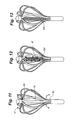
- FIG. 11 , FIG. 12 , and FIG. 13 show differing strategies to reduce blood clotting on the array. It is conventional practice to administer anticoagulants to a patient undergoing these procedures. However is very useful to eliminate blood clotting on the catheter itself.
- FIG. 11 , FIG. 12 , and FIG. 13 show several techniques that may be adopted to achieve this goal. Continuous or episodic injection of saline or heprinized saline are contemplated with the embodiments of FIG. 11 and FIG. 12 . It should be noted that various coating such as hydrophilic coatings, heprinized coatings, and parylene may also be applied to catheter surface alone or in combination with the techniques presented in the figures in order to reduce clot.
- FIG. 11 shows a distal segment having a fluid supply lumen associated with the pull wire feature 52 .
- Fluid 57 introduced into a hub at the proximal end of the catheter emerges from aperture 53 and aperture 55 to flood the array and prevent blood clots from adhering to the splines.
- FIG. 12 shows a porous membrane associated with the pull wire feature location in the distal array segment to allow fluid introduced into the catheter under pressure to emerge from the porous sheath 200 and flood the array to prevent blood clots from adhering to the splines.
- FIG. 13 shows a collapsible corrugated section 202 preventing blood from entering the catheter opening in the distal array structures.
- FIG. 14 shows a strategy for constraining the deployment providing tight control over the final shape of the deployed array.
- tether 300 may emerge from the central shaft in FIG. 14 to restrain the motion of the splines or limbs.
- FIG. 15 and FIG. 10 describe two different embodiments that meet this requirement.
- the mechanism in FIG. 15 relies on a spring to accomplish independence of the two mechanisms, while the mechanism of FIG. 10 relies on threads in distal array segment 36 to accomplish the same goals.
- FIG. 15 is a simplified schematic diagram of the catheter that serves to describe the interaction between the articulation and deflection aspects of the catheter.
- the figure serves to explain the operation of one embodiment of the array deployment construction.
- the array is pulled open with a pull wire.
- the array is biased by a spring 400 to return to the undeployed state.
- the pull wire 52 extends from the handle 30 where it is anchored to a proximal spring 402 to the distal tip 37 where it is anchored in the distal tip.
- the proximal spring 402 is in turn connected to the pommel or deployment control 33 .
- the deployment control 33 As the deployment control 33 is retracted the pull wire pulls the distal tip 37 toward the handle 30 .
- the tip motion is guided by tube 406 sliding over a bushing 408 .
- This motion can continue until the tube bottoms out on surface 404 .
- This mechanical stop determines the amount of shortening of the distal segment. As a consequence this stop also serves to limit the deployed state of the deployable array. In this figure the splines are not shown for clarity (for comparison see FIG. 16B ).
- This motion also compresses the distal spring 400 . If tension of the pull wire is eased then the distal spring 400 restores the array to the undeployed state.
- the pull wire 5 and the proximal compensator spring 402 have a nominal length that gets longer or increases as the deployment control moves into the locked 30 position. The increase in length comes from the tension supplied to the spring that increases spring length. This process is seen clearly comparing FIG. 16A to FIG. 16B .
- FIG. 16C corresponds to deflection or articulation of the catheter deflectable segment 34 .
- the deflection control causes the catheter to deflect in the plane of the figure and this displaces the pull wire 52 within the elongate catheter body 32 .
- the pull wire moves from a concentric to an offset position within the body 34 the relative length of the pull wire compared to the length of the shaft changes. This is seen most clearly at reference numeral 410 .
- the proximal spring 402 compensates for and takes up this motion by contracting slightly while still providing enough tension in the pull wire to keep the distal array fully deployed.
- FIG. 17 shows the interplay of tension in the pull wire and displacement of catheter components.
- tension rises in the wire as seen at panel A.
- the mechanical stop engages the proximal spring and force preferably remains constant as the control reaches the deployed state depicted in panel B. In this state, the catheter is in the state depicted in FIG. 16B .
- the relative motion of the pull wire and its housing causes the spring tension to fall off in the proximal spring as seen in panel C to D, while the distal array remains against its stop. In this fashion, the distal spring and its mechanical stop cooperate with the proximal spring force to stabilize the array deployment during catheter deflection.
- FIG. 10A and FIG. 10B show an alternative embodiment for deploying the array of the catheter.
- a screw 153 is positioned in the distal segment of the catheter.
- This screw 153 is rotated by a rotatable member or shaft 161 driven by a knob located in the handle which is not illustrated in the figures.
- the rotatable member 161 is keyed to the distal array segment 36 with the construction in section 155 .
- the construction provides the counter-force against which distal array segment 26 is deployed and retracted.
- This construction also isolates the screw 153 and prevents it from being influenced by tension in the rotatable member 161 .
- a complimentary nut forms a pull member 159 is free to slide over the stationary screw.
- the pull member 159 has an end anchored in the distal tip of the array and the traction supplied by the screw 153 causes the pull member 159 to move retrograde deploying the splines 50 of the array as seen in FIG. 10B .
- This construction renders the deployment function independent of the articulation function of the catheter since the deployment function is unaffected by the tension on rotatable member 161 .
- this embodiment permits the array to deploy to known continuous intermediate states or positions between the fully retracted and fully deployed states. These continuous intermediate positions are useful in mapping operations where it is desirable to introduce the catheter into structures smaller than its fully deployed diameter while maintaining accurate knowledge of electrode locations. Electrode locations are determined from the amount of deployment which can be derived from the number of rotations employed by the rotatable member during deployment.
Abstract
Description
Claims (21)
Priority Applications (10)
| Application Number | Priority Date | Filing Date | Title |
|---|---|---|---|
| US12/005,975 US8103327B2 (en) | 2007-12-28 | 2007-12-28 | Cardiac mapping catheter |
| JP2010540635A JP5419890B2 (en) | 2007-12-28 | 2008-12-10 | Non-contact mapping catheter |
| EP08867908.9A EP2234537B1 (en) | 2007-12-28 | 2008-12-10 | Non contact mapping catheter |
| CA2710769A CA2710769A1 (en) | 2007-12-28 | 2008-12-10 | Non contact mapping catheter |
| PCT/US2008/013553 WO2009085108A1 (en) | 2007-12-28 | 2008-12-10 | Non contact mapping catheter |
| US13/289,367 US8447377B2 (en) | 2007-12-28 | 2011-11-04 | Cardiac mapping catheter |
| US13/868,151 US8755861B2 (en) | 2007-12-28 | 2013-04-23 | Cardiac mapping catheter |
| JP2013207473A JP2014014713A (en) | 2007-12-28 | 2013-10-02 | Non contact mapping catheter |
| US14/277,122 US10034637B2 (en) | 2007-12-28 | 2014-05-14 | Cardiac mapping catheter |
| US16/046,844 US11272886B2 (en) | 2007-12-28 | 2018-07-26 | Cardiac mapping catheter |
Applications Claiming Priority (1)
| Application Number | Priority Date | Filing Date | Title |
|---|---|---|---|
| US12/005,975 US8103327B2 (en) | 2007-12-28 | 2007-12-28 | Cardiac mapping catheter |
Related Child Applications (1)
| Application Number | Title | Priority Date | Filing Date |
|---|---|---|---|
| US13/289,367 Continuation US8447377B2 (en) | 2007-12-28 | 2011-11-04 | Cardiac mapping catheter |
Publications (2)
| Publication Number | Publication Date |
|---|---|
| US20090171274A1 US20090171274A1 (en) | 2009-07-02 |
| US8103327B2 true US8103327B2 (en) | 2012-01-24 |
Family
ID=40799373
Family Applications (5)
| Application Number | Title | Priority Date | Filing Date |
|---|---|---|---|
| US12/005,975 Active 2030-06-22 US8103327B2 (en) | 2007-12-28 | 2007-12-28 | Cardiac mapping catheter |
| US13/289,367 Active 2028-03-19 US8447377B2 (en) | 2007-12-28 | 2011-11-04 | Cardiac mapping catheter |
| US13/868,151 Active US8755861B2 (en) | 2007-12-28 | 2013-04-23 | Cardiac mapping catheter |
| US14/277,122 Active 2030-09-26 US10034637B2 (en) | 2007-12-28 | 2014-05-14 | Cardiac mapping catheter |
| US16/046,844 Active 2030-06-09 US11272886B2 (en) | 2007-12-28 | 2018-07-26 | Cardiac mapping catheter |
Family Applications After (4)
| Application Number | Title | Priority Date | Filing Date |
|---|---|---|---|
| US13/289,367 Active 2028-03-19 US8447377B2 (en) | 2007-12-28 | 2011-11-04 | Cardiac mapping catheter |
| US13/868,151 Active US8755861B2 (en) | 2007-12-28 | 2013-04-23 | Cardiac mapping catheter |
| US14/277,122 Active 2030-09-26 US10034637B2 (en) | 2007-12-28 | 2014-05-14 | Cardiac mapping catheter |
| US16/046,844 Active 2030-06-09 US11272886B2 (en) | 2007-12-28 | 2018-07-26 | Cardiac mapping catheter |
Country Status (5)
| Country | Link |
|---|---|
| US (5) | US8103327B2 (en) |
| EP (1) | EP2234537B1 (en) |
| JP (2) | JP5419890B2 (en) |
| CA (1) | CA2710769A1 (en) |
| WO (1) | WO2009085108A1 (en) |
Cited By (110)
| Publication number | Priority date | Publication date | Assignee | Title |
|---|---|---|---|---|
| US20040167570A1 (en) * | 2002-02-21 | 2004-08-26 | Anthony Pantages | Sheath apparatus and methods for delivering a closure device |
| US20050237364A1 (en) * | 2004-04-26 | 2005-10-27 | Hirosumi Ito | Printed wiring board and electric device using the same |
| US20050274768A1 (en) * | 2004-05-25 | 2005-12-15 | Christy Cummins | Surgical stapler |
| US20060167484A1 (en) * | 2000-12-07 | 2006-07-27 | Carley Michael T | Closure device and methods for making and using them |
| US20060287674A1 (en) * | 2000-01-05 | 2006-12-21 | Ginn Richard S | Closure system and methods of use |
| US20070010853A1 (en) * | 2000-10-06 | 2007-01-11 | Integrated Vascular Systems, Inc. | Apparatus and methods for positioning a vascular sheath |
| US20070021778A1 (en) * | 2005-06-24 | 2007-01-25 | Abbott Laboratories Abbott Vascular Devices | Apparatus and method for delivering a closure element |
| US20070250080A1 (en) * | 2006-04-20 | 2007-10-25 | Integrated Vascular Systems, Inc. | Resettable clip applier and reset tools |
| US20070270904A1 (en) * | 2000-12-07 | 2007-11-22 | Integrated Vascular Systems, Inc. | Apparatus and Methods for Providing Tactile Feedback while Delivering a Closure Device |
| US20070282352A1 (en) * | 2000-12-07 | 2007-12-06 | Carley Michael T | Closure device and methods for making and using them |
| US20080004640A1 (en) * | 2006-06-28 | 2008-01-03 | Abbott Laboratories | Vessel closure device |
| US20080004636A1 (en) * | 2005-07-01 | 2008-01-03 | Abbott Laboratories | Clip applier and methods of use |
| US20080065152A1 (en) * | 2006-09-08 | 2008-03-13 | Abbott Laboratories | Apparatus and method for delivering a closure element |
| US20080210737A1 (en) * | 2000-01-05 | 2008-09-04 | Integrated Vascular Systems, Inc. | Integrated vascular device with puncture site closure component and sealant and methods of use |
| US20080269802A1 (en) * | 2000-09-08 | 2008-10-30 | Abbott Vascular Inc. | Surgical stapler |
| US20090157102A1 (en) * | 2007-12-17 | 2009-06-18 | Abbott Laboratories | Clip applier and methods of use |
| US20090187215A1 (en) * | 2007-12-19 | 2009-07-23 | Abbott Laboratories | Methods and apparatus to reduce a dimension of an implantable device in a smaller state |
| US20090287244A1 (en) * | 2008-05-16 | 2009-11-19 | Abbott Laboratories Vascular Enterprises Limited | Apparatus and methods for engaging tissue |
| US20100114159A1 (en) * | 2008-10-30 | 2010-05-06 | Abbott Vascular Inc. | Closure device |
| US20100160958A1 (en) * | 2008-12-22 | 2010-06-24 | Abbott Laboratories | Closure Device |
| US20100168647A1 (en) * | 2008-12-29 | 2010-07-01 | Tegg Troy T | Non-contact electrode basket catheters with irrigation |
| US20100168790A1 (en) * | 2008-12-22 | 2010-07-01 | Abbott Laboratories | Curved closure device |
| US20100179567A1 (en) * | 2009-01-09 | 2010-07-15 | Abbott Vascular Inc. | Closure devices, systems, and methods |
| US20100179572A1 (en) * | 2009-01-09 | 2010-07-15 | Abbott Vascular Inc. | Closure devices, systems, and methods |
| US20100179590A1 (en) * | 2009-01-09 | 2010-07-15 | Abbott Vascular Inc. | Vessel closure devices and methods |
| US20100179571A1 (en) * | 2009-01-09 | 2010-07-15 | Abbott Vascular Inc. | Closure devices, systems, and methods |
| US20100185234A1 (en) * | 2009-01-16 | 2010-07-22 | Abbott Vascular Inc. | Closure devices, systems, and methods |
| US20100217132A1 (en) * | 2009-02-26 | 2010-08-26 | Abbott Laboratories | Methods and apparatus for locating a surface of a body lumen |
| US20110066164A1 (en) * | 2007-12-18 | 2011-03-17 | Abbott Laboratories | Modular clip applier |
| US20110137340A1 (en) * | 2001-06-07 | 2011-06-09 | Abbott Vascular Inc. | Surgical staple |
| US20110166584A1 (en) * | 2003-01-30 | 2011-07-07 | Integrated Vascular Systems, Inc. | Clip applier and methods of use |
| US20110218568A1 (en) * | 2009-01-09 | 2011-09-08 | Voss Laveille K | Vessel closure devices, systems, and methods |
| US20110230897A1 (en) * | 2003-01-30 | 2011-09-22 | Integrated Vascular Systems, Inc. | Clip applier and methods of use |
| US20110238089A1 (en) * | 2007-12-17 | 2011-09-29 | Abbott Laboratories | Tissue closure system and methods of use |
| US20120078077A1 (en) * | 2007-12-28 | 2012-03-29 | Rhythmia Medical, Inc. | Cardiac mapping catheter |
| US20120296329A1 (en) * | 2011-05-18 | 2012-11-22 | St. Jude Medical, Inc. | Apparatus and method of assessing transvascular denervation |
| US8469995B2 (en) | 2002-06-04 | 2013-06-25 | Abbott Vascular Inc. | Blood vessel closure clip and delivery device |
| US8529587B2 (en) | 2003-01-30 | 2013-09-10 | Integrated Vascular Systems, Inc. | Methods of use of a clip applier |
| US8585836B2 (en) | 2002-12-31 | 2013-11-19 | Integrated Vascular Systems, Inc. | Methods for manufacturing a clip and clip |
| US8603116B2 (en) | 2010-08-04 | 2013-12-10 | Abbott Cardiovascular Systems, Inc. | Closure device with long tines |
| US20140148674A1 (en) * | 2007-05-09 | 2014-05-29 | St. Jude Medical, Cardiology Division, Inc. | Bendable catheter arms having varied flexibility |
| US8758396B2 (en) | 2000-01-05 | 2014-06-24 | Integrated Vascular Systems, Inc. | Vascular sheath with bioabsorbable puncture site closure apparatus and methods of use |
| US8758399B2 (en) | 2010-08-02 | 2014-06-24 | Abbott Cardiovascular Systems, Inc. | Expandable bioabsorbable plug apparatus and method |
| US8758400B2 (en) | 2000-01-05 | 2014-06-24 | Integrated Vascular Systems, Inc. | Closure system and methods of use |
| US8821534B2 (en) | 2010-12-06 | 2014-09-02 | Integrated Vascular Systems, Inc. | Clip applier having improved hemostasis and methods of use |
| US9149276B2 (en) | 2011-03-21 | 2015-10-06 | Abbott Cardiovascular Systems, Inc. | Clip and deployment apparatus for tissue closure |
| US9277872B2 (en) | 2011-01-13 | 2016-03-08 | Rhythmia Medical, Inc. | Electroanatomical mapping |
| WO2016039824A1 (en) * | 2014-09-12 | 2016-03-17 | X-Rhythm, Llc | Multi-electrode mapping catheter |
| US9314230B2 (en) | 2009-01-09 | 2016-04-19 | Abbott Vascular Inc. | Closure device with rapidly eroding anchor |
| US9332976B2 (en) | 2011-11-30 | 2016-05-10 | Abbott Cardiovascular Systems, Inc. | Tissue closure device |
| US9364209B2 (en) | 2012-12-21 | 2016-06-14 | Abbott Cardiovascular Systems, Inc. | Articulating suturing device |
| US20160302858A1 (en) * | 2015-04-17 | 2016-10-20 | Boston Scientific Scimed Inc. | Tissue diagnosis and treatment using electrodes and mini-electrodes |
| US9474467B2 (en) | 2008-04-02 | 2016-10-25 | Rhythmia Medical, Inc. | Intracardiac tracking system |
| US9486191B2 (en) | 2009-01-09 | 2016-11-08 | Abbott Vascular, Inc. | Closure devices |
| US9532725B2 (en) | 2014-03-07 | 2017-01-03 | Boston Scientific Scimed Inc. | Medical devices for mapping cardiac tissue |
| US9585647B2 (en) | 2009-08-26 | 2017-03-07 | Abbott Laboratories | Medical device for repairing a fistula |
| US9585588B2 (en) | 2014-06-03 | 2017-03-07 | Boston Scientific Scimed, Inc. | Electrode assembly having an atraumatic distal tip |
| US9603659B2 (en) | 2011-09-14 | 2017-03-28 | Boston Scientific Scimed Inc. | Ablation device with ionically conductive balloon |
| US20170112405A1 (en) * | 2015-10-21 | 2017-04-27 | St. Jude Medical, Cardiology Division, Inc. | High density electrode mapping catheter |
| US9687297B2 (en) | 2010-12-02 | 2017-06-27 | St. Jude Medical, Atrial Fibrillation Division, Inc. | Catheter electrode assemblies and methods of construction thereof |
| US9687167B2 (en) | 2014-03-11 | 2017-06-27 | Boston Scientific Scimed, Inc. | Medical devices for mapping cardiac tissue |
| US9687166B2 (en) | 2013-10-14 | 2017-06-27 | Boston Scientific Scimed, Inc. | High resolution cardiac mapping electrode array catheter |
| US9730600B2 (en) | 2013-10-31 | 2017-08-15 | Boston Scientific Scimed, Inc. | Medical device for high resolution mapping using localized matching |
| US9743854B2 (en) | 2014-12-18 | 2017-08-29 | Boston Scientific Scimed, Inc. | Real-time morphology analysis for lesion assessment |
| US9757191B2 (en) | 2012-01-10 | 2017-09-12 | Boston Scientific Scimed, Inc. | Electrophysiology system and methods |
| US20170303985A1 (en) * | 2014-09-23 | 2017-10-26 | Handok Kalos Medical Inc. | Catheter and manufacturing method therefor |
| US9848795B2 (en) | 2014-06-04 | 2017-12-26 | Boston Scientific Scimed Inc. | Electrode assembly |
| US9913589B2 (en) | 2008-01-17 | 2018-03-13 | Christoph Scharf | Device and method for the geometric determination of electrical dipole densities on the cardiac wall |
| US9968268B2 (en) | 2011-03-10 | 2018-05-15 | Acutus Medical, Inc. | Device and method for the geometric determination of electrical dipole densities on the cardiac wall |
| US10004459B2 (en) | 2012-08-31 | 2018-06-26 | Acutus Medical, Inc. | Catheter system and methods of medical uses of same, including diagnostic and treatment uses for the heart |
| US10076258B2 (en) | 2013-11-01 | 2018-09-18 | Boston Scientific Scimed, Inc. | Cardiac mapping using latency interpolation |
| US10172536B2 (en) | 2012-12-20 | 2019-01-08 | Boston Scientific Scimed, Inc. | Real-time feedback for electrode contact during mapping |
| US10201311B2 (en) | 2013-02-08 | 2019-02-12 | Acutus Medical, Inc. | Expandable catheter assembly with flexible printed circuit board (PCB) electrical pathways |
| US10271758B2 (en) | 2015-09-26 | 2019-04-30 | Boston Scientific Scimed, Inc. | Intracardiac EGM signals for beat matching and acceptance |
| US10335051B2 (en) | 2011-01-13 | 2019-07-02 | Rhythmia Medical, Inc. | Beat alignment and selection for cardiac mapping |
| US10362954B2 (en) | 2015-10-21 | 2019-07-30 | St. Jude Medical, Cardiology Division, Inc. | High density electrode mapping catheter |
| US10376171B2 (en) | 2006-08-03 | 2019-08-13 | Christoph Scharf | Method and device for determining and presenting surface charge and dipole densities on cardiac walls |
| US20190381274A1 (en) * | 2017-01-19 | 2019-12-19 | St. Jude Medical, Cardiology Division, Inc. | Sheath visualization |
| US10524684B2 (en) | 2014-10-13 | 2020-01-07 | Boston Scientific Scimed Inc | Tissue diagnosis and treatment using mini-electrodes |
| US10593234B2 (en) | 2015-05-12 | 2020-03-17 | Acutus Medical, Inc. | Cardiac virtualization test tank and testing system and method |
| US10603105B2 (en) | 2014-10-24 | 2020-03-31 | Boston Scientific Scimed Inc | Medical devices with a flexible electrode assembly coupled to an ablation tip |
| US10653318B2 (en) | 2015-05-13 | 2020-05-19 | Acutus Medical, Inc. | Localization system and method useful in the acquisition and analysis of cardiac information |
| US10758144B2 (en) | 2015-08-20 | 2020-09-01 | Boston Scientific Scimed Inc. | Flexible electrode for cardiac sensing and method for making |
| US10828011B2 (en) | 2013-09-13 | 2020-11-10 | Acutus Medical, Inc. | Devices and methods for determination of electrical dipole densities on a cardiac surface |
| US10912484B2 (en) | 2018-07-09 | 2021-02-09 | Biosense Webster (Israel) Ltd. | Multiplexing of high count electrode catheter(s) |
| US11147496B2 (en) | 2018-01-16 | 2021-10-19 | Boston Scientific Scimed Inc. | Systems and methods for mapping electrical activity in the heart |
| US11278231B2 (en) | 2014-03-25 | 2022-03-22 | Acutus Medical, Inc. | Cardiac analysis user interface system and method |
| US20220087735A1 (en) * | 2020-09-22 | 2022-03-24 | Biosense Webster (Israel) Ltd. | Basket catheter having insulated ablation electrodes and diagnostic electrodes |
| US11344366B2 (en) | 2015-05-12 | 2022-05-31 | Acutus Medical, Inc. | Ultrasound sequencing system and method |
| US11399759B2 (en) | 2016-05-03 | 2022-08-02 | Acutus Medical, Inc. | Cardiac mapping system with efficiency algorithm |
| US11433220B2 (en) | 2017-07-07 | 2022-09-06 | St. Jude Medical, Cardiology Division, Inc. | Layered high density electrode mapping catheter |
| US11540876B2 (en) | 2016-05-03 | 2023-01-03 | St. Jude Medical Cardiology Division, Inc. | Irrigated high density electrode catheter |
| US11617861B2 (en) | 2014-06-17 | 2023-04-04 | St. Jude Medical, Cardiology Division, Inc. | Triple coil catheter support |
| US11622806B2 (en) | 2010-04-09 | 2023-04-11 | St Jude Medical International Holding S.À R.L. | Control handle for a contact force ablation catheter |
| US11642063B2 (en) | 2018-08-23 | 2023-05-09 | St. Jude Medical, Cardiology Division, Inc. | Curved high density electrode mapping catheter |
| US11647935B2 (en) | 2017-07-24 | 2023-05-16 | St. Jude Medical, Cardiology Division, Inc. | Masked ring electrodes |
| US11672947B2 (en) | 2017-11-28 | 2023-06-13 | St. Jude Medical, Cardiology Division, Inc. | Lumen management catheter |
| US11684416B2 (en) | 2009-02-11 | 2023-06-27 | Boston Scientific Scimed, Inc. | Insulated ablation catheter devices and methods of use |
| US11707229B2 (en) | 2015-05-08 | 2023-07-25 | St Jude Medical International Holding S.À R.L. | Integrated sensors for medical devices and method of making integrated sensors for medical devices |
| US11786705B2 (en) | 2016-10-24 | 2023-10-17 | St. Jude Medical, Cardiology Division, Inc. | Catheter insertion devices |
| US11826172B2 (en) | 2014-05-06 | 2023-11-28 | St. Jude Medical, Cardiology Division, Inc. | Electrode support structure assembly |
| US11839424B2 (en) | 2010-05-05 | 2023-12-12 | St. Jude Medical, Atrial Fibrillation Division, Inc | Monitoring, managing and/or protecting system and method for non-targeted tissue |
| US11844615B2 (en) | 2015-03-12 | 2023-12-19 | The Regents Of The University Of Michigan | Catheter and method to localize ectopic and reentrant activity in the heart |
| US11844910B2 (en) | 2014-06-05 | 2023-12-19 | St. Jude Medical, Cardiology Division, Inc. | Deflectable catheter shaft section |
| US11850051B2 (en) | 2019-04-30 | 2023-12-26 | Biosense Webster (Israel) Ltd. | Mapping grid with high density electrode array |
| US11878095B2 (en) | 2018-12-11 | 2024-01-23 | Biosense Webster (Israel) Ltd. | Balloon catheter with high articulation |
| US11918762B2 (en) | 2018-10-03 | 2024-03-05 | St. Jude Medical, Cardiology Division, Inc. | Reduced actuation force electrophysiology catheter handle |
| US11918383B2 (en) | 2020-12-21 | 2024-03-05 | Biosense Webster (Israel) Ltd. | Visualizing performance of catheter electrodes |
| US11918341B2 (en) | 2019-12-20 | 2024-03-05 | Biosense Webster (Israel) Ltd. | Selective graphical presentation of electrophysiological parameters |
| US11950841B2 (en) * | 2020-09-22 | 2024-04-09 | Biosense Webster (Israel) Ltd. | Basket catheter having insulated ablation electrodes and diagnostic electrodes |
Families Citing this family (116)
| Publication number | Priority date | Publication date | Assignee | Title |
|---|---|---|---|---|
| WO2005067817A1 (en) | 2004-01-13 | 2005-07-28 | Remon Medical Technologies Ltd | Devices for fixing a sensor in a body lumen |
| US10390714B2 (en) * | 2005-01-12 | 2019-08-27 | Remon Medical Technologies, Ltd. | Devices for fixing a sensor in a lumen |
| US8060214B2 (en) | 2006-01-05 | 2011-11-15 | Cardiac Pacemakers, Inc. | Implantable medical device with inductive coil configurable for mechanical fixation |
| US7515954B2 (en) | 2006-06-13 | 2009-04-07 | Rhythmia Medical, Inc. | Non-contact cardiac mapping, including moving catheter and multi-beat integration |
| US7729752B2 (en) | 2006-06-13 | 2010-06-01 | Rhythmia Medical, Inc. | Non-contact cardiac mapping, including resolution map |
| US9119633B2 (en) | 2006-06-28 | 2015-09-01 | Kardium Inc. | Apparatus and method for intra-cardiac mapping and ablation |
| US10028783B2 (en) | 2006-06-28 | 2018-07-24 | Kardium Inc. | Apparatus and method for intra-cardiac mapping and ablation |
| US11389232B2 (en) | 2006-06-28 | 2022-07-19 | Kardium Inc. | Apparatus and method for intra-cardiac mapping and ablation |
| US8676349B2 (en) | 2006-09-15 | 2014-03-18 | Cardiac Pacemakers, Inc. | Mechanism for releasably engaging an implantable medical device for implantation |
| WO2008034077A2 (en) | 2006-09-15 | 2008-03-20 | Cardiac Pacemakers, Inc. | Anchor for an implantable sensor |
| US20080071248A1 (en) * | 2006-09-15 | 2008-03-20 | Cardiac Pacemakers, Inc. | Delivery stystem for an implantable physiologic sensor |
| US20080190438A1 (en) * | 2007-02-08 | 2008-08-14 | Doron Harlev | Impedance registration and catheter tracking |
| US8204599B2 (en) | 2007-05-02 | 2012-06-19 | Cardiac Pacemakers, Inc. | System for anchoring an implantable sensor in a vessel |
| US8906011B2 (en) | 2007-11-16 | 2014-12-09 | Kardium Inc. | Medical device for use in bodily lumens, for example an atrium |
| US8934987B2 (en) | 2008-07-15 | 2015-01-13 | Cardiac Pacemakers, Inc. | Implant assist apparatus for acoustically enabled implantable medical device |
| ES2871298T3 (en) | 2008-07-24 | 2021-10-28 | Massachusetts Inst Technology | Systems and Methods for Imaging Using Absorption |
| US8368649B2 (en) * | 2008-08-29 | 2013-02-05 | Siemens Medical Solutions Usa, Inc. | Control system for use within a sterile environment |
| US8167876B2 (en) | 2008-10-27 | 2012-05-01 | Rhythmia Medical, Inc. | Tracking system using field mapping |
| US8808345B2 (en) | 2008-12-31 | 2014-08-19 | Medtronic Ardian Luxembourg S.A.R.L. | Handle assemblies for intravascular treatment devices and associated systems and methods |
| WO2010093489A2 (en) * | 2009-02-13 | 2010-08-19 | Cardiac Pacemakers, Inc. | Deployable sensor platform on the lead system of an implantable device |
| US9398862B2 (en) * | 2009-04-23 | 2016-07-26 | Rhythmia Medical, Inc. | Multi-electrode mapping system |
| US8571647B2 (en) | 2009-05-08 | 2013-10-29 | Rhythmia Medical, Inc. | Impedance based anatomy generation |
| US8103338B2 (en) | 2009-05-08 | 2012-01-24 | Rhythmia Medical, Inc. | Impedance based anatomy generation |
| SE0901166A1 (en) * | 2009-09-10 | 2011-03-11 | Cathprint Ab | Flexible catheter lead carrier provided with such lead carrier |
| IT1399677B1 (en) * | 2009-10-26 | 2013-04-26 | Torino Politecnico | SENSOR FOR THE ACQUISITION OF BIOELECTRIC SIGNALS FROM AN ANULAR MUSCLE, IN PARTICULAR FOR MULTI-CHANNEL SURFACE ELECTROMYOGRAPHY |
| US8825129B2 (en) * | 2010-03-05 | 2014-09-02 | Sri International | Indwelling nerve block catheters |
| US9179968B2 (en) * | 2010-05-10 | 2015-11-10 | St. Jude Medical Luxembourg Holding S.À.R.L. | Irrigated finned ablation head |
| US8903473B2 (en) * | 2010-09-15 | 2014-12-02 | Medtronic, Inc. | Radiopaque markers for implantable medical devices |
| US9579200B2 (en) | 2010-09-15 | 2017-02-28 | The United States Of America, As Represented By The Secretary, Department Of Health & Human Services | Methods and devices for transcatheter cerclage annuloplasty |
| CA2764494A1 (en) | 2011-01-21 | 2012-07-21 | Kardium Inc. | Enhanced medical device for use in bodily cavities, for example an atrium |
| US9452016B2 (en) | 2011-01-21 | 2016-09-27 | Kardium Inc. | Catheter system |
| US9480525B2 (en) | 2011-01-21 | 2016-11-01 | Kardium, Inc. | High-density electrode-based medical device system |
| US11259867B2 (en) * | 2011-01-21 | 2022-03-01 | Kardium Inc. | High-density electrode-based medical device system |
| ITRM20110206A1 (en) * | 2011-04-21 | 2012-10-22 | Ab Medica Spa | ACQUISITION AND MONITORING SYSTEM OF BIOELECTRIC SIGNALS FROM THE BRAIN AND INTRACRANIC STIMULATION. |
| WO2012145072A1 (en) | 2011-04-22 | 2012-10-26 | Topera, Inc. | Basket style cardiac mapping catheter having a flexible electrode assembly for detection of cardiac rhythm disorders |
| AU2014202011B2 (en) * | 2011-04-22 | 2015-05-07 | Topera, Inc. | Basket style cardiac mapping catheter having an atraumatic, metallic two-part distal tip for detection of cardiac rhythm disorders |
| JP5579681B2 (en) * | 2011-10-06 | 2014-08-27 | 平河ヒューテック株式会社 | Electrode catheter |
| US8649880B1 (en) * | 2011-10-07 | 2014-02-11 | Autry J. Parker, Jr. | Deployable stimulator array and method of use |
| USD777926S1 (en) | 2012-01-20 | 2017-01-31 | Kardium Inc. | Intra-cardiac procedure device |
| USD777925S1 (en) | 2012-01-20 | 2017-01-31 | Kardium Inc. | Intra-cardiac procedure device |
| JP5936409B2 (en) * | 2012-03-26 | 2016-06-22 | 国立研究開発法人国立循環器病研究センター | Electrode unit and tissue stimulation system |
| WO2014066470A1 (en) * | 2012-10-24 | 2014-05-01 | Evergreen Medical Technologies, Inc. | Flex circuit ribbon based elongated members and attachments |
| EP2945683A4 (en) * | 2013-01-18 | 2017-03-15 | SRI International | Anchoring nerve block catheter |
| CN103932676A (en) * | 2013-01-23 | 2014-07-23 | 四川锦江电子科技有限公司 | Flexible circuit electrode |
| US9179971B2 (en) | 2013-02-11 | 2015-11-10 | St. Jude Medical, Atrial Fibrillation Division, Inc. | Printed electrode catheter |
| US10328238B2 (en) | 2013-03-12 | 2019-06-25 | St. Jude Medical, Cardiology Division, Inc. | Catheter system |
| WO2014162660A1 (en) * | 2013-04-01 | 2014-10-09 | テルモ株式会社 | Monitoring device and monitoring device kit |
| JP6240751B2 (en) | 2013-05-06 | 2017-11-29 | ボストン サイエンティフィック サイムド,インコーポレイテッドBoston Scientific Scimed,Inc. | Anatomic mapping system for continuous display of recent heart rate characteristics during real-time or playback electrophysiological data visualization |
| US9918649B2 (en) | 2013-05-14 | 2018-03-20 | Boston Scientific Scimed Inc. | Representation and identification of activity patterns during electro-physiology mapping using vector fields |
| US9814618B2 (en) * | 2013-06-06 | 2017-11-14 | Boston Scientific Scimed, Inc. | Devices for delivering energy and related methods of use |
| EP2818104B1 (en) * | 2013-06-25 | 2016-01-06 | VascoMed GmbH | Catheter and method for producing same |
| CN110547865B (en) * | 2013-08-09 | 2022-10-04 | 波士顿科学国际有限公司 | Expandable catheter and related methods of manufacture and use |
| US20150119878A1 (en) * | 2013-10-24 | 2015-04-30 | St. Jude Medical, Cardiology Division, Inc. | Electrode assembly having asymmetric electrode placement |
| US9987073B2 (en) * | 2013-11-19 | 2018-06-05 | Covidien Lp | Electrosurgical coagulation instrument including a suction pipe and a collapsible tip |
| US9993160B2 (en) * | 2014-01-07 | 2018-06-12 | Kardium Inc. | Medical device including manipulable portion with connected elongate members |
| EP3079580B1 (en) | 2014-01-28 | 2017-11-22 | St. Jude Medical International Holding S.à r.l. | Medical device with a packaged electronic subassembly and method for fabricating the same |
| EP3082588B8 (en) * | 2014-01-28 | 2018-12-19 | St. Jude Medical International Holding S.à r.l. | Elongate medical devices incorporating a flexible substrate, a sensor, and electrically-conductive traces |
| JP6419214B2 (en) | 2014-01-28 | 2018-11-07 | セント・ジュード・メディカル,カーディオロジー・ディヴィジョン,インコーポレイテッド | Catheter shaft with conductive traces |
| JP2016002282A (en) * | 2014-06-17 | 2016-01-12 | オリンパス株式会社 | Electrode unit and tissue stimulation system |
| WO2016044687A1 (en) * | 2014-09-18 | 2016-03-24 | University Of Utah Research Foundation | Cardiac mapping catheter |
| KR102033759B1 (en) | 2014-09-23 | 2019-10-17 | 주식회사 한독칼로스메디칼 | Catheter and manufacturing method thereof |
| KR102033760B1 (en) | 2014-09-23 | 2019-10-17 | 주식회사 한독칼로스메디칼 | Catheter and manufacturing method thereof |
| US9314208B1 (en) * | 2014-10-28 | 2016-04-19 | Biosense Webster (Israel) Ltd. | Basket catheter with microelectrode array distal tip |
| EP3212270B1 (en) | 2014-10-30 | 2019-09-04 | Kardium Inc. | Catheter system |
| US20180146925A1 (en) * | 2014-12-29 | 2018-05-31 | Jamil Mogul | Tentacular Electrode Catheter Apparatus |
| US9757574B2 (en) | 2015-05-11 | 2017-09-12 | Rainbow Medical Ltd. | Dual chamber transvenous pacemaker |
| WO2016181317A2 (en) | 2015-05-12 | 2016-11-17 | Navix International Limited | Calculation of an ablation plan |
| US9931487B2 (en) | 2015-08-06 | 2018-04-03 | Boston Scientific Scimed, Inc. | Bidirectional steering control apparatus for a catheter |
| US10492857B2 (en) | 2015-08-06 | 2019-12-03 | Boston Scientific Scimed Inc | Deployment control apparatus for a catheter with a deployable array |
| US10376170B2 (en) | 2015-08-10 | 2019-08-13 | Boston Scientific Scimed, Inc. | Catheter with annular lumen to provide distal flushing |
| AU2016210644A1 (en) * | 2015-08-12 | 2017-03-02 | Biosense Webster (Israel) Ltd. | High electrode density basket catheter |
| US20170071543A1 (en) * | 2015-09-14 | 2017-03-16 | Biosense Webster (Israel) Ltd. | Convertible basket catheter |
| EP3352648B1 (en) | 2015-09-26 | 2022-10-26 | Boston Scientific Scimed Inc. | Multiple rhythm template monitoring |
| EP3353753A1 (en) | 2015-09-26 | 2018-08-01 | Boston Scientific Scimed Inc. | Systems and methods for anatomical shell editing |
| US10405766B2 (en) | 2015-09-26 | 2019-09-10 | Boston Scientific Scimed, Inc. | Method of exploring or mapping internal cardiac structures |
| US10362952B2 (en) | 2015-12-10 | 2019-07-30 | Biosense Webster (Israel) Ltd. | Stabilized spine electrophysiologic catheter |
| US10362953B2 (en) * | 2015-12-11 | 2019-07-30 | Biosense Webster (Israel) Ltd. | Electrode array catheter with interconnected framework |
| KR20180094955A (en) * | 2015-12-15 | 2018-08-24 | 에이전시 포 사이언스, 테크놀로지 앤드 리서치 | Deployable multi-spinal device and method for catheter-based kidney nerve block |
| CN108366734A (en) * | 2015-12-20 | 2018-08-03 | 波士顿科学医学有限公司 | The automatic mapping of operating speed information |
| US10660700B2 (en) * | 2016-04-28 | 2020-05-26 | Biosense Webster (Israel) Ltd. | Irrigated balloon catheter with flexible circuit electrode assembly |
| EP3451954B1 (en) | 2016-05-02 | 2023-09-20 | Affera, Inc. | A system comprising a catheter, a sheath and an insertion sleeve |
| US11039923B2 (en) | 2016-05-06 | 2021-06-22 | Transmural Systems Llc | Annuloplasty procedures, related devices and methods |
| US11007059B2 (en) | 2016-05-06 | 2021-05-18 | Transmural Systems Llc | Annuloplasty procedures, related devices and methods |
| US10376320B2 (en) | 2016-05-11 | 2019-08-13 | Affera, Inc. | Anatomical model generation |
| WO2017197247A2 (en) | 2016-05-12 | 2017-11-16 | Affera, Inc. | Anatomical model controlling |
| US10772566B2 (en) * | 2016-05-17 | 2020-09-15 | Biosense Weber (Israel) Ltd. | Multi-electrode catheter spine and method of making the same |
| EP3484362A1 (en) | 2016-07-14 | 2019-05-22 | Navix International Limited | Characteristic track catheter navigation |
| US11266467B2 (en) | 2016-10-25 | 2022-03-08 | Navix International Limited | Systems and methods for registration of intra-body electrical readings with a pre-acquired three dimensional image |
| CN110177500B (en) * | 2016-11-16 | 2022-03-04 | 纳维斯国际有限公司 | Dynamic visual rendering of tissue models |
| US10709507B2 (en) | 2016-11-16 | 2020-07-14 | Navix International Limited | Real-time display of treatment-related tissue changes using virtual material |
| WO2018092071A1 (en) | 2016-11-16 | 2018-05-24 | Navix International Limited | Estimators for ablation effectiveness |
| WO2018092062A1 (en) | 2016-11-16 | 2018-05-24 | Navix International Limited | Real-time display of tissue deformation by interactions with an intra-body probe |
| US11331029B2 (en) | 2016-11-16 | 2022-05-17 | Navix International Limited | Esophagus position detection by electrical mapping |
| EP3554406A1 (en) * | 2016-12-19 | 2019-10-23 | Boston Scientific Scimed Inc. | Distally-facing electrode array with longitudinally mounted splines |
| US20180184982A1 (en) * | 2017-01-05 | 2018-07-05 | Biosense Webster (Israel) Ltd. | Hybrid balloon basket catheter |
| US11344259B2 (en) * | 2017-01-11 | 2022-05-31 | Abbott Cardiovascular Systems Inc. | Expandable member for an electrophysiology catheter |
| US11471067B2 (en) | 2017-01-12 | 2022-10-18 | Navix International Limited | Intrabody probe navigation by electrical self-sensing |
| US11246534B2 (en) * | 2017-01-23 | 2022-02-15 | Biosense Webster (Israel) Ltd. | Basket catheter made from flexible circuit board with mechanical strengthening |
| US11116450B2 (en) * | 2017-03-09 | 2021-09-14 | Biosense Webster (Israel) Ltd. | Electrode assembly having spines with controlled flexibility |
| US10881482B2 (en) * | 2017-05-09 | 2021-01-05 | Boston Scientific Scimed, Inc. | Operating room devices, methods, and systems |
| US9848906B1 (en) | 2017-06-20 | 2017-12-26 | Joe Michael Eskridge | Stent retriever having an expandable fragment guard |
| CN111050641B (en) | 2017-08-17 | 2023-06-09 | 纳维斯国际有限公司 | Remote imaging based on field gradients |
| KR20200051661A (en) | 2017-08-26 | 2020-05-13 | 트랜스뮤럴 시스템스 엘엘씨 | Cardiac annuloplasty and coordination procedures and related devices and methods |
| US10874824B2 (en) * | 2017-10-18 | 2020-12-29 | Biosense Webster (Israel) Ltd. | High-volume manufacturing of catheters comprising electrodes having low impedance at low frequency |
| US11864825B2 (en) | 2018-05-02 | 2024-01-09 | Biosense Webster (Israel) Ltd. | Ablation catheter with selective radial energy delivery |
| US20210121116A1 (en) * | 2018-06-26 | 2021-04-29 | Berne University Of Applied Sciences | Cuff with integrated signal recorder for long-term measurements of biosignals from a living body |
| US20200205737A1 (en) * | 2018-12-28 | 2020-07-02 | Biosense Webster (Israel) Ltd. | Flexible Nested Sensing Electrodes |
| EP3989837A1 (en) * | 2019-06-28 | 2022-05-04 | Boston Scientific Scimed, Inc. | Ultrasound device |
| US20210187241A1 (en) * | 2019-12-20 | 2021-06-24 | Biosense Webster (Israel) Ltd. | Expandable Assembly Catheter |
| US11553961B2 (en) * | 2020-04-30 | 2023-01-17 | Biosense Webster (Israel) Ltd. | Catheter with stretchable irrigation tube |
| US20220016394A1 (en) * | 2020-07-16 | 2022-01-20 | Canon U.S.A., Inc. | Medical Apparatus and Method of Use Thereof |
| IT202000020149A1 (en) | 2020-08-14 | 2022-02-14 | Francesco Solimene | HIGH DENSITY MAPPING CATHETER |
| US20220087736A1 (en) * | 2020-09-23 | 2022-03-24 | Biosense Webster (Israel) Ltd. | Electrode shorting |
| USD1014762S1 (en) | 2021-06-16 | 2024-02-13 | Affera, Inc. | Catheter tip with electrode panel(s) |
| US20230015298A1 (en) * | 2021-07-13 | 2023-01-19 | Biosense Webster (Isreal) Ltd. | Ablation electrodes made from electrical traces of flexible printed circuit board |
| WO2023053106A2 (en) | 2021-09-30 | 2023-04-06 | Biosense Webster (Israel) Ltd. | Devices and methods for an expandable assembly catheter |
Citations (32)
| Publication number | Priority date | Publication date | Assignee | Title |
|---|---|---|---|---|
| US4649924A (en) | 1984-08-14 | 1987-03-17 | Consiglio Nazionale Delle Ricerche | Method for the detection of intracardiac electrical potential fields |
| US4890623A (en) | 1988-03-14 | 1990-01-02 | C. R. Bard, Inc. | Biopotential sensing device and method for making |
| US4940064A (en) | 1986-11-14 | 1990-07-10 | Desai Jawahar M | Catheter for mapping and ablation and method therefor |
| US5156151A (en) | 1991-02-15 | 1992-10-20 | Cardiac Pathways Corporation | Endocardial mapping and ablation system and catheter probe |
| US5228442A (en) * | 1991-02-15 | 1993-07-20 | Cardiac Pathways Corporation | Method for mapping, ablation, and stimulation using an endocardial catheter |
| US5255679A (en) | 1992-06-02 | 1993-10-26 | Cardiac Pathways Corporation | Endocardial catheter for mapping and/or ablation with an expandable basket structure having means for providing selective reinforcement and pressure sensing mechanism for use therewith, and method |
| US5311866A (en) | 1992-09-23 | 1994-05-17 | Endocardial Therapeutics, Inc. | Heart mapping catheter |
| US5313943A (en) * | 1992-09-25 | 1994-05-24 | Ep Technologies, Inc. | Catheters and methods for performing cardiac diagnosis and treatment |
| US5345936A (en) | 1991-02-15 | 1994-09-13 | Cardiac Pathways Corporation | Apparatus with basket assembly for endocardial mapping |
| US5415166A (en) | 1991-02-15 | 1995-05-16 | Cardiac Pathways Corporation | Endocardial mapping apparatus and cylindrical semiconductor device mounting structure for use therewith and method |
| WO1995013111A1 (en) | 1993-11-10 | 1995-05-18 | Medtronic Cadiorhythm | Electrode array catheter |
| US5425364A (en) | 1991-02-15 | 1995-06-20 | Cardiac Pathways Corporation | Flexible strip assembly without feedthrough holes and device utilizing the same |
| US5456254A (en) | 1991-02-15 | 1995-10-10 | Cardiac Pathways Corp | Flexible strip assembly having insulating layer with conductive pads exposed through insulating layer and device utilizing the same |
| US5471982A (en) | 1992-09-29 | 1995-12-05 | Ep Technologies, Inc. | Cardiac mapping and ablation systems |
| WO1996034652A1 (en) | 1995-05-02 | 1996-11-07 | Heart Rhythm Technologies, Inc. | Catheter control system |
| US5647870A (en) | 1993-03-16 | 1997-07-15 | Ep Technologies, Inc. | Multiple electrode support structures |
| US5681308A (en) | 1994-06-24 | 1997-10-28 | Stuart D. Edwards | Ablation apparatus for cardiac chambers |
| US5697377A (en) | 1995-11-22 | 1997-12-16 | Medtronic, Inc. | Catheter mapping system and method |
| US5699796A (en) | 1993-01-29 | 1997-12-23 | Cardima, Inc. | High resolution intravascular signal detection |
| US5871443A (en) | 1992-09-25 | 1999-02-16 | Ep Technologies, Inc. | Cardiac mapping and ablation systems |
| US6064905A (en) | 1998-06-18 | 2000-05-16 | Cordis Webster, Inc. | Multi-element tip electrode mapping catheter |
| WO2002043789A2 (en) | 2000-11-29 | 2002-06-06 | Gmp|Vascular, Inc. | Multi-site thermographic profiling catheter |
| US6690963B2 (en) | 1995-01-24 | 2004-02-10 | Biosense, Inc. | System for determining the location and orientation of an invasive medical instrument |
| US6826421B1 (en) | 1992-09-23 | 2004-11-30 | Graydon Ernest Beatty | Endocardial mapping catheter |
| US6837886B2 (en) | 2000-05-03 | 2005-01-04 | C.R. Bard, Inc. | Apparatus and methods for mapping and ablation in electrophysiology procedures |
| US20050033136A1 (en) | 2003-08-01 | 2005-02-10 | Assaf Govari | Catheter with electrode strip |
| US20050033137A1 (en) | 2002-10-25 | 2005-02-10 | The Regents Of The University Of Michigan | Ablation catheters and methods for their use |
| WO2005114720A2 (en) | 2004-05-14 | 2005-12-01 | California Institute Of Technology | Parylene-based flexible multi-electrode arrays for neuronal stimulation and recording and methods for manufacturing the same |
| WO2006036408A1 (en) | 2004-09-24 | 2006-04-06 | Boston Scientific Limited | Rf ablation probe with unibody electrode element |
| US7198635B2 (en) | 2000-10-17 | 2007-04-03 | Asthmatx, Inc. | Modification of airways by application of energy |
| US20070083194A1 (en) | 2005-06-20 | 2007-04-12 | Kunis Christopher G | Ablation catheter |
| US20080140152A1 (en) * | 2006-12-06 | 2008-06-12 | Spinal Modulation, Inc. | Implantable flexible circuit leads and methods of use |
Family Cites Families (144)
| Publication number | Priority date | Publication date | Assignee | Title |
|---|---|---|---|---|
| US4674518A (en) | 1985-09-06 | 1987-06-23 | Cardiac Pacemakers, Inc. | Method and apparatus for measuring ventricular volume |
| US5231995A (en) | 1986-11-14 | 1993-08-03 | Desai Jawahar M | Method for catheter mapping and ablation |
| US4920490A (en) | 1988-01-28 | 1990-04-24 | Rensselaer Polytechnic Institute | Process and apparatus for distinguishing conductivities by electric current computed tomography |
| US4840182A (en) | 1988-04-04 | 1989-06-20 | Rhode Island Hospital | Conductance catheter |
| US5284142A (en) | 1991-12-16 | 1994-02-08 | Rensselaer Polytechnic Institute | Three-dimensional impedance imaging processes |
| US5588429A (en) | 1991-07-09 | 1996-12-31 | Rensselaer Polytechnic Institute | Process for producing optimal current patterns for electrical impedance tomography |
| US5381333A (en) | 1991-07-23 | 1995-01-10 | Rensselaer Polytechnic Institute | Current patterns for electrical impedance tomography |
| US5300068A (en) | 1992-04-21 | 1994-04-05 | St. Jude Medical, Inc. | Electrosurgical apparatus |
| US5341807A (en) | 1992-06-30 | 1994-08-30 | American Cardiac Ablation Co., Inc. | Ablation catheter positioning system |
| US5782239A (en) | 1992-06-30 | 1998-07-21 | Cordis Webster, Inc. | Unique electrode configurations for cardiovascular electrode catheter with built-in deflection method and central puller wire |
| US8728065B2 (en) | 2009-07-02 | 2014-05-20 | St. Jude Medical, Atrial Fibrillation Division, Inc. | Apparatus and methods for contactless electrophysiology studies |
| US5297549A (en) | 1992-09-23 | 1994-03-29 | Endocardial Therapeutics, Inc. | Endocardial mapping system |
| US6240307B1 (en) | 1993-09-23 | 2001-05-29 | Endocardial Solutions, Inc. | Endocardial mapping system |
| US6603996B1 (en) | 2000-06-07 | 2003-08-05 | Graydon Ernest Beatty | Software for mapping potential distribution of a heart chamber |
| US5553611A (en) | 1994-01-06 | 1996-09-10 | Endocardial Solutions, Inc. | Endocardial measurement method |
| US5662108A (en) | 1992-09-23 | 1997-09-02 | Endocardial Solutions, Inc. | Electrophysiology mapping system |
| US5687737A (en) | 1992-10-09 | 1997-11-18 | Washington University | Computerized three-dimensional cardiac mapping with interactive visual displays |
| US5490859A (en) * | 1992-11-13 | 1996-02-13 | Scimed Life Systems, Inc. | Expandable intravascular occlusion material removal devices and methods of use |
| US5476495A (en) | 1993-03-16 | 1995-12-19 | Ep Technologies, Inc. | Cardiac mapping and ablation systems |
| US5893847A (en) | 1993-03-16 | 1999-04-13 | Ep Technologies, Inc. | Multiple electrode support structures with slotted hub and hoop spline elements |
| WO1994021170A1 (en) | 1993-03-16 | 1994-09-29 | Ep Technologies, Inc. | Flexible circuit assemblies employing ribbon cable |
| US5746207A (en) * | 1993-03-23 | 1998-05-05 | Mclaughlin; James Andrew | Profiled biosignal electrode device |
| US5840031A (en) | 1993-07-01 | 1998-11-24 | Boston Scientific Corporation | Catheters for imaging, sensing electrical potentials and ablating tissue |
| IL116699A (en) | 1996-01-08 | 2001-09-13 | Biosense Ltd | Method of constructing cardiac map |
| US5391199A (en) | 1993-07-20 | 1995-02-21 | Biosense, Inc. | Apparatus and method for treating cardiac arrhythmias |
| US5921982A (en) | 1993-07-30 | 1999-07-13 | Lesh; Michael D. | Systems and methods for ablating body tissue |
| US6947785B1 (en) | 1993-09-23 | 2005-09-20 | Endocardial Solutions, Inc. | Interface system for endocardial mapping catheter |
| US5400783A (en) * | 1993-10-12 | 1995-03-28 | Cardiac Pathways Corporation | Endocardial mapping apparatus with rotatable arm and method |
| US5713367A (en) | 1994-01-26 | 1998-02-03 | Cambridge Heart, Inc. | Measuring and assessing cardiac electrical stability |
| US5417982A (en) * | 1994-02-17 | 1995-05-23 | Modi; Pankaj | Controlled release of drugs or hormones in biodegradable polymer microspheres |
| US5911739A (en) * | 1994-03-04 | 1999-06-15 | Ep Technologies, Inc. | Structures for supporting diagnostic or therapeutic elements in internal body regions |
| US5469858A (en) | 1994-03-15 | 1995-11-28 | Hewlett-Packard Corporation | ECG P-QRS-T onset/offset annotation method and apparatus |
| US5722402A (en) | 1994-10-11 | 1998-03-03 | Ep Technologies, Inc. | Systems and methods for guiding movable electrode elements within multiple-electrode structures |
| US5941251A (en) | 1994-10-11 | 1999-08-24 | Ep Technologies, Inc. | Systems for locating and guiding operative elements within interior body regions |
| US6246898B1 (en) | 1995-03-28 | 2001-06-12 | Sonometrics Corporation | Method for carrying out a medical procedure using a three-dimensional tracking and imaging system |
| DE19511532A1 (en) | 1995-03-29 | 1996-10-02 | Siemens Ag | Process for locating electrical cardiac activity |
| US5577502A (en) | 1995-04-03 | 1996-11-26 | General Electric Company | Imaging of interventional devices during medical procedures |
| WO1996034560A1 (en) * | 1995-05-02 | 1996-11-07 | Heart Rhythm Technologies, Inc. | Catheter with expandable probe |
| US5954665A (en) | 1995-06-07 | 1999-09-21 | Biosense, Inc. | Cardiac ablation catheter using correlation measure |
| US5702438A (en) * | 1995-06-08 | 1997-12-30 | Avitall; Boaz | Expandable recording and ablation catheter system |
| US6023638A (en) * | 1995-07-28 | 2000-02-08 | Scimed Life Systems, Inc. | System and method for conducting electrophysiological testing using high-voltage energy pulses to stun tissue |
| US6001065A (en) | 1995-08-02 | 1999-12-14 | Ibva Technologies, Inc. | Method and apparatus for measuring and analyzing physiological signals for active or passive control of physical and virtual spaces and the contents therein |
| US5848972A (en) | 1995-09-15 | 1998-12-15 | Children's Medical Center Corporation | Method for endocardial activation mapping using a multi-electrode catheter |
| NL1001890C2 (en) | 1995-12-13 | 1997-06-17 | Cordis Europ | Catheter with plate-shaped electrode array. |
| WO1997025917A1 (en) | 1996-01-19 | 1997-07-24 | Ep Technologies, Inc. | Multi-function electrode structures for electrically analyzing and heating body tissue |
| DE19622078A1 (en) | 1996-05-31 | 1997-12-04 | Siemens Ag | Active current localising appts. for heart |
| US6167296A (en) | 1996-06-28 | 2000-12-26 | The Board Of Trustees Of The Leland Stanford Junior University | Method for volumetric image navigation |
| US5971933A (en) | 1996-09-17 | 1999-10-26 | Cleveland Clinic Foundation | Method and apparatus to correct for electric field non-uniformity in conductance catheter volumetry |
| JPH10122056A (en) | 1996-10-18 | 1998-05-12 | Mitsubishi Heavy Ind Ltd | Liquefied fuel carburetor |
| RU2127075C1 (en) | 1996-12-11 | 1999-03-10 | Корженевский Александр Владимирович | Method for producing tomographic image of body and electrical-impedance tomographic scanner |
| US6314310B1 (en) | 1997-02-14 | 2001-11-06 | Biosense, Inc. | X-ray guided surgical location system with extended mapping volume |
| US6050267A (en) | 1997-04-28 | 2000-04-18 | American Cardiac Ablation Co. Inc. | Catheter positioning system |
| US6839588B1 (en) | 1997-07-31 | 2005-01-04 | Case Western Reserve University | Electrophysiological cardiac mapping system based on a non-contact non-expandable miniature multi-electrode catheter and method therefor |
| US7016719B2 (en) | 1997-07-31 | 2006-03-21 | Case Western Reserve University | System and methods for noninvasive electrocardiographic imaging (ECGI) using generalized minimum residual (GMRes) |
| US5928239A (en) | 1998-03-16 | 1999-07-27 | University Of Washington | Percutaneous surgical cavitation device and method |
| US6014581A (en) | 1998-03-26 | 2000-01-11 | Ep Technologies, Inc. | Interface for performing a diagnostic or therapeutic procedure on heart tissue with an electrode structure |
| US7263397B2 (en) | 1998-06-30 | 2007-08-28 | St. Jude Medical, Atrial Fibrillation Division, Inc. | Method and apparatus for catheter navigation and location and mapping in the heart |
| US6226542B1 (en) | 1998-07-24 | 2001-05-01 | Biosense, Inc. | Three-dimensional reconstruction of intrabody organs |
| WO2000010456A1 (en) | 1998-08-02 | 2000-03-02 | Super Dimension Ltd. | Intrabody navigation system for medical applications |
| US6701176B1 (en) | 1998-11-04 | 2004-03-02 | Johns Hopkins University School Of Medicine | Magnetic-resonance-guided imaging, electrophysiology, and ablation |
| US5986126A (en) | 1999-01-25 | 1999-11-16 | E. I. Du Pont De Nemours And Company | Process for the production of 6-aminocapronitrile and/or hexamethylenediamine |
| DE69920395T2 (en) | 1999-01-28 | 2005-10-06 | Ministèro dell' Universita' e della Ricerca Scientifica e Tecnologica | Device for localization of endocardial electrodes |
| US6556695B1 (en) | 1999-02-05 | 2003-04-29 | Mayo Foundation For Medical Education And Research | Method for producing high resolution real-time images, of structure and function during medical procedures |
| US6278894B1 (en) | 1999-06-21 | 2001-08-21 | Cardiac Pacemakers, Inc. | Multi-site impedance sensor using coronary sinus/vein electrodes |
| EP1069814A1 (en) | 1999-07-16 | 2001-01-17 | Ezio Babini | Support device for boards, in particular for printed circuit boards |
| US6317619B1 (en) | 1999-07-29 | 2001-11-13 | U.S. Philips Corporation | Apparatus, methods, and devices for magnetic resonance imaging controlled by the position of a moveable RF coil |
| US6360123B1 (en) | 1999-08-24 | 2002-03-19 | Impulse Dynamics N.V. | Apparatus and method for determining a mechanical property of an organ or body cavity by impedance determination |
| US6574498B1 (en) | 1999-09-16 | 2003-06-03 | Super Dimension Ltd. | Linking of an intra-body tracking system to external reference coordinates |
| US6368285B1 (en) | 1999-09-21 | 2002-04-09 | Biosense, Inc. | Method and apparatus for mapping a chamber of a heart |
| US6829497B2 (en) | 1999-09-21 | 2004-12-07 | Jamil Mogul | Steerable diagnostic catheters |
| US6298257B1 (en) | 1999-09-22 | 2001-10-02 | Sterotaxis, Inc. | Cardiac methods and system |
| US6308093B1 (en) | 1999-10-07 | 2001-10-23 | Massachusetts Institute Of Technology | Method and apparatus for guiding ablative therapy of abnormal biological electrical excitation |
| US6892091B1 (en) | 2000-02-18 | 2005-05-10 | Biosense, Inc. | Catheter, method and apparatus for generating an electrical map of a chamber of the heart |
| US6475214B1 (en) * | 2000-05-01 | 2002-11-05 | Biosense Webster, Inc. | Catheter with enhanced ablation electrode |
| US6400981B1 (en) | 2000-06-21 | 2002-06-04 | Biosense, Inc. | Rapid mapping of electrical activity in the heart |
| US6569160B1 (en) | 2000-07-07 | 2003-05-27 | Biosense, Inc. | System and method for detecting electrode-tissue contact |
| US6408199B1 (en) | 2000-07-07 | 2002-06-18 | Biosense, Inc. | Bipolar mapping of intracardiac potentials with electrode having blood permeable covering |
| US6650927B1 (en) | 2000-08-18 | 2003-11-18 | Biosense, Inc. | Rendering of diagnostic imaging data on a three-dimensional map |
| US6631290B1 (en) | 2000-10-25 | 2003-10-07 | Medtronic, Inc. | Multilayer ceramic electrodes for sensing cardiac depolarization signals |
| JP2002126096A (en) * | 2000-10-27 | 2002-05-08 | Aisin Seiki Co Ltd | Catheter equipped with electrode |
| US6807439B2 (en) | 2001-04-03 | 2004-10-19 | Medtronic, Inc. | System and method for detecting dislodgement of an implantable medical device |
| WO2002082375A2 (en) | 2001-04-06 | 2002-10-17 | Stephen Solomon | Cardiological mapping and navigation system |
| WO2002089686A1 (en) * | 2001-05-10 | 2002-11-14 | Rita Medical Systems, Inc. | Rf tissue ablation apparatus and method |
| US6397776B1 (en) | 2001-06-11 | 2002-06-04 | General Electric Company | Apparatus for large area chemical vapor deposition using multiple expanding thermal plasma generators |
| US6773402B2 (en) | 2001-07-10 | 2004-08-10 | Biosense, Inc. | Location sensing with real-time ultrasound imaging |
| US6847839B2 (en) | 2001-07-30 | 2005-01-25 | The Trustees Of Columbia University In The City Of New York | System and method for determining reentrant ventricular tachycardia isthmus location and shape for catheter ablation |
| US7615049B2 (en) * | 2001-09-19 | 2009-11-10 | Mederi Therapeutics, Inc. | Devices, systems and methods for treating tissue regions of the body |
| US7187964B2 (en) | 2001-09-27 | 2007-03-06 | Dirar S. Khoury | Cardiac catheter imaging system |
| GB0123772D0 (en) | 2001-10-03 | 2001-11-21 | Qinetiq Ltd | Apparatus for monitoring fetal heartbeat |
| JP3876680B2 (en) | 2001-10-19 | 2007-02-07 | コニカミノルタビジネステクノロジーズ株式会社 | Image display device |
| US6735465B2 (en) | 2001-10-24 | 2004-05-11 | Scimed Life Systems, Inc. | Systems and processes for refining a registered map of a body cavity |
| US6741878B2 (en) * | 2001-12-14 | 2004-05-25 | Biosense Webster, Inc. | Basket catheter with improved expansion mechanism |
| US7169107B2 (en) | 2002-01-25 | 2007-01-30 | Karen Jersey-Willuhn | Conductivity reconstruction based on inverse finite element measurements in a tissue monitoring system |
| DE10210645B4 (en) | 2002-03-11 | 2006-04-13 | Siemens Ag | A method of detecting and displaying a medical catheter inserted into an examination area of a patient |
| US20140018880A1 (en) | 2002-04-08 | 2014-01-16 | Medtronic Ardian Luxembourg S.A.R.L. | Methods for monopolar renal neuromodulation |
| US7043292B2 (en) | 2002-06-21 | 2006-05-09 | Tarjan Peter P | Single or multi-mode cardiac activity data collection, processing and display obtained in a non-invasive manner |
| US6892090B2 (en) | 2002-08-19 | 2005-05-10 | Surgical Navigation Technologies, Inc. | Method and apparatus for virtual endoscopy |
| US6957101B2 (en) | 2002-08-21 | 2005-10-18 | Joshua Porath | Transient event mapping in the heart |
| US7599730B2 (en) | 2002-11-19 | 2009-10-06 | Medtronic Navigation, Inc. | Navigation system for cardiac therapies |
| US6987995B2 (en) * | 2003-03-12 | 2006-01-17 | Biosense Webster, Inc. | Multifunctional catheter handle |
| US7003342B2 (en) | 2003-06-02 | 2006-02-21 | Biosense Webster, Inc. | Catheter and method for mapping a pulmonary vein |
| US6893588B2 (en) | 2003-06-23 | 2005-05-17 | Graham Packaging Company, L.P. | Nitrogen blow molding to enhance oxygen scavenger shelf-life |
| US7398116B2 (en) | 2003-08-11 | 2008-07-08 | Veran Medical Technologies, Inc. | Methods, apparatuses, and systems useful in conducting image guided interventions |
| DE10340546B4 (en) | 2003-09-01 | 2006-04-20 | Siemens Ag | Method and apparatus for visually assisting electrophysiology catheter application in the heart |
| US20050054918A1 (en) | 2003-09-04 | 2005-03-10 | Sra Jasbir S. | Method and system for treatment of atrial fibrillation and other cardiac arrhythmias |
| WO2005025401A2 (en) | 2003-09-09 | 2005-03-24 | Board Of Regents | Method and apparatus for determining cardiac performance in a patient with a conductance catheter |
| US20050107833A1 (en) | 2003-11-13 | 2005-05-19 | Freeman Gary A. | Multi-path transthoracic defibrillation and cardioversion |
| US20050154282A1 (en) | 2003-12-31 | 2005-07-14 | Wenguang Li | System and method for registering an image with a representation of a probe |
| US8945116B2 (en) * | 2004-05-17 | 2015-02-03 | Boston Scientific Scimed, Inc. | Mapping and ablation method for the treatment of ventricular tachycardia |
| US20050288599A1 (en) | 2004-05-17 | 2005-12-29 | C.R. Bard, Inc. | High density atrial fibrillation cycle length (AFCL) detection and mapping system |
| US7753906B2 (en) * | 2004-09-14 | 2010-07-13 | Richard Esposito | Catheter having anchoring and stabilizing devices |
| WO2006044868A1 (en) | 2004-10-20 | 2006-04-27 | Nervonix, Inc. | An active electrode, bio-impedance based, tissue discrimination system and methods and use |
| US7720520B2 (en) | 2004-12-01 | 2010-05-18 | Boston Scientific Scimed, Inc. | Method and system for registering an image with a navigation reference catheter |
| US7117030B2 (en) | 2004-12-02 | 2006-10-03 | The Research Foundation Of State University Of New York | Method and algorithm for spatially identifying sources of cardiac fibrillation |
| US7684850B2 (en) | 2005-01-07 | 2010-03-23 | Biosense Webster, Inc. | Reference catheter for impedance calibration |
| US7869865B2 (en) | 2005-01-07 | 2011-01-11 | Biosense Webster, Inc. | Current-based position sensing |
| US7722538B2 (en) | 2005-02-10 | 2010-05-25 | Dirar S. Khoury | Conductance-imaging catheter and determination of cavitary volume |
| US7848787B2 (en) | 2005-07-08 | 2010-12-07 | Biosense Webster, Inc. | Relative impedance measurement |
| US7536218B2 (en) | 2005-07-15 | 2009-05-19 | Biosense Webster, Inc. | Hybrid magnetic-based and impedance-based position sensing |
| US7610078B2 (en) | 2005-08-26 | 2009-10-27 | Boston Scientific Scimed, Inc. | System and method of graphically generating anatomical structures using ultrasound echo information |
| WO2007064810A2 (en) | 2005-11-29 | 2007-06-07 | Krishnan Subramaniam C | Method and apparatus for detecting and achieving closure of patent foramen ovale |
| US9629567B2 (en) | 2006-01-12 | 2017-04-25 | Biosense Webster, Inc. | Mapping of complex fractionated atrial electrogram |
| US7841986B2 (en) | 2006-05-10 | 2010-11-30 | Regents Of The University Of Minnesota | Methods and apparatus of three dimensional cardiac electrophysiological imaging |
| EP1857141A1 (en) | 2006-05-15 | 2007-11-21 | BIOTRONIK CRM Patent AG | Method for automatically monitoring the cardiac burden of sleep disordered breathing |
| US7729752B2 (en) | 2006-06-13 | 2010-06-01 | Rhythmia Medical, Inc. | Non-contact cardiac mapping, including resolution map |
| US7515954B2 (en) | 2006-06-13 | 2009-04-07 | Rhythmia Medical, Inc. | Non-contact cardiac mapping, including moving catheter and multi-beat integration |
| US7505810B2 (en) | 2006-06-13 | 2009-03-17 | Rhythmia Medical, Inc. | Non-contact cardiac mapping, including preprocessing |
| US20080190438A1 (en) | 2007-02-08 | 2008-08-14 | Doron Harlev | Impedance registration and catheter tracking |
| US8224416B2 (en) * | 2007-05-09 | 2012-07-17 | St. Jude Medical, Atrial Fibrillation Division, Inc. | Basket catheter having multiple electrodes |
| US10492729B2 (en) | 2007-05-23 | 2019-12-03 | St. Jude Medical, Cardiology Division, Inc. | Flexible high-density mapping catheter tips and flexible ablation catheter tips with onboard high-density mapping electrodes |
| US8103327B2 (en) | 2007-12-28 | 2012-01-24 | Rhythmia Medical, Inc. | Cardiac mapping catheter |
| US8538509B2 (en) | 2008-04-02 | 2013-09-17 | Rhythmia Medical, Inc. | Intracardiac tracking system |
| US8128617B2 (en) | 2008-05-27 | 2012-03-06 | Boston Scientific Scimed, Inc. | Electrical mapping and cryo ablating with a balloon catheter |
| US8167876B2 (en) | 2008-10-27 | 2012-05-01 | Rhythmia Medical, Inc. | Tracking system using field mapping |
| US8571647B2 (en) | 2009-05-08 | 2013-10-29 | Rhythmia Medical, Inc. | Impedance based anatomy generation |
| US9408661B2 (en) | 2010-07-30 | 2016-08-09 | Patrick A. Haverkost | RF electrodes on multiple flexible wires for renal nerve ablation |
| WO2012145072A1 (en) | 2011-04-22 | 2012-10-26 | Topera, Inc. | Basket style cardiac mapping catheter having a flexible electrode assembly for detection of cardiac rhythm disorders |
| JP6106669B2 (en) | 2011-07-22 | 2017-04-05 | ボストン サイエンティフィック サイムド,インコーポレイテッドBoston Scientific Scimed,Inc. | A neuromodulation system having a neuromodulation element that can be placed in a helical guide |
| US8825130B2 (en) | 2011-12-30 | 2014-09-02 | St. Jude Medical, Atrial Fibrillation Division, Inc. | Electrode support structure assemblies |
| WO2014052489A2 (en) | 2012-09-26 | 2014-04-03 | Boston Scientific Scimed, Inc. | Catheter having rib and spine structure supporting multiple electrodes for renal nerve ablation |
| WO2014110579A1 (en) | 2013-01-14 | 2014-07-17 | Boston Scientific Scimed, Inc. | Renal nerve ablation catheter |
| US9186080B2 (en) | 2013-05-07 | 2015-11-17 | Boston Scientific Scimed Inc. | System for identifying rotor propagation vectors |
| WO2015187386A1 (en) | 2014-06-03 | 2015-12-10 | Boston Scientific Scimed, Inc. | Electrode assembly having an atraumatic distal tip |
| WO2015187430A2 (en) | 2014-06-04 | 2015-12-10 | Boston Scientific Scimed, Inc. | Electrode assembly |
-
2007
- 2007-12-28 US US12/005,975 patent/US8103327B2/en active Active
-
2008
- 2008-12-10 WO PCT/US2008/013553 patent/WO2009085108A1/en active Application Filing
- 2008-12-10 CA CA2710769A patent/CA2710769A1/en not_active Abandoned
- 2008-12-10 EP EP08867908.9A patent/EP2234537B1/en active Active
- 2008-12-10 JP JP2010540635A patent/JP5419890B2/en not_active Expired - Fee Related
-
2011
- 2011-11-04 US US13/289,367 patent/US8447377B2/en active Active
-
2013
- 2013-04-23 US US13/868,151 patent/US8755861B2/en active Active
- 2013-10-02 JP JP2013207473A patent/JP2014014713A/en active Pending
-
2014
- 2014-05-14 US US14/277,122 patent/US10034637B2/en active Active
-
2018
- 2018-07-26 US US16/046,844 patent/US11272886B2/en active Active
Patent Citations (34)
| Publication number | Priority date | Publication date | Assignee | Title |
|---|---|---|---|---|
| US4649924A (en) | 1984-08-14 | 1987-03-17 | Consiglio Nazionale Delle Ricerche | Method for the detection of intracardiac electrical potential fields |
| US4940064A (en) | 1986-11-14 | 1990-07-10 | Desai Jawahar M | Catheter for mapping and ablation and method therefor |
| US4890623A (en) | 1988-03-14 | 1990-01-02 | C. R. Bard, Inc. | Biopotential sensing device and method for making |
| US5425364A (en) | 1991-02-15 | 1995-06-20 | Cardiac Pathways Corporation | Flexible strip assembly without feedthrough holes and device utilizing the same |
| US5456254A (en) | 1991-02-15 | 1995-10-10 | Cardiac Pathways Corp | Flexible strip assembly having insulating layer with conductive pads exposed through insulating layer and device utilizing the same |
| US5156151A (en) | 1991-02-15 | 1992-10-20 | Cardiac Pathways Corporation | Endocardial mapping and ablation system and catheter probe |
| US5279299A (en) | 1991-02-15 | 1994-01-18 | Cardiac Pathways Corporation | Endocardial mapping and ablation system and catheter probe |
| US5228442A (en) * | 1991-02-15 | 1993-07-20 | Cardiac Pathways Corporation | Method for mapping, ablation, and stimulation using an endocardial catheter |
| US5415166A (en) | 1991-02-15 | 1995-05-16 | Cardiac Pathways Corporation | Endocardial mapping apparatus and cylindrical semiconductor device mounting structure for use therewith and method |
| US5345936A (en) | 1991-02-15 | 1994-09-13 | Cardiac Pathways Corporation | Apparatus with basket assembly for endocardial mapping |
| US5255679A (en) | 1992-06-02 | 1993-10-26 | Cardiac Pathways Corporation | Endocardial catheter for mapping and/or ablation with an expandable basket structure having means for providing selective reinforcement and pressure sensing mechanism for use therewith, and method |
| US6826421B1 (en) | 1992-09-23 | 2004-11-30 | Graydon Ernest Beatty | Endocardial mapping catheter |
| US5311866A (en) | 1992-09-23 | 1994-05-17 | Endocardial Therapeutics, Inc. | Heart mapping catheter |
| US5313943A (en) * | 1992-09-25 | 1994-05-24 | Ep Technologies, Inc. | Catheters and methods for performing cardiac diagnosis and treatment |
| US5871443A (en) | 1992-09-25 | 1999-02-16 | Ep Technologies, Inc. | Cardiac mapping and ablation systems |
| US5471982A (en) | 1992-09-29 | 1995-12-05 | Ep Technologies, Inc. | Cardiac mapping and ablation systems |
| US5699796A (en) | 1993-01-29 | 1997-12-23 | Cardima, Inc. | High resolution intravascular signal detection |
| US5647870A (en) | 1993-03-16 | 1997-07-15 | Ep Technologies, Inc. | Multiple electrode support structures |
| WO1995013111A1 (en) | 1993-11-10 | 1995-05-18 | Medtronic Cadiorhythm | Electrode array catheter |
| US5681308A (en) | 1994-06-24 | 1997-10-28 | Stuart D. Edwards | Ablation apparatus for cardiac chambers |
| US6690963B2 (en) | 1995-01-24 | 2004-02-10 | Biosense, Inc. | System for determining the location and orientation of an invasive medical instrument |
| WO1996034652A1 (en) | 1995-05-02 | 1996-11-07 | Heart Rhythm Technologies, Inc. | Catheter control system |
| US5681280A (en) * | 1995-05-02 | 1997-10-28 | Heart Rhythm Technologies, Inc. | Catheter control system |
| US5697377A (en) | 1995-11-22 | 1997-12-16 | Medtronic, Inc. | Catheter mapping system and method |
| US6064905A (en) | 1998-06-18 | 2000-05-16 | Cordis Webster, Inc. | Multi-element tip electrode mapping catheter |
| US6837886B2 (en) | 2000-05-03 | 2005-01-04 | C.R. Bard, Inc. | Apparatus and methods for mapping and ablation in electrophysiology procedures |
| US7198635B2 (en) | 2000-10-17 | 2007-04-03 | Asthmatx, Inc. | Modification of airways by application of energy |
| WO2002043789A2 (en) | 2000-11-29 | 2002-06-06 | Gmp|Vascular, Inc. | Multi-site thermographic profiling catheter |
| US20050033137A1 (en) | 2002-10-25 | 2005-02-10 | The Regents Of The University Of Michigan | Ablation catheters and methods for their use |
| US20050033136A1 (en) | 2003-08-01 | 2005-02-10 | Assaf Govari | Catheter with electrode strip |
| WO2005114720A2 (en) | 2004-05-14 | 2005-12-01 | California Institute Of Technology | Parylene-based flexible multi-electrode arrays for neuronal stimulation and recording and methods for manufacturing the same |
| WO2006036408A1 (en) | 2004-09-24 | 2006-04-06 | Boston Scientific Limited | Rf ablation probe with unibody electrode element |
| US20070083194A1 (en) | 2005-06-20 | 2007-04-12 | Kunis Christopher G | Ablation catheter |
| US20080140152A1 (en) * | 2006-12-06 | 2008-06-12 | Spinal Modulation, Inc. | Implantable flexible circuit leads and methods of use |
Non-Patent Citations (2)
| Title |
|---|
| Authorized officer Patricia Jameson, Supplementary Partial European Search Report in Application No. 08867908.9, mailed Sep. 6, 2011, 12 pages. |
| Authorized officer Yoshiko Kuwahara, International Preliminary Report on Patentability in PCT/US2008/013553 mailed Jul. 8, 2010, 7 pages. |
Cited By (214)
| Publication number | Priority date | Publication date | Assignee | Title |
|---|---|---|---|---|
| US9579091B2 (en) | 2000-01-05 | 2017-02-28 | Integrated Vascular Systems, Inc. | Closure system and methods of use |
| US8758396B2 (en) | 2000-01-05 | 2014-06-24 | Integrated Vascular Systems, Inc. | Vascular sheath with bioabsorbable puncture site closure apparatus and methods of use |
| US8758400B2 (en) | 2000-01-05 | 2014-06-24 | Integrated Vascular Systems, Inc. | Closure system and methods of use |
| US8956388B2 (en) | 2000-01-05 | 2015-02-17 | Integrated Vascular Systems, Inc. | Integrated vascular device with puncture site closure component and sealant |
| US9050087B2 (en) | 2000-01-05 | 2015-06-09 | Integrated Vascular Systems, Inc. | Integrated vascular device with puncture site closure component and sealant and methods of use |
| US20060287674A1 (en) * | 2000-01-05 | 2006-12-21 | Ginn Richard S | Closure system and methods of use |
| US10111664B2 (en) | 2000-01-05 | 2018-10-30 | Integrated Vascular Systems, Inc. | Closure system and methods of use |
| US20080221616A1 (en) * | 2000-01-05 | 2008-09-11 | Integrated Vascular Systems, Inc. | Integrated vascular device with puncture site closure component and sealant and methods of use |
| US20080210737A1 (en) * | 2000-01-05 | 2008-09-04 | Integrated Vascular Systems, Inc. | Integrated vascular device with puncture site closure component and sealant and methods of use |
| US8784447B2 (en) | 2000-09-08 | 2014-07-22 | Abbott Vascular Inc. | Surgical stapler |
| US20080272173A1 (en) * | 2000-09-08 | 2008-11-06 | Abbott Vascular Inc. | Surgical stapler |
| US9060769B2 (en) | 2000-09-08 | 2015-06-23 | Abbott Vascular Inc. | Surgical stapler |
| US9402625B2 (en) | 2000-09-08 | 2016-08-02 | Abbott Vascular Inc. | Surgical stapler |
| US20090230168A1 (en) * | 2000-09-08 | 2009-09-17 | Abbott Vascular Inc. | Surgical stapler |
| US20080269802A1 (en) * | 2000-09-08 | 2008-10-30 | Abbott Vascular Inc. | Surgical stapler |
| US20080269801A1 (en) * | 2000-09-08 | 2008-10-30 | Abbott Vascular Inc. | Surgical stapler |
| US9089674B2 (en) | 2000-10-06 | 2015-07-28 | Integrated Vascular Systems, Inc. | Apparatus and methods for positioning a vascular sheath |
| US20070010853A1 (en) * | 2000-10-06 | 2007-01-11 | Integrated Vascular Systems, Inc. | Apparatus and methods for positioning a vascular sheath |
| US20090177212A1 (en) * | 2000-12-07 | 2009-07-09 | Integrated Vascular Systems, Inc. | Closure device and methods for making and using them |
| US8690910B2 (en) | 2000-12-07 | 2014-04-08 | Integrated Vascular Systems, Inc. | Closure device and methods for making and using them |
| US20060167484A1 (en) * | 2000-12-07 | 2006-07-27 | Carley Michael T | Closure device and methods for making and using them |
| US9585646B2 (en) | 2000-12-07 | 2017-03-07 | Integrated Vascular Systems, Inc. | Closure device and methods for making and using them |
| US8486092B2 (en) | 2000-12-07 | 2013-07-16 | Integrated Vascular Systems, Inc. | Closure device and methods for making and using them |
| US9320522B2 (en) | 2000-12-07 | 2016-04-26 | Integrated Vascular Systems, Inc. | Closure device and methods for making and using them |
| US8603136B2 (en) | 2000-12-07 | 2013-12-10 | Integrated Vascular Systems, Inc. | Apparatus and methods for providing tactile feedback while delivering a closure device |
| US8486108B2 (en) | 2000-12-07 | 2013-07-16 | Integrated Vascular Systems, Inc. | Closure device and methods for making and using them |
| US20070282352A1 (en) * | 2000-12-07 | 2007-12-06 | Carley Michael T | Closure device and methods for making and using them |
| US9554786B2 (en) | 2000-12-07 | 2017-01-31 | Integrated Vascular Systems, Inc. | Closure device and methods for making and using them |
| US20070270904A1 (en) * | 2000-12-07 | 2007-11-22 | Integrated Vascular Systems, Inc. | Apparatus and Methods for Providing Tactile Feedback while Delivering a Closure Device |
| US8236026B2 (en) | 2000-12-07 | 2012-08-07 | Integrated Vascular Systems, Inc. | Closure device and methods for making and using them |
| US10245013B2 (en) | 2000-12-07 | 2019-04-02 | Integrated Vascular Systems, Inc. | Closure device and methods for making and using them |
| US8597325B2 (en) | 2000-12-07 | 2013-12-03 | Integrated Vascular Systems, Inc. | Apparatus and methods for providing tactile feedback while delivering a closure device |
| US20060190038A1 (en) * | 2000-12-07 | 2006-08-24 | Carley Michael T | Closure device and methods for making and using them |
| US8728119B2 (en) | 2001-06-07 | 2014-05-20 | Abbott Vascular Inc. | Surgical staple |
| US20110137340A1 (en) * | 2001-06-07 | 2011-06-09 | Abbott Vascular Inc. | Surgical staple |
| US20110144691A1 (en) * | 2001-06-07 | 2011-06-16 | Abbott Vascular Inc. | Surgical staple |
| US8579932B2 (en) | 2002-02-21 | 2013-11-12 | Integrated Vascular Systems, Inc. | Sheath apparatus and methods for delivering a closure device |
| US20040167570A1 (en) * | 2002-02-21 | 2004-08-26 | Anthony Pantages | Sheath apparatus and methods for delivering a closure device |
| US9498196B2 (en) | 2002-02-21 | 2016-11-22 | Integrated Vascular Systems, Inc. | Sheath apparatus and methods for delivering a closure device |
| US10201340B2 (en) | 2002-02-21 | 2019-02-12 | Integrated Vascular Systems, Inc. | Sheath apparatus and methods for delivering a closure device |
| US9980728B2 (en) | 2002-06-04 | 2018-05-29 | Abbott Vascular Inc | Blood vessel closure clip and delivery device |
| US8469995B2 (en) | 2002-06-04 | 2013-06-25 | Abbott Vascular Inc. | Blood vessel closure clip and delivery device |
| US9295469B2 (en) | 2002-06-04 | 2016-03-29 | Abbott Vascular Inc. | Blood vessel closure clip and delivery device |
| US8585836B2 (en) | 2002-12-31 | 2013-11-19 | Integrated Vascular Systems, Inc. | Methods for manufacturing a clip and clip |
| US9271707B2 (en) | 2003-01-30 | 2016-03-01 | Integrated Vascular Systems, Inc. | Clip applier and methods of use |
| US9398914B2 (en) | 2003-01-30 | 2016-07-26 | Integrated Vascular Systems, Inc. | Methods of use of a clip applier |
| US8926656B2 (en) | 2003-01-30 | 2015-01-06 | Integated Vascular Systems, Inc. | Clip applier and methods of use |
| US10398418B2 (en) | 2003-01-30 | 2019-09-03 | Integrated Vascular Systems, Inc. | Clip applier and methods of use |
| US8398656B2 (en) | 2003-01-30 | 2013-03-19 | Integrated Vascular Systems, Inc. | Clip applier and methods of use |
| US20110166584A1 (en) * | 2003-01-30 | 2011-07-07 | Integrated Vascular Systems, Inc. | Clip applier and methods of use |
| US11589856B2 (en) | 2003-01-30 | 2023-02-28 | Integrated Vascular Systems, Inc. | Clip applier and methods of use |
| US8529587B2 (en) | 2003-01-30 | 2013-09-10 | Integrated Vascular Systems, Inc. | Methods of use of a clip applier |
| US20110230897A1 (en) * | 2003-01-30 | 2011-09-22 | Integrated Vascular Systems, Inc. | Clip applier and methods of use |
| US20050237364A1 (en) * | 2004-04-26 | 2005-10-27 | Hirosumi Ito | Printed wiring board and electric device using the same |
| US8590760B2 (en) | 2004-05-25 | 2013-11-26 | Abbott Vascular Inc. | Surgical stapler |
| US20050274768A1 (en) * | 2004-05-25 | 2005-12-15 | Christy Cummins | Surgical stapler |
| US20070021778A1 (en) * | 2005-06-24 | 2007-01-25 | Abbott Laboratories Abbott Vascular Devices | Apparatus and method for delivering a closure element |
| US8926633B2 (en) | 2005-06-24 | 2015-01-06 | Abbott Laboratories | Apparatus and method for delivering a closure element |
| US8518057B2 (en) | 2005-07-01 | 2013-08-27 | Abbott Laboratories | Clip applier and methods of use |
| US11344304B2 (en) | 2005-07-01 | 2022-05-31 | Abbott Laboratories | Clip applier and methods of use |
| US8313497B2 (en) | 2005-07-01 | 2012-11-20 | Abbott Laboratories | Clip applier and methods of use |
| US10085753B2 (en) | 2005-07-01 | 2018-10-02 | Abbott Laboratories | Clip applier and methods of use |
| US20080004636A1 (en) * | 2005-07-01 | 2008-01-03 | Abbott Laboratories | Clip applier and methods of use |
| US9050068B2 (en) | 2005-07-01 | 2015-06-09 | Abbott Laboratories | Clip applier and methods of use |
| US8808310B2 (en) | 2006-04-20 | 2014-08-19 | Integrated Vascular Systems, Inc. | Resettable clip applier and reset tools |
| US20070250080A1 (en) * | 2006-04-20 | 2007-10-25 | Integrated Vascular Systems, Inc. | Resettable clip applier and reset tools |
| US20080004640A1 (en) * | 2006-06-28 | 2008-01-03 | Abbott Laboratories | Vessel closure device |
| US9962144B2 (en) | 2006-06-28 | 2018-05-08 | Abbott Laboratories | Vessel closure device |
| US8556930B2 (en) | 2006-06-28 | 2013-10-15 | Abbott Laboratories | Vessel closure device |
| US11013444B2 (en) | 2006-08-03 | 2021-05-25 | Christoph Scharf | Method and device for determining and presenting surface charge and dipole densities on cardiac walls |
| US10413206B2 (en) | 2006-08-03 | 2019-09-17 | Christoph Scharf | Method and device for determining and presenting surface charge and dipole densities on cardiac walls |
| US10376171B2 (en) | 2006-08-03 | 2019-08-13 | Christoph Scharf | Method and device for determining and presenting surface charge and dipole densities on cardiac walls |
| US20080065152A1 (en) * | 2006-09-08 | 2008-03-13 | Abbott Laboratories | Apparatus and method for delivering a closure element |
| US8758398B2 (en) | 2006-09-08 | 2014-06-24 | Integrated Vascular Systems, Inc. | Apparatus and method for delivering a closure element |
| US10285756B2 (en) | 2007-05-09 | 2019-05-14 | St. Jude Medical, Atrial Fibrillation Division, Inc. | Bendable catheter arms having varied flexibility |
| US20140148674A1 (en) * | 2007-05-09 | 2014-05-29 | St. Jude Medical, Cardiology Division, Inc. | Bendable catheter arms having varied flexibility |
| US9408663B2 (en) * | 2007-05-09 | 2016-08-09 | St. Jude Medical, Atrial Fibrillation Division, Inc. | Bendable catheter arms having varied flexibility |
| US11154355B2 (en) | 2007-05-09 | 2021-10-26 | St. Jude Medical, Atrial Fibrillation Division, Inc. | Bendable catheter arms having varied flexibility |
| US20110238089A1 (en) * | 2007-12-17 | 2011-09-29 | Abbott Laboratories | Tissue closure system and methods of use |
| US8893947B2 (en) | 2007-12-17 | 2014-11-25 | Abbott Laboratories | Clip applier and methods of use |
| US20090157102A1 (en) * | 2007-12-17 | 2009-06-18 | Abbott Laboratories | Clip applier and methods of use |
| US8672953B2 (en) | 2007-12-17 | 2014-03-18 | Abbott Laboratories | Tissue closure system and methods of use |
| US8820602B2 (en) | 2007-12-18 | 2014-09-02 | Abbott Laboratories | Modular clip applier |
| US20110066164A1 (en) * | 2007-12-18 | 2011-03-17 | Abbott Laboratories | Modular clip applier |
| US20090187215A1 (en) * | 2007-12-19 | 2009-07-23 | Abbott Laboratories | Methods and apparatus to reduce a dimension of an implantable device in a smaller state |
| US20120078077A1 (en) * | 2007-12-28 | 2012-03-29 | Rhythmia Medical, Inc. | Cardiac mapping catheter |
| US10034637B2 (en) | 2007-12-28 | 2018-07-31 | Boston Scientific Scimed, Inc. | Cardiac mapping catheter |
| US8447377B2 (en) * | 2007-12-28 | 2013-05-21 | Rhythmia Medical, Inc. | Cardiac mapping catheter |
| US8755861B2 (en) * | 2007-12-28 | 2014-06-17 | Rhythmia Medical, Inc. | Cardiac mapping catheter |
| US10463267B2 (en) | 2008-01-17 | 2019-11-05 | Christoph Scharf | Device and method for the geometric determination of electrical dipole densities on the cardiac wall |
| US11116438B2 (en) | 2008-01-17 | 2021-09-14 | Christoph Scharf | Device and method for the geometric determination of electrical dipole densities on the cardiac wall |
| US9913589B2 (en) | 2008-01-17 | 2018-03-13 | Christoph Scharf | Device and method for the geometric determination of electrical dipole densities on the cardiac wall |
| US9474467B2 (en) | 2008-04-02 | 2016-10-25 | Rhythmia Medical, Inc. | Intracardiac tracking system |
| US20090287244A1 (en) * | 2008-05-16 | 2009-11-19 | Abbott Laboratories Vascular Enterprises Limited | Apparatus and methods for engaging tissue |
| US9282965B2 (en) | 2008-05-16 | 2016-03-15 | Abbott Laboratories | Apparatus and methods for engaging tissue |
| US10413295B2 (en) | 2008-05-16 | 2019-09-17 | Abbott Laboratories | Engaging element for engaging tissue |
| US20100114156A1 (en) * | 2008-10-30 | 2010-05-06 | Abbott Vascular Inc. | Closure device |
| US20100114159A1 (en) * | 2008-10-30 | 2010-05-06 | Abbott Vascular Inc. | Closure device |
| US8398676B2 (en) | 2008-10-30 | 2013-03-19 | Abbott Vascular Inc. | Closure device |
| US8657852B2 (en) | 2008-10-30 | 2014-02-25 | Abbott Vascular Inc. | Closure device |
| US9241696B2 (en) | 2008-10-30 | 2016-01-26 | Abbott Vascular Inc. | Closure device |
| US8858594B2 (en) | 2008-12-22 | 2014-10-14 | Abbott Laboratories | Curved closure device |
| US8323312B2 (en) | 2008-12-22 | 2012-12-04 | Abbott Laboratories | Closure device |
| US20100168790A1 (en) * | 2008-12-22 | 2010-07-01 | Abbott Laboratories | Curved closure device |
| US20100160958A1 (en) * | 2008-12-22 | 2010-06-24 | Abbott Laboratories | Closure Device |
| US11696716B2 (en) | 2008-12-29 | 2023-07-11 | St. Jude Medical, Atrial Fibrillation Division, Inc. | Non-contact electrode basket catheters with irrigation |
| US9339331B2 (en) * | 2008-12-29 | 2016-05-17 | St. Jude Medical, Atrial Fibrillation Division, Inc. | Non-contact electrode basket catheters with irrigation |
| US20100168647A1 (en) * | 2008-12-29 | 2010-07-01 | Tegg Troy T | Non-contact electrode basket catheters with irrigation |
| US9414820B2 (en) | 2009-01-09 | 2016-08-16 | Abbott Vascular Inc. | Closure devices, systems, and methods |
| US20100179590A1 (en) * | 2009-01-09 | 2010-07-15 | Abbott Vascular Inc. | Vessel closure devices and methods |
| US9314230B2 (en) | 2009-01-09 | 2016-04-19 | Abbott Vascular Inc. | Closure device with rapidly eroding anchor |
| US9486191B2 (en) | 2009-01-09 | 2016-11-08 | Abbott Vascular, Inc. | Closure devices |
| US20100179571A1 (en) * | 2009-01-09 | 2010-07-15 | Abbott Vascular Inc. | Closure devices, systems, and methods |
| US11439378B2 (en) | 2009-01-09 | 2022-09-13 | Abbott Cardiovascular Systems, Inc. | Closure devices and methods |
| US20100179572A1 (en) * | 2009-01-09 | 2010-07-15 | Abbott Vascular Inc. | Closure devices, systems, and methods |
| US10537313B2 (en) | 2009-01-09 | 2020-01-21 | Abbott Vascular, Inc. | Closure devices and methods |
| US9089311B2 (en) | 2009-01-09 | 2015-07-28 | Abbott Vascular Inc. | Vessel closure devices and methods |
| US20100179567A1 (en) * | 2009-01-09 | 2010-07-15 | Abbott Vascular Inc. | Closure devices, systems, and methods |
| US20110218568A1 (en) * | 2009-01-09 | 2011-09-08 | Voss Laveille K | Vessel closure devices, systems, and methods |
| US9173644B2 (en) | 2009-01-09 | 2015-11-03 | Abbott Vascular Inc. | Closure devices, systems, and methods |
| US9414824B2 (en) | 2009-01-16 | 2016-08-16 | Abbott Vascular Inc. | Closure devices, systems, and methods |
| US20100185234A1 (en) * | 2009-01-16 | 2010-07-22 | Abbott Vascular Inc. | Closure devices, systems, and methods |
| US11684416B2 (en) | 2009-02-11 | 2023-06-27 | Boston Scientific Scimed, Inc. | Insulated ablation catheter devices and methods of use |
| US20100217132A1 (en) * | 2009-02-26 | 2010-08-26 | Abbott Laboratories | Methods and apparatus for locating a surface of a body lumen |
| US8905937B2 (en) | 2009-02-26 | 2014-12-09 | Integrated Vascular Systems, Inc. | Methods and apparatus for locating a surface of a body lumen |
| US9585647B2 (en) | 2009-08-26 | 2017-03-07 | Abbott Laboratories | Medical device for repairing a fistula |
| US11622806B2 (en) | 2010-04-09 | 2023-04-11 | St Jude Medical International Holding S.À R.L. | Control handle for a contact force ablation catheter |
| US11839424B2 (en) | 2010-05-05 | 2023-12-12 | St. Jude Medical, Atrial Fibrillation Division, Inc | Monitoring, managing and/or protecting system and method for non-targeted tissue |
| US8758399B2 (en) | 2010-08-02 | 2014-06-24 | Abbott Cardiovascular Systems, Inc. | Expandable bioabsorbable plug apparatus and method |
| US8603116B2 (en) | 2010-08-04 | 2013-12-10 | Abbott Cardiovascular Systems, Inc. | Closure device with long tines |
| US9687297B2 (en) | 2010-12-02 | 2017-06-27 | St. Jude Medical, Atrial Fibrillation Division, Inc. | Catheter electrode assemblies and methods of construction thereof |
| US11065052B2 (en) | 2010-12-02 | 2021-07-20 | St. Jude Medical, Atrial Fibrillation Division, Inc. | Catheter electrode assemblies and methods of construction therefor |
| US10219861B2 (en) | 2010-12-02 | 2019-03-05 | St. Jude Medical, Atrial Fibrillation Division, Inc. | Catheter electrode assemblies and methods of construction thereof |
| US8821534B2 (en) | 2010-12-06 | 2014-09-02 | Integrated Vascular Systems, Inc. | Clip applier having improved hemostasis and methods of use |
| US9888862B2 (en) | 2011-01-13 | 2018-02-13 | Boston Scientific Scimed, Inc. | Electroanatomical mapping |
| US10335051B2 (en) | 2011-01-13 | 2019-07-02 | Rhythmia Medical, Inc. | Beat alignment and selection for cardiac mapping |
| US9498146B2 (en) | 2011-01-13 | 2016-11-22 | Rhythmia Medical, Inc. | Electroanatomical mapping |
| US9277872B2 (en) | 2011-01-13 | 2016-03-08 | Rhythmia Medical, Inc. | Electroanatomical mapping |
| US10314497B2 (en) | 2011-03-10 | 2019-06-11 | Acutus Medical Inc. | Device and method for the geometric determination of electrical dipole densities on the cardiac wall |
| US9968268B2 (en) | 2011-03-10 | 2018-05-15 | Acutus Medical, Inc. | Device and method for the geometric determination of electrical dipole densities on the cardiac wall |
| US11278209B2 (en) | 2011-03-10 | 2022-03-22 | Acutus Medical, Inc. | Device and method for the geometric determination of electrical dipole densities on the cardiac wall |
| US9149276B2 (en) | 2011-03-21 | 2015-10-06 | Abbott Cardiovascular Systems, Inc. | Clip and deployment apparatus for tissue closure |
| US20120296329A1 (en) * | 2011-05-18 | 2012-11-22 | St. Jude Medical, Inc. | Apparatus and method of assessing transvascular denervation |
| US8909316B2 (en) * | 2011-05-18 | 2014-12-09 | St. Jude Medical, Cardiology Division, Inc. | Apparatus and method of assessing transvascular denervation |
| US9603659B2 (en) | 2011-09-14 | 2017-03-28 | Boston Scientific Scimed Inc. | Ablation device with ionically conductive balloon |
| US9332976B2 (en) | 2011-11-30 | 2016-05-10 | Abbott Cardiovascular Systems, Inc. | Tissue closure device |
| US9757191B2 (en) | 2012-01-10 | 2017-09-12 | Boston Scientific Scimed, Inc. | Electrophysiology system and methods |
| US10004459B2 (en) | 2012-08-31 | 2018-06-26 | Acutus Medical, Inc. | Catheter system and methods of medical uses of same, including diagnostic and treatment uses for the heart |
| USD954970S1 (en) | 2012-08-31 | 2022-06-14 | Acutus Medical, Inc. | Set of transducer-electrode pairs for a catheter |
| US10667753B2 (en) | 2012-08-31 | 2020-06-02 | Acutus Medical, Inc. | Catheter system and methods of medical uses of same, including diagnostic and treatment uses for the heart |
| US10172536B2 (en) | 2012-12-20 | 2019-01-08 | Boston Scientific Scimed, Inc. | Real-time feedback for electrode contact during mapping |
| US10537312B2 (en) | 2012-12-21 | 2020-01-21 | Abbott Cardiovascular Systems, Inc. | Articulating suturing device |
| US9364209B2 (en) | 2012-12-21 | 2016-06-14 | Abbott Cardiovascular Systems, Inc. | Articulating suturing device |
| US11672518B2 (en) | 2012-12-21 | 2023-06-13 | Abbott Cardiovascular Systems, Inc. | Articulating suturing device |
| US10201311B2 (en) | 2013-02-08 | 2019-02-12 | Acutus Medical, Inc. | Expandable catheter assembly with flexible printed circuit board (PCB) electrical pathways |
| US10828011B2 (en) | 2013-09-13 | 2020-11-10 | Acutus Medical, Inc. | Devices and methods for determination of electrical dipole densities on a cardiac surface |
| US9687166B2 (en) | 2013-10-14 | 2017-06-27 | Boston Scientific Scimed, Inc. | High resolution cardiac mapping electrode array catheter |
| US9730600B2 (en) | 2013-10-31 | 2017-08-15 | Boston Scientific Scimed, Inc. | Medical device for high resolution mapping using localized matching |
| US10076258B2 (en) | 2013-11-01 | 2018-09-18 | Boston Scientific Scimed, Inc. | Cardiac mapping using latency interpolation |
| US9532725B2 (en) | 2014-03-07 | 2017-01-03 | Boston Scientific Scimed Inc. | Medical devices for mapping cardiac tissue |
| US9687167B2 (en) | 2014-03-11 | 2017-06-27 | Boston Scientific Scimed, Inc. | Medical devices for mapping cardiac tissue |
| US11931157B2 (en) | 2014-03-25 | 2024-03-19 | Acutus Medical, Inc. | Cardiac analysis user interface system and method |
| US11278231B2 (en) | 2014-03-25 | 2022-03-22 | Acutus Medical, Inc. | Cardiac analysis user interface system and method |
| US11826172B2 (en) | 2014-05-06 | 2023-11-28 | St. Jude Medical, Cardiology Division, Inc. | Electrode support structure assembly |
| US9585588B2 (en) | 2014-06-03 | 2017-03-07 | Boston Scientific Scimed, Inc. | Electrode assembly having an atraumatic distal tip |
| US9848795B2 (en) | 2014-06-04 | 2017-12-26 | Boston Scientific Scimed Inc. | Electrode assembly |
| US11844910B2 (en) | 2014-06-05 | 2023-12-19 | St. Jude Medical, Cardiology Division, Inc. | Deflectable catheter shaft section |
| US11617861B2 (en) | 2014-06-17 | 2023-04-04 | St. Jude Medical, Cardiology Division, Inc. | Triple coil catheter support |
| WO2016039824A1 (en) * | 2014-09-12 | 2016-03-17 | X-Rhythm, Llc | Multi-electrode mapping catheter |
| US10925542B2 (en) | 2014-09-12 | 2021-02-23 | X-Rhythm, Llc | Multi-electrode mapping catheter with continuous flex circuit |
| US10111623B2 (en) | 2014-09-12 | 2018-10-30 | X-Rhythm, Llc | Multi-electrode mapping catheter |
| US20210030462A1 (en) * | 2014-09-23 | 2021-02-04 | Handok Kalos Medical Inc. | Catheter device |
| US20170303985A1 (en) * | 2014-09-23 | 2017-10-26 | Handok Kalos Medical Inc. | Catheter and manufacturing method therefor |
| US11589768B2 (en) | 2014-10-13 | 2023-02-28 | Boston Scientific Scimed Inc. | Tissue diagnosis and treatment using mini-electrodes |
| US10524684B2 (en) | 2014-10-13 | 2020-01-07 | Boston Scientific Scimed Inc | Tissue diagnosis and treatment using mini-electrodes |
| US10603105B2 (en) | 2014-10-24 | 2020-03-31 | Boston Scientific Scimed Inc | Medical devices with a flexible electrode assembly coupled to an ablation tip |
| US9743854B2 (en) | 2014-12-18 | 2017-08-29 | Boston Scientific Scimed, Inc. | Real-time morphology analysis for lesion assessment |
| US11844615B2 (en) | 2015-03-12 | 2023-12-19 | The Regents Of The University Of Michigan | Catheter and method to localize ectopic and reentrant activity in the heart |
| US20160302858A1 (en) * | 2015-04-17 | 2016-10-20 | Boston Scientific Scimed Inc. | Tissue diagnosis and treatment using electrodes and mini-electrodes |
| US11707229B2 (en) | 2015-05-08 | 2023-07-25 | St Jude Medical International Holding S.À R.L. | Integrated sensors for medical devices and method of making integrated sensors for medical devices |
| US10593234B2 (en) | 2015-05-12 | 2020-03-17 | Acutus Medical, Inc. | Cardiac virtualization test tank and testing system and method |
| US11344366B2 (en) | 2015-05-12 | 2022-05-31 | Acutus Medical, Inc. | Ultrasound sequencing system and method |
| US10653318B2 (en) | 2015-05-13 | 2020-05-19 | Acutus Medical, Inc. | Localization system and method useful in the acquisition and analysis of cardiac information |
| US10758144B2 (en) | 2015-08-20 | 2020-09-01 | Boston Scientific Scimed Inc. | Flexible electrode for cardiac sensing and method for making |
| US11026618B2 (en) | 2015-09-26 | 2021-06-08 | Boston Scientific Scimed Inc. | Intracardiac EGM signals for beat matching and acceptance |
| US10271758B2 (en) | 2015-09-26 | 2019-04-30 | Boston Scientific Scimed, Inc. | Intracardiac EGM signals for beat matching and acceptance |
| US20170112405A1 (en) * | 2015-10-21 | 2017-04-27 | St. Jude Medical, Cardiology Division, Inc. | High density electrode mapping catheter |
| US10595738B2 (en) * | 2015-10-21 | 2020-03-24 | St. Jude Medical, Cardiology Division, Inc. | High density electrode mapping catheter |
| US10362954B2 (en) | 2015-10-21 | 2019-07-30 | St. Jude Medical, Cardiology Division, Inc. | High density electrode mapping catheter |
| US11642064B2 (en) | 2015-10-21 | 2023-05-09 | St. Jude Medical, Cardiology Division, Inc. | High density electrode mapping catheter |
| US11039773B2 (en) | 2015-10-21 | 2021-06-22 | St. Jude Medical Cardiology Division, Inc. | High density electrode mapping catheter |
| WO2017070559A1 (en) * | 2015-10-21 | 2017-04-27 | St. Jude, Cardiology Division, Inc. | High density electrode mapping catheter |
| JP2019063546A (en) * | 2015-10-21 | 2019-04-25 | セント・ジュード・メディカル,カーディオロジー・ディヴィジョン,インコーポレイテッド | High-density electrode mapping catheter |
| JP2019501679A (en) * | 2015-10-21 | 2019-01-24 | セント・ジュード・メディカル,カーディオロジー・ディヴィジョン,インコーポレイテッド | High density electrode mapping catheter |
| US11540876B2 (en) | 2016-05-03 | 2023-01-03 | St. Jude Medical Cardiology Division, Inc. | Irrigated high density electrode catheter |
| US11399759B2 (en) | 2016-05-03 | 2022-08-02 | Acutus Medical, Inc. | Cardiac mapping system with efficiency algorithm |
| US11786705B2 (en) | 2016-10-24 | 2023-10-17 | St. Jude Medical, Cardiology Division, Inc. | Catheter insertion devices |
| US20190381274A1 (en) * | 2017-01-19 | 2019-12-19 | St. Jude Medical, Cardiology Division, Inc. | Sheath visualization |
| US11433220B2 (en) | 2017-07-07 | 2022-09-06 | St. Jude Medical, Cardiology Division, Inc. | Layered high density electrode mapping catheter |
| US11647935B2 (en) | 2017-07-24 | 2023-05-16 | St. Jude Medical, Cardiology Division, Inc. | Masked ring electrodes |
| US11813410B2 (en) | 2017-11-28 | 2023-11-14 | St. Jude Medical, Cardiology Division, Inc. | Controllable expandable catheter |
| US11672947B2 (en) | 2017-11-28 | 2023-06-13 | St. Jude Medical, Cardiology Division, Inc. | Lumen management catheter |
| US11147496B2 (en) | 2018-01-16 | 2021-10-19 | Boston Scientific Scimed Inc. | Systems and methods for mapping electrical activity in the heart |
| US10912484B2 (en) | 2018-07-09 | 2021-02-09 | Biosense Webster (Israel) Ltd. | Multiplexing of high count electrode catheter(s) |
| US11642063B2 (en) | 2018-08-23 | 2023-05-09 | St. Jude Medical, Cardiology Division, Inc. | Curved high density electrode mapping catheter |
| US11918762B2 (en) | 2018-10-03 | 2024-03-05 | St. Jude Medical, Cardiology Division, Inc. | Reduced actuation force electrophysiology catheter handle |
| US11878095B2 (en) | 2018-12-11 | 2024-01-23 | Biosense Webster (Israel) Ltd. | Balloon catheter with high articulation |
| US11850051B2 (en) | 2019-04-30 | 2023-12-26 | Biosense Webster (Israel) Ltd. | Mapping grid with high density electrode array |
| US11950930B2 (en) | 2019-12-12 | 2024-04-09 | Biosense Webster (Israel) Ltd. | Multi-dimensional acquisition of bipolar signals from a catheter |
| US11918341B2 (en) | 2019-12-20 | 2024-03-05 | Biosense Webster (Israel) Ltd. | Selective graphical presentation of electrophysiological parameters |
| US20220087735A1 (en) * | 2020-09-22 | 2022-03-24 | Biosense Webster (Israel) Ltd. | Basket catheter having insulated ablation electrodes and diagnostic electrodes |
| US11950841B2 (en) * | 2020-09-22 | 2024-04-09 | Biosense Webster (Israel) Ltd. | Basket catheter having insulated ablation electrodes and diagnostic electrodes |
| US11950840B2 (en) | 2020-09-22 | 2024-04-09 | Biosense Webster (Israel) Ltd. | Basket catheter having insulated ablation electrodes |
| US11918383B2 (en) | 2020-12-21 | 2024-03-05 | Biosense Webster (Israel) Ltd. | Visualizing performance of catheter electrodes |
Also Published As
| Publication number | Publication date |
|---|---|
| US10034637B2 (en) | 2018-07-31 |
| JP5419890B2 (en) | 2014-02-19 |
| US11272886B2 (en) | 2022-03-15 |
| US8447377B2 (en) | 2013-05-21 |
| US20090171274A1 (en) | 2009-07-02 |
| WO2009085108A1 (en) | 2009-07-09 |
| EP2234537A4 (en) | 2011-10-05 |
| EP2234537A1 (en) | 2010-10-06 |
| JP2014014713A (en) | 2014-01-30 |
| US20140275921A1 (en) | 2014-09-18 |
| US20120078077A1 (en) | 2012-03-29 |
| CA2710769A1 (en) | 2010-07-09 |
| US8755861B2 (en) | 2014-06-17 |
| US20130253298A1 (en) | 2013-09-26 |
| US20180344251A1 (en) | 2018-12-06 |
| JP2011507656A (en) | 2011-03-10 |
| EP2234537B1 (en) | 2019-01-23 |
Similar Documents
| Publication | Publication Date | Title |
|---|---|---|
| US11272886B2 (en) | Cardiac mapping catheter | |
| US11051708B2 (en) | Catheter spine assembly with closely-spaced bipole microelectrodes | |
| US11039772B2 (en) | Catheter with stacked spine electrode assembly | |
| US20230210432A1 (en) | Masked Ring Electrodes | |
| EP3791816A2 (en) | Balloon catheter with force sensor | |
| CN108209986B (en) | Expandable catheter assembly with flexible printed circuit board | |
| CN106852708B (en) | Ablation catheter with light-based contact sensor | |
| US9585587B2 (en) | Fixed loop deflectable electrophysiology catheters having an extended braid primary shaft coupled to the periphery of the fixed loop and methods of manufacturing therefor | |
| JP2004160219A (en) | Catheter having plural operable tip end part | |
| JPH09503677A (en) | Imaging, Potential Sensing and Ablation Catheter | |
| US20210093377A1 (en) | Flexible multi-arm catheter with diametrically opposed sensing electrodes | |
| US20210093374A1 (en) | Wiring for Multi-Electrode Catheter | |
| JP2023094599A (en) | Cover for flexible-circuit electrodes of electrophysiology balloon catheter |
Legal Events
| Date | Code | Title | Description |
|---|---|---|---|
| AS | Assignment |
Owner name: RHYTHMIA MEDICAL, INC., MASSACHUSETTS Free format text: ASSIGNMENT OF ASSIGNORS INTEREST;ASSIGNORS:HARLEV, DORON;CALLAWAY, JUSTIN;SIGNING DATES FROM 20100809 TO 20100824;REEL/FRAME:024928/0565 |
|
| STCF | Information on status: patent grant |
Free format text: PATENTED CASE |
|
| CC | Certificate of correction | ||
| FEPP | Fee payment procedure |
Free format text: PAT HOLDER NO LONGER CLAIMS SMALL ENTITY STATUS, ENTITY STATUS SET TO UNDISCOUNTED (ORIGINAL EVENT CODE: STOL); ENTITY STATUS OF PATENT OWNER: LARGE ENTITY |
|
| FPAY | Fee payment |
Year of fee payment: 4 |
|
| AS | Assignment |
Owner name: BOSTON SCIENTIFIC SCIMED, INC., MINNESOTA Free format text: ASSIGNMENT OF ASSIGNORS INTEREST;ASSIGNOR:RHYTHMIA MEDICAL, INC.;REEL/FRAME:042339/0376 Effective date: 20121009 |
|
| MAFP | Maintenance fee payment |
Free format text: PAYMENT OF MAINTENANCE FEE, 8TH YEAR, LARGE ENTITY (ORIGINAL EVENT CODE: M1552); ENTITY STATUS OF PATENT OWNER: LARGE ENTITY Year of fee payment: 8 |
|
| MAFP | Maintenance fee payment |
Free format text: PAYMENT OF MAINTENANCE FEE, 12TH YEAR, LARGE ENTITY (ORIGINAL EVENT CODE: M1553); ENTITY STATUS OF PATENT OWNER: LARGE ENTITY Year of fee payment: 12 |