EP2033667B1 - Porous medicated stent - Google Patents
Porous medicated stent Download PDFInfo
- Publication number
- EP2033667B1 EP2033667B1 EP08010581A EP08010581A EP2033667B1 EP 2033667 B1 EP2033667 B1 EP 2033667B1 EP 08010581 A EP08010581 A EP 08010581A EP 08010581 A EP08010581 A EP 08010581A EP 2033667 B1 EP2033667 B1 EP 2033667B1
- Authority
- EP
- European Patent Office
- Prior art keywords
- stent
- porous region
- particles
- metal
- porous
- Prior art date
- Legal status (The legal status is an assumption and is not a legal conclusion. Google has not performed a legal analysis and makes no representation as to the accuracy of the status listed.)
- Expired - Lifetime
Links
Images


Classifications
-
- A—HUMAN NECESSITIES
- A61—MEDICAL OR VETERINARY SCIENCE; HYGIENE
- A61F—FILTERS IMPLANTABLE INTO BLOOD VESSELS; PROSTHESES; DEVICES PROVIDING PATENCY TO, OR PREVENTING COLLAPSING OF, TUBULAR STRUCTURES OF THE BODY, e.g. STENTS; ORTHOPAEDIC, NURSING OR CONTRACEPTIVE DEVICES; FOMENTATION; TREATMENT OR PROTECTION OF EYES OR EARS; BANDAGES, DRESSINGS OR ABSORBENT PADS; FIRST-AID KITS
- A61F2/00—Filters implantable into blood vessels; Prostheses, i.e. artificial substitutes or replacements for parts of the body; Appliances for connecting them with the body; Devices providing patency to, or preventing collapsing of, tubular structures of the body, e.g. stents
- A61F2/82—Devices providing patency to, or preventing collapsing of, tubular structures of the body, e.g. stents
- A61F2/92—Stents in the form of a rolled-up sheet expanding after insertion into the vessel, e.g. with a spiral shape in cross-section
-
- A—HUMAN NECESSITIES
- A61—MEDICAL OR VETERINARY SCIENCE; HYGIENE
- A61L—METHODS OR APPARATUS FOR STERILISING MATERIALS OR OBJECTS IN GENERAL; DISINFECTION, STERILISATION OR DEODORISATION OF AIR; CHEMICAL ASPECTS OF BANDAGES, DRESSINGS, ABSORBENT PADS OR SURGICAL ARTICLES; MATERIALS FOR BANDAGES, DRESSINGS, ABSORBENT PADS OR SURGICAL ARTICLES
- A61L31/00—Materials for other surgical articles, e.g. stents, stent-grafts, shunts, surgical drapes, guide wires, materials for adhesion prevention, occluding devices, surgical gloves, tissue fixation devices
- A61L31/02—Inorganic materials
- A61L31/022—Metals or alloys
-
- A—HUMAN NECESSITIES
- A61—MEDICAL OR VETERINARY SCIENCE; HYGIENE
- A61L—METHODS OR APPARATUS FOR STERILISING MATERIALS OR OBJECTS IN GENERAL; DISINFECTION, STERILISATION OR DEODORISATION OF AIR; CHEMICAL ASPECTS OF BANDAGES, DRESSINGS, ABSORBENT PADS OR SURGICAL ARTICLES; MATERIALS FOR BANDAGES, DRESSINGS, ABSORBENT PADS OR SURGICAL ARTICLES
- A61L31/00—Materials for other surgical articles, e.g. stents, stent-grafts, shunts, surgical drapes, guide wires, materials for adhesion prevention, occluding devices, surgical gloves, tissue fixation devices
- A61L31/08—Materials for coatings
- A61L31/10—Macromolecular materials
-
- A—HUMAN NECESSITIES
- A61—MEDICAL OR VETERINARY SCIENCE; HYGIENE
- A61L—METHODS OR APPARATUS FOR STERILISING MATERIALS OR OBJECTS IN GENERAL; DISINFECTION, STERILISATION OR DEODORISATION OF AIR; CHEMICAL ASPECTS OF BANDAGES, DRESSINGS, ABSORBENT PADS OR SURGICAL ARTICLES; MATERIALS FOR BANDAGES, DRESSINGS, ABSORBENT PADS OR SURGICAL ARTICLES
- A61L31/00—Materials for other surgical articles, e.g. stents, stent-grafts, shunts, surgical drapes, guide wires, materials for adhesion prevention, occluding devices, surgical gloves, tissue fixation devices
- A61L31/14—Materials characterised by their function or physical properties, e.g. injectable or lubricating compositions, shape-memory materials, surface modified materials
- A61L31/146—Porous materials, e.g. foams or sponges
-
- A—HUMAN NECESSITIES
- A61—MEDICAL OR VETERINARY SCIENCE; HYGIENE
- A61L—METHODS OR APPARATUS FOR STERILISING MATERIALS OR OBJECTS IN GENERAL; DISINFECTION, STERILISATION OR DEODORISATION OF AIR; CHEMICAL ASPECTS OF BANDAGES, DRESSINGS, ABSORBENT PADS OR SURGICAL ARTICLES; MATERIALS FOR BANDAGES, DRESSINGS, ABSORBENT PADS OR SURGICAL ARTICLES
- A61L31/00—Materials for other surgical articles, e.g. stents, stent-grafts, shunts, surgical drapes, guide wires, materials for adhesion prevention, occluding devices, surgical gloves, tissue fixation devices
- A61L31/14—Materials characterised by their function or physical properties, e.g. injectable or lubricating compositions, shape-memory materials, surface modified materials
- A61L31/16—Biologically active materials, e.g. therapeutic substances
-
- A—HUMAN NECESSITIES
- A61—MEDICAL OR VETERINARY SCIENCE; HYGIENE
- A61F—FILTERS IMPLANTABLE INTO BLOOD VESSELS; PROSTHESES; DEVICES PROVIDING PATENCY TO, OR PREVENTING COLLAPSING OF, TUBULAR STRUCTURES OF THE BODY, e.g. STENTS; ORTHOPAEDIC, NURSING OR CONTRACEPTIVE DEVICES; FOMENTATION; TREATMENT OR PROTECTION OF EYES OR EARS; BANDAGES, DRESSINGS OR ABSORBENT PADS; FIRST-AID KITS
- A61F2/00—Filters implantable into blood vessels; Prostheses, i.e. artificial substitutes or replacements for parts of the body; Appliances for connecting them with the body; Devices providing patency to, or preventing collapsing of, tubular structures of the body, e.g. stents
- A61F2/0077—Special surfaces of prostheses, e.g. for improving ingrowth
-
- A—HUMAN NECESSITIES
- A61—MEDICAL OR VETERINARY SCIENCE; HYGIENE
- A61F—FILTERS IMPLANTABLE INTO BLOOD VESSELS; PROSTHESES; DEVICES PROVIDING PATENCY TO, OR PREVENTING COLLAPSING OF, TUBULAR STRUCTURES OF THE BODY, e.g. STENTS; ORTHOPAEDIC, NURSING OR CONTRACEPTIVE DEVICES; FOMENTATION; TREATMENT OR PROTECTION OF EYES OR EARS; BANDAGES, DRESSINGS OR ABSORBENT PADS; FIRST-AID KITS
- A61F2/00—Filters implantable into blood vessels; Prostheses, i.e. artificial substitutes or replacements for parts of the body; Appliances for connecting them with the body; Devices providing patency to, or preventing collapsing of, tubular structures of the body, e.g. stents
- A61F2/82—Devices providing patency to, or preventing collapsing of, tubular structures of the body, e.g. stents
- A61F2/86—Stents in a form characterised by the wire-like elements; Stents in the form characterised by a net-like or mesh-like structure
- A61F2/90—Stents in a form characterised by the wire-like elements; Stents in the form characterised by a net-like or mesh-like structure characterised by a net-like or mesh-like structure
- A61F2/91—Stents in a form characterised by the wire-like elements; Stents in the form characterised by a net-like or mesh-like structure characterised by a net-like or mesh-like structure made from perforated sheet material or tubes, e.g. perforated by laser cuts or etched holes
-
- A—HUMAN NECESSITIES
- A61—MEDICAL OR VETERINARY SCIENCE; HYGIENE
- A61F—FILTERS IMPLANTABLE INTO BLOOD VESSELS; PROSTHESES; DEVICES PROVIDING PATENCY TO, OR PREVENTING COLLAPSING OF, TUBULAR STRUCTURES OF THE BODY, e.g. STENTS; ORTHOPAEDIC, NURSING OR CONTRACEPTIVE DEVICES; FOMENTATION; TREATMENT OR PROTECTION OF EYES OR EARS; BANDAGES, DRESSINGS OR ABSORBENT PADS; FIRST-AID KITS
- A61F2210/00—Particular material properties of prostheses classified in groups A61F2/00 - A61F2/26 or A61F2/82 or A61F9/00 or A61F11/00 or subgroups thereof
- A61F2210/0076—Particular material properties of prostheses classified in groups A61F2/00 - A61F2/26 or A61F2/82 or A61F9/00 or A61F11/00 or subgroups thereof multilayered, e.g. laminated structures
-
- A—HUMAN NECESSITIES
- A61—MEDICAL OR VETERINARY SCIENCE; HYGIENE
- A61F—FILTERS IMPLANTABLE INTO BLOOD VESSELS; PROSTHESES; DEVICES PROVIDING PATENCY TO, OR PREVENTING COLLAPSING OF, TUBULAR STRUCTURES OF THE BODY, e.g. STENTS; ORTHOPAEDIC, NURSING OR CONTRACEPTIVE DEVICES; FOMENTATION; TREATMENT OR PROTECTION OF EYES OR EARS; BANDAGES, DRESSINGS OR ABSORBENT PADS; FIRST-AID KITS
- A61F2250/00—Special features of prostheses classified in groups A61F2/00 - A61F2/26 or A61F2/82 or A61F9/00 or A61F11/00 or subgroups thereof
- A61F2250/0058—Additional features; Implant or prostheses properties not otherwise provided for
- A61F2250/0067—Means for introducing or releasing pharmaceutical products into the body
- A61F2250/0068—Means for introducing or releasing pharmaceutical products into the body the pharmaceutical product being in a reservoir
-
- A—HUMAN NECESSITIES
- A61—MEDICAL OR VETERINARY SCIENCE; HYGIENE
- A61L—METHODS OR APPARATUS FOR STERILISING MATERIALS OR OBJECTS IN GENERAL; DISINFECTION, STERILISATION OR DEODORISATION OF AIR; CHEMICAL ASPECTS OF BANDAGES, DRESSINGS, ABSORBENT PADS OR SURGICAL ARTICLES; MATERIALS FOR BANDAGES, DRESSINGS, ABSORBENT PADS OR SURGICAL ARTICLES
- A61L2300/00—Biologically active materials used in bandages, wound dressings, absorbent pads or medical devices
- A61L2300/40—Biologically active materials used in bandages, wound dressings, absorbent pads or medical devices characterised by a specific therapeutic activity or mode of action
- A61L2300/416—Anti-neoplastic or anti-proliferative or anti-restenosis or anti-angiogenic agents, e.g. paclitaxel, sirolimus
-
- A—HUMAN NECESSITIES
- A61—MEDICAL OR VETERINARY SCIENCE; HYGIENE
- A61L—METHODS OR APPARATUS FOR STERILISING MATERIALS OR OBJECTS IN GENERAL; DISINFECTION, STERILISATION OR DEODORISATION OF AIR; CHEMICAL ASPECTS OF BANDAGES, DRESSINGS, ABSORBENT PADS OR SURGICAL ARTICLES; MATERIALS FOR BANDAGES, DRESSINGS, ABSORBENT PADS OR SURGICAL ARTICLES
- A61L2300/00—Biologically active materials used in bandages, wound dressings, absorbent pads or medical devices
- A61L2300/40—Biologically active materials used in bandages, wound dressings, absorbent pads or medical devices characterised by a specific therapeutic activity or mode of action
- A61L2300/42—Anti-thrombotic agents, anticoagulants, anti-platelet agents
-
- A—HUMAN NECESSITIES
- A61—MEDICAL OR VETERINARY SCIENCE; HYGIENE
- A61L—METHODS OR APPARATUS FOR STERILISING MATERIALS OR OBJECTS IN GENERAL; DISINFECTION, STERILISATION OR DEODORISATION OF AIR; CHEMICAL ASPECTS OF BANDAGES, DRESSINGS, ABSORBENT PADS OR SURGICAL ARTICLES; MATERIALS FOR BANDAGES, DRESSINGS, ABSORBENT PADS OR SURGICAL ARTICLES
- A61L2300/00—Biologically active materials used in bandages, wound dressings, absorbent pads or medical devices
- A61L2300/60—Biologically active materials used in bandages, wound dressings, absorbent pads or medical devices characterised by a special physical form
- A61L2300/602—Type of release, e.g. controlled, sustained, slow
Definitions
- This invention generally relates to a medicated prosthesis or implant. More particularly, the invention relates to a medicated intra-vascular prosthesis, such as a stent, that is radially expandable in the vasculature of a patient and delivers a therapeutic agent to the site of the implantation.
- a medicated intra-vascular prosthesis such as a stent
- Stents are generally cylindrically-shaped prosthetic implants which function to hold open and sometimes expand a segment of a blood vessel or other anatomical lumen. They are particularly suitable for supporting and preventing a torn or injured arterial lining from occluding a fluid passageway. Intravascular stents increasingly are useful for treatment of coronary artery stenoses, and for reducing the likelihood of the development of restenosis or closure after balloon angioplasty.
- thrombosis thrombosis
- neointimal hyperplasia smooth muscle cell migration and proliferation following implantation of the stent
- injury to the artery wall overall loss of luminal patency
- stent diameter in vivo thickness of the stent
- leukocyte adhesion to the luminal lining of stented arteries.
- the chief areas of concern are early subacute thrombosis, and eventual restenosis of the blood vessel due to intimal hyperplasia.
- Therapeutic pharmacological agents have been developed to improve successful placement of the stent and are delivered to the site of stent implantation.
- Stents that are of a common metallic structure were previously unable to deliver localized therapeutic pharmacological agents to a blood vessel at the location being treated with the stent.
- polymeric materials that can be loaded with therapeutic agents including drugs or other pharmacological treatments which agents then can be released for drug delivery.
- these polymeric materials may not fulfill the structural and mechanical requirements of a stent, especially when the polymeric materials are loaded with a drug, since drug loading of a polymeric material can significantly reduce the structural and mechanical properties of the polymeric material.
- CA-A-2,178,541 discloses a coronary stent that includes a structure adapted for introduction into the vascular system; at least one layer of a bioactive material posited on one surface of the stent structure; and at least one porous layer posited over the bioactive material layer and the bioactive-material-free stent surface.
- the porous layer is comprised of a polymer applied preferably by vapour or plasma deposition and provides a controlled release of the bioactive material.
- US-A-4,374,669 describes a cardiovascular prosthetic device or implant comprising a porous surface and a network of interconnected interstitial pores below the surface in fluid flow communication with the surface pores.
- Tissue forms a smooth thin adherent coating of self-determining thickness on the porous surface making it resistant to the formation of the blood clots normally associated with the presence of foreign bodies in the blood stream.
- Certain preferred embodiments of the present invention provide for an implantable prosthesis that is made of metal and has pores or porous cavities in the metallic portion of the prosthesis so that therapeutic agents such as drugs can be loaded directly into the pores without substantially weakening the structural and mechanical characteristics of the prosthesis.
- the stent of one embodiment of the present invention can be implanted at a specific site of vascular injury, such as can result from balloon angioplasty or de novo lesions of a therosclerotic disease.
- the drugs in the pores of the stent can treat restenosis and tissue inflammation, and promote endothelialization or any other disease or condition that might detract from the success of a stent implantation.
- the pores of the stent are formed by sintering the stent material from metallic particles, filaments, fibers or other materials.
- the stent can be formed from a sintered wire that is coiled or otherwise is formed into a stent.
- the stent can be formed from a sintered cylindrical tube or sintered metal sheet which can be laser-cut or chemically etched into an expandable stent structure.
- the porosity of the stent metal can be increased by using particles that are not generally spherical such as fibrous particles, filaments or wires.
- the interwoven fibers and filaments also can be sintered after they have been woven into the desired shape.
- the stent is formed from a metal wire or strut that is comprised of a first layer of particles, oriented along a first axis, which forms a core, and an outer layer of particles that are arranged radially outward from the inner layer of particles.
- the particles in the outer layer have a smaller diameter than the particles in the inner layer.
- the core permits a greater volume of drugs in the center of the stent.
- the smaller diameter particles on the outside control the rate at which drugs are released into the walls of the vessel.
- the larger diameter particles result in a layer of greater porosity which thus is capable of carrying a higher volume of medication.
- a stent that has a solid core and a porous outer section. This can be accomplished by sintering particles to a solid, non-porous, metal wire.
- a stent so configured has a solid core which reinforces the structure of the stent and thereby adds strength to the device.
- the porous particles sintered to the surface of the stent absorb drugs for delivery.
- the stent is formed from a sheet or tube of sintered metal.
- the sheet or tube is cut according to a pattern that allows the stent to be expanded and thus deployed into the vasculature.
- the stent pattern of this embodiment can be stenciled onto the sheet or tube of sintered metal and then may be cut by laser-cutting the sheet into the desired shape.
- the stent can be chemically etched into its desired shape.
- the stent receives a coating on the surface of the stent.
- the coating be a biopolymer and in other applications, the coating preferably is a synthetic polymer or a hydrogel.
- the coating also can be a heparin coating that is affixed to the surface of the stent through ionic bonding, end-point attaching or photolinking the heparin.
- the coating preferably is permeable to the medication.
- the permeability of the coating should be selected to release the medication in the stent at a desired rate.
- a bioabsorbable coating is applied to the stent. This coating is dissolved by the body fluids. Further, it is desirable in certain applications to load medication into the coating applied to the stent. According to one application, the coating preferably is loaded with the same drug or medication that is loaded into the stent. In another embodiment, the coating is loaded with a medication that is different than that which is carried by the stent. In this configuration, the two medications are released sequentially.
- the method of using the stent of the present invention comprises providing a porous prosthesis, loading a drug into the pores of the prosthesis, positioning the prosthesis in an appropriate site and affixing the prosthesis to the site.
- the method further includes the step of applying a coating to the stent after the step of loading the drug.
- the prosthesis of one embodiment is a porous stent 12 that is radially expandable against the walls 14 of a vessel 16.
- a therapeutic agent is loaded into the pores 18 (See FIG. 2 ) of the stent.
- the therapeutic agent is delivered to the tissue that comes into contact with the stent.
- the stent of one preferred embodiment is formed of a stent wire that is porous.
- An example of a porous stent wire is a sintered metal wire.
- FIG. 2 illustrates a partial microscopic view of a sintered wire that is suitable for use in one embodiment of the present invention.
- the wire is porous and has several pores 18. The cavities preferably range in size between 0.01 and 20 ⁇ m.
- the metal is made porous by the process of sintering metal.
- Sintering is a process of fabrication where particles are bonded together to form a coherent mass without entirely melting the particles. Particles are pressed together or molded into a desired shape. A considerable amount of pressure first is applied to press the particles together. Then the metal is heated to temperatures slightly below the melting point of the metal. Without entirely melting, the particles bond to each other. Space remains between the lattice of the particles and this space defines the pores 18.
- FIG. 3 is a microscopic view of a packed lattice 22 of metallic particles 24. Gaps 26 exist between each particle despite the fact that the particles are pressurized and are in contact with adjacent particles. Particles preferably are sized between 0.02 ⁇ m and 20 ⁇ m in diameter. Prior to heating, there are no chemical bonds formed between the individual particles. When the metal is heated to slightly below the melting point of the metal, the particles bond with neighboring particles. The gaps in the packed lattice form pores 18 when the particles are sintered.
- the metal stent wire formed by the process of sintering has pores 18 extending throughout the entire wire, thereby interconnecting the cavities. The cavities then can be filled with a therapeutic agent as hereinafter described.
- the appropriate pressure and temperature of sintering a particular metal is specific to that particular metal. One skilled in the art of metal fabrication would understand how to sinter any given metal or alloy.
- the metal stent material member can be a suitable metal such as stainless steel, tantalum, nickel-titanium alloy, platinum-iridium alloy, molybdenum-rhenium alloy, gold, magnesium, combinations thereof, although other similar materials also may be suitable.
- the metal can be modified to exhibit different hardnesses, and thus varying stiffnesses, by well known annealing and manufacturing processes.
- Porosity is the total volume of pores in the sintered metal divided by the total volume of the metal. Porosity determines the amount of a therapeutic agent that can be loaded into a stent of predetermined dimensions. High porosity means that a stent can deliver more therapeutic agents or have a narrower profile because it is less dense. High porosity, according to some embodiments of the present invention, adversely affects the strength and elasticity of a metal. Consequently, there is an ongoing tradeoff between stent strength, on the one hand, and stent profile and stent load capacity on the other hand.
- Pore size is a function of the size of the particles which create the gaps that establish the pores.
- the particles 24 may be generally spherical.
- the size of each pore 18 is proportional to particle size, particularly with generally spherical particles.
- General uniformity of pore size also is important to ensure that drugs are dispersed evenly throughout the stent. A generally uniform distribution of pores insures that the tissue in contact with the stent will receive an evenly distributed dose of a therapeutic agent.
- therapeutic drugs include antiplatelets, anticoagulants, antifibrins, antithrombins and antiproliferatives.
- antiplatelets examples include but are not limited to sodium heparin, low molecular weight heparin, hirudin, argatroban, forskolin, vapiprost, prostacyclin and prostacyclin analogues, dextran, D-phe-pro-arg-chloromethylketone (synthetic antithrombin), dipyridamole, glycoprotein IIb/IIIa platelet membrane receptor antibody, recombinant hirudin, thrombin inhibitor (available from Biogen), and an antiplatelet drug sold under the trademark "7E-3B" by Centorcor, Inc.
- cytostatic or antiproliferative agents include angiopeptin, a somatostatin analogue; angiotensin-converting enzyme inhibitors, such as those manufactured under the trademarks "Captopril” (by Squibb Corp.), “Cilazapril” (by Hoffman-LaRoche, Inc.) and “Lisinopril” (by Merck & Co., Inc.); calcium channel blockers such as nifedipine; colchicine; fibroblast growth factor (FGF) antagonists; fish oils, such as omega 3 fatty acids; cholesterol-lowering drugs such as inhibitors of HMG-CoA reductase, one of which is sold under the trademark "Lovastatin” by Merck & Co., Inc.; methotrexate, monoclonal phosphodiesterase inhibitors, prostaglandin inhibitor (available from Glaxo Wellcome, Inc., PDGF antagonists such as seramin and triazolopyrimidine, serotonin blockers, steroids, thioprot
- therapeutic agents have been used to prevent or to treat restenosis, and each is identified by way of example and not by limitation, as other therapeutic drugs may be developed which equally are applicable for use with the present invention.
- Using such therapeutic agents to treat vessels or body lumens is known in the art, as is the calculation of dosages, dosage rates and appropriate duration of treatment.
- the therapeutic agent of one embodiment preferably is in liquid form and is loaded into a stent by immersing the stent in a medicated solution.
- the therapeutic agent may be dissolved in a solvent or suspended in a liquid mixture. If a suspension is used, it is important that the pore size of the stent is considerably larger than the suspended particles of the therapeutic agent. An average pore size that is more than ten (10) times the particle size of a suspended therapeutic agent is suitable.
- the therapeutic agent is absorbed into the pores of the stent.
- the loaded stent then can be removed from the solution and implanted into the vasculature of a patient.
- the loading of the therapeutic agent into the stent can be facilitated by applying pressure to the fluid in which the agent is dissolved or suspended.
- the applied pressure will aid the passage of medicated fluid into the pores of the stent. This technique might be likened to the physical process of forcing a fluid through the pores of a filter.
- the therapeutic agent remains in place by reason of the surface tension between the outer surfaces of the particles that form the pores 18 and the particles of the therapeutic agent.
- the loaded or medicated porous stent 12 then is deployed to the site of an arterial closure 13 and is expanded.
- the expanded stent engages the walls 14 of the vessel 16 to maintain the patency of the vessel.
- the therapeutic agent disseminates from the pores 18 and is absorbed into the tissue of the walls of the vessel that are in contact with the stent.
- a stent embodying the present invention over prior art medicated stents are its profile and strength.
- Metal including sintered metal, is stronger than synthetic materials, such as polymer blends, which are capable of being loaded with a therapeutic agent.
- the radial profile of the stent must be substantially larger than that of metal stents. This is true whether or not a metal stent is coated with a polymeric material to carry a therapeutic agent, or if the stent is made entirely of a plastic material.
- Sintered metal has strength and elasticity that is comparable to non-sintered metal. Sintered metal further has the added feature of porosity. Consequently, a sintered metal stent can be manufactured with a profile that is substantially comparable to that of a conventional metal stent, and a therapeutic agent can be loaded into the pores and delivered to the site of stent implantation without the aid of medicated polymer coatings.
- a medicated metal stent having a therapeutic agent loaded directly into the pores of the stent likely will be less apt to cause irritation at the site of implantation.
- FIG.4 illustrates a stent wire 30 constructed according to an embodiment of the invention.
- the stent is formed of elongated particles 32, i.e. , filaments and fibers.
- the resultant porosity typically is in the range of five to thirty percent.
- these filaments or fibers can result in a porosity of greater than thirty percent when sintered.
- the technique of fabricating a stent with elongated filaments or fibers is similar to the method described above for spherical particles or powders.
- the filaments or fibers are molded and pressurized. Then the fibers are heated to a temperature just below the melting point of the metal.
- a stent made of metal filaments or fibers 32 rather than spherical particles exhibits greater porosity because of the irregular shape of the particles.
- the particles can be packed less densely than uniformly-shaped particles but contact between the irregularly-shaped particles nevertheless can be maintained to allow sintering.
- the void space or pores 34 in the sintered metal tend to be larger than the pores 18 that result from spherical particle sintering.
- the strength of a stent wire 30 using filaments in FIG. 4 is improved because, upon sintering, the individual strands have a greater surface-area-to-volume ratio and will contact more neighboring strands than will spherical particles. Thus, each filament or fiber will have a greater surface area on which to bond with adjacent filaments or fibers. A matrix of overlapping filaments or fibers thus is formed exhibiting greater porosity and stronger inter-particle bonding.
- wire fibers 36 are woven or twined into a structure 38 as illustrated in FIG. 5 .
- the individual strands cooperate in a synergistic manner to reinforce the strength of the wire.
- the wire fibers can be woven into the form of a sintered metal sheet having improved and reinforced strength or into a sintered metal tube. Other combinations of particle size and shape can be employed to form a stent wire having different characteristics.
- the stent wire 42 is formed of an inner core 44 and an outer layer 46 of sintered particles.
- the outer layer is formed from particles having a different diameter than the diameter of the particles that form the inner core.
- the core of the metal is formed of particles that have a diameter in the range of 10-20 ⁇ m.
- Surrounding the core are particles that have a diameter in the range of: 2-4 ⁇ m.
- the larger particles create a core having larger pores 52. This results in higher porosity and thus a higher load capacity.
- the smaller particles on the outer layer form smaller pores 54 which reduce the rate of diffusion of drugs into the tissue of the vessel.
- a larger volume can be stored in the larger pores 52 at the core 44 of the stent wire.
- the therapeutic agent in the stent wire is delivered at a rate that is determined by the smaller pores 54 in the outer layer 46 of the stent wire.
- Such a structure is expected to be capable of storing a large amount of therapeutic agent at the core and of delivering the therapeutic agent at a slower rate than would be accomplished if the pores of the stent wire were of more uniform size. Consequently, this design is appropriate when long-term drug therapy at a low dosage rate is desired.
- FIG. 7 shows a stent wire 56 formed from sintered particles 58.
- the pores 62 formed between the sintered metal particles surrounding the solid core retain the therapeutic agent.
- the overall porosity of a stent having a solid core and porous outer layer is much less than that of a stent wire having similar proportions but which is composed entirely of sintered particles.
- the solid core reinforces the tensile strength and elasticity of the metal stent and is considerably stronger than a uniform-particle sintered stent.
- the sintered metal stent of the present invention can be made of material formed in the spherically-shaped or filament-like particles discussed previously.
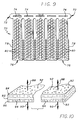
- the stent can be formed of a sheet of sintered metal 64 as shown in FIG. 8 or of a sintered metal tube.
- metal particles 66 are arranged and pressurized into a sheet. The sheet then is heated to a temperature below the melting point of the particles as described previously.
- the sheet of sintered metal is porous and has a plurality of pores 68.
- a sheet or tube of sintered metal can be cut in the desired shape to form the metal structural member with a laser, such as a continuous CO 2 laser, a pulsed YAG laser, or an excimer laser, for example, or alternatively, by chemical etching or stamping.
- a laser such as a continuous CO 2 laser, a pulsed YAG laser, or an excimer laser, for example, or alternatively, by chemical etching or stamping.
- the stent can be formed into a particular pattern known in the art for stents formed from metal sheets.
- One such pattern is a rolled locking design and is illustrated in FIG. 9 .
- the sheet is etched into a stent configuration 70 that has a head portion 72 that includes one Or more slots 74 for receipt of a number of tail sections 76 that correspond to each slot.
- the tail sections are received into the slots so as to form a cylindrical loop.
- Each tail section includes a plurality of teeth 78 that are adapted to cooperatively engage the slot of the head portion. When the teeth engage the slot, the tail sections are retained in place, holding the stent configuration in an expanded state.
- holes 80 are formed throughout the stent to reduce the metal-to-air ratio of the stent. The less metal that is in contact with the wall 14 of the vessel 16, the more likely the stent is to be compatible with the blood.
- the tail sections Prior to deployment, the tail sections are coiled into a retracted position in a "jelly-roll" fashion. Each tail section then is threaded through each corresponding slot and wound.
- the stent configuration is expanded by a balloon according to principles that are well known in the art for delivering and implanting stents. As the stent configuration 70 is expanded by a balloon during deployment, it unwinds and the teeth 78 lock into the slots 74 at a desired radial diameter to prevent the stent from returning to its original, retracted state.
- a benefit of the coiled stent shown in FIG. 9 is that the stent 70 can be etched to have a minimal surface area that comes in contact with the walls of the vessel. This may be an important feature when it is desired to cover the entire area of the walls of a blood vessel with a therapeutic agent because the coiled sheet metal stent can be configured to maintain maximum surface area contact with the wall of the blood vessel in contrast to wire stents.
- FIG. 10 there is shown a sheet that has particles that are sintered to both sides 84 and 86 of a metal sheet 82.
- the stent of FIG. 10 is similar in structure to the stent wire of FIG. 7 in that it has a solid core and particles sintered to the core forming a porous outer layer.
- the solid core reinforces the strength of the metal.
- the metal sheet also provides a barrier through which a therapeutic agent cannot pass.
- a therapeutic agent loaded into the pores 92 on the top side 84 of the sheet permeates in a first direction 88 outward from the solid core.
- a therapeutic agent loaded into the pores 94 on the bottom side 86 of the solid wire permeates only in a second direction 90 which is opposite to the direction of the therapeutic agent loaded into the pores on the top side.
- a stent as shown in FIG. 10 When a stent as shown in FIG. 10 is looped into a cylindrical formation and placed into a vessel, only the top side 84, which is directed radially outward, engages the walls of the vessel.
- the bottom side 86 faces radially inward and does not come in contact with the walls of the vessel.
- a first therapeutic agent can be loaded into the top side to treat the tissue in the wall of the vessel.
- a second therapeutic agent can be loaded into the bottom side to prevent coagulation of the blood flowing in the vessel.
- the stent can be formed so that particles are sintered only to one side of the stent.
- a therapeutic agent is loaded into the sintered metal on the porous side of the stent. When a stent is formed with only one porous side, that side can be oriented radially outward to deliver a therapeutic agent to the tissue in the wall of the stent.
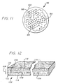
- FIG. 11 illustrates a cross-sectional view of a stent wire according to another arrangement.
- the sheet has a plurality of porous cavities or pores 98.
- a therapeutic agent is loaded into the pores of the sintered metal.
- a coating 100 is applied to the sintered metal.
- the coating may be used for several purposes as described hereinafter.
- the stent is formed of a sintered sheet 104 of metal having core 106 formed of larger-diameter particles 108 that form larger pores as a result of sintering.
- the core layer 106 is sandwiched between two layers 110 and 112 formed of smaller-diameter particles 114 or particles that result in the formation of smaller pores.
- Such a sheet is formed by orienting the middle or core layer 106 of larger-diameter particles 108 along a plane.
- a top layer of the smaller-diameter particles is arranged in a plane parallel to and above the core layer.
- a bottom layer of particles are arranged in a plane parallel to and below the core layer.
- the three layers then are pressed together and sintered into a single sheet.
- the sheet then can be cut or etched into a stent configuration.
- one of the benefits of certain embodiments is to provide a stent that does not require a coating for the purpose of delivering a therapeutic agent to the blood vessel
- the application of a coating after a therapeutic agent has been loaded into the pores of the sintered metal does not defeat the utility of the present invention.
- the profile of the polymeric coating can be reduced.
- a larger dosage of a therapeutic agent can be delivered to the site of stent implantation.
- Additional benefits can be obtained by loading a stent with a therapeutic agent in the pores of the metal and by then further applying a polymeric coating to the stent. Even if a polymeric coating is applied to the stent, the principles of reducing profile and reinforcing the stent still are realizable because a greater volume of therapeutic agent can be delivered by a polymeric-coated sintered stent than by a coated, all-polymeric stent with comparable dimensions.
- the polymeric material that coats a sintered metal stent preferably comprises a biodegradable, bioabsorbable polymeric film that is capable of being loaded with and subsequently releasing therapeutic drugs.
- the polymeric coatings preferably include, but are not limited to, polycaprolactone (PCL), poly-DL-lactic acid (DL-PLA) and poly-L-lactic acid (L-PLA) or lactide.
- PCL polycaprolactone
- DL-PLA poly-DL-lactic acid
- L-PLA poly-L-lactic acid
- Other biodegradable, bioabsorbable polymers such as polyorthoesters, polyiminocarbonates, aliphatic polycarbonates, and polyphosphazenes also may be suitable, and other non-degradable polymers capable of carrying and delivering therapeutic drugs might be appropriate as well.
- non-degradable synthetic polymers are those sold under the trade names "Parylene” and "Parylast” by Advanced Surface Technology of Billerica, MA, polyurethane, polyethylene, polyethylene teraphthalate, ethylene vinyl acetate, silicone and polyethylene oxide (PEO).
- the polymeric layer is loaded with a pharmacologic agent for use in localized drug therapy.
- biodegradable, bioabsorbable, reabsorbable, degradable, and absorbable collectively are meant to encompass materials that are broken down and gradually absorbed or eliminated by the body, whether these processes are due to hydrolysis, metabolic processes, or to bulk or surface erosion.
- one polymeric layer preferably is about 0.051 millimeters (0.002 inches) thick.
- the thin polymeric films used to coat the stent preferably first are intermixed with the drug or drugs to be delivered, and then typically are laminated or solvent cast to the surface of the metal structural member. Lamination processing methods and temperatures can vary widely depending on the polymers used and the temperature sensitivity of the loaded drugs. Alternatively, the metal structure of the stent can be encapsulated in the layers of polymeric material by solvent casting, melt processing, insert molding, and dip coating.
- the membrane is bioabsorbable, but no therapeutic agent is loaded into the polymer.
- the coating dissolves after implantation and this delays the time that a therapeutic agent is released into the vasculature of a patient.
- the thickness of the coating, as well as the rate at which the coating is bioabsorbed, determine the length of time that the stent is positioned in the vascular before a therapeutic agent is delivered from the pores of the stent.
- a therapeutic agent can be loaded into the bioabsorbable coating.
- a therapeutic agent will be delivered to the stent at a rate determined by the bioabsorbability of the coating.
- the therapeutic agent in the pores can be delivered at a rate determined by the pore size and porosity.
- the coating is permeable and non-absorbable.
- the rate at which the drugs permeate into the tissue is controlled by the physical properties of the particular coating selected.
- the coating may be selected to reduce restenosis, thrombosis or other tissue inflammation.
- a heparin coating is known in the art to reduce blood clotting. Heparin, when coated on a stent, reduces clotting of blood on the surface of the stent. The heparin coating is affixed to the surface of the stent through ionic bonding, end-point attaching, or by photo-linking the heparin.
- a first therapeutic agent is loaded into the coating and a second therapeutic agent is loaded into the pores of the stent. This may be the case when a series of drug dosages or concentrations are needed.
- the first therapeutic agent is absorbed first by the stent and a second therapeutic agent is absorbed later by the vasculature.
- This variation adds a further dimension to drug treatment allowing for sequential drug therapy at the site of placement of a stent.
Description
- This invention generally relates to a medicated prosthesis or implant. More particularly, the invention relates to a medicated intra-vascular prosthesis, such as a stent, that is radially expandable in the vasculature of a patient and delivers a therapeutic agent to the site of the implantation.
- Stents are generally cylindrically-shaped prosthetic implants which function to hold open and sometimes expand a segment of a blood vessel or other anatomical lumen. They are particularly suitable for supporting and preventing a torn or injured arterial lining from occluding a fluid passageway. Intravascular stents increasingly are useful for treatment of coronary artery stenoses, and for reducing the likelihood of the development of restenosis or closure after balloon angioplasty.
- The success of a stent can be assessed by evaluating a number of factors, such as thrombosis; neointimal hyperplasia, smooth muscle cell migration and proliferation following implantation of the stent; injury to the artery wall; overall loss of luminal patency; stent diameter in vivo; thickness of the stent; and leukocyte adhesion to the luminal lining of stented arteries. However, the chief areas of concern are early subacute thrombosis, and eventual restenosis of the blood vessel due to intimal hyperplasia.
- Therapeutic pharmacological agents have been developed to improve successful placement of the stent and are delivered to the site of stent implantation. Stents that are of a common metallic structure were previously unable to deliver localized therapeutic pharmacological agents to a blood vessel at the location being treated with the stent. There are polymeric materials that can be loaded with therapeutic agents including drugs or other pharmacological treatments which agents then can be released for drug delivery. However, these polymeric materials may not fulfill the structural and mechanical requirements of a stent, especially when the polymeric materials are loaded with a drug, since drug loading of a polymeric material can significantly reduce the structural and mechanical properties of the polymeric material.
-
CA-A-2,178,541 discloses a coronary stent that includes a structure adapted for introduction into the vascular system; at least one layer of a bioactive material posited on one surface of the stent structure; and at least one porous layer posited over the bioactive material layer and the bioactive-material-free stent surface. The porous layer is comprised of a polymer applied preferably by vapour or plasma deposition and provides a controlled release of the bioactive material. - By contrast
US-A-4,374,669 describes a cardiovascular prosthetic device or implant comprising a porous surface and a network of interconnected interstitial pores below the surface in fluid flow communication with the surface pores. Tissue forms a smooth thin adherent coating of self-determining thickness on the porous surface making it resistant to the formation of the blood clots normally associated with the presence of foreign bodies in the blood stream. - It is known in the art to coat a metallic stent with a polymeric material and to load the polymeric material with a drug. Alternatively, stents of polymeric materials have been reinforced with metal structure. These stent designs have the strength necessary to hold open the lumen of the vessel because of the reinforcement contributed by the metal. Stents made of both polymeric material and metal have a larger radial profile because the volume occupied by the metal portion of the stent cannot absorb and retain drugs. Reducing the profile of a stent is desirable because doing so increases the in vivo diameter of the lumen created by the stent. Thus it is desirable to configure a metallic stent to deliver drugs to the blood vessel walls without substantially increasing the profile of the stent.
- According to the present invention there is provided s stent as claimed in the appended claims.
- Certain preferred embodiments of the present invention provide for an implantable prosthesis that is made of metal and has pores or porous cavities in the metallic portion of the prosthesis so that therapeutic agents such as drugs can be loaded directly into the pores without substantially weakening the structural and mechanical characteristics of the prosthesis. The stent of one embodiment of the present invention can be implanted at a specific site of vascular injury, such as can result from balloon angioplasty or de novo lesions of a therosclerotic disease. The drugs in the pores of the stent can treat restenosis and tissue inflammation, and promote endothelialization or any other disease or condition that might detract from the success of a stent implantation.
- The pores of the stent are formed by sintering the stent material from metallic particles, filaments, fibers or other materials. The stent can be formed from a sintered wire that is coiled or otherwise is formed into a stent. The stent can be formed from a sintered cylindrical tube or sintered metal sheet which can be laser-cut or chemically etched into an expandable stent structure.
- Additionally, the porosity of the stent metal can be increased by using particles that are not generally spherical such as fibrous particles, filaments or wires. In one embodiment of the invention, the interwoven fibers and filaments also can be sintered after they have been woven into the desired shape.
- In one embodiment of the present invention, the stent is formed from a metal wire or strut that is comprised of a first layer of particles, oriented along a first axis, which forms a core, and an outer layer of particles that are arranged radially outward from the inner layer of particles. The particles in the outer layer have a smaller diameter than the particles in the inner layer. The core permits a greater volume of drugs in the center of the stent. The smaller diameter particles on the outside control the rate at which drugs are released into the walls of the vessel. The larger diameter particles result in a layer of greater porosity which thus is capable of carrying a higher volume of medication.
- In another embodiment it may be desirable to form a stent that has a solid core and a porous outer section. This can be accomplished by sintering particles to a solid, non-porous, metal wire. A stent so configured has a solid core which reinforces the structure of the stent and thereby adds strength to the device. The porous particles sintered to the surface of the stent absorb drugs for delivery.
- In one embodiment according to the present invention, the stent is formed from a sheet or tube of sintered metal. The sheet or tube is cut according to a pattern that allows the stent to be expanded and thus deployed into the vasculature. The stent pattern of this embodiment can be stenciled onto the sheet or tube of sintered metal and then may be cut by laser-cutting the sheet into the desired shape. Alternatively, the stent can be chemically etched into its desired shape.
- According to another embodiment of the invention, the stent receives a coating on the surface of the stent. In certain applications, it is desirable that the coating be a biopolymer and in other applications, the coating preferably is a synthetic polymer or a hydrogel. The coating also can be a heparin coating that is affixed to the surface of the stent through ionic bonding, end-point attaching or photolinking the heparin.
- According to one embodiment of the invention, the coating preferably is permeable to the medication. The permeability of the coating should be selected to release the medication in the stent at a desired rate.
- In another embodiment of the present invention, a bioabsorbable coating is applied to the stent. This coating is dissolved by the body fluids. Further, it is desirable in certain applications to load medication into the coating applied to the stent. According to one application, the coating preferably is loaded with the same drug or medication that is loaded into the stent. In another embodiment, the coating is loaded with a medication that is different than that which is carried by the stent. In this configuration, the two medications are released sequentially.
- The method of using the stent of the present invention comprises providing a porous prosthesis, loading a drug into the pores of the prosthesis, positioning the prosthesis in an appropriate site and affixing the prosthesis to the site. In another arrangement, the method further includes the step of applying a coating to the stent after the step of loading the drug.
- These and other features of the present invention will become apparent from the following more detailed description, when taken in conjunction with the accompanying drawings which illustrate, by way of example, the principles of the present invention.
-
-
FIGURE 1 is a longitudinal sectional view of a blood vessel with a stent manufactured according to one embodiment of the present invention. -
FIG. 2 is a porous stent wire or strut in a partially magnified, partially cut-away perspective, manufactured according to one embodiment of the present invention. -
FIG. 3 is a magnified, cross-sectional view of un-sintered, packed particle. -
FIG. 4 is a porous stent wire or strut in a partially magnified, partially cut-away perspective, manufactured according to one embodiment of the present invention. -
FIG. 5 is a porous stent wire or strut in a partially magnified, partially cut-away perspective, manufactured according to one embodiment of the present invention. -
FIG. 6 is a cross-sectional view of a stent wire or strut manufactured according to one embodiment of the present invention. -
FIG. 7 is a cross-sectional view of a stent wire or strut: formed from sintered metal particles surrounding a solid core. -
FIG. 8 is a sheet of sintered stent manufactured according to one embodiment of the present invention. -
FIG. 9 is a stent formed from a sheet of sintered metal according to one embodiment of the present invention. -
FIG. 10 is a cross-sectional, partially cut-away view of a sheet of sintered metal that has particles that are sintered to both sides of the sheet. -
FIG. 11 is a cross-sectional view of a stent wire or strut having a coating applied to the sintered metal. -
FIG. 12 is a cross-sectional view, partially cut- away of a sheet of sintered metal manufactured according to the principles of one embodiment of the present invention. - Referring to
FIGURE 1 , the prosthesis of one embodiment is aporous stent 12 that is radially expandable against thewalls 14 of avessel 16. A therapeutic agent is loaded into the pores 18 (SeeFIG. 2 ) of the stent. When placed in the vasculature, the therapeutic agent is delivered to the tissue that comes into contact with the stent. The stent of one preferred embodiment is formed of a stent wire that is porous. An example of a porous stent wire is a sintered metal wire.FIG. 2 illustrates a partial microscopic view of a sintered wire that is suitable for use in one embodiment of the present invention. The wire is porous and hasseveral pores 18. The cavities preferably range in size between 0.01 and 20 µm. - The metal is made porous by the process of sintering metal. Sintering is a process of fabrication where particles are bonded together to form a coherent mass without entirely melting the particles. Particles are pressed together or molded into a desired shape. A considerable amount of pressure first is applied to press the particles together. Then the metal is heated to temperatures slightly below the melting point of the metal. Without entirely melting, the particles bond to each other. Space remains between the lattice of the particles and this space defines the
pores 18. - The formation of sintered metal is illustrated with reference to
FIG. 3 and continued reference toFIG. 2. FIG. 3 is a microscopic view of a packedlattice 22 ofmetallic particles 24.Gaps 26 exist between each particle despite the fact that the particles are pressurized and are in contact with adjacent particles. Particles preferably are sized between 0.02 µm and 20 µm in diameter. Prior to heating, there are no chemical bonds formed between the individual particles. When the metal is heated to slightly below the melting point of the metal, the particles bond with neighboring particles. The gaps in the packed lattice form pores 18 when the particles are sintered. Thus inFIG. 2 , the metal stent wire formed by the process of sintering haspores 18 extending throughout the entire wire, thereby interconnecting the cavities. The cavities then can be filled with a therapeutic agent as hereinafter described. The appropriate pressure and temperature of sintering a particular metal is specific to that particular metal. One skilled in the art of metal fabrication would understand how to sinter any given metal or alloy. - For each of the embodiments, the metal stent material member can be a suitable metal such as stainless steel, tantalum, nickel-titanium alloy, platinum-iridium alloy, molybdenum-rhenium alloy, gold, magnesium, combinations thereof, although other similar materials also may be suitable. The metal can be modified to exhibit different hardnesses, and thus varying stiffnesses, by well known annealing and manufacturing processes.
- One of the factors to be considered when making a stent according to the present invention is the porosity of the metal. Porosity is the total volume of pores in the sintered metal divided by the total volume of the metal. Porosity determines the amount of a therapeutic agent that can be loaded into a stent of predetermined dimensions. High porosity means that a stent can deliver more therapeutic agents or have a narrower profile because it is less dense. High porosity, according to some embodiments of the present invention, adversely affects the strength and elasticity of a metal. Consequently, there is an ongoing tradeoff between stent strength, on the one hand, and stent profile and stent load capacity on the other hand.
- Pore size is a function of the size of the particles which create the gaps that establish the pores. As illustrated in
FIG. 3 , theparticles 24 may be generally spherical. The size of eachpore 18 is proportional to particle size, particularly with generally spherical particles. When theparticles 24 are not of uniform size, smaller particles tend to fill the gaps between larger particles. Thus, the porosity of the metal formed with such particles is less predictable than when more equally-sized particles are used. General uniformity of pore size also is important to ensure that drugs are dispersed evenly throughout the stent. A generally uniform distribution of pores insures that the tissue in contact with the stent will receive an evenly distributed dose of a therapeutic agent. - There are several types of drugs that currently are administered at the site that a stent is placed in the vessel. Examples of therapeutic drugs, or agents that can be combined with the particle layers, include antiplatelets, anticoagulants, antifibrins, antithrombins and antiproliferatives. Examples of antiplatelets, anticoagulants, antifibrins and antithrombins include but are not limited to sodium heparin, low molecular weight heparin, hirudin, argatroban, forskolin, vapiprost, prostacyclin and prostacyclin analogues, dextran, D-phe-pro-arg-chloromethylketone (synthetic antithrombin), dipyridamole, glycoprotein IIb/IIIa platelet membrane receptor antibody, recombinant hirudin, thrombin inhibitor (available from Biogen), and an antiplatelet drug sold under the trademark "7E-3B" by Centorcor, Inc. Examples of cytostatic or antiproliferative agents include angiopeptin, a somatostatin analogue; angiotensin-converting enzyme inhibitors, such as those manufactured under the trademarks "Captopril" (by Squibb Corp.), "Cilazapril" (by Hoffman-LaRoche, Inc.) and "Lisinopril" (by Merck & Co., Inc.); calcium channel blockers such as nifedipine; colchicine; fibroblast growth factor (FGF) antagonists; fish oils, such as omega 3 fatty acids; cholesterol-lowering drugs such as inhibitors of HMG-CoA reductase, one of which is sold under the trademark "Lovastatin" by Merck & Co., Inc.; methotrexate, monoclonal phosphodiesterase inhibitors, prostaglandin inhibitor (available from Glaxo Wellcome, Inc., PDGF antagonists such as seramin and triazolopyrimidine, serotonin blockers, steroids, thioprotease inhibitors, and nitric oxide. Other therapeutic drugs which may be appropriate include alpha-interferon and genetically-engineered epithelial cells, for example.
- The foregoing therapeutic agents have been used to prevent or to treat restenosis, and each is identified by way of example and not by limitation, as other therapeutic drugs may be developed which equally are applicable for use with the present invention. Using such therapeutic agents to treat vessels or body lumens is known in the art, as is the calculation of dosages, dosage rates and appropriate duration of treatment.
- The therapeutic agent of one embodiment preferably is in liquid form and is loaded into a stent by immersing the stent in a medicated solution. The therapeutic agent may be dissolved in a solvent or suspended in a liquid mixture. If a suspension is used, it is important that the pore size of the stent is considerably larger than the suspended particles of the therapeutic agent. An average pore size that is more than ten (10) times the particle size of a suspended therapeutic agent is suitable. After the stent is immersed in the medicated solution, the therapeutic agent is absorbed into the pores of the stent. The loaded stent then can be removed from the solution and implanted into the vasculature of a patient. Optionally, the loading of the therapeutic agent into the stent can be facilitated by applying pressure to the fluid in which the agent is dissolved or suspended. The applied pressure will aid the passage of medicated fluid into the pores of the stent. This technique might be likened to the physical process of forcing a fluid through the pores of a filter.
- Once loaded into the stent the therapeutic agent remains in place by reason of the surface tension between the outer surfaces of the particles that form the
pores 18 and the particles of the therapeutic agent. As shown inFIG. 1 , the loaded or medicatedporous stent 12 then is deployed to the site of anarterial closure 13 and is expanded. The expanded stent engages thewalls 14 of thevessel 16 to maintain the patency of the vessel. Once in the vessel and as is illustrated inFIG. 2 , the therapeutic agent disseminates from thepores 18 and is absorbed into the tissue of the walls of the vessel that are in contact with the stent. - Among the advantages of a stent embodying the present invention over prior art medicated stents are its profile and strength. Metal, including sintered metal, is stronger than synthetic materials, such as polymer blends, which are capable of being loaded with a therapeutic agent. Thus, in order for a medicated stent to deliver an appropriate amount of a therapeutic agent and structurally maintain vessel patency, the radial profile of the stent must be substantially larger than that of metal stents. This is true whether or not a metal stent is coated with a polymeric material to carry a therapeutic agent, or if the stent is made entirely of a plastic material.
- Sintered metal has strength and elasticity that is comparable to non-sintered metal. Sintered metal further has the added feature of porosity. Consequently, a sintered metal stent can be manufactured with a profile that is substantially comparable to that of a conventional metal stent, and a therapeutic agent can be loaded into the pores and delivered to the site of stent implantation without the aid of medicated polymer coatings.
- Additionally, many synthetic materials, including materials that are bioabsorbable, can cause inflammation of the tissue. A medicated metal stent having a therapeutic agent loaded directly into the pores of the stent likely will be less apt to cause irritation at the site of implantation.
-
FIG.4 illustrates astent wire 30 constructed according to an embodiment of the invention. The stent is formed ofelongated particles 32, i.e., filaments and fibers. When generally spherically-shaped particles of metal are used to compose sintered metal, the resultant porosity typically is in the range of five to thirty percent. When the particles are elongated, these filaments or fibers can result in a porosity of greater than thirty percent when sintered. The technique of fabricating a stent with elongated filaments or fibers is similar to the method described above for spherical particles or powders. The filaments or fibers are molded and pressurized. Then the fibers are heated to a temperature just below the melting point of the metal. - A stent made of metal filaments or
fibers 32 rather than spherical particles (such as those illustrated inFIG. 2 ) exhibits greater porosity because of the irregular shape of the particles. The particles can be packed less densely than uniformly-shaped particles but contact between the irregularly-shaped particles nevertheless can be maintained to allow sintering. Thus, the void space or pores 34 in the sintered metal tend to be larger than thepores 18 that result from spherical particle sintering. - The strength of a
stent wire 30 using filaments inFIG. 4 is improved because, upon sintering, the individual strands have a greater surface-area-to-volume ratio and will contact more neighboring strands than will spherical particles. Thus, each filament or fiber will have a greater surface area on which to bond with adjacent filaments or fibers. A matrix of overlapping filaments or fibers thus is formed exhibiting greater porosity and stronger inter-particle bonding. - In yet another embodiment,
wire fibers 36 are woven or twined into astructure 38 as illustrated inFIG. 5 . The individual strands cooperate in a synergistic manner to reinforce the strength of the wire. Additionally, the wire fibers can be woven into the form of a sintered metal sheet having improved and reinforced strength or into a sintered metal tube. Other combinations of particle size and shape can be employed to form a stent wire having different characteristics. - As illustrated in
FIG. 6 , thestent wire 42 is formed of aninner core 44 and anouter layer 46 of sintered particles. The outer layer is formed from particles having a different diameter than the diameter of the particles that form the inner core. For example, the core of the metal is formed of particles that have a diameter in the range of 10-20 µm. Surrounding the core are particles that have a diameter in the range of: 2-4 µm. The larger particles create a core having larger pores 52. This results in higher porosity and thus a higher load capacity. The smaller particles on the outer layer formsmaller pores 54 which reduce the rate of diffusion of drugs into the tissue of the vessel. - When a therapeutic agent is loaded into a stent formed of the
stent wire 42 illustrated inFIG. 6 , a larger volume can be stored in thelarger pores 52 at thecore 44 of the stent wire. Once the stent is placed into the vessel, the therapeutic agent in the stent wire is delivered at a rate that is determined by thesmaller pores 54 in theouter layer 46 of the stent wire. Such a structure is expected to be capable of storing a large amount of therapeutic agent at the core and of delivering the therapeutic agent at a slower rate than would be accomplished if the pores of the stent wire were of more uniform size. Consequently, this design is appropriate when long-term drug therapy at a low dosage rate is desired. -
FIG. 7 shows astent wire 56 formed fromsintered particles 58. Thepores 62 formed between the sintered metal particles surrounding the solid core retain the therapeutic agent. The overall porosity of a stent having a solid core and porous outer layer is much less than that of a stent wire having similar proportions but which is composed entirely of sintered particles. However, the solid core reinforces the tensile strength and elasticity of the metal stent and is considerably stronger than a uniform-particle sintered stent. Thus, it is desirable to use a sintered stent with a solid core for applications where maximum tensile strength and elasticity is desirable and only a relatively small amount of therapeutic agent is needed. - The sintered metal stent of the present invention can be made of material formed in the spherically-shaped or filament-like particles discussed previously. For example, the stent can be formed of a sheet of sintered
metal 64 as shown inFIG. 8 or of a sintered metal tube. By way of example,metal particles 66 are arranged and pressurized into a sheet. The sheet then is heated to a temperature below the melting point of the particles as described previously. The sheet of sintered metal is porous and has a plurality ofpores 68. - The same principles that apply to porosity and pore size of a wire apply equally to a sintered stent that is formed into a sheet or tube. The advantage of forming the stent from a sheet of metal is that the stent is radially expandable without placing a great deal of strain on the metal lattice when it is expanded. A sheet or tube of sintered metal can be cut in the desired shape to form the metal structural member with a laser, such as a continuous CO2 laser, a pulsed YAG laser, or an excimer laser, for example, or alternatively, by chemical etching or stamping. When cut from a flat sheet, the stent then is rolled into a cylindrical configuration and is laser welded along the longitudinal edges.
- The stent can be formed into a particular pattern known in the art for stents formed from metal sheets. One such pattern is a rolled locking design and is illustrated in
FIG. 9 . The sheet is etched into astent configuration 70 that has ahead portion 72 that includes one Ormore slots 74 for receipt of a number oftail sections 76 that correspond to each slot. The tail sections are received into the slots so as to form a cylindrical loop. Each tail section includes a plurality ofteeth 78 that are adapted to cooperatively engage the slot of the head portion. When the teeth engage the slot, the tail sections are retained in place, holding the stent configuration in an expanded state. Additionally, holes 80 are formed throughout the stent to reduce the metal-to-air ratio of the stent. The less metal that is in contact with thewall 14 of thevessel 16, the more likely the stent is to be compatible with the blood. - Prior to deployment, the tail sections are coiled into a retracted position in a "jelly-roll" fashion. Each tail section then is threaded through each corresponding slot and wound. The stent configuration is expanded by a balloon according to principles that are well known in the art for delivering and implanting stents. As the
stent configuration 70 is expanded by a balloon during deployment, it unwinds and theteeth 78 lock into theslots 74 at a desired radial diameter to prevent the stent from returning to its original, retracted state. - A benefit of the coiled stent shown in
FIG. 9 is that thestent 70 can be etched to have a minimal surface area that comes in contact with the walls of the vessel. This may be an important feature when it is desired to cover the entire area of the walls of a blood vessel with a therapeutic agent because the coiled sheet metal stent can be configured to maintain maximum surface area contact with the wall of the blood vessel in contrast to wire stents. - With reference to
FIG. 10 , there is shown a sheet that has particles that are sintered to bothsides metal sheet 82. The stent ofFIG. 10 is similar in structure to the stent wire ofFIG. 7 in that it has a solid core and particles sintered to the core forming a porous outer layer. The solid core reinforces the strength of the metal. The metal sheet also provides a barrier through which a therapeutic agent cannot pass. Thus, a therapeutic agent loaded into thepores 92 on thetop side 84 of the sheet permeates in afirst direction 88 outward from the solid core. A therapeutic agent loaded into thepores 94 on thebottom side 86 of the solid wire permeates only in asecond direction 90 which is opposite to the direction of the therapeutic agent loaded into the pores on the top side. - When a stent as shown in
FIG. 10 is looped into a cylindrical formation and placed into a vessel, only thetop side 84, which is directed radially outward, engages the walls of the vessel. Thebottom side 86 faces radially inward and does not come in contact with the walls of the vessel. Thus, if it is desired, a first therapeutic agent can be loaded into the top side to treat the tissue in the wall of the vessel. A second therapeutic agent can be loaded into the bottom side to prevent coagulation of the blood flowing in the vessel. Additionally, the stent can be formed so that particles are sintered only to one side of the stent. A therapeutic agent is loaded into the sintered metal on the porous side of the stent. When a stent is formed with only one porous side, that side can be oriented radially outward to deliver a therapeutic agent to the tissue in the wall of the stent. -
FIG. 11 illustrates a cross-sectional view of a stent wire according to another arrangement. The sheet has a plurality of porous cavities or pores 98. A therapeutic agent is loaded into the pores of the sintered metal. Then, acoating 100 is applied to the sintered metal. The coating may be used for several purposes as described hereinafter. - With reference to
FIG. 12 , another embodiment of the invention is shown wherein the stent is formed of asintered sheet 104 ofmetal having core 106 formed of larger-diameter particles 108 that form larger pores as a result of sintering. Thecore layer 106 is sandwiched between twolayers diameter particles 114 or particles that result in the formation of smaller pores. Such a sheet is formed by orienting the middle orcore layer 106 of larger-diameter particles 108 along a plane. A top layer of the smaller-diameter particles is arranged in a plane parallel to and above the core layer. A bottom layer of particles are arranged in a plane parallel to and below the core layer. The three layers then are pressed together and sintered into a single sheet. The sheet then can be cut or etched into a stent configuration. While one of the benefits of certain embodiments is to provide a stent that does not require a coating for the purpose of delivering a therapeutic agent to the blood vessel, the application of a coating after a therapeutic agent has been loaded into the pores of the sintered metal does not defeat the utility of the present invention. For example, when a therapeutic agent is loaded into the pores of the stent and also into a polymeric coating, the profile of the polymeric coating can be reduced. When such a polymeric coating is applied, a larger dosage of a therapeutic agent can be delivered to the site of stent implantation. Additional benefits can be obtained by loading a stent with a therapeutic agent in the pores of the metal and by then further applying a polymeric coating to the stent. Even if a polymeric coating is applied to the stent, the principles of reducing profile and reinforcing the stent still are realizable because a greater volume of therapeutic agent can be delivered by a polymeric-coated sintered stent than by a coated, all-polymeric stent with comparable dimensions. - The polymeric material that coats a sintered metal stent preferably comprises a biodegradable, bioabsorbable polymeric film that is capable of being loaded with and subsequently releasing therapeutic drugs. The polymeric coatings preferably include, but are not limited to, polycaprolactone (PCL), poly-DL-lactic acid (DL-PLA) and poly-L-lactic acid (L-PLA) or lactide. Other biodegradable, bioabsorbable polymers such as polyorthoesters, polyiminocarbonates, aliphatic polycarbonates, and polyphosphazenes also may be suitable, and other non-degradable polymers capable of carrying and delivering therapeutic drugs might be appropriate as well. Examples of non-degradable synthetic polymers are those sold under the trade names "Parylene" and "Parylast" by Advanced Surface Technology of Billerica, MA, polyurethane, polyethylene, polyethylene teraphthalate, ethylene vinyl acetate, silicone and polyethylene oxide (PEO). The polymeric layer, according to one embodiment, is loaded with a pharmacologic agent for use in localized drug therapy. As used in this description, the terms biodegradable, bioabsorbable, reabsorbable, degradable, and absorbable collectively are meant to encompass materials that are broken down and gradually absorbed or eliminated by the body, whether these processes are due to hydrolysis, metabolic processes, or to bulk or surface erosion. In each of the foregoing embodiments, one polymeric layer preferably is about 0.051 millimeters (0.002 inches) thick.
- The thin polymeric films used to coat the stent preferably first are intermixed with the drug or drugs to be delivered, and then typically are laminated or solvent cast to the surface of the metal structural member. Lamination processing methods and temperatures can vary widely depending on the polymers used and the temperature sensitivity of the loaded drugs. Alternatively, the metal structure of the stent can be encapsulated in the layers of polymeric material by solvent casting, melt processing, insert molding, and dip coating.
- In one embodiment of the present invention, the membrane is bioabsorbable, but no therapeutic agent is loaded into the polymer. The coating dissolves after implantation and this delays the time that a therapeutic agent is released into the vasculature of a patient. The thickness of the coating, as well as the rate at which the coating is bioabsorbed, determine the length of time that the stent is positioned in the vascular before a therapeutic agent is delivered from the pores of the stent. Additionally, a therapeutic agent can be loaded into the bioabsorbable coating. Thus a therapeutic agent will be delivered to the stent at a rate determined by the bioabsorbability of the coating. Once the bioabsorbable material has completely dissolved, the therapeutic agent in the pores can be delivered at a rate determined by the pore size and porosity.
- In another embodiment, it is preferred that the coating is permeable and non-absorbable. In such circumstances, the rate at which the drugs permeate into the tissue is controlled by the physical properties of the particular coating selected. Additionally, the coating may be selected to reduce restenosis, thrombosis or other tissue inflammation. For example, a heparin coating is known in the art to reduce blood clotting. Heparin, when coated on a stent, reduces clotting of blood on the surface of the stent. The heparin coating is affixed to the surface of the stent through ionic bonding, end-point attaching, or by photo-linking the heparin.
- In yet another embodiment, a first therapeutic agent is loaded into the coating and a second therapeutic agent is loaded into the pores of the stent. This may be the case when a series of drug dosages or concentrations are needed. When such a stent is placed into the vasculature, the first therapeutic agent is absorbed first by the stent and a second therapeutic agent is absorbed later by the vasculature. This variation adds a further dimension to drug treatment allowing for sequential drug therapy at the site of placement of a stent.
Claims (15)
- A stent comprising a body wherein at least a part of the body includes a first porous region (46) and a second porous region (44) disposed beneath the first porous region (46),
the second porous region (44) containing a therapeutic substance for the local delivery of the substance when the stent has been implanted in a vessel,
the average pore size of the first porous region (46) being less than the average pore size of the second porous region (44) for reducing the rate of release of the therapeutic substance from the second porous region (44), and
the first porous region (46) being formed by sintering metallic particles having a first diameter, and the second porous region (44) being formed by sintering metallic particles having a second diameter, the first diameter being smaller than the second diameter. - The stent of claim 1, additionally including a third porous region, such that the second porous region (44) is disposed between the first and third porous regions, wherein the average pore size of the third porous region is less than the average pore size of the second porous region (44).
- The stent of claim 1, wherein the first porous region (46) is capable of contacting the inner wall of the vessel when the stent has been implanted in the vessel.
- The stent of claim 1, additionally including a film layer disposed over the first porous region (46).
- The stent of claim 4, wherein the film layer comprises a biodegradable polymer.
- The stent of claim 4, wherein the film layer comprises a non-degradable polymer.
- The stent of claim 4, wherein the film layer includes a second therapeutic substance.
- The stent of claim 4, wherein the film layer includes heparin.
- The stent of claim 1, wherein the therapeutic substance is for the treatment of restenosis.
- The stent of claim 1, wherein the therapeutic substance comprises particles, and wherein the second porous region (44) includes pores (52) having an average pore size greater than ten times the particle size of the therapeutic substance.
- The stent of claim 1, wherein the first or second porous region (46,44) is formed by sintering a matrix of overlapping metallic filaments.
- The stent of claim 1, wherein the first or second porous region (46,44) is formed by sintering woven metallic wire fibers.
- The stent of claim 1, wherein the first diameter is 2 to 4 µm.
- The stent of claim 1, wherein the second diameter is 10 to 20 µm.
- The stent of claim 1, wherein an inner core of a strut of the body defines the second porous region (44) and an outer layer surrounding the inner core defines the first porous region (46).
Applications Claiming Priority (3)
| Application Number | Priority Date | Filing Date | Title |
|---|---|---|---|
| US08/842,660 US5843172A (en) | 1997-04-15 | 1997-04-15 | Porous medicated stent |
| EP98302905A EP0875218B1 (en) | 1997-04-15 | 1998-04-15 | Porous medicated stent |
| EP05002387A EP1527754B1 (en) | 1997-04-15 | 1998-04-15 | Porous medicated stent |
Related Parent Applications (3)
| Application Number | Title | Priority Date | Filing Date |
|---|---|---|---|
| EP05002387A Division EP1527754B1 (en) | 1997-04-15 | 1998-04-15 | Porous medicated stent |
| EP98302905.9 Division | 1998-04-15 | ||
| EP05002387.8 Division | 2005-02-04 |
Publications (3)
| Publication Number | Publication Date |
|---|---|
| EP2033667A2 EP2033667A2 (en) | 2009-03-11 |
| EP2033667A3 EP2033667A3 (en) | 2009-06-24 |
| EP2033667B1 true EP2033667B1 (en) | 2013-02-13 |
Family
ID=25287924
Family Applications (3)
| Application Number | Title | Priority Date | Filing Date |
|---|---|---|---|
| EP08010581A Expired - Lifetime EP2033667B1 (en) | 1997-04-15 | 1998-04-15 | Porous medicated stent |
| EP05002387A Expired - Lifetime EP1527754B1 (en) | 1997-04-15 | 1998-04-15 | Porous medicated stent |
| EP98302905A Expired - Lifetime EP0875218B1 (en) | 1997-04-15 | 1998-04-15 | Porous medicated stent |
Family Applications After (2)
| Application Number | Title | Priority Date | Filing Date |
|---|---|---|---|
| EP05002387A Expired - Lifetime EP1527754B1 (en) | 1997-04-15 | 1998-04-15 | Porous medicated stent |
| EP98302905A Expired - Lifetime EP0875218B1 (en) | 1997-04-15 | 1998-04-15 | Porous medicated stent |
Country Status (5)
| Country | Link |
|---|---|
| US (1) | US5843172A (en) |
| EP (3) | EP2033667B1 (en) |
| JP (1) | JP3614652B2 (en) |
| CA (1) | CA2235031A1 (en) |
| DE (2) | DE69829015T2 (en) |
Families Citing this family (476)
| Publication number | Priority date | Publication date | Assignee | Title |
|---|---|---|---|---|
| US6491965B1 (en) * | 1995-11-30 | 2002-12-10 | Hamilton Civic Hospitals Research Development, Inc. | Medical device comprising glycosaminoglycan-antithrombin III/heparin cofactor II conjugates |
| US7045585B2 (en) * | 1995-11-30 | 2006-05-16 | Hamilton Civic Hospital Research Development Inc. | Methods of coating a device using anti-thrombin heparin |
| US6562781B1 (en) | 1995-11-30 | 2003-05-13 | Hamilton Civic Hospitals Research Development Inc. | Glycosaminoglycan-antithrombin III/heparin cofactor II conjugates |
| JP4636634B2 (en) | 1996-04-26 | 2011-02-23 | ボストン サイエンティフィック サイムド,インコーポレイテッド | Intravascular stent |
| US6783543B2 (en) * | 2000-06-05 | 2004-08-31 | Scimed Life Systems, Inc. | Intravascular stent with increasing coating retaining capacity |
| US20040106985A1 (en) | 1996-04-26 | 2004-06-03 | Jang G. David | Intravascular stent |
| US6235053B1 (en) | 1998-02-02 | 2001-05-22 | G. David Jang | Tubular stent consists of chevron-shape expansion struts and contralaterally attached diagonal connectors |
| US7070590B1 (en) * | 1996-07-02 | 2006-07-04 | Massachusetts Institute Of Technology | Microchip drug delivery devices |
| US20030093143A1 (en) * | 1999-03-01 | 2003-05-15 | Yiju Zhao | Medical device having surface depressions containing nitric oxide releasing compound |
| US6099561A (en) * | 1996-10-21 | 2000-08-08 | Inflow Dynamics, Inc. | Vascular and endoluminal stents with improved coatings |
| US7341598B2 (en) * | 1999-01-13 | 2008-03-11 | Boston Scientific Scimed, Inc. | Stent with protruding branch portion for bifurcated vessels |
| ZA9710342B (en) * | 1996-11-25 | 1998-06-10 | Alza Corp | Directional drug delivery stent and method of use. |
| US7959664B2 (en) * | 1996-12-26 | 2011-06-14 | Medinol, Ltd. | Flat process of drug coating for stents |
| IT1289815B1 (en) * | 1996-12-30 | 1998-10-16 | Sorin Biomedica Cardio Spa | ANGIOPLASTIC STENT AND RELATED PRODUCTION PROCESS |
| US8172897B2 (en) | 1997-04-15 | 2012-05-08 | Advanced Cardiovascular Systems, Inc. | Polymer and metal composite implantable medical devices |
| US10028851B2 (en) | 1997-04-15 | 2018-07-24 | Advanced Cardiovascular Systems, Inc. | Coatings for controlling erosion of a substrate of an implantable medical device |
| US6240616B1 (en) | 1997-04-15 | 2001-06-05 | Advanced Cardiovascular Systems, Inc. | Method of manufacturing a medicated porous metal prosthesis |
| US8790391B2 (en) * | 1997-04-18 | 2014-07-29 | Cordis Corporation | Methods and devices for delivering therapeutic agents to target vessels |
| US6273913B1 (en) * | 1997-04-18 | 2001-08-14 | Cordis Corporation | Modified stent useful for delivery of drugs along stent strut |
| US6342051B1 (en) * | 1997-06-12 | 2002-01-29 | Gholam A. Peyman | Treatment of anoxic tissue with angiogenesis-inducing implants |
| DE19731021A1 (en) | 1997-07-18 | 1999-01-21 | Meyer Joerg | In vivo degradable metallic implant |
| US20030129215A1 (en) * | 1998-09-24 | 2003-07-10 | T-Ram, Inc. | Medical devices containing rapamycin analogs |
| US6890546B2 (en) | 1998-09-24 | 2005-05-10 | Abbott Laboratories | Medical devices containing rapamycin analogs |
| US8394398B2 (en) * | 1997-09-26 | 2013-03-12 | Abbott Laboratories | Methods of administering rapamycin analogs with anti-inflammatories using medical devices |
| US5972027A (en) * | 1997-09-30 | 1999-10-26 | Scimed Life Systems, Inc | Porous stent drug delivery system |
| US20040254635A1 (en) | 1998-03-30 | 2004-12-16 | Shanley John F. | Expandable medical device for delivery of beneficial agent |
| US6241762B1 (en) | 1998-03-30 | 2001-06-05 | Conor Medsystems, Inc. | Expandable medical device with ductile hinges |
| EP1920733A3 (en) † | 1998-03-30 | 2008-05-21 | Conor Medsystems, Inc. | Flexible medical device |
| US7208010B2 (en) | 2000-10-16 | 2007-04-24 | Conor Medsystems, Inc. | Expandable medical device for delivery of beneficial agent |
| US7713297B2 (en) * | 1998-04-11 | 2010-05-11 | Boston Scientific Scimed, Inc. | Drug-releasing stent with ceramic-containing layer |
| DE19916086B4 (en) * | 1998-04-11 | 2004-11-11 | Inflow Dynamics Inc. | Implantable prosthesis, especially vascular prosthesis (stent) |
| US20030040790A1 (en) | 1998-04-15 | 2003-02-27 | Furst Joseph G. | Stent coating |
| US20020099438A1 (en) | 1998-04-15 | 2002-07-25 | Furst Joseph G. | Irradiated stent coating |
| US20070087028A1 (en) * | 1998-04-16 | 2007-04-19 | Robert Falotico | Intraluminal devices for the prevention and treatment of vascular disease |
| US8029561B1 (en) | 2000-05-12 | 2011-10-04 | Cordis Corporation | Drug combination useful for prevention of restenosis |
| US6652581B1 (en) * | 1998-07-07 | 2003-11-25 | Boston Scientific Scimed, Inc. | Medical device with porous surface for controlled drug release and method of making the same |
| US7967855B2 (en) | 1998-07-27 | 2011-06-28 | Icon Interventional Systems, Inc. | Coated medical device |
| US8070796B2 (en) * | 1998-07-27 | 2011-12-06 | Icon Interventional Systems, Inc. | Thrombosis inhibiting graft |
| US7235096B1 (en) * | 1998-08-25 | 2007-06-26 | Tricardia, Llc | Implantable device for promoting repair of a body lumen |
| WO2000010623A1 (en) | 1998-08-25 | 2000-03-02 | Tricardia, L.L.C. | An implantable device for promoting repair of a body lumen |
| US6406488B1 (en) * | 1998-08-27 | 2002-06-18 | Heartstent Corporation | Healing transmyocardial implant |
| DE19855421C2 (en) | 1998-11-02 | 2001-09-20 | Alcove Surfaces Gmbh | Implant |
| US6375680B1 (en) * | 1998-12-01 | 2002-04-23 | St. Jude Medical, Inc. | Substrates for forming synthetic tissues |
| US6231597B1 (en) * | 1999-02-16 | 2001-05-15 | Mark E. Deem | Apparatus and methods for selectively stenting a portion of a vessel wall |
| DE19948783C2 (en) | 1999-02-18 | 2001-06-13 | Alcove Surfaces Gmbh | Implant |
| US6620192B1 (en) | 1999-03-16 | 2003-09-16 | Advanced Cardiovascular Systems, Inc. | Multilayer stent |
| US6364903B2 (en) * | 1999-03-19 | 2002-04-02 | Meadox Medicals, Inc. | Polymer coated stent |
| US6258117B1 (en) | 1999-04-15 | 2001-07-10 | Mayo Foundation For Medical Education And Research | Multi-section stent |
| DE19921088C2 (en) * | 1999-04-30 | 2003-08-07 | Magforce Applic Gmbh | Stent to keep aisle-like structures open |
| US6398808B1 (en) | 1999-06-15 | 2002-06-04 | Scimed Life Systems, Inc. | Localized delivery of genetic information from biostable materials |
| US6287628B1 (en) * | 1999-09-03 | 2001-09-11 | Advanced Cardiovascular Systems, Inc. | Porous prosthesis and a method of depositing substances into the pores |
| US7807211B2 (en) | 1999-09-03 | 2010-10-05 | Advanced Cardiovascular Systems, Inc. | Thermal treatment of an implantable medical device |
| US6379381B1 (en) | 1999-09-03 | 2002-04-30 | Advanced Cardiovascular Systems, Inc. | Porous prosthesis and a method of depositing substances into the pores |
| US20070032853A1 (en) | 2002-03-27 | 2007-02-08 | Hossainy Syed F | 40-O-(2-hydroxy)ethyl-rapamycin coated stent |
| DE60032912T2 (en) * | 1999-09-03 | 2007-10-25 | Advanced Cardiovascular Systems, Inc., Santa Clara | POROUS PROSTHESIS AND METHOD FOR THE DEPOSITION OF SUBSTANCES IN THE PORES |
| US6254631B1 (en) | 1999-09-23 | 2001-07-03 | Intratherapeutics, Inc. | Stent with enhanced friction |
| DE19951477A1 (en) * | 1999-10-26 | 2001-05-03 | Biotronik Mess & Therapieg | Stent |
| US6428569B1 (en) | 1999-11-09 | 2002-08-06 | Scimed Life Systems Inc. | Micro structure stent configurations |
| CA2392006C (en) | 1999-11-17 | 2011-03-15 | Microchips, Inc. | Microfabricated devices for the delivery of molecules into a carrier fluid |
| US6537310B1 (en) | 1999-11-19 | 2003-03-25 | Advanced Bio Prosthetic Surfaces, Ltd. | Endoluminal implantable devices and method of making same |
| US8458879B2 (en) | 2001-07-03 | 2013-06-11 | Advanced Bio Prosthetic Surfaces, Ltd., A Wholly Owned Subsidiary Of Palmaz Scientific, Inc. | Method of fabricating an implantable medical device |
| US6251136B1 (en) | 1999-12-08 | 2001-06-26 | Advanced Cardiovascular Systems, Inc. | Method of layering a three-coated stent using pharmacological and polymeric agents |
| US6508832B1 (en) | 1999-12-09 | 2003-01-21 | Advanced Cardiovascular Systems, Inc. | Implantable nickel-free stainless steel stents and method of making the same |
| US6585759B1 (en) | 1999-12-16 | 2003-07-01 | Israel Aircraft Industries Ltd. | Method and apparatus for manufacturing medical support devices |
| US7278195B2 (en) * | 1999-12-16 | 2007-10-09 | Israel Aircraft Industries Ltd. | Method for producing a coated medical support device |
| AU2599501A (en) * | 1999-12-29 | 2001-07-09 | Advanced Cardiovascular Systems Inc. | Device and active component for inhibiting formation of thrombus-inflammatory cell matrix |
| KR100346994B1 (en) | 2000-01-11 | 2002-07-31 | 한국과학기술연구원 | Biocompatible Metallic Materials Grafted with Sulfonated Poly(Ethylene Oxide) and Preparation Thereof |
| US20040010308A1 (en) * | 2000-01-18 | 2004-01-15 | Mindguard Ltd. | Implantable composite device and corresponding method for deflecting embolic material in blood flowing at an arterial bifurcation |
| IL140869A0 (en) * | 2001-01-11 | 2002-02-10 | Mind Guard Ltd | Implantable expanding stroke preventing device and method of maufacturing |
| US20040010307A1 (en) * | 2000-01-18 | 2004-01-15 | Mindguard Ltd. | Implantable integral device and corresponding method for deflecting embolic material in blood flowing at an arterial bifurcation |
| US6312463B1 (en) | 2000-02-01 | 2001-11-06 | Endotex Interventional Systems, Inc. | Micro-porous mesh stent with hybrid structure |
| ES2420279T3 (en) | 2000-03-02 | 2013-08-23 | Microchips, Inc. | Microfabricated devices and methods for storage and selective exposure of chemicals |
| US6290722B1 (en) | 2000-03-13 | 2001-09-18 | Endovascular Technologies, Inc. | Tacky attachment method of covered materials on stents |
| US9522217B2 (en) | 2000-03-15 | 2016-12-20 | Orbusneich Medical, Inc. | Medical device with coating for capturing genetically-altered cells and methods for using same |
| US8088060B2 (en) | 2000-03-15 | 2012-01-03 | Orbusneich Medical, Inc. | Progenitor endothelial cell capturing with a drug eluting implantable medical device |
| AU2001253479A1 (en) * | 2000-04-13 | 2001-10-30 | Sts Biopolymers, Inc. | Targeted therapeutic agent release devices and methods of making and using the same |
| US8236048B2 (en) | 2000-05-12 | 2012-08-07 | Cordis Corporation | Drug/drug delivery systems for the prevention and treatment of vascular disease |
| US6776796B2 (en) | 2000-05-12 | 2004-08-17 | Cordis Corportation | Antiinflammatory drug and delivery device |
| EP1280571B1 (en) * | 2000-05-12 | 2005-10-26 | Cordis Corporation | Delivery devices for treatment of vascular disease |
| DE60124772T2 (en) | 2000-05-19 | 2007-09-13 | Advanced Bio Prothestic Surfaces, Ltd., San Antonio | METHOD AND DEVICE FOR PRODUCING AN INTRAVASCULAR STENT |
| US8252044B1 (en) * | 2000-11-17 | 2012-08-28 | Advanced Bio Prosthestic Surfaces, Ltd. | Device for in vivo delivery of bioactive agents and method of manufacture thereof |
| US6395326B1 (en) | 2000-05-31 | 2002-05-28 | Advanced Cardiovascular Systems, Inc. | Apparatus and method for depositing a coating onto a surface of a prosthesis |
| US7682648B1 (en) | 2000-05-31 | 2010-03-23 | Advanced Cardiovascular Systems, Inc. | Methods for forming polymeric coatings on stents |
| US20040211362A1 (en) * | 2000-05-31 | 2004-10-28 | Daniel Castro | System for coating a stent |
| US6800089B1 (en) * | 2000-05-31 | 2004-10-05 | Advanced Cardiovascular Systems, Inc. | Mechanical attachment method of cover materials on stents |
| AU6539101A (en) * | 2000-06-05 | 2001-12-17 | G David Jang | Intravascular stent with increasing coating retaining capacity |
| EP1299590A2 (en) * | 2000-06-29 | 2003-04-09 | Johnson & Johnson Consumer Companies, Inc. | Electrostatic impregnation of powders on substrates |
| US6585765B1 (en) | 2000-06-29 | 2003-07-01 | Advanced Cardiovascular Systems, Inc. | Implantable device having substances impregnated therein and a method of impregnating the same |
| IL137090A (en) * | 2000-06-29 | 2010-04-15 | Pentech Medical Devices Ltd | Polymeric stent |
| US6709451B1 (en) | 2000-07-14 | 2004-03-23 | Norman Noble, Inc. | Channeled vascular stent apparatus and method |
| IL137326A0 (en) * | 2000-07-17 | 2001-07-24 | Mind Guard Ltd | Implantable braided stroke preventing device and method of manufacturing |
| US20040158317A1 (en) * | 2000-07-18 | 2004-08-12 | Pharmasonics, Inc. | Coated stent with ultrasound therapy |
| US6555157B1 (en) * | 2000-07-25 | 2003-04-29 | Advanced Cardiovascular Systems, Inc. | Method for coating an implantable device and system for performing the method |
| FR2812498B1 (en) | 2000-07-28 | 2003-03-14 | Sagem | AUDIO AND / OR VIDEO SEQUENCE SELECTION DEVICE |
| US6451373B1 (en) | 2000-08-04 | 2002-09-17 | Advanced Cardiovascular Systems, Inc. | Method of forming a therapeutic coating onto a surface of an implantable prosthesis |
| EP1179353A1 (en) * | 2000-08-11 | 2002-02-13 | B. Braun Melsungen Ag | Antithrombogenic implants with coating of polyphosphazenes and a pharmacologically active agent |
| US20090004240A1 (en) * | 2000-08-11 | 2009-01-01 | Celonova Biosciences, Inc. | Implants with a phosphazene-containing coating |
| MY128992A (en) * | 2000-08-25 | 2007-03-30 | Merck Patent Gmbh | Saratin for inhibiting platelet adhesion to collagen |
| US7766956B2 (en) | 2000-09-22 | 2010-08-03 | Boston Scientific Scimed, Inc. | Intravascular stent and assembly |
| US6254632B1 (en) * | 2000-09-28 | 2001-07-03 | Advanced Cardiovascular Systems, Inc. | Implantable medical device having protruding surface structures for drug delivery and cover attachment |
| US6805898B1 (en) * | 2000-09-28 | 2004-10-19 | Advanced Cardiovascular Systems, Inc. | Surface features of an implantable medical device |
| ATE343969T1 (en) | 2000-09-29 | 2006-11-15 | Cordis Corp | COATED MEDICAL DEVICES |
| DE20122506U1 (en) | 2000-10-16 | 2005-12-08 | Conor Medsystems, Inc., Menlo Park | Expandable medical device for delivering a beneficial agent |
| US6764507B2 (en) | 2000-10-16 | 2004-07-20 | Conor Medsystems, Inc. | Expandable medical device with improved spatial distribution |
| US6506437B1 (en) | 2000-10-17 | 2003-01-14 | Advanced Cardiovascular Systems, Inc. | Methods of coating an implantable device having depots formed in a surface thereof |
| US6979347B1 (en) * | 2000-10-23 | 2005-12-27 | Advanced Cardiovascular Systems, Inc. | Implantable drug delivery prosthesis |
| US6558733B1 (en) | 2000-10-26 | 2003-05-06 | Advanced Cardiovascular Systems, Inc. | Method for etching a micropatterned microdepot prosthesis |
| US6783793B1 (en) | 2000-10-26 | 2004-08-31 | Advanced Cardiovascular Systems, Inc. | Selective coating of medical devices |
| US6758859B1 (en) | 2000-10-30 | 2004-07-06 | Kenny L. Dang | Increased drug-loading and reduced stress drug delivery device |
| KR20030045847A (en) * | 2000-10-31 | 2003-06-11 | 쿡 인코포레이티드 | Coated implantable medical device |
| US9107605B2 (en) * | 2000-11-17 | 2015-08-18 | Advanced Bio Prosthetic Surfaces, Ltd., A Wholly Owned Subsidiary Of Palmaz Scientific, Inc. | Device for in vivo delivery of bioactive agents and method of manufacture thereof |
| US10398830B2 (en) * | 2000-11-17 | 2019-09-03 | Vactronix Scientific, Llc | Device for in vivo delivery of bioactive agents and method of manufacture thereof |
| US7244272B2 (en) | 2000-12-19 | 2007-07-17 | Nicast Ltd. | Vascular prosthesis and method for production thereof |
| US20020084178A1 (en) | 2000-12-19 | 2002-07-04 | Nicast Corporation Ltd. | Method and apparatus for manufacturing polymer fiber shells via electrospinning |
| JP2002306518A (en) * | 2000-12-21 | 2002-10-22 | Yuichi Mori | Indwelling implement |
| US20020082678A1 (en) * | 2000-12-22 | 2002-06-27 | Motasim Sirhan | Intravascular delivery of mizoribine |
| US6939375B2 (en) | 2000-12-22 | 2005-09-06 | Avantac Vascular Corporation | Apparatus and methods for controlled substance delivery from implanted prostheses |
| US20020082679A1 (en) * | 2000-12-22 | 2002-06-27 | Avantec Vascular Corporation | Delivery or therapeutic capable agents |
| US7018405B2 (en) | 2000-12-22 | 2006-03-28 | Avantec Vascular Corporation | Intravascular delivery of methylprednisolone |
| US7077859B2 (en) | 2000-12-22 | 2006-07-18 | Avantec Vascular Corporation | Apparatus and methods for variably controlled substance delivery from implanted prostheses |
| US7083642B2 (en) | 2000-12-22 | 2006-08-01 | Avantec Vascular Corporation | Delivery of therapeutic capable agents |
| US6471980B2 (en) | 2000-12-22 | 2002-10-29 | Avantec Vascular Corporation | Intravascular delivery of mycophenolic acid |
| US6913617B1 (en) * | 2000-12-27 | 2005-07-05 | Advanced Cardiovascular Systems, Inc. | Method for creating a textured surface on an implantable medical device |
| US6641607B1 (en) | 2000-12-29 | 2003-11-04 | Advanced Cardiovascular Systems, Inc. | Double tube stent |
| US6544582B1 (en) | 2001-01-05 | 2003-04-08 | Advanced Cardiovascular Systems, Inc. | Method and apparatus for coating an implantable device |
| US9080146B2 (en) | 2001-01-11 | 2015-07-14 | Celonova Biosciences, Inc. | Substrates containing polyphosphazene as matrices and substrates containing polyphosphazene with a micro-structured surface |
| US6752829B2 (en) | 2001-01-30 | 2004-06-22 | Scimed Life Systems, Inc. | Stent with channel(s) for containing and delivering a biologically active material and method for manufacturing the same |
| US20040073294A1 (en) | 2002-09-20 | 2004-04-15 | Conor Medsystems, Inc. | Method and apparatus for loading a beneficial agent into an expandable medical device |
| DE10127011A1 (en) * | 2001-06-05 | 2002-12-12 | Jomed Gmbh | Implant used for treating vascular narrowing or occlusion, especially for controlling restenosis contains FK506 in chemically bound or physically fixed form |
| KR100994543B1 (en) | 2001-02-16 | 2010-11-16 | 아스텔라스세이야쿠 가부시키가이샤 | 506 implants with fk506 |
| US7771468B2 (en) * | 2001-03-16 | 2010-08-10 | Angiotech Biocoatings Corp. | Medicated stent having multi-layer polymer coating |
| JP2004530054A (en) | 2001-03-20 | 2004-09-30 | ナイキャスト リミテッド | Method and apparatus for improving the mechanical properties of nonwoven materials |
| US6764505B1 (en) | 2001-04-12 | 2004-07-20 | Advanced Cardiovascular Systems, Inc. | Variable surface area stent |
| US20040022853A1 (en) * | 2001-04-26 | 2004-02-05 | Control Delivery Systems, Inc. | Polymer-based, sustained release drug delivery system |
| WO2002087586A1 (en) * | 2001-04-26 | 2002-11-07 | Control Delivery Systems, Inc. | Sustained release drug delivery system containing codrugs |
| US7041046B2 (en) * | 2001-05-07 | 2006-05-09 | Xoft, Inc. | Combination ionizing radiation and immunomodulator delivery devices and methods for inhibiting hyperplasia |
| US6537195B2 (en) | 2001-05-07 | 2003-03-25 | Xoft, Microtube, Inc. | Combination x-ray radiation and drug delivery devices and methods for inhibiting hyperplasia |
| US8182527B2 (en) | 2001-05-07 | 2012-05-22 | Cordis Corporation | Heparin barrier coating for controlled drug release |
| EP1393601B1 (en) * | 2001-05-11 | 2010-09-29 | Exogenesis Corporation | Method for improving the effectiveness of medical devices by adhering drugs to the surface thereof |
| US7923055B2 (en) | 2001-05-11 | 2011-04-12 | Exogenesis Corporation | Method of manufacturing a drug delivery system |
| US7666462B2 (en) * | 2001-05-11 | 2010-02-23 | Exogenesis Corporation | Method of controlling a drug release rate |
| US8889169B2 (en) * | 2001-05-11 | 2014-11-18 | Exogenesis Corporation | Drug delivery system and method of manufacturing thereof |
| US7862495B2 (en) | 2001-05-31 | 2011-01-04 | Advanced Cardiovascular Systems, Inc. | Radiation or drug delivery source with activity gradient to minimize edge effects |
| US6695920B1 (en) | 2001-06-27 | 2004-02-24 | Advanced Cardiovascular Systems, Inc. | Mandrel for supporting a stent and a method of using the mandrel to coat a stent |
| US7247313B2 (en) * | 2001-06-27 | 2007-07-24 | Advanced Cardiovascular Systems, Inc. | Polyacrylates coatings for implantable medical devices |
| AU2002345328A1 (en) | 2001-06-27 | 2003-03-03 | Remon Medical Technologies Ltd. | Method and device for electrochemical formation of therapeutic species in vivo |
| US6656216B1 (en) | 2001-06-29 | 2003-12-02 | Advanced Cardiovascular Systems, Inc. | Composite stent with regioselective material |
| US20030100945A1 (en) | 2001-11-23 | 2003-05-29 | Mindguard Ltd. | Implantable intraluminal device and method of using same in treating aneurysms |
| ES2278952T3 (en) * | 2001-07-26 | 2007-08-16 | Avantec Vascular Corporation | DEVICES FOR MANAGING THERAPEUTIC AGENTS WITH VARIABLE LIBERATION PROFILE. |
| US7056338B2 (en) * | 2003-03-28 | 2006-06-06 | Conor Medsystems, Inc. | Therapeutic agent delivery device with controlled therapeutic agent release rates |
| US7842083B2 (en) | 2001-08-20 | 2010-11-30 | Innovational Holdings, Llc. | Expandable medical device with improved spatial distribution |
| KR100455343B1 (en) * | 2001-08-29 | 2004-11-12 | 학교법인 인하학원 | Covering composition for drug releasing stent and drug releasing stent manufactured using same |
| DE10142881A1 (en) * | 2001-09-03 | 2003-04-03 | Jomed Gmbh | Implant containing combretastatin A-4, useful for inhibiting growth of muscle cells, particularly prevention of restenosis, especially a stent or stent-graft |
| IN2014DN10834A (en) * | 2001-09-17 | 2015-09-04 | Psivida Inc | |
| US20030060873A1 (en) * | 2001-09-19 | 2003-03-27 | Nanomedical Technologies, Inc. | Metallic structures incorporating bioactive materials and methods for creating the same |
| US7776379B2 (en) * | 2001-09-19 | 2010-08-17 | Medlogics Device Corporation | Metallic structures incorporating bioactive materials and methods for creating the same |
| WO2003035135A1 (en) | 2001-09-24 | 2003-05-01 | Boston Scientific Limited | Optimized dosing for drug coated stents |
| WO2003028590A1 (en) * | 2001-09-24 | 2003-04-10 | Medtronic Ave Inc. | Rational drug therapy device and methods |
| US20030059520A1 (en) | 2001-09-27 | 2003-03-27 | Yung-Ming Chen | Apparatus for regulating temperature of a composition and a method of coating implantable devices |
| US6753071B1 (en) | 2001-09-27 | 2004-06-22 | Advanced Cardiovascular Systems, Inc. | Rate-reducing membrane for release of an agent |
| US20030065382A1 (en) * | 2001-10-02 | 2003-04-03 | Fischell Robert E. | Means and method for the treatment of coronary artery obstructions |
| DE10150995A1 (en) * | 2001-10-08 | 2003-04-10 | Biotronik Mess & Therapieg | Implant e.g. a stent, comprises a decomposable substance which allows contact between the cell proliferation inhibitor and the stent surroundings only after a specified time |
| US20030077310A1 (en) | 2001-10-22 | 2003-04-24 | Chandrashekhar Pathak | Stent coatings containing HMG-CoA reductase inhibitors |
| US8740973B2 (en) | 2001-10-26 | 2014-06-03 | Icon Medical Corp. | Polymer biodegradable medical device |
| US20030083614A1 (en) * | 2001-10-30 | 2003-05-01 | Boehringer Ingelheim Pharma Kg | Controlled release endoprosthetic device |
| EP1308179A1 (en) * | 2001-10-30 | 2003-05-07 | Boehringer Ingelheim Pharma GmbH & Co.KG | Improved endoprosthetic device |
| US20030088307A1 (en) * | 2001-11-05 | 2003-05-08 | Shulze John E. | Potent coatings for stents |
| CA2466432A1 (en) * | 2001-11-08 | 2003-05-15 | Atrium Medical Corporation | Intraluminal device with a coating containing a therapeutic agent |
| US6663880B1 (en) * | 2001-11-30 | 2003-12-16 | Advanced Cardiovascular Systems, Inc. | Permeabilizing reagents to increase drug delivery and a method of local delivery |
| US7014654B2 (en) * | 2001-11-30 | 2006-03-21 | Scimed Life Systems, Inc. | Stent designed for the delivery of therapeutic substance or other agents |
| US7892273B2 (en) | 2001-12-03 | 2011-02-22 | Xtent, Inc. | Custom length stent apparatus |
| US7137993B2 (en) | 2001-12-03 | 2006-11-21 | Xtent, Inc. | Apparatus and methods for delivery of multiple distributed stents |
| US6945994B2 (en) | 2001-12-05 | 2005-09-20 | Boston Scientific Scimed, Inc. | Combined balloon-expanding and self-expanding stent |
| US20030114919A1 (en) * | 2001-12-10 | 2003-06-19 | Mcquiston Jesse | Polymeric stent with metallic rings |
| DE10200387B4 (en) * | 2002-01-08 | 2009-11-26 | Translumina Gmbh | stent |
| US7291165B2 (en) * | 2002-01-31 | 2007-11-06 | Boston Scientific Scimed, Inc. | Medical device for delivering biologically active material |
| US7445629B2 (en) * | 2002-01-31 | 2008-11-04 | Boston Scientific Scimed, Inc. | Medical device for delivering biologically active material |
| US7326245B2 (en) * | 2002-01-31 | 2008-02-05 | Boston Scientific Scimed, Inc. | Medical device for delivering biologically active material |
| CN101717410B (en) | 2002-02-01 | 2015-04-29 | 阿里亚德医药股份有限公司 | Phosphorus-containing compounds & uses thereof |
| US7691461B1 (en) * | 2002-04-01 | 2010-04-06 | Advanced Cardiovascular Systems, Inc. | Hybrid stent and method of making |
| DE10216971A1 (en) * | 2002-04-16 | 2003-10-30 | Lothar Sellin | Medical implant, e.g. stent, has drug combined directly or by drug release system with biocompatible e.g. hemocompatible surface coating e.g. of carbon, silicon carbide or pyrolytic carbon |
| US20030199887A1 (en) * | 2002-04-23 | 2003-10-23 | David Ferrera | Filamentous embolization device and method of use |
| US20030229392A1 (en) * | 2002-06-03 | 2003-12-11 | Wong Samuel J. | Drug eluted vascular graft |
| AU2003242773A1 (en) * | 2002-06-28 | 2004-01-19 | Novartis Ag | Use of organic compounds |
| WO2004004602A1 (en) * | 2002-07-08 | 2004-01-15 | Abbott Laboratories Vascular Enterprises Limited | Drug eluting stent and methods of manufacture |
| EP1521603B1 (en) * | 2002-07-12 | 2011-01-19 | Cook Incorporated | Coated medical device |
| US8920826B2 (en) | 2002-07-31 | 2014-12-30 | Boston Scientific Scimed, Inc. | Medical imaging reference devices |
| US8016881B2 (en) | 2002-07-31 | 2011-09-13 | Icon Interventional Systems, Inc. | Sutures and surgical staples for anastamoses, wound closures, and surgical closures |
| US7232828B2 (en) | 2002-08-10 | 2007-06-19 | Bethesda Pharmaceuticals, Inc. | PPAR Ligands that do not cause fluid retention, edema or congestive heart failure |
| DE10237572A1 (en) * | 2002-08-13 | 2004-02-26 | Biotronik Meß- und Therapiegeräte GmbH & Co. Ingenieurbüro Berlin | Stent with a polymer coating |
| DE10237981A1 (en) * | 2002-08-14 | 2004-03-11 | Strecker, Ernst Peter, Prof. Dr.med. | Meandering stent comprises long wire adopting form of looped spiral with series of interrupted single turns |
| US7029495B2 (en) * | 2002-08-28 | 2006-04-18 | Scimed Life Systems, Inc. | Medical devices and methods of making the same |
| US20040049261A1 (en) * | 2002-09-09 | 2004-03-11 | Yixin Xu | Medical devices |
| US7758636B2 (en) | 2002-09-20 | 2010-07-20 | Innovational Holdings Llc | Expandable medical device with openings for delivery of multiple beneficial agents |
| DE60226236T3 (en) * | 2002-09-20 | 2011-12-15 | Abbott Laboratories Vascular Enterprises Ltd. | A rough-surfaced stent and its manufacturing process |
| EP1549253A4 (en) | 2002-09-26 | 2010-11-03 | Endovascular Devices Inc | Apparatus and method for delivery of mitomycin through an eluting biocompatible implantable medical device |
| US20060100695A1 (en) * | 2002-09-27 | 2006-05-11 | Peacock James C Iii | Implantable stent with modified ends |
| US7135038B1 (en) | 2002-09-30 | 2006-11-14 | Advanced Cardiovascular Systems, Inc. | Drug eluting stent |
| US20040088038A1 (en) * | 2002-10-30 | 2004-05-06 | Houdin Dehnad | Porous metal for drug-loaded stents |
| AU2003291311A1 (en) * | 2002-11-07 | 2004-06-03 | Carbon Medical Technologies, Inc. | Biocompatible medical device coatings |
| US20040143321A1 (en) * | 2002-11-08 | 2004-07-22 | Conor Medsystems, Inc. | Expandable medical device and method for treating chronic total occlusions with local delivery of an angiogenic factor |
| AU2003287633A1 (en) * | 2002-11-08 | 2004-06-03 | Innovational Holdings, Llc | Method and apparatus for reducing tissue damage after ischemic injury |
| US7169178B1 (en) | 2002-11-12 | 2007-01-30 | Advanced Cardiovascular Systems, Inc. | Stent with drug coating |
| US20050070989A1 (en) * | 2002-11-13 | 2005-03-31 | Whye-Kei Lye | Medical devices having porous layers and methods for making the same |
| US7435255B1 (en) | 2002-11-13 | 2008-10-14 | Advnaced Cardiovascular Systems, Inc. | Drug-eluting stent and methods of making |
| US9770349B2 (en) * | 2002-11-13 | 2017-09-26 | University Of Virginia Patent Foundation | Nanoporous stents with enhanced cellular adhesion and reduced neointimal formation |
| US7144422B1 (en) * | 2002-11-13 | 2006-12-05 | Advanced Cardiovascular Systems, Inc. | Drug-eluting stent and methods of making the same |
| DE60322581D1 (en) * | 2002-11-13 | 2008-09-11 | Setagon Inc | MEDICAL DEVICES WITH POROUS LAYERS AND MANUFACTURING METHOD THEREFOR |
| US20060121080A1 (en) | 2002-11-13 | 2006-06-08 | Lye Whye K | Medical devices having nanoporous layers and methods for making the same |
| EP1565159A1 (en) | 2002-11-14 | 2005-08-24 | Universität Duisburg-Essen | Implant, therapeutic agent and micelle |
| PL376169A1 (en) * | 2002-11-15 | 2005-12-27 | Novartis Ag | Drug delivery system |
| US7074276B1 (en) | 2002-12-12 | 2006-07-11 | Advanced Cardiovascular Systems, Inc. | Clamp mandrel fixture and a method of using the same to minimize coating defects |
| US8435550B2 (en) | 2002-12-16 | 2013-05-07 | Abbot Cardiovascular Systems Inc. | Anti-proliferative and anti-inflammatory agent combination for treatment of vascular disorders with an implantable medical device |
| US7338557B1 (en) * | 2002-12-17 | 2008-03-04 | Advanced Cardiovascular Systems, Inc. | Nozzle for use in coating a stent |
| US7105018B1 (en) * | 2002-12-30 | 2006-09-12 | Advanced Cardiovascular Systems, Inc. | Drug-eluting stent cover and method of use |
| US7455687B2 (en) * | 2002-12-30 | 2008-11-25 | Advanced Cardiovascular Systems, Inc. | Polymer link hybrid stent |
| US20040236415A1 (en) * | 2003-01-02 | 2004-11-25 | Richard Thomas | Medical devices having drug releasing polymer reservoirs |
| US7156869B1 (en) | 2003-01-27 | 2007-01-02 | Advanced Cardiovascular Systems, Inc. | Drug-eluting stent and delivery system with tapered stent in shoulder region |
| US7087115B1 (en) * | 2003-02-13 | 2006-08-08 | Advanced Cardiovascular Systems, Inc. | Nozzle and method for use in coating a stent |
| EP1603485A4 (en) * | 2003-02-26 | 2011-03-30 | Medivas Llc | Bioactive stents and methods for use thereof |
| US6699282B1 (en) | 2003-03-06 | 2004-03-02 | Gelsus Research And Consulting, Inc. | Method and apparatus for delivery of medication |
| US8281737B2 (en) * | 2003-03-10 | 2012-10-09 | Boston Scientific Scimed, Inc. | Coated medical device and method for manufacturing the same |
| EP1610823B1 (en) | 2003-03-28 | 2011-09-28 | Innovational Holdings, LLC | Implantable medical device with continuous agent concentration gradient |
| US7163555B2 (en) * | 2003-04-08 | 2007-01-16 | Medtronic Vascular, Inc. | Drug-eluting stent for controlled drug delivery |
| US20050070996A1 (en) * | 2003-04-08 | 2005-03-31 | Dinh Thomas Q. | Drug-eluting stent for controlled drug delivery |
| US8109987B2 (en) | 2003-04-14 | 2012-02-07 | Tryton Medical, Inc. | Method of treating a lumenal bifurcation |
| DE10318803B4 (en) | 2003-04-17 | 2005-07-28 | Translumina Gmbh | Device for applying active substances to surfaces of medical implants, in particular stents |
| US7951557B2 (en) | 2003-04-27 | 2011-05-31 | Protalix Ltd. | Human lysosomal proteins from plant cell culture |
| US7288084B2 (en) * | 2003-04-28 | 2007-10-30 | Boston Scientific Scimed, Inc. | Drug-loaded medical device |
| US7323209B1 (en) | 2003-05-15 | 2008-01-29 | Advanced Cardiovascular Systems, Inc. | Apparatus and method for coating stents |
| US20050060022A1 (en) * | 2003-05-21 | 2005-03-17 | Felt Jeffrey C. | Polymer stent |
| US20060217797A1 (en) * | 2003-05-23 | 2006-09-28 | Wong Samuel J | Asymmetric drug eluting hemodialysis graft |
| US8303642B1 (en) | 2003-05-23 | 2012-11-06 | Advanced Cardiovascular Systems, Inc. | Metal injection molded tubing for drug eluting stents |
| US6904658B2 (en) * | 2003-06-02 | 2005-06-14 | Electroformed Stents, Inc. | Process for forming a porous drug delivery layer |
| DE10329260A1 (en) * | 2003-06-23 | 2005-01-13 | Biotronik Meß- und Therapiegeräte GmbH & Co. Ingenieurbüro Berlin | Stent with a coating system |
| US20050118344A1 (en) | 2003-12-01 | 2005-06-02 | Pacetti Stephen D. | Temperature controlled crimping |
| US7341630B1 (en) * | 2003-06-26 | 2008-03-11 | Advanced Cardiovascular Systems, Inc. | Stent coating system |
| AU2004266642A1 (en) * | 2003-08-12 | 2005-03-03 | University Of Utah Research Foundation | Heparin binding proteins: sensors for heparin detection |
| US7488343B2 (en) * | 2003-09-16 | 2009-02-10 | Boston Scientific Scimed, Inc. | Medical devices |
| US7785653B2 (en) | 2003-09-22 | 2010-08-31 | Innovational Holdings Llc | Method and apparatus for loading a beneficial agent into an expandable medical device |
| US7198675B2 (en) | 2003-09-30 | 2007-04-03 | Advanced Cardiovascular Systems | Stent mandrel fixture and method for selectively coating surfaces of a stent |
| US7208172B2 (en) * | 2003-11-03 | 2007-04-24 | Medlogics Device Corporation | Metallic composite coating for delivery of therapeutic agents from the surface of implantable devices |
| US8333798B2 (en) * | 2003-11-07 | 2012-12-18 | Merlin Md Pte Ltd. | Implantable medical devices with enhanced visibility, mechanical properties and biocompatability |
| US20060085062A1 (en) * | 2003-11-28 | 2006-04-20 | Medlogics Device Corporation | Implantable stent with endothelialization factor |
| US20050119723A1 (en) * | 2003-11-28 | 2005-06-02 | Medlogics Device Corporation | Medical device with porous surface containing bioerodable bioactive composites and related methods |
| DE10357281A1 (en) * | 2003-12-05 | 2005-07-14 | Hassel, Thomas, Dipl.-Ing. | Degradable stent for blood vessel support made of magnesium material, comprises degradation-inhibiting biocompatible coating |
| US20050149174A1 (en) * | 2003-12-18 | 2005-07-07 | Medtronic Vascular, Inc. | Medical devices to treat or inhibit restenosis |
| US20050154455A1 (en) * | 2003-12-18 | 2005-07-14 | Medtronic Vascular, Inc. | Medical devices to treat or inhibit restenosis |
| US20050137683A1 (en) * | 2003-12-19 | 2005-06-23 | Medtronic Vascular, Inc. | Medical devices to treat or inhibit restenosis |
| US7326236B2 (en) | 2003-12-23 | 2008-02-05 | Xtent, Inc. | Devices and methods for controlling and indicating the length of an interventional element |
| US20050152942A1 (en) * | 2003-12-23 | 2005-07-14 | Medtronic Vascular, Inc. | Medical devices to treat or inhibit restenosis |
| AU2005206920B2 (en) * | 2004-01-16 | 2011-04-14 | Osteobiologics, Inc. | Bone-tendon-bone implant |
| EP1711213B1 (en) * | 2004-02-06 | 2014-11-19 | Biotronik VI Patent AG | Implant for releasing an active substance into a vessel through which a body medium flows |
| DE102004029611A1 (en) | 2004-02-06 | 2005-08-25 | Restate Patent Ag | Implant for e.g. releasing active substances into a vessel through which body fluids flow, comprises a base consisting of a biodegradable material as the carrier of the active substances |
| US20050197691A1 (en) * | 2004-02-18 | 2005-09-08 | Medtronic Vascular, Inc. | Medical devices to treat or inhibit restenosis |
| US8137397B2 (en) | 2004-02-26 | 2012-03-20 | Boston Scientific Scimed, Inc. | Medical devices |
| EP1735042B1 (en) * | 2004-03-19 | 2011-11-23 | Abbott Laboratories | Multiple drug delivery from a balloon and a prosthesis |
| US8431145B2 (en) | 2004-03-19 | 2013-04-30 | Abbott Laboratories | Multiple drug delivery from a balloon and a prosthesis |
| JP4620109B2 (en) * | 2004-03-23 | 2011-01-26 | イソフラックス・インコーポレイテッド | Radiopaque coatings for biomedical devices |
| US8500751B2 (en) | 2004-03-31 | 2013-08-06 | Merlin Md Pte Ltd | Medical device |
| US8715340B2 (en) | 2004-03-31 | 2014-05-06 | Merlin Md Pte Ltd. | Endovascular device with membrane |
| WO2005094725A1 (en) | 2004-03-31 | 2005-10-13 | Merlin Md Pte Ltd | A method for treating aneurysms |
| US20050228473A1 (en) * | 2004-04-05 | 2005-10-13 | David Brown | Device and method for delivering a treatment to an artery |
| WO2005106866A1 (en) * | 2004-04-27 | 2005-11-10 | Konica Minolta Opto, Inc. | Objective lens and optical pickup |
| US20050261762A1 (en) * | 2004-05-21 | 2005-11-24 | Medtronic Vascular, Inc. | Medical devices to prevent or inhibit restenosis |
| EP1753369B1 (en) | 2004-06-08 | 2013-05-29 | Advanced Stent Technologies, Inc. | Stent with protruding branch portion for bifurcated vessels |
| WO2006002399A2 (en) * | 2004-06-24 | 2006-01-05 | Surmodics, Inc. | Biodegradable implantable medical devices, methods and systems |
| US20050288766A1 (en) | 2004-06-28 | 2005-12-29 | Xtent, Inc. | Devices and methods for controlling expandable prostheses during deployment |
| US8317859B2 (en) | 2004-06-28 | 2012-11-27 | J.W. Medical Systems Ltd. | Devices and methods for controlling expandable prostheses during deployment |
| US8709469B2 (en) | 2004-06-30 | 2014-04-29 | Abbott Cardiovascular Systems Inc. | Anti-proliferative and anti-inflammatory agent combination for treatment of vascular disorders with an implantable medical device |
| US20060025848A1 (en) * | 2004-07-29 | 2006-02-02 | Jan Weber | Medical device having a coating layer with structural elements therein and method of making the same |
| US7648727B2 (en) | 2004-08-26 | 2010-01-19 | Advanced Cardiovascular Systems, Inc. | Methods for manufacturing a coated stent-balloon assembly |
| US7901451B2 (en) | 2004-09-24 | 2011-03-08 | Biosensors International Group, Ltd. | Drug-delivery endovascular stent and method for treating restenosis |
| EP1811933B1 (en) | 2004-09-28 | 2016-03-23 | Atrium Medical Corporation | Barrier layer |
| US9000040B2 (en) | 2004-09-28 | 2015-04-07 | Atrium Medical Corporation | Cross-linked fatty acid-based biomaterials |
| US9012506B2 (en) | 2004-09-28 | 2015-04-21 | Atrium Medical Corporation | Cross-linked fatty acid-based biomaterials |
| US20060083770A1 (en) * | 2004-10-15 | 2006-04-20 | Specialty Coating Systems, Inc. | Medical devices and methods of preparation and use |
| US7455688B2 (en) * | 2004-11-12 | 2008-11-25 | Con Interventional Systems, Inc. | Ostial stent |
| US9339403B2 (en) * | 2004-11-12 | 2016-05-17 | Icon Medical Corp. | Medical adhesive for medical devices |
| US7588642B1 (en) | 2004-11-29 | 2009-09-15 | Advanced Cardiovascular Systems, Inc. | Abluminal stent coating apparatus and method using a brush assembly |
| WO2006063430A1 (en) * | 2004-12-16 | 2006-06-22 | Miv Therapeutics Inc. | Multi-layer drug delivery device and method of manufacturing same |
| US9788978B2 (en) * | 2004-12-20 | 2017-10-17 | Nicholas A. Rojo | Implantable systems and stents containing cells for therapeutic uses |
| US20060155367A1 (en) * | 2005-01-07 | 2006-07-13 | Hines Richard A | Micro-pleated stent assembly |
| US8287583B2 (en) | 2005-01-10 | 2012-10-16 | Taheri Laduca Llc | Apparatus and method for deploying an implantable device within the body |
| JP2008528196A (en) * | 2005-01-26 | 2008-07-31 | マイクルス コーポレイション | Microcoil for implants having a fine pore surface |
| US20110054511A1 (en) * | 2005-01-26 | 2011-03-03 | Micrus Endovascular Corporation | Adding microscopic porosity to the surface of a microcoil to be used for medical implantation |
| US20070003589A1 (en) * | 2005-02-17 | 2007-01-04 | Irina Astafieva | Coatings for implantable medical devices containing attractants for endothelial cells |
| US7540995B2 (en) | 2005-03-03 | 2009-06-02 | Icon Medical Corp. | Process for forming an improved metal alloy stent |
| WO2006110197A2 (en) | 2005-03-03 | 2006-10-19 | Icon Medical Corp. | Polymer biodegradable medical device |
| AU2006221046B2 (en) * | 2005-03-03 | 2012-02-02 | Icon Medical Corp. | Improved metal alloys for medical device |
| US8323333B2 (en) | 2005-03-03 | 2012-12-04 | Icon Medical Corp. | Fragile structure protective coating |
| US7452502B2 (en) * | 2005-03-03 | 2008-11-18 | Icon Medical Corp. | Metal alloy for a stent |
| US20060198869A1 (en) * | 2005-03-03 | 2006-09-07 | Icon Medical Corp. | Bioabsorable medical devices |
| US9107899B2 (en) | 2005-03-03 | 2015-08-18 | Icon Medical Corporation | Metal alloys for medical devices |
| WO2006096230A2 (en) * | 2005-03-03 | 2006-09-14 | Icon Interventional Systems, Inc. | Metal alloy for a stent |
| US20070255094A1 (en) * | 2005-03-14 | 2007-11-01 | Oepen Randolf V | Crack/fatigue resistant endoprosthesis |
| US7837726B2 (en) | 2005-03-14 | 2010-11-23 | Abbott Laboratories | Visible endoprosthesis |
| US20060275341A1 (en) * | 2005-06-02 | 2006-12-07 | Miv Therapeutics Inc. | Thin foam coating comprising discrete, closed-cell capsules |
| US7622070B2 (en) * | 2005-06-20 | 2009-11-24 | Advanced Cardiovascular Systems, Inc. | Method of manufacturing an implantable polymeric medical device |
| US7901462B2 (en) * | 2005-06-23 | 2011-03-08 | Depuy Products, Inc. | Implants with textured surface and methods for producing the same |
| US7823533B2 (en) | 2005-06-30 | 2010-11-02 | Advanced Cardiovascular Systems, Inc. | Stent fixture and method for reducing coating defects |
| US7735449B1 (en) | 2005-07-28 | 2010-06-15 | Advanced Cardiovascular Systems, Inc. | Stent fixture having rounded support structures and method for use thereof |
| WO2007024649A2 (en) * | 2005-08-19 | 2007-03-01 | X-Cell Medical Incorporated | Methods of treating and preventing acute myocardial infarction |
| US20070055352A1 (en) * | 2005-09-07 | 2007-03-08 | Wendy Naimark | Stent with pockets for containing a therapeutic agent |
| US9278161B2 (en) | 2005-09-28 | 2016-03-08 | Atrium Medical Corporation | Tissue-separating fatty acid adhesion barrier |
| US9427423B2 (en) | 2009-03-10 | 2016-08-30 | Atrium Medical Corporation | Fatty-acid based particles |
| US7867547B2 (en) | 2005-12-19 | 2011-01-11 | Advanced Cardiovascular Systems, Inc. | Selectively coating luminal surfaces of stents |
| US7540881B2 (en) * | 2005-12-22 | 2009-06-02 | Boston Scientific Scimed, Inc. | Bifurcation stent pattern |
| US8840660B2 (en) | 2006-01-05 | 2014-09-23 | Boston Scientific Scimed, Inc. | Bioerodible endoprostheses and methods of making the same |
| US8089029B2 (en) | 2006-02-01 | 2012-01-03 | Boston Scientific Scimed, Inc. | Bioabsorbable metal medical device and method of manufacture |
| US9327056B2 (en) * | 2006-02-14 | 2016-05-03 | Washington State University | Bone replacement materials |
| US8828077B2 (en) | 2006-03-15 | 2014-09-09 | Medinol Ltd. | Flat process of preparing drug eluting stents |
| CA2646885A1 (en) | 2006-03-20 | 2007-09-27 | Xtent, Inc. | Apparatus and methods for deployment of linked prosthetic segments |
| US20070224235A1 (en) * | 2006-03-24 | 2007-09-27 | Barron Tenney | Medical devices having nanoporous coatings for controlled therapeutic agent delivery |
| US8187620B2 (en) | 2006-03-27 | 2012-05-29 | Boston Scientific Scimed, Inc. | Medical devices comprising a porous metal oxide or metal material and a polymer coating for delivering therapeutic agents |
| US8048150B2 (en) | 2006-04-12 | 2011-11-01 | Boston Scientific Scimed, Inc. | Endoprosthesis having a fiber meshwork disposed thereon |
| US8916198B2 (en) * | 2006-04-25 | 2014-12-23 | Washington State University | Mesoporous calcium silicate compositions and methods for synthesis of mesoporous calcium silicate for controlled release of bioactive agents |
| US9028871B2 (en) * | 2006-04-25 | 2015-05-12 | Washington State University | Resorbable ceramics with controlled strength loss rates |
| US20080051335A1 (en) * | 2006-05-02 | 2008-02-28 | Kleiner Lothar W | Methods, compositions and devices for treating lesioned sites using bioabsorbable carriers |
| US8003156B2 (en) | 2006-05-04 | 2011-08-23 | Advanced Cardiovascular Systems, Inc. | Rotatable support elements for stents |
| US7985441B1 (en) | 2006-05-04 | 2011-07-26 | Yiwen Tang | Purification of polymers for coating applications |
| US8603530B2 (en) | 2006-06-14 | 2013-12-10 | Abbott Cardiovascular Systems Inc. | Nanoshell therapy |
| US8048448B2 (en) | 2006-06-15 | 2011-11-01 | Abbott Cardiovascular Systems Inc. | Nanoshells for drug delivery |
| US8017237B2 (en) | 2006-06-23 | 2011-09-13 | Abbott Cardiovascular Systems, Inc. | Nanoshells on polymers |
| US8815275B2 (en) | 2006-06-28 | 2014-08-26 | Boston Scientific Scimed, Inc. | Coatings for medical devices comprising a therapeutic agent and a metallic material |
| US8771343B2 (en) | 2006-06-29 | 2014-07-08 | Boston Scientific Scimed, Inc. | Medical devices with selective titanium oxide coatings |
| US7559137B2 (en) * | 2006-07-17 | 2009-07-14 | Potomac Photonics, Inc. | Method for providing electrically conductive paths in polymer tubing |
| US9265866B2 (en) * | 2006-08-01 | 2016-02-23 | Abbott Cardiovascular Systems Inc. | Composite polymeric and metallic stent with radiopacity |
| EP2054537A2 (en) | 2006-08-02 | 2009-05-06 | Boston Scientific Scimed, Inc. | Endoprosthesis with three-dimensional disintegration control |
| EP1891995A1 (en) * | 2006-08-08 | 2008-02-27 | Debiotech S.A. | Drug loading of porous coating |
| US9248121B2 (en) * | 2006-08-21 | 2016-02-02 | Abbott Laboratories | Medical devices for controlled drug release |
| WO2008033711A2 (en) | 2006-09-14 | 2008-03-20 | Boston Scientific Limited | Medical devices with drug-eluting coating |
| JP2010503490A (en) | 2006-09-15 | 2010-02-04 | ボストン サイエンティフィック リミテッド | Endoprosthesis with adjustable surface features |
| WO2008034048A2 (en) | 2006-09-15 | 2008-03-20 | Boston Scientific Limited | Bioerodible endoprosthesis with biostable inorganic layers |
| JP2010503494A (en) | 2006-09-15 | 2010-02-04 | ボストン サイエンティフィック リミテッド | Biodegradable endoprosthesis and method for producing the same |
| CA2663220A1 (en) * | 2006-09-15 | 2008-03-20 | Boston Scientific Limited | Medical devices and methods of making the same |
| WO2008034007A2 (en) * | 2006-09-15 | 2008-03-20 | Boston Scientific Limited | Medical devices |
| WO2008034031A2 (en) | 2006-09-15 | 2008-03-20 | Boston Scientific Limited | Bioerodible endoprostheses and methods of making the same |
| EP2068962B1 (en) | 2006-09-18 | 2013-01-30 | Boston Scientific Limited | Endoprostheses |
| EP2066271A1 (en) * | 2006-09-18 | 2009-06-10 | Boston Scientific Limited | Medical device with porous surface |
| EP2073765A2 (en) * | 2006-09-25 | 2009-07-01 | Boston Scientific Limited | Injection of therapeutic into porous regions of a medical device |
| US7780730B2 (en) | 2006-09-25 | 2010-08-24 | Iyad Saidi | Nasal implant introduced through a non-surgical injection technique |
| US7951191B2 (en) * | 2006-10-10 | 2011-05-31 | Boston Scientific Scimed, Inc. | Bifurcated stent with entire circumferential petal |
| WO2008051867A2 (en) * | 2006-10-20 | 2008-05-02 | Elixir Medical Corporation | Luminal prostheses and methods for coating thereof |
| US20080097591A1 (en) | 2006-10-20 | 2008-04-24 | Biosensors International Group | Drug-delivery endovascular stent and method of use |
| US8067055B2 (en) | 2006-10-20 | 2011-11-29 | Biosensors International Group, Ltd. | Drug-delivery endovascular stent and method of use |
| CA2668408A1 (en) * | 2006-11-03 | 2008-05-15 | Boston Scientific Limited | Ion bombardment of medical devices |
| US7981150B2 (en) | 2006-11-09 | 2011-07-19 | Boston Scientific Scimed, Inc. | Endoprosthesis with coatings |
| US7842082B2 (en) | 2006-11-16 | 2010-11-30 | Boston Scientific Scimed, Inc. | Bifurcated stent |
| DE602007011822D1 (en) * | 2006-11-16 | 2011-02-17 | Boston Scient Ltd | STENT WITH FUNCTION FOR DIFFERENT TIMES OF ABLUMINAL AND LUMINOUS RELEASE OF A TREATMENT |
| WO2008061431A1 (en) * | 2006-11-20 | 2008-05-29 | Lepu Medical Technology (Beijing) Co., Ltd. | Vessel stent with multi drug-coatings |
| CN101578078B (en) * | 2006-11-22 | 2013-01-02 | 印斯拜尔Md有限公司 | Optimized stent jacket |
| CA2678611A1 (en) * | 2006-12-26 | 2008-07-24 | Boston Scientific Limited | Differential drug release from a medical device |
| ES2506144T3 (en) | 2006-12-28 | 2014-10-13 | Boston Scientific Limited | Bioerodible endoprosthesis and their manufacturing procedure |
| US7575593B2 (en) | 2007-01-30 | 2009-08-18 | Medtronic Vascular, Inc. | Implantable device with reservoirs for increased drug loading |
| US8221496B2 (en) | 2007-02-01 | 2012-07-17 | Cordis Corporation | Antithrombotic and anti-restenotic drug eluting stent |
| US9526642B2 (en) | 2007-02-09 | 2016-12-27 | Taheri Laduca Llc | Vascular implants and methods of fabricating the same |
| EP2120807A2 (en) * | 2007-02-13 | 2009-11-25 | Cinvention Ag | Degradable reservoir implants |
| WO2008098922A2 (en) * | 2007-02-13 | 2008-08-21 | Cinvention Ag | Biodegradable porous stent |
| EP2111193A1 (en) * | 2007-02-13 | 2009-10-28 | Cinvention Ag | Reservoir implants and stents |
| US20080199510A1 (en) | 2007-02-20 | 2008-08-21 | Xtent, Inc. | Thermo-mechanically controlled implants and methods of use |
| US20080208352A1 (en) * | 2007-02-27 | 2008-08-28 | Medtronic Vascular, Inc. | Stent Having Controlled Porosity for Improved Ductility |
| US8070797B2 (en) * | 2007-03-01 | 2011-12-06 | Boston Scientific Scimed, Inc. | Medical device with a porous surface for delivery of a therapeutic agent |
| US8431149B2 (en) | 2007-03-01 | 2013-04-30 | Boston Scientific Scimed, Inc. | Coated medical devices for abluminal drug delivery |
| CA2680229A1 (en) * | 2007-03-09 | 2008-09-18 | Jay S. Yadav | Bioabsorbable coatings for medical devices |
| US8486132B2 (en) | 2007-03-22 | 2013-07-16 | J.W. Medical Systems Ltd. | Devices and methods for controlling expandable prostheses during deployment |
| US8646444B2 (en) * | 2007-03-27 | 2014-02-11 | Electrolux Home Products, Inc. | Glide rack |
| US20080243234A1 (en) * | 2007-03-27 | 2008-10-02 | Medtronic Vascular, Inc. | Magnesium Alloy Stent |
| US8067054B2 (en) * | 2007-04-05 | 2011-11-29 | Boston Scientific Scimed, Inc. | Stents with ceramic drug reservoir layer and methods of making and using the same |
| ES2660667T3 (en) | 2007-05-07 | 2018-03-23 | Protalix Ltd. | Large-scale disposable bioreactor |
| WO2008139442A2 (en) * | 2007-05-10 | 2008-11-20 | Sedelnikov, Nikolai | Medical implanting devices provided with anti-trombogenic coating and method for obtaining such coating |
| US7976915B2 (en) | 2007-05-23 | 2011-07-12 | Boston Scientific Scimed, Inc. | Endoprosthesis with select ceramic morphology |
| US8048441B2 (en) | 2007-06-25 | 2011-11-01 | Abbott Cardiovascular Systems, Inc. | Nanobead releasing medical devices |
| DE102007030438A1 (en) * | 2007-06-29 | 2009-01-08 | Biotronik Vi Patent Ag | Implant for use in modern medical technology, is made of bio-corrosive magnesium alloy and having coating of polyorthoester that is hydrophob and is wet by water such that hydrolytic dismantling of polymer in aqueous media is retarded |
| US8002823B2 (en) | 2007-07-11 | 2011-08-23 | Boston Scientific Scimed, Inc. | Endoprosthesis coating |
| US7942926B2 (en) | 2007-07-11 | 2011-05-17 | Boston Scientific Scimed, Inc. | Endoprosthesis coating |
| US9284409B2 (en) | 2007-07-19 | 2016-03-15 | Boston Scientific Scimed, Inc. | Endoprosthesis having a non-fouling surface |
| US8815273B2 (en) | 2007-07-27 | 2014-08-26 | Boston Scientific Scimed, Inc. | Drug eluting medical devices having porous layers |
| US7931683B2 (en) | 2007-07-27 | 2011-04-26 | Boston Scientific Scimed, Inc. | Articles having ceramic coated surfaces |
| US20090030504A1 (en) | 2007-07-27 | 2009-01-29 | Boston Scientific Scimed, Inc. | Medical devices comprising porous inorganic fibers for the release of therapeutic agents |
| US8221822B2 (en) | 2007-07-31 | 2012-07-17 | Boston Scientific Scimed, Inc. | Medical device coating by laser cladding |
| WO2009020520A1 (en) | 2007-08-03 | 2009-02-12 | Boston Scientific Scimed, Inc. | Coating for medical device having increased surface area |
| US7883736B2 (en) | 2007-09-06 | 2011-02-08 | Boston Scientific Scimed, Inc. | Endoprostheses having porous claddings prepared using metal hydrides |
| US7959669B2 (en) * | 2007-09-12 | 2011-06-14 | Boston Scientific Scimed, Inc. | Bifurcated stent with open ended side branch support |
| US8052745B2 (en) | 2007-09-13 | 2011-11-08 | Boston Scientific Scimed, Inc. | Endoprosthesis |
| US20090076591A1 (en) * | 2007-09-19 | 2009-03-19 | Boston Scientific Scimed, Inc. | Stent Design Allowing Extended Release of Drug and/or Enhanced Adhesion of Polymer to OD Surface |
| US20090081272A1 (en) * | 2007-09-24 | 2009-03-26 | John Clarke | Medical devices having a metal particulate composition for controlled diffusion |
| US7938855B2 (en) | 2007-11-02 | 2011-05-10 | Boston Scientific Scimed, Inc. | Deformable underlayer for stent |
| US8216632B2 (en) | 2007-11-02 | 2012-07-10 | Boston Scientific Scimed, Inc. | Endoprosthesis coating |
| US8029554B2 (en) | 2007-11-02 | 2011-10-04 | Boston Scientific Scimed, Inc. | Stent with embedded material |
| US7833266B2 (en) | 2007-11-28 | 2010-11-16 | Boston Scientific Scimed, Inc. | Bifurcated stent with drug wells for specific ostial, carina, and side branch treatment |
| DE102007060497A1 (en) | 2007-12-06 | 2009-06-10 | Joline Gmbh & Co. Kg | Implantable vascular support |
| US8277501B2 (en) * | 2007-12-21 | 2012-10-02 | Boston Scientific Scimed, Inc. | Bi-stable bifurcated stent petal geometry |
| US20090198321A1 (en) * | 2008-02-01 | 2009-08-06 | Boston Scientific Scimed, Inc. | Drug-Coated Medical Devices for Differential Drug Release |
| US20100042206A1 (en) * | 2008-03-04 | 2010-02-18 | Icon Medical Corp. | Bioabsorbable coatings for medical devices |
| US9101503B2 (en) | 2008-03-06 | 2015-08-11 | J.W. Medical Systems Ltd. | Apparatus having variable strut length and methods of use |
| US8252048B2 (en) * | 2008-03-19 | 2012-08-28 | Boston Scientific Scimed, Inc. | Drug eluting stent and method of making the same |
| US20090240318A1 (en) * | 2008-03-19 | 2009-09-24 | Boston Scientific Scimed, Inc. | Stent expansion column, strut and connector slit design |
| JP5581311B2 (en) | 2008-04-22 | 2014-08-27 | ボストン サイエンティフィック サイムド,インコーポレイテッド | MEDICAL DEVICE HAVING INORGANIC MATERIAL COATING AND MANUFACTURING METHOD THEREOF |
| US8932346B2 (en) | 2008-04-24 | 2015-01-13 | Boston Scientific Scimed, Inc. | Medical devices having inorganic particle layers |
| US8114151B2 (en) * | 2008-05-08 | 2012-02-14 | Boston Scientific Scimed, Inc. | Stent with tabs and holes for drug delivery |
| US7998192B2 (en) | 2008-05-09 | 2011-08-16 | Boston Scientific Scimed, Inc. | Endoprostheses |
| US8932340B2 (en) * | 2008-05-29 | 2015-01-13 | Boston Scientific Scimed, Inc. | Bifurcated stent and delivery system |
| US8236046B2 (en) | 2008-06-10 | 2012-08-07 | Boston Scientific Scimed, Inc. | Bioerodible endoprosthesis |
| US10022164B2 (en) | 2008-06-11 | 2018-07-17 | Eventions, Llc | Orthopedic fastener device |
| US10245165B2 (en) | 2009-04-02 | 2019-04-02 | Q3 Medical Devices Limited | Stent |
| US20100256731A1 (en) | 2009-04-02 | 2010-10-07 | Mangiardi Eric K | Stent |
| US10117760B2 (en) | 2009-04-02 | 2018-11-06 | Q3 Medical Devices Limited | Stent |
| US11207199B2 (en) | 2008-06-11 | 2021-12-28 | Q3 Medical Devices Limited | Stent with anti-migration devices |
| DE102008002395A1 (en) * | 2008-06-12 | 2009-12-17 | Biotronik Vi Patent Ag | Drug-loaded implant |
| WO2009155328A2 (en) * | 2008-06-18 | 2009-12-23 | Boston Scientific Scimed, Inc. | Endoprosthesis coating |
| US7951193B2 (en) | 2008-07-23 | 2011-05-31 | Boston Scientific Scimed, Inc. | Drug-eluting stent |
| US7985252B2 (en) | 2008-07-30 | 2011-07-26 | Boston Scientific Scimed, Inc. | Bioerodible endoprosthesis |
| JP2011530347A (en) * | 2008-08-07 | 2011-12-22 | エクソジェネシス コーポレーション | Drug delivery system and method of manufacturing the drug delivery system |
| EP2326356B1 (en) * | 2008-08-07 | 2017-10-11 | Exogenesis Corporation | Medical device for bone implant and method for producing such a device |
| DE102008037200B4 (en) * | 2008-08-11 | 2015-07-09 | Aap Implantate Ag | Use of a die-casting method for producing a magnesium implant and magnesium alloy |
| US8337878B2 (en) | 2008-08-27 | 2012-12-25 | Boston Scientific Scimed, Inc. | Medical devices having coatings for therapeutic agent delivery |
| US8262692B2 (en) | 2008-09-05 | 2012-09-11 | Merlin Md Pte Ltd | Endovascular device |
| JP2012501806A (en) * | 2008-09-12 | 2012-01-26 | ボストン サイエンティフィック サイムド,インコーポレイテッド | Stent layer manufacturing of stents |
| AU2009295963A1 (en) * | 2008-09-29 | 2010-04-01 | Robert Ndondo-Lay | Matrix coated stent |
| US8382824B2 (en) | 2008-10-03 | 2013-02-26 | Boston Scientific Scimed, Inc. | Medical implant having NANO-crystal grains with barrier layers of metal nitrides or fluorides |
| US9597220B2 (en) | 2008-11-19 | 2017-03-21 | Spirox, Inc. | Apparatus and methods for correcting nasal valve collapse |
| US8231980B2 (en) | 2008-12-03 | 2012-07-31 | Boston Scientific Scimed, Inc. | Medical implants including iridium oxide |
| WO2010101901A2 (en) | 2009-03-02 | 2010-09-10 | Boston Scientific Scimed, Inc. | Self-buffering medical implants |
| US8071156B2 (en) | 2009-03-04 | 2011-12-06 | Boston Scientific Scimed, Inc. | Endoprostheses |
| US8696759B2 (en) * | 2009-04-15 | 2014-04-15 | DePuy Synthes Products, LLC | Methods and devices for implants with calcium phosphate |
| US8287937B2 (en) | 2009-04-24 | 2012-10-16 | Boston Scientific Scimed, Inc. | Endoprosthese |
| US8382818B2 (en) | 2009-07-02 | 2013-02-26 | Tryton Medical, Inc. | Ostium support for treating vascular bifurcations |
| US20110038910A1 (en) | 2009-08-11 | 2011-02-17 | Atrium Medical Corporation | Anti-infective antimicrobial-containing biomaterials |
| US8381774B2 (en) * | 2009-09-20 | 2013-02-26 | Medtronic Vascular, Inc. | Methods for loading a drug eluting medical device |
| US8951595B2 (en) | 2009-12-11 | 2015-02-10 | Abbott Cardiovascular Systems Inc. | Coatings with tunable molecular architecture for drug-coated balloon |
| US8480620B2 (en) | 2009-12-11 | 2013-07-09 | Abbott Cardiovascular Systems Inc. | Coatings with tunable solubility profile for drug-coated balloon |
| US8361140B2 (en) * | 2009-12-29 | 2013-01-29 | Boston Scientific Scimed, Inc. | High strength low opening pressure stent design |
| WO2011097098A1 (en) * | 2010-02-03 | 2011-08-11 | Boston Scientific Scimed, Inc. | A therapeutic balloon with systemic drug loss protection and controlled particle size release |
| US8992411B2 (en) | 2010-02-16 | 2015-03-31 | Ams Research Corporation | Bioabsorbable mesh for surgical implants |
| US8398916B2 (en) * | 2010-03-04 | 2013-03-19 | Icon Medical Corp. | Method for forming a tubular medical device |
| US8668732B2 (en) | 2010-03-23 | 2014-03-11 | Boston Scientific Scimed, Inc. | Surface treated bioerodible metal endoprostheses |
| EP2593141B1 (en) | 2010-07-16 | 2018-07-04 | Atrium Medical Corporation | Composition and methods for altering the rate of hydrolysis of cured oil-based materials |
| SG10201508163UA (en) | 2010-10-01 | 2015-11-27 | Univ Tennessee Res Foundation | Multigraft copolymers as superelastomers |
| KR101236638B1 (en) * | 2010-10-21 | 2013-02-22 | 서울대학교산학협력단 | Implant for drug delivery and the method for manufacturing the same |
| US8329021B2 (en) | 2010-10-28 | 2012-12-11 | Palmaz Scientific, Inc. | Method for mass transfer of micro-patterns onto medical devices |
| US20120130480A1 (en) * | 2010-11-18 | 2012-05-24 | Robert Falotico | Local vascular delivery of adenosine a2a receptor agonists to reduce myocardial injury |
| EP2677960A4 (en) * | 2011-02-23 | 2016-11-16 | Boston Scient Scimed Inc | Pelvic implant and therapeutic agent system and method |
| US8927047B2 (en) | 2011-02-25 | 2015-01-06 | Abbott Cardiovascular Systems Inc. | Methods of drug loading a hollow stent with a high viscosity formulation |
| US8757219B2 (en) | 2011-02-25 | 2014-06-24 | Abbott Cardiovascular Systems Inc. | Suction pump and apparatus for loading material into a stent strut |
| US8936827B2 (en) | 2011-02-25 | 2015-01-20 | Abbott Cardiovascular Systems Inc. | Methods of loading a hollow stent with a drug or drug formulation |
| US9238514B2 (en) | 2011-02-25 | 2016-01-19 | Abbott Cardiovascular Systems Inc. | Vacuum chamber and apparatus for loading material into a stent strut |
| US20120216908A1 (en) | 2011-02-25 | 2012-08-30 | Abbott Cardiovascular Systems Inc. | Methods Of Drug Loading A Hollow Stent By Immersion |
| US9585780B2 (en) | 2011-02-25 | 2017-03-07 | Abbott Cardiovascular Systems Inc. | Pressure chamber and apparatus for loading material into a stent strut |
| US8733408B2 (en) | 2011-02-25 | 2014-05-27 | Abbott Cardiovascular Systems Inc. | Cover sleeve and apparatus for loading material into a stent strut |
| KR101302557B1 (en) * | 2011-08-16 | 2013-09-02 | 충북대학교 산학협력단 | Method For Preparing Polymeric Biomaterials Having Immobilized Drug Delivery System Comprising Bioactive Molecules Loaded Particulate Carrier |
| US9220759B2 (en) | 2012-02-23 | 2015-12-29 | Abbott Cardiovascular Systems Inc. | Treatment of diabetic patients with a drug eluting stent and adjunctive therapy |
| US9220584B2 (en) | 2012-03-30 | 2015-12-29 | Abbott Cardiovascular Systems Inc. | Treatment of diabetic patients with a stent and locally administered adjunctive therapy |
| ES2943709T3 (en) | 2012-04-06 | 2023-06-15 | Merlin Md Pte Ltd | Devices to treat an aneurysm |
| US8998977B2 (en) | 2012-04-13 | 2015-04-07 | Medtronic Vascular, Inc. | Hollow drug-filled stent and method of forming hollow drug-filled stent |
| US9867880B2 (en) | 2012-06-13 | 2018-01-16 | Atrium Medical Corporation | Cured oil-hydrogel biomaterial compositions for controlled drug delivery |
| US9155645B2 (en) | 2012-06-26 | 2015-10-13 | Abbott Cardiovascular Systems Inc. | Implantable prosthesis with radiopaque particles and method of making same |
| US9149375B2 (en) * | 2012-06-26 | 2015-10-06 | Abbott Cardiovascular Systems Inc. | Radiopaque drug-filled prosthesis and method of making same |
| CN105142687B (en) | 2013-02-15 | 2017-12-08 | 波士顿科学国际有限公司 | Bioerodible magnesium alloy micro-structural for interior prosthese |
| PL2961350T3 (en) | 2013-02-27 | 2018-07-31 | Spirox, Inc. | Nasal implants and systems |
| WO2014182542A1 (en) | 2013-05-06 | 2014-11-13 | Abbott Cardiovascular Systems Inc. | A hollow stent filled with a therapeutic agent formulation |
| JP2017501756A (en) | 2013-10-29 | 2017-01-19 | ボストン サイエンティフィック サイムド,インコーポレイテッドBoston Scientific Scimed,Inc. | Bioerodible magnesium alloy microstructure for internal prostheses |
| SG11201610439VA (en) | 2014-06-20 | 2017-01-27 | Univ Tennessee Res Foundation | Multigraft copolymer superelastomers by emulsion polymerization |
| BR112016030273A2 (en) | 2014-06-24 | 2017-08-22 | Icon Medical Corp | MEDICAL DEVICE AND METHOD FOR FORMING SAID DEVICE |
| WO2016033196A1 (en) | 2014-08-26 | 2016-03-03 | Spirox, Inc. | Nasal implants and systems and method of use |
| WO2016145368A1 (en) | 2015-03-11 | 2016-09-15 | Boston Scientific Scimed, Inc. | Bioerodible magnesium alloy microstructures for endoprostheses |
| US20180200185A1 (en) | 2015-07-23 | 2018-07-19 | Novaflux, Inc | Implants and constructs including hollow fibers |
| CN105031748B (en) * | 2015-08-03 | 2018-06-26 | 苏州康力丰纳米科技有限公司 | It is prepared by a kind of aspirin metallic composite of controllable sustained-release |
| AU2016309948B2 (en) | 2015-08-14 | 2021-05-20 | The University Of Sydney | Connexin 45 inhibition for therapy |
| CN113349985A (en) | 2015-09-25 | 2021-09-07 | 斯贝洛克斯公司 | Nasal implants and systems and methods of use |
| US11766506B2 (en) | 2016-03-04 | 2023-09-26 | Mirus Llc | Stent device for spinal fusion |
| JP6934889B2 (en) | 2016-05-02 | 2021-09-15 | エンテラス メディカル インコーポレイテッドEntellus Medical,Inc. | Nasal flap implant and its transplant method |
| EP3457985B1 (en) | 2016-05-16 | 2021-02-17 | Elixir Medical Corporation | Uncaging stent |
| US11622872B2 (en) | 2016-05-16 | 2023-04-11 | Elixir Medical Corporation | Uncaging stent |
| US11325991B2 (en) | 2017-04-25 | 2022-05-10 | University Of Tennessee Research Foundation | All-acrylic multigraft copolymer superelastomers |
| CA3072324A1 (en) | 2017-09-29 | 2019-04-04 | Toray Industries, Inc. | Antithrombotic medical material using nickel titanium alloy |
Family Cites Families (22)
| Publication number | Priority date | Publication date | Assignee | Title |
|---|---|---|---|---|
| CA962806A (en) * | 1970-06-04 | 1975-02-18 | Ontario Research Foundation | Surgical prosthetic device |
| US4374669A (en) * | 1975-05-09 | 1983-02-22 | Mac Gregor David C | Cardiovascular prosthetic devices and implants with porous systems |
| US4281669A (en) * | 1975-05-09 | 1981-08-04 | Macgregor David C | Pacemaker electrode with porous system |
| US4101984A (en) * | 1975-05-09 | 1978-07-25 | Macgregor David C | Cardiovascular prosthetic devices and implants with porous systems |
| US5522894A (en) * | 1984-12-14 | 1996-06-04 | Draenert; Klaus | Bone replacement material made of absorbable beads |
| IT1196836B (en) * | 1986-12-12 | 1988-11-25 | Sorin Biomedica Spa | Polymeric or metal alloy prosthesis with biocompatible carbon coating |
| US5163958A (en) * | 1989-02-02 | 1992-11-17 | Cordis Corporation | Carbon coated tubular endoprosthesis |
| US5649951A (en) * | 1989-07-25 | 1997-07-22 | Smith & Nephew Richards, Inc. | Zirconium oxide and zirconium nitride coated stents |
| US5545208A (en) * | 1990-02-28 | 1996-08-13 | Medtronic, Inc. | Intralumenal drug eluting prosthesis |
| US5236457A (en) * | 1992-02-27 | 1993-08-17 | Zimmer, Inc. | Method of making an implant having a metallic porous surface |
| US5599352A (en) * | 1992-03-19 | 1997-02-04 | Medtronic, Inc. | Method of making a drug eluting stent |
| US5342348A (en) * | 1992-12-04 | 1994-08-30 | Kaplan Aaron V | Method and device for treating and enlarging body lumens |
| US5419760A (en) * | 1993-01-08 | 1995-05-30 | Pdt Systems, Inc. | Medicament dispensing stent for prevention of restenosis of a blood vessel |
| US5630840A (en) * | 1993-01-19 | 1997-05-20 | Schneider (Usa) Inc | Clad composite stent |
| US5441515A (en) * | 1993-04-23 | 1995-08-15 | Advanced Cardiovascular Systems, Inc. | Ratcheting stent |
| US5464650A (en) * | 1993-04-26 | 1995-11-07 | Medtronic, Inc. | Intravascular stent and method |
| DE4336705C2 (en) * | 1993-10-27 | 1996-11-28 | Hoffmann Elektrokohle | Sliding contact element and method for connecting an electrical connecting conductor to a sliding contact element |
| CA2140053C (en) * | 1994-02-09 | 2000-04-04 | Joel S. Rosenblatt | Collagen-based injectable drug delivery system and its use |
| US5707385A (en) * | 1994-11-16 | 1998-01-13 | Advanced Cardiovascular Systems, Inc. | Drug loaded elastic membrane and method for delivery |
| EP0814729B1 (en) * | 1995-03-10 | 2000-08-09 | Impra, Inc. | Endoluminal encapsulated stent and methods of manufacture |
| US5605696A (en) * | 1995-03-30 | 1997-02-25 | Advanced Cardiovascular Systems, Inc. | Drug loaded polymeric material and method of manufacture |
| AU716005B2 (en) * | 1995-06-07 | 2000-02-17 | Cook Medical Technologies Llc | Implantable medical device |
-
1997
- 1997-04-15 US US08/842,660 patent/US5843172A/en not_active Expired - Lifetime
-
1998
- 1998-04-15 EP EP08010581A patent/EP2033667B1/en not_active Expired - Lifetime
- 1998-04-15 JP JP10519398A patent/JP3614652B2/en not_active Expired - Fee Related
- 1998-04-15 EP EP05002387A patent/EP1527754B1/en not_active Expired - Lifetime
- 1998-04-15 DE DE69829015T patent/DE69829015T2/en not_active Expired - Lifetime
- 1998-04-15 DE DE69839854T patent/DE69839854D1/en not_active Expired - Lifetime
- 1998-04-15 EP EP98302905A patent/EP0875218B1/en not_active Expired - Lifetime
- 1998-04-16 CA CA002235031A patent/CA2235031A1/en not_active Abandoned
Also Published As
| Publication number | Publication date |
|---|---|
| DE69839854D1 (en) | 2008-09-18 |
| US5843172A (en) | 1998-12-01 |
| EP2033667A2 (en) | 2009-03-11 |
| DE69829015D1 (en) | 2005-03-24 |
| EP1527754A1 (en) | 2005-05-04 |
| JPH10295823A (en) | 1998-11-10 |
| EP0875218A3 (en) | 2000-08-30 |
| EP1527754B1 (en) | 2008-08-06 |
| EP0875218A2 (en) | 1998-11-04 |
| CA2235031A1 (en) | 1998-10-15 |
| JP3614652B2 (en) | 2005-01-26 |
| EP2033667A3 (en) | 2009-06-24 |
| DE69829015T2 (en) | 2006-03-16 |
| EP0875218B1 (en) | 2005-02-16 |
Similar Documents
| Publication | Publication Date | Title |
|---|---|---|
| EP2033667B1 (en) | Porous medicated stent | |
| US6240616B1 (en) | Method of manufacturing a medicated porous metal prosthesis | |
| US6379381B1 (en) | Porous prosthesis and a method of depositing substances into the pores | |
| EP1214108B1 (en) | A porous prosthesis and a method of depositing substances into the pores | |
| US6287628B1 (en) | Porous prosthesis and a method of depositing substances into the pores | |
| US6979347B1 (en) | Implantable drug delivery prosthesis | |
| EP1925270B1 (en) | EPTFE covering for endovascular prostheses | |
| US8252044B1 (en) | Device for in vivo delivery of bioactive agents and method of manufacture thereof | |
| EP0756853A1 (en) | Composite metal and polymer locking stents for drug delivery | |
| US20050060020A1 (en) | Covered stent with biologically active material | |
| US20050149163A1 (en) | Reduced restenosis drug containing stents | |
| WO2001041678A2 (en) | Open-celled microcellular polymeric foams for use as vascular grafts and stent covers | |
| MXPA98002936A (en) | Porosa medicated endoprotesis and method of | |
| MXPA98002937A (en) | Method for making a porosomedic metal protesis | |
| AU2002322505A1 (en) | ePTFE covering for endovascular prostheses |
Legal Events
| Date | Code | Title | Description |
|---|---|---|---|
| PUAI | Public reference made under article 153(3) epc to a published international application that has entered the european phase |
Free format text: ORIGINAL CODE: 0009012 |
|
| AC | Divisional application: reference to earlier application |
Ref document number: 0875218 Country of ref document: EP Kind code of ref document: P Ref document number: 1527754 Country of ref document: EP Kind code of ref document: P |
|
| AK | Designated contracting states |
Kind code of ref document: A2 Designated state(s): DE FR GB NL |
|
| PUAL | Search report despatched |
Free format text: ORIGINAL CODE: 0009013 |
|
| AK | Designated contracting states |
Kind code of ref document: A3 Designated state(s): DE FR GB NL |
|
| RIC1 | Information provided on ipc code assigned before grant |
Ipc: A61L 31/14 20060101ALI20090515BHEP Ipc: A61F 2/06 20060101AFI20090515BHEP Ipc: A61L 31/00 20060101ALI20090515BHEP Ipc: A61L 31/02 20060101ALI20090515BHEP Ipc: A61L 31/16 20060101ALI20090515BHEP |
|
| 17P | Request for examination filed |
Effective date: 20091216 |
|
| AKX | Designation fees paid |
Designated state(s): DE FR GB NL |
|
| 17Q | First examination report despatched |
Effective date: 20100219 |
|
| GRAP | Despatch of communication of intention to grant a patent |
Free format text: ORIGINAL CODE: EPIDOSNIGR1 |
|
| GRAS | Grant fee paid |
Free format text: ORIGINAL CODE: EPIDOSNIGR3 |
|
| GRAA | (expected) grant |
Free format text: ORIGINAL CODE: 0009210 |
|
| AC | Divisional application: reference to earlier application |
Ref document number: 1527754 Country of ref document: EP Kind code of ref document: P Ref document number: 0875218 Country of ref document: EP Kind code of ref document: P |
|
| AK | Designated contracting states |
Kind code of ref document: B1 Designated state(s): DE FR GB NL |
|
| REG | Reference to a national code |
Ref country code: GB Ref legal event code: FG4D |
|
| REG | Reference to a national code |
Ref country code: DE Ref legal event code: R096 Ref document number: 69842961 Country of ref document: DE Effective date: 20130411 |
|
| REG | Reference to a national code |
Ref country code: NL Ref legal event code: T3 |
|
| PLBE | No opposition filed within time limit |
Free format text: ORIGINAL CODE: 0009261 |
|
| STAA | Information on the status of an ep patent application or granted ep patent |
Free format text: STATUS: NO OPPOSITION FILED WITHIN TIME LIMIT |
|
| 26N | No opposition filed |
Effective date: 20131114 |
|
| REG | Reference to a national code |
Ref country code: DE Ref legal event code: R097 Ref document number: 69842961 Country of ref document: DE Effective date: 20131114 |
|
| REG | Reference to a national code |
Ref country code: FR Ref legal event code: PLFP Year of fee payment: 19 |
|
| PGFP | Annual fee paid to national office [announced via postgrant information from national office to epo] |
Ref country code: GB Payment date: 20160329 Year of fee payment: 19 Ref country code: FR Payment date: 20160331 Year of fee payment: 19 |
|
| PGFP | Annual fee paid to national office [announced via postgrant information from national office to epo] |
Ref country code: NL Payment date: 20160404 Year of fee payment: 19 |
|
| PGFP | Annual fee paid to national office [announced via postgrant information from national office to epo] |
Ref country code: DE Payment date: 20160414 Year of fee payment: 19 |
|
| REG | Reference to a national code |
Ref country code: DE Ref legal event code: R119 Ref document number: 69842961 Country of ref document: DE |
|
| REG | Reference to a national code |
Ref country code: NL Ref legal event code: MM Effective date: 20170501 |
|
| GBPC | Gb: european patent ceased through non-payment of renewal fee |
Effective date: 20170415 |
|
| REG | Reference to a national code |
Ref country code: FR Ref legal event code: ST Effective date: 20171229 |
|
| PG25 | Lapsed in a contracting state [announced via postgrant information from national office to epo] |
Ref country code: FR Free format text: LAPSE BECAUSE OF NON-PAYMENT OF DUE FEES Effective date: 20170502 Ref country code: DE Free format text: LAPSE BECAUSE OF NON-PAYMENT OF DUE FEES Effective date: 20171103 Ref country code: NL Free format text: LAPSE BECAUSE OF NON-PAYMENT OF DUE FEES Effective date: 20170501 |
|
| PG25 | Lapsed in a contracting state [announced via postgrant information from national office to epo] |
Ref country code: GB Free format text: LAPSE BECAUSE OF NON-PAYMENT OF DUE FEES Effective date: 20170415 |