EP1531874B1 - Embolic particles - Google Patents
Embolic particles Download PDFInfo
- Publication number
- EP1531874B1 EP1531874B1 EP03751943A EP03751943A EP1531874B1 EP 1531874 B1 EP1531874 B1 EP 1531874B1 EP 03751943 A EP03751943 A EP 03751943A EP 03751943 A EP03751943 A EP 03751943A EP 1531874 B1 EP1531874 B1 EP 1531874B1
- Authority
- EP
- European Patent Office
- Prior art keywords
- particle
- microns
- percent
- less
- particles
- Prior art date
- Legal status (The legal status is an assumption and is not a legal conclusion. Google has not performed a legal analysis and makes no representation as to the accuracy of the status listed.)
- Expired - Fee Related
Links
Images


Classifications
-
- A—HUMAN NECESSITIES
- A61—MEDICAL OR VETERINARY SCIENCE; HYGIENE
- A61L—METHODS OR APPARATUS FOR STERILISING MATERIALS OR OBJECTS IN GENERAL; DISINFECTION, STERILISATION OR DEODORISATION OF AIR; CHEMICAL ASPECTS OF BANDAGES, DRESSINGS, ABSORBENT PADS OR SURGICAL ARTICLES; MATERIALS FOR BANDAGES, DRESSINGS, ABSORBENT PADS OR SURGICAL ARTICLES
- A61L24/00—Surgical adhesives or cements; Adhesives for colostomy devices
- A61L24/02—Surgical adhesives or cements; Adhesives for colostomy devices containing inorganic materials
-
- A—HUMAN NECESSITIES
- A61—MEDICAL OR VETERINARY SCIENCE; HYGIENE
- A61B—DIAGNOSIS; SURGERY; IDENTIFICATION
- A61B18/00—Surgical instruments, devices or methods for transferring non-mechanical forms of energy to or from the body
- A61B18/18—Surgical instruments, devices or methods for transferring non-mechanical forms of energy to or from the body by applying electromagnetic radiation, e.g. microwaves
-
- A—HUMAN NECESSITIES
- A61—MEDICAL OR VETERINARY SCIENCE; HYGIENE
- A61K—PREPARATIONS FOR MEDICAL, DENTAL OR TOILETRY PURPOSES
- A61K49/00—Preparations for testing in vivo
- A61K49/04—X-ray contrast preparations
- A61K49/0433—X-ray contrast preparations containing an organic halogenated X-ray contrast-enhancing agent
- A61K49/0447—Physical forms of mixtures of two different X-ray contrast-enhancing agents, containing at least one X-ray contrast-enhancing agent which is a halogenated organic compound
- A61K49/0452—Solutions, e.g. for injection
-
- A—HUMAN NECESSITIES
- A61—MEDICAL OR VETERINARY SCIENCE; HYGIENE
- A61K—PREPARATIONS FOR MEDICAL, DENTAL OR TOILETRY PURPOSES
- A61K51/00—Preparations containing radioactive substances for use in therapy or testing in vivo
- A61K51/12—Preparations containing radioactive substances for use in therapy or testing in vivo characterised by a special physical form, e.g. emulsion, microcapsules, liposomes, characterized by a special physical form, e.g. emulsions, dispersions, microcapsules
- A61K51/1241—Preparations containing radioactive substances for use in therapy or testing in vivo characterised by a special physical form, e.g. emulsion, microcapsules, liposomes, characterized by a special physical form, e.g. emulsions, dispersions, microcapsules particles, powders, lyophilizates, adsorbates, e.g. polymers or resins for adsorption or ion-exchange resins
- A61K51/1244—Preparations containing radioactive substances for use in therapy or testing in vivo characterised by a special physical form, e.g. emulsion, microcapsules, liposomes, characterized by a special physical form, e.g. emulsions, dispersions, microcapsules particles, powders, lyophilizates, adsorbates, e.g. polymers or resins for adsorption or ion-exchange resins microparticles or nanoparticles, e.g. polymeric nanoparticles
-
- A—HUMAN NECESSITIES
- A61—MEDICAL OR VETERINARY SCIENCE; HYGIENE
- A61K—PREPARATIONS FOR MEDICAL, DENTAL OR TOILETRY PURPOSES
- A61K9/00—Medicinal preparations characterised by special physical form
- A61K9/14—Particulate form, e.g. powders, Processes for size reducing of pure drugs or the resulting products, Pure drug nanoparticles
- A61K9/16—Agglomerates; Granulates; Microbeadlets ; Microspheres; Pellets; Solid products obtained by spray drying, spray freeze drying, spray congealing,(multiple) emulsion solvent evaporation or extraction
- A61K9/1605—Excipients; Inactive ingredients
- A61K9/1629—Organic macromolecular compounds
- A61K9/1635—Organic macromolecular compounds obtained by reactions only involving carbon-to-carbon unsaturated bonds, e.g. polyvinyl pyrrolidone, poly(meth)acrylates
-
- A—HUMAN NECESSITIES
- A61—MEDICAL OR VETERINARY SCIENCE; HYGIENE
- A61K—PREPARATIONS FOR MEDICAL, DENTAL OR TOILETRY PURPOSES
- A61K9/00—Medicinal preparations characterised by special physical form
- A61K9/14—Particulate form, e.g. powders, Processes for size reducing of pure drugs or the resulting products, Pure drug nanoparticles
- A61K9/16—Agglomerates; Granulates; Microbeadlets ; Microspheres; Pellets; Solid products obtained by spray drying, spray freeze drying, spray congealing,(multiple) emulsion solvent evaporation or extraction
- A61K9/1605—Excipients; Inactive ingredients
- A61K9/1629—Organic macromolecular compounds
- A61K9/1652—Polysaccharides, e.g. alginate, cellulose derivatives; Cyclodextrin
-
- A—HUMAN NECESSITIES
- A61—MEDICAL OR VETERINARY SCIENCE; HYGIENE
- A61L—METHODS OR APPARATUS FOR STERILISING MATERIALS OR OBJECTS IN GENERAL; DISINFECTION, STERILISATION OR DEODORISATION OF AIR; CHEMICAL ASPECTS OF BANDAGES, DRESSINGS, ABSORBENT PADS OR SURGICAL ARTICLES; MATERIALS FOR BANDAGES, DRESSINGS, ABSORBENT PADS OR SURGICAL ARTICLES
- A61L24/00—Surgical adhesives or cements; Adhesives for colostomy devices
- A61L24/001—Use of materials characterised by their function or physical properties
- A61L24/0015—Medicaments; Biocides
-
- A—HUMAN NECESSITIES
- A61—MEDICAL OR VETERINARY SCIENCE; HYGIENE
- A61L—METHODS OR APPARATUS FOR STERILISING MATERIALS OR OBJECTS IN GENERAL; DISINFECTION, STERILISATION OR DEODORISATION OF AIR; CHEMICAL ASPECTS OF BANDAGES, DRESSINGS, ABSORBENT PADS OR SURGICAL ARTICLES; MATERIALS FOR BANDAGES, DRESSINGS, ABSORBENT PADS OR SURGICAL ARTICLES
- A61L24/00—Surgical adhesives or cements; Adhesives for colostomy devices
- A61L24/001—Use of materials characterised by their function or physical properties
- A61L24/0036—Porous materials, e.g. foams or sponges
-
- A—HUMAN NECESSITIES
- A61—MEDICAL OR VETERINARY SCIENCE; HYGIENE
- A61L—METHODS OR APPARATUS FOR STERILISING MATERIALS OR OBJECTS IN GENERAL; DISINFECTION, STERILISATION OR DEODORISATION OF AIR; CHEMICAL ASPECTS OF BANDAGES, DRESSINGS, ABSORBENT PADS OR SURGICAL ARTICLES; MATERIALS FOR BANDAGES, DRESSINGS, ABSORBENT PADS OR SURGICAL ARTICLES
- A61L24/00—Surgical adhesives or cements; Adhesives for colostomy devices
- A61L24/0047—Composite materials, i.e. containing one material dispersed in a matrix of the same or different material
- A61L24/0073—Composite materials, i.e. containing one material dispersed in a matrix of the same or different material with a macromolecular matrix
- A61L24/0089—Composite materials, i.e. containing one material dispersed in a matrix of the same or different material with a macromolecular matrix containing inorganic fillers not covered by groups A61L24/0078 or A61L24/0084
-
- A—HUMAN NECESSITIES
- A61—MEDICAL OR VETERINARY SCIENCE; HYGIENE
- A61P—SPECIFIC THERAPEUTIC ACTIVITY OF CHEMICAL COMPOUNDS OR MEDICINAL PREPARATIONS
- A61P43/00—Drugs for specific purposes, not provided for in groups A61P1/00-A61P41/00
-
- A—HUMAN NECESSITIES
- A61—MEDICAL OR VETERINARY SCIENCE; HYGIENE
- A61L—METHODS OR APPARATUS FOR STERILISING MATERIALS OR OBJECTS IN GENERAL; DISINFECTION, STERILISATION OR DEODORISATION OF AIR; CHEMICAL ASPECTS OF BANDAGES, DRESSINGS, ABSORBENT PADS OR SURGICAL ARTICLES; MATERIALS FOR BANDAGES, DRESSINGS, ABSORBENT PADS OR SURGICAL ARTICLES
- A61L2300/00—Biologically active materials used in bandages, wound dressings, absorbent pads or medical devices
- A61L2300/10—Biologically active materials used in bandages, wound dressings, absorbent pads or medical devices containing or releasing inorganic materials
- A61L2300/102—Metals or metal compounds, e.g. salts such as bicarbonates, carbonates, oxides, zeolites, silicates
-
- A—HUMAN NECESSITIES
- A61—MEDICAL OR VETERINARY SCIENCE; HYGIENE
- A61L—METHODS OR APPARATUS FOR STERILISING MATERIALS OR OBJECTS IN GENERAL; DISINFECTION, STERILISATION OR DEODORISATION OF AIR; CHEMICAL ASPECTS OF BANDAGES, DRESSINGS, ABSORBENT PADS OR SURGICAL ARTICLES; MATERIALS FOR BANDAGES, DRESSINGS, ABSORBENT PADS OR SURGICAL ARTICLES
- A61L2300/00—Biologically active materials used in bandages, wound dressings, absorbent pads or medical devices
- A61L2300/40—Biologically active materials used in bandages, wound dressings, absorbent pads or medical devices characterised by a specific therapeutic activity or mode of action
- A61L2300/418—Agents promoting blood coagulation, blood-clotting agents, embolising agents
-
- A—HUMAN NECESSITIES
- A61—MEDICAL OR VETERINARY SCIENCE; HYGIENE
- A61L—METHODS OR APPARATUS FOR STERILISING MATERIALS OR OBJECTS IN GENERAL; DISINFECTION, STERILISATION OR DEODORISATION OF AIR; CHEMICAL ASPECTS OF BANDAGES, DRESSINGS, ABSORBENT PADS OR SURGICAL ARTICLES; MATERIALS FOR BANDAGES, DRESSINGS, ABSORBENT PADS OR SURGICAL ARTICLES
- A61L2300/00—Biologically active materials used in bandages, wound dressings, absorbent pads or medical devices
- A61L2300/60—Biologically active materials used in bandages, wound dressings, absorbent pads or medical devices characterised by a special physical form
- A61L2300/606—Coatings
-
- A—HUMAN NECESSITIES
- A61—MEDICAL OR VETERINARY SCIENCE; HYGIENE
- A61L—METHODS OR APPARATUS FOR STERILISING MATERIALS OR OBJECTS IN GENERAL; DISINFECTION, STERILISATION OR DEODORISATION OF AIR; CHEMICAL ASPECTS OF BANDAGES, DRESSINGS, ABSORBENT PADS OR SURGICAL ARTICLES; MATERIALS FOR BANDAGES, DRESSINGS, ABSORBENT PADS OR SURGICAL ARTICLES
- A61L2400/00—Materials characterised by their function or physical properties
- A61L2400/04—Materials for stopping bleeding
-
- A—HUMAN NECESSITIES
- A61—MEDICAL OR VETERINARY SCIENCE; HYGIENE
- A61L—METHODS OR APPARATUS FOR STERILISING MATERIALS OR OBJECTS IN GENERAL; DISINFECTION, STERILISATION OR DEODORISATION OF AIR; CHEMICAL ASPECTS OF BANDAGES, DRESSINGS, ABSORBENT PADS OR SURGICAL ARTICLES; MATERIALS FOR BANDAGES, DRESSINGS, ABSORBENT PADS OR SURGICAL ARTICLES
- A61L2430/00—Materials or treatment for tissue regeneration
- A61L2430/36—Materials or treatment for tissue regeneration for embolization or occlusion, e.g. vaso-occlusive compositions or devices
Definitions
- This invention relates to embolization.
- Therapeutic vascular occlusions are used to prevent or treat pathological conditions in situ.
- Compositions including embolic particles are used for occluding vessels in a variety of medical applications. Delivery of embolic particles through a catheter is dependent on size uniformity, density and compressibility of the embolic particles.
- US-B1-6 315 709 discloses a device and method for treating vascular defects by filling the defect with magnetically controlled devices.
- Vascular defects include aneurysms and arteriovenous malformations (AVMs), particularly in the head and neck of the patient.
- the magnetic devices fall into categories of ferro-fluids, magnetic objects and magnetic coils.
- Ferro-fluids consist of a magnetic particulate that is combined with a glue or polymer agent. The ferro-fluid is magnetically held and solidified, while the adhesive agent cures.
- Magnetic objects such as polyvinyl alcohol (PVA) coated magnetic particles can be injected into a vascular defect such as an AVM. The magnetic objects are attracted into pre-selected AVM branches by the application of a magnetic gradient.
- PVA polyvinyl alcohol
- Magnetic coils are magnetically steered and held within an aneurysm to improve coil delivery.
- Large neck aneurysms are particularly assisted by this method, due to the need to steer the coil towards the center of the aneurysm and to hold the coils within the aneurysm.
- Various methods of magnetically controlling these devices are described to cover a broad range of new treatment approaches.
- the objective technical problem of the present invention is to provide an improved particle, in particularly a particle with a better handling.
- a substantially spherical particle 10 includes a matrix 12, a material 14 and pores 16.
- Pores 16 are regions of particle 10 that are substantially devoid of matrix 12 and material 14. In some embodiments, pores 16 contain a gas, such as air.
- particle 10 has a diameter of about 3,000 microns or less (e.g., about 2,500 microns or less; about 2,000 microns or less; about 1,500 microns or less; about 1,200 microns or less; about 1,000 microns or less; about 900 microns or less; about 700 microns or less; about 500 microns or less; about 400 microns or less; about 300 microns or less; about 100 microns or less) and/or about ten microns or more (e.g., about 100 microns or more; about 300 microns or more; about 400 microns or more; about 500 microns or more; about 700 microns or more; about 900 microns or more; about 1,000 microns or more; about 1,200 microns or more; about 1,500 microns or more; about 2,000 microns or more; about 2,500 microns or more).
- 3,000 microns or less e.g., about 2,500 microns or less; about 2,000 microns
- the diameter of particle 10 can be from about 100 microns to about 700 microns; from about 500 microns to about 700 microns; from about 100 microns to about 500 microns; from about 100 microns to about 300 microns; from about 300 microns to about 500 microns; from about 500 microns to about 1,200 microns; from about 500 microns to about 700 microns; from about 700 microns to about 900 microns; from about 900 microns to about 1,200 microns.
- particle 10 can be considered to include a center region, C, from the center c' of particle 10 to a radius of about r/3, a body region, B, from about r/3 to about 2 r/3, and a surface region, S, from about 2r/3 to r.
- the regions can be characterized by the relative size of pores 16 present in particle 10 in each region, the density of pores 16 (the number of pores 16 per unit volume of particle 10) in each region, and/or the mass density (the density of the matrix 12 and material 14 mass per unit volume of particle 10) in each region.
- the mean size of pores 16 in region C of particle 10 is greater than the mean size of pores 16 at region S of particle 10.
- the mean size of pores 16 in region C of particle 10 is greater than the mean size of pores 16 in region B particle 10
- the mean size of pores 16 in region B of particle 10 is greater than the mean size of pores 16 at region S particle 10.
- the mean size of pores 16 in region C is about 20 microns or more (e.g., about 30 microns or more, from about 20 microns to about 35 microns).
- the mean size of pores 16 in region B is about 18 microns or less (e.g. about 15 microns or less, from about 18 microns to about two microns).
- the mean size of pores 16 in region S is about one micron or less (e.g. from about 0.1 micron to about 0.01 micron). In certain embodiments, the mean size of pores 16 in region B is from about 50 percent to about 70 percent of the mean size of pores 16 in region C, and/or the mean size of pores 16 at region S is about ten percent or less (e.g., about two percent or less) of the mean size of pores 16 in region B. In some embodiments, the surface of particle 10 and/or its region S is/are substantially free of pores having a diameter greater than about one micron (e.g., greater than about ten microns).
- the mean size of pores 16 in the region from 0.8r to r is about one micron or less (e.g., about 0.5 micron or less, about 0.1 micron or less). In some embodiments, pores 16 in the region from the center of particle 10 to 0.9r (e.g., from the center of particle 10 to 0.8r) are about ten microns or greater and/or have a mean size of from about two microns to about 35 microns.
- the mean size of pores 16 in the region from 0.8r to r is about five percent or less (e.g., about one percent or less, about 0.3 percent or less) of the mean size of pores 16 in the region from the center to 0.9r.
- the largest pores in particle 10 can have a size in the range of about one percent or more (e.g., about five percent or more, about ten percent or more) of the diameter of particle 10.
- the size of pores 16 in particle 10 can be measured by viewing a cross-section of particle 10. For irregularly shaped (nonspherical) pores, the maximum visible cross-section is used.
- the density of pores 16 in region C of particle 10 is greater than the density of pores 16 at region S of particle 10. In some embodiments, the density of pores 16 in region C of particle 10 is greater than the density of pores 16 in region B of particle 10, and/or the density of pores 16 in region B of particle 10 is greater than the density of pores 16 at region S of particle 10.
- the mass density in region C of particle 10 is less than the mass density at region S of particle 10. In some embodiments, the mass density in region C of particle 10 is less than the mass density in region B of particle 10, and/or the mass density in region B of particle 10 is less than the mass density at region S of particle 10.
- the density of particle 10 (e.g., as measured in grams of material per unit volume) is such that it can be readily suspended in a carrier fluid (e.g., a pharmaceutically acceptable carrier, such as a saline solution, a contrast solution, or a mixture thereof) and remain suspended during delivery.
- a carrier fluid e.g., a pharmaceutically acceptable carrier, such as a saline solution, a contrast solution, or a mixture thereof
- the density of particle 10 is from about 1.1 grams per cubic centimeter to about 1.4 grams per cubic centimeter.
- the density of particle 10 can be from about 1.2 grams per cubic centimeter to about 1.3 grams per cubic centimeter.
- the region of small pores near the surface of particle 10 can be relatively stiff and incompressible, which can enhance resistance to shear forces and abrasion.
- the variable pore size profile can produce a symmetric compressibility and, it is believed, a compressibility profile.
- particle 10 can be relatively easily compressed from a maximum, at rest diameter to a smaller, compressed first diameter. Compression to an even smaller diameter, however, may involve substantially greater force.
- a variable compressibility profile can be the result of a relatively weak, collapsible inter-pore wall structure in the center region of particle 10 (where the pores are relatively large), and a stiffer inter-pore wall structure near the surface of particle 10 (where the pores are more numerous and relatively small).
- a variable pore size profile can enhance elastic recovery after compression.
- the pore structure can influence the density of particle 10 and the rate of carrier fluid or body fluid uptake.
- a plurality of the particles can be delivered through a catheter having a lumen with a cross-sectional area that is smaller (e.g., about 50 percent or less) than the uncompressed cross-sectional area of the particles.
- the particles are compressed to pass through the catheter for delivery into the body.
- the compression force is provided indirectly, by depressing the syringe plunger to increase the pressure applied to the carrier fluid.
- the particles are relatively easily compressed to diameters sufficient for delivery through the catheter into the body.
- the relatively robust, rigid surface region of the particles can resist abrasion when the particles contact hard surfaces such as syringe surfaces, hard plastic or metal stopcock surfaces, and/or the catheter lumen wall (made of, e.g., Teflon) during delivery.
- the particles can substantially recover to original diameter and shape for efficient transport in the carrier and body fluid stream.
- the particles can again compress as they aggregate in the occlusion region.
- the particles can form a relatively dense occluding mass.
- the compression of the particles in the body is generally determined by the force provided by body fluid flow in the lumen. In some embodiments, the compression may be limited by the compression profile of the particles, and the number of particles needed to occlude a given diameter may be reduced.
- the sphericity of particle 10 after compression in a catheter is about 0.8 or more (e.g., about 0.85 or more, about 0.9 or more, about 0.95 or more, about 0.97 or more).
- Particle 10 can be, for example, manually compressed, essentially flattened, while wet to about 50 percent or less of its original diameter and then, upon exposure to fluid, regain a sphericity of about 0.8 or more (e.g., about 0.85 or more, about 0.9 or more, about 0.95 or more, about 0.97 or more).
- the sphericity of a particle is calculated using the equations in Appendix A.
- the relevant parameters of a particle can be determined using a Beckman Coulter RapidVUE Image Analyzer version 2.06 (Beckman Coulter, Miami, FL).
- Porous particles are described, for example, in U.S. Publication No. 2004/0096662 filed on August 8, 2003 , and entitled "Embolization”.
- matrix 12 is formed of one or more polymers.
- polymers include polyvinyl alcohols, polyacrylic acids, polymethacrylic acids, poly vinyl sulfonates, carboxymethyl celluloses, hydroxyethyl celluloses, substituted celluloses, polyacrylamides, polyethylene glycols, polyamides, polyureas, polyurethanes, polyesters, polyethers, polystyrenes, polysaccharides, polylactic acids, polyethylenes, polymethylmethacrylates, polycaprolactones, polyglycolic acids, poly(lactic-co-glycolic) acids (e.g., poly(d-lactic-co-glycolic) acids), and copolymers or mixtures thereof.
- matrix 12 can be substantially formed of a highly water insoluble, high molecular weight polymer.
- a polymer is a high molecular weight polyvinyl alcohol (PVA) that has been acetalized.
- Matrix 12 can be substantially pure intrachain 1,3-acetalized PVA and substantially free of animal derived residue such as collagen.
- particle 10 includes a minor amount (e.g., about 2.5 weight percent or less, about one weight percent or less, about 0.2 weight percent or less) of a gelling material (e.g., a polysaccharide, such as alginate).
- a gelling material e.g., a polysaccharide, such as alginate
- the majority (e.g., at least about 75 weight percent, at least about 90 weight percent, at least about 95 weight percent) of matrix 12 is formed of a bioabsorbable polymer (e.g., polysaccharide, such as alginate).
- a bioabsorbable polymer e.g., polysaccharide, such as alginate
- particle 10 can include about 99.9 percent by weight or less (e.g., about 99.5 percent by weight or less, about 99 percent by weight or less, about 95 percent by weight or less, about 90 percent by weight or less, about 80 percent by weight or less, about 70 percent by weight or less, about 60 percent by weight or less, about 50 percent by weight or less, about 40 percent by weight or less, about 30 percent by weight or less, about 20 percent by weight or less) and/or about ten percent by weight or more (e.g., about 20 percent by weight or more, about 30 percent by weight or more, about 40 percent by weight or more, about 50 percent by weight or more, about 60 percent by weight or more, about 70 percent by weight or more, about 80 percent by weight or more, about 90 percent by weight or more, about 95 percent by weight or more) of matrix 12.
- about 99.9 percent by weight or less e.g., about 99.5 percent by weight or less, about 99 percent by weight or less, about 95 percent by weight or less, about 90 percent by weight or less, about 80 percent by
- Material 14 is formed of one or more radiopaque materials.
- a radiopaque material refers to a material having a density of about ten grams per cubic centimeter or greater (e.g., about 25 grams per cubic centimeter or greater, about 50 grams per cubic centimeter or greater).
- a radiopaque material can be, for example, a metal (e.g., tungsten, tantalum, platinum, palladium, lead, gold, titanium, silver), a metal alloy (e.g., stainless steel, an alloy of tungsten, an alloy of tantalum, an alloy of platinum, an alloy of palladium, an alloy of lead, an alloy of gold, an alloy of titanium, an alloy of silver), a metal oxide (e.g., titanium dioxide, zirconium oxide, aluminum oxide), bismuth subcarbonate, or barium sulfate.
- a radiopaque material is a radiopaque contrast agent.
- radiopaque contrast agents examples include Omnipaque TM , Renocal ® , iodiamide meglumine, diatrizoate meglumine, ipodate calcium, ipodate sodium, iodamide sodium, iothalamate sodium, iopamidol, and metrizamide. Radiopaque contrast agents are commercially available from, for example, Bracco Diagnostic.
- Particle 10 can exhibit enhanced visibility under X-ray fluoroscopy, such as when particle 10 is in a subject (see discussion below).
- X-ray fluoroscopy can be performed without the use of a radiopaque contrast agent.
- material 14 can be biocompatible.
- Material 14 can be a biocompatible material (e.g., magnetite).
- material 14 can be bioerodable, such that material 14 can eventually break down in the body and either be dispersed throughout the body or excreted from the body.
- both material 14 and matrix 12 can be biocompatible.
- matrix 12 can be a polysaccharide (e.g., alginate), while material 14 is a biocompatible material (e.g., magnetite).
- particle 10 can include more than about 0.1 percent by weight (e.g., more than about 0.5 percent by weight, more than about one percent by weight, more than about five percent by weight, more than about ten percent by weight, more than about 20 percent by weight, more than about 30 percent by weight, more than about 40 percent by weight, more than about 50 percent by weight, more than about 60 percent by weight, more than about 70 percent by weight, more than about 80 percent by weight) and/or less than about 90 percent by weight (e.g., less than about 80 percent by weight, less than about 70 percent by weight, less than about 60 percent by weight, less than about 50 percent by weight, less than about 40 percent by weight, less than about 30 percent by weight, less than about 20 percent by weight, less than about ten percent by weight, less than about five percent by weight, less than about one percent by weight, less than about 0.5 percent by weight) of material 14.
- more than about 0.1 percent by weight e.g., more than about 0.5 percent by weight, more than about one percent by weight, more than about five
- Material 14 includes one or more radiopaque materials
- particle 10 can include from about 0.1 percent by weight to about 50 percent by weight (e.g., from about 0.1 percent by weight to about 20 percent by weight, from about one percent by weight to about 20 percent by weight) of the radiopaque material(s).
- material 14 can be in any desired form (e.g., a solid, a liquid) and any desired shape (e.g., one or more particles, one or more fibers, one or more flakes, and/or one or more powders).
- material 14 e.g., a particle of material 14, a fiber of material 14, a flake of material 14, a powder of material 14
- material 14 e.g., a particle of material 14, a fiber of material 14, a flake of material 14, a powder of material 14
- material 14 can have a width or diameter, and/or a length, of from about two microns to about 20 microns (e.g., from about ten microns to about 12 microns).
- a fiber of material 14 has a ratio of its largest linear dimension to its smallest linear dimension of at least about 2:1 (e.g., at least about 3:1, at least about 5:1, at least about 10:1, at least about 15:1). In some embodiments, a fiber of material 14 has a ratio of its largest linear dimension to its smallest linear dimension of at most about 20:1 (e.g., at most about 15:1, at most about 10:1, about most about 5:1, at most about 3:1). In some embodiments, material 14 includes a mixture of fibers having two or more different aspect ratios.
- particle 10 is formed using a drop generator.
- FIG. 2A shows an embodiment of a system for producing particle 10.
- the system includes a flow controller 300, a drop generator 310, a gelling vessel 320, a reactor vessel 330, a gel dissolution chamber 340 and a filter 350.
- flow controller 300 delivers a solution that contains the material of matrix 12 (e.g., one or more polymers) and a gelling precursor (e.g., alginate) to a viscosity controller 305, which heats the solution to reduce viscosity prior to delivery to drop generator 310.
- the solution passes through an ori fice in a nozzle in drop generator 310, forming drops of the solution.
- the drops are then directed into gelling vessel 320, where the drops contact a gelling agent (e.g., calcium chloride) and are stabilized by gel formation.
- a gelling agent e.g., calcium chloride
- the gel-stabilized drops are transferred from gelling vessel 320 to rector vessel 330, where the polymer in the gel-stabilized drops is reacted (e.g., cross-linked), forming precursor particles.
- the precursor particles are transferred to gel dissolution chamber 340, where the gelling precursor is removed.
- the particles are then filtered in filter 350 to remove debris, and are sterilized and packaged as an embolic composition including the particles.
- material 14 is included in the solution delivered by the drop generator, and the solution is processed as described above to form particle 10.
- material 14 is included in the gelling vessel so that material 14 is incorporated into the drop when the drop contacts the gelling agent. Combinations of these methods can be used.
- material 14 is added to particle 10 in a separate operation.
- material 14 can be applied to the surface of particle 10 by compounding matrix material 12 with one or more of the coating materials (described below) and then applying the compounded coating material to the surface of particle 10.
- material 14 can be placed in particle 10 (e.g., in one or more pores 16 or cavities of particle 10).
- material 14 in liquid form (e.g., a contrast agent) prior to being incorporated into particle 10
- material 14 can be incorporated into the particles by, for example, absorption. Combinations of these methods can be used.
- one material can be incorporated into a cavity in a particle, while another material (either the same as, or different from, the first material) can be absorbed through the surface of the particle.
- multiple particles are combined with a carrier fluid (e.g., a saline solution, a contrast agent, or both) to form an embolic composition.
- a carrier fluid e.g., a saline solution, a contrast agent, or both
- embolic compositions can be used in, for example, neural, pulmonary, and/or AAA (abdominal aortic aneurysm) applications.
- the compositions can be used in the treatment of, for example, fibroids, tumors, internal bleeding, arteriovenous malformations (AVMs), and/or hypervascular tumors.
- compositions can be used as, for example, fillers for aneurysm sacs, AAA sac (Type II endoleaks), endoleak sealants, arterial sealants, and/or puncture sealants, and/or can be used to provide occlusion of other lumens such as fallopian tubes.
- Fibroids can include uterine fibroids which grow within the uterine wall (intramural type), on the outside of the uterus (subserosal type), inside the uterine cavity (submucosal type), between the layers of broad ligament supporting the uterus (interligamentous type), attached to another organ (parasitic type), or on a mushroom-like stalk (pedunculated type).
- AVMs are for example, abnormal collections of blood vessels, e.g. in the brain, which shunt blood from a high pressure artery to a low pressure vein, resulting in hypoxia and malnutrition of those regions from which the blood is diverted.
- a composition containing the particles can be used to prophylactically treat a condition.
- the magnitude of a dose of an embolic composition can vary based on the nature, location and severity of the condition to be treated, as well as the route of administration.
- a physician treating the condition, disease or disorder can determine an effective amount of embolic composition.
- An effective amount of embolic composition refers to the amount sufficient to result in amelioration of symptoms or a prolongation of survival of the subject.
- the embolic compositions can be administered as pharmaceutically acceptable compositions to a subject in any therapeutically acceptable dosage, including those administered to a subject intravenously, subcutaneously, percutaneously, intratrachealy, intramuscularly, intramucosaly, intracutaneously, intra-articularly, orally or parenterally.
- An embolic composition can be prepared in calibrated concentrations of the particles for ease of delivery by the physician.
- Suspensions of the particles in saline solution can be prepared to remain stable (e.g., to not precipitate) over a duration of time.
- a suspension of the particles can be stable, for example, for from about one minute to about 20 minutes (e.g. from about one minute to about ten minutes, from about two minutes to about seven minutes, from about three minutes to about six minutes).
- the concentration of particles can be determined by adjusting the weight ratio of the particles to the physiological solution. If the weight ratio of the particles is too small, then too much liquid could be injected into a blood vessel, possibly allowing the particles to stray into lateral vessels.
- the physiological solution can contain from about 0.01 weight percent to about 15 weight percent of the particles.
- a composition can include a mixture of particles, such as particles including ferromagnetic material, and particles including radiopaque material.
- an embolic composition including embolic particles 111 and a carrier fluid, is injected into a vessel through an instrument such as a catheter 150.
- Catheter 150 is connected to a syringe barrel 110 with a plunger 160.
- Catheter 150 is inserted, for example, into a femoral artery 120 of a subject.
- Catheter 150 delivers the embolic composition to, for example, occlude a uterine artery 130 leading to a fibroid 140.
- Fibroid 140 is located in the uterus of a female subject.
- the embolic composition is initially loaded into syringe 110.
- Plunger 160 of syringe 110 is then compressed to deliver the embolic composition through catheter 150 into a lumen 165 of uterine artery 130.
- uterine artery 130 is subdivided into smaller uterine vessels 170 (e.g., having a diameter of about two millimeters or less) which feed fibroid 140.
- the embolic particles 111 in the embolic composition partially or totally fill the lumen of uterine artery 130, either partially or completely occluding the lumen of the uterine artery 130 that feeds uterine fibroid 140.
- the majority (e.g., about 50 percent or more, about 60 percent or more, about 70 percent or more, about 80 percent or more, about 90 percent or more) of the particles have a diameter of about 3,000 microns or less (e.g., about 2,500 microns or less; about 2,000 microns or less; about 1,500 microns or less; about 1,200 microns or less; about 900 microns or less; about 700 microns or less; about 500 microns or less; about 400 microns or less; about 300 microns or less; about 100 microns or less) and/or about ten microns or more (e.g., about 100 microns or more; about 300 microns or more; about 400 microns or more; about 500 microns or more; about 700 microns or more; about 900 microns or more; about 1,200 microns or more; about 1,500 microns or more; about 2,000 microns
- the particles delivered to a subject in an embolic composition have a mean diameter of about 3,000 microns or less (e.g., about 2,500 microns or less; about 2,000 microns or less; about 1,500 microns or less; about 1,200 microns or less; about 900 microns or less; about 700 microns or less; about 500 microns or less; about 400 microns or less; about 300 microns or less; about 100 microns or less) and/or about ten microns or more (e.g., about 100 microns or more; about 300 microns or more; about 400 microns or more; about 500 microns or more; about 700 microns or more; about 900 microns or more; about 1,200 microns or more; about 1,500 microns or more; about 2,000 microns or more; about 2,500 microns or more).
- a mean diameter of about 3,000 microns or less e.g., about 2,500 microns or less; about 2,000 micro
- Exemplary ranges for the mean diameter of particles delivered to a subject include from about 100 microns to about 300 microns; from about 300 microns to about 500 microns; from about 500 microns to about 700 microns; and from about 900 microns to about 1,200 microns.
- the particles delivered to a subject in an embolic composition have a mean diameter in approximately the middle of the range of the diameters of the individual particles, and a variance of about 20 percent or less (e.g. about 15 percent or less, about ten percent or less).
- the mean size of the particles delivered to a subject in an embolic composition can vary depending upon the particular condition to be treated.
- the particles delivered to the subject can have a mean diameter of about 500 microns or less (e.g., from about 100 microns to about 300 microns; from about 300 microns to about 500 microns).
- the particles delivered to the subject in an embolic composition can have a mean diameter of about 1,200 microns or less (e.g., from about 500 microns to about 700 microns; from about 700 microns to about 900 microns; from about 900 microns to about 1,200 microns).
- a particle can contain combinations of different types of materials (e.g., one or more ferromagnetic materials and one or more radiopaque materials; one or more radiopaque materials and one or more MRI-visible materials; one or more MRI-visible materials, one or more ferromagnetic materials, and one or more radiopaque materials).
- materials e.g., one or more ferromagnetic materials and one or more radiopaque materials; one or more radiopaque materials and one or more MRI-visible materials; one or more MRI-visible materials, one or more ferromagnetic materials, and one or more radiopaque materials).
- a particle can be prepared (e.g., for use in an embolic composition) without removal of the gelling precursor (e.g. alginate).
- Such particles can be prepared, for example, using a drop generator as described above, but without removing the gelling precursor from the particle after cross-linking.
- a particle can include one or more therapeutic agents (e.g., drugs).
- the therapeutic agent(s) can be in and/or on the particle.
- Therapeutic agents include agents that are negatively charged, positively r charged, amphoteric, or neutral.
- Therapeutic agents can be, for example, materials that are biologically active to treat physiological conditions; pharmaceutically active compounds; gene therapies; nucleic acids with and without carrier vectors; oligonucleotides; gene/vector systems; DNA chimeras; compacting agents (e.g., DNA compacting agents); viruses; polymers; hyaluronic acid; proteins (e.g., enzymes such as ribozymes); cells (of human origin, from an animal source, or genetically engineered); stem cells; immunologic species; nonsteroidal anti-inflammatory medications; oral contraceptives; progestins; gonadotrophin-releasing hormone agonists; chemotherapeutic agents; and radioactive species (e.g., radioisotopes, radioactive molecules).
- compacting agents e.g., DNA compacting agents
- viruses e.g., DNA compacting agents
- proteins e.g., enzymes such as ribozymes
- cells of human origin, from an animal source, or genetically engineered
- stem cells immunologic
- Non-limiting examples of therapeutic agents include anti-thrombogenic agents; antioxidants; angiogenic and anti-angiogenic agents and factors; anti-proliferative agents (e.g., agents capable of blocking smooth muscle cell proliferation); anti-inflammatory agents; calcium entry blockers; antineoplastic/antiproliferative/anti-mitotic agents (e.g., paclitaxel, doxorubicin, cisplatin); antimicrobials; anesthetic agents; anti-coagulants; vascular cell growth promoters; vascular cell growth inhibitors; cholesterol-lowering agents; vasodilating agents; agents which interfere with endogenous vasoactive mechanisms; and survival genes which protect against cell death.
- Therapeutic agents are described, for example, in co-pending U.S. Publication No. 2004/0076582, filed on July 8, 2003 , and entitled "Agent Delivery Particle".
- a particle can be coated (e.g., with a bioabsorable material).
- a particle can include a polyvinyl alcohol matrix polymer with a sodium alginate coating.
- the coating can contain, for example, one or more therapeutic agents.
- a particle can be coated to include a high concentration of one or more therapeutic agents and/or loaded into the interior of the particle.
- the surface can release an initial dosage of therapeutic agent after which the body of the particle can provide a burst release of therapeutic agent.
- the therapeutic agent on the surface can be the same as or different from the therapeutic agent in the body of the particle.
- the therapeutic agent on the surface can be applied by exposing the particle to a high concentration solution of the therapeutic agent.
- the therapeutic agent coated particle can include another coating over the surface the therapeutic agent (e.g., a degradable and/or bioabsorbable polymer which erodes when the particle is administered).
- the coating can assist in controlling the rate at which therapeutic agent is released from the particle.
- the coating can be in the form of a porous membrane.
- the coating can delay an initial burst of therapeutic agent release.
- the coating can be applied by dipping or spraying the particle.
- the erodible polymer can be a polysaccharide (such as an alginate).
- the coating can be an inorganic, ionic salt.
- erodible coatings include water soluble polymers (such as polyvinyl alcohol, e.g., that has not been cross-linked), biodegradable poly DL-lactide-poly ethylene glycol (PELA), hydrogels (e.g., polyacrylic acid, haluronic acid, gelatin, carboxymethyl cellulose), polyethylene glycols (PEG), chitosan, polyesters (e.g., polycaprolactones), and poly(lactic-co-glycolic) acids (e.g., poly(d-lactic-co-co-glycolic) acids).
- the coating can include therapeutic agent or can be substantially free of therapeutic agent.
- the therapeutic agent in the coating can be the same as or different from an agent on a surface layer of the particle and/or within the particle.
- a polymer coating e.g. an erodible coating, can be applied to the particle surface in cases in which a high concentration of therapeutic agent has not been applied to the particle surface.
- the coating can include a ferromagnetic material, a radiopaque material, and/or an MRI-visible material.
- the particle interior can include a ferromagnetic material, a radiopaque material, and/or an MRI-visible material.
- the coating can include a higher, equal, or lower concentration of ferromagnetic material, radiopaque material, and/or MRI-visible material relative to the particle interior.
- the interior of the particle can include one type of material (e.g., a ferromagnetic material), while the coating includes a different type of material (e.g., a radiopaque material).
- a ferromagnetic material e.g., a ferromagnetic material
- a radiopaque material e.g., a radiopaque material
- one or more particles is/are substantially nonspherical.
- particles can be shaped (e.g., molded, compressed, punched, and/or agglomerated with other particles) at different points in the particle manufacturing process.
- the matrix polymer e.g., polyvinyl alcohol and the gelling precursor is sodium alginate
- the particles after contacting the particles with the gelling agent but before cross-linking, the particles can be physically deformed into a specific shape and/or size.
- the matrix polymer e.g., polyvinyl alcohol
- the matrix polymer e.g., polyvinyl alcohol
- the matrix polymer e.g., polyvinyl alcohol
- the gelling precursor e.g., alginate
- non-spherical particles can be manufactured and formed by controlling, for example, drop formation conditions.
- nonspherical particles can be formed by post-processing the particles (e.g., by cutting or dicing into other shapes). Particle shaping is described, for example, in co-pending U.S. Publication No. 2003/203985, filed March 28, 2003 , and entitled "Forming a Chemically Cross-Linked Particle of a Desired Shape and Diameter".
- the particles can be used for tissue bulking.
- the particles can be placed (e.g., injected) into tissue adjacent to a body passageway.
- the particles can narrow the passageway, thereby providing bulk and allowing the tissue to constrict the passageway more easily.
- the particles can be placed in the tissue according to a number of different methods, for example, percutaneously, laparoscopically, and/or through a catheter.
- a cavity can be formed in the tissue, and the particles can be placed in the cavity.
- Particle tissue bulking can be used to treat, for example, intrinsic sphincteric deficiency (ISD), vesicoureteral reflux, gastroesophageal reflux disease (GERD), and/or vocal cord paralysis (e.g., to restore glottic competence in cases of paralytic dysphonia).
- particle tissue bulking can be used to treat urinary incontinence and/or fecal incontinence.
- the particles can be used as a graft material or a filler to fill and/or to smooth out soft tissue defects, such as for reconstructive or cosmetic applications (e.g., surgery).
- soft tissue defect applications include cleft lips, scars(e.g., depressed scars from chicken pox or acne scars), indentations resulting from liposuction, wrinkles (e.g., glabella frown wrinkles), and soft tissue augmentation of thin lips.
- Tissue bulking is described, for example, in co-pending U.S. Publication No. 2003/0233150, filed on August 30, 2002 , and entitled "Tissue Treatment".
- one or more ferromagnetic materials, and one or more MRI-visible materials and one or more radiopaque materials can be nonhomogeneously distributed in a particle.
- the density of the ferromagnetic, MRI-visible and/or radiopaque material(s) can be higher in the center region of the particle than at the surface region of the particle.
- the density of the ferromagnetic, MRI-visible and/or radiopaque material(s) can be higher at the surface region of the particle than in the center region of the particle.
- a particle can have a cavity (a portion that is substantially devoid of a matrix material such as a matrix polymer) that has a diameter of at least about 50 microns (e.g., at least about 100 microns, at least about 150 microns).
- a cavity can contain one or more ferromagnetic materials, one or more MRI-visible materials and/or one or more radiopaque materials.
- the ferromagnetic, MRI-visibte and/or radiopaque material(s) can be nonhomogeneously distributed in the particle.
- one or more ferromagnetic materials, and one or more MRI-visible materials and one or more radiopaque materials can be located at the surface of the particle.
- the interior of the particle can be substantially devoid the ferromagnetic, MRI-visible and radiopaque material(s), or the interior of the particle can further include the ferromagnetic, MRI-visible and radiopaque material(s).
- one or more ferromagnetic materials, one or more MRI-visible materials and/or one or more radiopaque materials can be attached to the surface of a particle (e.g., via a chemical linker).
- Arithmetic mean ⁇ k D k / N Average diameter of all the particles in the sample.
- Surface mean ⁇ k D k 2 / N 1 / 2 The diameter of a particle whose surface area, if multiplied by the total number of particles, will equal the total surface area of the sample.
- Volume mean ⁇ k D k 3 / N 1 / 3 The diameter of a particle whose volume, if multiplied by the total number of Particles, equals the total volume of the sample.
- Sauter mean ⁇ k D k 3 / ⁇ k D k 2 The diameter of a particle whose ratio of volume to surface area is the same as the complete sample.
- Sample size check ⁇ k Dmax 3 / ⁇ k D k 3 Fraction of the total volume represented by the largest particle.
Abstract
Description
- This invention relates to embolization.
- Therapeutic vascular occlusions (embolizations) are used to prevent or treat pathological conditions in situ. Compositions including embolic particles are used for occluding vessels in a variety of medical applications. Delivery of embolic particles through a catheter is dependent on size uniformity, density and compressibility of the embolic particles.
-
US-B1-6 315 709 discloses a device and method for treating vascular defects by filling the defect with magnetically controlled devices. Vascular defects include aneurysms and arteriovenous malformations (AVMs), particularly in the head and neck of the patient. The magnetic devices fall into categories of ferro-fluids, magnetic objects and magnetic coils. Ferro-fluids consist of a magnetic particulate that is combined with a glue or polymer agent. The ferro-fluid is magnetically held and solidified, while the adhesive agent cures. Magnetic objects such as polyvinyl alcohol (PVA) coated magnetic particles can be injected into a vascular defect such as an AVM. The magnetic objects are attracted into pre-selected AVM branches by the application of a magnetic gradient. Magnetic coils are magnetically steered and held within an aneurysm to improve coil delivery. Large neck aneurysms are particularly assisted by this method, due to the need to steer the coil towards the center of the aneurysm and to hold the coils within the aneurysm. Various methods of magnetically controlling these devices are described to cover a broad range of new treatment approaches. - The objective technical problem of the present invention is to provide an improved particle, in particularly a particle with a better handling.
- This problem is solved according to the invention by a particle as defined in claim 1. Advantageous embodiments are defined in the dependent claims.
-
-
FIG. 1 is a cross-sectional view of an embodiment of a particle. -
FIG. 2A is a schematic of an embodiment of a system for manufacturing particles, andFIG. 2B is an enlarged schematic ofregion 2B inFIG. 2A . -
FIG. 3A is a schematic illustrating an embodiment of injection of an embolic composition including embolic particles into a vessel, andFIG. 3B is an enlarged view of theregion 3B inFIG. 3A . - Referring to
FIG. 1 , a substantiallyspherical particle 10 includes amatrix 12, amaterial 14 andpores 16.Material 14, which is formed of one or more radiopaque materials, and one or more MRI-visible materials, and/or one or more ferromagnetic materials, is substantially homogeneously distributed inmatrix 12.Pores 16 are regions ofparticle 10 that are substantially devoid ofmatrix 12 andmaterial 14. In some embodiments,pores 16 contain a gas, such as air. - In general,
particle 10 has a diameter of about 3,000 microns or less (e.g., about 2,500 microns or less; about 2,000 microns or less; about 1,500 microns or less; about 1,200 microns or less; about 1,000 microns or less; about 900 microns or less; about 700 microns or less; about 500 microns or less; about 400 microns or less; about 300 microns or less; about 100 microns or less) and/or about ten microns or more (e.g., about 100 microns or more; about 300 microns or more; about 400 microns or more; about 500 microns or more; about 700 microns or more; about 900 microns or more; about 1,000 microns or more; about 1,200 microns or more; about 1,500 microns or more; about 2,000 microns or more; about 2,500 microns or more). In certain embodiments, the diameter ofparticle 10 can be from about 100 microns to about 700 microns; from about 500 microns to about 700 microns; from about 100 microns to about 500 microns; from about 100 microns to about 300 microns; from about 300 microns to about 500 microns; from about 500 microns to about 1,200 microns; from about 500 microns to about 700 microns; from about 700 microns to about 900 microns; from about 900 microns to about 1,200 microns. - As shown in
FIG. 1 ,particle 10 can be considered to include a center region, C, from the center c' ofparticle 10 to a radius of about r/3, a body region, B, from about r/3 to about 2 r/3, and a surface region, S, from about 2r/3 to r. The regions can be characterized by the relative size ofpores 16 present inparticle 10 in each region, the density of pores 16 (the number ofpores 16 per unit volume of particle 10) in each region, and/or the mass density (the density of thematrix 12 andmaterial 14 mass per unit volume of particle 10) in each region. - In general, the mean size of
pores 16 in region C ofparticle 10 is greater than the mean size ofpores 16 at region S ofparticle 10. In some embodiments, the mean size ofpores 16 in region C ofparticle 10 is greater than the mean size ofpores 16 inregion B particle 10, and/or the mean size ofpores 16 in region B ofparticle 10 is greater than the mean size ofpores 16 atregion S particle 10. In some embodiments, the mean size ofpores 16 in region C is about 20 microns or more (e.g., about 30 microns or more, from about 20 microns to about 35 microns). In certain embodiments, the mean size ofpores 16 in region B is about 18 microns or less (e.g. about 15 microns or less, from about 18 microns to about two microns). In some embodiments, the mean size ofpores 16 in region S is about one micron or less (e.g. from about 0.1 micron to about 0.01 micron). In certain embodiments, the mean size ofpores 16 in region B is from about 50 percent to about 70 percent of the mean size ofpores 16 in region C, and/or the mean size ofpores 16 at region S is about ten percent or less (e.g., about two percent or less) of the mean size ofpores 16 in region B. In some embodiments, the surface ofparticle 10 and/or its region S is/are substantially free of pores having a diameter greater than about one micron (e.g., greater than about ten microns). In certain embodiments, the mean size ofpores 16 in the region from 0.8r to r (e.g., from 0.9r to r) is about one micron or less (e.g., about 0.5 micron or less, about 0.1 micron or less). In some embodiments,pores 16 in the region from the center ofparticle 10 to 0.9r (e.g., from the center ofparticle 10 to 0.8r) are about ten microns or greater and/or have a mean size of from about two microns to about 35 microns. In certain embodiments, the mean size ofpores 16 in the region from 0.8r to r (e.g., from 0.9r to r) is about five percent or less (e.g., about one percent or less, about 0.3 percent or less) of the mean size ofpores 16 in the region from the center to 0.9r. In some embodiments, the largest pores inparticle 10 can have a size in the range of about one percent or more (e.g., about five percent or more, about ten percent or more) of the diameter ofparticle 10. The size ofpores 16 inparticle 10 can be measured by viewing a cross-section ofparticle 10. For irregularly shaped (nonspherical) pores, the maximum visible cross-section is used. - Generally, the density of
pores 16 in region C ofparticle 10 is greater than the density ofpores 16 at region S ofparticle 10. In some embodiments, the density ofpores 16 in region C ofparticle 10 is greater than the density ofpores 16 in region B ofparticle 10, and/or the density ofpores 16 in region B ofparticle 10 is greater than the density ofpores 16 at region S ofparticle 10. - In general, the mass density in region C of
particle 10 is less than the mass density at region S ofparticle 10. In some embodiments, the mass density in region C ofparticle 10 is less than the mass density in region B ofparticle 10, and/or the mass density in region B ofparticle 10 is less than the mass density at region S ofparticle 10. - In general, the density of particle 10 (e.g., as measured in grams of material per unit volume) is such that it can be readily suspended in a carrier fluid (e.g., a pharmaceutically acceptable carrier, such as a saline solution, a contrast solution, or a mixture thereof) and remain suspended during delivery. In some embodiments, the density of
particle 10 is from about 1.1 grams per cubic centimeter to about 1.4 grams per cubic centimeter. As an example, for suspension in a saline-contrast solution, the density ofparticle 10 can be from about 1.2 grams per cubic centimeter to about 1.3 grams per cubic centimeter. - In certain embodiments the region of small pores near the surface of
particle 10 can be relatively stiff and incompressible, which can enhance resistance to shear forces and abrasion. In addition, the variable pore size profile can produce a symmetric compressibility and, it is believed, a compressibility profile. As a result,particle 10 can be relatively easily compressed from a maximum, at rest diameter to a smaller, compressed first diameter. Compression to an even smaller diameter, however, may involve substantially greater force. It is believed that a variable compressibility profile can be the result of a relatively weak, collapsible inter-pore wall structure in the center region of particle 10 (where the pores are relatively large), and a stiffer inter-pore wall structure near the surface of particle 10 (where the pores are more numerous and relatively small). It is further believed that a variable pore size profile can enhance elastic recovery after compression. It is also believed that the pore structure can influence the density ofparticle 10 and the rate of carrier fluid or body fluid uptake. - In some embodiments, a plurality of the particles (e.g., in an embolic composition) can be delivered through a catheter having a lumen with a cross-sectional area that is smaller (e.g., about 50 percent or less) than the uncompressed cross-sectional area of the particles. In such embodiments, the particles are compressed to pass through the catheter for delivery into the body. Typically, the compression force is provided indirectly, by depressing the syringe plunger to increase the pressure applied to the carrier fluid. In general, the particles are relatively easily compressed to diameters sufficient for delivery through the catheter into the body. The relatively robust, rigid surface region of the particles can resist abrasion when the particles contact hard surfaces such as syringe surfaces, hard plastic or metal stopcock surfaces, and/or the catheter lumen wall (made of, e.g., Teflon) during delivery. Once in the body, the particles can substantially recover to original diameter and shape for efficient transport in the carrier and body fluid stream. At the point of occlusion, the particles can again compress as they aggregate in the occlusion region. The particles can form a relatively dense occluding mass. The compression of the particles in the body is generally determined by the force provided by body fluid flow in the lumen. In some embodiments, the compression may be limited by the compression profile of the particles, and the number of particles needed to occlude a given diameter may be reduced.
- In certain embodiments, the sphericity of
particle 10 after compression in a catheter (e.g., after compression to about 50 percent or more of the cross-sectional area of particle 10) is about 0.8 or more (e.g., about 0.85 or more, about 0.9 or more, about 0.95 or more, about 0.97 or more).Particle 10 can be, for example, manually compressed, essentially flattened, while wet to about 50 percent or less of its original diameter and then, upon exposure to fluid, regain a sphericity of about 0.8 or more (e.g., about 0.85 or more, about 0.9 or more, about 0.95 or more, about 0.97 or more). As referred to herein, the sphericity of a particle is calculated using the equations in Appendix A. The relevant parameters of a particle can be determined using a Beckman Coulter RapidVUE Image Analyzer version 2.06 (Beckman Coulter, Miami, FL). - Porous particles are described, for example, in
U.S. Publication No. 2004/0096662 filed on August 8, 2003 , and entitled "Embolization". - In general,
matrix 12 is formed of one or more polymers. Examples of polymers include polyvinyl alcohols, polyacrylic acids, polymethacrylic acids, poly vinyl sulfonates, carboxymethyl celluloses, hydroxyethyl celluloses, substituted celluloses, polyacrylamides, polyethylene glycols, polyamides, polyureas, polyurethanes, polyesters, polyethers, polystyrenes, polysaccharides, polylactic acids, polyethylenes, polymethylmethacrylates, polycaprolactones, polyglycolic acids, poly(lactic-co-glycolic) acids (e.g., poly(d-lactic-co-glycolic) acids), and copolymers or mixtures thereof. In some embodiments,matrix 12 can be substantially formed of a highly water insoluble, high molecular weight polymer. An example of such a polymer is a high molecular weight polyvinyl alcohol (PVA) that has been acetalized.Matrix 12 can be substantially pure intrachain 1,3-acetalized PVA and substantially free of animal derived residue such as collagen. In some embodiments,particle 10 includes a minor amount (e.g., about 2.5 weight percent or less, about one weight percent or less, about 0.2 weight percent or less) of a gelling material (e.g., a polysaccharide, such as alginate). In certain embodiments, the majority (e.g., at least about 75 weight percent, at least about 90 weight percent, at least about 95 weight percent) ofmatrix 12 is formed of a bioabsorbable polymer (e.g., polysaccharide, such as alginate). - In general, the amount of
matrix 12 contained inparticle 10 can be varied as desired. In some embodiments,particle 10 can include about 99.9 percent by weight or less (e.g., about 99.5 percent by weight or less, about 99 percent by weight or less, about 95 percent by weight or less, about 90 percent by weight or less, about 80 percent by weight or less, about 70 percent by weight or less, about 60 percent by weight or less, about 50 percent by weight or less, about 40 percent by weight or less, about 30 percent by weight or less, about 20 percent by weight or less) and/or about ten percent by weight or more (e.g., about 20 percent by weight or more, about 30 percent by weight or more, about 40 percent by weight or more, about 50 percent by weight or more, about 60 percent by weight or more, about 70 percent by weight or more, about 80 percent by weight or more, about 90 percent by weight or more, about 95 percent by weight or more) ofmatrix 12. -
Material 14 is formed of one or more radiopaque materials. As used herein, a radiopaque material refers to a material having a density of about ten grams per cubic centimeter or greater (e.g., about 25 grams per cubic centimeter or greater, about 50 grams per cubic centimeter or greater). A radiopaque material can be, for example, a metal (e.g., tungsten, tantalum, platinum, palladium, lead, gold, titanium, silver), a metal alloy (e.g., stainless steel, an alloy of tungsten, an alloy of tantalum, an alloy of platinum, an alloy of palladium, an alloy of lead, an alloy of gold, an alloy of titanium, an alloy of silver), a metal oxide (e.g., titanium dioxide, zirconium oxide, aluminum oxide), bismuth subcarbonate, or barium sulfate. In some embodiments, a radiopaque material is a radiopaque contrast agent. Examples of radiopaque contrast agents include Omnipaque™, Renocal®, iodiamide meglumine, diatrizoate meglumine, ipodate calcium, ipodate sodium, iodamide sodium, iothalamate sodium, iopamidol, and metrizamide. Radiopaque contrast agents are commercially available from, for example, Bracco Diagnostic. -
Particle 10 can exhibit enhanced visibility under X-ray fluoroscopy, such as whenparticle 10 is in a subject (see discussion below). In some embodiments, X-ray fluoroscopy can be performed without the use of a radiopaque contrast agent. - In certain embodiments,
material 14 can be biocompatible.Material 14 can be a biocompatible material (e.g., magnetite). - In some embodiments,
material 14 can be bioerodable, such thatmaterial 14 can eventually break down in the body and either be dispersed throughout the body or excreted from the body. - In some embodiments, both
material 14 andmatrix 12 can be biocompatible. For example,matrix 12 can be a polysaccharide (e.g., alginate), whilematerial 14 is a biocompatible material (e.g., magnetite). - Generally, the amount of
material 14 contained withinparticle 10 can be varied as desired. In some embodiments,particle 10 can include more than about 0.1 percent by weight (e.g., more than about 0.5 percent by weight, more than about one percent by weight, more than about five percent by weight, more than about ten percent by weight, more than about 20 percent by weight, more than about 30 percent by weight, more than about 40 percent by weight, more than about 50 percent by weight, more than about 60 percent by weight, more than about 70 percent by weight, more than about 80 percent by weight) and/or less than about 90 percent by weight (e.g., less than about 80 percent by weight, less than about 70 percent by weight, less than about 60 percent by weight, less than about 50 percent by weight, less than about 40 percent by weight, less than about 30 percent by weight, less than about 20 percent by weight, less than about ten percent by weight, less than about five percent by weight, less than about one percent by weight, less than about 0.5 percent by weight) ofmaterial 14. -
Material 14 includes one or more radiopaque materials,particle 10 can include from about 0.1 percent by weight to about 50 percent by weight (e.g., from about 0.1 percent by weight to about 20 percent by weight, from about one percent by weight to about 20 percent by weight) of the radiopaque material(s). - In general,
material 14 can be in any desired form (e.g., a solid, a liquid) and any desired shape (e.g., one or more particles, one or more fibers, one or more flakes, and/or one or more powders). In some embodiments, material 14 (e.g., a particle ofmaterial 14, a fiber ofmaterial 14, a flake ofmaterial 14, a powder of material 14) can have a width or diameter, and/or length, of less than about 40 microns (e.g., less than about 35 microns, less than about 30 microns, less than about 25 microns, less than about 20 microns, less than about 15 microns, less than about ten microns, less than about five microns, less than about one micron, less than about 0.5 micron, less than about 0.1 micron, less than about 0.05 micron, less than about 0.03 micron, less than about 0.01 micron) and/or more than about 0.005 micron (e.g., more than about 0.01 micron, more than about 0.03 micron, more than about 0.05 micron, more than about 0.1 micron, more than about 0.5 micron, more than about one micron, more than about five microns, more than about ten microns, more than about 15 microns, more than about 20 microns, more than about 25 microns, more than about 30 microns, more than about 35 microns). In some embodiments, material 14 (e.g., a particle ofmaterial 14, a fiber ofmaterial 14, a flake ofmaterial 14, a powder of material 14) can have a width or diameter, and/or a length, of from about two microns to about 20 microns (e.g., from about ten microns to about 12 microns). - As used herein, a fiber of
material 14 has a ratio of its largest linear dimension to its smallest linear dimension of at least about 2:1 (e.g., at least about 3:1, at least about 5:1, at least about 10:1, at least about 15:1). In some embodiments, a fiber ofmaterial 14 has a ratio of its largest linear dimension to its smallest linear dimension of at most about 20:1 (e.g., at most about 15:1, at most about 10:1, about most about 5:1, at most about 3:1). In some embodiments,material 14 includes a mixture of fibers having two or more different aspect ratios. - In general, various methods can be used to prepare
particle 10. In some embodiments,particle 10 is formed using a drop generator. -
FIG. 2A shows an embodiment of a system for producingparticle 10. The system includes aflow controller 300, adrop generator 310, a gellingvessel 320, areactor vessel 330, agel dissolution chamber 340 and afilter 350. As shown inFIG. 2B ,flow controller 300 delivers a solution that contains the material of matrix 12 (e.g., one or more polymers) and a gelling precursor (e.g., alginate) to aviscosity controller 305, which heats the solution to reduce viscosity prior to delivery to dropgenerator 310. The solution passes through an ori fice in a nozzle indrop generator 310, forming drops of the solution. The drops are then directed into gellingvessel 320, where the drops contact a gelling agent (e.g., calcium chloride) and are stabilized by gel formation. The gel-stabilized drops are transferred from gellingvessel 320 torector vessel 330, where the polymer in the gel-stabilized drops is reacted (e.g., cross-linked), forming precursor particles. The precursor particles are transferred to geldissolution chamber 340, where the gelling precursor is removed. The particles are then filtered infilter 350 to remove debris, and are sterilized and packaged as an embolic composition including the particles. Methods of making particles are described, for example, inU.S. Publication No. 2004/0096662, filed on August 8, 2003 , and entitled "Embolization", - In some embodiments in which a drop generator is used in the preparation of
particle 10,material 14 is included in the solution delivered by the drop generator, and the solution is processed as described above toform particle 10. In certain embodiments in which a drop generator is used in the preparation ofparticle 10,material 14 is included in the gelling vessel so thatmaterial 14 is incorporated into the drop when the drop contacts the gelling agent. Combinations of these methods can be used. - In some embodiments,
material 14 is added toparticle 10 in a separate operation. For example,material 14 can be applied to the surface ofparticle 10 by compoundingmatrix material 12 with one or more of the coating materials (described below) and then applying the compounded coating material to the surface ofparticle 10. In certain embodiments,material 14 can be placed in particle 10 (e.g., in one ormore pores 16 or cavities of particle 10). In embodiments in whichmaterial 14 is in liquid form (e.g., a contrast agent) prior to being incorporated intoparticle 10,material 14 can be incorporated into the particles by, for example, absorption. Combinations of these methods can be used. For example, in some embodiments, one material can be incorporated into a cavity in a particle, while another material (either the same as, or different from, the first material) can be absorbed through the surface of the particle. - In some embodiments, multiple particles are combined with a carrier fluid (e.g., a saline solution, a contrast agent, or both) to form an embolic composition. Such embolic compositions can be used in, for example, neural, pulmonary, and/or AAA (abdominal aortic aneurysm) applications. The compositions can be used in the treatment of, for example, fibroids, tumors, internal bleeding, arteriovenous malformations (AVMs), and/or hypervascular tumors. The compositions can be used as, for example, fillers for aneurysm sacs, AAA sac (Type II endoleaks), endoleak sealants, arterial sealants, and/or puncture sealants, and/or can be used to provide occlusion of other lumens such as fallopian tubes. Fibroids can include uterine fibroids which grow within the uterine wall (intramural type), on the outside of the uterus (subserosal type), inside the uterine cavity (submucosal type), between the layers of broad ligament supporting the uterus (interligamentous type), attached to another organ (parasitic type), or on a mushroom-like stalk (pedunculated type). Internal bleeding includes gastrointestinal, urinary, renal and varicose bleeding. AVMs are for example, abnormal collections of blood vessels, e.g. in the brain, which shunt blood from a high pressure artery to a low pressure vein, resulting in hypoxia and malnutrition of those regions from which the blood is diverted. In some embodiments, a composition containing the particles can be used to prophylactically treat a condition.
- The magnitude of a dose of an embolic composition can vary based on the nature, location and severity of the condition to be treated, as well as the route of administration. A physician treating the condition, disease or disorder can determine an effective amount of embolic composition. An effective amount of embolic composition refers to the amount sufficient to result in amelioration of symptoms or a prolongation of survival of the subject. The embolic compositions can be administered as pharmaceutically acceptable compositions to a subject in any therapeutically acceptable dosage, including those administered to a subject intravenously, subcutaneously, percutaneously, intratrachealy, intramuscularly, intramucosaly, intracutaneously, intra-articularly, orally or parenterally.
- An embolic composition can be prepared in calibrated concentrations of the particles for ease of delivery by the physician. Suspensions of the particles in saline solution can be prepared to remain stable (e.g., to not precipitate) over a duration of time. A suspension of the particles can be stable, for example, for from about one minute to about 20 minutes (e.g. from about one minute to about ten minutes, from about two minutes to about seven minutes, from about three minutes to about six minutes). The concentration of particles can be determined by adjusting the weight ratio of the particles to the physiological solution. If the weight ratio of the particles is too small, then too much liquid could be injected into a blood vessel, possibly allowing the particles to stray into lateral vessels. In some embodiments, the physiological solution can contain from about 0.01 weight percent to about 15 weight percent of the particles. A composition can include a mixture of particles, such as particles including ferromagnetic material, and particles including radiopaque material.
- Referring to
FIGS. 3A and3B , an embolic composition, includingembolic particles 111 and a carrier fluid, is injected into a vessel through an instrument such as acatheter 150.Catheter 150 is connected to asyringe barrel 110 with aplunger 160.Catheter 150 is inserted, for example, into afemoral artery 120 of a subject.Catheter 150 delivers the embolic composition to, for example, occlude auterine artery 130 leading to afibroid 140.Fibroid 140 is located in the uterus of a female subject. The embolic composition is initially loaded intosyringe 110.Plunger 160 ofsyringe 110 is then compressed to deliver the embolic composition throughcatheter 150 into alumen 165 ofuterine artery 130. - Referring particularly to
FIG. 3B , which is an enlarged view ofsection 3B ofFIG. 3A ,uterine artery 130 is subdivided into smaller uterine vessels 170 (e.g., having a diameter of about two millimeters or less) which feedfibroid 140. Theembolic particles 111 in the embolic composition partially or totally fill the lumen ofuterine artery 130, either partially or completely occluding the lumen of theuterine artery 130 that feedsuterine fibroid 140. - In some embodiments, among the particles delivered to a subject in an embolic composition, the majority (e.g., about 50 percent or more, about 60 percent or more, about 70 percent or more, about 80 percent or more, about 90 percent or more) of the particles have a diameter of about 3,000 microns or less (e.g., about 2,500 microns or less; about 2,000 microns or less; about 1,500 microns or less; about 1,200 microns or less; about 900 microns or less; about 700 microns or less; about 500 microns or less; about 400 microns or less; about 300 microns or less; about 100 microns or less) and/or about ten microns or more (e.g., about 100 microns or more; about 300 microns or more; about 400 microns or more; about 500 microns or more; about 700 microns or more; about 900 microns or more; about 1,200 microns or more; about 1,500 microns or more; about 2,000 microns or more; about 2,500 microns or more).
- In certain embodiments, the particles delivered to a subject in an embolic composition have a mean diameter of about 3,000 microns or less (e.g., about 2,500 microns or less; about 2,000 microns or less; about 1,500 microns or less; about 1,200 microns or less; about 900 microns or less; about 700 microns or less; about 500 microns or less; about 400 microns or less; about 300 microns or less; about 100 microns or less) and/or about ten microns or more (e.g., about 100 microns or more; about 300 microns or more; about 400 microns or more; about 500 microns or more; about 700 microns or more; about 900 microns or more; about 1,200 microns or more; about 1,500 microns or more; about 2,000 microns or more; about 2,500 microns or more). Exemplary ranges for the mean diameter of particles delivered to a subject include from about 100 microns to about 300 microns; from about 300 microns to about 500 microns; from about 500 microns to about 700 microns; and from about 900 microns to about 1,200 microns. In general, the particles delivered to a subject in an embolic composition have a mean diameter in approximately the middle of the range of the diameters of the individual particles, and a variance of about 20 percent or less (e.g. about 15 percent or less, about ten percent or less).
- In some embodiments, the mean size of the particles delivered to a subject in an embolic composition can vary depending upon the particular condition to be treated. As an example, in embodiments in which the particles in an embolic composition are used to treat a liver tumor, the particles delivered to the subject can have a mean diameter of about 500 microns or less (e.g., from about 100 microns to about 300 microns; from about 300 microns to about 500 microns). As another example, in embodiments in which the particles in an embolic composition are used to treat a uterine fibroid, the particles delivered to the subject in an embolic composition can have a mean diameter of about 1,200 microns or less (e.g., from about 500 microns to about 700 microns; from about 700 microns to about 900 microns; from about 900 microns to about 1,200 microns).
- As an example, in some embodiments, a particle can contain combinations of different types of materials (e.g., one or more ferromagnetic materials and one or more radiopaque materials; one or more radiopaque materials and one or more MRI-visible materials; one or more MRI-visible materials, one or more ferromagnetic materials, and one or more radiopaque materials).
- As another example, a particle can be prepared (e.g., for use in an embolic composition) without removal of the gelling precursor (e.g. alginate). Such particles can be prepared, for example, using a drop generator as described above, but without removing the gelling precursor from the particle after cross-linking.
- As an additional example, in some embodiments a particle can include one or more therapeutic agents (e.g., drugs). The therapeutic agent(s) can be in and/or on the particle. Therapeutic agents include agents that are negatively charged, positively r charged, amphoteric, or neutral. Therapeutic agents can be, for example, materials that are biologically active to treat physiological conditions; pharmaceutically active compounds; gene therapies; nucleic acids with and without carrier vectors; oligonucleotides; gene/vector systems; DNA chimeras; compacting agents (e.g., DNA compacting agents); viruses; polymers; hyaluronic acid; proteins (e.g., enzymes such as ribozymes); cells (of human origin, from an animal source, or genetically engineered); stem cells; immunologic species; nonsteroidal anti-inflammatory medications; oral contraceptives; progestins; gonadotrophin-releasing hormone agonists; chemotherapeutic agents; and radioactive species (e.g., radioisotopes, radioactive molecules). Non-limiting examples of therapeutic agents include anti-thrombogenic agents; antioxidants; angiogenic and anti-angiogenic agents and factors; anti-proliferative agents (e.g., agents capable of blocking smooth muscle cell proliferation); anti-inflammatory agents; calcium entry blockers; antineoplastic/antiproliferative/anti-mitotic agents (e.g., paclitaxel, doxorubicin, cisplatin); antimicrobials; anesthetic agents; anti-coagulants; vascular cell growth promoters; vascular cell growth inhibitors; cholesterol-lowering agents; vasodilating agents; agents which interfere with endogenous vasoactive mechanisms; and survival genes which protect against cell death. Therapeutic agents are described, for example, in co-pending
U.S. Publication No. 2004/0076582, filed on July 8, 2003 , and entitled "Agent Delivery Particle". - As a further example, in some embodiments a particle can be coated (e.g., with a bioabsorable material). For example, a particle can include a polyvinyl alcohol matrix polymer with a sodium alginate coating. The coating can contain, for example, one or more therapeutic agents. In certain embodiments, a particle can be coated to include a high concentration of one or more therapeutic agents and/or loaded into the interior of the particle. The surface can release an initial dosage of therapeutic agent after which the body of the particle can provide a burst release of therapeutic agent. The therapeutic agent on the surface can be the same as or different from the therapeutic agent in the body of the particle. The therapeutic agent on the surface can be applied by exposing the particle to a high concentration solution of the therapeutic agent. The therapeutic agent coated particle can include another coating over the surface the therapeutic agent (e.g., a degradable and/or bioabsorbable polymer which erodes when the particle is administered). The coating can assist in controlling the rate at which therapeutic agent is released from the particle. For example, the coating can be in the form of a porous membrane. The coating can delay an initial burst of therapeutic agent release. The coating can be applied by dipping or spraying the particle. The erodible polymer can be a polysaccharide (such as an alginate). In some embodiments, the coating can be an inorganic, ionic salt. Other erodible coatings include water soluble polymers (such as polyvinyl alcohol, e.g., that has not been cross-linked), biodegradable poly DL-lactide-poly ethylene glycol (PELA), hydrogels (e.g., polyacrylic acid, haluronic acid, gelatin, carboxymethyl cellulose), polyethylene glycols (PEG), chitosan, polyesters (e.g., polycaprolactones), and poly(lactic-co-glycolic) acids (e.g., poly(d-lactic-co-co-glycolic) acids). The coating can include therapeutic agent or can be substantially free of therapeutic agent. The therapeutic agent in the coating can be the same as or different from an agent on a surface layer of the particle and/or within the particle. A polymer coating, e.g. an erodible coating, can be applied to the particle surface in cases in which a high concentration of therapeutic agent has not been applied to the particle surface. In some embodiments, the coating can include a ferromagnetic material, a radiopaque material, and/or an MRI-visible material. Alternatively or in addition, the particle interior can include a ferromagnetic material, a radiopaque material, and/or an MRI-visible material. The coating can include a higher, equal, or lower concentration of ferromagnetic material, radiopaque material, and/or MRI-visible material relative to the particle interior. In some embodiments, the interior of the particle can include one type of material (e.g., a ferromagnetic material), while the coating includes a different type of material (e.g., a radiopaque material). Coatings are described, for example, in
U.S. Publication No. 2004/0076582, filed on July 8, 2003 , and entitled "Agent Delivery Particle". - As an additional example, in some embodiments one or more particles is/are substantially nonspherical. In some embodiments, particles can be shaped (e.g., molded, compressed, punched, and/or agglomerated with other particles) at different points in the particle manufacturing process. In some embodiments (e.g., where the matrix polymer is a polyvinyl alcohol and the gelling precursor is sodium alginate), after contacting the particles with the gelling agent but before cross-linking, the particles can be physically deformed into a specific shape and/or size. After shaping, the matrix polymer (e.g., polyvinyl alcohol) can be cross-linked, optionally followed by substantial removal of the gelling precursor (e.g., alginate). While substantially spherical particles are preferred, non-spherical particles can be manufactured and formed by controlling, for example, drop formation conditions. In some embodiments, nonspherical particles can be formed by post-processing the particles (e.g., by cutting or dicing into other shapes). Particle shaping is described, for example, in co-pending
U.S. Publication No. 2003/203985, filed March 28, 2003 , and entitled "Forming a Chemically Cross-Linked Particle of a Desired Shape and Diameter". - As a further example, in some embodiments the particles can be used for tissue bulking. As an example, the particles can be placed (e.g., injected) into tissue adjacent to a body passageway. The particles can narrow the passageway, thereby providing bulk and allowing the tissue to constrict the passageway more easily. The particles can be placed in the tissue according to a number of different methods, for example, percutaneously, laparoscopically, and/or through a catheter. In certain embodiments, a cavity can be formed in the tissue, and the particles can be placed in the cavity. Particle tissue bulking can be used to treat, for example, intrinsic sphincteric deficiency (ISD), vesicoureteral reflux, gastroesophageal reflux disease (GERD), and/or vocal cord paralysis (e.g., to restore glottic competence in cases of paralytic dysphonia). In some embodiments, particle tissue bulking can be used to treat urinary incontinence and/or fecal incontinence. The particles can be used as a graft material or a filler to fill and/or to smooth out soft tissue defects, such as for reconstructive or cosmetic applications (e.g., surgery). Examples of soft tissue defect applications include cleft lips, scars(e.g., depressed scars from chicken pox or acne scars), indentations resulting from liposuction, wrinkles (e.g., glabella frown wrinkles), and soft tissue augmentation of thin lips. Tissue bulking is described, for example, in co-pending
U.S. Publication No. 2003/0233150, filed on August 30, 2002 , and entitled "Tissue Treatment". - As an additional example, in certain embodiments one or more ferromagnetic materials, and one or more MRI-visible materials and one or more radiopaque materials can be nonhomogeneously distributed in a particle. As an example, the density of the ferromagnetic, MRI-visible and/or radiopaque material(s) can be higher in the center region of the particle than at the surface region of the particle. As another example, the density of the ferromagnetic, MRI-visible and/or radiopaque material(s) can be higher at the surface region of the particle than in the center region of the particle.
- As another example, in certain embodiments a particle can have a cavity (a portion that is substantially devoid of a matrix material such as a matrix polymer) that has a diameter of at least about 50 microns (e.g., at least about 100 microns, at least about 150 microns). In some embodiments, such a cavity can contain one or more ferromagnetic materials, one or more MRI-visible materials and/or one or more radiopaque materials. In such embodiments, the ferromagnetic, MRI-visibte and/or radiopaque material(s) can be nonhomogeneously distributed in the particle.
- As a further example, in some embodiments one or more ferromagnetic materials, and one or more MRI-visible materials and one or more radiopaque materials can be located at the surface of the particle. In such embodiments, the interior of the particle can be substantially devoid the ferromagnetic, MRI-visible and radiopaque material(s), or the interior of the particle can further include the ferromagnetic, MRI-visible and radiopaque material(s).
- As an additional example, in certain embodiments one or more ferromagnetic materials, one or more MRI-visible materials and/or one or more radiopaque materials can be attached to the surface of a particle (e.g., via a chemical linker).
-
Arithmetic mean: 
Average diameter of all the particles in the sample. Surface mean: 
The diameter of a particle whose surface area, if multiplied by the total number of particles, will equal the total surface area of the sample. Volume mean: 
The diameter of a particle whose volume, if multiplied by the total number of Particles, equals the total volume of the sample. Sauter mean: 
The diameter of a particle whose ratio of volume to surface area is the same as the complete sample. Sample size check: 
Fraction of the total volume represented by the largest particle. -
- DVXX:
- the XXth percentile by volume. It is computed as the diameter such that the collection of particles having that size or less represents XX % of the total volume. The commonly used ones are DV10. DV50 and DV90.
DV50 is also called the volume median. It is the diameter that divides the sample into two equal halves, by mass or volume. -
Deviation: 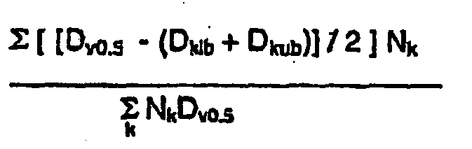
klb = lower bound of bin k
kub = upper bound of bin kRelative span: 
Sphericity: a value from 0 to 1, with 1 representing a perfect circle. Computed as Da/Dp, where Da = square root (4A/π), Dp=P/π; A=pixel area,
P = pixel perimeter.
Claims (14)
- A particle (10), comprising a polymeric matrix (12) and a radiopaque material distributed in the polymeric matrix, wherein the particle (10) has a diameter of from ten microns to 3,000 microns, and wherein the particle (10) has an interior with a density of large pores (16) and a surface region with a density of large pores (16), and the density of large pores (16) of the interior is greater than the density of large pores (16) of the surface region.
- The particle (10) according to claim 1, wherein the radiopaque material is selected from the group consisting of metals, metal alloys, and contrast agents.
- The particle (10) according to one of the preceding claims, wherein the radiopaque material comprises a member selected from the group consisting of titanium dioxide and bismuth subcarbonate.
- The particle (10) according to one of the preceding claims, wherein the radiopaque material comprises platinum or barium sulfate.
- The particle (10) according to one of the preceding claims, wherein the radiopaque material is homogeneously distributed throughout the polymeric matrix.
- The particle (10) according to claim 1 or 5, wherein the polymeric matrix (12) comprises a polysaccharide.
- The particle (10) according to one of the preceding claims, wherein the polymeric matrix (12) comprises a member selected from the group consisting of polyvinyl alcohols, polyacrylic acids, polymethacrylic acids, poly vinyl sulfonates, carboxymethyl celluloses, hydroxyethyl celluloses, substituted celluloses, polyacrylamides, polyethylene glycols, polyamides, polyureas, polyurethanes, polyesters, polyethers, polystyrenes, polysaccharides, polylactic acids, polyethylenes, polymethylmethacrylates, polycaprolactones, polyglycolic acids, poly (lactic-co-glycolic) acids, and combinations thereof.
- The particle (10) according to claim 1, wherein the particle (10) further comprises a therapeutic agent.
- The particle (10) according to one of the preceding claims, wherein the polymeric matrix (12) comprises a first polymer and a second polymer.
- The particle (10) according to one of the preceding claims, wherein the second polymer forms a coating over the first polymer.
- The particle (10) according to claim 1 or 8, wherein the particle (10) is spherical.
- The particle (10) of according to one of the preceding claims, wherein the particle (10) comprises from 0.1 percent to 50 percent by weight of the radiopaque material.
- The particle (10) of according to one of the preceding claims, wherein the particle (10) comprises a ferromagnetic material distributed in the polymer matrix.
- The particle (10) of according to one of the preceding claims, wherein the particle (10) comprises an MRI-visible material distributed in the polymer matrix.
Applications Claiming Priority (3)
| Application Number | Priority Date | Filing Date | Title |
|---|---|---|---|
| US10/232,265 US7462366B2 (en) | 2002-03-29 | 2002-08-30 | Drug delivery particle |
| US232265 | 2002-08-30 | ||
| PCT/US2003/027358 WO2004020011A1 (en) | 2002-08-30 | 2003-08-29 | Embolization |
Publications (2)
| Publication Number | Publication Date |
|---|---|
| EP1531874A1 EP1531874A1 (en) | 2005-05-25 |
| EP1531874B1 true EP1531874B1 (en) | 2009-04-22 |
Family
ID=31976966
Family Applications (1)
| Application Number | Title | Priority Date | Filing Date |
|---|---|---|---|
| EP03751943A Expired - Fee Related EP1531874B1 (en) | 2002-08-30 | 2003-08-29 | Embolic particles |
Country Status (8)
| Country | Link |
|---|---|
| US (4) | US7462366B2 (en) |
| EP (1) | EP1531874B1 (en) |
| JP (1) | JP2005537070A (en) |
| AU (1) | AU2003270050A1 (en) |
| CA (1) | CA2496612A1 (en) |
| DE (1) | DE60327326D1 (en) |
| WO (1) | WO2004020011A1 (en) |
| ZA (1) | ZA200502434B (en) |
Families Citing this family (168)
| Publication number | Priority date | Publication date | Assignee | Title |
|---|---|---|---|---|
| US7713297B2 (en) | 1998-04-11 | 2010-05-11 | Boston Scientific Scimed, Inc. | Drug-releasing stent with ceramic-containing layer |
| US7776310B2 (en) | 2000-11-16 | 2010-08-17 | Microspherix Llc | Flexible and/or elastic brachytherapy seed or strand |
| US20110301569A1 (en) | 2001-01-20 | 2011-12-08 | Gordon Wayne Dyer | Methods and apparatus for the CVCS |
| US7700819B2 (en) | 2001-02-16 | 2010-04-20 | Kci Licensing, Inc. | Biocompatible wound dressing |
| US7763769B2 (en) | 2001-02-16 | 2010-07-27 | Kci Licensing, Inc. | Biocompatible wound dressing |
| WO2003002243A2 (en) | 2001-06-27 | 2003-01-09 | Remon Medical Technologies Ltd. | Method and device for electrochemical formation of therapeutic species in vivo |
| US7462366B2 (en) | 2002-03-29 | 2008-12-09 | Boston Scientific Scimed, Inc. | Drug delivery particle |
| US7094369B2 (en) * | 2002-03-29 | 2006-08-22 | Scimed Life Systems, Inc. | Processes for manufacturing polymeric microspheres |
| US7053134B2 (en) * | 2002-04-04 | 2006-05-30 | Scimed Life Systems, Inc. | Forming a chemically cross-linked particle of a desired shape and diameter |
| US7449236B2 (en) * | 2002-08-09 | 2008-11-11 | Boston Scientific Scimed, Inc. | Porous polymeric particle comprising polyvinyl alcohol and having interior to surface porosity-gradient |
| US7842377B2 (en) * | 2003-08-08 | 2010-11-30 | Boston Scientific Scimed, Inc. | Porous polymeric particle comprising polyvinyl alcohol and having interior to surface porosity-gradient |
| US8012454B2 (en) | 2002-08-30 | 2011-09-06 | Boston Scientific Scimed, Inc. | Embolization |
| US7883490B2 (en) | 2002-10-23 | 2011-02-08 | Boston Scientific Scimed, Inc. | Mixing and delivery of therapeutic compositions |
| US20050075625A1 (en) * | 2003-07-18 | 2005-04-07 | Kinh-Luan Dao | Medical devices |
| US7976823B2 (en) | 2003-08-29 | 2011-07-12 | Boston Scientific Scimed, Inc. | Ferromagnetic particles and methods |
| US20050070990A1 (en) * | 2003-09-26 | 2005-03-31 | Stinson Jonathan S. | Medical devices and methods of making same |
| US7901770B2 (en) | 2003-11-04 | 2011-03-08 | Boston Scientific Scimed, Inc. | Embolic compositions |
| US20070031339A1 (en) | 2003-11-28 | 2007-02-08 | Oskar Axelsson | Contrast agents |
| US20050175709A1 (en) * | 2003-12-11 | 2005-08-11 | Baty Ace M.Iii | Therapeutic microparticles |
| US7854756B2 (en) * | 2004-01-22 | 2010-12-21 | Boston Scientific Scimed, Inc. | Medical devices |
| US8620406B2 (en) * | 2004-01-23 | 2013-12-31 | Boston Scientific Scimed, Inc. | Medical devices visible by magnetic resonance imaging |
| US8992592B2 (en) | 2004-12-29 | 2015-03-31 | Boston Scientific Scimed, Inc. | Medical devices including metallic films |
| US7901447B2 (en) | 2004-12-29 | 2011-03-08 | Boston Scientific Scimed, Inc. | Medical devices including a metallic film and at least one filament |
| US8998973B2 (en) | 2004-03-02 | 2015-04-07 | Boston Scientific Scimed, Inc. | Medical devices including metallic films |
| US8632580B2 (en) | 2004-12-29 | 2014-01-21 | Boston Scientific Scimed, Inc. | Flexible medical devices including metallic films |
| US7736671B2 (en) | 2004-03-02 | 2010-06-15 | Boston Scientific Scimed, Inc. | Embolization |
| US8591568B2 (en) | 2004-03-02 | 2013-11-26 | Boston Scientific Scimed, Inc. | Medical devices including metallic films and methods for making same |
| TWI434676B (en) | 2004-03-19 | 2014-04-21 | Merck Sharp & Dohme | X-ray visible drug delivery device |
| US20050215950A1 (en) * | 2004-03-26 | 2005-09-29 | Scimed Life Systems, Inc. | Balloon catheter with radiopaque portion |
| US8173176B2 (en) | 2004-03-30 | 2012-05-08 | Boston Scientific Scimed, Inc. | Embolization |
| US20050238870A1 (en) * | 2004-04-22 | 2005-10-27 | Marcia Buiser | Embolization |
| US7311861B2 (en) | 2004-06-01 | 2007-12-25 | Boston Scientific Scimed, Inc. | Embolization |
| US7585318B2 (en) * | 2004-06-18 | 2009-09-08 | Boston Scientific Scimed, Inc. | Medical devices and methods of making the same |
| US7344560B2 (en) | 2004-10-08 | 2008-03-18 | Boston Scientific Scimed, Inc. | Medical devices and methods of making the same |
| CA3066594A1 (en) * | 2004-10-08 | 2006-04-20 | Georgia Tech Research Corporation | Microencapsulation of cells in hydrogels using electrostatic potentials |
| JP3857295B2 (en) * | 2004-11-10 | 2006-12-13 | 三菱電機株式会社 | Semiconductor light emitting device |
| US7201918B2 (en) * | 2004-11-16 | 2007-04-10 | Microvention, Inc. | Compositions, systems and methods for treatment of defects in blood vessels |
| US8425550B2 (en) | 2004-12-01 | 2013-04-23 | Boston Scientific Scimed, Inc. | Embolic coils |
| US20060122694A1 (en) * | 2004-12-03 | 2006-06-08 | Stinson Jonathan S | Medical devices and methods of making the same |
| AU2005321543A1 (en) * | 2004-12-30 | 2006-07-06 | Cinvention Ag | Combination comprising an agent providing a signal, an implant material and a drug |
| US7727273B2 (en) * | 2005-01-13 | 2010-06-01 | Boston Scientific Scimed, Inc. | Medical devices and methods of making the same |
| US7918795B2 (en) | 2005-02-02 | 2011-04-05 | Gynesonics, Inc. | Method and device for uterine fibroid treatment |
| US20060177489A1 (en) * | 2005-02-07 | 2006-08-10 | Massouda Debora F | Attachment of chitosan to surfaces using rehydration process |
| US8048028B2 (en) * | 2005-02-17 | 2011-11-01 | Boston Scientific Scimed, Inc. | Reinforced medical balloon |
| US20060182873A1 (en) * | 2005-02-17 | 2006-08-17 | Klisch Leo M | Medical devices |
| US7727555B2 (en) * | 2005-03-02 | 2010-06-01 | Boston Scientific Scimed, Inc. | Particles |
| US7858183B2 (en) | 2005-03-02 | 2010-12-28 | Boston Scientific Scimed, Inc. | Particles |
| US8435280B2 (en) * | 2005-03-31 | 2013-05-07 | Boston Scientific Scimed, Inc. | Flexible stent with variable width elements |
| US7641983B2 (en) * | 2005-04-04 | 2010-01-05 | Boston Scientific Scimed, Inc. | Medical devices including composites |
| EA013680B1 (en) * | 2005-04-15 | 2010-06-30 | Бейцзин Шенияо Сайнс Энд Текнолоджи Девелопмент Ко., Лтд. | Microspherical vascular embolus of sodium alginate containing a gynecological pharmaceutics and preparation thereof |
| US7963287B2 (en) | 2005-04-28 | 2011-06-21 | Boston Scientific Scimed, Inc. | Tissue-treatment methods |
| US8071155B2 (en) * | 2005-05-05 | 2011-12-06 | Boston Scientific Scimed, Inc. | Medical devices and methods of making the same |
| US20060259126A1 (en) * | 2005-05-05 | 2006-11-16 | Jason Lenz | Medical devices and methods of making the same |
| US7854760B2 (en) | 2005-05-16 | 2010-12-21 | Boston Scientific Scimed, Inc. | Medical devices including metallic films |
| US7449505B2 (en) * | 2005-05-20 | 2008-11-11 | Xerox Corporation | Method of making porous microspheres with geometric standard deviation of 1.25 or less |
| US20060276875A1 (en) * | 2005-05-27 | 2006-12-07 | Stinson Jonathan S | Medical devices |
| US20060276910A1 (en) * | 2005-06-01 | 2006-12-07 | Jan Weber | Endoprostheses |
| US9463426B2 (en) * | 2005-06-24 | 2016-10-11 | Boston Scientific Scimed, Inc. | Methods and systems for coating particles |
| US7778684B2 (en) * | 2005-08-08 | 2010-08-17 | Boston Scientific Scimed, Inc. | MRI resonator system with stent implant |
| US20070048383A1 (en) * | 2005-08-25 | 2007-03-01 | Helmus Michael N | Self-assembled endovascular structures |
| US20070073374A1 (en) * | 2005-09-29 | 2007-03-29 | Anderl Steven F | Endoprostheses including nickel-titanium alloys |
| US8007509B2 (en) | 2005-10-12 | 2011-08-30 | Boston Scientific Scimed, Inc. | Coil assemblies, components and methods |
| US8101197B2 (en) | 2005-12-19 | 2012-01-24 | Stryker Corporation | Forming coils |
| US8152839B2 (en) | 2005-12-19 | 2012-04-10 | Boston Scientific Scimed, Inc. | Embolic coils |
| US7947368B2 (en) | 2005-12-21 | 2011-05-24 | Boston Scientific Scimed, Inc. | Block copolymer particles |
| US8840660B2 (en) | 2006-01-05 | 2014-09-23 | Boston Scientific Scimed, Inc. | Bioerodible endoprostheses and methods of making the same |
| US7874986B2 (en) | 2006-04-20 | 2011-01-25 | Gynesonics, Inc. | Methods and devices for visualization and ablation of tissue |
| US11259825B2 (en) | 2006-01-12 | 2022-03-01 | Gynesonics, Inc. | Devices and methods for treatment of tissue |
| US7815571B2 (en) | 2006-04-20 | 2010-10-19 | Gynesonics, Inc. | Rigid delivery systems having inclined ultrasound and needle |
| US10058342B2 (en) | 2006-01-12 | 2018-08-28 | Gynesonics, Inc. | Devices and methods for treatment of tissue |
| US9357977B2 (en) | 2006-01-12 | 2016-06-07 | Gynesonics, Inc. | Interventional deployment and imaging system |
| EP1810698A1 (en) * | 2006-01-24 | 2007-07-25 | Biocompatibles UK Limited | Process for producing radiopaque embolic microspheres |
| EP2368581B1 (en) * | 2006-01-30 | 2018-08-29 | Biosphere Medical, Inc. | Porous intravascular embolization particles and related methods |
| US20080033366A1 (en) | 2006-01-30 | 2008-02-07 | Surgica Corporation | Compressible intravascular embolization particles and related methods and delivery systems |
| US8089029B2 (en) | 2006-02-01 | 2012-01-03 | Boston Scientific Scimed, Inc. | Bioabsorbable metal medical device and method of manufacture |
| US20070239256A1 (en) * | 2006-03-22 | 2007-10-11 | Jan Weber | Medical devices having electrical circuits with multilayer regions |
| US20070224235A1 (en) | 2006-03-24 | 2007-09-27 | Barron Tenney | Medical devices having nanoporous coatings for controlled therapeutic agent delivery |
| US8187620B2 (en) | 2006-03-27 | 2012-05-29 | Boston Scientific Scimed, Inc. | Medical devices comprising a porous metal oxide or metal material and a polymer coating for delivering therapeutic agents |
| US8048150B2 (en) | 2006-04-12 | 2011-11-01 | Boston Scientific Scimed, Inc. | Endoprosthesis having a fiber meshwork disposed thereon |
| US8206300B2 (en) | 2008-08-26 | 2012-06-26 | Gynesonics, Inc. | Ablation device with articulated imaging transducer |
| US10595819B2 (en) | 2006-04-20 | 2020-03-24 | Gynesonics, Inc. | Ablation device with articulated imaging transducer |
| JP2009540946A (en) * | 2006-06-20 | 2009-11-26 | ボストン サイエンティフィック リミテッド | Medical devices containing composite materials |
| US8815275B2 (en) | 2006-06-28 | 2014-08-26 | Boston Scientific Scimed, Inc. | Coatings for medical devices comprising a therapeutic agent and a metallic material |
| CA2655793A1 (en) | 2006-06-29 | 2008-01-03 | Boston Scientific Limited | Medical devices with selective coating |
| US10675298B2 (en) | 2006-07-27 | 2020-06-09 | Boston Scientific Scimed Inc. | Particles |
| WO2008014060A2 (en) * | 2006-07-27 | 2008-01-31 | Boston Scientific Limited | Microparticle |
| EP2054537A2 (en) | 2006-08-02 | 2009-05-06 | Boston Scientific Scimed, Inc. | Endoprosthesis with three-dimensional disintegration control |
| CA2662808A1 (en) * | 2006-09-14 | 2008-03-20 | Boston Scientific Limited | Medical devices with drug-eluting coating |
| EP2081616B1 (en) | 2006-09-15 | 2017-11-01 | Boston Scientific Scimed, Inc. | Bioerodible endoprostheses and methods of making the same |
| JP2010503494A (en) | 2006-09-15 | 2010-02-04 | ボストン サイエンティフィック リミテッド | Biodegradable endoprosthesis and method for producing the same |
| CA2663220A1 (en) | 2006-09-15 | 2008-03-20 | Boston Scientific Limited | Medical devices and methods of making the same |
| WO2008034047A2 (en) | 2006-09-15 | 2008-03-20 | Boston Scientific Limited | Endoprosthesis with adjustable surface features |
| EP2210625B8 (en) | 2006-09-15 | 2012-02-29 | Boston Scientific Scimed, Inc. | Bioerodible endoprosthesis with biostable inorganic layers |
| JP2010503468A (en) * | 2006-09-18 | 2010-02-04 | ボストン サイエンティフィック リミテッド | Medical device with a porous surface |
| US8002821B2 (en) | 2006-09-18 | 2011-08-23 | Boston Scientific Scimed, Inc. | Bioerodible metallic ENDOPROSTHESES |
| US7780798B2 (en) | 2006-10-13 | 2010-08-24 | Boston Scientific Scimed, Inc. | Medical devices including hardened alloys |
| US20100311638A1 (en) * | 2006-10-27 | 2010-12-09 | Paul Tiege | Method for Altering the Shape of Polymer Particles |
| US8414927B2 (en) | 2006-11-03 | 2013-04-09 | Boston Scientific Scimed, Inc. | Cross-linked polymer particles |
| US7981150B2 (en) | 2006-11-09 | 2011-07-19 | Boston Scientific Scimed, Inc. | Endoprosthesis with coatings |
| ES2506144T3 (en) | 2006-12-28 | 2014-10-13 | Boston Scientific Limited | Bioerodible endoprosthesis and their manufacturing procedure |
| US8431149B2 (en) | 2007-03-01 | 2013-04-30 | Boston Scientific Scimed, Inc. | Coated medical devices for abluminal drug delivery |
| US8070797B2 (en) | 2007-03-01 | 2011-12-06 | Boston Scientific Scimed, Inc. | Medical device with a porous surface for delivery of a therapeutic agent |
| JP5578655B2 (en) * | 2007-03-02 | 2014-08-27 | ザ ボード オブ トラスティーズ オブ ザ ユニヴァーシティー オブ イリノイ | Drug delivery by microparticles |
| US8067054B2 (en) | 2007-04-05 | 2011-11-29 | Boston Scientific Scimed, Inc. | Stents with ceramic drug reservoir layer and methods of making and using the same |
| US7976915B2 (en) | 2007-05-23 | 2011-07-12 | Boston Scientific Scimed, Inc. | Endoprosthesis with select ceramic morphology |
| US8211162B2 (en) * | 2007-05-25 | 2012-07-03 | Boston Scientific Scimed, Inc. | Connector node for durable stent |
| US8002823B2 (en) | 2007-07-11 | 2011-08-23 | Boston Scientific Scimed, Inc. | Endoprosthesis coating |
| US7942926B2 (en) | 2007-07-11 | 2011-05-17 | Boston Scientific Scimed, Inc. | Endoprosthesis coating |
| US20090018644A1 (en) * | 2007-07-13 | 2009-01-15 | Jan Weber | Boron-Enhanced Shape Memory Endoprostheses |
| US7604662B2 (en) * | 2007-07-13 | 2009-10-20 | Boston Scientific Scimed, Inc. | Endoprostheses containing boride intermetallic phases |
| EP2187988B1 (en) | 2007-07-19 | 2013-08-21 | Boston Scientific Limited | Endoprosthesis having a non-fouling surface |
| US8815273B2 (en) | 2007-07-27 | 2014-08-26 | Boston Scientific Scimed, Inc. | Drug eluting medical devices having porous layers |
| US7931683B2 (en) | 2007-07-27 | 2011-04-26 | Boston Scientific Scimed, Inc. | Articles having ceramic coated surfaces |
| WO2009018340A2 (en) | 2007-07-31 | 2009-02-05 | Boston Scientific Scimed, Inc. | Medical device coating by laser cladding |
| EP2185103B1 (en) | 2007-08-03 | 2014-02-12 | Boston Scientific Scimed, Inc. | Coating for medical device having increased surface area |
| US20090068279A1 (en) * | 2007-09-12 | 2009-03-12 | Boston Scientific Scimed, Inc. | Microspheres with surface projections |
| US8052745B2 (en) | 2007-09-13 | 2011-11-08 | Boston Scientific Scimed, Inc. | Endoprosthesis |
| US20090088723A1 (en) * | 2007-09-28 | 2009-04-02 | Accessclosure, Inc. | Apparatus and methods for treating pseudoaneurysms |
| WO2009046281A2 (en) * | 2007-10-05 | 2009-04-09 | Boston Scientific Scimed, Inc. | Injectable particles |
| US9107828B2 (en) * | 2007-10-05 | 2015-08-18 | Boston Scientific Scimed, Inc. | Multi-component particles for injection and processes for forming the same |
| US20090092675A1 (en) * | 2007-10-05 | 2009-04-09 | Boston Scientific Scimed, Inc. | Compositions containing multiple polymers and particles made using the compositions |
| US8088072B2 (en) | 2007-10-12 | 2012-01-03 | Gynesonics, Inc. | Methods and systems for controlled deployment of needles in tissue |
| US8216632B2 (en) | 2007-11-02 | 2012-07-10 | Boston Scientific Scimed, Inc. | Endoprosthesis coating |
| US8029554B2 (en) | 2007-11-02 | 2011-10-04 | Boston Scientific Scimed, Inc. | Stent with embedded material |
| US7938855B2 (en) | 2007-11-02 | 2011-05-10 | Boston Scientific Scimed, Inc. | Deformable underlayer for stent |
| US8821931B2 (en) | 2007-11-16 | 2014-09-02 | Bridgestone Corporation | Micro-particles containing a 3-D polymeric structure |
| US20090169641A1 (en) * | 2007-12-26 | 2009-07-02 | Boston Scientifice Scimed, Inc. | Compressible particles |
| WO2009086067A2 (en) * | 2007-12-28 | 2009-07-09 | Boston Scientific Scimed, Inc. | Particles for injection and processes fo forming the same |
| WO2009086076A2 (en) * | 2007-12-28 | 2009-07-09 | Boston Scientific Scimed, Inc. | Particles for injection and processes for forming the same |
| WO2009086098A1 (en) * | 2007-12-28 | 2009-07-09 | Boston Scientific Scimed, Inc. | Porous microparticles for injection and processes for forming the same |
| US20090169591A1 (en) * | 2007-12-28 | 2009-07-02 | Boston Scientific Scimed, Inc. | Medical articles for the treatment of tumors |
| WO2009131911A2 (en) | 2008-04-22 | 2009-10-29 | Boston Scientific Scimed, Inc. | Medical devices having a coating of inorganic material |
| WO2009132176A2 (en) | 2008-04-24 | 2009-10-29 | Boston Scientific Scimed, Inc. | Medical devices having inorganic particle layers |
| US7998192B2 (en) | 2008-05-09 | 2011-08-16 | Boston Scientific Scimed, Inc. | Endoprostheses |
| US8236046B2 (en) | 2008-06-10 | 2012-08-07 | Boston Scientific Scimed, Inc. | Bioerodible endoprosthesis |
| EP2303350A2 (en) | 2008-06-18 | 2011-04-06 | Boston Scientific Scimed, Inc. | Endoprosthesis coating |
| US7985252B2 (en) | 2008-07-30 | 2011-07-26 | Boston Scientific Scimed, Inc. | Bioerodible endoprosthesis |
| US8382824B2 (en) | 2008-10-03 | 2013-02-26 | Boston Scientific Scimed, Inc. | Medical implant having NANO-crystal grains with barrier layers of metal nitrides or fluorides |
| JP2012507562A (en) * | 2008-10-30 | 2012-03-29 | ダビド リウ | Microspherical porous biocompatible scaffold and manufacturing method and apparatus thereof |
| US8231980B2 (en) | 2008-12-03 | 2012-07-31 | Boston Scientific Scimed, Inc. | Medical implants including iridium oxide |
| US20100217370A1 (en) * | 2009-02-20 | 2010-08-26 | Boston Scientific Scimed, Inc. | Bioerodible Endoprosthesis |
| US8262574B2 (en) | 2009-02-27 | 2012-09-11 | Gynesonics, Inc. | Needle and tine deployment mechanism |
| EP2403546A2 (en) | 2009-03-02 | 2012-01-11 | Boston Scientific Scimed, Inc. | Self-buffering medical implants |
| US8071156B2 (en) | 2009-03-04 | 2011-12-06 | Boston Scientific Scimed, Inc. | Endoprostheses |
| US8287937B2 (en) | 2009-04-24 | 2012-10-16 | Boston Scientific Scimed, Inc. | Endoprosthese |
| US20100292776A1 (en) * | 2009-05-14 | 2010-11-18 | Boston Scientific Scimed, Inc. | Bioerodible Endoprosthesis |
| CN102741336B (en) | 2009-12-01 | 2014-03-19 | 株式会社普利司通 | Modified rubber compositions and methods of preparation |
| US20110160839A1 (en) * | 2009-12-29 | 2011-06-30 | Boston Scientific Scimed, Inc. | Endoprosthesis |
| US8668732B2 (en) | 2010-03-23 | 2014-03-11 | Boston Scientific Scimed, Inc. | Surface treated bioerodible metal endoprostheses |
| US8709072B2 (en) | 2010-05-13 | 2014-04-29 | Boston Scientific Scimed, Inc. | Endoprosthesis |
| JP5834071B2 (en) | 2010-05-14 | 2015-12-16 | ボストン サイエンティフィック サイムド,インコーポレイテッドBoston Scientific Scimed,Inc. | Stent |
| WO2012006101A2 (en) | 2010-06-28 | 2012-01-12 | The General Hospital Corporation | Blood substitutes and uses thereof |
| US8765725B2 (en) | 2012-05-08 | 2014-07-01 | Aciex Therapeutics, Inc. | Preparations of hydrophobic therapeutic agents, methods of manufacture and use thereof |
| CA2872845C (en) | 2012-05-08 | 2021-11-09 | Aciex Therapeutics, Inc. | Preparation of nanocrystals of fluticasone propionate |
| ES2655656T3 (en) * | 2012-05-24 | 2018-02-21 | Biosphere Medical Inc. | Biomaterials suitable for use as implants detectable by magnetic resonance imaging, eluents of drugs for vascular occlusion |
| EP2882459A1 (en) | 2012-08-10 | 2015-06-17 | University of North Texas Health Science Center | Drug delivery vehicle comprising conjugates between targeting polyamino acids and fatty acids |
| US9815865B2 (en) | 2013-01-07 | 2017-11-14 | Nicox Ophthalmics, Inc. | Preparations of hydrophobic therapeutic agents, methods of manufacture and use thereof |
| KR102505172B1 (en) | 2013-09-19 | 2023-02-28 | 마이크로벤션, 인코포레이티드 | Polymer films |
| JP6405369B2 (en) | 2013-09-19 | 2018-10-17 | テルモ株式会社 | Polymer particles |
| EP3065719A4 (en) | 2013-11-08 | 2017-08-09 | Terumo Corporation | Polymer particles |
| WO2015161211A1 (en) * | 2014-04-17 | 2015-10-22 | Boston Scientific Scimed, Inc. | Devices and methods for therapeutic heat treatment |
| CN104383357A (en) * | 2014-10-30 | 2015-03-04 | 四川金堂海纳生物医药技术研究所 | Medicinal decoction for treating vocal cord paralysis and preparation method thereof |
| WO2016154592A1 (en) | 2015-03-26 | 2016-09-29 | Microvention, Inc. | Embiolic particles |
| CN107708736A (en) | 2015-06-15 | 2018-02-16 | 波士顿科学国际有限公司 | Apparatus and method for therapeutic heat processing |
| JP6866298B2 (en) | 2015-11-24 | 2021-04-28 | Jsr株式会社 | Method for producing porous particles, method for separating porous particles, carriers, columns, and target substances |
| EP3518994B1 (en) | 2016-09-28 | 2024-02-07 | Terumo Corporation | Polymer particles |
| AU2017359338B2 (en) | 2016-11-11 | 2022-09-08 | Gynesonics, Inc. | Controlled treatment of tissue and dynamic interaction with, and comparison of, tissue and/or treatment data |
| WO2019169108A1 (en) * | 2018-02-28 | 2019-09-06 | Celista Pharmaceuticals Llc | Oxycodone and methylnaltrexone multi-particulates and suspensions containing them |
Family Cites Families (341)
| Publication number | Priority date | Publication date | Assignee | Title |
|---|---|---|---|---|
| US2275154A (en) * | 1940-07-10 | 1942-03-03 | United Drug Company | Method for making capsules |
| US2609347A (en) | 1948-05-27 | 1952-09-02 | Wilson Christopher Lumley | Method of making expanded polyvinyl alcohol-formaldehyde reaction product and product resulting therefrom |
| JPS4820019B1 (en) * | 1969-06-05 | 1973-06-18 | ||
| US3737398A (en) | 1969-11-13 | 1973-06-05 | D Yamaguchi | Method of making a polyvinyl acetal sponge buff |
| CS179075B1 (en) | 1974-11-26 | 1977-10-31 | Stoy Vladimir | Mode of manufacture of spherical particles from polymer |
| US4076640A (en) * | 1975-02-24 | 1978-02-28 | Xerox Corporation | Preparation of spheroidized particles |
| US3957933A (en) * | 1975-03-05 | 1976-05-18 | General Atomic Company | Apparatus for producing microspherical particles and method for operating such apparatus |
| JPS51135958A (en) | 1975-05-20 | 1976-11-25 | Fuji Photo Film Co Ltd | Method of making fine powder polymer having pores |
| US4025686A (en) * | 1975-06-26 | 1977-05-24 | Owens-Corning Fiberglas Corporation | Molded composite article and method for making the article |
| US4034759A (en) | 1975-08-27 | 1977-07-12 | Xomed, Inc. | Moisture-expandable prosthesis |
| US4098728A (en) | 1976-01-02 | 1978-07-04 | Solomon Rosenblatt | Medical surgical sponge and method of making same |
| US4055377A (en) | 1976-08-03 | 1977-10-25 | Minnesota Mining And Manufacturing Company | Magnetically orientable retroreflectorization particles |
| GB1591924A (en) * | 1976-10-25 | 1981-07-01 | Berger Jenson & Nicholson Ltd | Polymer aggregates |
| US4159719A (en) | 1977-05-09 | 1979-07-03 | Xomed, Inc. | Moisture-expandable ear wick |
| ES478736A1 (en) | 1978-03-23 | 1979-06-01 | Hoechst Ag | Polyvinyl alcohol pellets containing a plasticizer, and method for their preparation. |
| DE2834539A1 (en) * | 1978-08-07 | 1980-02-21 | Basf Ag | MACROPOROUS POLYMERS AS CARRIER MATERIAL FOR THE COVALENT BINDING OF PROTEINS |
| US4793980A (en) | 1978-09-21 | 1988-12-27 | Torobin Leonard B | Hollow porous microspheres as substrates and containers for catalyst |
| US4243794A (en) * | 1978-10-10 | 1981-01-06 | Minnesota Mining And Manufacturing Company | Mixture of rough and spheroidized resin particles |
| US4198318A (en) * | 1978-11-24 | 1980-04-15 | Conoco, Inc. | Production of high strength alumina spheres by hydrogelling corresponding slurries |
| US4268495A (en) * | 1979-01-08 | 1981-05-19 | Ethicon, Inc. | Injectable embolization and occlusion solution |
| US4246208A (en) * | 1979-03-22 | 1981-01-20 | Xerox Corporation | Dust-free plasma spheroidization |
| US4346712A (en) | 1979-04-06 | 1982-08-31 | Kuraray Company, Ltd. | Releasable balloon catheter |
| HU184722B (en) | 1980-02-18 | 1984-10-29 | Laszlo Lazar | Therapeutically suitable silicone rubber mixture and therapeuticaid |
| US4271281A (en) | 1980-05-29 | 1981-06-02 | American Hoechst Corporation | Process for preparing styrenic polymer particles |
| CA1113532A (en) * | 1980-05-30 | 1981-12-01 | Bernard Desourdy | Endless track assembly |
| DE3031737A1 (en) * | 1980-08-22 | 1982-04-01 | Bayer Ag, 5090 Leverkusen | METHOD FOR PRODUCING PEARL POLYMERISATS OF UNIFORM PARTICLE SIZE |
| CA1166413A (en) * | 1980-10-30 | 1984-05-01 | Edward E. Timm | Process and apparatus for preparing uniform size polymer beads |
| US4681119A (en) | 1980-11-17 | 1987-07-21 | Schering Aktiengesellschaft | Method of production and use of microbubble precursors |
| US4442843A (en) * | 1980-11-17 | 1984-04-17 | Schering, Ag | Microbubble precursors and methods for their production and use |
| US4657756A (en) * | 1980-11-17 | 1987-04-14 | Schering Aktiengesellschaft | Microbubble precursors and apparatus for their production and use |
| NZ199916A (en) | 1981-03-11 | 1985-07-12 | Unilever Plc | Low density polymeric block material for use as carrier for included liquids |
| US4413070A (en) | 1981-03-30 | 1983-11-01 | California Institute Of Technology | Polyacrolein microspheres |
| US4622362A (en) | 1981-03-30 | 1986-11-11 | California Institute Of Technology | Polyacrolein microspheres |
| US4678814A (en) | 1981-03-30 | 1987-07-07 | California Institute Of Technology | Polyacrolein microspheres |
| CA1177811A (en) | 1981-04-13 | 1984-11-13 | Theo G. Spek | Process for the preparation of silica particles; silica particles with a narrow pore diameter distribution, catalysts made therefrom and use of these catalysts |
| US4428869A (en) * | 1981-08-20 | 1984-01-31 | International Flavors & Fragrances Inc. | Cologne consisting of microcapsule suspension |
| US4456693A (en) | 1982-03-08 | 1984-06-26 | W. R. Grace & Co. | Hydrocracking catalyst |
| US4452773A (en) | 1982-04-05 | 1984-06-05 | Canadian Patents And Development Limited | Magnetic iron-dextran microspheres |
| US4472552A (en) | 1982-09-27 | 1984-09-18 | W. R. Grace & Co. | Continuous process for making solid, free-flowing water dispersible PVA-aldehyde reaction product |
| US4459145A (en) | 1982-09-30 | 1984-07-10 | The United States Of America As Represented By The United States Department Of Energy | Fabrication of glass microspheres with conducting surfaces |
| JPS59131355A (en) | 1983-01-17 | 1984-07-28 | 森下仁丹株式会社 | Multiple soft capsule |
| US4515906A (en) * | 1983-02-28 | 1985-05-07 | Bend Research, Inc. | Anisotropic microporous supports impregnated with polymeric ion-exchange materials |
| DE3313947A1 (en) | 1983-04-15 | 1984-10-18 | Schering AG, 1000 Berlin und 4709 Bergkamen | MICROPARTICLES AND GAS BUBBLES CONTAINING ULTRASONIC CONTRASTING AGENTS |
| DE3834705A1 (en) | 1988-10-07 | 1990-04-12 | Schering Ag | ULTRASONIC CONTRASTING AGENTS FROM GAS BUBBLES AND MICROPARTICLES CONTAINING FATTY ACID |
| DE3313946A1 (en) | 1983-04-15 | 1984-10-18 | Schering AG, 1000 Berlin und 4709 Bergkamen | MICROPARTICLES AND GAS BUBBLES CONTAINING ULTRASONIC CONTRASTING AGENTS |
| CA1225585A (en) * | 1983-06-30 | 1987-08-18 | Maria T. Litvinova | Composition for embolization of blood vessels |
| US4492720A (en) * | 1983-11-15 | 1985-01-08 | Benjamin Mosier | Method of preparing microspheres for intravascular delivery |
| US4573967A (en) * | 1983-12-06 | 1986-03-04 | Eli Lilly And Company | Vacuum vial infusion system |
| US4671954A (en) | 1983-12-13 | 1987-06-09 | University Of Florida | Microspheres for incorporation of therapeutic substances and methods of preparation thereof |
| US4551436A (en) | 1984-04-11 | 1985-11-05 | General Electric Company | Fabrication of small dense silicon carbide spheres |
| DE3414924A1 (en) | 1984-04-19 | 1985-10-31 | Klaus Dr.med. Dr.med.habil. 8000 München Draenert | COATED ANCHORAGE PART FOR IMPLANTS |
| US4674480A (en) | 1984-05-25 | 1987-06-23 | Lemelson Jerome H | Drug compositions and methods of applying same |
| FR2566384B1 (en) * | 1984-06-21 | 1986-09-05 | Saint Gobain Vitrage | IMPROVEMENTS IN TECHNIQUES FOR THE PRODUCTION OF GLASS MICROSPHERES |
| DE3527482A1 (en) | 1984-07-31 | 1986-02-06 | Fuji Spinning Co., Ltd., Tokio/Tokyo | METHOD FOR PRODUCING GRAINY POROUS CHITOSAN |
| GB8419708D0 (en) * | 1984-08-02 | 1984-09-05 | Shell Int Research | Preparation of silica spheres |
| US4623706A (en) | 1984-08-23 | 1986-11-18 | The Dow Chemical Company | Process for preparing uniformly sized polymer particles by suspension polymerization of vibratorily excited monomers in a gaseous or liquid stream |
| US4629464A (en) | 1984-09-25 | 1986-12-16 | Tdk Corporation | Porous hydroxyapatite material for artificial bone substitute |
| JPS61101242A (en) | 1984-10-22 | 1986-05-20 | Showa Denko Kk | Production of coated substance |
| US4789501A (en) * | 1984-11-19 | 1988-12-06 | The Curators Of The University Of Missouri | Glass microspheres |
| US4675113A (en) | 1984-11-28 | 1987-06-23 | University Patents, Inc. | Affinity chromatography using dried calcium alginate-magnetite separation media in a magnetically stabilized fluidized bed |
| DE3568442D1 (en) | 1984-12-06 | 1989-04-06 | Kanegafuchi Chemical Ind | A method of preparation of droplets |
| US5106903A (en) * | 1984-12-17 | 1992-04-21 | Lehigh University | Preparation of large particle size monodisperse latexes |
| US4897255A (en) * | 1985-01-14 | 1990-01-30 | Neorx Corporation | Metal radionuclide labeled proteins for diagnosis and therapy |
| JPH0678460B2 (en) * | 1985-05-01 | 1994-10-05 | 株式会社バイオマテリアル・ユニバース | Porous transparent polyvinyl alcohol gel |
| JPS61293911A (en) * | 1985-06-24 | 1986-12-24 | Teisan Seiyaku Kk | Sustained release preparation |
| SE459005B (en) * | 1985-07-12 | 1989-05-29 | Aake Rikard Lindahl | SET TO MANUFACTURE SPHERICAL POLYMER PARTICLES |
| USH915H (en) | 1985-07-22 | 1991-05-07 | Gibbs Marylu B | Controlled macroporous copolymer properties by removal of impurities in the diluent |
| US4742086A (en) | 1985-11-02 | 1988-05-03 | Lion Corporation | Process for manufacturing porous polymer |
| DE3543348A1 (en) | 1985-12-07 | 1987-06-11 | Bayer Ag | PEARL-SHAPED CROSS-NETWORKED MIXED POLYMERS WITH EPOXY AND BASIC AMINO GROUPS, METHOD FOR THE PRODUCTION THEREOF AND THEIR USE |
| JPS62169723A (en) * | 1986-01-22 | 1987-07-25 | Teisan Seiyaku Kk | Sustained release preparation |
| GB8610024D0 (en) | 1986-04-24 | 1986-05-29 | Unilever Plc | Porous structures |
| US4929400A (en) | 1986-04-28 | 1990-05-29 | California Institute Of Technology | Production of monodisperse, polymeric microspheres |
| US5262176A (en) * | 1986-07-03 | 1993-11-16 | Advanced Magnetics, Inc. | Synthesis of polysaccharide covered superparamagnetic oxide colloids |
| JPS6317904A (en) | 1986-07-09 | 1988-01-25 | Mitsubishi Chem Ind Ltd | Production of crosslinked porous polyvinyl alcohol particle |
| US4743507A (en) | 1986-09-12 | 1988-05-10 | Franses Elias I | Nonspherical microparticles and method therefor |
| EP0265061B1 (en) | 1986-09-18 | 1992-01-08 | London School of Pharmacy Innovations Ltd | Pharmaceutical formulation |
| US5114421A (en) | 1986-09-22 | 1992-05-19 | Polak Robert B | Medicament container/dispenser assembly |
| CA1287459C (en) | 1986-10-01 | 1991-08-13 | Mukesh Jain | Process for the preparation of hollow microspheres |
| US4859711A (en) | 1986-10-01 | 1989-08-22 | Alcan International Limited | Hollow microspheres |
| US5263992A (en) | 1986-10-17 | 1993-11-23 | Bio-Metric Systems, Inc. | Biocompatible device with covalently bonded biocompatible agent |
| DE3787700T3 (en) | 1986-10-29 | 1998-12-24 | Kanegafuchi Chemical Ind | Uniform polymer particles. |
| US5292814A (en) * | 1987-04-29 | 1994-03-08 | Ernst Bayer | Process for the preparation of monodispersed polymer beads |
| US4795741A (en) * | 1987-05-06 | 1989-01-03 | Biomatrix, Inc. | Compositions for therapeutic percutaneous embolization and the use thereof |
| GB8713263D0 (en) | 1987-06-05 | 1987-07-08 | Unilever Plc | Spheroidal silica |
| JPH0612993B2 (en) | 1987-08-10 | 1994-02-23 | 株式会社クラレ | Method for producing spherical microbe-immobilized moldings |
| US4819637A (en) * | 1987-09-01 | 1989-04-11 | Interventional Therapeutics Corporation | System for artificial vessel embolization and devices for use therewith |
| JPH0762054B2 (en) | 1987-10-13 | 1995-07-05 | 倉敷紡績株式会社 | Crosslinked polymer particles |
| US4850978A (en) | 1987-10-29 | 1989-07-25 | Baxter International Inc. | Drug delivery cartridge with protective cover |
| US4804366A (en) * | 1987-10-29 | 1989-02-14 | Baxter International Inc. | Cartridge and adapter for introducing a beneficial agent into an intravenous delivery system |
| US4981625A (en) * | 1988-03-14 | 1991-01-01 | California Institute Of Technology | Monodisperse, polymeric microspheres produced by irradiation of slowly thawing frozen drops |
| FR2634376B1 (en) * | 1988-07-21 | 1992-04-17 | Farmalyoc | NOVEL SOLID AND POROUS UNIT FORM COMPRISING MICROPARTICLES AND / OR NANOPARTICLES, AS WELL AS ITS PREPARATION |
| WO1990001969A1 (en) | 1988-08-24 | 1990-03-08 | Slepian Marvin J | Biodegradable polymeric endoluminal sealing |
| DE3829938A1 (en) | 1988-09-02 | 1990-03-29 | Hermann Hofmann | ORGANO-MINERAL DUENGER AND METHOD FOR THE PRODUCTION THEREOF |
| US4933372A (en) | 1988-09-26 | 1990-06-12 | Supelco, Inc. | Porous rigid resins and process of preparation |
| US5047438A (en) | 1988-09-26 | 1991-09-10 | Supelco, Inc. | Porous rigid resins and process of preparation |
| US5681576A (en) | 1988-11-16 | 1997-10-28 | Mdv Technologies, Inc. | Method and composition for post surgical adhesion reduction |
| DE3841401A1 (en) | 1988-12-08 | 1990-06-13 | Martin Lemperle | ALLOPLASTIC IMPLANT |
| US5258028A (en) | 1988-12-12 | 1993-11-02 | Ersek Robert A | Textured micro implants |
| US4946899A (en) | 1988-12-16 | 1990-08-07 | The University Of Akron | Thermoplastic elastomers of isobutylene and process of preparation |
| GB8900376D0 (en) | 1989-01-09 | 1989-03-08 | Nycomed As | Iodinated esters |
| US5091205A (en) * | 1989-01-17 | 1992-02-25 | Union Carbide Chemicals & Plastics Technology Corporation | Hydrophilic lubricious coatings |
| FR2641692A1 (en) * | 1989-01-17 | 1990-07-20 | Nippon Zeon Co | Plug for closing an opening for a medical application, and device for the closure plug making use thereof |
| US5032117A (en) | 1989-01-30 | 1991-07-16 | Motta Louis J | Tandem syringe |
| GB8905934D0 (en) * | 1989-03-15 | 1989-04-26 | Dow Europ Sa | A process for preparing adsorptive porous resin beads |
| US5888930A (en) * | 1989-03-27 | 1999-03-30 | Bend Research, Inc. | Asymmetric microporous beads for controlled release |
| US5354290A (en) | 1989-05-31 | 1994-10-11 | Kimberly-Clark Corporation | Porous structure of an absorbent polymer |
| CA2017570C (en) * | 1989-05-31 | 2000-12-19 | James R. Gross | Porous structure of an absorbent polymer |
| US5007940A (en) * | 1989-06-09 | 1991-04-16 | American Medical Systems, Inc. | Injectable polymeric bodies |
| US5158573A (en) | 1989-06-09 | 1992-10-27 | American Medical Systems, Inc. | Injectable polymeric bodies |
| US5116387A (en) | 1989-06-09 | 1992-05-26 | American Medical Systems, Inc. | Preparation of injectable polymeric bodies |
| US5190760A (en) * | 1989-07-08 | 1993-03-02 | Coopers Animal Health Limited | Solid pharmaceutical composition |
| US5698271A (en) * | 1989-08-22 | 1997-12-16 | Immunivest Corporation | Methods for the manufacture of magnetically responsive particles |
| US5253991A (en) | 1989-11-20 | 1993-10-19 | Sumitomo Cement Co., Ltd. | Apparatus for producing spheroidal inorganic particulate material |
| US5542935A (en) | 1989-12-22 | 1996-08-06 | Imarx Pharmaceutical Corp. | Therapeutic delivery systems related applications |
| US5580575A (en) | 1989-12-22 | 1996-12-03 | Imarx Pharmaceutical Corp. | Therapeutic drug delivery systems |
| US5922304A (en) | 1989-12-22 | 1999-07-13 | Imarx Pharmaceutical Corp. | Gaseous precursor filled microspheres as magnetic resonance imaging contrast agents |
| US5585112A (en) | 1989-12-22 | 1996-12-17 | Imarx Pharmaceutical Corp. | Method of preparing gas and gaseous precursor-filled microspheres |
| US5469854A (en) | 1989-12-22 | 1995-11-28 | Imarx Pharmaceutical Corp. | Methods of preparing gas-filled liposomes |
| US6306427B1 (en) | 1989-12-28 | 2001-10-23 | Rhone-Poulenc Nutrition Animale | Pellets containing active ingredients protected against degradation in the rumen of ruminants |
| US5435645A (en) | 1989-12-29 | 1995-07-25 | Tecres Spa | Process and apparatus for the mixing and direct emplacement of a two-component bone cement |
| US5147937A (en) | 1990-03-22 | 1992-09-15 | Rohm And Haas Company | Process for making controlled, uniform-sized particles in the 1 to 50 micrometer range |
| US5556610A (en) | 1992-01-24 | 1996-09-17 | Bracco Research S.A. | Gas mixtures useful as ultrasound contrast media, contrast agents containing the media and method |
| JPH03297475A (en) * | 1990-04-16 | 1991-12-27 | Ken Ishihara | Controlling method for emission of medicine by means of resonance sound wave |
| US5514090A (en) | 1990-04-24 | 1996-05-07 | Science Incorporated | Closed drug delivery system |
| US5137928A (en) | 1990-04-26 | 1992-08-11 | Hoechst Aktiengesellschaft | Ultrasonic contrast agents, processes for their preparation and the use thereof as diagnostic and therapeutic agents |
| CA2016870C (en) | 1990-05-15 | 1994-03-29 | Arnie Drudik | Dispenser for storing and mixing several components |
| AU636481B2 (en) | 1990-05-18 | 1993-04-29 | Bracco International B.V. | Polymeric gas or air filled microballoons usable as suspensions in liquid carriers for ultrasonic echography |
| JP2514087Y2 (en) * | 1990-05-25 | 1996-10-16 | 幸三 牧田 | Balloon with detachable double-sided check valve |
| US6291605B1 (en) | 1990-06-06 | 2001-09-18 | Clarence S. Freeman | Polymerization process with spraying step |
| WO1991019483A1 (en) | 1990-06-20 | 1991-12-26 | Advanced Polymer Systems, Inc. | Compositions and methods for the controlled release of soluble active substances |
| JP2931879B2 (en) * | 1990-06-27 | 1999-08-09 | レンゴー株式会社 | Cellulose-based porous spherical particles comprising chitin or deacetylated chitin and cellulose and a method for producing the same |
| ATE130517T1 (en) | 1990-08-08 | 1995-12-15 | Takeda Chemical Industries Ltd | INTRAVASCULAR EMBOLIZING AGENT CONTAINING A SUBSTANCE INHIBITING ANGIOGENESIS. |
| US5484584A (en) * | 1990-10-02 | 1996-01-16 | Board Of Regents, The University Of Texas System | Therapeutic and diagnostic use of modified polymeric microcapsules |
| US5149543A (en) | 1990-10-05 | 1992-09-22 | Massachusetts Institute Of Technology | Ionically cross-linked polymeric microcapsules |
| US5120349A (en) | 1990-12-07 | 1992-06-09 | Landec Labs, Inc. | Microcapsule having temperature-dependent permeability profile |
| US5171214A (en) | 1990-12-26 | 1992-12-15 | Abbott Laboratories | Drug storage and delivery system |
| US5171217A (en) | 1991-02-28 | 1992-12-15 | Indiana University Foundation | Method for delivery of smooth muscle cell inhibitors |
| US5147631A (en) | 1991-04-30 | 1992-09-15 | Du Pont Merck Pharmaceutical Company | Porous inorganic ultrasound contrast agents |
| FR2676927B1 (en) | 1991-05-29 | 1995-06-23 | Ibf | MICROSPHERES FOR USE IN THERAPEUTIC VASCULAR OCCLUSIONS AND INJECTABLE SOLUTIONS CONTAINING THEM. |
| DK0586524T3 (en) | 1991-06-03 | 1997-05-20 | Nycomed Imaging As | |
| GB9116610D0 (en) | 1991-08-01 | 1991-09-18 | Danbiosyst Uk | Preparation of microparticles |
| US5216096A (en) | 1991-09-24 | 1993-06-01 | Japan Synthetic Rubber Co., Ltd. | Process for the preparation of cross-linked polymer particles |
| US5811447A (en) * | 1993-01-28 | 1998-09-22 | Neorx Corporation | Therapeutic inhibitor of vascular smooth muscle cells |
| ATE154241T1 (en) * | 1991-10-01 | 1997-06-15 | Takeda Chemical Industries Ltd | MICROPARTICLE SUMMARY FOR EXTENDED RELEASE AND PRODUCTION THE SAME |
| JPH0718357A (en) * | 1991-10-02 | 1995-01-20 | Yasubumi Furuya | Combined functional material device |
| JP3356447B2 (en) * | 1991-10-16 | 2002-12-16 | テルモ株式会社 | Vascular lesion embolic material composed of dried polymer gel |
| US5258042A (en) | 1991-12-16 | 1993-11-02 | Henry Ford Health System | Intravascular hydrogel implant |
| DE69226203T2 (en) * | 1991-12-20 | 1998-12-10 | Allied Signal Inc | MATERIALS WITH LOW DENSITY AND HIGH SPECIFIC SURFACE AND ARTICLES MOLDED THEREOF FOR USE IN METAL RECOVERY |
| US5260002A (en) | 1991-12-23 | 1993-11-09 | Vanderbilt University | Method and apparatus for producing uniform polymeric spheres |
| CA2125647C (en) | 1991-12-24 | 1997-12-30 | Joseph A. Antonelli | Dual stabilized microparticles |
| GB9200388D0 (en) | 1992-01-09 | 1992-02-26 | Nycomed As | Improvements in or relating to contrast agents |
| GB9200391D0 (en) | 1992-01-09 | 1992-02-26 | Nycomed As | Improvements in or relating to contrast agents |
| US6537574B1 (en) | 1992-02-11 | 2003-03-25 | Bioform, Inc. | Soft tissue augmentation material |
| US5480644A (en) * | 1992-02-28 | 1996-01-02 | Jsf Consultants Ltd. | Use of injectable biomaterials for the repair and augmentation of the anal sphincters |
| ES2135466T3 (en) | 1992-03-06 | 1999-11-01 | Nycomed Imaging As | IMPROVEMENTS INTRODUCED IN CONTRAST AGENTS. |
| JP3004724B2 (en) | 1992-04-06 | 2000-01-31 | ユーロプラスティ インコーポレイテッド | Treatment of reflux obstruction by microparticle injection |
| DE69306844T2 (en) | 1992-04-10 | 1997-07-10 | Mitsubishi Chem Corp | Process for the preparation of spherical cross-linked acrylonitrile copolymers |
| US6235313B1 (en) | 1992-04-24 | 2001-05-22 | Brown University Research Foundation | Bioadhesive microspheres and their use as drug delivery and imaging systems |
| AU4198793A (en) * | 1992-07-24 | 1994-01-27 | Takeda Chemical Industries Ltd. | Microparticle preparation and production thereof |
| US5514379A (en) * | 1992-08-07 | 1996-05-07 | The General Hospital Corporation | Hydrogel compositions and methods of use |
| US5807323A (en) | 1992-08-13 | 1998-09-15 | Science Incorporated | Mixing and delivery syringe assembly |
| US6592859B1 (en) | 1992-08-20 | 2003-07-15 | Ethicon, Inc. | Controlled expansion sphincter augmentation media |
| US5512604A (en) * | 1992-08-28 | 1996-04-30 | The Dow Chemical Company | Porous copolymers having a cellular polymeric structure suitable for preparing ion-exchange resins and adsorbents |
| WO1994006477A1 (en) * | 1992-09-16 | 1994-03-31 | Holmes, Michael, John | Improvements in or relating to contrast agents |
| WO1994006460A1 (en) | 1992-09-21 | 1994-03-31 | Vitaphore Corporation | Embolization plugs for blood vessels |
| DE4232755A1 (en) | 1992-09-26 | 1994-03-31 | Schering Ag | Microparticle preparations made from biodegradable copolymers |
| KR960001417B1 (en) | 1992-09-26 | 1996-01-27 | 한국과학기술원 | Method for preparing an improved porous polymer bead |
| GB9221329D0 (en) | 1992-10-10 | 1992-11-25 | Delta Biotechnology Ltd | Preparation of further diagnostic agents |
| US5382260A (en) | 1992-10-30 | 1995-01-17 | Interventional Therapeutics Corp. | Embolization device and apparatus including an introducer cartridge and method for delivering the same |
| US5369163A (en) | 1992-11-13 | 1994-11-29 | Rohm And Haas Company | Process for preparing large dimension emulsion polymer particles, polymer product and uses thereof |
| US5690666A (en) | 1992-11-18 | 1997-11-25 | Target Therapeutics, Inc. | Ultrasoft embolism coils and process for using them |
| US5349957A (en) * | 1992-12-02 | 1994-09-27 | Sterling Winthrop Inc. | Preparation and magnetic properties of very small magnetite-dextran particles |
| JP3256583B2 (en) | 1992-12-10 | 2002-02-12 | 株式会社リコー | Electrophotographic toner and method for producing the same |
| US5288763A (en) * | 1992-12-23 | 1994-02-22 | The Johns Hopkins University School Of Medicine | Porous, polymer beads and process of their preparation |
| US6482436B1 (en) | 1993-01-29 | 2002-11-19 | Ferx Incorporated | Magnetically responsive composition |
| US5328936A (en) | 1993-02-01 | 1994-07-12 | Rohm And Haas Company | Polymerization process for making porous polymeric particles |
| US6090925A (en) | 1993-03-09 | 2000-07-18 | Epic Therapeutics, Inc. | Macromolecular microparticles and methods of production and use |
| US5320639A (en) | 1993-03-12 | 1994-06-14 | Meadox Medicals, Inc. | Vascular plug delivery system |
| US5567415A (en) | 1993-05-12 | 1996-10-22 | The Board Of Regents Of The University Of Nebraska | Ultrasound contrast agents and methods for their manufacture and use |
| US5701899A (en) | 1993-05-12 | 1997-12-30 | The Board Of Regents Of The University Of Nebraska | Perfluorobutane ultrasound contrast agent and methods for its manufacture and use |
| US5695740A (en) | 1993-05-12 | 1997-12-09 | The Board Of Regents Of The University Of Nebraska | Perfluorocarbon ultrasound contrast agent comprising microbubbles containing a filmogenic protein and a saccharide |
| US5344867A (en) | 1993-06-14 | 1994-09-06 | The Bfgoodrich Company | Vinylidene chloride emulsion interpolymer composition |
| US5716981A (en) * | 1993-07-19 | 1998-02-10 | Angiogenesis Technologies, Inc. | Anti-angiogenic compositions and methods of use |
| ATE154757T1 (en) | 1993-07-19 | 1997-07-15 | Angiotech Pharm Inc | ANTI-ANGIogenic AGENTS AND METHODS OF USE THEREOF |
| US5397303A (en) * | 1993-08-06 | 1995-03-14 | River Medical, Inc. | Liquid delivery device having a vial attachment or adapter incorporated therein |
| US5398851A (en) | 1993-08-06 | 1995-03-21 | River Medical, Inc. | Liquid delivery device |
| US5443495A (en) | 1993-09-17 | 1995-08-22 | Scimed Lifesystems Inc. | Polymerization angioplasty balloon implant device |
| US5531716A (en) | 1993-09-29 | 1996-07-02 | Hercules Incorporated | Medical devices subject to triggered disintegration |
| DE1258262T1 (en) * | 1993-10-28 | 2003-04-10 | Medrad Inc | Contrast delivery system |
| PT682530E (en) | 1993-12-15 | 2003-06-30 | Bracco Research Sa | UTEIS GAS MIXTURES AS CONTRAST MEANS FOR ULTRASSONS |
| WO1995019841A1 (en) | 1994-01-21 | 1995-07-27 | Bruce Nathaniel Gray | Particulate material |
| WO1995022318A1 (en) | 1994-02-17 | 1995-08-24 | Pankaj Modi | Drugs, vaccines and hormones in polylactide coated microspheres |
| US5569468A (en) | 1994-02-17 | 1996-10-29 | Modi; Pankaj | Vaccine delivery system for immunization, using biodegradable polymer microspheres |
| US5417982A (en) | 1994-02-17 | 1995-05-23 | Modi; Pankaj | Controlled release of drugs or hormones in biodegradable polymer microspheres |
| WO1995025480A1 (en) | 1994-03-18 | 1995-09-28 | Cook Incorporated | Helical embolization coil |
| US5431174A (en) | 1994-04-04 | 1995-07-11 | Via Medical Corporation | Method of fluid delivery and collection |
| WO1995029661A1 (en) | 1994-04-28 | 1995-11-09 | Primed Halberstadt Medizintechnik Gmbh | One-piece dispensing device for the contamination-free administration of medicaments (cytostatica) |
| US5534589A (en) * | 1994-05-04 | 1996-07-09 | Minnesota Mining And Manufacturing Company | Repulpable plastic films |
| WO1995031485A1 (en) | 1994-05-15 | 1995-11-23 | Pharmacia Biotech Ab | A method of manufacturing particles, and particles that can be produced in accordance with the method |
| JP2535785B2 (en) | 1994-06-03 | 1996-09-18 | 工業技術院長 | Vascular embolic agent |
| US5583162A (en) | 1994-06-06 | 1996-12-10 | Biopore Corporation | Polymeric microbeads and method of preparation |
| US5639710A (en) | 1994-07-06 | 1997-06-17 | Zeneca Limited | Solid microspheres for agriculturally active compounds and process for their production |
| ES2096521B1 (en) | 1994-08-10 | 1997-11-16 | Univ La Laguna | BIODEGRADABLE SYNTHETIC POLYMER MICROSPHERES IN THE MANUFACTURE AND ELABORATION OF REACTIVE EQUIPMENT FOR THE PREPARATION OF RADIOPHARMACEUTICAL MEDICINES. |
| WO1996004954A1 (en) | 1994-08-17 | 1996-02-22 | Boston Scientific Corporation | Implant, and method and device for inserting the implant |
| US5827531A (en) | 1994-12-02 | 1998-10-27 | The United States Of America As Represented By The Administrator Of The National Aeronautics And Space Administration | Microcapsules and methods for making |
| US6099864A (en) | 1994-12-02 | 2000-08-08 | The United States Of America As Represented By The Administrator Of The National Aeronautics And Space Administration | In situ activation of microcapsules |
| DK175166B1 (en) * | 1995-01-03 | 2004-06-21 | Cook William Europ | Method of manufacturing an assembly for placing an embolization coil in the vascular system and such assembly as well as an apparatus for advancing the assembly |
| JP3625837B2 (en) * | 1995-01-27 | 2005-03-02 | シメッド ライフ システムズ,インコーポレイテッド | Embolic device |
| US6179817B1 (en) * | 1995-02-22 | 2001-01-30 | Boston Scientific Corporation | Hybrid coating for medical devices |
| EP0730847B1 (en) | 1995-03-07 | 2001-05-23 | Menlo Care Inc. | Controlled expansion sphincter augmentation media |
| US5637087A (en) | 1995-03-22 | 1997-06-10 | Abbott Laboratories | Prefilled, two-constituent syringe |
| US5785682A (en) | 1995-03-22 | 1998-07-28 | Abbott Laboratories | Pre-filled syringe drug delivery system |
| US5569193A (en) | 1995-03-22 | 1996-10-29 | Abbott Laboratories | Syringe system accommodating separately storable prefilled containers for two constituents |
| US5876372A (en) * | 1995-03-22 | 1999-03-02 | Abbott Laboratories | Syringe system accomodating seperate prefilled barrels for two constituents |
| ES2170853T3 (en) * | 1995-03-28 | 2002-08-16 | Fidia Advanced Biopolymers Srl | NANOSPHERES THAT UNDERSTAND A BIOCOMPATIBLE POLISACARIDE. |
| US5779668A (en) | 1995-03-29 | 1998-07-14 | Abbott Laboratories | Syringe barrel for lyophilization, reconstitution and administration |
| US6428771B1 (en) | 1995-05-15 | 2002-08-06 | Pharmaceutical Discovery Corporation | Method for drug delivery to the pulmonary system |
| US6312407B1 (en) | 1995-06-05 | 2001-11-06 | Medtronic Percusurge, Inc. | Occlusion of a vessel |
| US6214331B1 (en) * | 1995-06-06 | 2001-04-10 | C. R. Bard, Inc. | Process for the preparation of aqueous dispersions of particles of water-soluble polymers and the particles obtained |
| US5766147A (en) | 1995-06-07 | 1998-06-16 | Winfield Medical | Vial adaptor for a liquid delivery device |
| US5657756A (en) | 1995-06-07 | 1997-08-19 | Ctf Systems Inc. | Method and systems for obtaining higher order gradiometer measurements with lower order gradiometers |
| US6143211A (en) | 1995-07-21 | 2000-11-07 | Brown University Foundation | Process for preparing microparticles through phase inversion phenomena |
| US5877224A (en) * | 1995-07-28 | 1999-03-02 | Rutgers, The State University Of New Jersey | Polymeric drug formulations |
| US6096344A (en) | 1995-07-28 | 2000-08-01 | Advanced Polymer Systems, Inc. | Bioerodible porous compositions |
| US5840387A (en) | 1995-07-28 | 1998-11-24 | Aegis Biosciences L.L.C. | Sulfonated multiblock copolymer and uses therefor |
| US5558822A (en) | 1995-08-16 | 1996-09-24 | Gas Research Institute | Method for production of spheroidized particles |
| US5888546A (en) * | 1995-08-28 | 1999-03-30 | The Regents Of The University Of California | Embolic material for endovascular occlusion of abnormal vasculature and method for using the same |
| US5833361A (en) | 1995-09-07 | 1998-11-10 | Funk; James E. | Apparatus for the production of small spherical granules |
| CA2161863A1 (en) | 1995-10-31 | 1997-05-01 | Michael Vivian Sefton | Angiogenic material and uses thereof |
| JPH09165328A (en) * | 1995-12-15 | 1997-06-24 | Noboru Harada | Microsphere and composition for medicine containing the same |
| US5752974A (en) | 1995-12-18 | 1998-05-19 | Collagen Corporation | Injectable or implantable biomaterials for filling or blocking lumens and voids of the body |
| EP0928182B1 (en) | 1996-01-11 | 2002-05-08 | Duoject Medical Systems Inc | Delivery system for pharmaceuticals packed in pharmaceutical vials |
| US5702361A (en) * | 1996-01-31 | 1997-12-30 | Micro Therapeutics, Inc. | Method for embolizing blood vessels |
| US5895398A (en) | 1996-02-02 | 1999-04-20 | The Regents Of The University Of California | Method of using a clot capture coil |
| US6051247A (en) * | 1996-05-30 | 2000-04-18 | University Of Florida Research Foundation, Inc. | Moldable bioactive compositions |
| US5855615A (en) * | 1996-06-07 | 1999-01-05 | Menlo Care, Inc. | Controller expansion sphincter augmentation media |
| US5792478A (en) | 1996-07-08 | 1998-08-11 | Advanced Uro Science | Tissue injectable composition and method of use |
| US5695480A (en) * | 1996-07-29 | 1997-12-09 | Micro Therapeutics, Inc. | Embolizing compositions |
| US5741331A (en) * | 1996-07-29 | 1998-04-21 | Corvita Corporation | Biostable elastomeric polymers having quaternary carbons |
| US5830178A (en) | 1996-10-11 | 1998-11-03 | Micro Therapeutics, Inc. | Methods for embolizing vascular sites with an emboilizing composition comprising dimethylsulfoxide |
| DE69729569T2 (en) * | 1996-07-29 | 2005-06-23 | Micro Therapeutics, Inc., San Clemente | KIT OF THREE GLASS VASES FOR VASCULAR EMBOLIZATION USING A MEDIUM CONTAINING DIMETHYL SUULFOXYD |
| TW421658B (en) | 1996-07-31 | 2001-02-11 | Kanebo Ltd | Porious spherical particles and the preparation process for preparing thereof |
| US5823198A (en) | 1996-07-31 | 1998-10-20 | Micro Therapeutics, Inc. | Method and apparatus for intravasculer embolization |
| US5813411A (en) | 1996-08-20 | 1998-09-29 | Menlo Care, Inc. | Method of deforming tissue with a swollen hydrogel |
| US5902832A (en) | 1996-08-20 | 1999-05-11 | Menlo Care, Inc. | Method of synthesizing swollen hydrogel for sphincter augmentation |
| US5964797A (en) * | 1996-08-30 | 1999-10-12 | Target Therapeutics, Inc. | Electrolytically deployable braided vaso-occlusion device |
| US5785642A (en) | 1996-10-18 | 1998-07-28 | Micro Therapeutics, Inc. | Methods for treating urinary incontinence in mammals |
| US5756127A (en) | 1996-10-29 | 1998-05-26 | Wright Medical Technology, Inc. | Implantable bioresorbable string of calcium sulfate beads |
| JPH10127754A (en) * | 1996-11-06 | 1998-05-19 | Koken Co Ltd | Occlusion material composed of collagen |
| JP3635316B2 (en) * | 1996-11-25 | 2005-04-06 | 株式会社アドバンス | Method for producing spherical ceramic porous body |
| US6139963A (en) | 1996-11-28 | 2000-10-31 | Kuraray Co., Ltd. | Polyvinyl alcohol hydrogel and process for producing the same |
| DE29724255U1 (en) | 1996-12-18 | 2000-10-05 | Alpha Bioverfahrenstechnik Gmb | Microcapsules |
| US6090800A (en) * | 1997-05-06 | 2000-07-18 | Imarx Pharmaceutical Corp. | Lipid soluble steroid prodrugs |
| WO1998045191A1 (en) | 1997-04-10 | 1998-10-15 | Johns Hopkins University | Gaz syringe and package therefor |
| JP4102459B2 (en) | 1997-05-14 | 2008-06-18 | 森下仁丹株式会社 | Seamless capsule for synthesizing biopolymer and method for producing the same |
| US6056844A (en) | 1997-06-06 | 2000-05-02 | Triton Systems, Inc. | Temperature-controlled induction heating of polymeric materials |
| JPH10337335A (en) * | 1997-06-06 | 1998-12-22 | Nippon Electric Glass Co Ltd | Implant material for thermal therapy |
| WO1998056435A1 (en) * | 1997-06-13 | 1998-12-17 | Micro Therapeutics, Inc. | Contoured syringe and novel luer hub and methods for embolizing blood vessels |
| US6048908A (en) | 1997-06-27 | 2000-04-11 | Biopore Corporation | Hydrophilic polymeric material |
| US5959073A (en) | 1997-07-07 | 1999-09-28 | Southwest Research Institute | Method for preparing polymeric beads |
| JP2933580B2 (en) * | 1997-07-22 | 1999-08-16 | 鐘紡株式会社 | Sponge-like spherical particles and method for producing the same |
| US6056721A (en) | 1997-08-08 | 2000-05-02 | Sunscope International, Inc. | Balloon catheter and method |
| AU8913998A (en) | 1997-08-28 | 1999-03-22 | Boston Scientific Corporation | System for implanting a cross-linked polysaccharide fiber and methods of formingand inserting the fiber |
| US6638621B2 (en) * | 2000-08-16 | 2003-10-28 | Lyotropic Therapeutics, Inc. | Coated particles, methods of making and using |
| US6476069B2 (en) | 1997-09-11 | 2002-11-05 | Provasis Therapeutics Inc. | Compositions for creating embolic agents and uses thereof |
| US6538026B1 (en) | 1997-09-11 | 2003-03-25 | Provasis Therapeutics, Inc. | Compositions useful for remodeling body spaces |
| WO1999023954A1 (en) | 1997-11-07 | 1999-05-20 | Salviac Limited | Implantable occluder devices for medical use |
| US5951160A (en) | 1997-11-20 | 1999-09-14 | Biomet, Inc. | Method and apparatus for packaging, mixing and delivering bone cement |
| DE19752585B4 (en) | 1997-11-27 | 2007-06-28 | Inotech Ag | Device and method for encapsulating microbial, plant and animal cells or of biological and chemical substances |
| US5989237A (en) | 1997-12-04 | 1999-11-23 | Baxter International Inc. | Sliding reconstitution device with seal |
| WO1999042147A1 (en) | 1998-02-23 | 1999-08-26 | Massachusetts Institute Of Technology | Biodegradable shape memory polymers |
| ES2265186T3 (en) | 1998-02-23 | 2007-02-01 | Mnemoscience Gmbh | POLYMERS WITH FORM MEMORY. |
| US6003566A (en) | 1998-02-26 | 1999-12-21 | Becton Dickinson And Company | Vial transferset and method |
| US6059766A (en) | 1998-02-27 | 2000-05-09 | Micro Therapeutics, Inc. | Gynecologic embolotherapy methods |
| AU2617099A (en) | 1998-03-07 | 1999-09-20 | Inotech Ag | Method and device for capsulating microbial, plant and animal cells or biological and chemical substances |
| AU4067999A (en) | 1998-04-03 | 1999-10-25 | Du Pont Pharmaceuticals Company | Inorganic material for radioactive drug delivery |
| US6047861A (en) * | 1998-04-15 | 2000-04-11 | Vir Engineering, Inc. | Two component fluid dispenser |
| US6224794B1 (en) | 1998-05-06 | 2001-05-01 | Angiotech Pharmaceuticals, Inc. | Methods for microsphere production |
| WO1999062079A1 (en) * | 1998-05-26 | 1999-12-02 | Bar-Ilan University | Nucleation and growth of magnetic metal oxide nanoparticles and its use |
| US6224630B1 (en) * | 1998-05-29 | 2001-05-01 | Advanced Bio Surfaces, Inc. | Implantable tissue repair device |
| EP1082072B8 (en) | 1998-06-04 | 2014-03-05 | New York University | Endovascular thin film devices for treating and preventing stroke |
| US6267154B1 (en) | 1998-06-05 | 2001-07-31 | Abbott Laboratories | System for storing mixing and administering a drug |
| US6165193A (en) | 1998-07-06 | 2000-12-26 | Microvention, Inc. | Vascular embolization with an expansible implant |
| US6264861B1 (en) | 1998-08-05 | 2001-07-24 | Xeikon Nv | Method for producing rounded polymeric particles |
| US6315709B1 (en) * | 1998-08-07 | 2001-11-13 | Stereotaxis, Inc. | Magnetic vascular defect treatment system |
| US6152943A (en) * | 1998-08-14 | 2000-11-28 | Incept Llc | Methods and apparatus for intraluminal deposition of hydrogels |
| CA2248592A1 (en) | 1998-08-31 | 2000-02-29 | Christopher D. Batich | Microspheres for use in the treatment of cancer |
| US6296622B1 (en) | 1998-12-21 | 2001-10-02 | Micrus Corporation | Endoluminal device delivery system using axially recovering shape memory material |
| FR2784580B1 (en) * | 1998-10-16 | 2004-06-25 | Biosepra Inc | POLYVINYL-ALCOHOL MICROSPHERES AND METHODS OF MAKING THE SAME |
| US6238335B1 (en) | 1998-12-11 | 2001-05-29 | Enteric Medical Technologies, Inc. | Method for treating gastroesophageal reflux disease and apparatus for use therewith |
| JP2000189511A (en) | 1998-12-25 | 2000-07-11 | Kaneka Medeikkusu:Kk | Embolization material |
| US6162377A (en) | 1999-02-23 | 2000-12-19 | Alberta Research Council Inc. | Apparatus and method for the formation of uniform spherical particles |
| US6495161B1 (en) | 1999-03-09 | 2002-12-17 | Vivorx, Inc. | Cytoprotective biocompatible containment systems for biologically active materials and methods of making same |
| US6296604B1 (en) * | 1999-03-17 | 2001-10-02 | Stereotaxis, Inc. | Methods of and compositions for treating vascular defects |
| US6306425B1 (en) | 1999-04-09 | 2001-10-23 | Southern Research Institute | Injectable naltrexone microsphere compositions and their use in reducing consumption of heroin and alcohol |
| US6368658B1 (en) * | 1999-04-19 | 2002-04-09 | Scimed Life Systems, Inc. | Coating medical devices using air suspension |
| US6280457B1 (en) | 1999-06-04 | 2001-08-28 | Scimed Life Systems, Inc. | Polymer covered vaso-occlusive devices and methods of producing such devices |
| CZ2002728A3 (en) | 1999-08-27 | 2002-08-14 | Southern Research Institute | Injectable buprenorphine microparticle composition, medicinal form, and use thereof |
| FR2797769B1 (en) | 1999-09-01 | 2003-07-25 | Cis Bio Int | RADIOPHARMACEUTICAL PRODUCTS AND THEIR PREPARATION PROCESS |
| JP2001079011A (en) | 1999-09-14 | 2001-03-27 | Akira Morimoto | Embolization coil and its manufacture |
| US6277392B1 (en) | 1999-09-16 | 2001-08-21 | Carbon Medical Technologies, Inc. | Tissue injectable composition |
| US6602261B2 (en) | 1999-10-04 | 2003-08-05 | Microvention, Inc. | Filamentous embolic device with expansile elements |
| US6238403B1 (en) | 1999-10-04 | 2001-05-29 | Microvention, Inc. | Filamentous embolic device with expansible elements |
| KR100335866B1 (en) * | 2000-01-06 | 2002-05-10 | 박호군 | Microspheric Embolic Materials Having Duel Structure of Poly(Vinyl Acetate) Core/Poly(Vinyl Alcohol) Shell, and Method for Preparing The Same |
| US6306419B1 (en) | 2000-02-23 | 2001-10-23 | Aegis Biosciences, Llc | Medical uses of styrene sulfonate polymers |
| DE60139112D1 (en) * | 2000-03-06 | 2009-08-13 | Boston Scient Ltd | ULTRALSCHALL VISIBLE EMBOLIZING SUBSTANCES |
| US6652883B2 (en) * | 2000-03-13 | 2003-11-25 | Biocure, Inc. | Tissue bulking and coating compositions |
| ATE373682T1 (en) | 2000-03-13 | 2007-10-15 | Biocure Inc | EMBOLIC COMPOSITIONS |
| US6423332B1 (en) | 2000-05-26 | 2002-07-23 | Ethicon, Inc. | Method and composition for deforming soft tissues |
| DE10026620A1 (en) | 2000-05-29 | 2002-03-07 | Gerhard Quelle | Biocompatible material for cell and tissue implantation, useful e.g. for drug release or cosmetic tissue augmentation, consisting of spherical particles having (semi-)permeable or porous outer shell and internal cavity |
| US6486822B1 (en) * | 2000-06-07 | 2002-11-26 | The Boeing Company | Chemically modified radar absorbing materials and an associated fabrication method |
| US6355275B1 (en) * | 2000-06-23 | 2002-03-12 | Carbon Medical Technologies, Inc. | Embolization using carbon coated microparticles |
| JP2002017848A (en) | 2000-07-12 | 2002-01-22 | Terumo Corp | Intravitally injectable particulate and method for preparing the same |
| WO2002011696A2 (en) * | 2000-08-08 | 2002-02-14 | Ev & M | Active tissue augmentation materials and method |
| JP2004505761A (en) | 2000-08-15 | 2004-02-26 | ボード・オブ・トラスティーズ・オブ・ザ・ユニバーシティ・オブ・イリノイ | Small particles |
| US6394965B1 (en) | 2000-08-15 | 2002-05-28 | Carbon Medical Technologies, Inc. | Tissue marking using biocompatible microparticles |
| US6703127B2 (en) | 2000-09-27 | 2004-03-09 | Microtek Laboratories, Inc. | Macrocapsules containing microencapsulated phase change materials |
| AUPR098200A0 (en) | 2000-10-25 | 2000-11-16 | Sirtex Medical Limited | Production of low density radionuclide containing microspheres |
| AUPR098300A0 (en) * | 2000-10-25 | 2000-11-16 | Sirtex Medical Limited | Polymer based radionuclide containing microspheres |
| US6545097B2 (en) | 2000-12-12 | 2003-04-08 | Scimed Life Systems, Inc. | Drug delivery compositions and medical devices containing block copolymer |
| WO2003053325A2 (en) | 2000-12-13 | 2003-07-03 | Purdue Research Foundation | Microencapsulation of drugs by solvent exchange |
| US6632531B2 (en) | 2001-02-15 | 2003-10-14 | Rohm And Haas Company | Porous particles, their aqueous dispersions, and method of preparation |
| US6887857B2 (en) | 2001-04-27 | 2005-05-03 | Scimed Life Systems, Inc. | Microparticle protection of therapeutic agents |
| US6997863B2 (en) * | 2001-07-25 | 2006-02-14 | Triton Biosystems, Inc. | Thermotherapy via targeted delivery of nanoscale magnetic particles |
| US20030032935A1 (en) * | 2001-08-10 | 2003-02-13 | Scimed Life Systems, Inc. | Packages facilitating convenient mixing and delivery of liquids |
| US7094369B2 (en) | 2002-03-29 | 2006-08-22 | Scimed Life Systems, Inc. | Processes for manufacturing polymeric microspheres |
| EP1490119A1 (en) * | 2002-03-29 | 2004-12-29 | Boston Scientific Limited | Tissue treatment |
| US7131997B2 (en) * | 2002-03-29 | 2006-11-07 | Scimed Life Systems, Inc. | Tissue treatment |
| US7462366B2 (en) | 2002-03-29 | 2008-12-09 | Boston Scientific Scimed, Inc. | Drug delivery particle |
| US7218962B2 (en) | 2002-03-29 | 2007-05-15 | Boston Scientific Scimed, Inc. | Magnetically enhanced injection catheter |
| US7008979B2 (en) * | 2002-04-30 | 2006-03-07 | Hydromer, Inc. | Coating composition for multiple hydrophilic applications |
| US7838699B2 (en) | 2002-05-08 | 2010-11-23 | Biosphere Medical | Embolization using degradable crosslinked hydrogels |
| US7449236B2 (en) | 2002-08-09 | 2008-11-11 | Boston Scientific Scimed, Inc. | Porous polymeric particle comprising polyvinyl alcohol and having interior to surface porosity-gradient |
| US20040076582A1 (en) | 2002-08-30 | 2004-04-22 | Dimatteo Kristian | Agent delivery particle |
| WO2004014446A1 (en) * | 2002-08-09 | 2004-02-19 | Boston Scientific Limited | Embolization |
| US8012454B2 (en) | 2002-08-30 | 2011-09-06 | Boston Scientific Scimed, Inc. | Embolization |
| US7883490B2 (en) | 2002-10-23 | 2011-02-08 | Boston Scientific Scimed, Inc. | Mixing and delivery of therapeutic compositions |
| US7588825B2 (en) | 2002-10-23 | 2009-09-15 | Boston Scientific Scimed, Inc. | Embolic compositions |
| US7792568B2 (en) | 2003-03-17 | 2010-09-07 | Boston Scientific Scimed, Inc. | MRI-visible medical devices |
| US7906148B2 (en) * | 2003-07-31 | 2011-03-15 | Boston Scientific Scimed, Inc. | Latex medical articles for release of antimicrobial agents |
| US20050037047A1 (en) * | 2003-08-11 | 2005-02-17 | Young-Ho Song | Medical devices comprising spray dried microparticles |
-
2002
- 2002-08-30 US US10/232,265 patent/US7462366B2/en not_active Expired - Fee Related
-
2003
- 2003-08-29 CA CA002496612A patent/CA2496612A1/en not_active Abandoned
- 2003-08-29 JP JP2004532036A patent/JP2005537070A/en active Pending
- 2003-08-29 DE DE60327326T patent/DE60327326D1/en not_active Expired - Lifetime
- 2003-08-29 WO PCT/US2003/027358 patent/WO2004020011A1/en active Application Filing
- 2003-08-29 AU AU2003270050A patent/AU2003270050A1/en not_active Abandoned
- 2003-08-29 EP EP03751943A patent/EP1531874B1/en not_active Expired - Fee Related
-
2005
- 2005-03-24 ZA ZA200502434A patent/ZA200502434B/en unknown
-
2008
- 2008-09-23 US US12/235,978 patent/US7951402B2/en not_active Expired - Fee Related
-
2012
- 2012-11-20 US US13/682,359 patent/US8974829B2/en not_active Expired - Lifetime
-
2015
- 2015-03-10 US US14/643,992 patent/US9931429B2/en not_active Expired - Lifetime
Also Published As
| Publication number | Publication date |
|---|---|
| US20090035352A1 (en) | 2009-02-05 |
| US8974829B2 (en) | 2015-03-10 |
| US20030185895A1 (en) | 2003-10-02 |
| EP1531874A1 (en) | 2005-05-25 |
| CA2496612A1 (en) | 2004-03-11 |
| DE60327326D1 (en) | 2009-06-04 |
| US20150182658A1 (en) | 2015-07-02 |
| AU2003270050A1 (en) | 2004-03-19 |
| US20130079767A1 (en) | 2013-03-28 |
| US9931429B2 (en) | 2018-04-03 |
| US7951402B2 (en) | 2011-05-31 |
| WO2004020011A1 (en) | 2004-03-11 |
| ZA200502434B (en) | 2005-10-10 |
| JP2005537070A (en) | 2005-12-08 |
| US7462366B2 (en) | 2008-12-09 |
Similar Documents
| Publication | Publication Date | Title |
|---|---|---|
| EP1531874B1 (en) | Embolic particles | |
| US8012454B2 (en) | Embolization | |
| EP1730077B1 (en) | Embolic composition | |
| US7666333B2 (en) | Embolization | |
| US9339591B2 (en) | Embolization | |
| US7842377B2 (en) | Porous polymeric particle comprising polyvinyl alcohol and having interior to surface porosity-gradient | |
| CA2563263A1 (en) | Embolization | |
| EP1534351B1 (en) | Embolization | |
| US20040096662A1 (en) | Embolization |
Legal Events
| Date | Code | Title | Description |
|---|---|---|---|
| PUAI | Public reference made under article 153(3) epc to a published international application that has entered the european phase |
Free format text: ORIGINAL CODE: 0009012 |
|
| 17P | Request for examination filed |
Effective date: 20050223 |
|
| AK | Designated contracting states |
Kind code of ref document: A1 Designated state(s): AT BE BG CH CY CZ DE DK EE ES FI FR GB GR HU IE IT LI LU MC NL PT RO SE SI SK TR |
|
| AX | Request for extension of the european patent |
Extension state: AL LT LV MK |
|
| DAX | Request for extension of the european patent (deleted) | ||
| RBV | Designated contracting states (corrected) |
Designated state(s): DE FR GB IE NL |
|
| RIN1 | Information on inventor provided before grant (corrected) |
Inventor name: LANPHERE, JANEL Inventor name: RIOUX, ROBERT F. Inventor name: CASEY, THOMAS, V., II |
|
| RBV | Designated contracting states (corrected) |
Designated state(s): DE FR GB IE NL |
|
| 17Q | First examination report despatched |
Effective date: 20061204 |
|
| GRAP | Despatch of communication of intention to grant a patent |
Free format text: ORIGINAL CODE: EPIDOSNIGR1 |
|
| RTI1 | Title (correction) |
Free format text: EMBOLIC PARTICLES |
|
| GRAS | Grant fee paid |
Free format text: ORIGINAL CODE: EPIDOSNIGR3 |
|
| RBV | Designated contracting states (corrected) |
Designated state(s): DE FR GB IE NL |
|
| GRAA | (expected) grant |
Free format text: ORIGINAL CODE: 0009210 |
|
| RAP1 | Party data changed (applicant data changed or rights of an application transferred) |
Owner name: BOSTON SCIENTIFIC LIMITED |
|
| AK | Designated contracting states |
Kind code of ref document: B1 Designated state(s): DE FR GB IE NL |
|
| REG | Reference to a national code |
Ref country code: GB Ref legal event code: FG4D |
|
| REG | Reference to a national code |
Ref country code: IE Ref legal event code: FG4D |
|
| REF | Corresponds to: |
Ref document number: 60327326 Country of ref document: DE Date of ref document: 20090604 Kind code of ref document: P |
|
| PLBE | No opposition filed within time limit |
Free format text: ORIGINAL CODE: 0009261 |
|
| STAA | Information on the status of an ep patent application or granted ep patent |
Free format text: STATUS: NO OPPOSITION FILED WITHIN TIME LIMIT |
|
| 26N | No opposition filed |
Effective date: 20100125 |
|
| PGFP | Annual fee paid to national office [announced via postgrant information from national office to epo] |
Ref country code: NL Payment date: 20100809 Year of fee payment: 8 |
|
| PGFP | Annual fee paid to national office [announced via postgrant information from national office to epo] |
Ref country code: FR Payment date: 20100819 Year of fee payment: 8 |
|
| PGFP | Annual fee paid to national office [announced via postgrant information from national office to epo] |
Ref country code: GB Payment date: 20100708 Year of fee payment: 8 |
|
| REG | Reference to a national code |
Ref country code: NL Ref legal event code: V1 Effective date: 20120301 |
|
| GBPC | Gb: european patent ceased through non-payment of renewal fee |
Effective date: 20110829 |
|
| REG | Reference to a national code |
Ref country code: FR Ref legal event code: ST Effective date: 20120430 |
|
| PG25 | Lapsed in a contracting state [announced via postgrant information from national office to epo] |
Ref country code: NL Free format text: LAPSE BECAUSE OF NON-PAYMENT OF DUE FEES Effective date: 20120301 |
|
| PG25 | Lapsed in a contracting state [announced via postgrant information from national office to epo] |
Ref country code: FR Free format text: LAPSE BECAUSE OF NON-PAYMENT OF DUE FEES Effective date: 20110831 Ref country code: GB Free format text: LAPSE BECAUSE OF NON-PAYMENT OF DUE FEES Effective date: 20110829 |
|
| REG | Reference to a national code |
Ref country code: DE Ref legal event code: R082 Ref document number: 60327326 Country of ref document: DE Representative=s name: VOSSIUS & PARTNER PATENTANWAELTE RECHTSANWAELT, DE |
|
| REG | Reference to a national code |
Ref country code: DE Ref legal event code: R082 Ref document number: 60327326 Country of ref document: DE Representative=s name: VOSSIUS & PARTNER PATENTANWAELTE RECHTSANWAELT, DE |
|
| REG | Reference to a national code |
Ref country code: DE Ref legal event code: R082 Ref document number: 60327326 Country of ref document: DE Representative=s name: VOSSIUS & PARTNER PATENTANWAELTE RECHTSANWAELT, DE Effective date: 20141026 Ref country code: DE Ref legal event code: R082 Ref document number: 60327326 Country of ref document: DE Representative=s name: VOSSIUS & PARTNER PATENTANWAELTE RECHTSANWAELT, DE Effective date: 20150202 Ref country code: DE Ref legal event code: R081 Ref document number: 60327326 Country of ref document: DE Owner name: BOSTON SCIENTIFIC LIMITED, BM Free format text: FORMER OWNER: BOSTON SCIENTIFIC LTD., ST. MICHAEL, BARBADOS, BB Effective date: 20150202 |
|
| PGFP | Annual fee paid to national office [announced via postgrant information from national office to epo] |
Ref country code: DE Payment date: 20180814 Year of fee payment: 16 Ref country code: IE Payment date: 20180809 Year of fee payment: 16 |
|
| REG | Reference to a national code |
Ref country code: DE Ref legal event code: R119 Ref document number: 60327326 Country of ref document: DE |
|
| PG25 | Lapsed in a contracting state [announced via postgrant information from national office to epo] |
Ref country code: DE Free format text: LAPSE BECAUSE OF NON-PAYMENT OF DUE FEES Effective date: 20200303 Ref country code: IE Free format text: LAPSE BECAUSE OF NON-PAYMENT OF DUE FEES Effective date: 20190829 |