EP0144534A2 - prosthetic devices - Google Patents
prosthetic devices Download PDFInfo
- Publication number
- EP0144534A2 EP0144534A2 EP84109787A EP84109787A EP0144534A2 EP 0144534 A2 EP0144534 A2 EP 0144534A2 EP 84109787 A EP84109787 A EP 84109787A EP 84109787 A EP84109787 A EP 84109787A EP 0144534 A2 EP0144534 A2 EP 0144534A2
- Authority
- EP
- European Patent Office
- Prior art keywords
- bioresorbable
- prosthetic device
- graft
- fabric
- polyester
- Prior art date
- Legal status (The legal status is an assumption and is not a legal conclusion. Google has not performed a legal analysis and makes no representation as to the accuracy of the status listed.)
- Withdrawn
Links
- 239000000463 material Substances 0.000 claims abstract description 39
- 238000001727 in vivo Methods 0.000 claims abstract description 24
- 230000000694 effects Effects 0.000 claims abstract description 18
- 210000002950 fibroblast Anatomy 0.000 claims abstract description 12
- 210000002540 macrophage Anatomy 0.000 claims abstract description 7
- 210000004962 mammalian cell Anatomy 0.000 claims abstract description 5
- 238000003780 insertion Methods 0.000 claims abstract description 4
- 230000037431 insertion Effects 0.000 claims abstract description 4
- 229920000728 polyester Polymers 0.000 claims description 52
- 238000000576 coating method Methods 0.000 claims description 44
- 230000002792 vascular Effects 0.000 claims description 42
- 239000011248 coating agent Substances 0.000 claims description 37
- -1 poly(2,3-butylene succinate) Polymers 0.000 claims description 27
- 239000004744 fabric Substances 0.000 claims description 23
- 150000002009 diols Chemical class 0.000 claims description 19
- 238000000034 method Methods 0.000 claims description 18
- 238000002513 implantation Methods 0.000 claims description 14
- OFOBLEOULBTSOW-UHFFFAOYSA-N Malonic acid Chemical compound OC(=O)CC(O)=O OFOBLEOULBTSOW-UHFFFAOYSA-N 0.000 claims description 11
- 230000004102 tricarboxylic acid cycle Effects 0.000 claims description 11
- 229920001244 Poly(D,L-lactide) Polymers 0.000 claims description 7
- 239000000126 substance Substances 0.000 claims description 7
- 239000002131 composite material Substances 0.000 claims description 6
- 238000006482 condensation reaction Methods 0.000 claims description 6
- 239000006185 dispersion Substances 0.000 claims description 6
- 230000001939 inductive effect Effects 0.000 claims description 5
- 239000003054 catalyst Substances 0.000 claims description 4
- 238000007598 dipping method Methods 0.000 claims description 4
- 238000006116 polymerization reaction Methods 0.000 claims description 4
- 230000008595 infiltration Effects 0.000 claims description 3
- 238000001764 infiltration Methods 0.000 claims description 3
- 238000010521 absorption reaction Methods 0.000 claims description 2
- 230000015572 biosynthetic process Effects 0.000 abstract description 25
- 210000001519 tissue Anatomy 0.000 abstract description 19
- 239000008280 blood Substances 0.000 abstract description 12
- 210000004369 blood Anatomy 0.000 abstract description 12
- QVGXLLKOCUKJST-UHFFFAOYSA-N atomic oxygen Chemical compound [O] QVGXLLKOCUKJST-UHFFFAOYSA-N 0.000 abstract description 7
- 229910052760 oxygen Inorganic materials 0.000 abstract description 7
- 239000001301 oxygen Substances 0.000 abstract description 7
- 210000002808 connective tissue Anatomy 0.000 abstract description 5
- 102000008186 Collagen Human genes 0.000 abstract description 4
- 108010035532 Collagen Proteins 0.000 abstract description 4
- 229920001436 collagen Polymers 0.000 abstract description 4
- 102000004190 Enzymes Human genes 0.000 abstract description 3
- 108090000790 Enzymes Proteins 0.000 abstract description 3
- 239000007857 degradation product Substances 0.000 abstract description 3
- 235000015097 nutrients Nutrition 0.000 abstract description 3
- 239000010410 layer Substances 0.000 description 16
- 239000005020 polyethylene terephthalate Substances 0.000 description 15
- 229920004934 Dacron® Polymers 0.000 description 14
- 210000000269 carotid artery external Anatomy 0.000 description 13
- 241001465754 Metazoa Species 0.000 description 12
- JVTAAEKCZFNVCJ-REOHCLBHSA-N L-lactic acid Chemical group C[C@H](O)C(O)=O JVTAAEKCZFNVCJ-REOHCLBHSA-N 0.000 description 8
- 241001494479 Pecora Species 0.000 description 8
- 229920000954 Polyglycolide Polymers 0.000 description 8
- 230000017531 blood circulation Effects 0.000 description 7
- BJEPYKJPYRNKOW-UHFFFAOYSA-N malic acid Chemical group OC(=O)C(O)CC(O)=O BJEPYKJPYRNKOW-UHFFFAOYSA-N 0.000 description 7
- 238000001878 scanning electron micrograph Methods 0.000 description 7
- 230000003872 anastomosis Effects 0.000 description 6
- 229920001519 homopolymer Polymers 0.000 description 6
- 239000004633 polyglycolic acid Substances 0.000 description 6
- XLYOFNOQVPJJNP-UHFFFAOYSA-N water Substances O XLYOFNOQVPJJNP-UHFFFAOYSA-N 0.000 description 6
- 208000007536 Thrombosis Diseases 0.000 description 5
- 150000001991 dicarboxylic acids Chemical class 0.000 description 5
- 230000008569 process Effects 0.000 description 5
- OKTJSMMVPCPJKN-UHFFFAOYSA-N Carbon Chemical group [C] OKTJSMMVPCPJKN-UHFFFAOYSA-N 0.000 description 4
- WERYXYBDKMZEQL-UHFFFAOYSA-N butane-1,4-diol Chemical compound OCCCCO WERYXYBDKMZEQL-UHFFFAOYSA-N 0.000 description 4
- 229910052799 carbon Inorganic materials 0.000 description 4
- 125000004432 carbon atom Chemical group C* 0.000 description 4
- 125000003178 carboxy group Chemical group [H]OC(*)=O 0.000 description 4
- 210000004027 cell Anatomy 0.000 description 4
- 229920001577 copolymer Polymers 0.000 description 4
- 238000002788 crimping Methods 0.000 description 4
- 230000001965 increasing effect Effects 0.000 description 4
- 229920000642 polymer Polymers 0.000 description 4
- 239000011148 porous material Substances 0.000 description 4
- 241000282472 Canis lupus familiaris Species 0.000 description 3
- 229920001634 Copolyester Polymers 0.000 description 3
- 238000004833 X-ray photoelectron spectroscopy Methods 0.000 description 3
- 230000001464 adherent effect Effects 0.000 description 3
- 210000001367 artery Anatomy 0.000 description 3
- 238000000151 deposition Methods 0.000 description 3
- 230000003993 interaction Effects 0.000 description 3
- 230000007774 longterm Effects 0.000 description 3
- 229920005604 random copolymer Polymers 0.000 description 3
- 230000009257 reactivity Effects 0.000 description 3
- 230000004044 response Effects 0.000 description 3
- 238000007789 sealing Methods 0.000 description 3
- 241000894007 species Species 0.000 description 3
- 238000001228 spectrum Methods 0.000 description 3
- 230000001954 sterilising effect Effects 0.000 description 3
- 238000004659 sterilization and disinfection Methods 0.000 description 3
- 238000003786 synthesis reaction Methods 0.000 description 3
- 230000001732 thrombotic effect Effects 0.000 description 3
- CSCPPACGZOOCGX-UHFFFAOYSA-N Acetone Chemical compound CC(C)=O CSCPPACGZOOCGX-UHFFFAOYSA-N 0.000 description 2
- 241000282465 Canis Species 0.000 description 2
- 206010053567 Coagulopathies Diseases 0.000 description 2
- LFQSCWFLJHTTHZ-UHFFFAOYSA-N Ethanol Chemical compound CCO LFQSCWFLJHTTHZ-UHFFFAOYSA-N 0.000 description 2
- IAYPIBMASNFSPL-UHFFFAOYSA-N Ethylene oxide Chemical compound C1CO1 IAYPIBMASNFSPL-UHFFFAOYSA-N 0.000 description 2
- AEMRFAOFKBGASW-UHFFFAOYSA-M Glycolate Chemical compound OCC([O-])=O AEMRFAOFKBGASW-UHFFFAOYSA-M 0.000 description 2
- AEMRFAOFKBGASW-UHFFFAOYSA-N Glycolic acid Chemical compound OCC(O)=O AEMRFAOFKBGASW-UHFFFAOYSA-N 0.000 description 2
- 230000003213 activating effect Effects 0.000 description 2
- 210000000988 bone and bone Anatomy 0.000 description 2
- OWBTYPJTUOEWEK-UHFFFAOYSA-N butane-2,3-diol Chemical compound CC(O)C(C)O OWBTYPJTUOEWEK-UHFFFAOYSA-N 0.000 description 2
- 230000015556 catabolic process Effects 0.000 description 2
- 238000012668 chain scission Methods 0.000 description 2
- 230000035602 clotting Effects 0.000 description 2
- 230000007423 decrease Effects 0.000 description 2
- 238000006731 degradation reaction Methods 0.000 description 2
- 230000032798 delamination Effects 0.000 description 2
- 230000008021 deposition Effects 0.000 description 2
- 238000010586 diagram Methods 0.000 description 2
- 238000001035 drying Methods 0.000 description 2
- 230000008030 elimination Effects 0.000 description 2
- 238000003379 elimination reaction Methods 0.000 description 2
- 238000002474 experimental method Methods 0.000 description 2
- 239000000835 fiber Substances 0.000 description 2
- 238000005194 fractionation Methods 0.000 description 2
- 230000035876 healing Effects 0.000 description 2
- 125000002887 hydroxy group Chemical group [H]O* 0.000 description 2
- 239000002207 metabolite Substances 0.000 description 2
- 230000008520 organization Effects 0.000 description 2
- 230000000704 physical effect Effects 0.000 description 2
- 230000008929 regeneration Effects 0.000 description 2
- 238000011069 regeneration method Methods 0.000 description 2
- 238000009738 saturating Methods 0.000 description 2
- 210000004872 soft tissue Anatomy 0.000 description 2
- 239000000243 solution Substances 0.000 description 2
- 230000000707 stereoselective effect Effects 0.000 description 2
- 239000000758 substrate Substances 0.000 description 2
- KDYFGRWQOYBRFD-UHFFFAOYSA-N succinic acid Chemical compound OC(=O)CCC(O)=O KDYFGRWQOYBRFD-UHFFFAOYSA-N 0.000 description 2
- 239000002344 surface layer Substances 0.000 description 2
- 229920002994 synthetic fiber Polymers 0.000 description 2
- 239000011800 void material Substances 0.000 description 2
- BJEPYKJPYRNKOW-REOHCLBHSA-N (S)-malic acid Chemical compound OC(=O)[C@@H](O)CC(O)=O BJEPYKJPYRNKOW-REOHCLBHSA-N 0.000 description 1
- ZMKVBUOZONDYBW-UHFFFAOYSA-N 1,6-dioxecane-2,5-dione Chemical compound O=C1CCC(=O)OCCCCO1 ZMKVBUOZONDYBW-UHFFFAOYSA-N 0.000 description 1
- IHEOXPWZFCUHOQ-UHFFFAOYSA-N 2,3-dimethyl-1,4-dioxocane-5,8-dione Chemical class CC1OC(=O)CCC(=O)OC1C IHEOXPWZFCUHOQ-UHFFFAOYSA-N 0.000 description 1
- 206010002091 Anaesthesia Diseases 0.000 description 1
- 206010002329 Aneurysm Diseases 0.000 description 1
- 206010015548 Euthanasia Diseases 0.000 description 1
- 102000009123 Fibrin Human genes 0.000 description 1
- 108010073385 Fibrin Proteins 0.000 description 1
- BWGVNKXGVNDBDI-UHFFFAOYSA-N Fibrin monomer Chemical compound CNC(=O)CNC(=O)CN BWGVNKXGVNDBDI-UHFFFAOYSA-N 0.000 description 1
- VZCYOOQTPOCHFL-OWOJBTEDSA-N Fumaric acid Chemical compound OC(=O)\C=C\C(O)=O VZCYOOQTPOCHFL-OWOJBTEDSA-N 0.000 description 1
- HTTJABKRGRZYRN-UHFFFAOYSA-N Heparin Chemical compound OC1C(NC(=O)C)C(O)OC(COS(O)(=O)=O)C1OC1C(OS(O)(=O)=O)C(O)C(OC2C(C(OS(O)(=O)=O)C(OC3C(C(O)C(O)C(O3)C(O)=O)OS(O)(=O)=O)C(CO)O2)NS(O)(=O)=O)C(C(O)=O)O1 HTTJABKRGRZYRN-UHFFFAOYSA-N 0.000 description 1
- 206010061218 Inflammation Diseases 0.000 description 1
- 238000005481 NMR spectroscopy Methods 0.000 description 1
- 208000034827 Neointima Diseases 0.000 description 1
- 206010029113 Neovascularisation Diseases 0.000 description 1
- 208000002565 Open Fractures Diseases 0.000 description 1
- 229910000831 Steel Inorganic materials 0.000 description 1
- 229920006362 Teflon® Polymers 0.000 description 1
- DOOTYTYQINUNNV-UHFFFAOYSA-N Triethyl citrate Chemical compound CCOC(=O)CC(O)(C(=O)OCC)CC(=O)OCC DOOTYTYQINUNNV-UHFFFAOYSA-N 0.000 description 1
- 229920004890 Triton X-100 Polymers 0.000 description 1
- 239000013504 Triton X-100 Substances 0.000 description 1
- 239000002253 acid Substances 0.000 description 1
- 230000001476 alcoholic effect Effects 0.000 description 1
- 230000037005 anaesthesia Effects 0.000 description 1
- 238000004458 analytical method Methods 0.000 description 1
- 230000033115 angiogenesis Effects 0.000 description 1
- 230000002491 angiogenic effect Effects 0.000 description 1
- 238000010171 animal model Methods 0.000 description 1
- 230000010100 anticoagulation Effects 0.000 description 1
- 210000000702 aorta abdominal Anatomy 0.000 description 1
- 239000007864 aqueous solution Substances 0.000 description 1
- 238000005452 bending Methods 0.000 description 1
- 238000006065 biodegradation reaction Methods 0.000 description 1
- 230000004071 biological effect Effects 0.000 description 1
- 239000012620 biological material Substances 0.000 description 1
- 210000004204 blood vessel Anatomy 0.000 description 1
- 230000036760 body temperature Effects 0.000 description 1
- 150000001721 carbon Chemical group 0.000 description 1
- 125000002915 carbonyl group Chemical group [*:2]C([*:1])=O 0.000 description 1
- 210000001715 carotid artery Anatomy 0.000 description 1
- 239000013043 chemical agent Substances 0.000 description 1
- 238000004581 coalescence Methods 0.000 description 1
- 230000008602 contraction Effects 0.000 description 1
- 238000007334 copolymerization reaction Methods 0.000 description 1
- 230000009089 cytolysis Effects 0.000 description 1
- 230000000593 degrading effect Effects 0.000 description 1
- 239000008367 deionised water Substances 0.000 description 1
- 229910021641 deionized water Inorganic materials 0.000 description 1
- 239000004053 dental implant Substances 0.000 description 1
- 230000001419 dependent effect Effects 0.000 description 1
- 210000004207 dermis Anatomy 0.000 description 1
- 239000003599 detergent Substances 0.000 description 1
- 238000003618 dip coating Methods 0.000 description 1
- 210000002919 epithelial cell Anatomy 0.000 description 1
- 125000004185 ester group Chemical group 0.000 description 1
- 230000001747 exhibiting effect Effects 0.000 description 1
- 229950003499 fibrin Drugs 0.000 description 1
- 239000003527 fibrinolytic agent Substances 0.000 description 1
- 230000003480 fibrinolytic effect Effects 0.000 description 1
- 235000011087 fumaric acid Nutrition 0.000 description 1
- 150000002238 fumaric acids Chemical class 0.000 description 1
- 238000005227 gel permeation chromatography Methods 0.000 description 1
- 230000002489 hematologic effect Effects 0.000 description 1
- 230000000004 hemodynamic effect Effects 0.000 description 1
- 229960002897 heparin Drugs 0.000 description 1
- 229920000669 heparin Polymers 0.000 description 1
- OHMBHFSEKCCCBW-UHFFFAOYSA-N hexane-2,5-diol Chemical compound CC(O)CCC(C)O OHMBHFSEKCCCBW-UHFFFAOYSA-N 0.000 description 1
- 229910052739 hydrogen Inorganic materials 0.000 description 1
- 239000001257 hydrogen Substances 0.000 description 1
- 230000003301 hydrolyzing effect Effects 0.000 description 1
- 206010020718 hyperplasia Diseases 0.000 description 1
- 230000008076 immune mechanism Effects 0.000 description 1
- 210000000987 immune system Anatomy 0.000 description 1
- 239000007943 implant Substances 0.000 description 1
- 230000006698 induction Effects 0.000 description 1
- 230000036512 infertility Effects 0.000 description 1
- 230000004054 inflammatory process Effects 0.000 description 1
- 230000000977 initiatory effect Effects 0.000 description 1
- 208000014674 injury Diseases 0.000 description 1
- 230000010354 integration Effects 0.000 description 1
- 230000009545 invasion Effects 0.000 description 1
- 238000002955 isolation Methods 0.000 description 1
- JVTAAEKCZFNVCJ-UHFFFAOYSA-N lactic acid Chemical group CC(O)C(O)=O JVTAAEKCZFNVCJ-UHFFFAOYSA-N 0.000 description 1
- 230000033001 locomotion Effects 0.000 description 1
- 230000003211 malignant effect Effects 0.000 description 1
- 238000004519 manufacturing process Methods 0.000 description 1
- 230000007246 mechanism Effects 0.000 description 1
- 239000000155 melt Substances 0.000 description 1
- 239000000203 mixture Substances 0.000 description 1
- 230000004048 modification Effects 0.000 description 1
- 238000012986 modification Methods 0.000 description 1
- 210000003205 muscle Anatomy 0.000 description 1
- 230000007886 mutagenicity Effects 0.000 description 1
- 231100000299 mutagenicity Toxicity 0.000 description 1
- 230000008692 neointimal formation Effects 0.000 description 1
- 230000003647 oxidation Effects 0.000 description 1
- 238000007254 oxidation reaction Methods 0.000 description 1
- 125000004430 oxygen atom Chemical group O* 0.000 description 1
- 238000002559 palpation Methods 0.000 description 1
- 230000000149 penetrating effect Effects 0.000 description 1
- 230000035515 penetration Effects 0.000 description 1
- 229920001432 poly(L-lactide) Polymers 0.000 description 1
- 229920000139 polyethylene terephthalate Polymers 0.000 description 1
- 229920001343 polytetrafluoroethylene Polymers 0.000 description 1
- 239000004810 polytetrafluoroethylene Substances 0.000 description 1
- 239000002510 pyrogen Substances 0.000 description 1
- 238000009877 rendering Methods 0.000 description 1
- 238000010183 spectrum analysis Methods 0.000 description 1
- 230000002269 spontaneous effect Effects 0.000 description 1
- 230000006641 stabilisation Effects 0.000 description 1
- 238000011105 stabilization Methods 0.000 description 1
- 229910001220 stainless steel Inorganic materials 0.000 description 1
- 239000010935 stainless steel Substances 0.000 description 1
- 239000010959 steel Substances 0.000 description 1
- 238000007920 subcutaneous administration Methods 0.000 description 1
- KDYFGRWQOYBRFD-UHFFFAOYSA-L succinate(2-) Chemical compound [O-]C(=O)CCC([O-])=O KDYFGRWQOYBRFD-UHFFFAOYSA-L 0.000 description 1
- 239000001384 succinic acid Substances 0.000 description 1
- 231100000331 toxic Toxicity 0.000 description 1
- 230000002588 toxic effect Effects 0.000 description 1
- 230000001988 toxicity Effects 0.000 description 1
- 231100000419 toxicity Toxicity 0.000 description 1
- VZCYOOQTPOCHFL-UHFFFAOYSA-N trans-butenedioic acid Natural products OC(=O)C=CC(O)=O VZCYOOQTPOCHFL-UHFFFAOYSA-N 0.000 description 1
- 230000008733 trauma Effects 0.000 description 1
- 239000001069 triethyl citrate Substances 0.000 description 1
- VMYFZRTXGLUXMZ-UHFFFAOYSA-N triethyl citrate Natural products CCOC(=O)C(O)(C(=O)OCC)C(=O)OCC VMYFZRTXGLUXMZ-UHFFFAOYSA-N 0.000 description 1
- 235000013769 triethyl citrate Nutrition 0.000 description 1
- 238000009941 weaving Methods 0.000 description 1
Images
Classifications
-
- A—HUMAN NECESSITIES
- A61—MEDICAL OR VETERINARY SCIENCE; HYGIENE
- A61L—METHODS OR APPARATUS FOR STERILISING MATERIALS OR OBJECTS IN GENERAL; DISINFECTION, STERILISATION OR DEODORISATION OF AIR; CHEMICAL ASPECTS OF BANDAGES, DRESSINGS, ABSORBENT PADS OR SURGICAL ARTICLES; MATERIALS FOR BANDAGES, DRESSINGS, ABSORBENT PADS OR SURGICAL ARTICLES
- A61L27/00—Materials for grafts or prostheses or for coating grafts or prostheses
- A61L27/50—Materials characterised by their function or physical properties, e.g. injectable or lubricating compositions, shape-memory materials, surface modified materials
- A61L27/58—Materials at least partially resorbable by the body
-
- A—HUMAN NECESSITIES
- A61—MEDICAL OR VETERINARY SCIENCE; HYGIENE
- A61F—FILTERS IMPLANTABLE INTO BLOOD VESSELS; PROSTHESES; DEVICES PROVIDING PATENCY TO, OR PREVENTING COLLAPSING OF, TUBULAR STRUCTURES OF THE BODY, e.g. STENTS; ORTHOPAEDIC, NURSING OR CONTRACEPTIVE DEVICES; FOMENTATION; TREATMENT OR PROTECTION OF EYES OR EARS; BANDAGES, DRESSINGS OR ABSORBENT PADS; FIRST-AID KITS
- A61F2/00—Filters implantable into blood vessels; Prostheses, i.e. artificial substitutes or replacements for parts of the body; Appliances for connecting them with the body; Devices providing patency to, or preventing collapsing of, tubular structures of the body, e.g. stents
- A61F2/02—Prostheses implantable into the body
- A61F2/04—Hollow or tubular parts of organs, e.g. bladders, tracheae, bronchi or bile ducts
- A61F2/06—Blood vessels
-
- A—HUMAN NECESSITIES
- A61—MEDICAL OR VETERINARY SCIENCE; HYGIENE
- A61L—METHODS OR APPARATUS FOR STERILISING MATERIALS OR OBJECTS IN GENERAL; DISINFECTION, STERILISATION OR DEODORISATION OF AIR; CHEMICAL ASPECTS OF BANDAGES, DRESSINGS, ABSORBENT PADS OR SURGICAL ARTICLES; MATERIALS FOR BANDAGES, DRESSINGS, ABSORBENT PADS OR SURGICAL ARTICLES
- A61L27/00—Materials for grafts or prostheses or for coating grafts or prostheses
- A61L27/14—Macromolecular materials
- A61L27/18—Macromolecular materials obtained otherwise than by reactions only involving carbon-to-carbon unsaturated bonds
-
- A—HUMAN NECESSITIES
- A61—MEDICAL OR VETERINARY SCIENCE; HYGIENE
- A61F—FILTERS IMPLANTABLE INTO BLOOD VESSELS; PROSTHESES; DEVICES PROVIDING PATENCY TO, OR PREVENTING COLLAPSING OF, TUBULAR STRUCTURES OF THE BODY, e.g. STENTS; ORTHOPAEDIC, NURSING OR CONTRACEPTIVE DEVICES; FOMENTATION; TREATMENT OR PROTECTION OF EYES OR EARS; BANDAGES, DRESSINGS OR ABSORBENT PADS; FIRST-AID KITS
- A61F2210/00—Particular material properties of prostheses classified in groups A61F2/00 - A61F2/26 or A61F2/82 or A61F9/00 or A61F11/00 or subgroups thereof
- A61F2210/0004—Particular material properties of prostheses classified in groups A61F2/00 - A61F2/26 or A61F2/82 or A61F9/00 or A61F11/00 or subgroups thereof bioabsorbable
Definitions
- This invention relates to prosthetic devices, and more particularly to prosthetic devices suitable for use in mammalian bodies in place of a part thereof.
- Various prosthesis such as grafts and bypasses for insertion into the vascular system must have certain properties including physical and mechanical compatibility with the vessel to which they are connected, saturability, conformability to contours, ability to withstand pressure and pressure fluctuations, and flexibility; biocompatibility-including such aspects as sterility and absence of toxicity, pyrogenicity, allergenicity, and mutagenicity; and adequate durability--both in terms of "shelf life" after fabrication and durability after implantation.
- Mechanical problems which can develop from mismatch of native vessel and prosthesis include elongation, kinking, aneurysm formation and anastomotic hyperplasia.
- Vascular prosthesis made of biodurable materials have been so far the relatively most successful substrates, providing a conduit for maintaining continuous blood flow while inflicting preferably minimized and clinically tolerable hemotologic trauma.
- Most vascular grafts made of synthetic materials and in current clinical use are formed of knitted or woven nonbiodegradable fibers with open pores in the fabric which are typically closed by thrombosis (preclotting) before implantation.
- Such prosthesis have been used both as vascular replacements and as bypass grafts but only the relatively larger arteries such as the abdominal aorta.
- bioresorbable materials have been proposed for use in such prosthesis, the practical uses of biresorbable - materials are, in general, currently limited to temporary devices such as fasteners (sutures and pins).
- a sterile, shaped prosthetic device suitable for insertion into a mammalian body, at least a portion of which is of a bioresorbable material.
- This material is digestible by macrophages, and other cells of the mammalian immune system its surface properties favor the attachment and adhesion of fibroblast cells capable of producing collagen for organized connective tissues, and the material itself or its degradation products can induce in vivo the formation of new capillarly vessels (termed a "vasotropic" effect herein).
- Preferred prosthetic devices have through passages, the interspatial dimensions which are sufficient to permit several layers of mammalian cells to form within each passage.
- the invading fibroblast cells commence formation of collagen leading to connective tissue while macrophages and extracellular enzymes degrade the material, and newly formed capillary vessels penetrate the prosthesis and provide blood containing oxygen and nutrients which further the formation of organized tissue around as well as within the prosthetic device.
- bioresorbable is used herein to mean not only biodegradable but that the degradation products, formed in vivo from those materials, are metabolizable as well by the mammalian body, without any toxic or otherwise harmful side effects.
- nonbioresorbability implies nonbiodegradability as well. It has been discovered that certain bioresorbable polyesters promote the regrowth of vascular tissues in and around a partially or completely metabolizable synthetic scaffold designed to lead to tissue organization that is comparable to intact native arterial vessels. These polyesters also exert a "vasotropic" effect in vivo by inducing the formation of new capillary vessels within surrounding tissues. The adjective “vasotropic” is used here with the intent to distinguish the effect which can be arbitrarily induced by synthetic materials, from the "angiogenic” effect or, “angiogenesis” that is a spontaneous phenomenon generally associated with the growth of malignant tissues.
- a vascular prosthesis that includes a sterilized, flexible, tubular device of fabric scaffold structure, with at least 25 percent of the yarns of the fabric scaffold structure consisting solely of bioresorbable material in an arrangement such that, on implantation of the vascular prosthesis in a mammalian body, the bioresorbable material undergoes absorption and provides passages through the scaffold structure that have interspatial dimensions sufficient to allow several layers of mammalian cells to form within each passage.
- bioresorbable material encourage not only the invasion of fibroblast cells onto and into the fabric of the vascular graft, but also encourage the attachment as well as the adhesion of fibroblast cells. Not only the shape of fibroblast cells but the incidence of their adhesion and attachment are a function of the surface properties of the porous substrate.
- the surface properties of bioresorbable materials in accordance with an aspect of the invention are computable as well as characterizable in terms of the total surface free energy, Ys , which can be expressed as the sum of ydand , the dispersion (i.e., Van der Waals-London) force and the polar (i.e., hydrogen bonding, dipole-dipole, etc.) force components, respectively.
- Fibroblast cell attachment and adhesion is favored if (i) the total surface free energy of the prosthesis, ⁇ s is in the range of 35-60 dyne/cm and preferably, in the range of 40-55 dyne/cm, and (ii) the dispersion force component, ⁇ d s is predominant, amounting to more than one-half of y , and preferably constituting 60%-80% of y .
- the surface properties of the bioresorbable material also encourage the infiltration of the porous prosthesis by macrophages which are cells of the mammalian immune mechanism and are capable of digesting the material in bioresorbable vascular grafts, in accordance with the invention, the formation of organized collageneous tissue is part of the reconstructive processes ultimately leading to vascular wall tissue organization which is similar or, comparable to that of intact native arterial vessel walls.
- Certain bioresorbable polyesters impart "vasotropic properties to vascular grafts which are manifested in vivo by the induction of the formation of entirely new capillary blood vessels in living soft tissues.
- a bioresorbable coating which is either composed of a single substance having "vasotropic” properties or, which has a component exhibiting "vasotropic” properties, the induced formation of new capillary vessels penetrating the interstices of the graft occurs.
- bioresorbable materials capable of exerting "vasotropic" effects either in the short or, in the long term can be of significant value by initiating, aiding or accelerating natural reconstructive processes, in both soft and hard damaged tissues of the mammalian body, which would otherwise not occur or, would take place only at slow rates as a result of inadequate transport of metabolites to and from the regeneration site.
- the transport of metabolites and oxygen needed for, at least, the partial reconstruction of damaged tissues becomes facilitated.
- An appropriate deposition of a bioresorbable coating generally smoothens the luminal blood-contacting surface of both woven and knitted fabric vascular grafts. In turn, this results in a smaller amount of deposits from blood (viz., platelets and fibrin) as on uncoated surfaces, ultimately leading to relatively thin pseudoneointimal and neointimal layers covering the luminal surface of the graft.
- the adherence of pseudoneointimal and neointimal layers formed on the luminal surface does not only occur to the graft alone, but to the collageneous and connective tissues as well, which are organized within the graft interstices, irrespective of whether the fabric graft is made of either a bioresorbable or a nonbioresorbable material.
- Establishment of continuity in the biological material formed on the luminal side of the graft and the organized tissues formed within and on the outside surface of the graft are measures rendering a potential delamination of the luminal layer more difficult than when this continuity does not or cannot exist.
- Neointimal delamination is known to be one of the modes of failure of current nonbioresorbable vascular grafts.
- bioresorbable coatings preferably exert their effects both in the short and in the long term.
- the desired "vasotropic" properties might be imparted by means of a single substance which, however, undergoes bioresorption at a rapid rate.
- Another single substance might be less “vasotropic” than-the former but its bioresorption rate in vivo is also less than that of the former.
- a composite of both of these bioresorbable substances is more preferred as a coating than a single-component coating consisting only of either of the two substances.
- Polyesters are, in general, constituted of a dicarboxylic acid and a diol, which are reacted with each other in a so-called condensation reaction with the elimination of water. Upon hydrolytic attack, the dicarboxylic acid and diol components are reconstituted with the uptake of water.
- Bioresorbable polyesters in accordance with the invention are derived from Krebs-Cycle dicarboxylic acids and diols that are also metabolizable.
- the diols incorporated into these polyesters preferably consist of an even number of carbon atoms (since chain scission occurs in pairs of carbon atoms), and the number of carbon atoms is greater than two.
- diols such as 2,3-butanediol, 1,4-butanediol and 2,5-hexanediol, etc., are preferred.
- Vascular grafts made completely either of nonresorbable (viz., Dacron) or bioresorbable yarns (i.e., polyglycolic acid), or grafts made using any combination of these yarn types (viz., partially resorbable grafts) can be coated with bioresorbable polyesters which are capable of eliciting the interstitial penetration of cells in vivo. This effect can be well considered to promote the integration as well as the stabilization of vascular prosthesis. Since, in addition, some of these polyesters demonstrably exert a "vasotropic" effect, this can ultimately result in the ingrowth of capillary vessels or, vascularization of the graft.
- polyester graft coatings become degraded and ultimately vanish, gradually increasing the biological porosity of the graft over its initial porosity.
- the rate at which a given amount of bioresorbable coating becomes metabolized is a function of the mass of the coating applied, as well as of the molecular weight and structural characteristics of polyesters used.
- the mass of coating that is deposited over unit nominal area of graft surface has, among others, some practical limitations in order to preserve the compliance, conformability, flexibility, etc. of the woven or knitted fabric tubular structure while still sealing the interstitial pores of both types of fabric to the extent that sealing by graft preclotting is alleviated.
- the mass of the coating applied and the molecular structural characteristics of polyesters used, or a combination of both may be utilized to control the in vivo longevity of the coating while simultaneously achieving the sufficient sealing of fabric porosity.
- the mass of uniformly distributed coating required to seal these grafts without compromising the fabric's physical properties is typically on the order of 10 to 15 mg per unit nominal area of graft surface.
- a total of about 200 to 250 mg of polyester coating is sufficient.
- the mass of coating applied represents a secondary means of controlling the rate at which a bioresorbable coating is metabolized. Control over the in vivo longevity of woven-graft coatings is primarily exerted via the molecular structural characteristics of polyesters used.
- the porosity of knitted grafts is about one order of magnitude greater (i.e., approx. 1,500 ml cm -2 min -1 vs. 150 ml cm -2 min- 1 ).
- the total amount of coating which has to be deposited on a knitted graft until its interstitial volume becomes sealed by the coalescence of individual yarn coatings is large enough to compromise the desirable conformability of this type of graft, converting it into a rigid pipe.
- Another aspect of the invention is to cause the polyester coating to spread over and form a film above the individual yarns of knitted grafts, without saturating their interstitial spaces and thereby preserving their desirable mechanical properties.
- the mass of uniformly distributed coating that is necessary to sufficiently seal knitted grafts is on the order of 35 to 45 mg per unit nominal area of graft surface.
- a total polyester coating of about 1.0 g is required. As a result of the relatively greater mass of coating taken up, this factor becomes significant to exerting control over the in vivo longevity of polyester coatings deposited on knitted grafts.
- polyesters derived from Krebs-Cycle dicarboxylic acids can be influenced to the extent as to have these materials metabolized either within relatively short periods of time, i.e., four to six weeks, or over relatively long periods on the order of four to six months.
- Preferred polyesters are, in general, homo- and copolyesters derived from succinic, L-hydroxysuccinic (or malic), and fumaric acids, as well as from 2,3-butanediol and 1,4-butanediol, and include poly(2,3-butylene succinate) and copoly[(l:3)-l,4-butylene/2,3-butylene succinate].
- Poly(2,3-butylene succinate), shown below, is an amorphous homopolyester at ambient temperature was with a relatively low softening point
- copolyester because the sequence of 1,4- and 2,3-butylene moieties in the polyester backbone may not be necessarily monotonous throughout the entire length of polymer chains.
- a similar "hybridized" polymer with a relatively high 2,3-butylene moiety content, copoly[(l:3) 1,4-butylene/2,3-butylene succinate] has the good film-forming properties of the poly(2,3-butylene succinate) homopolymer but undergoes bioresorption at rates comparable to that of the (1,4-butylene succinate) homopolyester.
- in vivo bioresorption rates may be influenced via the nature of chain segments introduced by the diols.
- Another homopolyester is poly(2,3-butylene fumarate) shown below
- This homopolyester is an unsaturated analogue of poly (2,3-butylene succinate), has a moderately high softening point (75° to 85°C), and its all-trans structure has been confirmed by NMR spectroscopy.
- the subcutaneous bioresorption of the fumarate polyester is significantly slower under identical conditions, and is on the order of three to four months.
- the introduction of trans double bonds into the polyester backbone represents another means to influence in vivo bioresorption rates.
- Still another homopolyester is poly(2,3-butylene hydroxy succinate), its molecular structure being shown below
- the hydroxy succinic acid moiety has an asymmetrically substituted, optically active carbon atom which allows for the use of either the L- or the D-stereoisomers, or the D,L-lacemic mixture of the two enantiomorphs in the polyesters formed.
- Poly(2,3-butylene D,L-hydroxy succinate) is an amorphous substance with a moderately high softening point (75° to 80°C). The bioresorption rate of this polyester is significantly slower than that of poly(2,3-butylene succinate) under identical conditions.
- polyesters composed of Krebs-Cycle dicarboxylic acids and the diols specified generally involves a straightforward condensation reaction with the concurrent elimination of water.
- the molecular weight of these polymers has to be sufficiently high (i.e., at least 10,000 Daltons or greater) or, preferably, the maximum achievable with a given dicarboxylic acid or diol.
- the condensation reaction between Krebs-Cycle dicarboxylic acids and the selected diols is performed, at elevated temperatures (viz., 160° to 190°C), with the vacuum-melt polymerization technique in the absence of any catalyst. Elevated temperatures and polymerization periods extending up to 72 or 96 hours are generally employed to attain polyester molecular weights in the range of 10,000 to 20,000 Daltons.
- crude polymers are purified, at least twice, by reprecipitative fractionation before any biological application.
- the weight-average molecular weight of polyesters is also of importance. In the absence of any catalyst and with increasing molecular weights, the rate of condensation reaction between a given dicarboxylic acid and diol gradually decreases such that a plateau molecular weight characteristic of this system is approached. If desired, further increases in weight-average molecular weight can be achieved by chain extension performed as follows.
- the reactivity of different Krebs-Cycle dicarboxylic acids toward a given diol, HO-D l -OH, is different because the electronic structure of the acid residues is different and affects the reactivity of the carboxylic groups in different ways.
- the reactivity of hydroxy succinic acid toward diols of interest is generally greater than that of succinic acid because of the activating effect of the alpha hydroxy group in the former.
- the activating effect is independent of the stereochemical conformation of the hydroxy succinic acid, which can be either D-, of L- or the D,L racemate.
- the hydroxy-terminated polyester can be subjected to chain extension by reacting it with small amounts of another Krebs-Cycle dicarboxylic acid, HOOC-R 2 -COOH which is more reactive than the dicarboxylic acid HOOC-R l -COOH.
- the chain-extension is also performed under the conditions of vacuum-melt polymerization at elevated temperatures, yielding essentially the same polyester but with substantially increased weight-average molecular weight, and with the general formula of HOOC-R 2 -COO-[-D 1 -OOC-R 1 -...-[D 1 -OOC-R 2 -COO-. -D 1 ]-OOC-R 1 ... Dl ...-R 2 -COOH where the bracketed chain segments correspond to the average molecular weight of the original polyester.
- tubular vascular graft prosthesis in accordance with the invention are composite structures of nonbioresorbable multifilament yarns in a fabric array with through passages that contain bioresorbable multifilament yarns.
- the fabric is woven or knitted in a desired fabric pattern (for example, either a helical or rectangular weave) and is of a desired porosity.
- the pores of the tubular scaffold of nonbioresorbable material have interspacial dimensions of about 200 microns dimensions, such that several layers of cells can form through and within each pore.
- An overcoating of a bioresorbable, vasotropic effect-inducing material having a coating thickness typically in the range of l5 to 25 microns may be used on some or all of the yarns or on the tubular structure itself.
- Suitable bioresorbable yarns are composed of polyglycolic acid; polyglycolic acid/poly(D,L-lactic acid) random copolymers having any ratio between its glycolate and D,L-lactate moieties; polyglycolic acid/poly(L-lactic acid) random copolymers having any ratio between its glycolate and L-lactate moieties; poly(D,L-lactic acid) homopolymers in which the abundance of L-lactate moieties can comprise 50% to 99.99% of lactate moieties; and poly(L-lactic acid) stereospecific homopolymers.
- the homo- and copolymers named above are selected such as to impart, by means of their molecular structure, controllably variable rates of in vivo bioresorbability into the fibers and yarns made from them.
- one means of achieving the gradual bioresorption of the vascular graft scaffold is by interditation of yarns which exhibit, under identical conditions, different rates of bioresorption as a result of the particulars of their molecular structure.
- copolymerization increases the rate of bioresorption.
- poly(D,L-lactic acid) containing 50% or more of L-lactate moieties is a copolymer of D- and L-lactic acids, and resorbs faster than the pure poly(L-lactic acid) homopolymer. Random copolymers of glycolic acid and D,L-lactic acid resorb faster than their corresponding homopolymers.
- the tubular vascular prosthesis 10 shown in Fig. 1 has a length of about five centimeters and is composed of nonbioresorbable (Dacron) multifilament longitudinal yarns 12 (each yarn 12 being approximately 150 microns in diameter and being composed of filaments about 20 microns in diameter) at a density of 100 yarns per inch, and multifilament bioresorbable (a 9:1 copolymer of [PGA] polyglycolic acid and poly(L-(-)-lactic acid) cross yarns 14 interdigitated with nonbioresorbable (Dacron) multifilament cross yarns 16 a density of 100 yarns per inch--the cross-sectional diameter of the cross yarns 14,16 and their filaments being the same as those of longitudinal yarns 12.
- nonbioresorbable (Dacron) multifilament longitudinal yarns 12 each yarn 12 being approximately 150 microns in diameter and being composed of filaments about 20 microns in diameter
- the interdigitation of bioresorbable (B) cross yarns 14 with nonbioresorbable (N) cross yarns 16 is in yarn pairs with a resulting cross yarn pattern of NNBBBBBBNN... , as indicated diagrammatically in Fig. 2, providing a 75 percent bioresorbable cross yarn content.
- the tubular woven prosthesis had an internal diameter of six millimeter before crimping and a void area (or porosity) of about 18 percent. Crimping (a temperature-controlled process, without the use of any chemical agent) reached a depth of about one millimeter, which reduced the narrowest diameter of the prosthesis before implantation to five millimeters. Control prosthesis were made of Dacron polyfilament yarn of the same dimensions and using the same weaving technique.
- the bioresorbable and control prosthesis were ultrasonicated in 0.1 percent aqueous solution of nonionic detergent (Triton X-100) for sixty minutes at room temperature and the vascular prosthesis were then rinsed with pyrogen-free deionized water, followed by several rinses with 95 percent ethanol and drying in vacuo.
- the vascular prosthesis were then packaged under a laminar flow hood equipped with high efficiency air filters and the packaged prosthesis were subjected to standard ethylene oxide sterilization.
- Example l Under anesthesia, light anticoagulation and sterile operating room conditions, the vascular graft 10 was implanted by end-to-end anastomoses into the right external carotid artery of an adult sheep. Under identical conditions, an approximately five centimeter length of sterilized control graft consisting solely of woven Dacron multifilament yarns was implanted into the left external carotid artery of the same animal during the same operation.
- both grafts were open to blood flow when the animal was anticoagulated and sacrificed by euthanasia, and the grafts were excised.
- the recovered graft with 75 percent bioresorbable cross yarn content remained patent, displayed on a smooth pseudoneointimal layer on its luminal blood-containing surface, and showed a partial degradation of bioresorbable-yarns and transmural interstitial vascularization.
- Example 2 Under the operative conditions described in Example 1, an approximately five centimeter long and five millimeter diameter piece of tubular partially bioresorbable prosthesis with a 75 percent bioresorbable cross yarn (polyglycolic acid multifilament) content--the other cross yarns and the longitudinal yarns were of multifilament Dacron--was implanted into the right external carotid artery of a sheep. Into the left external carotid artery of the sheep, a control prosthesis of all-Dacron, also of approximately five centimeters length, was implanted. Under conditions described in Example 1, the experimental animal was electively terminated after fifteen weeks.
- bioresorbable cross yarn polyglycolic acid multifilament
- the recovered nonbioresorbable (all-Dacron) graft was occluded while the recovered biorresorbable graft remained patent, displayed smooth pseudoneointimal layers on its luminal blood-contacting surface, and displayed a partial degradation of bioresorbable yarns and transmural interstitial vascularization.
- Example 3 A 7.5 centimeter long piece of a human-implantable grade, crimped, woven Dacron DeBakey-type vascular graft having an eight millimeter diameter, was subjected to cleansing and sterilization as described in Example 1. Using a dipping apparatus which was enclosed in a dust-free chamber and had controllable speeds of vertical motion, the dried vascular graft was immersed, at a rate of about 0.5 centimeter per second, into a solution of five grams of double-precipitated poly( D , L - lactic acid) (approximately 70,000 Daltons weight-average molecular weight) and five grams of double-precipitated poly(2,3-butylene succinate) (approximately 17,500 Daltons weight-average molecular weight) in 100 milliliters of pure acetone.
- double-precipitated poly( D , L - lactic acid) approximately 70,000 Daltons weight-average molecular weight
- poly(2,3-butylene succinate) approximately
- the vascular graft was inverted and redipped under the same conditions.
- the vascular graft was dried and packaged under a laminar flow hood equipped with high efficiency air filters, and the packaged prosthesis was subjected to standard ethylene oxide sterilization.
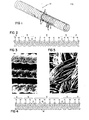
- Fig. 3 is a scanning electron micrograph (SEM) showing, at 50X magnification, the appearance of three ribs (or three crimpings) of the coated graft, which were taken from the end zone of the coated graft segment. On the ribs appearing at left and in center, the coating has covered the inter-yarn voids smoothening the surface. As determined from SEM's taken from cross sections, the thickness of the bioresorbable polyester composite coating was on the order of 15-25 microns. When deposited at this layer thickness, the coating filled the graft interstitial spaces to the extent that it did not only reduce the fraying of cut ends (a known propensity of grafts having this type of weave pattern), but it also eliminated the need for pre-clotting.
- SEM scanning electron micrograph
- Example 2 Under operative conditions described in Example 1, an approximately five centimeter piece of the coated vascular graft was implanted by end-to-end anastomoses into the right external carotid artery of an adult sheep. Under identical conditions, an approximately five centimeter piece of the same human-implantable Dacron graft which had not been treated or coated but only sterilized, was pre-clotted and implanted into the left external carotid artery of the same animal during the same operation. After five weeks of indwelling time, both grafts were open to blood flow when the animal was electively terminated under conditions described in Example 1, and the grafts were excised.
- the coated vascular graft On its luminal blood-contacting surface, the coated vascular graft had a smooth glistening pseudoneointimal layer virtually free of any red or white thrombotic deposit, whereas the same surface of the uncoated Dacron graft had a comparatively thick layer of adherent red and white thrombotic deposits. On its external surface, the coated Dacron had induced a significantly less severe tissue response and inflammatory reaction than the uncoated graft. Histopathology of segments taken from both grafts indicated a much stronger interstitial permeation of fibroblast cells into the coated graft.
- the attachment of fibroblasts can be, in part, attributed to the specific surface properties displayed by the polyester coating.
- the total surface free energy 1s of cast films of the polyester is generally in the range of 40 to 50 dynes/cm.
- the predominant component of this surface force field is constituted by Van der Waals-London type dispersion forces, leading to a computable dispersion force component, equal to 60% to 75% of the total ⁇ s .
- polar forces arising from H-bonding, dipole-dipole interactions, etc. represent the minor fraction of the total surface force field, leading to a computable polar force component, equal to 25% to 50% of ⁇ s .
- Example 4 An approximately seven centimeters long length of seven millimeter diameter tubing which was prepared to have a 75 percent bioresorbable cross yarn content as described in Example 2, was cleansed and subsequently overcoated with a 1:1 composite of poly(D,L-lactic acid) and poly(2,3-butylene succinate) under conditions indentical to those described in Example 3 to provide coating 18 as indicated diagrammatically in Fig. 4. Implantation of this coated partially bioresorbable vascular graft into the right external carotid artery of a sheep was performed in the same manner as given above. As a control implanted into the left external carotid artery, a 75 percent bioresorbable cross yarn containing graft was used which had not been coated.
- Histopathological analyses of grafts both of which were patent when excised after five weeks of indwelling time, indicated comparatively smoother pseudoneointimal linings on the luminal surface of the coated graft, as well as a comparatively milder tissue response on the external surface of the coated graft.
- Example 5 The in vivo performance characteristics of vascular grafts include, among others, the incidence of adherent thrombus formation potentially leading to occulusion, the rate of formation of neointimal lining, the interaction with perivascular tissues, etc. In a given species, these characteristics or, in general, graft healing and patency are known to be affected not only by the properties of a particular graft, but potentially by other factors as well, such as the implantation technique applied which can be a surgeon- dependent human factor. This example is to demonstrate that, if implanted in the same species via the same type of anastomoses by a different but equally competent surgeon, the results obtained with a coated woven graft are approximately the same as those described in Example 3.
- Example 6 Another factor know to influence the in vivo performance characteristics of vascular grafts and especially, their so-called "healing" properties including, among others, the intensity of interactions with the blood stream and the rate of neointima formation, is the type of animal species used since the hematologic profiles of different species are different. For example, the propensity of sheep blood for clotting induced by surfaces foreign to blood is somewhat less than than of canine blood, implying that, on these surfaces, canine blood clots easier. On the other hand, the fibrinolytic system that can dissolve clots as a result of lysis performed by certain blood enzymes, is weak or almost nonexistent in the sheep, whereas it is quite effective in dogs.
- the woven graft was shaped into an inverted "U" form such that it traversed through several layers of muscle, with the bottom of the loop brought close to the subdermis where blood flow through the graft could be easily detected by palpation or a Doppler flow meter.
- both grafts were open to blood flow when the animal was electively terminated under conditions as described in Example 1, and the grafts were excised.
- the recovered coated graft displayed a smooth, white pseudoneointimal lining on its blood-contacting surface, whereas this lining in the uncoated control graft appeared to be in small patches which were interspersed with red thrombotic deposits partially occluding the graft lumen.
- a kink had evolved in the coated graft, for causes mostly attributable to the contraction of tissues the graft was tunnelled through.
- Example 7 As a result of their fabric texture and comparatively smaller yarn density, vascular grafts knitted of multifiliament Dacron yarn are generally more conformable and compliant than grafts that were woven of the same yarn. For the same structural reasons, however, the porosity of knitted grafts is significantly greater. As measured in terms of leak rate at a constant pressure head of 100 mm Hg, the flow of water through a knitted graft can be typically on the order of 1,500 ml cm -2 min -1 , whereas it is only about 150 ml cm -2 min -1 for woven grafts. To avoid large losses of blood, all types of currently available knitted grafts, without exception, must be sealed by preclotting prior to implantation.
- the graft was dipped twice into an acetonic solution of 2.5% by weight double precipitated poly(D,L-lactic acid) (approximately 75,000 Daltons weight-average molecular weight), 2.5% by weight double-precipitated poly(2,3-butylene succinate) (approximately 17,500 Daltons weight-average molecular weight) and 0.10% by weight triethyl citrate purified by vacuum fractionation.
- the graft was dipped thrice more but in a moderately stretched state. This condition forces the polyester coating to form a continuous film between the individual yarns, covering over the interstitial space as shown, at a magnification of 200X in Figure 5, in a scanning electron micrograph (SEM) of the coated graft. If dip- coating of the knitted graft were to be continued without the conditions forcing this film formation, the graft can be sealed only by saturating its interstitial volume with the coating.
- the amount of coating so deposited is sufficient to completely alleviate the need of preclotting, as substantiated by, among others, the experimentally established decrease of the original leak rate of 1,500 ml cm -2 min -1 of water to about 60 to 80 ml cm min 1.
- the amount of polyester coating required to adequately seal for example, an 8 mm diameter woven graft is in the range of 0.020 to 0.025 g, and on the order of 1.0 g for a knitted graft which has the same diameter and has been coated by the forced film formation technique.
- Example 2 Under operative conditions described in Example 1, an approximately twenty-kilogram mongrel dog was implanted in its right external carotid artery with an 8 mm diameter knitted Sauvage-type graft which had been coated by means of the forced film formation technique described above. Connections to the artery were accomplished by end-to-side anastomoses, and the graft was positioned in the inverted "U" form, in the same manner as described in Example 6.
- a human-implantable grade, crimped, knitted Sauvage-type "Bionit" graft was placed in an identical manner as control, which was of the same diameter and approximately of the same length, and which had been sterilized only but not coated.
- the coated graft required no preclotting, whereas the uncoated graft could be preclotted only with difficulty since, according to standard implantation protocol, the animal was lightly anticoagulated with heparin.
- both grafts were open to blood flow when the animal was electively terminated under conditions described in Example 1, and the grafts were excised.
- Microscopic histapothological examination revealed an advanced degree of neondothelialization on the luminal surface of the coated knitted graft, whereas the luminal lining of the same but uncoated graft consisted mostly of fibrinaceous deposits.
Abstract
Description
- This invention relates to prosthetic devices, and more particularly to prosthetic devices suitable for use in mammalian bodies in place of a part thereof.
- Successful prosthetic devices of various shapes and applications have, in general, been made of materials relatively resistant to biodegradation such as stainless steel or inert biodurable polymeric materials. For example, replacement of injured, diseased, or nonfunctioning blood vessels has been conducted for many years, and nonresorbable synthetic permanent vascular grafts are currently made of either Dacron (R) (polyethylene terephthalate) or microporous Teflon (R) (polytetrafluoroethylene). Various prosthesis such as grafts and bypasses for insertion into the vascular system must have certain properties including physical and mechanical compatibility with the vessel to which they are connected, saturability, conformability to contours, ability to withstand pressure and pressure fluctuations, and flexibility; biocompatibility-including such aspects as sterility and absence of toxicity, pyrogenicity, allergenicity, and mutagenicity; and adequate durability--both in terms of "shelf life" after fabrication and durability after implantation. Mechanical problems which can develop from mismatch of native vessel and prosthesis include elongation, kinking, aneurysm formation and anastomotic hyperplasia. Vascular prosthesis made of biodurable materials have been so far the relatively most successful substrates, providing a conduit for maintaining continuous blood flow while inflicting preferably minimized and clinically tolerable hemotologic trauma. Most vascular grafts made of synthetic materials and in current clinical use are formed of knitted or woven nonbiodegradable fibers with open pores in the fabric which are typically closed by thrombosis (preclotting) before implantation. Such prosthesis have been used both as vascular replacements and as bypass grafts but only the relatively larger arteries such as the abdominal aorta. While bioresorbable materials have been proposed for use in such prosthesis, the practical uses of biresorbable - materials are, in general, currently limited to temporary devices such as fasteners (sutures and pins).
- In accordance with one aspect of the-'invention, there is provided a sterile, shaped prosthetic device suitable for insertion into a mammalian body, at least a portion of which is of a bioresorbable material. This material is digestible by macrophages, and other cells of the mammalian immune system its surface properties favor the attachment and adhesion of fibroblast cells capable of producing collagen for organized connective tissues, and the material itself or its degradation products can induce in vivo the formation of new capillarly vessels (termed a "vasotropic" effect herein). Preferred prosthetic devices have through passages, the interspatial dimensions which are sufficient to permit several layers of mammalian cells to form within each passage. The invading fibroblast cells commence formation of collagen leading to connective tissue while macrophages and extracellular enzymes degrade the material, and newly formed capillary vessels penetrate the prosthesis and provide blood containing oxygen and nutrients which further the formation of organized tissue around as well as within the prosthetic device.
- The term bioresorbable is used herein to mean not only biodegradable but that the degradation products, formed in vivo from those materials, are metabolizable as well by the mammalian body, without any toxic or otherwise harmful side effects. Also, as used herein, the term nonbioresorbability implies nonbiodegradability as well. It has been discovered that certain bioresorbable polyesters promote the regrowth of vascular tissues in and around a partially or completely metabolizable synthetic scaffold designed to lead to tissue organization that is comparable to intact native arterial vessels. These polyesters also exert a "vasotropic" effect in vivo by inducing the formation of new capillary vessels within surrounding tissues. The adjective "vasotropic" is used here with the intent to distinguish the effect which can be arbitrarily induced by synthetic materials, from the "angiogenic" effect or, "angiogenesis" that is a spontaneous phenomenon generally associated with the growth of malignant tissues.
- In accordance with another feature, there is provided a vascular prosthesis that includes a sterilized, flexible, tubular device of fabric scaffold structure, with at least 25 percent of the yarns of the fabric scaffold structure consisting solely of bioresorbable material in an arrangement such that, on implantation of the vascular prosthesis in a mammalian body, the bioresorbable material undergoes absorption and provides passages through the scaffold structure that have interspatial dimensions sufficient to allow several layers of mammalian cells to form within each passage.
- Surface properties of the bioresorbable material encourage not only the invasion of fibroblast cells onto and into the fabric of the vascular graft, but also encourage the attachment as well as the adhesion of fibroblast cells. Not only the shape of fibroblast cells but the incidence of their adhesion and attachment are a function of the surface properties of the porous substrate. Using wettability spectrum analysis, the surface properties of bioresorbable materials in accordance with an aspect of the invention are computable as well as characterizable in terms of the total surface free energy, Ys, which can be expressed as the sum of ydand

- The surface properties of the bioresorbable material also encourage the infiltration of the porous prosthesis by macrophages which are cells of the mammalian immune mechanism and are capable of digesting the material in bioresorbable vascular grafts, in accordance with the invention, the formation of organized collageneous tissue is part of the reconstructive processes ultimately leading to vascular wall tissue organization which is similar or, comparable to that of intact native arterial vessel walls.
- Certain bioresorbable polyesters impart "vasotropic properties to vascular grafts which are manifested in vivo by the induction of the formation of entirely new capillary blood vessels in living soft tissues. In the presence of a bioresorbable coating which is either composed of a single substance having "vasotropic" properties or, which has a component exhibiting "vasotropic" properties, the induced formation of new capillary vessels penetrating the interstices of the graft occurs. In addition to vascular grafts, bioresorbable materials capable of exerting "vasotropic" effects either in the short or, in the long term can be of significant value by initiating, aiding or accelerating natural reconstructive processes, in both soft and hard damaged tissues of the mammalian body, which would otherwise not occur or, would take place only at slow rates as a result of inadequate transport of metabolites to and from the regeneration site. As a result of the formation of new capillary vessels or neovascularization, the transport of metabolites and oxygen needed for, at least, the partial reconstruction of damaged tissues becomes facilitated. The establishment of transport channels for nutrients can be especially significant where the damaged tissue had a relatively low level of original vascularization such as in bones or, the original vascularization of the tissues was destroyed or rendered nonfunctional such as in burnt soft tissues of the dermis and subdermal layers. Thus, for those familiar with the art, the spectrum of applicability of these bioresorbable vasotropic materials can involve intraosseous implants to aid the regrowth of missing bone segments after compound fractures, dental implants, and burn covers aiding, at least the partial, regeneration of devitalized subdermal layers until they become covered by epithelial cells.
- An appropriate deposition of a bioresorbable coating generally smoothens the luminal blood-contacting surface of both woven and knitted fabric vascular grafts. In turn, this results in a smaller amount of deposits from blood (viz., platelets and fibrin) as on uncoated surfaces, ultimately leading to relatively thin pseudoneointimal and neointimal layers covering the luminal surface of the graft.
- As a result of the formation of organized tissues within the interstices of the graft, as noted above, the adherence of pseudoneointimal and neointimal layers formed on the luminal surface does not only occur to the graft alone, but to the collageneous and connective tissues as well, which are organized within the graft interstices, irrespective of whether the fabric graft is made of either a bioresorbable or a nonbioresorbable material. Establishment of continuity in the biological material formed on the luminal side of the graft and the organized tissues formed within and on the outside surface of the graft are measures rendering a potential delamination of the luminal layer more difficult than when this continuity does not or cannot exist. Neointimal delamination is known to be one of the modes of failure of current nonbioresorbable vascular grafts.
- To initiate, maintain and accomplish the processes of vascular wall reconstruction, bioresorbable coatings preferably exert their effects both in the short and in the long term. For example, the desired "vasotropic" properties might be imparted by means of a single substance which, however, undergoes bioresorption at a rapid rate. Another single substance might be less "vasotropic" than-the former but its bioresorption rate in vivo is also less than that of the former. Thus, to maintain the vascular wall reconstructive processes both in the short and long term, a composite of both of these bioresorbable substances is more preferred as a coating than a single-component coating consisting only of either of the two substances.
- Polyesters are, in general, constituted of a dicarboxylic acid and a diol, which are reacted with each other in a so-called condensation reaction with the elimination of water. Upon hydrolytic attack, the dicarboxylic acid and diol components are reconstituted with the uptake of water. Bioresorbable polyesters in accordance with the invention are derived from Krebs-Cycle dicarboxylic acids and diols that are also metabolizable. With respect to the known mechanisms of the mammalian body degrading carbon chains, namely, beta oxidation followed by chain scission, the diols incorporated into these polyesters preferably consist of an even number of carbon atoms (since chain scission occurs in pairs of carbon atoms), and the number of carbon atoms is greater than two. Thus, for the synthesis of our bioresorbable polyesters, diols such as 2,3-butanediol, 1,4-butanediol and 2,5-hexanediol, etc., are preferred.
- Vascular grafts made completely either of nonresorbable (viz., Dacron) or bioresorbable yarns (i.e., polyglycolic acid), or grafts made using any combination of these yarn types (viz., partially resorbable grafts) can be coated with bioresorbable polyesters which are capable of eliciting the interstitial penetration of cells in vivo. This effect can be well considered to promote the integration as well as the stabilization of vascular prosthesis. Since, in addition, some of these polyesters demonstrably exert a "vasotropic" effect, this can ultimately result in the ingrowth of capillary vessels or, vascularization of the graft.
- As a result of their bioresorbability, the polyester graft coatings become degraded and ultimately vanish, gradually increasing the biological porosity of the graft over its initial porosity. The rate at which a given amount of bioresorbable coating becomes metabolized is a function of the mass of the coating applied, as well as of the molecular weight and structural characteristics of polyesters used. The mass of coating that is deposited over unit nominal area of graft surface has, among others, some practical limitations in order to preserve the compliance, conformability, flexibility, etc. of the woven or knitted fabric tubular structure while still sealing the interstitial pores of both types of fabric to the extent that sealing by graft preclotting is alleviated. In accordance with another aspect of the invention, depending on the type of fabric structure to be coated, the mass of the coating applied and the molecular structural characteristics of polyesters used, or a combination of both, may be utilized to control the in vivo longevity of the coating while simultaneously achieving the sufficient sealing of fabric porosity.
- As a result of the generally greater yarn density of woven grafts (or their smaller porosity compared to knitted grafts), the mass of uniformly distributed coating required to seal these grafts without compromising the fabric's physical properties is typically on the order of 10 to 15 mg per unit nominal area of graft surface. For example, for an 8 mm woven graft measuring 10 cm in length, a total of about 200 to 250 mg of polyester coating is sufficient. Thus, for woven grafts, the mass of coating applied represents a secondary means of controlling the rate at which a bioresorbable coating is metabolized. Control over the in vivo longevity of woven-graft coatings is primarily exerted via the molecular structural characteristics of polyesters used.
- In contrast to woven grafts, the porosity of knitted grafts, as measured by standard leak rate methods, is about one order of magnitude greater (i.e., approx. 1,500 ml cm-2min-1 vs. 150 ml cm-2 min-1). With the coating initially depositing on individual yarns, the total amount of coating which has to be deposited on a knitted graft until its interstitial volume becomes sealed by the coalescence of individual yarn coatings, is large enough to compromise the desirable conformability of this type of graft, converting it into a rigid pipe. Another aspect of the invention is to cause the polyester coating to spread over and form a film above the individual yarns of knitted grafts, without saturating their interstitial spaces and thereby preserving their desirable mechanical properties. Under these film-forming conditions, the mass of uniformly distributed coating that is necessary to sufficiently seal knitted grafts is on the order of 35 to 45 mg per unit nominal area of graft surface. For the same 8
mm diameter 10 cm long but knitted graft, a total polyester coating of about 1.0 g is required. As a result of the relatively greater mass of coating taken up, this factor becomes significant to exerting control over the in vivo longevity of polyester coatings deposited on knitted grafts. - Independent of the different masses of coating required by different types of grafts, a feasible way of exerting control over the in vivo longevity of graft coatings is via the molecular structural characteristics of polyesters used. As discussed below, the rate of bioresorption of polyesters derived from Krebs-Cycle dicarboxylic acids can be influenced to the extent as to have these materials metabolized either within relatively short periods of time, i.e., four to six weeks, or over relatively long periods on the order of four to six months.
- Preferred polyesters are, in general, homo- and copolyesters derived from succinic, L-hydroxysuccinic (or malic), and fumaric acids, as well as from 2,3-butanediol and 1,4-butanediol, and include poly(2,3-butylene succinate) and copoly[(l:3)-l,4-butylene/2,3-butylene succinate].
- Poly(2,3-butylene succinate), shown below, is an amorphous homopolyester at ambient temperature was with a relatively low softening point
-

- The structure of a random copolyester, copoly[(l:l) l,4-butylene/2,3-butylene succinate] is shown below

- The adjective "random" is used for this copolyester because the sequence of 1,4- and 2,3-butylene moieties in the polyester backbone may not be necessarily monotonous throughout the entire length of polymer chains. A similar "hybridized" polymer with a relatively high 2,3-butylene moiety content, copoly[(l:3) 1,4-butylene/2,3-butylene succinate] has the good film-forming properties of the poly(2,3-butylene succinate) homopolymer but undergoes bioresorption at rates comparable to that of the (1,4-butylene succinate) homopolyester. Thus, in vivo bioresorption rates may be influenced via the nature of chain segments introduced by the diols.
- Another homopolyester is poly(2,3-butylene fumarate) shown below
-

- Still another homopolyester is poly(2,3-butylene hydroxy succinate), its molecular structure being shown below

- As indicated by the asterisk, the hydroxy succinic acid moiety has an asymmetrically substituted, optically active carbon atom which allows for the use of either the L- or the D-stereoisomers, or the D,L-lacemic mixture of the two enantiomorphs in the polyesters formed. Poly(2,3-butylene D,L-hydroxy succinate) is an amorphous substance with a moderately high softening point (75° to 80°C). The bioresorption rate of this polyester is significantly slower than that of poly(2,3-butylene succinate) under identical conditions.
- The synthesis of polyesters composed of Krebs-Cycle dicarboxylic acids and the diols specified generally involves a straightforward condensation reaction with the concurrent elimination of water. To obtain polyesters with adequate physical properties, however, the molecular weight of these polymers has to be sufficiently high (i.e., at least 10,000 Daltons or greater) or, preferably, the maximum achievable with a given dicarboxylic acid or diol.
- To alleviate any potential catalyst-related .physiologic effects or artifacts of unknown origin, the condensation reaction between Krebs-Cycle dicarboxylic acids and the selected diols is performed, at elevated temperatures (viz., 160° to 190°C), with the vacuum-melt polymerization technique in the absence of any catalyst. Elevated temperatures and polymerization periods extending up to 72 or 96 hours are generally employed to attain polyester molecular weights in the range of 10,000 to 20,000 Daltons. To further assure the alleviation of any potential artifacts capable of obscuring the true in vivo biological effects of these polyesters, crude polymers are purified, at least twice, by reprecipitative fractionation before any biological application.
- As a means of exerting control over the in vivo longevity of polyester coatings on vascular grafts, the weight-average molecular weight of polyesters is also of importance. In the absence of any catalyst and with increasing molecular weights, the rate of condensation reaction between a given dicarboxylic acid and diol gradually decreases such that a plateau molecular weight characteristic of this system is approached. If desired, further increases in weight-average molecular weight can be achieved by chain extension performed as follows.
- The reactivity of different Krebs-Cycle dicarboxylic acids toward a given diol, HO-Dl-OH, is different because the electronic structure of the acid residues is different and affects the reactivity of the carboxylic groups in different ways. For example, the reactivity of hydroxy succinic acid toward diols of interest is generally greater than that of succinic acid because of the activating effect of the alpha hydroxy group in the former. The activating effect is independent of the stereochemical conformation of the hydroxy succinic acid, which can be either D-, of L- or the D,L racemate.
- In the synthesis of a polyester which is desired for graft coating and which is formed from the Krebs-Cycle dicarboxylic acid HOOC-Ri-COOH and the diol HO-Dl-OH, a slight excess in the range of 5 to 10 mole percent of the diol is employed in the condensation reaction. This excess renders the polyester molecules formed to be overwhelmingly terminated by the alcoholic hydroxyl groups of the diol, HO-D1-OOC-R1-COO- ....-D1-OH. If the plateau value of the weight-average molecular weight that is attainable by the polyester under these conditions, is insufficient to meet in vivo graft coating longevity requirements, following its isolation, the hydroxy-terminated polyester can be subjected to chain extension by reacting it with small amounts of another Krebs-Cycle dicarboxylic acid, HOOC-R2-COOH which is more reactive than the dicarboxylic acid HOOC-Rl-COOH. The chain-extension is also performed under the conditions of vacuum-melt polymerization at elevated temperatures, yielding essentially the same polyester but with substantially increased weight-average molecular weight, and with the general formula of HOOC-R2-COO-[-D1-OOC-R1-...-[D1-OOC-R2-COO-. -D1]-OOC-R1... Dl ...-R2-COOH where the bracketed chain segments correspond to the average molecular weight of the original polyester. For example, upon treatment under these conditions with 0.005% to 0.01% by weight of hydroxy succinic acid which was homogeneously dispersed in the melt of a hydroxy-terminated poly(2,3-butylene succinate) of 12,000 Daltons, the weight-average molecular weight of this polyester increased to 28,500 Daltons, as determined by gel permeation chromatography.
- Particular embodiments of tubular vascular graft prosthesis in accordance with the invention are composite structures of nonbioresorbable multifilament yarns in a fabric array with through passages that contain bioresorbable multifilament yarns. The fabric is woven or knitted in a desired fabric pattern (for example, either a helical or rectangular weave) and is of a desired porosity. The pores of the tubular scaffold of nonbioresorbable material have interspacial dimensions of about 200 microns dimensions, such that several layers of cells can form through and within each pore. An overcoating of a bioresorbable, vasotropic effect-inducing material having a coating thickness typically in the range of l5 to 25 microns may be used on some or all of the yarns or on the tubular structure itself.
- Suitable bioresorbable yarns are composed of polyglycolic acid; polyglycolic acid/poly(D,L-lactic acid) random copolymers having any ratio between its glycolate and D,L-lactate moieties; polyglycolic acid/poly(L-lactic acid) random copolymers having any ratio between its glycolate and L-lactate moieties; poly(D,L-lactic acid) homopolymers in which the abundance of L-lactate moieties can comprise 50% to 99.99% of lactate moieties; and poly(L-lactic acid) stereospecific homopolymers. The homo- and copolymers named above are selected such as to impart, by means of their molecular structure, controllably variable rates of in vivo bioresorbability into the fibers and yarns made from them. Thus, one means of achieving the gradual bioresorption of the vascular graft scaffold is by interditation of yarns which exhibit, under identical conditions, different rates of bioresorption as a result of the particulars of their molecular structure. While absolute in vivo resorption rates vary according to the site of implantation in the mammalian body, under identical conditions, i.e., in identical implantation sites, the homopolymers, poly(glycolic acid) and stereospecific poly(L-lactic acid) undergo bioresorption at rates which are substantially slower than those of their copolymers. In general, copolymerization increases the rate of bioresorption. For example, poly(D,L-lactic acid) containing 50% or more of L-lactate moieties, is a copolymer of D- and L-lactic acids, and resorbs faster than the pure poly(L-lactic acid) homopolymer. Random copolymers of glycolic acid and D,L-lactic acid resorb faster than their corresponding homopolymers.
- Other features and advantages of the invention will be seen as the following description of particular embodiments progresses, in conjunction with the drawings, in which:
- Fig. 1 is a perspective view (partially in section) of a tubular prosthesis in accordance with one embodiment of the invention;
- Fig. 2 is a diagram indicating the weave pattern employed in the prosthesis of Fig. 1;
- Fig. 3 is a scanning electron micrograph of another embodiment of a vascular prosthesis in accordance with the invention; and
- Fig. 4 is a diagram, similar to Fig. 2, illustrating aspects of still another vascular prosthesis in accordance with the invention.
- The tubular
vascular prosthesis 10 shown in Fig. 1 has a length of about five centimeters and is composed of nonbioresorbable (Dacron) multifilament longitudinal yarns 12 (eachyarn 12 being approximately 150 microns in diameter and being composed of filaments about 20 microns in diameter) at a density of 100 yarns per inch, and multifilament bioresorbable (a 9:1 copolymer of [PGA] polyglycolic acid and poly(L-(-)-lactic acid)cross yarns 14 interdigitated with nonbioresorbable (Dacron) multifilament cross yarns 16 a density of 100 yarns per inch--the cross-sectional diameter of thecross yarns longitudinal yarns 12. The interdigitation of bioresorbable (B)cross yarns 14 with nonbioresorbable (N)cross yarns 16 is in yarn pairs with a resulting cross yarn pattern of NNBBBBBBNN... , as indicated diagrammatically in Fig. 2, providing a 75 percent bioresorbable cross yarn content. The tubular woven prosthesis had an internal diameter of six millimeter before crimping and a void area (or porosity) of about 18 percent. Crimping (a temperature-controlled process, without the use of any chemical agent) reached a depth of about one millimeter, which reduced the narrowest diameter of the prosthesis before implantation to five millimeters. Control prosthesis were made of Dacron polyfilament yarn of the same dimensions and using the same weaving technique. - After crimping, the bioresorbable and control prosthesis were ultrasonicated in 0.1 percent aqueous solution of nonionic detergent (Triton X-100) for sixty minutes at room temperature and the vascular prosthesis were then rinsed with pyrogen-free deionized water, followed by several rinses with 95 percent ethanol and drying in vacuo. The vascular prosthesis were then packaged under a laminar flow hood equipped with high efficiency air filters and the packaged prosthesis were subjected to standard ethylene oxide sterilization.
- Example l. Under anesthesia, light anticoagulation and sterile operating room conditions, the
vascular graft 10 was implanted by end-to-end anastomoses into the right external carotid artery of an adult sheep. Under identical conditions, an approximately five centimeter length of sterilized control graft consisting solely of woven Dacron multifilament yarns was implanted into the left external carotid artery of the same animal during the same operation. - After five weeks of indwelling time, both grafts were open to blood flow when the animal was anticoagulated and sacrificed by euthanasia, and the grafts were excised. The recovered graft with 75 percent bioresorbable cross yarn content remained patent, displayed on a smooth pseudoneointimal layer on its luminal blood-containing surface, and showed a partial degradation of bioresorbable-yarns and transmural interstitial vascularization.
- Example 2. Under the operative conditions described in Example 1, an approximately five centimeter long and five millimeter diameter piece of tubular partially bioresorbable prosthesis with a 75 percent bioresorbable cross yarn (polyglycolic acid multifilament) content--the other cross yarns and the longitudinal yarns were of multifilament Dacron--was implanted into the right external carotid artery of a sheep. Into the left external carotid artery of the sheep, a control prosthesis of all-Dacron, also of approximately five centimeters length, was implanted. Under conditions described in Example 1, the experimental animal was electively terminated after fifteen weeks. The recovered nonbioresorbable (all-Dacron) graft was occluded while the recovered biorresorbable graft remained patent, displayed smooth pseudoneointimal layers on its luminal blood-contacting surface, and displayed a partial degradation of bioresorbable yarns and transmural interstitial vascularization.
- Example 3. A 7.5 centimeter long piece of a human-implantable grade, crimped, woven Dacron DeBakey-type vascular graft having an eight millimeter diameter, was subjected to cleansing and sterilization as described in Example 1. Using a dipping apparatus which was enclosed in a dust-free chamber and had controllable speeds of vertical motion, the dried vascular graft was immersed, at a rate of about 0.5 centimeter per second, into a solution of five grams of double-precipitated poly(D,L- lactic acid) (approximately 70,000 Daltons weight-average molecular weight) and five grams of double-precipitated poly(2,3-butylene succinate) (approximately 17,500 Daltons weight-average molecular weight) in 100 milliliters of pure acetone. Following the first dip and a partial drying of the coating deposited, the vascular graft was inverted and redipped under the same conditions. After the second dip, the vascular graft was dried and packaged under a laminar flow hood equipped with high efficiency air filters, and the packaged prosthesis was subjected to standard ethylene oxide sterilization.
- Fig. 3 is a scanning electron micrograph (SEM) showing, at 50X magnification, the appearance of three ribs (or three crimpings) of the coated graft, which were taken from the end zone of the coated graft segment. On the ribs appearing at left and in center, the coating has covered the inter-yarn voids smoothening the surface. As determined from SEM's taken from cross sections, the thickness of the bioresorbable polyester composite coating was on the order of 15-25 microns. When deposited at this layer thickness, the coating filled the graft interstitial spaces to the extent that it did not only reduce the fraying of cut ends (a known propensity of grafts having this type of weave pattern), but it also eliminated the need for pre-clotting.
- Under operative conditions described in Example 1, an approximately five centimeter piece of the coated vascular graft was implanted by end-to-end anastomoses into the right external carotid artery of an adult sheep. Under identical conditions, an approximately five centimeter piece of the same human-implantable Dacron graft which had not been treated or coated but only sterilized, was pre-clotted and implanted into the left external carotid artery of the same animal during the same operation. After five weeks of indwelling time, both grafts were open to blood flow when the animal was electively terminated under conditions described in Example 1, and the grafts were excised. On its luminal blood-contacting surface, the coated vascular graft had a smooth glistening pseudoneointimal layer virtually free of any red or white thrombotic deposit, whereas the same surface of the uncoated Dacron graft had a comparatively thick layer of adherent red and white thrombotic deposits. On its external surface, the coated Dacron had induced a significantly less severe tissue response and inflammatory reaction than the uncoated graft. Histopathology of segments taken from both grafts indicated a much stronger interstitial permeation of fibroblast cells into the coated graft.
- Although a well-defined cause and effect relationship for this in vivo phenomenon has not yet been established, the attachment of fibroblasts can be, in part, attributed to the specific surface properties displayed by the polyester coating. As deduced from wettability spectrum determinations, the total surface free energy 1s of cast films of the polyester is generally in the range of 40 to 50 dynes/cm. However, the predominant component of this surface force field is constituted by Van der Waals-London type dispersion forces, leading to a computable dispersion force component,


- The surface characteristics deduced from wettability spectra are consistent with the results of Electron Spectroscopy for Chemical Analysis. These results have consistently indicated that the carbon/oxygen ratio, found within the 20 to 40 A° thick surface layer analyzed by ESCA, is appreciably greater than the stoichiometric carbon/oxygen ratio of the particular polyester. In these polyesters, most of the carbon atoms are present in CH3-, CH2- and CH = moieties, i.e., nonpolar groups capable of exerting only dispersion forces that contribute to the

- Example 4. An approximately seven centimeters long length of seven millimeter diameter tubing which was prepared to have a 75 percent bioresorbable cross yarn content as described in Example 2, was cleansed and subsequently overcoated with a 1:1 composite of poly(D,L-lactic acid) and poly(2,3-butylene succinate) under conditions indentical to those described in Example 3 to provide
coating 18 as indicated diagrammatically in Fig. 4. Implantation of this coated partially bioresorbable vascular graft into the right external carotid artery of a sheep was performed in the same manner as given above. As a control implanted into the left external carotid artery, a 75 percent bioresorbable cross yarn containing graft was used which had not been coated. Histopathological analyses of grafts, both of which were patent when excised after five weeks of indwelling time, indicated comparatively smoother pseudoneointimal linings on the luminal surface of the coated graft, as well as a comparatively milder tissue response on the external surface of the coated graft. - Example 5. The in vivo performance characteristics of vascular grafts include, among others, the incidence of adherent thrombus formation potentially leading to occulusion, the rate of formation of neointimal lining, the interaction with perivascular tissues, etc. In a given species, these characteristics or, in general, graft healing and patency are known to be affected not only by the properties of a particular graft, but potentially by other factors as well, such as the implantation technique applied which can be a surgeon- dependent human factor. This example is to demonstrate that, if implanted in the same species via the same type of anastomoses by a different but equally competent surgeon, the results obtained with a coated woven graft are approximately the same as those described in Example 3.
- Two 7.5 cm long pieces of an 8 mm diameter human-implantable grade, crimped, woven DeBakey-type vascular graft were coated as describe in Example 3, with double-precipitated poly(D,L-lactic acid). Each of these grafts was placed by end-to-end anastomoses into the right external carotid artery of each of two adult sheep under the same operative conditions as described in Example 1, but with all implantations performed by a different surgeon. Under identical conditions, each of two approximately 7.5 cm pieces of the same sterilized 0, but uncoated graft were preclotted and placed into the left external carotid artery of the same animals during the same operations. After six weeks of indwelling time, all grafts were open to blood flow when both animals were electively terminated under conditions described in Example 1, and all grafts were excised. To within the limits of reproducibility of biological experiments, the condition of coated grafts and uncoated controls recovered from these implantations was, respectively, the same in terms of luminal lining and perivascular tissue response, as of those grafts which were recovered from the implantation experiment described in Example 3.
- Example 6. Another factor know to influence the in vivo performance characteristics of vascular grafts and especially, their so-called "healing" properties including, among others, the intensity of interactions with the blood stream and the rate of neointima formation, is the type of animal species used since the hematologic profiles of different species are different. For example, the propensity of sheep blood for clotting induced by surfaces foreign to blood is somewhat less than than of canine blood, implying that, on these surfaces, canine blood clots easier. On the other hand, the fibrinolytic system that can dissolve clots as a result of lysis performed by certain blood enzymes, is weak or almost nonexistent in the sheep, whereas it is quite effective in dogs.
- A piece of a 6 mm diameter human-implantable grade, crimped, woven DeBakey-type vascular graft which had been cleansed and coated as described in Example 5, was end-to-side anastomosed.to the right external carotid artery of an approximately fifteen-kilogram, adult mongrel dog, under the operative conditions described in Example 1. With the two anastomoses separated by a distance of about 10 cm, the segment of the artery between them was tied off by ligatures. By simultaneous stretching and bending, the woven graft was shaped into an inverted "U" form such that it traversed through several layers of muscle, with the bottom of the loop brought close to the subdermis where blood flow through the graft could be easily detected by palpation or a Doppler flow meter. In an identical manner, a piece of the same human-implantable woven Dacron graft which had not been coated but has preclotted, was placed as a control into the left carotid artery of the same animal during the same operation.
- After six weeks of indwelling time, both grafts were open to blood flow when the animal was electively terminated under conditions as described in Example 1, and the grafts were excised. The recovered coated graft displayed a smooth, white pseudoneointimal lining on its blood-contacting surface, whereas this lining in the uncoated control graft appeared to be in small patches which were interspersed with red thrombotic deposits partially occluding the graft lumen. In addition, during the indwelling period, a kink had evolved in the coated graft, for causes mostly attributable to the contraction of tissues the graft was tunnelled through. While kinks, in general, give rise to the perturbation of flow and the formation of separated flow regions, the incidence of these flow phenomena in blood conduits which were made of material foreign to blood, is known to be a common hemodynamic cause of thrombus formation. In spite of the kink evolved, the luminal surface of the layer which was formed in this coated graft, was void of any adherent thrombus, indicating the dynamic in vivo hemocompatibility of that layer.
- Example 7. As a result of their fabric texture and comparatively smaller yarn density, vascular grafts knitted of multifiliament Dacron yarn are generally more conformable and compliant than grafts that were woven of the same yarn. For the same structural reasons, however, the porosity of knitted grafts is significantly greater. As measured in terms of leak rate at a constant pressure head of 100 mm Hg, the flow of water through a knitted graft can be typically on the order of 1,500 ml cm-2 min-1, whereas it is only about 150 ml cm -2 min-1 for woven grafts. To avoid large losses of blood, all types of currently available knitted grafts, without exception, must be sealed by preclotting prior to implantation.
- A piece of an 8 mm diameter human-implantable grade, crimped, knitted Sauvage-type "Bionit" graft having a length of approximately 10 cm in the unstretched state, was cleansed ultrasonically and dried as described in Example 1. Using the apparatus and conditions described in Example 3, the graft was dipped twice into an acetonic solution of 2.5% by weight double precipitated poly(D,L-lactic acid) (approximately 75,000 Daltons weight-average molecular weight), 2.5% by weight double-precipitated poly(2,3-butylene succinate) (approximately 17,500 Daltons weight-average molecular weight) and 0.10% by weight triethyl citrate purified by vacuum fractionation. Following these initial dippings which served to deposit a thin coating over the individual yarns of the knitted fabric, the graft was dipped thrice more but in a moderately stretched state. This condition forces the polyester coating to form a continuous film between the individual yarns, covering over the interstitial space as shown, at a magnification of 200X in Figure 5, in a scanning electron micrograph (SEM) of the coated graft. If dip- coating of the knitted graft were to be continued without the conditions forcing this film formation, the graft can be sealed only by saturating its interstitial volume with the coating. This, however, results in the deposition of amounts of coating which convert the originally conformable graft into a rock-hard, brittle and nonconformant conduit ("steel pipe" effect). In contrast, the forced film formation disclosed here is manifested by the fact that, as seen from the SEM of the coated graft, the contours of most of the individual filaments are still discernible, indicative of the absence of saturation. The knitted graft that was coated according to this forced film formation technique essentially retains, in an aqueous environment and at body temperature, the original conformability of the same but uncoated graft. The amount of coating so deposited is sufficient to completely alleviate the need of preclotting, as substantiated by, among others, the experimentally established decrease of the original leak rate of 1,500 ml cm-2 min-1 of water to about 60 to 80 ml cm min 1. Normalized to a unit length of 10 cm of graft in the unstretched state, the amount of polyester coating required to adequately seal, for example, an 8 mm diameter woven graft is in the range of 0.020 to 0.025 g, and on the order of 1.0 g for a knitted graft which has the same diameter and has been coated by the forced film formation technique.
- Under operative conditions described in Example 1, an approximately twenty-kilogram mongrel dog was implanted in its right external carotid artery with an 8 mm diameter knitted Sauvage-type graft which had been coated by means of the forced film formation technique described above. Connections to the artery were accomplished by end-to-side anastomoses, and the graft was positioned in the inverted "U" form, in the same manner as described in Example 6. During the same operation but into the left external carotid artery of the same animal, a human-implantable grade, crimped, knitted Sauvage-type "Bionit" graft was placed in an identical manner as control, which was of the same diameter and approximately of the same length, and which had been sterilized only but not coated. The coated graft required no preclotting, whereas the uncoated graft could be preclotted only with difficulty since, according to standard implantation protocol, the animal was lightly anticoagulated with heparin.
- - After six weeks of indwelling time, both grafts were open to blood flow when the animal was electively terminated under conditions described in Example 1, and the grafts were excised. Microscopic histapothological examination revealed an advanced degree of neondothelialization on the luminal surface of the coated knitted graft, whereas the luminal lining of the same but uncoated graft consisted mostly of fibrinaceous deposits.
- While particular embodiments of the invention have been shown and described, various modifications will be apparent to those skilled in the art, and therefore it is not intended that the invention be limited to the disclosed embodiments or to details thereof and departures may be made therefrom within the spirit and scope of the invention.
Claims (15)
The prosthetic device of claim 3 wherein γs is in the range of 40-55 dynes per centimeter and y constitutes 60-80 percent of y .
Applications Claiming Priority (2)
| Application Number | Priority Date | Filing Date | Title |
|---|---|---|---|
| US533877 | 1983-09-20 | ||
| US06/533,877 US4594407A (en) | 1983-09-20 | 1983-09-20 | Prosthetic devices derived from krebs-cycle dicarboxylic acids and diols |
Publications (2)
| Publication Number | Publication Date |
|---|---|
| EP0144534A2 true EP0144534A2 (en) | 1985-06-19 |
| EP0144534A3 EP0144534A3 (en) | 1986-12-17 |
Family
ID=24127802
Family Applications (1)
| Application Number | Title | Priority Date | Filing Date |
|---|---|---|---|
| EP84109787A Withdrawn EP0144534A3 (en) | 1983-09-20 | 1984-08-16 | Prosthetic devices |
Country Status (5)
| Country | Link |
|---|---|
| US (1) | US4594407A (en) |
| EP (1) | EP0144534A3 (en) |
| JP (1) | JPS6096257A (en) |
| AU (1) | AU3265684A (en) |
| CA (1) | CA1239875A (en) |
Cited By (34)
| Publication number | Priority date | Publication date | Assignee | Title |
|---|---|---|---|---|
| WO1988004557A1 (en) * | 1986-12-17 | 1988-06-30 | Allied Corporation | Implantable devices having hydrophobic component |
| DE3835099A1 (en) * | 1987-10-14 | 1989-04-27 | Debiopharm Sa | PHARMACEUTICAL COMPOSITION |
| WO1991014407A1 (en) * | 1990-03-26 | 1991-10-03 | Keshelava Viktor V | Prosthesis of a tubular organ |
| WO1992005747A1 (en) * | 1990-10-09 | 1992-04-16 | Moskovsky Institut Stali I Splavov | Appliance for implantation in hollow organs and device for its introduction |
| US5413601A (en) * | 1990-03-26 | 1995-05-09 | Keshelava; Viktor V. | Tubular organ prosthesis |
| EP0689807A3 (en) * | 1994-06-27 | 1996-08-07 | Advanced Cardiovascular System | Biodegradable mesh-and-film stent |
| US6527801B1 (en) | 2000-04-13 | 2003-03-04 | Advanced Cardiovascular Systems, Inc. | Biodegradable drug delivery material for stent |
| US7867547B2 (en) | 2005-12-19 | 2011-01-11 | Advanced Cardiovascular Systems, Inc. | Selectively coating luminal surfaces of stents |
| US7875283B2 (en) | 2000-04-13 | 2011-01-25 | Advanced Cardiovascular Systems, Inc. | Biodegradable polymers for use with implantable medical devices |
| US7964210B2 (en) | 2006-03-31 | 2011-06-21 | Abbott Cardiovascular Systems Inc. | Degradable polymeric implantable medical devices with a continuous phase and discrete phase |
| US8003156B2 (en) | 2006-05-04 | 2011-08-23 | Advanced Cardiovascular Systems, Inc. | Rotatable support elements for stents |
| US8017237B2 (en) | 2006-06-23 | 2011-09-13 | Abbott Cardiovascular Systems, Inc. | Nanoshells on polymers |
| US8048441B2 (en) | 2007-06-25 | 2011-11-01 | Abbott Cardiovascular Systems, Inc. | Nanobead releasing medical devices |
| US8048448B2 (en) | 2006-06-15 | 2011-11-01 | Abbott Cardiovascular Systems Inc. | Nanoshells for drug delivery |
| US8099849B2 (en) | 2006-12-13 | 2012-01-24 | Abbott Cardiovascular Systems Inc. | Optimizing fracture toughness of polymeric stent |
| US8197879B2 (en) | 2003-09-30 | 2012-06-12 | Advanced Cardiovascular Systems, Inc. | Method for selectively coating surfaces of a stent |
| US8241554B1 (en) | 2004-06-29 | 2012-08-14 | Advanced Cardiovascular Systems, Inc. | Method of forming a stent pattern on a tube |
| US8333000B2 (en) | 2006-06-19 | 2012-12-18 | Advanced Cardiovascular Systems, Inc. | Methods for improving stent retention on a balloon catheter |
| US8343530B2 (en) | 2006-05-30 | 2013-01-01 | Abbott Cardiovascular Systems Inc. | Polymer-and polymer blend-bioceramic composite implantable medical devices |
| US8585754B2 (en) | 2000-04-13 | 2013-11-19 | Abbott Cardiovascular Systems Inc. | Stent formed of a Biodegradable material |
| US8603530B2 (en) | 2006-06-14 | 2013-12-10 | Abbott Cardiovascular Systems Inc. | Nanoshell therapy |
| US9038260B2 (en) | 2006-05-26 | 2015-05-26 | Abbott Cardiovascular Systems Inc. | Stent with radiopaque markers |
| US9072820B2 (en) | 2006-06-26 | 2015-07-07 | Advanced Cardiovascular Systems, Inc. | Polymer composite stent with polymer particles |
| US9763818B2 (en) | 2010-01-30 | 2017-09-19 | Abbott Cardiovascular Systems Inc. | Method of crimping stent on catheter delivery assembly |
| US9827119B2 (en) | 2010-01-30 | 2017-11-28 | Abbott Cardiovascular Systems Inc. | Polymer scaffolds having a low crossing profile |
| US9833342B2 (en) | 2006-08-21 | 2017-12-05 | Abbott Cardiovascular Systems Inc. | Tracheobronchial implantable medical device and methods of use |
| US9999527B2 (en) | 2015-02-11 | 2018-06-19 | Abbott Cardiovascular Systems Inc. | Scaffolds having radiopaque markers |
| US10028851B2 (en) | 1997-04-15 | 2018-07-24 | Advanced Cardiovascular Systems, Inc. | Coatings for controlling erosion of a substrate of an implantable medical device |
| US10070975B2 (en) | 2006-01-04 | 2018-09-11 | Abbott Cardiovascular Systems Inc. | Stents with radiopaque markers |
| US10145811B2 (en) | 2006-07-13 | 2018-12-04 | Abbott Cardiovascular Systems Inc. | Radio frequency identification monitoring of stents |
| US10166131B2 (en) | 2001-09-19 | 2019-01-01 | Abbott Laboratories Vascular Enterprises Limited | Process for loading a stent onto a stent delivery system |
| US10307274B2 (en) | 2011-07-29 | 2019-06-04 | Abbott Cardiovascular Systems Inc. | Methods for uniform crimping and deployment of a polymer scaffold |
| WO2019108523A1 (en) * | 2017-11-28 | 2019-06-06 | Medtronic Vascular, Inc. | Framed biodegradable yarn structure and method |
| US10610387B2 (en) | 2015-06-12 | 2020-04-07 | Abbott Cardiovascular Systems Inc. | Scaffolds having a radiopaque marker and methods for attaching a marker to a scaffold |
Families Citing this family (73)
| Publication number | Priority date | Publication date | Assignee | Title |
|---|---|---|---|---|
| US4923470A (en) * | 1985-04-25 | 1990-05-08 | American Cyanamid Company | Prosthetic tubular article made with four chemically distinct fibers |
| US4699141A (en) * | 1986-01-16 | 1987-10-13 | Rhode Island Hospital | Neovascularization |
| EP0259536A3 (en) * | 1986-09-11 | 1989-01-25 | BAXTER INTERNATIONAL INC. (a Delaware corporation) | Biological implant with textured surface |
| US4843112A (en) * | 1987-03-12 | 1989-06-27 | The Beth Israel Hospital Association | Bioerodable implant composition |
| US5085861A (en) * | 1987-03-12 | 1992-02-04 | The Beth Israel Hospital Association | Bioerodable implant composition comprising crosslinked biodegradable polyesters |
| US5187150A (en) * | 1987-10-14 | 1993-02-16 | Debiopharm S.A. | Polyester-based composition for the controlled release of polypeptide medicinal substances |
| ES2056978T3 (en) * | 1988-03-22 | 1994-10-16 | American Cyanamid Co | PROSTHETIC ARTICLE. |
| US5019393A (en) * | 1988-08-03 | 1991-05-28 | New England Deaconess Hospital Corporation | Biocompatible substance with thromboresistance |
| US5112615A (en) * | 1988-08-03 | 1992-05-12 | New England Deaconess Hospital Corporation | Soluble hirudin conjugates |
| US5281662A (en) * | 1988-08-03 | 1994-01-25 | New England Deaconess Hospital Corporation | Anthraquinone dye treated materials |
| US5167960A (en) * | 1988-08-03 | 1992-12-01 | New England Deaconess Hospital Corporation | Hirudin-coated biocompatible substance |
| WO1992009312A1 (en) * | 1990-11-22 | 1992-06-11 | Toray Industries, Inc. | Implant material |
| CA2114290C (en) * | 1993-01-27 | 2006-01-10 | Nagabushanam Totakura | Post-surgical anti-adhesion device |
| US5480962A (en) * | 1993-07-22 | 1996-01-02 | Eastman Chemical Company | Copolyesters having repeat units derived from succinic acid |
| US20030086975A1 (en) * | 2001-11-08 | 2003-05-08 | Timothy Ringeisen | Method for making a porous Polymeric material |
| US8697108B2 (en) * | 1994-05-13 | 2014-04-15 | Kensey Nash Corporation | Method for making a porous polymeric material |
| US5480963A (en) * | 1994-07-22 | 1996-01-02 | United States Surgical Corporation | Absorbable copolymers derived from tricarboxylic acids and surgical articles made therefrom |
| US20020055786A1 (en) * | 1994-08-16 | 2002-05-09 | Anthony Atala | Reconstruction of urological structures with polymeric matrices |
| US8172897B2 (en) | 1997-04-15 | 2012-05-08 | Advanced Cardiovascular Systems, Inc. | Polymer and metal composite implantable medical devices |
| US6240616B1 (en) | 1997-04-15 | 2001-06-05 | Advanced Cardiovascular Systems, Inc. | Method of manufacturing a medicated porous metal prosthesis |
| AU747166B2 (en) * | 1997-10-31 | 2002-05-09 | Children's Medical Center Corporation | Bladder reconstruction |
| US20040167634A1 (en) * | 1999-05-26 | 2004-08-26 | Anthony Atala | Prosthetic kidney and its use for treating kidney disease |
| EP1372531A2 (en) * | 2001-03-30 | 2004-01-02 | Terumo Kabushiki Kaisha | Stent cover and stent |
| US7285304B1 (en) | 2003-06-25 | 2007-10-23 | Advanced Cardiovascular Systems, Inc. | Fluid treatment of a polymeric coating on an implantable medical device |
| US7989018B2 (en) | 2001-09-17 | 2011-08-02 | Advanced Cardiovascular Systems, Inc. | Fluid treatment of a polymeric coating on an implantable medical device |
| BR0214217A (en) * | 2001-11-16 | 2004-09-21 | Childrens Medical Center | Increased Organ Function |
| US20050214339A1 (en) | 2004-03-29 | 2005-09-29 | Yiwen Tang | Biologically degradable compositions for medical applications |
| US8568469B1 (en) | 2004-06-28 | 2013-10-29 | Advanced Cardiovascular Systems, Inc. | Stent locking element and a method of securing a stent on a delivery system |
| US7971333B2 (en) | 2006-05-30 | 2011-07-05 | Advanced Cardiovascular Systems, Inc. | Manufacturing process for polymetric stents |
| US7731890B2 (en) | 2006-06-15 | 2010-06-08 | Advanced Cardiovascular Systems, Inc. | Methods of fabricating stents with enhanced fracture toughness |
| US8747878B2 (en) | 2006-04-28 | 2014-06-10 | Advanced Cardiovascular Systems, Inc. | Method of fabricating an implantable medical device by controlling crystalline structure |
| US8778256B1 (en) | 2004-09-30 | 2014-07-15 | Advanced Cardiovascular Systems, Inc. | Deformation of a polymer tube in the fabrication of a medical article |
| US8747879B2 (en) | 2006-04-28 | 2014-06-10 | Advanced Cardiovascular Systems, Inc. | Method of fabricating an implantable medical device to reduce chance of late inflammatory response |
| US9283099B2 (en) | 2004-08-25 | 2016-03-15 | Advanced Cardiovascular Systems, Inc. | Stent-catheter assembly with a releasable connection for stent retention |
| US7229471B2 (en) | 2004-09-10 | 2007-06-12 | Advanced Cardiovascular Systems, Inc. | Compositions containing fast-leaching plasticizers for improved performance of medical devices |
| US8043553B1 (en) | 2004-09-30 | 2011-10-25 | Advanced Cardiovascular Systems, Inc. | Controlled deformation of a polymer tube with a restraining surface in fabricating a medical article |
| US8173062B1 (en) | 2004-09-30 | 2012-05-08 | Advanced Cardiovascular Systems, Inc. | Controlled deformation of a polymer tube in fabricating a medical article |
| US7875233B2 (en) | 2004-09-30 | 2011-01-25 | Advanced Cardiovascular Systems, Inc. | Method of fabricating a biaxially oriented implantable medical device |
| US8143368B2 (en) * | 2004-12-23 | 2012-03-27 | Massachusetts Institute Of Technology | Disposable medical supplies from hydrolytically biodegradable plastics |
| US7381048B2 (en) | 2005-04-12 | 2008-06-03 | Advanced Cardiovascular Systems, Inc. | Stents with profiles for gripping a balloon catheter and molds for fabricating stents |
| US20060251612A1 (en) * | 2005-05-09 | 2006-11-09 | Dimiter Kotzev | Bioresorbable cyanoacrylate adhesives |
| US7658880B2 (en) | 2005-07-29 | 2010-02-09 | Advanced Cardiovascular Systems, Inc. | Polymeric stent polishing method and apparatus |
| US9248034B2 (en) | 2005-08-23 | 2016-02-02 | Advanced Cardiovascular Systems, Inc. | Controlled disintegrating implantable medical devices |
| US7951185B1 (en) | 2006-01-06 | 2011-05-31 | Advanced Cardiovascular Systems, Inc. | Delivery of a stent at an elevated temperature |
| US7761968B2 (en) | 2006-05-25 | 2010-07-27 | Advanced Cardiovascular Systems, Inc. | Method of crimping a polymeric stent |
| US7951194B2 (en) | 2006-05-26 | 2011-05-31 | Abbott Cardiovascular Sysetms Inc. | Bioabsorbable stent with radiopaque coating |
| US7842737B2 (en) | 2006-09-29 | 2010-11-30 | Abbott Cardiovascular Systems Inc. | Polymer blend-bioceramic composite implantable medical devices |
| US7959940B2 (en) | 2006-05-30 | 2011-06-14 | Advanced Cardiovascular Systems, Inc. | Polymer-bioceramic composite implantable medical devices |
| US8486135B2 (en) | 2006-06-01 | 2013-07-16 | Abbott Cardiovascular Systems Inc. | Implantable medical devices fabricated from branched polymers |
| US8034287B2 (en) | 2006-06-01 | 2011-10-11 | Abbott Cardiovascular Systems Inc. | Radiation sterilization of medical devices |
| US8535372B1 (en) | 2006-06-16 | 2013-09-17 | Abbott Cardiovascular Systems Inc. | Bioabsorbable stent with prohealing layer |
| US8128688B2 (en) | 2006-06-27 | 2012-03-06 | Abbott Cardiovascular Systems Inc. | Carbon coating on an implantable device |
| US7794776B1 (en) | 2006-06-29 | 2010-09-14 | Abbott Cardiovascular Systems Inc. | Modification of polymer stents with radiation |
| US7740791B2 (en) | 2006-06-30 | 2010-06-22 | Advanced Cardiovascular Systems, Inc. | Method of fabricating a stent with features by blow molding |
| US7823263B2 (en) | 2006-07-11 | 2010-11-02 | Abbott Cardiovascular Systems Inc. | Method of removing stent islands from a stent |
| US7998404B2 (en) | 2006-07-13 | 2011-08-16 | Advanced Cardiovascular Systems, Inc. | Reduced temperature sterilization of stents |
| US7794495B2 (en) | 2006-07-17 | 2010-09-14 | Advanced Cardiovascular Systems, Inc. | Controlled degradation of stents |
| US7886419B2 (en) | 2006-07-18 | 2011-02-15 | Advanced Cardiovascular Systems, Inc. | Stent crimping apparatus and method |
| US8016879B2 (en) | 2006-08-01 | 2011-09-13 | Abbott Cardiovascular Systems Inc. | Drug delivery after biodegradation of the stent scaffolding |
| US7923022B2 (en) | 2006-09-13 | 2011-04-12 | Advanced Cardiovascular Systems, Inc. | Degradable polymeric implantable medical devices with continuous phase and discrete phase |
| US8262723B2 (en) | 2007-04-09 | 2012-09-11 | Abbott Cardiovascular Systems Inc. | Implantable medical devices fabricated from polymer blends with star-block copolymers |
| US7829008B2 (en) | 2007-05-30 | 2010-11-09 | Abbott Cardiovascular Systems Inc. | Fabricating a stent from a blow molded tube |
| US7959857B2 (en) | 2007-06-01 | 2011-06-14 | Abbott Cardiovascular Systems Inc. | Radiation sterilization of medical devices |
| US8202528B2 (en) | 2007-06-05 | 2012-06-19 | Abbott Cardiovascular Systems Inc. | Implantable medical devices with elastomeric block copolymer coatings |
| US8293260B2 (en) | 2007-06-05 | 2012-10-23 | Abbott Cardiovascular Systems Inc. | Elastomeric copolymer coatings containing poly (tetramethyl carbonate) for implantable medical devices |
| EP2162529B1 (en) | 2007-06-08 | 2019-03-27 | Wake Forest University Health Sciences | Selective cell therapy for the treatment of renal failure |
| US9580688B2 (en) * | 2007-06-08 | 2017-02-28 | Wake Forest University Health Sciences | Kidney structures and methods of forming the same |
| US10590391B2 (en) * | 2007-06-08 | 2020-03-17 | Wake Forest University Health Sciences | Selective cell therapy for the treatment of renal failure |
| US8425591B1 (en) | 2007-06-11 | 2013-04-23 | Abbott Cardiovascular Systems Inc. | Methods of forming polymer-bioceramic composite medical devices with bioceramic particles |
| US7901452B2 (en) | 2007-06-27 | 2011-03-08 | Abbott Cardiovascular Systems Inc. | Method to fabricate a stent having selected morphology to reduce restenosis |
| US7955381B1 (en) | 2007-06-29 | 2011-06-07 | Advanced Cardiovascular Systems, Inc. | Polymer-bioceramic composite implantable medical device with different types of bioceramic particles |
| GB2541154B (en) * | 2015-06-09 | 2019-06-12 | Cook Medical Technologies Llc | Bioactive material coated medical device |
| WO2016205383A1 (en) | 2015-06-17 | 2016-12-22 | Hollister Incorporated | Selectively water disintegrable materials and catheters made of such materials |
Citations (4)
| Publication number | Priority date | Publication date | Assignee | Title |
|---|---|---|---|---|
| US3883901A (en) * | 1972-12-01 | 1975-05-20 | Rhone Poulenc Sa | Method of replacing or repairing the body with bioresorbable surgical articles |
| DE2461370A1 (en) * | 1974-01-02 | 1975-07-03 | Sauvage Lester R | POROESE VASCULAR PROSTHESIS |
| DE2654658A1 (en) * | 1975-12-02 | 1977-06-08 | Rhone Poulenc Ind | IMPLANTABLE SURGICAL LEAD |
| EP0018496A1 (en) * | 1979-04-28 | 1980-11-12 | MERCK PATENT GmbH | Surgical netting |
Family Cites Families (42)
| Publication number | Priority date | Publication date | Assignee | Title |
|---|---|---|---|---|
| US2703316A (en) * | 1951-06-05 | 1955-03-01 | Du Pont | Polymers of high melting lactide |
| US2668162A (en) * | 1952-03-20 | 1954-02-02 | Du Pont | Preparation of high molecular weight polyhydroxyacetic ester |
| US2976576A (en) * | 1956-04-24 | 1961-03-28 | Wichterle Otto | Process for producing shaped articles from three-dimensional hydrophilic high polymers |
| US3337673A (en) * | 1958-07-31 | 1967-08-22 | Us Catheter & Instr Corp | Uniformly corrugated prosthesis and process of making same |
| US3318656A (en) * | 1958-07-31 | 1967-05-09 | Us Catheter & Instr Corp | Bleaching of tetrafluoroethylene filaments |
| US3068206A (en) * | 1958-11-03 | 1962-12-11 | Union Carbide Corp | Polyester synthetic resin reaction system |
| US3095017A (en) * | 1959-07-14 | 1963-06-25 | Us Catheter & Instr Corp | Woven articles |
| NL290659A (en) * | 1962-04-03 | |||
| US3463158A (en) * | 1963-10-31 | 1969-08-26 | American Cyanamid Co | Polyglycolic acid prosthetic devices |
| US3297033A (en) * | 1963-10-31 | 1967-01-10 | American Cyanamid Co | Surgical sutures |
| US3531561A (en) * | 1965-04-20 | 1970-09-29 | Ethicon Inc | Suture preparation |
| US3468853A (en) * | 1966-06-15 | 1969-09-23 | American Cyanamid Co | Process of polymerizing a glycolide |
| US3632416A (en) * | 1967-10-27 | 1972-01-04 | Nat Patent Dev Corp | Fibrous textile materials impregnated with hydroxyalkyl methacrylate casting syrups |
| US3565869A (en) * | 1968-12-23 | 1971-02-23 | American Cyanamid Co | Extrudable and stretchable polyglycolic acid and process for preparing same |
| US3663288A (en) * | 1969-09-04 | 1972-05-16 | American Cyanamid Co | Physiologically acceptible elastomeric article |
| US4074039A (en) * | 1970-03-26 | 1978-02-14 | Ceskoslovenska Akademie Ved | Hydrophilic N,N-diethyl acrylamide copolymers |
| US3797499A (en) * | 1970-05-13 | 1974-03-19 | Ethicon Inc | Polylactide fabric graphs for surgical implantation |
| BE758156R (en) * | 1970-05-13 | 1971-04-28 | Ethicon Inc | ABSORBABLE SUTURE ELEMENT AND ITS |
| US3867190A (en) * | 1971-10-18 | 1975-02-18 | American Cyanamid Co | Reducing capillarity of polyglycolic acid sutures |
| US3839297A (en) * | 1971-11-22 | 1974-10-01 | Ethicon Inc | Use of stannous octoate catalyst in the manufacture of l(-)lactide-glycolide copolymer sutures |
| US3792010A (en) * | 1972-03-27 | 1974-02-12 | Ethicon Inc | Plasticized polyester sutures |
| US4042978A (en) * | 1972-06-30 | 1977-08-23 | Imperial Chemical Industries Limited | Prosthetics |
| US3959200A (en) * | 1973-10-25 | 1976-05-25 | E. I. Du Pont De Nemours And Company | Copolyester composition |
| FR2278348A2 (en) * | 1974-06-28 | 1976-02-13 | Rhone Poulenc Ind | BIORESORBABLE SURGICAL ARTICLES |
| US4173689A (en) * | 1976-02-03 | 1979-11-06 | University Of Utah | Synthetic polymer prosthesis material |
| US4095600A (en) * | 1976-06-01 | 1978-06-20 | American Cyanamid Company | Normally-solid, bioabsorbable, hydrolyzable, polymeric reaction product |
| US4118470A (en) * | 1976-06-01 | 1978-10-03 | American Cyanamid Company | Normally-solid, bioabsorbable, hydrolyzable, polymeric reaction product |
| US4193138A (en) * | 1976-08-20 | 1980-03-18 | Sumitomo Electric Industries, Ltd. | Composite structure vascular prostheses |
| US4201216A (en) * | 1976-12-15 | 1980-05-06 | Ethicon, Inc. | Absorbable coating composition for sutures |
| US4141087A (en) * | 1977-01-19 | 1979-02-27 | Ethicon, Inc. | Isomorphic copolyoxalates and sutures thereof |
| US4208511A (en) * | 1977-01-19 | 1980-06-17 | Ethicon, Inc. | Isomorphic copolyoxalates and sutures thereof |
| US4124027A (en) * | 1977-03-04 | 1978-11-07 | Ethicon, Inc. | Controlled release sutures |
| US4140678A (en) * | 1977-06-13 | 1979-02-20 | Ethicon, Inc. | Synthetic absorbable surgical devices of poly(alkylene oxalates) |
| US4137921A (en) * | 1977-06-24 | 1979-02-06 | Ethicon, Inc. | Addition copolymers of lactide and glycolide and method of preparation |
| US4307472A (en) * | 1978-06-12 | 1981-12-29 | Glasrock Products, Inc. | Prosthetic device with rigid implantable member having bonded porous coating |
| US4268495A (en) * | 1979-01-08 | 1981-05-19 | Ethicon, Inc. | Injectable embolization and occlusion solution |
| US4286341A (en) * | 1979-04-16 | 1981-09-01 | Iowa State University Research Foundation, Inc. | Vascular prosthesis and method of making the same |
| US4273920A (en) * | 1979-09-12 | 1981-06-16 | Eli Lilly And Company | Polymerization process and product |
| US4343931A (en) * | 1979-12-17 | 1982-08-10 | Minnesota Mining And Manufacturing Company | Synthetic absorbable surgical devices of poly(esteramides) |
| US4496446A (en) * | 1980-10-20 | 1985-01-29 | American Cyanamid Company | Modification of polyglycolic acid structural elements to achieve variable in-vivo physical properties |
| US4388926A (en) * | 1980-12-22 | 1983-06-21 | Ethicon, Inc. | High compliance monofilament surgical sutures comprising poly[alkylene terephthalate-co-(2-alkenyl or alkyl)succinate] |
| US4481353A (en) * | 1983-10-07 | 1984-11-06 | The Children's Medical Center Corporation | Bioresorbable polyesters and polyester composites |
-
1983
- 1983-09-20 US US06/533,877 patent/US4594407A/en not_active Expired - Lifetime
-
1984
- 1984-08-16 EP EP84109787A patent/EP0144534A3/en not_active Withdrawn
- 1984-09-03 AU AU32656/84A patent/AU3265684A/en not_active Abandoned
- 1984-09-19 CA CA000463569A patent/CA1239875A/en not_active Expired
- 1984-09-20 JP JP59197768A patent/JPS6096257A/en active Pending
Patent Citations (4)
| Publication number | Priority date | Publication date | Assignee | Title |
|---|---|---|---|---|
| US3883901A (en) * | 1972-12-01 | 1975-05-20 | Rhone Poulenc Sa | Method of replacing or repairing the body with bioresorbable surgical articles |
| DE2461370A1 (en) * | 1974-01-02 | 1975-07-03 | Sauvage Lester R | POROESE VASCULAR PROSTHESIS |
| DE2654658A1 (en) * | 1975-12-02 | 1977-06-08 | Rhone Poulenc Ind | IMPLANTABLE SURGICAL LEAD |
| EP0018496A1 (en) * | 1979-04-28 | 1980-11-12 | MERCK PATENT GmbH | Surgical netting |
Cited By (57)
| Publication number | Priority date | Publication date | Assignee | Title |
|---|---|---|---|---|
| WO1988004557A1 (en) * | 1986-12-17 | 1988-06-30 | Allied Corporation | Implantable devices having hydrophobic component |
| DE3835099A1 (en) * | 1987-10-14 | 1989-04-27 | Debiopharm Sa | PHARMACEUTICAL COMPOSITION |
| FR2622105A1 (en) * | 1987-10-14 | 1989-04-28 | Debiopharm Sa | POLYESTER COMPOSITION FOR CONTROLLED RELEASE OF DRUG SUBSTANCES |
| BE1001687A5 (en) * | 1987-10-14 | 1990-02-06 | Debio Rech Pharma Sa | Based composition for polyesters controlled release of drug substances. |
| GR1000266B (en) * | 1987-10-14 | 1992-05-12 | Debiopharm Sa | Preparation method of a composition based on a polyester for the controlled emission of pharmaceutical substances |
| WO1991014407A1 (en) * | 1990-03-26 | 1991-10-03 | Keshelava Viktor V | Prosthesis of a tubular organ |
| US5413601A (en) * | 1990-03-26 | 1995-05-09 | Keshelava; Viktor V. | Tubular organ prosthesis |
| WO1992005747A1 (en) * | 1990-10-09 | 1992-04-16 | Moskovsky Institut Stali I Splavov | Appliance for implantation in hollow organs and device for its introduction |
| US5766710A (en) * | 1994-06-27 | 1998-06-16 | Advanced Cardiovascular Systems, Inc. | Biodegradable mesh and film stent |
| EP0689807A3 (en) * | 1994-06-27 | 1996-08-07 | Advanced Cardiovascular System | Biodegradable mesh-and-film stent |
| US5629077A (en) * | 1994-06-27 | 1997-05-13 | Advanced Cardiovascular Systems, Inc. | Biodegradable mesh and film stent |
| US10028851B2 (en) | 1997-04-15 | 2018-07-24 | Advanced Cardiovascular Systems, Inc. | Coatings for controlling erosion of a substrate of an implantable medical device |
| US8414642B2 (en) | 2000-04-13 | 2013-04-09 | Advanced Cardiovascular Systems, Inc. | Biodegradable stent of a polyorthoester polymer or a polyanhydride polymer |
| US6527801B1 (en) | 2000-04-13 | 2003-03-04 | Advanced Cardiovascular Systems, Inc. | Biodegradable drug delivery material for stent |
| US8585754B2 (en) | 2000-04-13 | 2013-11-19 | Abbott Cardiovascular Systems Inc. | Stent formed of a Biodegradable material |
| US7875283B2 (en) | 2000-04-13 | 2011-01-25 | Advanced Cardiovascular Systems, Inc. | Biodegradable polymers for use with implantable medical devices |
| US10166131B2 (en) | 2001-09-19 | 2019-01-01 | Abbott Laboratories Vascular Enterprises Limited | Process for loading a stent onto a stent delivery system |
| US8197879B2 (en) | 2003-09-30 | 2012-06-12 | Advanced Cardiovascular Systems, Inc. | Method for selectively coating surfaces of a stent |
| US8241554B1 (en) | 2004-06-29 | 2012-08-14 | Advanced Cardiovascular Systems, Inc. | Method of forming a stent pattern on a tube |
| US7867547B2 (en) | 2005-12-19 | 2011-01-11 | Advanced Cardiovascular Systems, Inc. | Selectively coating luminal surfaces of stents |
| US10070975B2 (en) | 2006-01-04 | 2018-09-11 | Abbott Cardiovascular Systems Inc. | Stents with radiopaque markers |
| US7964210B2 (en) | 2006-03-31 | 2011-06-21 | Abbott Cardiovascular Systems Inc. | Degradable polymeric implantable medical devices with a continuous phase and discrete phase |
| US8596215B2 (en) | 2006-05-04 | 2013-12-03 | Advanced Cardiovascular Systems, Inc. | Rotatable support elements for stents |
| US8003156B2 (en) | 2006-05-04 | 2011-08-23 | Advanced Cardiovascular Systems, Inc. | Rotatable support elements for stents |
| US8741379B2 (en) | 2006-05-04 | 2014-06-03 | Advanced Cardiovascular Systems, Inc. | Rotatable support elements for stents |
| US8637110B2 (en) | 2006-05-04 | 2014-01-28 | Advanced Cardiovascular Systems, Inc. | Rotatable support elements for stents |
| US8465789B2 (en) | 2006-05-04 | 2013-06-18 | Advanced Cardiovascular Systems, Inc. | Rotatable support elements for stents |
| US9038260B2 (en) | 2006-05-26 | 2015-05-26 | Abbott Cardiovascular Systems Inc. | Stent with radiopaque markers |
| US9694116B2 (en) | 2006-05-26 | 2017-07-04 | Abbott Cardiovascular Systems Inc. | Stents with radiopaque markers |
| US8343530B2 (en) | 2006-05-30 | 2013-01-01 | Abbott Cardiovascular Systems Inc. | Polymer-and polymer blend-bioceramic composite implantable medical devices |
| US8603530B2 (en) | 2006-06-14 | 2013-12-10 | Abbott Cardiovascular Systems Inc. | Nanoshell therapy |
| US8808342B2 (en) | 2006-06-14 | 2014-08-19 | Abbott Cardiovascular Systems Inc. | Nanoshell therapy |
| US8048448B2 (en) | 2006-06-15 | 2011-11-01 | Abbott Cardiovascular Systems Inc. | Nanoshells for drug delivery |
| US8333000B2 (en) | 2006-06-19 | 2012-12-18 | Advanced Cardiovascular Systems, Inc. | Methods for improving stent retention on a balloon catheter |
| US10342688B2 (en) | 2006-06-19 | 2019-07-09 | Abbott Cardiovascular Systems Inc. | Methods for improving stent retention on a balloon catheter |
| US8592036B2 (en) | 2006-06-23 | 2013-11-26 | Abbott Cardiovascular Systems Inc. | Nanoshells on polymers |
| US8293367B2 (en) | 2006-06-23 | 2012-10-23 | Advanced Cardiovascular Systems, Inc. | Nanoshells on polymers |
| US8017237B2 (en) | 2006-06-23 | 2011-09-13 | Abbott Cardiovascular Systems, Inc. | Nanoshells on polymers |
| US9072820B2 (en) | 2006-06-26 | 2015-07-07 | Advanced Cardiovascular Systems, Inc. | Polymer composite stent with polymer particles |
| US10145811B2 (en) | 2006-07-13 | 2018-12-04 | Abbott Cardiovascular Systems Inc. | Radio frequency identification monitoring of stents |
| US9833342B2 (en) | 2006-08-21 | 2017-12-05 | Abbott Cardiovascular Systems Inc. | Tracheobronchial implantable medical device and methods of use |
| US8099849B2 (en) | 2006-12-13 | 2012-01-24 | Abbott Cardiovascular Systems Inc. | Optimizing fracture toughness of polymeric stent |
| US8048441B2 (en) | 2007-06-25 | 2011-11-01 | Abbott Cardiovascular Systems, Inc. | Nanobead releasing medical devices |
| US9867728B2 (en) | 2010-01-30 | 2018-01-16 | Abbott Cardiovascular Systems Inc. | Method of making a stent |
| US9763818B2 (en) | 2010-01-30 | 2017-09-19 | Abbott Cardiovascular Systems Inc. | Method of crimping stent on catheter delivery assembly |
| US10123894B2 (en) | 2010-01-30 | 2018-11-13 | Abbott Cardiovascular Systems Inc. | Method of crimping stent on catheter delivery assembly |
| US9827119B2 (en) | 2010-01-30 | 2017-11-28 | Abbott Cardiovascular Systems Inc. | Polymer scaffolds having a low crossing profile |
| US9770351B2 (en) | 2010-01-30 | 2017-09-26 | Abbott Cardiovascular Systems Inc. | Crush recoverable polymer scaffolds |
| US11324614B2 (en) | 2010-01-30 | 2022-05-10 | Abbott Cardiovascular Systems Inc. | Balloon expanded polymer stent |
| US10307274B2 (en) | 2011-07-29 | 2019-06-04 | Abbott Cardiovascular Systems Inc. | Methods for uniform crimping and deployment of a polymer scaffold |
| US9999527B2 (en) | 2015-02-11 | 2018-06-19 | Abbott Cardiovascular Systems Inc. | Scaffolds having radiopaque markers |
| US10610387B2 (en) | 2015-06-12 | 2020-04-07 | Abbott Cardiovascular Systems Inc. | Scaffolds having a radiopaque marker and methods for attaching a marker to a scaffold |
| US11478370B2 (en) | 2015-06-12 | 2022-10-25 | Abbott Cardiovascular Systems Inc. | Scaffolds having a radiopaque marker and methods for attaching a marker to a scaffold |
| WO2019108523A1 (en) * | 2017-11-28 | 2019-06-06 | Medtronic Vascular, Inc. | Framed biodegradable yarn structure and method |
| US10856965B2 (en) | 2017-11-28 | 2020-12-08 | Medtronic Vascular, Inc. | Graft material having heated puncture structure and method |
| US10925714B2 (en) | 2017-11-28 | 2021-02-23 | Medtronic Vascular, Inc. | Variable permeability layered structure and method |
| US10939990B2 (en) | 2017-11-28 | 2021-03-09 | Medtronic Vascular, Inc. | Graft material having selectively advanced permeability structure and method |
Also Published As
| Publication number | Publication date |
|---|---|
| JPS6096257A (en) | 1985-05-29 |
| AU3265684A (en) | 1985-03-28 |
| EP0144534A3 (en) | 1986-12-17 |
| US4594407A (en) | 1986-06-10 |
| CA1239875A (en) | 1988-08-02 |
Similar Documents
| Publication | Publication Date | Title |
|---|---|---|
| US4594407A (en) | Prosthetic devices derived from krebs-cycle dicarboxylic acids and diols | |
| US4670286A (en) | Method of forming prosthetic devices | |
| US4416028A (en) | Blood vessel prosthesis | |
| US6514283B2 (en) | Intraluminal lining | |
| CA2084057C (en) | Composite artificial blood vessel | |
| Guidoin et al. | Albumin coating of a knitted polyester arterial prosthesis: an alternative to preclotting | |
| US5716660A (en) | Tubular polytetrafluoroethylene implantable prostheses | |
| US6136024A (en) | Artificial blood vessel | |
| US5246452A (en) | Vascular graft with removable sheath | |
| AU5989780A (en) | Blood vessel prosthesis | |
| US4863668A (en) | Method of forming fibrin-collagen nerve and body tissue repair material | |
| Leon et al. | Vascular grafts | |
| Ikada | Bioabsorbable fibers for medical use | |
| Guidoin et al. | A compound arterial prosthesis: the importance of the sterilization procedure on the healing and stability of albuminated polyester grafts | |
| JONAS et al. | Unsatisfactory clinical experience with a collagen‐sealed knitted Dacron extracardiac conduit | |
| Guidoin et al. | Polyester prostheses as substitutes in the thoracic aorta of dogs. II. Evaluation of albuminated polyester grafts stored in ethanol | |
| Guidoin et al. | An albumin-coated polyester arterial prosthesis made read to anastomose: in vivo evaluation in dogs | |
| EP0504262A1 (en) | Hollow viscus prosthesis and method of implantation | |
| Shalaby et al. | Synthetic vascular constructs | |
| Hermens et al. | Influence of luminal pore size on the patency rate and endothelialization of polymeric microvenous prostheses | |
| Zdrahala | Hydrogels and vascular grafts‐state of the art and beyond | |
| CN113993555A (en) | Method for manufacturing ePTFE artificial vascular grafts with improved hemocompatibility by selective plasma etching | |
| AU724790B2 (en) | Tubular polytetrafluoroethylene implantable prostheses | |
| Baquey et al. | Fluorinated biomaterials for cardiovascular surgery | |
| Lee et al. | Blood leak-proof porous vascular grafts |
Legal Events
| Date | Code | Title | Description |
|---|---|---|---|
| PUAI | Public reference made under article 153(3) epc to a published international application that has entered the european phase |
Free format text: ORIGINAL CODE: 0009012 |
|
| AK | Designated contracting states |
Designated state(s): DE FR GB IT |
|
| PUAL | Search report despatched |
Free format text: ORIGINAL CODE: 0009013 |
|
| AK | Designated contracting states |
Kind code of ref document: A3 Designated state(s): DE FR GB IT |
|
| 17P | Request for examination filed |
Effective date: 19870519 |
|
| STAA | Information on the status of an ep patent application or granted ep patent |
Free format text: STATUS: THE APPLICATION IS DEEMED TO BE WITHDRAWN |
|
| 18D | Application deemed to be withdrawn |
Effective date: 19890301 |
|
| RIN1 | Information on inventor provided before grant (corrected) |
Inventor name: NYILAS, AMERY Inventor name: TIN-HO, CHIU |